RESEARCH Open Access Effect of ω-3 and ω-9 fatty acid rich oils on lipoxygenases and cyclooxygenases enzymes and on the growth of a mammary adenocarcinoma model Andrea Comba 1,3 , Damian M Maestri 2,3 , María A Berra 1 , Carolina Paola Garcia 1 , Undurti N Das 4,5,6* , Aldo R Eynard 1,3 , María E Pasqualini 1* Abstract Background: Nutritional factors play a major role in cancer initiation and development. Dietary polyunsaturated fatty acids (PUFAs) have the ability to induce modifications in the activity of lipoxygenase (LOX) and cyclooxygenase (COX) enzymes that affect tumour growth. We studied the effect of two diets enriched in 6% Walnut and Peanut oils that are rich in ω-3 and ω9 PUFAs respectively on a murine mammary gland adenocarcinoma as compared with the control (C) that received commercial diet. Results: Peanut oil enriched diet induced an increase in membrane arachidonic acid (AA) content and the cyclooxygenase enzyme derived 12-HHT (p < 0.05) and simultaneously showed decrease in 12-LOX, 15-LOX-2, 15-LOX-1 and PGE activities (p < 0.05) that corresponded to higher apoptosis and lower mitosis seen in this group (p < 0.05). Furthermore, Peanut oil group showed lower T-cell infiltration (p < 0.05), number of metastasis (p < 0.05) and tumour volume (p < 0.05) and longer survival rate compared to other groups. Conclusions: The results of the present study showed that Peanut oil-enriched diet protects against mammary cancer development by modulating tumour membrane fatty acids composition and LOX and COX enzyme activities. Introduction Epidemiological studies showed that breast cancer inci- dence is increasing and is the third leading cause of death due to cancer [1]. Of all the environmental fac- tors, nutrition has a significant role in the initiation and progression of breast cancer [2]. Dietary ω-3 and ω-6 polyunsaturated fatty acids (PUFAs) have been shown to play an important role in human breast, colon, prostate, pancreas, and stomach cancers [3]. Several studies sug- gested that ω-3 and ω-6 PUFAs are cytotoxic to differ- ent types of cancer cells and may act synergistically with current chemotherapeutic drugs [4]. Arachidonic acid (20:4, ω-6, AA) derived from the dietary essential fatty acid linoleic acid (18:2 ω-6, LA) can induce apoptosis of tumor cells by its ability to con- vert sphingomyelin to ceramide that triggers the release of pro-apoptotic proteins [5]. In addition, AA is con- verted by the catalytic activities of cyclooxygenase (COX), lipoxygenase (LOX) and cytochrome P450 (CYP450) enzymes to several eicosanoids that have potent biological actions [6]. Eicosanoids may act as active carcinogens or tumour promoters in view of their pro-inflammatory actions and by modulating the expres- sion of various oncogenes and anti-oncogenes and thus, participate in cancer development [7,8]. Hence, inhibi- tion or modulation of the AA cascade may suppress inflammatory events to bring about their anti-carcino- genic effects. Tumour cell-derived PGE 2 inhibit the pro- duction of immune regulatory lymphokines, T-cell and B cell proliferation, and the cytotoxic activity of natural * Correspondence: [email protected]; [email protected] 1 Cátedra de Biología Celular, Histología y Embriología, Instituto de Biología Celular, Facultad de Ciencias Médicas, Universidad Nacional de Córdoba. Argentina 4 Jawaharlal Nehru Technological University, Kakinada 533 003, India Full list of author information is available at the end of the article Comba et al. Lipids in Health and Disease 2010, 9:112 http://www.lipidworld.com/content/9/1/112 © 2010 Comba et al; licensee BioMed Central Ltd. This is an Open Access article distributed under the terms of the Creative Commons Attribution License (http://creativecommons.org/licenses/by/2.0), which permits unrestricted use, distribution, and reproduction in any medium, provided the original work is properly cited.

Welcome message from author
This document is posted to help you gain knowledge. Please leave a comment to let me know what you think about it! Share it to your friends and learn new things together.
Transcript

RESEARCH Open Access
Effect of ω-3 and ω-9 fatty acid rich oils onlipoxygenases and cyclooxygenases enzymesand on the growth of a mammaryadenocarcinoma modelAndrea Comba1,3, Damian M Maestri2,3, María A Berra1, Carolina Paola Garcia1, Undurti N Das4,5,6*, Aldo R Eynard1,3
, María E Pasqualini1*
Abstract
Background: Nutritional factors play a major role in cancer initiation and development. Dietary polyunsaturatedfatty acids (PUFAs) have the ability to induce modifications in the activity of lipoxygenase (LOX) andcyclooxygenase (COX) enzymes that affect tumour growth. We studied the effect of two diets enriched in 6%Walnut and Peanut oils that are rich in ω-3 and ω9 PUFAs respectively on a murine mammary glandadenocarcinoma as compared with the control (C) that received commercial diet.
Results: Peanut oil enriched diet induced an increase in membrane arachidonic acid (AA) content and thecyclooxygenase enzyme derived 12-HHT (p < 0.05) and simultaneously showed decrease in 12-LOX, 15-LOX-2,15-LOX-1 and PGE activities (p < 0.05) that corresponded to higher apoptosis and lower mitosis seen in this group(p < 0.05). Furthermore, Peanut oil group showed lower T-cell infiltration (p < 0.05), number of metastasis(p < 0.05) and tumour volume (p < 0.05) and longer survival rate compared to other groups.
Conclusions: The results of the present study showed that Peanut oil-enriched diet protects against mammarycancer development by modulating tumour membrane fatty acids composition and LOX and COX enzymeactivities.
IntroductionEpidemiological studies showed that breast cancer inci-dence is increasing and is the third leading cause ofdeath due to cancer [1]. Of all the environmental fac-tors, nutrition has a significant role in the initiation andprogression of breast cancer [2]. Dietary ω-3 and ω-6polyunsaturated fatty acids (PUFAs) have been shown toplay an important role in human breast, colon, prostate,pancreas, and stomach cancers [3]. Several studies sug-gested that ω-3 and ω-6 PUFAs are cytotoxic to differ-ent types of cancer cells and may act synergistically withcurrent chemotherapeutic drugs [4].
Arachidonic acid (20:4, ω-6, AA) derived from thedietary essential fatty acid linoleic acid (18:2 ω-6, LA)can induce apoptosis of tumor cells by its ability to con-vert sphingomyelin to ceramide that triggers the releaseof pro-apoptotic proteins [5]. In addition, AA is con-verted by the catalytic activities of cyclooxygenase(COX), lipoxygenase (LOX) and cytochrome P450(CYP450) enzymes to several eicosanoids that havepotent biological actions [6]. Eicosanoids may act asactive carcinogens or tumour promoters in view of theirpro-inflammatory actions and by modulating the expres-sion of various oncogenes and anti-oncogenes and thus,participate in cancer development [7,8]. Hence, inhibi-tion or modulation of the AA cascade may suppressinflammatory events to bring about their anti-carcino-genic effects. Tumour cell-derived PGE2 inhibit the pro-duction of immune regulatory lymphokines, T-cell andB cell proliferation, and the cytotoxic activity of natural
* Correspondence: [email protected]; [email protected]átedra de Biología Celular, Histología y Embriología, Instituto de BiologíaCelular, Facultad de Ciencias Médicas, Universidad Nacional de Córdoba.Argentina4Jawaharlal Nehru Technological University, Kakinada 533 003, IndiaFull list of author information is available at the end of the article
Comba et al. Lipids in Health and Disease 2010, 9:112http://www.lipidworld.com/content/9/1/112
© 2010 Comba et al; licensee BioMed Central Ltd. This is an Open Access article distributed under the terms of the Creative CommonsAttribution License (http://creativecommons.org/licenses/by/2.0), which permits unrestricted use, distribution, and reproduction inany medium, provided the original work is properly cited.

killer cells, thus favoring tumour growth [9]. Dietarymanipulation of lipid sources may induce modificationof PUFAs composition and physical properties of cellmembranes that, in turn, influence eicosanoid synthesisand thus, affect tumour growth. Hence, we studied theeffects of two dietary oils, one enriched in ω-3 and ω-6from Walnuts and the other from Peanuts rich in ω-6and ω-9 PUFAs on LOX and COX enzyme activitiesand their influence on the growth of a murine mam-mary gland adenocarcinoma and the results are reportedhere.
MethodsPlant material, oil extraction and analysisWalnut (Juglans regia L. var. Chandler) and Peanut(Arachis hypogaea L., Runner market type) seeds wereobtained from local markets of Argentina. Oils wereextracted according to the procedure described byTobares et al. [10]
TumourMurine transplantable mammary adenocarcinoma (M3)in weaning BALB/c mice was used in the present study.M3 tumour is associated with 40% incidence of lungmetastasis with a latency period of 6 ± 2 days afterinoculation [11]. Three months after feeding the experi-mental animals with Walnut and Peanut oils, mice wereinoculated with the tumour. Transplantation of tumoursand other animal studies were conducted in accordancewith the guidelines of the National Institutes of Health(NIH) Guide for the Care and Use of Laboratory Ani-mals and all the procedures were approved by the Ani-mal Research Committee of the Institute of OncologyA.H. Roffo, Buenos Aires, Argentina.
Diets, Feeding Protocol, M3 tumour inoculum, TumourAnalysisThree different isocaloric diets (caloric density 4,3 kcal/g)were used. The control group was fed on a commercialdiet (GEPSA- Grupo Pilar, Argentina), considered as nor-mal-fat diet (6% fat) for rodents containing ω-9: ω-6: ω-3ratio = .1.4: 1.5: 0.1 The other two experimental groupswere fed on a basic semi-synthetic diet, adjusted to thecontrol diet in relation to nutrient content. The finalcomposition of the experimental diets were: 6% tested oil,17% casein, 33% sucrose, 38% corn starch, 2% fiber, 2%salt mixture and 0.5% vitamin mixture. The fatty acid(FA) oil composition is shown in Table 1. Diet 1 = sup-plemented with Walnut oil (Walnut) that has ω-9: ω-6:ω-3 ratio = 0.7: 1.7: 0.6; while diet 2 was supplementedwith Peanut oil (Peanut) that has ω-9: ω-6: ω-3 ratio =1.6: 1.4: 0.01.Sixty post-weaned male and female BALB/c mice were
randomly distributed among three groups (20 mice
each). Food and water were provided ad libitum. Ani-mals were kept in a light and temperature-controlledroom. After three months of feeding with respectivediets, mice were inoculated subcutaneously with 1 mm3
of tumour tissue and were sacrificed 35 days after theinoculation of the tumor. At the end of the study,tumour volume was measured by a digital caliper andtumour cell (TC) suspensions were isolated from pri-mary tumours for further studies [12]. The number ofmacroscopic metastasis was recorded in all the organsof the three groups of animals with the aid of a magni-fying lens.
Fatty acids analysis of oils and tumor cell membranes ofanimals fed Walnut and Peanut oilsBoth Walnut and Peanut oils were subjected to alkalinesaponification (1 N KOH) and the unsaponifiable matterwas extracted with n-hexane and fatty acid methyl esters(FAME) were obtained using 1N sulfuric acid in metha-nol [13].Tumor cell plasma membrane purification was
achieved as described by Calderon et al [14]. Briefly,tumor cells (1 × 10⁶) were placed in homogenizationsolution (hypotonic Hepes-Manitol buffer) (Sigma-Aldrich, St. Louis, MO, USA) and homogenized using aPolytron (7 s. at setting 7). The homogenate was treatedwith 10 mM of CaCl2 and centrifuged at 3000 g for15 min. The supernatant was saved and centrifuged at48000 g for 30 min, and the pellet containing theplasma membrane fragments, was collected in deionizedwater and the lipids in the lower phase were extractedwith chloroform: methanol: H2O (3:48:47 v/v). FAMEswere obtained with toluene and sodium metoxide at 4°Covernight [14].The identification of the FAMEs was carried out by
GLC using a capillary column of Polyethylene Glycol(30 m × 320 μm × 0.50 μm) (Phenomenex, Inc. U.S.A)using a Claurus 500 Perkin Elmers with an FID detector.FAME was identified by comparison of retention timeswith the corresponding commercial standards (Nu-ChekPrep, Inc. MN, U.S.A) [14].
LOXs and COXs Enzyme Activities in TumoursThe activities of 12-LOX, 15-LOX-2 and COX-2 withAA as the substrate and 15-LOX-1 with LA as the sub-strate were estimated as described previously by Kelav-kar UP et al, with slight modifications [15]. Briefly,tumour cell (TC) suspensions (1 × 10⁷ TC/ml) wereprepared from primary tumour that is free of necroticareas, blood clots and connective tissue, with 0.01% pro-nase and 0.24% Type1-deoxyribonuclease in DMEM(Sigma-Aldrich, St. Louis, MO, USA) and were washedtwice and resuspended in Ca2+ and Mg2+ free phosphatebuffer saline (PBS) and stimulated with ionophore
Comba et al. Lipids in Health and Disease 2010, 9:112http://www.lipidworld.com/content/9/1/112
Page 2 of 11

A23187 (2 M) (Sigma-Aldrich, St. Louis, MO, USA) for15 min at 37°C. The metabolites were extracted using aSTRATA C-18 cartridge (1 ml) (Phenomenex, Inc. U.S.A) and detected by Reverse-phase high performanceliquid chromatography (HPLC). Analysis were con-ducted with a C18 Phenosphere-Next column (5 μm;4.6 × 250 mm); (Phenomenex, Inc. U.S.A) equippedwith a Beckman System Gold Programmable ModuleModel 126. Metabolic separation was achieved using atime program. A linear gradient from solvent A: metha-nol: water: acetic acid, 50:50:0.02 (v/v), pH 6 to solventB: methanol, over 20 min. UV Programmable DetectorBeckman System Gold Model 166 linked with a compu-ter for data processing. UV analysis absorbance of theeicosanoid PGE2 was at 196 nm, and 12(S)-HHT, 12(S)-HETE, 15(S)-HETE and 13(S)-HODE was at 235 nm.Quantifications of eicosanoids PGE2, and 12(S)-HHTfrom COX-2 activity, and 12(S)-HETE, (12-LOX); 15(S)-HETE (15-LOX-2) and 13(S)-HODE (15-LOX-1) wereobtained by using standard curves (Biomol InternationalLP Plymouth Meeting, U.S.A) and expressed as (ng/1 ×107 TC) [16,17].
Evaluation of Apoptosis and mitosisApoptosis of TC was analyzed by flow cytometry (FC)(Coulter® XL EPICS® Flow Cytometer), using an AnnexinV- Fitc apoptosis detection kit (Sigma-Aldrich, St. Louis,MO, USA). The procedure is based on the bindingproperties of conjugated Annexin V-Fitc to plasmamembrane phosphatidylserine (PS) in combination withpropidium iodide (PI) staining [18]. A complementaryapproach to evaluate the same parameters was per-formed by counting the apoptotic and mitotic figures inslides of tumour tissue of 10 animals for each dietarycondition per 10 high-power fields scored descriptivelyor semiquantitatively in a blinded manner [12].
Tumour leukocyte infiltration indexThis study was assayed using CD3 complex expressedon mature T lymphocytes and NK-T cells in tumour tis-sue sections (4 μm) as the markers. The cells werewashed with PBS and incubated with the CD3
monoclonal hamster antimouse antibody (1:100 PBS)(BD Biosciences Pharmingen, Philadelphia, U.S.A),Immunoreactivity was revealed using an avidin-biotin-peroxidase technique (Vectastain Elite ABC kit; VectorLaboratories, Burlingame U.S.A). Infiltrating leukocytespositive for CD3 were counted in high power fields at ×400 and expressed as cells per unit area ± SEM(n [microscopic fields per section] = 10 [19].
Statistical analysisData obtained were analyzed using the ANOVA Test,and a generalized linear model with random componentgamma, canonic link function (covariates) was used toevaluate the significance for diets, apoptosis, mitosis,eicosanoids, tumour leukocyte infiltration, tumourvolume and metastases [20].
ResultsTumour cell membrane Fatty Acid AnalysesThe levels of AA found in TC membranes isolated fromPeanut group was significantly higher (14.96%) thanWalnut (3.15%) and Control (1.72%) (p < 0.05). Never-theless, LA content found in Walnut TC membraneswas higher (8.88%) than those measured in TC mem-branes from Peanut (8.22%) and Control (6.31%) groups(Fig 1 and Table 2).The percentage ω-3 PUFAs in Peanut TC membranes
was lower (1.13%) in comparison to Walnut (4.90%) andControl (5.26).The degree of membrane FA unsaturation (DBI/S) of
Peanut TC membranes was significantly higher (2.50)than Walnut (1.50) and Control (1.27) dietary treatment(Table 2).
Effect of Diets on Tumour growth, Metastasisdevelopment and Animal SurvivalTumour volume from Peanut and Walnut group waslower (13.23 ± 1.24 cm3 and 13.39 ± 1.08 cm3, respec-tively) than Control group (22.59 ± 1.6 cm3) (p < 0.05)(Figure 2-a). The number of metastases was lower inPeanut group (2.47 ± 0.67) than in Walnut (4.44 ± 1.05)
Table 1 Fatty acids composition of Commercial diet, Walnut and Peanut oils. Fatty acids were determined as indicatedin Methods section.
Diet Saturated Fatty Acids Unsaturated Fatty Acids
ω-7 ω-9 ω-6 ω-3 DBI/S
14:0 16:0 18:0 20:0 22:0 24:0 16:1 18:1 20:1 22:1 18.2 18:3
Control 0.65 18.41 5.78 0.0 0.0 0.0 3.00 33.27 0.0 0.0 36.54 2.34 4.68
Walnut 0.0 7.21 2.29 0.08 0.0 0.0 0.07 22.04 0.07 0.0 51.08 17.15 18.35
Peanut 0.0 9.34 1.20 0.65 2.23 1.18 0.0 43.56 2.62 0.46 38.45 0.30 8.92
Values are the average of at least three determinations (SEM was less than 6% in all cases). The degree of membrane FA unsaturation is expressed as DBI/Swhich means the sum of % each fatty acid × number of double bonds/% of saturated fatty acids.
Comba et al. Lipids in Health and Disease 2010, 9:112http://www.lipidworld.com/content/9/1/112
Page 3 of 11

Figure 1 Membrane Fatty acids profile of commercial standards A) and TC obtained from murine mammary adenocarcinoma cells ofmice fed with Control diet B) or basic diet enrichment with Walnut oil C) or Peanut oil D).
Comba et al. Lipids in Health and Disease 2010, 9:112http://www.lipidworld.com/content/9/1/112
Page 4 of 11

and Control (7.07 ± 0.92) mice (p < 0.05) (Figure 2-b)and were mainly located in lung, peritoneum and liver.Furthermore, mice from Peanut and Walnut groups
showed the highest survival time (60 ± 4.92 and 52 ±3.76 days after inoculum, respectively) compared toControl (38 ± 4.62 days) (p < 0.05) (Figure 2-c).
Effects of Diets on tumour cell proliferation andapoptosisAs shown in Figure3 a and Figure 3b, TC from Peanutgroup showed significantly higher percentage of apopto-tic cells (32.74 ± 5.99%) compared to Walnut (25.32 ±6.12%) and Control groups (20.77 ± 4.27%) (p < 0.05).
Table 2 Fatty acid profile of TC membranes from host bearing M3 adenocarcinoma fed on different diets.
Diet Saturated Fatty acids Unsaturated Fatty Acid
ω-7 ω-9 ω-6 ω-3 DBI/S
14:0 16:0 18:0 22:0 24:0 16:1 18:1 20:1 22:1 18:2 20:2 20:4 20:3 20:5 22:5
Control 1.70 29:0 21.62 3.00 2.03 2.50 20.5 3.57 1.49 6.31 1.30 1.72 1.13 1.55 2.58 1.27
Walnut 1.50 27.35 21.66 1.83 2.80 1.65 21.45 1.92 1.16 8.88 1.75. 3.15 0.0 0.85 4.05 1.50
Peanut 0.92 23.70 18.14 1.66 0.77 2.79 22.60 2.09 0.71 8.33 2..21 14.96 0.0 0.00 1.13 2.50
Fatty acids composition of TC membrane was determined as indicate in Methods section. Values represent the mean in percentage of at least threedeterminations from TC suspensions of 108 cells/ml (SEM was less than 6% in all cases). The degree of membrane FA unsaturation expressed as DBI/S: sum of %each fatty acid × number of double bonds/% of saturated fatty acids.
Figure 2 Different parameters of tumour development and metastasis from M3 hosts fed on Control, Walnut and Peanut diets.Different letters represent significant differences (p < 0.05): a) Tumour volume recorded during necropsy at 35 days after inoculation. Used asvolume = tumour height × width × height. Values represent the mean ± SEM of 18 samples. b) Metastasis number of M3 host fed on diets.Values represent the mean ± SEM of 18 samples. c) Survival evaluation of M3 hosts fed on Control, Walnut and Peanut diets. Values representthe mean ± SEM of 10 animals. Different letters represent significant differences (p < 0.05).
Comba et al. Lipids in Health and Disease 2010, 9:112http://www.lipidworld.com/content/9/1/112
Page 5 of 11
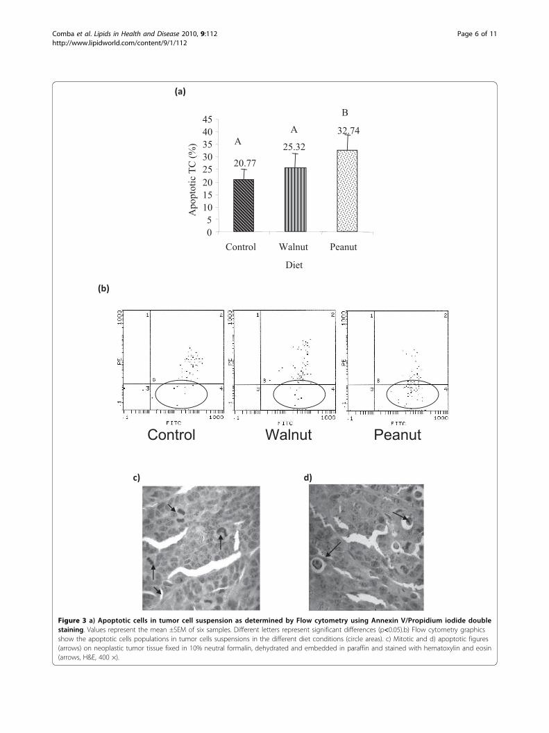
Figure 3 a) Apoptotic cells in tumor cell suspension as determined by Flow cytometry using Annexin V/Propidium iodide doublestaining. Values represent the mean ±SEM of six samples. Different letters represent significant differences (p<0.05).b) Flow cytometry graphicsshow the apoptotic cells populations in tumor cells suspensions in the different diet conditions (circle areas). c) Mitotic and d) apoptotic figures(arrows) on neoplastic tumor tissue fixed in 10% neutral formalin, dehydrated and embedded in paraffin and stained with hematoxylin and eosin(arrows, H&E, 400 ×).
Comba et al. Lipids in Health and Disease 2010, 9:112http://www.lipidworld.com/content/9/1/112
Page 6 of 11

These results agree with the number of apoptotic figuresrecorded in tumour tissue: 2.03 ± 0.14/field; 1.40 ± 0.12/field and 1.16 ± 0.12/field in Peanut, Walnut and Con-trol, respectively (p < 0.05) (Table 3). Moreover, themitotic figures were inversely related to apoptoticvalues. We observed lowest mitosis values (1.56 ± 0.36/field) in Peanut group tumour tissue compared to Wal-nut and Control groups (2.62 ± 0.30 and 3.3 ± 0.25/field, respectively) (p < 0.05) (Table 3).
Modulation of LOX and COX Enzymes of the ω-6Pathway in tumour cellsPUFAs of ω-6, ω-3 and ω-9 series modulated the for-mation and release of COX and LOX products of ω-6-AA and LOX products formed from w-6 LA aftertumor cell stimulation with the ionophore A 23187 asshown in Figures 4. COX-derived-12(S)-HHT was sig-nificantly higher in Peanut group (50.33 ng) than Wal-nut (21.26 ng) and Control (23.12 ng) (Figure 4-a) (p <0.05). On the other hand, PGE2, derived from theaction of COX-2, released from the tumor cells of Con-trol group were higher (1169.74 ng) compared withPeanut (799.40 ng) and Walnut (807.45 ng) groups(Figure 4-b), while tumor cells from the Peanut groupreleased significantly lower levels LOX-derived AAeicosanoids: 12(S)-HETE (9.31 ng) and 15(S)-HETE(8.88 ng) compared with those released by tumor cellsfrom Walnut (9.81 ng; 9.07 ng (p < 0.05) and Controlgroups (10.35 ng; 9.16 ng, respectively (p < 0.05) (Fig.4-c and Figure 4d). 13 (S)-HODE product derived fromLA by the action of LOX formed by the tumor cells ofPeanut group was significantly lower (10.51 ng) com-pared to those formed from Walnut (10.90 ng) group.Though 13(S)-HODE formed by the tumor cells of thecontrol group was higher compared to the Peanutgroup it was not significantly different (Figure 4e)Nevertheless, 13(S)-HODE/12(S)-HETE ratio was sig-nificantly higher in Peanut group (1.13) compared withboth Walnut (1.11) and Control (1.04) groups (p <0.05) (Figure 4-f).
Effect of ω-6, ω-3 and ω-9 dietary PUFAs on Tumourleukocyte infiltration indexIn order to know whether different oils could signifi-cantly influence tumor infiltrating leukocyte number,the number of lymphocyte T and NK-T cells in thetumor tissue was analyzed by CD3 inmunolabeling intumour tissue sections (Table 4). The Peanut oil treatedgroup showed the lowest leukocyte infiltration index(3.03 ± 1.05%) with respect to Walnut (16.71 ± 4.66%)and Control (29.23 ± 4.90%) (p < 0.05) (Table 4).
DiscussionEpigenetic factors have emerged as key mechanisms incancer development. Of all the environmental factors,diet plays a critical role in the prevention and pathobiol-ogy of cancer [21]. In the present, we evaluated whetherpost-weaning diet of mice modulates LOXs and COXsactivities through the eicosanoid release and their impacton certain parameters related to a mammary gland ade-nocarcinoma development. The comparative analysis ofthe three dietary conditions showed that mice fed onsimilar supplemented ω-6 fatty acids diets differing inω-9 and ω-3 content showed that ω-9 enriched Peanutoil diet exhibited a protective effect on tumour develop-ment in comparison to the other dietary oil treatments.It was noted that Peanut oil group showed the lowestnumber of mitosis, the highest amount of apoptosis,decreased leukocyte infiltration and increased survivaltime (Table 3 and Table 4; Figure 2 and Figure 3).Unexpectedly, as shown in Table 2, Peanut oil treated
tumor cell membranes exhibited the highest ω-6 AAlevels compared to Walnut and Control (25.50 vs. 13.78and 9.33 respectively) groups despite the fact that(Table 1) these oils do not contain any AA, while LAcontent of Walnut oil is the highest of the three oilsused in the study (see Table 1). It is also surprising tonote that even though the Walnut treated tumor cellmembranes contained significantly higher amounts of22:5 ω-3 fatty acid, the tumor volume and metastasisnumber were higher, survival of animals bearing thetumors was lower, and tumor cell apoptosis was lessand tumor cell mitotic numbers were higher in thisgroup compared to the Peanut oil treated group. Theseresults suggest that presence of higher amounts of ω-6AA in the tumor cell membrane is responsible for thebeneficial effects seen.It is known that dietary ω-6 LA is converted to AA by
the action of Δ6 and Δ5 desaturases and the correspond-ing elongases. It is likely that the high levels of AA notedin Peanut group may be linked to the up-regulation ofΔ6 and Δ5 desaturases [21] that could explain the higherlevels of unsaturated fatty acids in the tumor cell
Table 3 Number of apoptosis and mitosis recorded intumour sections from hosts fed on the different dietaryconditions.
Diets TUMOUR M3
Mitosis Apoptosis
Control 3.3 ± 0.26A 1.16 ± 0.12A
Walnut 2.62 ± 0.30A 1.40 ± 0.12A
Peanut 1.56 ± 0.36B 2.03 ± 0.14B
Values represent the mean ± SEM of apoptotic and mitotic figures countedfrom 10 animals of each dietary condition (10 high-power fields). Differentletters represent significant differences (p < 0.05).
Comba et al. Lipids in Health and Disease 2010, 9:112http://www.lipidworld.com/content/9/1/112
Page 7 of 11

Figure 4 Different eicosanoids released from M3 TC of hosts fed on different diets after stimulation with ionophore A 23287 (2 M).Values represent the means ± SEM of 15 samples. Different letters represent significant differences (p < 0.05): a) 12 (S)-HHT; b) PGE2;c) 12 (S)-HETE; d) 15 (S)-HETE; e) 13 (S)-HODE and f) 13 (S)-HODE/12 (S)-HETE ratio.
Comba et al. Lipids in Health and Disease 2010, 9:112http://www.lipidworld.com/content/9/1/112
Page 8 of 11

membranes [22] of Peanut oil treated group [23]. AAforms substrate to COXs and LOXs enzymes and someof their metabolites are: 12(S)-HHT and PGE2 via COXs;12(S)-HETE and 15(S)-HETE via LOXs, while LA is asubstrate for 15-LOX-1 and 13-HODE is its major meta-bolite. In the Peanut group, we observed the highest levelof 12(S)-HHT, a lipoperoxide marker, and lower levels ofPGE2 compared to control group. Both 12(S)-HHT andPGE2 are products of the activity of COX enzymes,whereas only PGE2 is formed due to the activity of thespecific enzyme PGE synthase that explains changes inthe levels of various eicosanoids in the cells. It has beenshown that enhanced formation of lipid peroxides andlow PGE2 production inhibit cancer progression andtumour growth [24]. Moreover, the lower percentage oftumour leukocyte infiltration exhibited in Peanut group(Table 4) correlated well with the low PGE2 levelsobserved, which is considered as an activator of inflam-matory process and related to pro-carcinogenic events[25]. There is evidence to suggest that tumor infiltratingmacrophages enhance tumor growth, promote tumor cellmotility and angiogenesis [26,27]. Though the exactmechanism(s) by which tumor-infiltrating macrophagesenhance tumor growth is not clear, it is possible thatthese macrophages produce excess of PGE2, TNF-a andother pro-inflammatory molecules that aid tumor cellgrowth. PGE2 is derived from AA, while TNF-a enhancesPGE2 production [28]. On the other hand, PGE2 sup-presses TNF-a production [29-32]. Inhibition of PGE2synthesis was found to enhance TNF-a production andaugment macrophage tumoricidal activity [29,31]. In con-trast, phospholipase A2 activity and AA was reported tobe essential for the tumoricidal action of TNF-a [33-36].It is known that both ω-3 and ω-6 fatty acids inhibit theproduction of TNF-a [37-39]. Thus, the relationshipamong PUFAs, eicosanoids and TNF-a and their actionson tumor cells is complex. In this complex net work ofevents, in general, PGE2 serves as an immunosuppressorand inhibits the tumoricidal action of macrophages;TNF-a needs phospholipase A2 activity and free AA tobring about its anti-tumor action; while free AA andother PUFAs have direct tumoricidal action [28-41].
In the present study, we found that 12(S)-HETElevels were significantly lower, higher degree of apop-tosis of tumor cells and lowest rate of mitosis countsin the (Figure 4 and Table 3) Peanut oil group suggest-ing that inhibition of 12(S)-HETE productiondiminishes cell proliferation and induces apoptosis aspreviously described [42]. Coincidently, high level of12(S)-HHT could be related to increased apoptosis.These results are in agreement with previous studiesthat showed that lipid peroxides are selectively toxic totumour cells by triggering apoptosis through nuclearcaspase activation [43,44]. Indeed, oxidation is theinitial mechanism for inducing the phosphatidyl-serinetranslocation from the cytosol domain to the externalmembrane, considered to be a key step in the apopto-tic process [45]. Thus, lower tumor cell proliferationand enhanced apoptosis could have resulted in reducedtumour volume (Figure 2 and Figure 3). With respectto 15-LOX-1 activity, Peanut group showed signifi-cantly low levels of its major metabolite, 13-HODE.Also, this group exhibited the lowest metastasis num-ber (Figure 2b). Although 13-HODE levels were lowerin Peanut group, 13-HODE/12-HETE ratio was signifi-cantly higher (1.13) than Walnut (1.11) and Control(1.04) groups (Figure 4f). Previously, we showed that13-HODE has anti-metastatic action, while 12(S)-HETE enhances metastasis [17]. In addition, it wasobserved that mammary gland tumour cells from micefed with ω-6 enriched diet released higher levels of 12(S)-HETE which could be linked to the high numberof metastasis in this animal model [12]. Several othersshowed that 12(S)-HETE is produced in large amountsby various epithelial cancer cell lines that positivelycorrelated to their metastatic potential [46]. The loweramounts of 15(S)-HETE noted in Peanut oil fed tumorcell group could be attributed to its anti-tumour actionlending support to the previous evidence that thismetabolite is present in low concentrations in differenttumour tissues [47].In summary, the present study showed that diets
enriched with high levels of ω-9 fatty acids reducetumour growth, metastasis and tumor leukocyte infil-tration by: 1) inhibiting LOXs activity, reducing theformation of pro-tumorigenic eicosanoids such as 12(S)-HETE and 15 (S)-HETE, 2) increasing the synthesisof 12 (S)-HHT that induces apoptosis and 3) decreas-ing the production of pro-inflammatory PGE2. Under-standing the mechanisms by which ω-3, ω-6 and ω-9PUFAs alter growth and trigger apoptosis of breastcancer cells is essential to devise newer dietary thera-peutic strategies to prevent cancer and employ variousfatty acid rich oils to potentiate the actions of the cur-rent anti-cancer therapies.
Table 4 Percentage of positive immunolabeling for CD3-T-lymphocytes and NK-T cells infiltration in tumor tissue.
Diets % of Tumor Infiltration
Control 29.23 ± 4.90A
Walnut 16.71 ± 4.66A
Peanut 3.03 ± 1.05B
The values indicate immunolabeling and represent the mean ± SEM of gold-silver particles showing positive labeling of CD3 in 15 fields evaluated in 10tumor sections of each dietary treatment. Different letters representsignificant differences (p < 0.05).
Comba et al. Lipids in Health and Disease 2010, 9:112http://www.lipidworld.com/content/9/1/112
Page 9 of 11

AcknowledgementsWe are indebted to Mr. Ricardo Mattos for animal care. We are grateful toDr. Constancio Giraudo for technical assistance in flow cytometry at theFundación para el Progreso de la Medicina de la Ciudad de Córdoba-Argentina. Dr. U.N. Das is in receipt of Ramalingaswami fellowship of theDepartment of Biotechnology, India during the tenure of this study. Thiswork was supported, in part, under the bilateral cooperation projectbetween governments of India and Argentina (DST- MINCYT), ARG-RPO033/2006). This work was supported by Grants from FONCYT, Ministerio deCiencia y Técnica de la Provincia de Córdoba- Argentina, CONICET-Argentina and SECYT-UNC.
Author details1Cátedra de Biología Celular, Histología y Embriología, Instituto de BiologíaCelular, Facultad de Ciencias Médicas, Universidad Nacional de Córdoba.Argentina. 2Instituto Multidisciplinario de Biología Vegetal (IMBIV-CONICET),Cátedra de Química Orgánica, FCEF y N. Universidad Nacional de Córdoba,Argentina. 3Consejo Nacional de Investigaciones Científicas y Técnicas(CONICET), Avenida Rivadavia 1917 - CP C1033AAJ - Qty. Buenos Aires,Argentina. 4Jawaharlal Nehru Technological University, Kakinada 533 003,India. 5UND Life Sciences, 13800 Fairhill Road, #321, Shaker Heights, OH44120, USA. 6Krishna Institute of Medical Sciences, Secunderabad-500 003,India.
Authors’ contributionsARE and MEP conceived the study, participated in its design andcoordination; AC, DMM, MAB and CPG performed various experiments; ARE,MEP and UND interpreted the data and wrote the manuscript; all authorsread and approved the final manuscript
Competing interestsThe authors declare that they have no competing interests.
Received: 24 September 2010 Accepted: 8 October 2010Published: 8 October 2010
References1. International Agency for Research on Cancer. Globocan 2008, Lyon 2008
[http://www-dep.iarc.fr/].2. Bidgoli SA, Ahmadi R, Zavarhei MD: Role of hormonal and environmental
factors on early incidence of breast cancer in Iran. Scie Total Enviorn 2010,408:4056-4061.
3. Thiébaut AC, Chajès V, Gerber M, Boutron-Ruault MC, Joulin V, Lenoir G,Berrino F, Riboli E, Bénichou J, Clavel-Chapelon F: Dietary intakes ofomega-6 and omega-3 polyunsaturated fatty acids and the risk ofbreast cancer. Int J Cancer 2009, 124:924-931.
4. Shaikh IA, Brown I, Wahle KW, Heys SD: Enhancing cytotoxic therapies forbreast and prostate cancers with polyunsaturated fatty acids. NutrCancer 2010, 62:284-96.
5. Hyde CAC, Missailidis S: Inhibition of arachidonic acid metabolism and itsimplication on cell proliferation and tumour-angiogenesis. InternImmunoph 2009, 9:701-715.
6. Comba A, Pasqualini ME: Primers on Molecular Pathways - Lipoxygenases:Their Role as an Oncogenic Pathway in Pancreatic Cancer. Pancreatology2009, 9:724-728.
7. Moreno JJ: New aspects of the role of hydroxyeicosatetraenoic acids incell growth and cancer development. Biochem Pharmacol 2009, 77:1-10.
8. Das UN: Influence of polyunsaturated fatty acids and their metaboliteson stem cell biology. Nutrition .
9. Zeidler R, Csanady M, Gires O, Lang S, Schmitt B, Wollenberg B: Tumourcell-derived prostaglandin E2 inhibits monocyte function by interferingwith CCR5 and Mac-1. FASEB J 2000, 17:661-668.
10. Tobares L, Guzmán C, Maestri DM: Effect of the Extraction and Bleaching-Processes on Jojoba (Simmondsia chinensis) Wax Quality. Eur J Lipid SciTechnol 2003, 105:749-753.
11. Galli S, Labato MI, Bal de Kier Joffé E, Carreras MC, Poderoso JJ: Decreasedmitochondrial nitric oxide synthase activity and hydrogen peroxiderelate persistent tumoral proliferation to embryonic behavior. Cancer Res2003, 63:6370-6377.
12. Espada CE, Berra MA, Martinez MJ, Eynard AR, Pasqualini ME: Effect of ChiaOil (Salvia Hispanica) rich in n-3 fatty acids on the eicosanoid release,
apoptosis and T-lymphocyte tumour infiltration in a murine mammarygland. Prost Leukot Essent Fatty Acids 2007, 77:21-28.
13. Maestri DM, Guzmán CA: A comparative study of seed lipid componentsof Nicotianeae (Solanaceae). Biochem Syst Ecl 1995, 23:201-207.
14. Calderón RO, Eynard AR: Fatty acids specifically related to the anisotropicproperties of plasma membrane from rat urothelium. Biochim BiophysActa 2000, 1483:174-84.
15. Kelavkar UP, Hutzley J, Mc Hugh K, Allen KGD, Parwani A: Prostate tumourgrowth can be modulated by dietary targeting the 15-lipoxygenase- Iand cyclooxygenase-2 enzymes. Neoplasia 2009, 11:692-699.
16. Pasqualini ME, Mohn CE, Petiti JP, Eynard AR: COX and LOX eicosanoidsmodulate platelet activation and procoagulation induced by two murinecancer cells. Prost Leukot Essent Fatty Acids 2000, 63:377-383.
17. Pasqualini ME, Heyd VL, Manzo P, Eynard AR: Association between E-cadherin expression by human colon, bladder and breast cancer cellsand the 13-HODE: 15-HETE ratio. A possible role of their metastaticpotential. Prost Leukot Essent Fatty Acids 2003, 68:9-16.
18. Vermmes C, Haanen CP, Reutelingsperger M: A novel assay for apoptosis:flow cytometric detection of phosphatidylserine expression on earlyapoptotic cells using fluorescein labelled annexin V. J Immunol Methods1995, 180:38-52.
19. Maruoka H, Shinohara K, Miyata M, Sugimura T, Terada M, Wakasugi H:A novel monoclonal antibody permitting recognition of NKT cells invarious mouse strains. Biochem Biophys Res Commun 1998, 242:413-418.
20. In-Kwon Y: Generalized weighted additive models based on distributionfunctions. Statis Probab Lett 2007, 77:1394-1402.
21. Das UN: Essential Fatty acids a review. Curr Pharm Biotechnol 2006, 7:467-82.22. Das UN: Can essential fatty acids reduce the burden of disease(s)? Lipids
Health Dis 2008, 7:9-27.23. Tapiero H, Ba GN, Coureur P, Tew KD: Polyunsaturated fatty acids (PUFAs)
and eicosanoids in human health and pathologies. Biomed Pharmacol2000, 56:215-222.
24. Pair R, Soreghan B, Szabo IL, Pavelka M, Baatar D, Tarnawski AS:Prostagalndin E2 transactivates EGF receptor: a novel mechanism forpromoting colon cancer growth and gastrointestinal hypertrophy. NatMed 2002, 8:289-293.
25. Wang D, Dubois RN: The role of COX-2 in intestinal inflammation andcolorectal cancer. Oncogene 2010, 29:781-788.
26. Ruhrberg C, De Palma M: Deciphering macrophage roles in humantumors. Nat Med 2010, 16:861-862.
27. Qualls JE, Murray PJ: Stopping macrophages wounds tumors. Nat Med2010, 16:863-864.
28. Tashjian AH Jr, Voelkel EF, Lazzaro M, Goad D, Bosma T, Levine L: Tumornecrosis factor-alpha (cachectin) stimulates bone resorption in mousecalvaria via a prostaglandin-mediated mechanism. Endocrinology 1987,120:2029-2036.
29. Alleva DG, Burger CJ, Elgert KD: Tumor growth increases Ia- macrophagesynthesis of tumor necrosis factor-alpha and prostaglandin E2: changesin macrophage suppressor activity. J Leukoc Biol 1993, 53:550-558.
30. Karck U, Peters T, Decker K: The release of tumor necrosis factor fromendotoxin-stimulated rat Kupffer cells is regulated by prostaglandin E2and dexamethasone. J Hepatol 1988, 7:352-361.
31. Ben-Efraim S, Bonta IL: Modulation of antitumour activity of macrophagesby regulation of eicosanoids and cytokine production. Int JImmunopharmacol 1994, 16:397-399.
32. Chae BS, Shin TY, Kim DK, Eun JS, Leem JY, Yang JH: Prostaglandin E2-mediated dysregulation of proinflammatory cytokine production inpristane-induced lupus mice. Arch Pharm Res 2008, 31:503-510.
33. Palombella VJ, Vilcek J: Mitogenic and cytotoxic actions of tumor necrosisfactor in BALB/c 3T3 cells. Role of phospholipase activation. J Biol Chem1989, 264:18128-18136.
34. Suffys P, Beyaert R, Van Roy F, Fiers W: Reduced tumour necrosis factor-induced cytotoxicity by inhibitors of the arachidonic acid metabolism.Biochem Biophys Res Commun 1987, 149:735-743.
35. Suffys P, Beyaert R, De Valck D, Vanhaesebroeck B, Van Roy F, Fiers W:Tumour-necrosis-factor-mediated cytotoxicity is correlated withphospholipase-A2 activity, but not with arachidonic acid release per se.Eur J Biochem 1991, 195:465-475.
36. Neale ML, Fiera RA, Matthews N: Involvement of phospholipase A2activation in tumour cell killing by tumour necrosis factor. Immunology1988, 64:81-85.
Comba et al. Lipids in Health and Disease 2010, 9:112http://www.lipidworld.com/content/9/1/112
Page 10 of 11

37. Watanabe S, Onozaki K, Yamamoto S, Okuyama H: Regulation by dietaryessential fatty acid balance of tumor necrosis factor production inmouse macrophages. J Leukoc Biol 1993, 53:151-156.
38. Tappia PS, Man WJ, Grimble RF: Influence of unsaturated fatty acids onthe production of tumour necrosis factor and interleukin-6 by ratperitoneal macrophages. Mol Cell Biochem 1995, 143:89-98.
39. Kumar GS, Das UN: Effect of prostaglandins and their precursors on theproliferation of human lymphocytes and their secretion of tumornecrosis factor and various interleukins. Prostaglandins Leukot Essent FattyAcids 1994, 50:331-334.
40. Das UN: Tumoricidal action of cis-unsaturated fatty acids and theirrelationship to free radicals and lipid peroxidation. Cancer Lett 1991,56:235-243.
41. Das UN, Prasad VV, Reddy DR: Local application of gamma-linolenic acidin the treatment of human gliomas. Cancer Lett 1995, 94:147-155.
42. Nazarewicz RR, Zenebe WJ, Parihar A, Parihar MS, Vaccaro M, Rink C,Sen CK, Ghafourifar P: 12(S)-hydroperoxyeicosatetraenoic acid (12-HETE)increases mitochondrial nitric oxide by increasing intramitochondrialcalcium. Arch Biochem Biophys 2007, 468:114-20.
43. Das UN: Essential fatty acids, lipid peroxidation and apoptosis.Prostaglandins Leukot Essen Fatty Acids 1999, 61:157-63.
44. Uauy R, Solomons N: Diet, Nutrition, and the life-course approach tocancer prevention. J Nutr 2005, 135:2934S-2945S.
45. Zuo X, Wu Y, Morris JS, Stimmel JB, Leesnitzer LM, Fischer SM, Lippman SM,Shureiqi I: Oxidative metabolism of linoleic acid modulates PPAR-beta/delta suppression of PPAR-gamma activity. Oncogene 2006, 25:1225-1241.
46. Nie D, Krishnamoorthy S, Jin R, Tang K, Chen Y, Qiao Y, Zacharek A, Guo Y,Milanini J, Pages G, Honn KV: Mechanisms regulating tumourangiogenesis by 12-lipoxygenase in prostate cancer cells. J Biol Chem2006, 281:18601-18609.
47. Srivastava K, Kundumani-Sridharan V, Zhang B, Bajpai AK, Rao GN: 15(S)-Hydroxyeicosatetraenoic Acid-Induced Angiogenesis Requires STAT3-Dependent Expression of VEGF. Cancer Res 2007, 67:4328-4336.
doi:10.1186/1476-511X-9-112Cite this article as: Comba et al.: Effect of ωω-3 and ωω-9 fatty acid richoils on lipoxygenases and cyclooxygenases enzymes and on thegrowth of a mammary adenocarcinoma model. Lipids in Health andDisease 2010 9:112.
Submit your next manuscript to BioMed Centraland take full advantage of:
• Convenient online submission
• Thorough peer review
• No space constraints or color figure charges
• Immediate publication on acceptance
• Inclusion in PubMed, CAS, Scopus and Google Scholar
• Research which is freely available for redistribution
Submit your manuscript at www.biomedcentral.com/submit
Comba et al. Lipids in Health and Disease 2010, 9:112http://www.lipidworld.com/content/9/1/112
Page 11 of 11
Related Documents