ISSN 1757-6180 R ESEARCH S POTLIGHT | N EWS & A NALYSIS 1065 10.4155/BIO.11.64 © 2011 Future Science Ltd Bioanalysis (2011) 3(10), 1065–1076 High-performance affinity separations & chromatographic immunoassays One area of active research at the University of Nebraska – Lincoln (UNL) is the development of novel separations for biological and environ� n� mental samples based on high�performance affinity chromatography (HPAC) and affinity CE (ACE). This work is carried out in the labo� ratory of David S Hage. Both HPAC and ACE are based on the use of a biologically related agent (e.g., an antibody, aptamer, receptor or transport protein) for the selective recognition and binding of a target analyte in a sample [1–4] . In HPAC, the selective binding agent is immo� bilized within a column and used as a station� ary phase for analyte retention [1,2,4] . In ACE, the binding agent is combined with the sample or placed within the running buffer to bind to the target and alter its electrophoretic mobility, thereby leading to the separation of this target from other sample components [3] . The selective, strong interactions present in many biological systems help to make HPAC and ACE powerful methods for the rapid study of specific targets in complex samples. These methods can be used alone for the detection and measurement of ana� lytes, or can be combined with other techniques to develop multidimensional schemes for separa� develop multidimensional schemes for separa� tion and analysis (e.g., the use of immunoaffin� analysis (e.g., the use of immunoaffin� ity columns with reversed�phase LC or as part of an LC–MS system) [1–4] . A large number of routine and custom�designed HPLC systems are available for such work in the Hage labora� for such work in the Hage labora� tory and in the facilities within the Chemistry Department at UNL. A variety of detection formats are possible with these HPLC systems, including absorbance, fluorescence or MS and more specialized modes such as chemilumines� s� cence or near�infrared fluorescence. Several CE systems are also available for use in this research with either absorbance or fluorescence detection. A specific topic area of emphasis in the Hage laboratory is the use of antibodies and other selective binding agents in HPLC systems to produce ‘chromatographic immunoassays’ [5,6] . It has been demonstrated in the past that these methods can be used for the rapid and selective measurements of drugs, hormones, peptides, pro� teins and many other targets in biological sam� ogical sam� ples. These properties make chromatographic immunoassays appealing for use in clinical test� ing, pharmaceutical analysis, biotechnology and environmental studies [5–15] . When used in com� bination with methods such as reversed�phase LC or CE, chromatographic immunoassays can also be adapted for the simultaneous analysis of several compounds within a given class of chemicals [5–9] . One application of Hage’s work in this area has been the creation of an automated and por� table system for the determination of triazine herbicides in environmental samples [7,8] . This method combined the use of a small antibody column with a reversed�phase HPLC column and absorbance detection. This approach has been used in both the laboratory and in a port� port� able device for field work. In work with the por� In work with the por� table system, it was possible to provide results at the site of a river or stream within 10 min of sample injection. This approach has been used not only for triazine herbicides, such as atrazine, but has been adapted for use with other environ� s been adapted for use with other environ� mental agents (e.g., chloroacetic acid herbicides such as 2,4�D) by changing the type of antibody column used in the system [7–9] . The Chemistry Department at the University of Nebraska – Lincoln (UNL) is located in Hamilton Hall on the main campus of UNL in Lincoln, NE, USA. This department houses the primary graduate and research program in chemistry in the state of Nebraska. This program includes the traditional fields of analytical chemistry, biochemistry, inorganic chemistry, organic chemistry and physical chemistry. However, this program also contains a great deal of multidisciplinary research in fields that range from bioanalytical and biophysical chemistry to nanomaterials, energy research, catalysis and computational chemistry. Current research in bioanalytical and biophysical chemistry at UNL includes work with separation methods such as HPLC and CE, as well as with techniques such as MS and LC–MS, NMR spectroscopy, electrochemical biosensors, scanning probe microscopy and laser spectroscopy. This article will discuss several of these areas, with an emphasis being placed on research in bioanalytical separations, binding assays and related fields. Research in bioanalysis and separations at the University of Nebraska – Lincoln David S Hage †1 , Eric D Dodds 1 , Liangcheng Du 1 & Robert Powers 1 1 Chemistry Department, University of Nebraska – Lincoln, Lincoln, NE 68588-0304, USA † Author for correspondence: Tel.: +1 402 472 2744 Fax: +1 402 472 9402 E-mail: [email protected]

Welcome message from author
This document is posted to help you gain knowledge. Please leave a comment to let me know what you think about it! Share it to your friends and learn new things together.
Transcript
ISSN 1757-6180
ReseaRch spotlight | News & aNalysis
106510.4155/BIO.11.64 © 2011 Future Science Ltd Bioanalysis (2011) 3(10), 1065–1076
High-performance affinity separations & chromatographic immunoassaysOne area of active research at the University of Nebraska – Lincoln (UNL) is the development of novel separations for biological and environ�n�mental samples based on high�performance affinity chromatography (HPAC) and affinity CE (ACE). This work is carried out in the labo�ratory of David S Hage. Both HPAC and ACE are based on the use of a biologically related agent (e.g., an antibody, aptamer, receptor or transport protein) for the selective recognition and binding of a target analyte in a sample [1–4]. In HPAC, the selective binding agent is immo�bilized within a column and used as a station�ary phase for analyte retention [1,2,4]. In ACE, the binding agent is combined with the sample or placed within the running buffer to bind to the target and alter its electrophoretic mobility, thereby leading to the separation of this target from other sample components [3]. The selective, strong interactions present in many biological systems help to make HPAC and ACE powerful methods for the rapid study of specific targets in complex samples. These methods can be used alone for the detection and measurement of ana�lytes, or can be combined with other techniques to develop multidimensional schemes for separa� develop multidimensional schemes for separa�tion and ana lysis (e.g., the use of immunoaffi n�ana lysis (e.g., the use of immunoaffin�ity columns with reversed�phase LC or as part of an LC–MS system) [1–4]. A large number of routine and custom�designed HPLC systems are available for such work in the Hage labora� for such work in the Hage labora�tory and in the facilities within the Chemistry Department at UNL. A variety of detection formats are possible with these HPLC systems, including absorbance, fluorescence or MS and
more specialized modes such as chemilumines�s�cence or near�infrared fluorescence. Several CE systems are also available for use in this research with either absorbance or fluorescence detection.
A specific topic area of emphasis in the Hage laboratory is the use of antibodies and other selective binding agents in HPLC systems to produce ‘chromatographic immunoassays’ [5,6]. It has been demonstrated in the past that these methods can be used for the rapid and selective measurements of drugs, hormones, peptides, pro�teins and many other targets in biological sam�ogical sam�ples. These properties make chromatographic immunoassays appealing for use in clinical test�ing, pharmaceutical ana lysis, biotechnology and environmental studies [5–15]. When used in com�bination with methods such as reversed�phase LC or CE, chromatographic immunoassays can also be adapted for the simultaneous ana lysis of several compounds within a given class of chemicals [5–9].
One application of Hage’s work in this area has been the creation of an automated and por�table system for the determination of triazine herbicides in environmental samples [7,8]. This method combined the use of a small antibody column with a reversed�phase HPLC column and absorbance detection. This approach has been used in both the laboratory and in a port� port�able device for field work. In work with the por� In work with the por�table system, it was possible to provide results at the site of a river or stream within 10 min of sample injection. This approach has been used not only for triazine herbicides, such as atrazine, but has been adapted for use with other environ�s been adapted for use with other environ�mental agents (e.g., chloroacetic acid herbicides such as 2,4�D) by changing the type of antibody column used in the system [7–9].
The Chemistry Department at the University of Nebraska – Lincoln (UNL) is located in Hamilton Hall on the main campus of UNL in Lincoln, NE, USA. This department houses the primary graduate and research program in chemistry in the state of Nebraska. This program includes the traditional fields of analytical chemistry, biochemistry, inorganic chemistry, organic chemistry and physical chemistry. However, this program also contains a great deal of multidisciplinary research in fields that range from bioanalytical and biophysical chemistry to nanomaterials, energy research, catalysis and computational chemistry. Current research in bioanalytical and biophysical chemistry at UNL includes work with separation methods such as HPLC and CE, as well as with techniques such as MS and LC–MS, NMR spectroscopy, electrochemical biosensors, scanning probe microscopy and laser spectroscopy. This article will discuss several of these areas, with an emphasis being placed on research in bioanalytical separations, binding assays and related fields.
Research in bioana lysis and separations at the University of Nebraska – Lincoln
David S Hage†1, Eric D Dodds1, Liangcheng Du1 & Robert Powers1
1Chemistry Department, University of Nebraska – Lincoln, Lincoln, NE 68588-0304, USA †Author for correspondence:Tel.: +1 402 472 2744 Fax: +1 402 472 9402 E-mail: [email protected]
1066 Bioanalysis (2011) 3(10) future science group
News & aNalysis | ReseaRch spotlight
Other chromatographic immunoassays that have been developed by the Hage laboratory include techniques for measuring specific hor�mones, peptides, drugs and proteins in clinical samples [10–15]. These methods have been shown in numerous studies to be precise, easy to auto�mate and to take only a small period of time to perform (i.e., typically only a few minutes). In addition, Hage’s group has explored and devel�oped many formats for use in these methods. These formats range from competitive binding assays to sandwich immunoassays, displace�ment immunoassays and one�site immunometric assays. As part of these studies, the Hage group has also been active in working with chromato�graphic theory and computer models to describe the response and behavior of these assays under various operating conditions [6,9,10,15].
A recent application of chromatographic immunoassays has been the development of ultra�fast immunoextraction and the use of this approach to measure the ‘free fraction’ of a drug or hormone in a biological sample (Figure 1) [10–12,16]. Ultrafast immunoextraction is based on the use of an affinity microcolumn with a small layer of antibodies that can bind quickly and selectively to the desired target. When used to measure the free fraction of a drug or hormone, this free fraction is extracted by the antibody column in a very short period of time (80–120 ms), which makes it possible to isolate this fraction without disturbing the fraction of the drug or hormone that is bound
by other sample components. The results of this approach have been found to give good correla�tion with reference methods such as ultrafiltra�tion. However, this information can be obtained with chromatographic immunoassays in much less ana lysis time than is required by other methods, making it an attractive approach for use in pharmacological studies and in clinical chemistry for personalized medicine [10–12].
Chiral separations & studies of biological interactionsTwo other ways that HPAC and ACE can be used is for chiral separations or to study biologi�cal interactions (e.g., protein–protein binding or protein–drug binding). This research is of cur�rent interest for the high�throughput screening of drug candidates for a given protein and for predicting the behavior of drugs and proteins in the body by using separation systems as models for these biological systems [4,17–20].
One protein the Hage group has studied extensively with regard to chiral separations and drug–protein binding is human serum albumin (HSA). HSA is the most abundant protein in serum and plays an important role in deliver�ing many types of drugs throughout the body. The Hage group has shown that HSA can be immobilized within HPLC columns and used in rapid chiral separations [21–24] or to study how this protein binds to various drugs, hor�mones and other small solutes in serum [21–32]. Chemicals that have been examined with these
Figure 1. (A) General design of a small affinity column for use in ultrafast immunoextraction and (B) the extraction of fluorescein as a model target by immobilized antibodies within such a column. The plot in (B) indicates that 95% extraction of the target could be obtained in 100 ms by this approach. The components in (A) were as follows: (1) connection tubing, (2) fitting, (3) end fitting assembly, (4) column casing, (5) frit, (6) sample distributor, and (7) affinity column or affinity disk. Adapted with permission from [16].
12
3
4
567
Flo
w d
irect
ion
A B
Residence time (ms)
% E
xtra
cted
50 100 150 20050
70
90
110
1067www.future-science.comfuture science group
ReseaRch spotlight | News & aNalysis
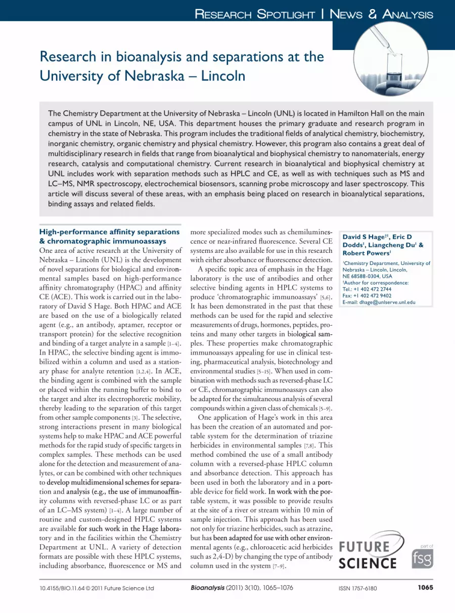
HSA columns include warfarin, thyroxine, tryptophan, ibuprofen, digitoxin, clomiphene, tamoxifen, phenytoin and carbamazepine. In addition, the Hage group has recently created columns that contained the serum protein a
1�acid glycoprotein (AGP) in a fully active
form that can be used in chiral separations or binding studies (Figure 2) [33–36]. The group has also developed columns containing complex particles such as high�density lipoprotein and low�density lipoprotein for the investigation of drug interactions with these binding agents in serum [37,38].
As part of this work, the Hage group has developed several new approaches for examin�ing the biological interactions by HPAC or ACE. Examples of these methods are approaches for the study of low�solubility drugs [28–30], site�selective studies of agents with multiple bind�ing sites on a protein [25,31] and techniques for examining the interactions of drugs or solutes that have allosteric interactions on a protein [39]. A variety of methods for examining the kinet�ics of drug–protein interactions have also been created for use with HPAC [40–43], along with HPAC and ACE approaches for examining the interactions of drugs with the soluble form of a protein [10–12,19,20,44]. These approaches have been used with both native proteins and with modi�fied proteins, as has recently been demonstrated in studies using HSA that has undergone non�enzymatic glycation [45–47]. Related work that has been associated with these efforts has involved the creation of affinity microcolumns, which can be used in binding studies with small amounts of proteins that have been obtained from individu�als and that can be examined in detail by HPAC over the course of hundreds of experiments [45–49].
LC–MS, GC–MS & related methodsResearch facilities are also available in the Chemistry Department for work in LC–MS and GC–MS within the Nebraska Center for Mass Spectrometry. The GC–MS system consists of an Agilent 6890N instrument with an auto�injection system and a Waters GCT TOF�MS for work with either electron ionization or chem�ical ionization. There are also LC–MS systems that use ESI, including Waters Q�TOF Ultima and Synapt G2 Q�TOF systems and a Thermo LCQ ion�trap mass spectrometer. Equipment for conducting MALDI�TOF�MS is also available in the center.
The group of Liangcheng Du makes use of many of these facilities as part of their research. The Du group is studying the molecular
mechanisms by which a variety of organisms (e.g., bacteria, fungi and plants) make structurally complex and biologically active natural products. The goal of this work is to use this knowledge to produce new products through genetic engineer�ing that will be beneficial to humans. A specific example of this work concerns the mechanism of biosynthesis for fungal polyketides. Polyketides are probably the most significant group of natural products in terms of their importance to human medicine. Most studies to date with these compounds have focused on polyketides from bacteria. Although fungi produce numer�ous polyketides, their biosynthetic mechanism remains largely unknown. The Du group has been studying mycotoxin fumonisins, which are produced by a number of economically significant fungi such as the pathogenic fungus Fusarium verticillioides, which is a widespread contaminant of corn and maize�derived food and feeds. The ingestion of fumonisin�contaminated corn causes fatal diseases in livestock and imposes a cancer risk to humans. By using HPLC, GC–MS and LC–MS, the Du group has identified multiple biosynthetic intermediates from various fungal strains, along with mutants that are generated by specifically changing the biosynthetic genes for fumonisins. This research has helped to reveal the pathway for the biosynthesis of fumonisin [50–52].
CH2NH-NHC(CH2)nCNH-N=CH
==
O O
Time (min)0 1 3 5 72 4 6
R
S
Res
po
nse
(22
5 n
m)
α1-acid glycoprotein
Figure 2. General structure of a silica support containing immobilized a1-acid glycoprotein, and the use of this support in the chiral separation of (R)- and (S)-propranolol. The conditions used in this separation were as follows: mobile phase: pH 7.4; 0.067 M phosphate buffer with 2% (v/v) 2-propanol; temperature: 37°C; flow-rate: 2.00 ml/min. Adapted with permission from [34].
1068 Bioanalysis (2011) 3(10) future science group
News & aNalysis | ReseaRch spotlight
In another project, Du has been studying the genetics and function of novel antifungal natu�ral products such as heat�stable antifungal fac� heat�stable antifungal fac�heat�stable antifungal fac�tor (HSAF; i.e., dihydromaltophilin). HSAF has been isolated from the biocontrol agent Lysobacter enzymogenes C3 [53]. This compound is a potent inhibitor of a wide range of fungi. The goals of the Du group in this area have been to develop novel antifungal drugs for humans and ‘green’ strategies for fungal disease control in agriculture. HPLC, MS and NMR have been used to deter�mine the structure of HSAF, which has a unique macrocyclic lactam system that contains a tetra�mic acid moiety and a 5,5,6�tricyclic skeleton [54]. Du has also determined the biosynthetic genes for this compound and has identified several com�pounds that are produced by gene disruption and gene�replacement mutants [55].
The use of LC–MS or separation methods in combination with MALDI�TOF�MS is also of interest to the Hage group. For instance, the group is using binding studies based on HPAC to provide information on the function of a biological molecule and its interactions with
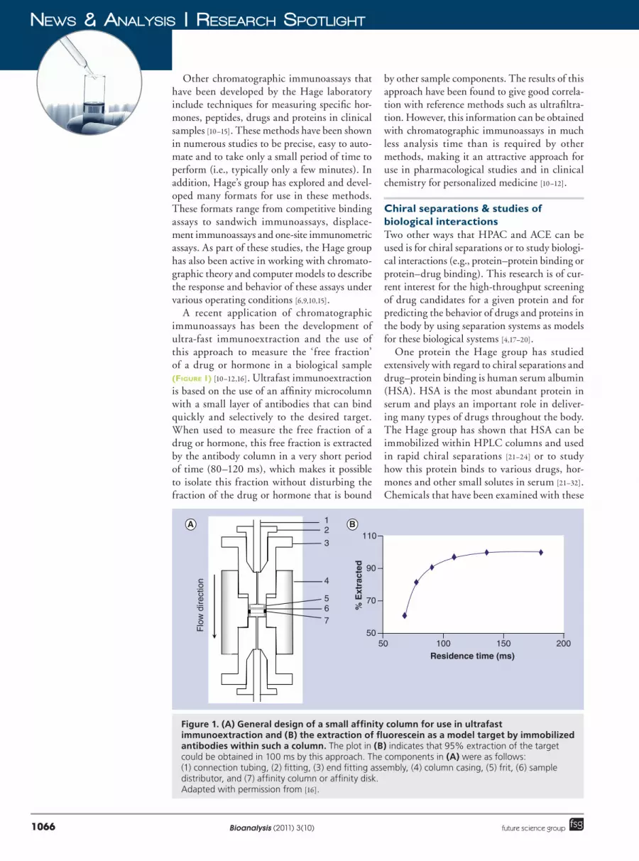
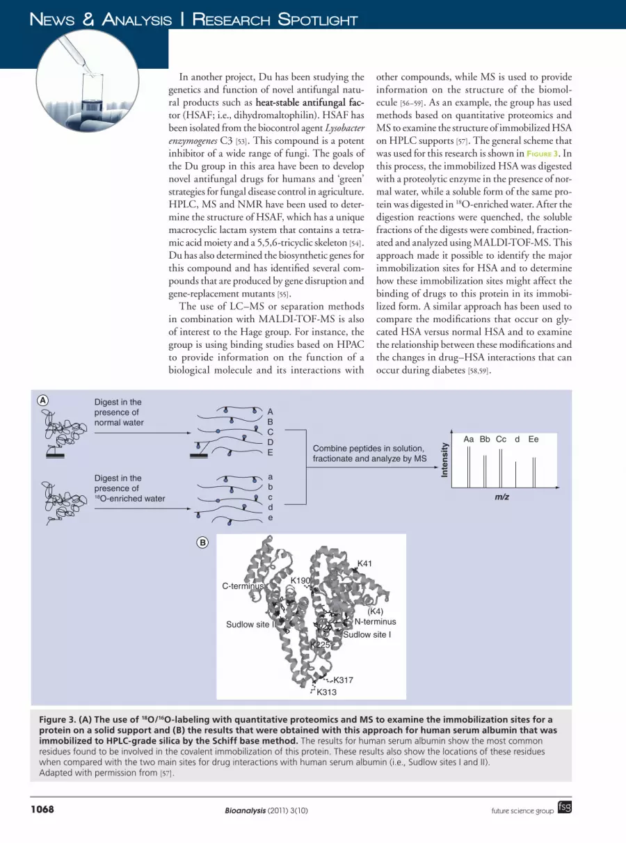
other compounds, while MS is used to provide information on the structure of the biomol�ecule [56–59]. As an example, the group has used methods based on quantitative proteomics and MS to examine the structure of immobilized HSA on HPLC supports [57]. The general scheme that was used for this research is shown in Figure 3. In this process, the immobilized HSA was digested with a proteolytic enzyme in the presence of nor�mal water, while a soluble form of the same pro�tein was digested in 18O�enriched water. After the digestion reactions were quenched, the soluble fractions of the digests were combined, fraction�ated and analyzed using MALDI�TOF�MS. This approach made it possible to identify the major immobilization sites for HSA and to determine how these immobilization sites might affect the binding of drugs to this protein in its immobi�lized form. A similar approach has been used to compare the modifications that occur on gly�cated HSA versus normal HSA and to examine the relationship between these modifications and the changes in drug–HSA interactions that can occur during diabetes [58,59].
Digest in the presence of normal water
Digest in the presence of 18O-enriched water m/z
Inte
nsi
tyAa Bb Cc d Ee
K41
K313K317
K225
K190
(K4)N-terminus
C-terminus
Sudlow site ISudlow site II
abcde
ABCDE Combine peptides in solution,
fractionate and analyze by MS
A
B
Figure 3. (A) The use of 18O/16O-labeling with quantitative proteomics and MS to examine the immobilization sites for a protein on a solid support and (B) the results that were obtained with this approach for human serum albumin that was immobilized to HPLC-grade silica by the Schiff base method. The results for human serum albumin show the most common residues found to be involved in the covalent immobilization of this protein. These results also show the locations of these residues when compared with the two main sites for drug interactions with human serum albumin (i.e., Sudlow sites I and II). Adapted with permission from [57].
1069www.future-science.comfuture science group
ReseaRch spotlight | News & aNalysis
Ion mobility MSAnother capability that has recently been added at UNL is the use of separations based on gas�phase ion mobility (IM) in conjunction with MS. This technique is the subject of research in the laboratory of Eric D Dodds. The equip�ment used for this work is a Waters Synapt G2 HDMS IM�enabled Q�TOF hybrid mass spec�trometer. This instrument offers the high mass accuracy (error < 1 ppm) and high mass resolution (m/Dm > 40,000) of a state�of�the�art Q�TOF, as well as the ability to carry out MS/MS by means of collision�induced dissociation or electron trans�fer dissociation. The IM feature of the instrument further provides for the separation and collisional cross�section measurement of analyte ions. IM is well�suited for use with LC and TOF�MS because the timescale of an IM separation (10�3 s) lies between those for typical LC runs (103 s) and TOF�MS (10�6 s). This means that IM can be used with LC and TOF�MS to provide an addi�tional dimension of separation without increas�ing ana lysis time. The trade�off in combining these methods is a reduced duty cycle because ions must be gated to the IM device rather than continuously sampled as a beam by the TOF�MS.
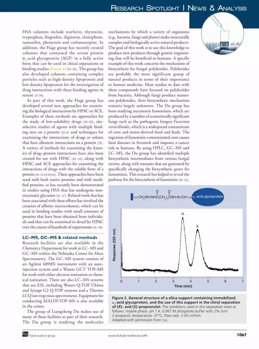
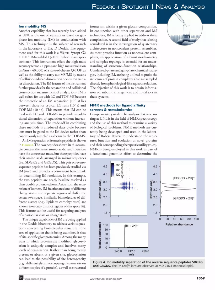
An IM separation of isomeric peptides is shown in Figure 4. The two peptides shown in this exam�ple contain the same amino acids, and therefore have the same exact mass, but these peptides have their amino acids arranged in mirror sequences (i.e., SDGRG and GRGDS). This pair of reverse�sequence peptides has been previously studied via IM [60,61] and provides a convenient benchmark for determining IM resolution. In this example, the two peptides are nearly baseline resolved as their doubly protonated ions. Aside from the sepa�ration of isomers, IM fractionates ions of different charge states into separate regions of drift time versus m/z space. Similarly, biomolecules of dif�ferent classes (e.g., lipids vs carbohydrates) are known to occupy distinct regions of this space [62]. This feature can be useful for targeting analytes of a particular class or charge state.
The unique capabilities of IM are being applied in the Dodds laboratory to address various ques�tions concerning biomolecular structure. One area of application that is being examined is that of site�specific glycoproteomics. Among the many ways in which proteins are modified, glycosyl�ation is uniquely complex and involves many levels of organization. Rather than being merely present or absent at a given site, glycosylation can lead to the possibility of site heterogeneity (e.g., different glycans occupying the same site on different copies of a protein), as well as structural
isomerism within a given glycan composition. In conjunction with other separation and MS techniques, IM is being applied to address these complexities. A second field of study that is being considered is in the interrogation of quaternary architecture in noncovalent protein assemblies. As most proteins function as noncovalent com�plexes, an appreciation of subunit stoichiometry and complex topology is essential for an under�standing of structure–function relationships. Condensed�phase and gas�phase chemical strate�gies, including IM, are being utilized to probe the structures of protein complexes that are sampled directly from physiological�like aqueous solutions. The objective of this work is to obtain informa�tion on subunit arrangement and interfaces in these systems.
NMR methods for ligand affinity screens & metabolomicsComplementary work in bioanalysis that is occur�ring at UNL is in the field of NMR spectroscopy and the use of this method to examine a variety of biological problems. NMR methods are cur�rently being developed and used in the labora�tory of Robert Powers to understand the struc�ture, function and evolution of novel proteins and their corresponding therapeutic utility [63–65]. NMR is being employed in this work as part of a functional genomics effort to determine the
100
80
60
40
20
0Rel
ativ
e ab
un
dan
ce
m/z245.0 247.5 250.0
4.5
4.0
3.5
3.0
2.5
2.0
1.5
Dri
ft t
ime
(ms)
Log
(inte
nsity
)
[M + 2H]2+
Dri
ft t
ime
(ms)
200
160
120
[GRGDS + 2H]2+
[SDGRG + 2H]2+
4.5
4.0
3.5
3.0
2.5
2.0
1.5
Relative abundance
0 20 40 60 80 100
Figure 4. Ion mobility separation of the reverse sequence peptides SDGRG and GRGDS. The [M+2H]2+ ions are observed at m/z 246.1 (monoisotopic).
1070 Bioanalysis (2011) 3(10) future science group
News & aNalysis | ReseaRch spotlight
structures of biomolecules, to investigate biomo�lecular and small�molecule interactions, to screen for functional ligands and potential drugs, and to monitor in vivo protein and drug activity.
These NMR studies are carried out using equipment in the Chemistry Department’s Research Instrumentation Facility. This facility operates and maintains several NMR spectrom�eters, including a 600�MHz Bruker AVANCE spectrometer, which is a five�channel NMR sys�tem capable of working with both solids and liq�uids. This instrument has triple�axis gradients, enabling sophisticated gradient experiments involving multiple nuclei, full broadband capa�bilities and a variable temperature unit for per�forming high and low temperature experiments. This instrument is also equipped with a range of probes for use with liquids, including a TXI triple resonance 1H/13C/15N probe optimized for protein experiments. A second NMR instrument
that is available is a three�channel 500�MHz Bruker DRX NMR spectrometer with a vari�able temperature unit that features a 5 mm 1H/13C/15N cryoprobe. This system excels at the direct detection of protons and as a platform for the indirect detection of 13C or 15N through 2D experiments. The addition of a Bruker BACS�120 sample changer to this instrument allows for the high�throughput screening of ligand�affinity interactions and work in metabolomics.
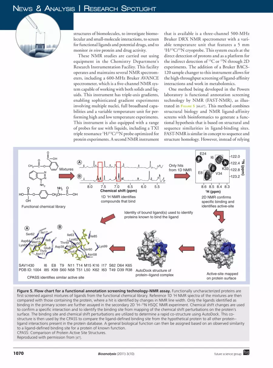
One method being developed in the Powers laboratory is functional annotation screening technology by NMR (FAST�NMR), as illus�trated in Figure 5 [66,67]. This method combines structural biology and NMR ligand�affinity screens with bioinformatics to generate a func�tional hypothesis that is based on structural and sequence similarities in ligand�binding sites. FAST�NMR is similar in concept to sequence and structure homology. However, instead of relying
A
B
A B
Functional chemical library
CPASS identifies similar active site
AutoDock structure of protein–ligand complex
Identity of bound ligand(s) used to identifyproteins known to bind the ligand
1D 1H NMR identifiescompounds that bind
2D NMR confirms specific binding and identifies active-site
Active-site mapped on protein surface
MixturesOnly hitsfrom 1D NMR
Ser62
Asp64Lys65
Ile17
Ile6
Ile63Lys62
Leu50
Thr51
Arg38Asp39Ser60
Asn58
Lys99
Ile85
Thr49
Glu8Thr9
Asn11
Lys16Met15
Thr14
SAV1430PDB ID: 1004
D64D39
S62T49
I17I63
K16K62
M15L50
T14T51
N11N58
T9S60
E8K99
I6I85
K65R38
Chemical shift (ppm)8.0 7.5 7.0 6.5 5.56.0
OCCH2
OOC H
O
N
N
N
NH2
OPHO
O
OH
CH2CH
NH2
C
O
OH1H (ppm)
15N (p
pm
)
8.5 8.4 8.38.6
122.0
122.4
122.8
123.2
E24I6
V34K33
E8
Figure 5. Flow chart for a functional annotation screening technology-NMR assay. Functionally uncharacterized proteins are first screened against mixtures of ligands from the functional chemical library. Reference 1D 1H NMR spectra of the mixtures are then compared with those containing the protein, where a hit is identified by changes in NMR line width. Only the ligands identified as binding in the primary screen are further assayed in the secondary 2D 1H–15N HSQC NMR experiment. Chemical shift changes are used to confirm a specific interaction and to identify the binding site from mapping of the chemical shift perturbations on the protein’s surface. The binding site and chemical shift perturbations are utilized to determine a rapid co-structure using AutoDock. This co-structure is then used by the CPASS to compare the ligand-defined binding site from the hypothetical protein to all other protein–ligand interactions present in the protein database. A general biological function can then be assigned based on an observed similarity to a ligand-defined binding site for a protein of known function. CPASS: Comparison of Protein Active Site Structures. Reproduced with permission from [67].
1071www.future-science.comfuture science group
ReseaRch spotlight | News & aNalysis
on global similarities, a functional assignment is made by focusing on evolutionary stable func�tional regions. FAST�NMR incorporates a multi�step NMR ligand�affinity screening technique [68] with a rapid approach to generate protein–ligand co�structures [69] and a technique to measure dis�sociation constants from NMR screens [70], along with Comparison of Protein Active Site Structures and Protein Function, Evolution, Structure and Sequence as tools for bioinformatics [71,72]. Over 24 proteins have been examined by this FAST�NMR assay. These proteins have included Staphylococcus aureus protein SAV1430, Pseudomonas aeruginosa protein PA1324, Pyrococcus horikoshii OT3 pro�tein PH1320, human protein Q13206, Bacillus subtilis protein YndB and Salmonella typhimurium PrgI protein [66,67,73,74]. Recent efforts with this approach have focused on the ana lysis of proteins of unknown function that are associated with pancreatic cancer.
NMR spectroscopy has also been used for dif�ferential metabolomics by the Powers group [75–78]. In this work, NMR is used to examine the
metabolome of lyzed cells or body fluids and to study changes that result from the influence of disease, drug treatments or environmental stress. NMR combined with principal component ana�lysis is used to measure global changes between cells under various conditions. Similarly, NMR combined with 13C�labeled metabolites permits a detailed ana lysis of metabolite concentration changes. This differential NMR metabolomics methodology is currently being used to under�stand the biofilm of staphylococcus, to under�stand mechanisms of drug activity and resis�tance in tuberculosis, to identify biomarkers for multiple sclerosis, to improve the industrial�scale production of diphtheria toxin and to aid in drug discovery for pancreatic cancer.
Development of new supports & separation media for bioana lysisOne unique capability of researchers in the Chemistry Department at UNL is their ability to create, characterize and evaluate a variety of novel materials for use in separation methods. For
Imm
ob
ilize
d R
IgG
(m
g/g
)
DoOH content (vol %)
70
60
50
40
30
20
10
070 80 90 1006050403020100
10% DoOH 30% DoOH 50% DoOH 100% DoOH
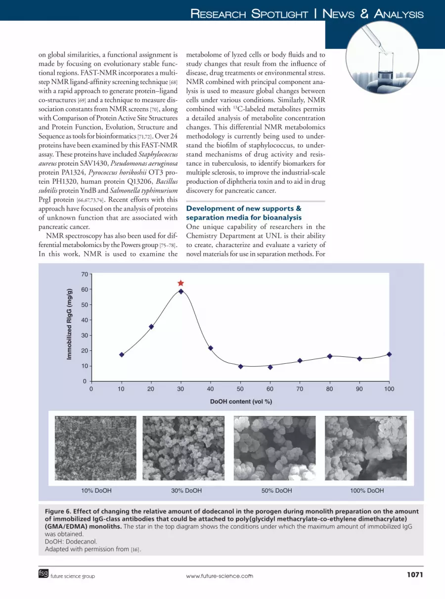
Figure 6. Effect of changing the relative amount of dodecanol in the porogen during monolith preparation on the amount of immobilized IgG-class antibodies that could be attached to poly(glycidyl methacrylate-co-ethylene dimethacrylate) (GMA/EDMA) monoliths. The star in the top diagram shows the conditions under which the maximum amount of immobilized IgG was obtained. DoOH: Dodecanol. Adapted with permission from [16].
1072 Bioanalysis (2011) 3(10) future science group
News & aNalysis | ReseaRch spotlight
instance, Hage has been active for many years in the optimization and creation of new schemes for immobilizing and modifying biological agents for use in HPAC or other bioanalytical meth�ods [16,23,24,33,37,79–85]. Facilities in Hage’s group can be used with both covalent and noncova�lent immobilization methods to place proteins, drugs, nucleic acids or other agents on inorganic materials, organic polymers or carbohydrate�based materials (e.g., silica or glass, methacrylate or agarose). As one example, Hage’s group has developed a method that can be used for the site�selective immobilization of antibodies to silica through their carbohydrate chains [79]. The same group has optimized techniques for immobiliza�tion of the proteins HSA, a
1�acid glycoprotein
and high�density lipoprotein , and has recently reported a noncovalent entrapment method for protein and ligand immobilization that can be used with silica or other porous HPLC sup�ports [21,23,24,33–35,37,80,82,83,85]. A variety of tools have been used in the creation and optimization of these immobilization methods. These tools have included protein assays, infrared spectros�copy, solid�state NMR spectroscopy and MS, as well as HPLC and CE [57,79–81,83–87].
Along with new methods for preparing station�ary phases, additional work is being conducted in the Hage group for the creation of improved support materials for HPLC and related methods. A set of supports that are of current interest are monolithic materials [88,89]. Monolith columns consist of a continuous bed of a porous material that contains a stationary phase, but also allows flow of a mobile phase through the bed. Monolith
columns are of great interest in HPLC because of their good mass transfer properties and low back pressures, allowing these materials to provide efficient separations at high�flow rates. The Hage group has been developing affinity monolith sup�ports for use with antibodies, proteins and other binding agents [88,89]. These studies have included monoliths based on organic copolymers (e.g., gly�cidyl methacrylate and ethylene dimethacrylate) and silica monoliths (Figure 6). These materials have been used in applications that include ultra�fast immunoextraction, HPAC assays for phar�maceutical agents, chiral separations and studies of drug–protein binding [16,23,24,35,49].
The Center for Nanohybrid Functional Materials has recently been created at UNL to further investigate and create novel materials for use in applications such as HPLC, biosens�ing and bioanalytical separations. This center consists of 15 faculty members from UNL, the University of Nebraska Medical Center, the University of Nebraska – Kearney, Creighton University (NE, USA) and Doane College (NE, USA). The goal of this center is to create, test and utilize new materials that consist of ordered nanostructural scaffolds that are functionalized or hybridized with chemical and biological rec�ognition elements. These nanoscaffolds are being prepared with control of 3D morphology and with a variety of frameworks, one set of which includes an expanded set of supports for mono�lithic columns. As these scaffolds are developed they will be combined with binding agents such as antibodies or aptamers for use in flow�based biosensors or bioanalytical separations.
Executive summary
� Research efforts in the field of bioana lysis and separations at the Chemistry Department of University of Nebraska – Lincoln (UNL) include work with HPLC, CE, MS, NMR spectroscopy and several other techniques.
� One area of active research at UNL is in the development of novel separation methods based on high-performance affinity chromatography and affinity CE, such as the use of antibodies in HPLC systems to create chromatographic immunoassays or multidimensional separation methods.
� The use of separation systems such as high-performance affinity chromatography or affinity CE for the study of biological interactions and for chiral separations is another field of ongoing research at UNL. This work is of interest for the creation of new methods for the high-throughput screening of drug candidates or for predicting and modeling the behavior of drugs and proteins in the body.
� The techniques of LC–MS, GC–MS and NMR are being used to discover new natural products and to study their biosynthetic mechanisms. In addition, affinity separations are being used with MS and LC–MS to study and relate the effects of protein modifications with changes in protein function.
� Gas-phase ion mobility is being used for the gas-phase separation of isomeric biomolecules. This method is also being explored as a means for sorting different classes of biomolecules according to their masses, charges and collisional cross-sections.
� The combined use of structural, biological and NMR ligand-affinity screens with bioinformatics is being used to generate functional assignments for proteins. NMR spectroscopy is also being used for studies in metabolomics.
� Several efforts are underway at UNL regarding the creation of new supports and separation media for bioana lysis. Two examples are the generation of improved immobilization methods for use with biomolecules and the development of monolith materials for use in high-performance affinity separations. A new center aimed at the creation of nanohybrid functional materials has also been created at UNL.
1073www.future-science.comfuture science group
ReseaRch spotlight | News & aNalysis
AcknowledgmentsThe authors thank Ronald L Cerny, Joseph Dumais and Patrick H Dussault for providing information on some of the facilities and centers that are described in this report.
Financial & competing interests disclosurePart of the work described in this review was supported by the NIH under grants R01 GM044931, R01 DK069629, R01 AI087668, R21 AI087561, R21 AI081154, P20 RR-17675, RR015468-01 and R03AI073510; the American Heart Association under grant 0860033Z; the NSF or NSF/EPSCoR program under grants MCB-0614916 and EPS-1004094; the NSFC under grants 30428023, 30970244 and 31028019; the USDA-ARS Wheat and Barley Scab Initiative; the University of Nebraska Research Council; and the Nebraska Tobacco Settlement Biochemical Research Development Fund. The authors have no other relevant affiliations or financial involvement with any organization or entity with a financial interest in or financial conflict with the subject matter or materials discussed in the manuscript apart from those disclosed.
No writing assistance was utilized in the production of this manuscript.
BibliographyPapers of special note have been highlighted as:� of interest�� of considerable interest
1 Hage DS. Handbook of Affinity Chromatography. CRC Press/Taylor & Francis (2006).
2 Hage DS. Affinity chromatography: a review of clinical applications. Clin. Chem. 45, 593–615 (1999).
3 Heegaard NHH, Schou C. Affinity ligands in capillary electrophoresis. In: Handbook of Affinity Chromatography. Hage DS (Ed.). CRC Press/Taylor & Francis (2006).
4 Hage DS. High�performance affinity chromatography: a powerful tool for studying serum protein binding. J. Chromatogr. B 768, 3–30 (2002).
� Provides a review on the use of high-performance affinity chromatography in the study of drug–protein interactions.
5 Hage DS, Nelson MA. Chromatographic immunoassays. Anal. Chem. 73, 198A–205A (2001).
� Reviews the use and applications of chromatographic immunoassays.
6 Moser AC, Hage DS. Chromatographic immunoassays. In: Handbook of Affinity Chromatography. Hage DS (Ed.). CRC Press/Taylor & Francis (2006).
7 Nelson MA, Gates A, Dodlinger M, Hage DS. Development of a portable immunoextraction/RPLC system for field studies of herbicide residues. Anal. Chem. 76, 805–813 (2004).
8 Thomas DH, Beck�Westermeyer M, Hage DS. Determination of atrazine in water using high�performance immunoaffinity chromatography and reversed�phase liquid chromatography. Anal. Chem. 66, 3823–3829 (1994).
9 Nelson MA, Papastavros E, Dodlinger M, Hage DS. Environmental ana lysis by on�line immunoextraction and reversed�phase liquid chromatography: optimization of the immunoextraction/RPLC interface. J. Agric. Food Chem. 55, 3788–3797 (2007).
10 Clarke W, Choudhuri AR, Hage DS. Analysis of free drug fractions by ultra�fast immunoaffinity chromatography. Anal. Chem. 73, 2157–2164 (2001).
11 Clarke W, Schiel JE, Moser A, Hage DS. Analysis of free hormone fractions by an ultrafast immunoextraction/displacement immunoassay: studies using free thyroxine as a model system. Anal. Chem. 77, 1859–1866 (2005).
12 Ohnmacht CM, Schiel JE, Hage DS. Analysis of free drug fractions using near infrared fluorescent labels and an ultrafast immunoextraction/displacement assay. Anal. Chem. 78, 7547–7556 (2006).
13 Hage DS, Kao PC. High�performance immunoaffinity chromatography and chemiluminescence in the automation of a parathyroid hormone sandwich immunoassay. Anal. Chem. 63, 586–595 (1991).
14 Oates MR, Clarke W, Zimlich A, Hage DS. Optimization and development of an HPLC�based one�site immunometric assay with chemiluminescence detection. Anal. Chim. Acta 470, 37–50 (2002).
15 Hage DS, Thomas DH, Beck MS. Theory of a sequential addition competitive binding immunoassay based on high�performance immunoaffinity chromatography. Anal. Chem. 65, 1622–1630 (1993).
16 Jiang T, Mallik R, Hage DS. Affinity monoliths for ultrafast immunoextraction. Anal. Chem. 77, 2362–2372 (2005).
17 Hage DS, Chen J. Quantitative affinity chromatography: practical aspects. In: Handbook of Affinity Chromatography. Hage DS (Ed.). CRC Press/Taylor & Francis (2006).
18 Hage DS. Chromatographic and electrophoretic studies of protein binding to chiral solutes. J. Chromatogr. A 906, 459–481 (2001).
19 Hage DS, Tweed SA. Recent advances in chromatographic and electrophoretic methods for the study of drug–protein interactions. J. Chromatogr. B 699, 499–528 (1997).
20 Hage DS. Chiral separations in capillary electrophoresis using proteins as stereoselective binding agents. Electrophoresis 18, 2311–2321 (1997).
21 Loun B, Hage DS. Chiral separation mechanisms in protein�based HPLC columns. I. Thermodynamic studies of (R)� and (S)�warfarin binding to immobilized human serum albumin. Anal. Chem. 66, 3814–3822 (1994).
1074 Bioanalysis (2011) 3(10) future science group
News & aNalysis | ReseaRch spotlight
22 Yang J, Hage DS. Role of binding capacity versus binding strength in the separation of chiral compounds by protein�based HPLC columns. J. Chromatogr. A 725, 273–285 (1996).
23 Mallik R, Jiang T, Hage DS. High�performance affinity monolith chromatography: development and evaluation of human serum albumin columns. Anal. Chem. 76, 7013–7022 (2004).
24 Mallik R, Hage DS. Development of an affinity silica monolith containing human serum albumin for chiral separations. J. Pharm. Biomed. Anal. 46, 820–830 (2008).
25 Loun B, Hage DS. Characterization of thyroxine�albumin binding using high�performance affinity chromatography. J. Chromatogr. 579, 225–235 (1992).
26 Loun B, Hage DS. Characterization of thyroxine�albumin binding using high�performance affinity chromatography. II. Comparison of the binding of thyroxine, triiodothyronines and related compounds at the warfarin and indole sites of human serum albumin. J. Chromatogr. B 665, 303–314 (1995).
27 Hage DS, Noctor TAG, Wainer IW. Characterization of the protein binding of chiral drugs by high�performance affinity chromatography: interactions of (R)� and (S)�ibuprofen with human serum albumin. J. Chromatogr. A 693, 23–32 (1995).
28 Sengupta A, Hage DS. Characterization of the binding of digitoxin and acetyldigitoxin to human serum albumin by high�performance affinity chromatography. J. Chromatogr. B 725, 91–100 (1999).
29 Hage DS, Sengupta A. Studies of protein binding to nonpolar solutes by using zonal elution and high�performance affinity chromatography: interactions of cis� and trans�clomiphene with human serum albumin in the presence of b�cyclodextrin. Anal. Chem. 70, 4602–4609 (1998).
30 Chen J, Hage DS. Quantitative studies of allosteric effects by biointeraction chromatography: ana lysis of protein binding to low solubility drugs. Anal. Chem. 78, 2672–2683 (2006).
31 Chen J, Ohnmacht C, Hage DS. Studies of phenytoin binding to human serum albumin by high�performance affinity chromatography. J. Chromatogr. B 809, 137–145 (2004).
32 Kim H, Hage DS. Chromatographic ana lysis of carbamazepine binding to human serum albumin. J. Chromatogr. B 816, 57–66 (2005).
33 Xuan H, Hage DS. Immobilization of a1�acid
glycoprotein for chromatographic studies of drug–protein binding. Anal. Biochem. 346, 300–310 (2005).
34 Xuan H, Hage DS. Evaluation of a hydrazide�linked alpha
1�acid glycoprotein chiral stationary phase:
separation of (R)� and (S)�propranolol. J. Sep. Sci. 29, 1412–1422 (2006).
35 Mallik R, Xuan H, Hage DS. Development of an affinity silica monolith containing a
1�acid
glycoprotein for chiral separations. J. Chromatogr. A 1149, 294–304 (2007).
36 Mallik R, Xuan H, Guiochon G, Hage DS. Immobilization of a
1�acid glycoprotein for
chromatographic studies of drug�protein binding. II. Correction for errors in association constant measurements. Anal. Biochem. 376, 154–156 (2008).
37 Chen S, Sobansky MR, Hage DS. Analysis of drug interactions with high�density lipoprotein by high�performance affinity chromatography. Anal. Biochem. 397, 107–114 (2010).
38 Hage DS, Anguizola J, Barnaby O et al. Characterization of drug interactions with serum proteins by using high�performance affinity chromatography. Curr. Drug Metab. DOI: 10.2174/138920011795202938 (2011) (Epub ahead of print).
39 Chen J, Hage DS. Quantitative studies of allosteric effects by biointeraction chromatography: ana lysis of protein binding to low solubility drugs. Anal. Chem. 78, 2672–2683 (2006).
40 Schiel JE, Ohnmacht CM, Hage DS. Measurement of drug�protein dissociation rates by high�performance affinity chromatography and peak profiling. Anal. Chem. 81, 4320–4333 (2009).
41 Schiel JE, Hage DS. Kinetic studies of biological interactions by affinity chromatography. J. Sep. Sci. 32, 1507–1522 (2009).
42 Loun B, Hage DS. Chiral separation mechanisms in protein�based HPLC columns. II. kinetic studies of (R)� and (S)�warfarin binding to immobilized human serum albumin. Anal. Chem. 68, 1218–1225 (1996).
43 Chen J, Schiel JE, Hage DS. Noncompetitive peak decay ana lysis of drug–protein dissociation by high�performance affinity chromatography and peak profiling. J. Sep. Sci. 32, 1632–1641 (2009).
44 Mallik R, Yoo MJ, Briscoe CJ, Hage DS. Analysis of drug�protein binding by ultrafast affinity chromatography using immobilized human serum albumin. J. Chromatogr. A 1217, 2796–2803 (2010).
45 Joseph KS, Hage DS. The effects of glycation on the binding of human serum albumin to warfarin and l�tryptophan. J. Pharm. Biomed. Anal. 53, 811–818 (2010).
46 Joseph KS, Anguizola J, Hage DS. Chromatographic ana lysis of acetohexamide binding to glycated human serum albumin. J. Chromatogr. B 287, 2775–2781 (2010).
47 Joseph KS, Anguizola J, Hage DS. Binding of tolbutamide to glycated human serum albumin. J. Pharm. Biomed. Anal. 54, 426–432 (2011).
48 Yoo MJ, Schiel JS, Hage DS. Evaluation of affinity microcolumns containing human serum albumin for rapid ana lysis of drug–protein binding. J. Chromatogr. B 878, 1707–1713 (2010).
49 Yoo MJ, Hage DS. Evaluation of silica monoliths in affinity microcolumns for high�throughput ana lysis of drug–protein interactions. J. Sep. Sci. 32, 2776–2785 (2009).
1075www.future-science.comfuture science group
ReseaRch spotlight | News & aNalysis
50 Gerber R, Lou L, Du L. A PLP�dependent polyketide chain releasing mechanism in the biosynthesis of mycotoxin fumonisins in Fusarium verticillioides. J. Am. Chem. Soc. 131, 3148–3149 (2009).
�� Describes an unprecedented mechanism for polyketide chain-releasing from polyketide synthase.
51 Huffman J, Gerber R, Du L. Recent advancements in the biosynthetic mechanisms for polyketide�derived mycotoxins. Biopolymers 93, 764–776 (2010).
52 Zhu X, Yu F, Li XC, Du L. Production of dihydroisocoumarins in Fusarium verticillioides by swapping ketosynthase domain of the fungal iterative polyketide synthase Fum1p with that of lovastatin diketide synthase. J. Am. Chem. Soc. 129, 36–37 (2007).
53 Li S, Du L, Yuen G, Harris SD. Distinct ceramide synthases regulate polarized growth in the filamentous fungus Aspergillus nidulans. Mol. Biol. Cell 17, 1218–1227 (2006).
54 Yu F, Zaleta�Rivera K, Zhu X et al. Structure and biosynthesis of heat�stable antifungal factor (HSAF), a broad�spectrum antimycotic with a novel mode of action. Antimicrob. Agents Chemother. 51, 64–72 (2007).
55 Lou L, Qian G, Xie Y et al. Biosynthesis of HSAF, a tetramic acid�containing macrolactam from Lysobacter enzymogenes. J. Am. Chem. Soc. 133, 643–645(2011).
� Revealed a new biosynthetic mechanism for hybrid polyketide-peptide antibiotics, in which a single nonribosomal peptide synthetase was demonstrated to catalyze the formation of two amide bonds and a carbon–carbon bond that led to a very unusual molecular scaffold.
56 Wa C, Cerny RL, Hage DS. Obtaining high sequence coverage in MALDI�TOF�MS for studies of protein modification: ana lysis of human serum albumin as a model. Anal. Biochem. 349, 229–241 (2006).
57 Wa C, Cerny RL, Hage DS. Identification and quantitative studies of protein immobilization sites by stable isotope labeling and mass spectrometry. Anal. Chem. 78, 7967–7977 (2006).
�� Describes the identification and quantitative analysis of protein immobilization sites by MS.
58 Wa C, Cerny RL, Clarke WA, Hage DS. Characterization of glycated adducts on human serum albumin by MALDI�TOF�MS. Clin. Chim. Acta 385, 48–60 (2007).
59 Barnaby O, Wa C, Cerny RL, Clarke W, Hage DS. Quantitative ana lysis of human serum albumin using 16O/18O�labeling and matrix�assisted laser desorption/ionization time�of�flight mass spectrometry. Clin. Chim. Acta 411, 1102–1110 (2010).
60 Wu C, Siems WF, Klasmeier J, Hill HH. Separation of isomeric peptides using electrospray ionization/high�resolution ion mobility spectrometry. Anal. Chem. 72, 391–395 (2000).
61 Kemper PR, Dupuis NF, Bowers MT. A new, higher resolution, ion mobility mass spectrometer. Int. J. Mass Spectrom. 287, 46–57 (2009).
62 Fenn LS, McLean JA. Biomolecular structure separations by ion mobility�mass spectrometry. Anal. Bioanal. Chem. 391, 905–909 (2009).
63 Montelione GT, Arrowsmith C, Girvin ME et al. Unique opportunities for NMR methods in structural genomics. J. Struct. Funct. Genomics 10, 101–106 (2009).
64 Powers R. Advances in nuclear magnetic resonance for drug discovery. Expert Opin. Drug Discovery 4, 1077–1098 (2009).
65 Powers R. NMR metabolomics and drug discovery. Magn. Reson. Chem. 47, S2–S11 (2009).
66 Mercier KA, Baran M, Ramanathan V et al. FAST�NMR: functional annotation ccreening technology using NMR spectroscopy. J. Am. Chem. Soc. 128, 15292–15299 (2006).
�� Provides a detailed description of the functional annotation screening technology-NMR assay for the functional annotation of uncharacterized proteins along with the first illustrative example.
67 Powers R, Copeland J, Mercier KA. Application of FAST�NMR in drug discovery. Drug Discovery Today 13, 172–179 (2008).
68 Mercier KA, Shortridge MD, Powers R. A multistep NMR screen for the identification and evaluation of chemical leads for drug discovery. Comb. Chem. High Throughput Screening 12, 285–295 (2009).
69 Stark J, Powers R. Rapid protein�ligand costructures using chemical shift perturbations. J. Am. Chem. Soc. 130, 535–545 (2008).
70 Shortridge MD, Hage DS, Harbison GS, Powers R. Estimating protein–ligand binding affinity using high�throughput screening by NMR. J. Combin. Chem. 10, 948–958 (2008).
71 Powers R, Copeland JC, Germer K, Mercier KA, Ramanathan V, Revesz P. Comparison of protein active site structures for functional annotation of proteins and drug design. Proteins: Struct. Funct. Bioinformatics 65, 124–135 (2006).
72 Powers R, Copeland JC, Stark JL, Caprez A, Guru A, Swanson D. Searching the protein structure database for ligand�binding site similarities using CPASS v.2. BMC Res. Notes 4, 17 (2011).
73 Mercier KA, Cort JR, Kennedy MA et al. Structure and function of Pseudomonas aeruginosa protein PA1324 (21–170). Protein Sci. 18, 606–618 (2009).
74 Shortridge MD, Powers R. Structural and functional similarity between the bacterial type III secretion system needle protein PrgI and the eukaryotic apoptosis Bcl�2 proteins. PLoS One 4, e7442 (2009).
1076 Bioanalysis (2011) 3(10) future science group
News & aNalysis | ReseaRch spotlight
75 Forgue P, Halouska S, Werth M, Xu K, Harris S, Powers R. NMR metabolic profiling of Aspergillus nidulans to monitor drug and protein activity. J. Proteome Res. 5, 1916–1923 (2006).
76 Halouska S, Powers R. Negative impact of noise on the principal component ana lysis of NMR data. J. Magnetic Reson. 178, 88–95 (2006).
77 Halouska S, Chacon, Fenton RJ, Zinniel DK, Barletta RG, Powers R. Use of NMR metabolomics to analyze the targets of d�cycloserine in mycobacteria: role of d�alanine racemase. J. Proteome Res. 6, 4608–4614 (2007).
� The differential NMR metabolomics methodology is described and demonstrated for monitoring the in vivo activity and selectivity of potential drug candidates.
78 Sadykov MR, Zhang B, Halouska S et al. Using NMR metabolomics to investigate tricarboxylic acid cycle dependent signal transduction in Staphylococcus epidermidis. J. Biol. Chem. 285, 36616–36624 (2010).
79 Ruhn PF, Garver S, Hage DS. Development of dihydrazide�activated silica supports for high�performance affinity chromatography. J. Chromatogr. A 669, 9–19 (1994).
80 Kim HS, Kye YS, Hage DS. Development and evaluation of N�hydroxysuccinimide�activated silica for immobilization of human serum albumin in HPLC columns. J. Chromatogr. A 1049, 51–61 (2004).
81 Kim HS, Hage DS. Immobilization methods for affinity chromatography. In: Handbook of Affinity Chromatography. Hage DS (Ed.). CRC Press/Taylor & Francis (2006).
82 Kim HS, Mallik R, Hage DS. Chromatographic ana lysis of carbamazepine binding to human serum albumin. II. Comparison of the Schiff base and N�hydroxysuccinimide immobilization methods. J. Chromatogr. B 837, 138–146 (2006).
83 Mallik R, Wa C, Hage DS. Development of sulfhydryl�reactive silica for protein immobilization in high�performance affinity chromatography. Anal. Chem. 79, 1411–1424 (2007).
84 Xuan H, Joseph KS, Wa C, Hage DS. Biointeraction ana lysis of carbamazepine binding to a
1�acid
glycoprotein by high�performance affinity chromatography. J. Sep. Sci. 33, 2294–2301 (2010).
85 Jackson AJ, Xuan H, Hage DS. Entrapment of proteins in glycogen�capped and hydrazide�activated supports. Anal. Biochem. 404, 106–108 (2010).
86 Nelson MA, Moser AC, Hage DS, Biointeraction ana lysis by high�performance affinity chromatography: kinetic studies of immobilized antibodies. J. Chromatogr. B 878, 165–171 (2010).
87 Chattopadhyay A, Hage DS, Determination of the diol content of chromatographic supports by capillary electrophoresis. J. Chromatogr. A 758, 255–261 (1997).
88 Mallik R, Hage DS. Affinity monolith chromatography. J. Sep. Sci. 29, 1686–1704 (2006).
� Provides an overview on the use of monolithic supports in the field of affinity-based separations.
89 Yoo MJ, Hage DS. Affinity monolith chromatography: principles and recent developments. In: Monolithic Chromatography and Its Modern Applications. Wang P (Ed.). ILM Publications, UK (2010).
Related Documents