158 https://doi.org/10.1107/S2056989018000488 Acta Cryst. (2018). E74, 158–162 research communications Received 23 December 2017 Accepted 8 January 2018 Edited by H. Stoeckli-Evans, University of Neucha ˆtel, Switzerland Keywords: crystal structure; pyridine–triazole; Alzheimer’s disease; copper(II) complex; hydrogen bonding; C—H interactions; offset – interactions. CCDC reference: 1815501 Supporting information: this article has supporting information at journals.iucr.org/e Crystal structure of catena-poly[[[dichlorido- copper(II)]-{l-tert-butyl N-methyl-N-[4-(6-{[4- (pyridin-2-yl-jN)-1H-1,2,3-triazol-1-yl-jN 3 ]- methyl}-1,3-benzothiazol-2-yl)phenyl]carbamato}] acetonitrile monosolvate] Alexandre Pocinho, Carine Duhayon, Emmanuel Gras* and Christelle Hureau CNRS LCC, Universite ´ de Toulouse, 205 route de Narbonne, F-31077 Toulouse, France. *Correspondence e-mail: [email protected] In the title coordination polymer, {[CuCl 2 (C 27 H 26 N 6 O 2 S)]CH 3 CN} n , the copper(II) ion is fivefold coordinated, with an almost perfect square-pyramidal coordination sphere. In the equatorial plane, it is ligated to a pyridine N atom and an N atom of the triazole unit and to two Cl ions, while the apical position is occupied by the carbonyl O atom of the tert-butyl carbamate group. In the crystal, the polymer chains propagate in the [11 1] direction, with the acetonitrile solvent molecules linked to the chain by C—HN hydrogen bonds. The chains are linked by C—HCl hydrogen bonds forming sheets parallel to the plane (011). The crystal packing is further consolidated by C—H interactions and offset – stacking interactions [intercentroid distance = 3.6805 (15) A ˚ ], forming a three-dimensional supramolecular structure. 1. Chemical context Alzheimer’s Disease (AD) is a neurodegenerative disease characterized by aggregation of amyloid peptide and extensive inflammation related to a strong oxidative stress (Cheignon et al., 2018). Metals are known to play a key role in this oxidative stress and also to be associated with peptide aggregation, at the core of the pathology (Faller et al., 2013; Viles, 2012). More specifically, Cu II has been found to form a complex with the amyloid peptide for which aggregation is one of the major hallmarks of AD (Eury et al. , 2011; Faller et al., 2014). This has triggered significant ongoing interest in the development of chelators able to interact with metals in the context of AD (Santos et al. , 2016; Conte-Daban et al., 2017). In the course of our studies on the development of bifunctional molecules able to target amyloid fibrils, for example via a 2-arylbenzothiazole core (Noel et al., 2013), and interact with copper ions found within the senile plaques, we have designed and synthesized a benzothiazole moiety deco- rated with a triazole-pyridine subunit, viz. tert-butyl methyl[4- (6-{[4-(pyridin-2-yl)-1H-1,2,3-triazol-1-yl]methyl} benzo[d]thiazol-2-yl]phenyl}carbamate (L). Indeed inte- grating the N-binding from the triazole moiety in the binding site of a chelator has been shown to be a successful approach (Jones et al. , 2012, 2017). Compared to these seminal works, the additional aryl-benzothiazole moiety in compound L is expected to enhance the ability of the chelator to interact with amyloid aggregates and thus to retrieve deleterious Cu II ions ISSN 2056-9890

Welcome message from author
This document is posted to help you gain knowledge. Please leave a comment to let me know what you think about it! Share it to your friends and learn new things together.
Transcript

158 https://doi.org/10.1107/S2056989018000488 Acta Cryst. (2018). E74, 158–162
research communications
Received 23 December 2017
Accepted 8 January 2018
Edited by H. Stoeckli-Evans, University of
Neuchatel, Switzerland
Keywords: crystal structure; pyridine–triazole;
Alzheimer’s disease; copper(II) complex;
hydrogen bonding; C—H� � �� interactions; offset
�–� interactions.
CCDC reference: 1815501
Supporting information: this article has
supporting information at journals.iucr.org/e
Crystal structure of catena-poly[[[dichlorido-copper(II)]-{l-tert-butyl N-methyl-N-[4-(6-{[4-(pyridin-2-yl-jN)-1H-1,2,3-triazol-1-yl-jN3]-methyl}-1,3-benzothiazol-2-yl)phenyl]carbamato}]acetonitrile monosolvate]
Alexandre Pocinho, Carine Duhayon, Emmanuel Gras* and Christelle Hureau
CNRS LCC, Universite de Toulouse, 205 route de Narbonne, F-31077 Toulouse, France. *Correspondence e-mail:
In the title coordination polymer, {[CuCl2(C27H26N6O2S)]�CH3CN}n, the
copper(II) ion is fivefold coordinated, with an almost perfect square-pyramidal
coordination sphere. In the equatorial plane, it is ligated to a pyridine N atom
and an N atom of the triazole unit and to two Cl� ions, while the apical position
is occupied by the carbonyl O atom of the tert-butyl carbamate group. In the
crystal, the polymer chains propagate in the [111] direction, with the acetonitrile
solvent molecules linked to the chain by C—H� � �N hydrogen bonds. The chains
are linked by C—H� � �Cl hydrogen bonds forming sheets parallel to the plane
(011). The crystal packing is further consolidated by C—H� � �� interactions and
offset �–� stacking interactions [intercentroid distance = 3.6805 (15) A],
forming a three-dimensional supramolecular structure.
1. Chemical context
Alzheimer’s Disease (AD) is a neurodegenerative disease
characterized by aggregation of amyloid peptide and extensive
inflammation related to a strong oxidative stress (Cheignon et
al., 2018). Metals are known to play a key role in this oxidative
stress and also to be associated with peptide aggregation, at
the core of the pathology (Faller et al., 2013; Viles, 2012). More
specifically, CuII has been found to form a complex with the
amyloid peptide for which aggregation is one of the major
hallmarks of AD (Eury et al., 2011; Faller et al., 2014). This has
triggered significant ongoing interest in the development of
chelators able to interact with metals in the context of AD
(Santos et al., 2016; Conte-Daban et al., 2017).
In the course of our studies on the development of
bifunctional molecules able to target amyloid fibrils, for
example via a 2-arylbenzothiazole core (Noel et al., 2013), and
interact with copper ions found within the senile plaques, we
have designed and synthesized a benzothiazole moiety deco-
rated with a triazole-pyridine subunit, viz. tert-butyl methyl[4-
(6-{[4-(pyridin-2-yl)-1H-1,2,3-triazol-1-yl]methyl}
benzo[d]thiazol-2-yl]phenyl}carbamate (L). Indeed inte-
grating the N-binding from the triazole moiety in the binding
site of a chelator has been shown to be a successful approach
(Jones et al., 2012, 2017). Compared to these seminal works,
the additional aryl-benzothiazole moiety in compound L is
expected to enhance the ability of the chelator to interact with
amyloid aggregates and thus to retrieve deleterious CuII ions
ISSN 2056-9890

from A� fibrils. Investigation of the ability to chelate CuII ions,
by studying the reaction of L with CuCl2, led to the formation
of the title coordination polymer whose synthesis and mol-
ecular and crystal structures are described herein.
2. Structural commentary
The molecular structure of the asymmetric unit of the title
coordination polymer is shown in Fig. 1. Selected bond lengths
and bond angles are given in Table 1. The ligand is L-shaped
with the benzothiazole ring system (S1/N3/C2/C4–C9; r.m.s.
deviation = 0.01 A) being inclined to the triazole ring (N17-
N197C20/C21) by 79.54 (12)�. The benzene ring is inclined to
the benzothiazole ring system by 12.27 (11)�, while the pyri-
dine ring is inclined to the triazole ring by 4.07 (14)�. The
copper(II) ion is fivefold coordinate with an almost perfect
square-pyramidal coordination sphere. In the equatorial
plane, the copper(II) ion coordinates the pyridine N atom N27
and atom N19 of the triazole unit and two Cl� anions, while
the apical position is occupied by the carbonyl O atom, O31, of
the tert-butyloxycarbamate group. The �5 descriptor for the
fivefold coordination sphere is 0.08 (�5 = 0 for an ideal square-
pyramidal coordination sphere, and = 1 for an ideal trigonal–
pyramidal coordination sphere; Addison et al., 1984). The
triazole ring (N17–N19/C20/C21) exhibits a slightly shorter
Cu1—N19 bond length [2.004 (2) A] than the pyridine Cu1—-
N27 bond length [2.054 (2) A], yet no trans effect is observed
as the two Cu—-Cl bond lengths are very close [2.2344 (7) and
2.2380 (7) A]. These bond lengths are similar to those
observed for a related complex, viz. dichloro-(4-{2-[4-(pyridin-
2-yl)-1H-1,2,3-triazol-1-yl]ethyl}morpholine)copper(II)
(Jones et al., 2012).
3. Supramolecular features
In the crystal, the polymer chains propagate in the [111]
direction (Fig. 2). They are linked by C—H� � �Cl hydrogen
bonds, forming sheets parallel to (011); see Fig. 3 and Table 2.
The acetonitrile solvent molecules are linked to the polymer
chains within the network by C—H� � �N hydrogen bonds (Figs.
2 and 3; Table 2). The crystal packing is further consolidated
research communications
Acta Cryst. (2018). E74, 158–162 Pocinho et al. � [CuCl2(C27H26N6O2S)]�CH3CN 159
Table 1Selected geometric parameters (A, �).
Cu1—O31i 2.508 (2) Cu1—Cl1 2.2344 (7)Cu1—N19 2.004 (2) Cu1—Cl2 2.2380 (7)Cu1—N27 2.054 (2)
Cl1—Cu1—N19 168.01 (7) Cl2—Cu1—N27 172.70 (6)
Symmetry code: (i) x� 1; y� 1; zþ 1.
Figure 2A view along the a axis of the acetonitrile solvent molecules (ball andstick) linked to the polymer chains, that propagate along direction [111],via a C—H� � �N hydrogen bond (see Table 2 for details). Other H atomshave been omitted for clarity.
Figure 1The molecular structure of the asymmetric unit of the title coordinationpolymer, with atom labelling. Displacement ellipsoids are drawn at the50% probability level. The H atoms have been omitted for clarity.[Symmetry codes: (i) x � 1, y � 1, z + 1; (ii) x + 1, y + 1, z � 1.]
Table 2Hydrogen-bond geometry (A, �).
Cg is the centroid of the C4–C9 ring.
D—H� � �A D—H H� � �A D� � �A D—H� � �A
C16—H162� � �N39ii 0.97 2.52 3.451 (6) 161C16—H161� � �Cl2iii 0.97 2.72 3.606 (3) 152C21—H211� � �Cl1iii 0.94 2.81 3.633 (3) 147C23—H231� � �Cl1iii 0.94 2.62 3.494 (3) 155C26—H261� � �Cl1 0.94 2.55 3.154 (3) 122C29—H291� � �Cl2iv 0.95 2.80 3.741 (3) 172C25—H251� � �Cgv 0.94 2.85 3.583 (3) 135
Symmetry codes: (ii) �x;�y;�zþ 2; (iii) x þ 1; y; z; (iv) x þ 1; yþ 1; z� 1; (v)�x;�y � 1;�zþ 2.

by C—H� � �� interactions (Table 2) and offset �–� stacking
interactions, forming a three-dimensional supramolecular
structure (Fig. 4). The offset �–� interactions involve inver-
sion-related triazole and pyridine rings with interplanar
distances of 3.3848 (11) and 3.300 (1) A [Cg3� � �Cg4i =
3.6805 (15) A, � = 4.07 (14)�, slippages are 1.63 and 1.45 A;
Cg3 and Cg4 are the centroids of rings N17–N19/C20/C21 and
N27/C22–C26, respectively; symmetry code: (i) �x, �y � 1,
�z + 2].
4. Database survey
A search of the Cambridge Structural Database (CSD,
Version 5.38, update May 2017; Groom et al., 2016) for pyri-
dine-triazole copper(II) dichloride complexes gave seven hits.
Two of these compounds have a similar geometry involving
the copper(II) atom, viz. dichloro-(4-{2-[4-(pyridin-2-yl)-1H-
1,2,3-triazol-1-yl]ethyl}morpholine)copper(II) (CSD refcode
MEHHEO; Jones et al., 2012) and bis(�-chloro)dichloro-
bis(2-{[4-(pyridin-2-yl)-1H-1,2,3-triazol-1-yl]methyl}benzo-
nitrile)di-copper (UMIYEW; Bai et al., 2016). As in the title
compound (see Table 1), the CuII ions have fivefold coordin-
ation spheres with a square-pyramidal geometry. In addition,
the Cu—Npyridine bond lengths [2.063 (3) and 2.075 (2) A,
respectively] are slightly longer than the Cu—Ntriazole bond
lengths [2.024 (3) and 2.005 (3) A, respectively], while the
Cu—Cl bonds lengths are very similar in both complexes
[2.265 (1) and 2.242 (1) A in MEHHEO, and 2.246 (1) and
160 Pocinho et al. � [CuCl2(C27H26N6O2S)]�CH3CN Acta Cryst. (2018). E74, 158–162
research communications
Figure 3A view along the c axis of the crystal packing of the title compound, showing the hydrogen bonds (dashed lines; see Table 2 for details) forming sheetsparallel to (011). H atoms not involved in these interactions have been omitted.
Figure 4A view along the a axis of the crystal packing of the title compound, showing the hydrogen bonds as dashed lines (see Table 2 for details). H atoms notinvolved in these interactions have been omitted.

2.264 (1) A in UMIYEW]. However, both of these compounds
are binuclear complexes, possessing inversion symmetry, with
bis(�-chloro) Cl� anions bridging the metal ions.
5. Synthesis and crystallization
The synthesis of the ligand, tert-butyl methyl[4-(6-{[4-(pyridin-
2-yl)-1H-1,2,3-triazol-1-yl]methyl}benzo[d]thiazol-2-yl)phen-
yl]carbamate (L), was performed according to literature
precedents (Noel et al., 2013; Jones et al., 2012). A mixture of
15 mg of L dissolved in 1 ml of acetonitrile, and 1.1 equiv. of
CuCl2 dissolved in 10 ml of a mixture acetonitrile/H2O (6/3)
was heated to 353 K. The mixture was cooled at room
temperature, allowing a precipitate to form. The supernatant
was removed and the precipitate was dissolved with a
minimum volume of hot acetonitrile, filtered and left at room
temperature in a closed vessel producing overnight pale-green
plate-like crystals.
6. Refinement
Crystal data, data collection and structure refinement details
are summarized in Table 3. The H atoms were all located in
difference-Fourier maps, but those attached to carbon atoms
were repositioned geometrically. The H atoms were initially
refined with soft restraints on the bond lengths and angles to
regularize their geometry [C—H = 0.93–0.98 A with Uiso(H) =
1.5Ueq(C-methyl) and 1.2Ueq(C) for other H atoms], after
which the positions were refined with riding constraints
(Cooper et al., 2010).
Funding information
The French Alzheimer Association is gratefully acknowledged
for its financial support.
References
Addison, A. W., Rao, T. N., Reedijk, J., van Rijn, J. & Verschoor, G. C.(1984). J. Chem. Soc. Dalton Trans. pp. 1349–1356.
Bai, S.-Q., Jiang, L., Young, D. J. & Hor, T. S. A. (2016). Aust. J. Chem.69, 372–378.
Betteridge, P. W., Carruthers, J. R., Cooper, R. I., Prout, K. & Watkin,D. J. (2003). J. Appl. Cryst. 36, 1487.
Bruker (2006). APEX2, SAINT and SADABS. Bruker AXS Inc.,Madison, Wisconsin, USA.
Cheignon, C., Tomas, M., Bonnefont-Rousselot, D., Faller, P., Hureau,C. & Collin, F. (2018). Redox Biology. 14, 450–464.
Conte-Daban, A., Boff, B., Candido Matias, A., Aparicio, C. N. M.,Gateau, C., Lebrun, C., Cerchiaro, G., Kieffer, I., Sayen, S., Guillon,E., Delangle, P. & Hureau, C. (2017). Chem. Eur. J. 23, 17078–17088.
Cooper, R. I., Thompson, A. L. & Watkin, D. J. (2010). J. Appl. Cryst.43, 1100–1107.
Eury, H., Bijani, C., Faller, P. & Hureau, C. (2011). Angew. Chem. Int.Ed. 50, 901–905.
Faller, P., Hureau, C. & Berthoumieu, O. (2013). Inorg. Chem. 52,12193–12206.
Faller, P., Hureau, C. & La Penna, G. (2014). Acc. Chem. Res. 47,2252–2259.
Groom, C. R., Bruno, I. J., Lightfoot, M. P. & Ward, S. C. (2016). ActaCryst. B72, 171–179.
Jones, M. R., Mathieu, E., Dyrager, C., Faissner, S., Vaillancourt, Z.,Korshavn, K. J., Lim, M. H., Ramamoorthy, A., Wee Yong, V.,Tsutsui, S., Stys, P. K. & Storr, T. (2017). Chem. Sci. 8, 5636–5643.
Jones, M. R., Service, E. L., Thompson, J. R., Wang, M. C., Kimsey,I. J., DeToma, A. S., Ramamoorthy, A., Lim, M. H. & Storr, T.(2012). Metallomics, 4, 910–920.
Macrae, C. F., Bruno, I. J., Chisholm, J. A., Edgington, P. R., McCabe,P., Pidcock, E., Rodriguez-Monge, L., Taylor, R., van de Streek, J. &Wood, P. A. (2008). J. Appl. Cryst. 41, 466–470.
Noel, S., Cadet, S., Gras, E. & Hureau, C. (2013). Chem. Soc. Rev. 42,7747–7762.
Palatinus, L. & Chapuis, G. (2007). J. Appl. Cryst. 40, 786–790.
research communications
Acta Cryst. (2018). E74, 158–162 Pocinho et al. � [CuCl2(C27H26N6O2S)]�CH3CN 161
Table 3Experimental details.
Crystal dataChemical formula [CuCl2(C27H26N6O2S)]�CH3CNMr 674.11Crystal system, space group Triclinic, P1Temperature (K) 100a, b, c (A) 8.6374 (7), 13.1553 (10),
14.2243 (11)�, �, � (�) 73.755 (3), 73.863 (3), 84.226 (3)V (A3) 1490.1 (2)Z 2Radiation type Mo K�� (mm�1) 1.02Crystal size (mm) 0.12 � 0.09 � 0.02
Data collectionDiffractometer Bruker Kappa APEXIIAbsorption correction Multi-scan (SADABS; Bruker,
2006)Tmin, Tmax 0.91, 0.98No. of measured, independent and
observed [I > 2.0�(I)] reflections26982, 5475, 4358
Rint 0.053(sin /)max (A�1) 0.603
RefinementR[F 2 > 2�(F 2)], wR(F 2), S 0.037, 0.036, 1.05No. of reflections 4062No. of parameters 379H-atom treatment H-atom parameters constrained��max, ��min (e A�3) 0.45, �0.36
Computer programs: APEX2 and SAINT (Bruker, 2006), SUPERFLIP (Palatinus &Chapuis, 2007), Mercury (Macrae et al., 2008), CRYSTALS (Betteridge et al., 2003) andPLATON (Spek, 2009). Weighting scheme: Chebychev polynomial (Watkin, 1994; Prince,1982)

Prince, E. (1982). Mathematical Techniques in Crystallography andMaterials Science, pp. 96–106. New York: Springer-Verlag.
Santos, M. A., Chand, K. & Chaves, S. (2016). Coord. Chem. Rev.327–328, 287–303.
Spek, A. L. (2009). Acta Cryst. D65, 148–155.Viles, J. H. (2012). Coord. Chem. Rev. 256, 2271–2284.Watkin, D. (1994). Acta Cryst. A50, 411–437.
162 Pocinho et al. � [CuCl2(C27H26N6O2S)]�CH3CN Acta Cryst. (2018). E74, 158–162
research communications

supporting information
sup-1Acta Cryst. (2018). E74, 158-162
supporting information
Acta Cryst. (2018). E74, 158-162 [https://doi.org/10.1107/S2056989018000488]
Crystal structure of catena-poly[[[dichloridocopper(II)]-{µ-tert-butyl N-methyl-
N-[4-(6-{[4-(pyridin-2-yl-κN)-1H-1,2,3-triazol-1-yl-κN3]methyl}-1,3-benzothia-
zol-2-yl)phenyl]carbamato}] acetonitrile monosolvate]
Alexandre Pocinho, Carine Duhayon, Emmanuel Gras and Christelle Hureau
Computing details
Data collection: APEX2 (Bruker, 2006); cell refinement: SAINT (Bruker, 2006); data reduction: SAINT (Bruker, 2006);
program(s) used to solve structure: SUPERFLIP (Palatinus & Chapuis, 2007); program(s) used to refine structure:
CRYSTALS (Betteridge et al., 2003); molecular graphics: Mercury (Macrae et al., 2008); software used to prepare
material for publication: CRYSTALS (Betteridge et al., 2003) and PLATON (Spek, 2009).
catena-Poly[[[dichloridocopper(II)]-{µ-tert-butyl N-methyl-N-[4-(6-{[4-(pyridin-2-yl-κN)-1H-1,2,3-triazol-1-yl-
κN3]methyl}-1,3-benzothiazol-2-yl)phenyl]carbamato}] acetonitrile monosolvate]
Crystal data
[CuCl2(C27H26N6O2S)]·CH3CNMr = 674.11Triclinic, P1Hall symbol: -P 1a = 8.6374 (7) Åb = 13.1553 (10) Åc = 14.2243 (11) Åα = 73.755 (3)°β = 73.863 (3)°γ = 84.226 (3)°V = 1490.1 (2) Å3
Z = 2F(000) = 694Dx = 1.502 Mg m−3
Mo Kα radiation, λ = 0.71073 ÅCell parameters from 7700 reflectionsθ = 2–25°µ = 1.02 mm−1
T = 100 KPlate, pale green0.12 × 0.09 × 0.02 mm
Data collection
Bruker Kappa APEXII diffractometer
Graphite monochromatorφ & ω scansAbsorption correction: multi-scan
(SADABS; Bruker, 2006)Tmin = 0.91, Tmax = 0.9826982 measured reflections
5475 independent reflections4358 reflections with I > 2.0σ(I)Rint = 0.053θmax = 25.4°, θmin = 1.6°h = −10→8k = −15→15l = −17→17
Refinement
Refinement on FLeast-squares matrix: fullR[F2 > 2σ(F2)] = 0.037wR(F2) = 0.036S = 1.05
4062 reflections379 parameters0 restraintsPrimary atom site location: other

supporting information
sup-2Acta Cryst. (2018). E74, 158-162
Secondary atom site location: difference Fourier map
Hydrogen site location: difference Fourier mapH-atom parameters constrained
Method, part 1, Chebychev polynomial, (Watkin, 1994; Prince, 1982) [weight] = 1.0/[A0*T0(x) + A1*T1(x) ··· + An-1]*Tn-1(x)] where Ai are the Chebychev coefficients listed below and x = F /Fmax Method = Robust Weighting (Prince, 1982) W = [weight] * [1-(deltaF/6*sigmaF)2]2 Ai are: 0.270 0.160 0.128
(Δ/σ)max = 0.001Δρmax = 0.45 e Å−3
Δρmin = −0.36 e Å−3
Special details
Experimental. The crystal was placed in the cold stream of an Oxford Cryosystems open-flow nitrogen cryostat (Cosier & Glazer, 1986) with a nominal stability of 0.1 K. Cosier, J. & Glazer, A.M., 1986. J. Appl. Cryst. 105-107.
Fractional atomic coordinates and isotropic or equivalent isotropic displacement parameters (Å2)
x y z Uiso*/Ueq
S1 0.49721 (8) −0.10533 (5) 0.91493 (5) 0.0178Cu1 −0.26343 (4) −0.55390 (3) 1.17008 (3) 0.0153Cl1 −0.43826 (8) −0.62080 (6) 1.11470 (6) 0.0283C2 0.4057 (3) 0.0195 (2) 0.8788 (2) 0.0166Cl2 −0.45219 (8) −0.45258 (6) 1.24664 (5) 0.0241N3 0.2719 (3) 0.03835 (18) 0.94172 (17) 0.0182C4 0.0978 (3) −0.0538 (2) 1.1089 (2) 0.0198C5 0.0743 (3) −0.1456 (2) 1.1862 (2) 0.0184C6 0.1821 (3) −0.2326 (2) 1.18434 (19) 0.0154C7 0.3190 (3) −0.2270 (2) 1.1035 (2) 0.0168C8 0.3417 (3) −0.1348 (2) 1.0253 (2) 0.0157C9 0.2337 (3) −0.0477 (2) 1.0261 (2) 0.0164C10 0.4785 (3) 0.0928 (2) 0.7807 (2) 0.0167C11 0.6322 (3) 0.0724 (2) 0.7241 (2) 0.0196C12 0.6982 (3) 0.1394 (2) 0.6295 (2) 0.0218C13 0.6083 (3) 0.2264 (2) 0.5891 (2) 0.0205C14 0.4567 (4) 0.2492 (2) 0.6463 (2) 0.0226C15 0.3921 (3) 0.1834 (2) 0.7420 (2) 0.0197C16 0.1480 (3) −0.3355 (2) 1.2670 (2) 0.0176N17 0.0898 (3) −0.41369 (17) 1.22896 (16) 0.0142N18 −0.0676 (3) −0.41805 (17) 1.23796 (16) 0.0156N19 −0.0798 (2) −0.48723 (16) 1.18938 (17) 0.0149C20 0.0688 (3) −0.5257 (2) 1.14907 (19) 0.0143C21 0.1801 (3) −0.4783 (2) 1.17465 (19) 0.0165C22 0.0753 (3) −0.6034 (2) 1.09231 (19) 0.0153C23 0.2169 (3) −0.6451 (2) 1.0407 (2) 0.0177C24 0.2065 (3) −0.7194 (2) 0.9903 (2) 0.0206C25 0.0551 (3) −0.7488 (2) 0.9927 (2) 0.0191C26 −0.0798 (3) −0.7017 (2) 1.0431 (2) 0.0171N27 −0.0723 (3) −0.63013 (17) 1.09261 (16) 0.0145

supporting information
sup-3Acta Cryst. (2018). E74, 158-162
N28 0.6716 (3) 0.29292 (18) 0.48943 (17) 0.0230C29 0.6889 (4) 0.4067 (2) 0.4767 (2) 0.0320C30 0.7287 (4) 0.2548 (2) 0.4066 (2) 0.0228O31 0.7877 (3) 0.30981 (16) 0.32214 (15) 0.0289O32 0.7071 (3) 0.15044 (16) 0.43036 (15) 0.0304C33 0.7874 (4) 0.0873 (2) 0.3586 (2) 0.0286C34 0.7093 (4) 0.1108 (3) 0.2724 (2) 0.0303C35 0.9671 (4) 0.1073 (3) 0.3221 (3) 0.0478C36 0.7533 (6) −0.0256 (3) 0.4247 (3) 0.0546C37 0.3253 (6) 0.2890 (4) 0.3651 (4) 0.0763C38 0.1812 (5) 0.2908 (3) 0.4432 (3) 0.0502N39 0.0648 (6) 0.2875 (4) 0.5048 (4) 0.0862H41 0.0246 0.0045 1.1119 0.0245*H51 −0.0172 −0.1504 1.2437 0.0226*H71 0.3946 −0.2857 1.1026 0.0214*H111 0.6940 0.0122 0.7511 0.0246*H121 0.8046 0.1270 0.5925 0.0270*H141 0.3970 0.3095 0.6193 0.0274*H151 0.2892 0.2001 0.7807 0.0247*H161 0.2462 −0.3640 1.2865 0.0221*H162 0.0655 −0.3234 1.3253 0.0219*H211 0.2925 −0.4869 1.1601 0.0196*H231 0.3174 −0.6224 1.0400 0.0224*H241 0.2997 −0.7503 0.9546 0.0264*H251 0.0439 −0.7998 0.9599 0.0235*H261 −0.1829 −0.7191 1.0427 0.0217*H291 0.6493 0.4483 0.4217 0.0499*H292 0.6300 0.4275 0.5370 0.0497*H293 0.8010 0.4230 0.4655 0.0508*H341 0.7619 0.0674 0.2266 0.0456*H342 0.5970 0.0936 0.2989 0.0468*H343 0.7196 0.1844 0.2362 0.0455*H351 1.0195 0.0571 0.2860 0.0706*H352 1.0120 0.0978 0.3794 0.0711*H353 0.9903 0.1784 0.2784 0.0709*H361 0.7925 −0.0372 0.4838 0.0841*H362 0.8064 −0.0739 0.3855 0.0839*H363 0.6376 −0.0356 0.4445 0.0840*H371 0.3941 0.2312 0.3908 0.1152*H372 0.3774 0.3554 0.3457 0.1152*H373 0.2956 0.2769 0.3077 0.1154*
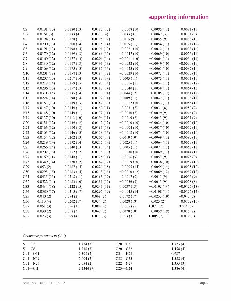
Atomic displacement parameters (Å2)
U11 U22 U33 U12 U13 U23
S1 0.0155 (3) 0.0172 (3) 0.0189 (3) 0.0004 (2) −0.0021 (3) −0.0046 (3)Cu1 0.01124 (16) 0.01627 (17) 0.02098 (18) −0.00100 (12) −0.00448 (12) −0.00854 (13)Cl1 0.0169 (3) 0.0348 (4) 0.0440 (4) 0.0014 (3) −0.0124 (3) −0.0236 (3)
supporting information
sup-4Acta Cryst. (2018). E74, 158-162
C2 0.0181 (13) 0.0180 (13) 0.0193 (13) −0.0008 (10) −0.0095 (11) −0.0091 (11)Cl2 0.0161 (3) 0.0283 (4) 0.0327 (4) 0.0033 (3) −0.0062 (3) −0.0174 (3)N3 0.0194 (11) 0.0178 (11) 0.0196 (12) 0.0015 (9) −0.0055 (9) −0.0086 (10)C4 0.0200 (13) 0.0200 (14) 0.0228 (14) 0.0015 (11) −0.0054 (11) −0.0121 (12)C5 0.0191 (13) 0.0198 (14) 0.0191 (13) −0.0021 (10) −0.0042 (11) −0.0098 (11)C6 0.0170 (12) 0.0169 (13) 0.0166 (13) −0.0047 (10) −0.0069 (10) −0.0073 (11)C7 0.0160 (12) 0.0177 (13) 0.0206 (14) −0.0011 (10) −0.0064 (11) −0.0094 (11)C8 0.0130 (12) 0.0187 (13) 0.0191 (13) −0.0032 (10) −0.0049 (10) −0.0090 (11)C9 0.0169 (12) 0.0175 (13) 0.0187 (13) −0.0023 (10) −0.0063 (10) −0.0087 (11)C10 0.0201 (13) 0.0158 (13) 0.0184 (13) −0.0029 (10) −0.0073 (11) −0.0077 (11)C11 0.0207 (13) 0.0217 (14) 0.0188 (14) 0.0003 (11) −0.0075 (11) −0.0071 (11)C12 0.0218 (14) 0.0259 (15) 0.0192 (14) −0.0016 (11) −0.0054 (11) −0.0082 (12)C13 0.0286 (15) 0.0157 (13) 0.0188 (14) −0.0040 (11) −0.0058 (11) −0.0064 (11)C14 0.0311 (15) 0.0185 (14) 0.0210 (14) 0.0044 (12) −0.0105 (12) −0.0081 (12)C15 0.0224 (14) 0.0198 (14) 0.0192 (14) 0.0009 (11) −0.0042 (11) −0.0106 (11)C16 0.0187 (13) 0.0189 (13) 0.0182 (13) −0.0012 (10) −0.0053 (11) −0.0088 (11)N17 0.0147 (10) 0.0149 (11) 0.0140 (11) −0.0031 (8) −0.0031 (8) −0.0050 (9)N18 0.0148 (10) 0.0149 (11) 0.0172 (11) −0.0030 (8) −0.0029 (9) −0.0046 (9)N19 0.0137 (10) 0.0113 (10) 0.0194 (11) −0.0010 (8) −0.0043 (9) −0.0031 (9)C20 0.0131 (12) 0.0139 (12) 0.0147 (12) −0.0010 (10) −0.0024 (10) −0.0029 (10)C21 0.0166 (12) 0.0180 (13) 0.0161 (13) −0.0004 (10) −0.0037 (10) −0.0072 (11)C22 0.0163 (12) 0.0146 (13) 0.0159 (13) −0.0012 (10) −0.0074 (10) −0.0019 (10)C23 0.0154 (12) 0.0202 (13) 0.0205 (14) 0.0019 (10) −0.0069 (11) −0.0087 (11)C24 0.0219 (14) 0.0192 (14) 0.0215 (14) 0.0025 (11) −0.0064 (11) −0.0068 (11)C25 0.0266 (14) 0.0140 (13) 0.0187 (14) 0.0005 (11) −0.0074 (11) −0.0062 (11)C26 0.0202 (13) 0.0152 (12) 0.0176 (13) −0.0030 (10) −0.0069 (11) −0.0044 (11)N27 0.0169 (11) 0.0148 (11) 0.0125 (11) −0.0016 (9) −0.0057 (9) −0.0025 (9)N28 0.0349 (14) 0.0170 (12) 0.0162 (12) −0.0019 (10) −0.0036 (10) −0.0052 (10)C29 0.053 (2) 0.0167 (14) 0.0221 (15) −0.0005 (14) −0.0055 (14) −0.0035 (12)C30 0.0293 (15) 0.0183 (14) 0.0213 (15) −0.0010 (12) −0.0069 (12) −0.0057 (12)O31 0.0433 (13) 0.0218 (11) 0.0165 (10) −0.0017 (9) −0.0011 (9) −0.0033 (9)O32 0.0522 (14) 0.0183 (10) 0.0181 (10) −0.0036 (9) −0.0013 (9) −0.0078 (8)C33 0.0434 (18) 0.0222 (15) 0.0241 (16) 0.0037 (13) −0.0105 (14) −0.0125 (13)C34 0.0380 (17) 0.0315 (17) 0.0265 (16) −0.0045 (14) −0.0108 (14) −0.0125 (13)C35 0.040 (2) 0.054 (2) 0.068 (3) 0.0172 (17) −0.0253 (19) −0.042 (2)C36 0.110 (4) 0.0202 (17) 0.037 (2) 0.0028 (19) −0.023 (2) −0.0102 (15)C37 0.051 (3) 0.056 (3) 0.084 (4) −0.005 (2) 0.021 (2) 0.004 (3)C38 0.038 (2) 0.058 (3) 0.049 (2) 0.0078 (18) −0.0059 (19) −0.015 (2)N39 0.073 (3) 0.099 (4) 0.072 (3) 0.013 (3) 0.005 (2) −0.029 (3)
Geometric parameters (Å, º)
S1—C2 1.754 (3) C20—C21 1.373 (4)S1—C8 1.736 (3) C20—C22 1.458 (4)Cu1—O31i 2.508 (2) C21—H211 0.937Cu1—N19 2.004 (2) C22—C23 1.388 (4)Cu1—N27 2.054 (2) C22—N27 1.355 (3)Cu1—Cl1 2.2344 (7) C23—C24 1.386 (4)

supporting information
sup-5Acta Cryst. (2018). E74, 158-162
Cu1—Cl2 2.2380 (7) C23—H231 0.943C2—N3 1.300 (3) C24—C25 1.390 (4)C2—C10 1.468 (4) C24—H241 0.946N3—C9 1.387 (4) C25—C26 1.377 (4)C4—C5 1.375 (4) C25—H251 0.943C4—C9 1.403 (4) C26—N27 1.339 (3)C4—H41 0.947 C26—H261 0.944C5—C6 1.402 (4) N28—C29 1.474 (4)C5—H51 0.959 N28—C30 1.356 (4)C6—C7 1.393 (4) C29—H291 0.949C6—C16 1.516 (4) C29—H292 0.964C7—C8 1.385 (4) C29—H293 0.973C7—H71 0.961 C30—O31 1.214 (3)C8—C9 1.403 (4) C30—O32 1.338 (3)C10—C11 1.392 (4) O32—C33 1.476 (3)C10—C15 1.401 (4) C33—C34 1.503 (4)C11—C12 1.386 (4) C33—C35 1.518 (5)C11—H111 0.959 C33—C36 1.526 (5)C12—C13 1.395 (4) C34—H341 0.974C12—H121 0.948 C34—H342 0.963C13—C14 1.390 (4) C34—H343 0.962C13—N28 1.431 (4) C35—H351 0.952C14—C15 1.389 (4) C35—H352 0.969C14—H141 0.948 C35—H353 0.972C15—H151 0.948 C36—H361 0.960C16—N17 1.473 (3) C36—H362 0.961C16—H161 0.973 C36—H363 0.972C16—H162 0.971 C37—C38 1.422 (6)N17—N18 1.336 (3) C37—H371 0.970N17—C21 1.352 (3) C37—H372 0.957N18—N19 1.315 (3) C37—H373 0.977N19—C20 1.362 (3) C38—N39 1.131 (6)
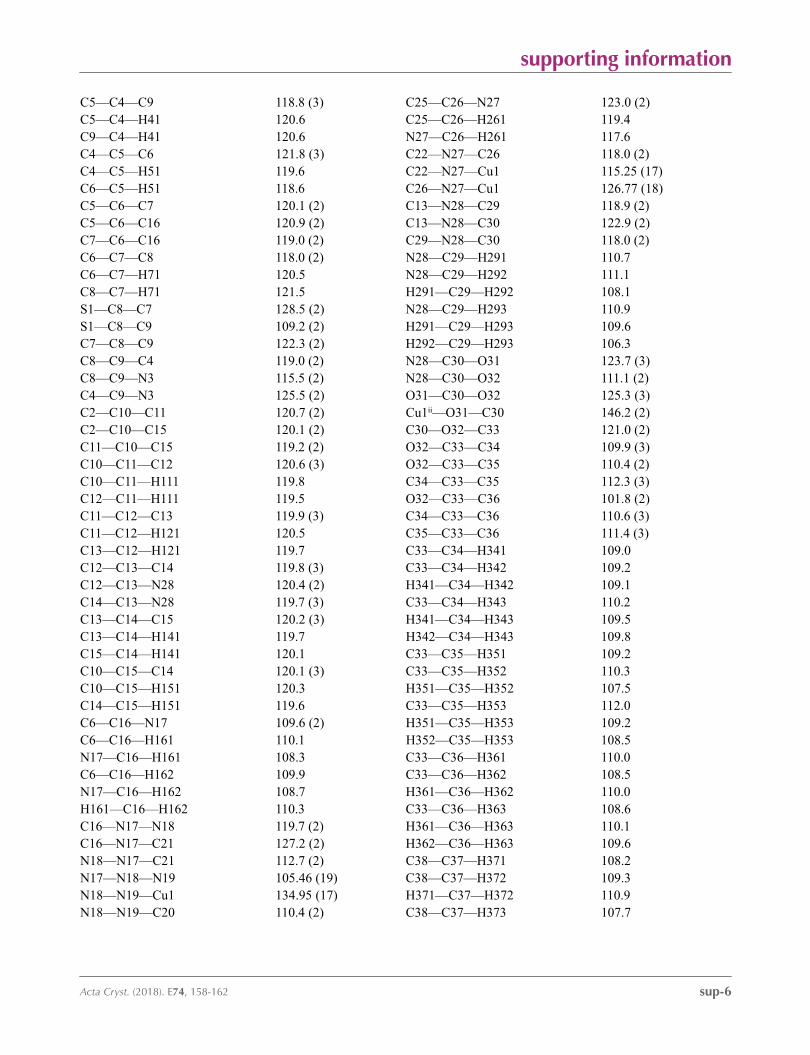
C2—S1—C8 89.01 (13) C20—C21—N17 103.8 (2)O31i—Cu1—Cl1 107.68 (6) C20—C21—H211 130.3O31i—Cu1—Cl2 100.13 (5) N17—C21—H211 125.8Cl1—Cu1—Cl2 93.31 (3) C20—C22—C23 124.4 (2)O31i—Cu1—N19 80.21 (8) C20—C22—N27 113.2 (2)Cl1—Cu1—N19 168.01 (7) C23—C22—N27 122.4 (2)Cl2—Cu1—N19 94.14 (6) C22—C23—C24 118.6 (2)O31i—Cu1—N27 83.26 (8) C22—C23—H231 119.9Cl1—Cu1—N27 91.79 (6) C24—C23—H231 121.5Cl2—Cu1—N27 172.70 (6) C23—C24—C25 119.0 (2)N19—Cu1—N27 80.00 (8) C23—C24—H241 121.6S1—C2—N3 115.9 (2) C25—C24—H241 119.5S1—C2—C10 119.49 (19) C24—C25—C26 118.9 (2)N3—C2—C10 124.6 (2) C24—C25—H251 121.0C2—N3—C9 110.5 (2) C26—C25—H251 120.0
supporting information
sup-6Acta Cryst. (2018). E74, 158-162
C5—C4—C9 118.8 (3) C25—C26—N27 123.0 (2)C5—C4—H41 120.6 C25—C26—H261 119.4C9—C4—H41 120.6 N27—C26—H261 117.6C4—C5—C6 121.8 (3) C22—N27—C26 118.0 (2)C4—C5—H51 119.6 C22—N27—Cu1 115.25 (17)C6—C5—H51 118.6 C26—N27—Cu1 126.77 (18)C5—C6—C7 120.1 (2) C13—N28—C29 118.9 (2)C5—C6—C16 120.9 (2) C13—N28—C30 122.9 (2)C7—C6—C16 119.0 (2) C29—N28—C30 118.0 (2)C6—C7—C8 118.0 (2) N28—C29—H291 110.7C6—C7—H71 120.5 N28—C29—H292 111.1C8—C7—H71 121.5 H291—C29—H292 108.1S1—C8—C7 128.5 (2) N28—C29—H293 110.9S1—C8—C9 109.2 (2) H291—C29—H293 109.6C7—C8—C9 122.3 (2) H292—C29—H293 106.3C8—C9—C4 119.0 (2) N28—C30—O31 123.7 (3)C8—C9—N3 115.5 (2) N28—C30—O32 111.1 (2)C4—C9—N3 125.5 (2) O31—C30—O32 125.3 (3)C2—C10—C11 120.7 (2) Cu1ii—O31—C30 146.2 (2)C2—C10—C15 120.1 (2) C30—O32—C33 121.0 (2)C11—C10—C15 119.2 (2) O32—C33—C34 109.9 (3)C10—C11—C12 120.6 (3) O32—C33—C35 110.4 (2)C10—C11—H111 119.8 C34—C33—C35 112.3 (3)C12—C11—H111 119.5 O32—C33—C36 101.8 (2)C11—C12—C13 119.9 (3) C34—C33—C36 110.6 (3)C11—C12—H121 120.5 C35—C33—C36 111.4 (3)C13—C12—H121 119.7 C33—C34—H341 109.0C12—C13—C14 119.8 (3) C33—C34—H342 109.2C12—C13—N28 120.4 (2) H341—C34—H342 109.1C14—C13—N28 119.7 (3) C33—C34—H343 110.2C13—C14—C15 120.2 (3) H341—C34—H343 109.5C13—C14—H141 119.7 H342—C34—H343 109.8C15—C14—H141 120.1 C33—C35—H351 109.2C10—C15—C14 120.1 (3) C33—C35—H352 110.3C10—C15—H151 120.3 H351—C35—H352 107.5C14—C15—H151 119.6 C33—C35—H353 112.0C6—C16—N17 109.6 (2) H351—C35—H353 109.2C6—C16—H161 110.1 H352—C35—H353 108.5N17—C16—H161 108.3 C33—C36—H361 110.0C6—C16—H162 109.9 C33—C36—H362 108.5N17—C16—H162 108.7 H361—C36—H362 110.0H161—C16—H162 110.3 C33—C36—H363 108.6C16—N17—N18 119.7 (2) H361—C36—H363 110.1C16—N17—C21 127.2 (2) H362—C36—H363 109.6N18—N17—C21 112.7 (2) C38—C37—H371 108.2N17—N18—N19 105.46 (19) C38—C37—H372 109.3N18—N19—Cu1 134.95 (17) H371—C37—H372 110.9N18—N19—C20 110.4 (2) C38—C37—H373 107.7

supporting information
sup-7Acta Cryst. (2018). E74, 158-162
Cu1—N19—C20 114.51 (16) H371—C37—H373 109.9N19—C20—C21 107.6 (2) H372—C37—H373 110.6N19—C20—C22 116.9 (2) C37—C38—N39 176.6 (5)C21—C20—C22 135.4 (2)
Symmetry codes: (i) x−1, y−1, z+1; (ii) x+1, y+1, z−1.
Hydrogen-bond geometry (Å, º)
Cg is the centroid of the C4–C9 ring.
D—H···A D—H H···A D···A D—H···A
C16—H162···N39iii 0.97 2.52 3.451 (6) 161C16—H161···Cl2iv 0.97 2.72 3.606 (3) 152C21—H211···Cl1iv 0.94 2.81 3.633 (3) 147C23—H231···Cl1iv 0.94 2.62 3.494 (3) 155C26—H261···Cl1 0.94 2.55 3.154 (3) 122C29—H291···Cl2ii 0.95 2.80 3.741 (3) 172C25—H251···Cgv 0.94 2.85 3.583 (3) 135
Symmetry codes: (ii) x+1, y+1, z−1; (iii) −x, −y, −z+2; (iv) x+1, y, z; (v) −x, −y−1, −z+2.
Related Documents


