Research Article Serotonin- and Dopamine-Related Gene Expression in db/db Mice Islets and in MIN6 -Cells Treated with Palmitate and Oleate L. R. Cataldo, 1,2 M. L. Mizgier, 1 D. Busso, 1 P. Olmos, 1 J. E. Galgani, 1,3 R. Valenzuela, 4 D. Mezzano, 5 E. Aranda, 5 V. A. Cortés, 1 and J. L. Santos 1 1 Departamento de Nutrici´ on, Diabetes y Metabolismo, Escuela de Medicina, Pontificia Universidad Cat´ olica de Chile, 8331150 Santiago, Chile 2 Facultad de Medicina, Universidad de los Andes, 7620001 Santiago, Chile 3 UDA-Ciencias de la Salud, Carrera de Nutrici´ on y Diet´ etica, Escuela de Medicina, Pontificia Universidad Cat´ olica de Chile, 8331150 Santiago, Chile 4 Departamento de Nutrici´ on, Facultad de Medicina, Universidad de Chile, 7550367 Santiago, Chile 5 Laboratorio de Hemostasia, Escuela de Medicina, Pontificia Universidad Cat´ olica de Chile, 8331150 Santiago, Chile Correspondence should be addressed to J. L. Santos; [email protected] Received 27 January 2016; Revised 26 April 2016; Accepted 10 May 2016 Academic Editor: Andrea Tura Copyright © 2016 L. R. Cataldo et al. is is an open access article distributed under the Creative Commons Attribution License, which permits unrestricted use, distribution, and reproduction in any medium, provided the original work is properly cited. High circulating nonesterified fatty acids (NEFAs) concentration, oſten reported in diabetes, leads to impaired glucose-stimulated insulin secretion (GSIS) through not yet well-defined mechanisms. Serotonin and dopamine might contribute to NEFA-dependent -cell dysfunction, since extracellular signal of these monoamines decreases GSIS. Moreover, palmitate-treated -cells may enhance the expression of the serotonin receptor Htr2c, affecting insulin secretion. Additionally, the expression of monoamine-oxidase type B (Maob) seems to be lower in islets from humans and mice with diabetes compared to nondiabetic islets, which may lead to increased monoamine concentrations. We assessed the expression of serotonin- and dopamine-related genes in islets from db/db and wild-type (WT) mice. In addition, the effect of palmitate and oleate on the expression of such genes, 5HT content, and GSIS in MIN6 -cell was determined. Lower Maob expression was found in islets from db/db versus WT mice and in MIN6 -cells in response to palmitate and oleate treatment compared to vehicle. Reduced 5HT content and impaired GSIS in response to palmitate (−25%; < 0.0001) and oleate (−43%; < 0.0001) were detected in MIN6 -cells. In conclusion, known defects of GSIS in islets from db/db mice and MIN6 -cells treated with NEFAs are accompanied by reduced Maob expression and reduced 5HT content. 1. Introduction Both insulin resistance and -cell failure are involved in the etiology of type-2 diabetes (DM2) phenotype [1]. High circulating nonesterified fatty acids (NEFAs) concentration is oſten reported in fasting physiological state [2] or phys- iopathological conditions such as stressed states [3] and DM2 [4–6]. Lipid infusion in nondiabetic subjects lead- ing to circulating NEFAs levels similar to those found in diabetic people (500–800 M) decreased glucose-stimulated insulin secretion (GSIS) [7]. Similar results were obtained in animal models [8]. Moreover, several in vitro studies have demonstrated that long-term exposure of -cells lines and murine islets to palmitic (C16:0) and oleic (C18:1) acids leads to impaired GSIS [5, 8–10]. In rat pancreatic islets, the mechanism of -cell lipodysfunction has been associated with reduced glucose uptake and mitochondrial oxidation caused by decreased activity of pyruvate dehydrogenase (PDH) and increased expression of UCP2 [11]. Other studies have explained impaired GSIS as a consequence of excessive production of radical oxygen species (ROS), which is depen- dent on mitochondrial fatty acid metabolism [9]. However, these mechanisms do not fully explain such decreased GSIS, and alternative mechanisms may be taking place. Hindawi Publishing Corporation Journal of Diabetes Research Volume 2016, Article ID 3793781, 12 pages http://dx.doi.org/10.1155/2016/3793781

Welcome message from author
This document is posted to help you gain knowledge. Please leave a comment to let me know what you think about it! Share it to your friends and learn new things together.
Transcript

Research ArticleSerotonin- and Dopamine-Related GeneExpression in db/db Mice Islets and in MIN6 𝛽-CellsTreated with Palmitate and Oleate
L. R. Cataldo,1,2 M. L. Mizgier,1 D. Busso,1 P. Olmos,1 J. E. Galgani,1,3 R. Valenzuela,4
D. Mezzano,5 E. Aranda,5 V. A. Cortés,1 and J. L. Santos1
1Departamento de Nutricion, Diabetes y Metabolismo, Escuela de Medicina, Pontificia Universidad Catolica de Chile,8331150 Santiago, Chile2Facultad de Medicina, Universidad de los Andes, 7620001 Santiago, Chile3UDA-Ciencias de la Salud, Carrera de Nutricion y Dietetica, Escuela de Medicina, Pontificia Universidad Catolica de Chile,8331150 Santiago, Chile4Departamento de Nutricion, Facultad de Medicina, Universidad de Chile, 7550367 Santiago, Chile5Laboratorio de Hemostasia, Escuela de Medicina, Pontificia Universidad Catolica de Chile, 8331150 Santiago, Chile
Correspondence should be addressed to J. L. Santos; [email protected]
Received 27 January 2016; Revised 26 April 2016; Accepted 10 May 2016
Academic Editor: Andrea Tura
Copyright © 2016 L. R. Cataldo et al. This is an open access article distributed under the Creative Commons Attribution License,which permits unrestricted use, distribution, and reproduction in any medium, provided the original work is properly cited.
High circulating nonesterified fatty acids (NEFAs) concentration, often reported in diabetes, leads to impaired glucose-stimulatedinsulin secretion (GSIS) through not yet well-definedmechanisms. Serotonin and dopamine might contribute to NEFA-dependent𝛽-cell dysfunction, since extracellular signal of thesemonoamines decreasesGSIS.Moreover, palmitate-treated𝛽-cellsmay enhancethe expression of the serotonin receptor Htr2c, affecting insulin secretion. Additionally, the expression of monoamine-oxidase typeB (Maob) seems to be lower in islets from humans and mice with diabetes compared to nondiabetic islets, which may lead toincreased monoamine concentrations. We assessed the expression of serotonin- and dopamine-related genes in islets from db/dband wild-type (WT) mice. In addition, the effect of palmitate and oleate on the expression of such genes, 5HT content, and GSISin MIN6 𝛽-cell was determined. Lower Maob expression was found in islets from db/db versus WT mice and in MIN6 𝛽-cells inresponse to palmitate and oleate treatment compared to vehicle. Reduced 5HT content and impaired GSIS in response to palmitate(−25%; 𝑝 < 0.0001) and oleate (−43%; 𝑝 < 0.0001) were detected in MIN6 𝛽-cells. In conclusion, known defects of GSIS in isletsfrom db/dbmice and MIN6 𝛽-cells treated with NEFAs are accompanied by reduced Maob expression and reduced 5HT content.
1. Introduction
Both insulin resistance and 𝛽-cell failure are involved inthe etiology of type-2 diabetes (DM2) phenotype [1]. Highcirculating nonesterified fatty acids (NEFAs) concentrationis often reported in fasting physiological state [2] or phys-iopathological conditions such as stressed states [3] andDM2 [4–6]. Lipid infusion in nondiabetic subjects lead-ing to circulating NEFAs levels similar to those found indiabetic people (500–800 𝜇M) decreased glucose-stimulatedinsulin secretion (GSIS) [7]. Similar results were obtainedin animal models [8]. Moreover, several in vitro studies
have demonstrated that long-term exposure of 𝛽-cells linesand murine islets to palmitic (C16:0) and oleic (C18:1) acidsleads to impaired GSIS [5, 8–10]. In rat pancreatic islets,the mechanism of 𝛽-cell lipodysfunction has been associatedwith reduced glucose uptake and mitochondrial oxidationcaused by decreased activity of pyruvate dehydrogenase(PDH) and increased expression of UCP2 [11]. Other studieshave explained impaired GSIS as a consequence of excessiveproduction of radical oxygen species (ROS), which is depen-dent on mitochondrial fatty acid metabolism [9]. However,these mechanisms do not fully explain such decreased GSIS,and alternative mechanisms may be taking place.
Hindawi Publishing CorporationJournal of Diabetes ResearchVolume 2016, Article ID 3793781, 12 pageshttp://dx.doi.org/10.1155/2016/3793781
2 Journal of Diabetes Research
Other molecular signals including serotonin and dopam-ine might contribute to NEFA-dependent 𝛽-cell dysfunction.In this context, high circulating levels of serotonin areobserved under fasting conditions [2] and also in patientswith DM2 [12, 13]. Furthermore, dietary fat compositionmayalter serotonin and dopamine concentration in the brain[14]. Moreover, NEFAs may directly modify the expressionof some monoamines signaling genes in 𝛽-cells. It has beenreported that expression of the serotonin receptor Htr2cis higher in pancreatic islets from diabetic versus controlmice, and it is positively regulated in mice 𝛽-cells exposedto palmitic acid [15]. Moreover, agonists of Htr2c reducethe GSIS and may contribute to NEFAs-dependent 𝛽-celldysfunction [15]. In addition, it has been recently reportedthat the activity of monoamine-oxidase type B (Maob),which degrades the monoamines, is reduced in diabetichuman and mice islets [16], suggesting that higher content ofmonoamines in 𝛽-cells contributes to reduced GSIS in DM2.In support of this hypothesis, gut-derived serotonin anddopamine [2, 17–19] or synthesized [18, 20] and coreleasedfrom 𝛽-cells could, in an endocrine or auto/paracrine man-ner, negatively modulate GSIS [15, 18, 19, 21–27]. In fact, sero-tonin acting through Htr2c receptor and dopamine throughD2 or D3 receptor decrease GSIS in 𝛽-cell lines and humanor mice islets [27–31]. In addition, antagonism of dopamineD2 receptors signaling in human islets enhanced GSIS [27].
In this regard, long-term exposure of 𝛽-cells to NEFAsmay change the expression of some monoamines-relatedgenes leading to increased serotonin and dopamine contentand chronic autocrine signaling that consequently impairGSIS.The aims of this studywere (i) to assess and qualitativelycompare the expression of serotonin- and dopamine-relatedgenes in pancreatic islets from wild-type (WT) and db/dbmice and (ii) to evaluate the effect of palmitate and oleate onexpression of such genes, 5HT content, and GSIS in MIN6𝛽-cells.
2. Material and Methods
2.1. MIN6 Cell Culture. MIN6 𝛽-cells were cultured in Dul-becco’s modified Eagle medium (DMEM) (Gibco; Life Tech-nologies Co., Grand Island, NY) containing 10% fetal bovineserum (FBS), 25mmol/L glucose, 3.7 g/L sodiumbicarbonate,100 U/mL penicillin, and 100 𝜇g/mL streptomycin. MIN6𝛽-cells were cultured at 37∘C in a humidified atmospherecontaining 95% air and 5% CO
2. These cells were provided
by Professor Francisco Perez-Bravo (University of Chile,Santiago, Chile).
2.2. NEFAs Preparation andTreatment Protocol. Sodium saltssolutions of nonesterified palmitic (Sigma, code P9767) andoleic (Sigma, code O7501) acids were prepared as previouslyreported [34]. Briefly, 1mL of 100mM palmitic or oleicsolutions (27.8mg of palmitate and 30.4mg oleate) wasdissolved in NaOH 0.1M and warmed up to 60∘–70∘C andgently shaken. Then, the solutions were diluted twenty timesin DMEM medium with 10% fatty acid-free Bovine SerumAlbumin (BSA). Diluted NEFAs solutions were filtered
(0.45 𝜇m), aliquoted in amber tubes, and frozen at −20∘C.Experiments were then conducted by diluting 10x with freshand sterile DMEMmedium (10% fetal bovine serum, FBS) toreach the final concentration of 0.5mM of NEFAs salts and1% BSA.
MIN6 𝛽-cells were incubated with palmitate or oleate(0.5mM, 1% BSA) in DMEM medium (10% FBS) for 24hours, and then glucose-stimulated insulin secretion wasassessed. In parallel, the viability of MIN6 𝛽-cells after 24 hof exposure to fatty acids was measured by Trypan Blueexclusion assay and the percentage of viability was quantifiedby an automated cell counter (Luna, Logos Biosystems).
2.3. Glucose-Stimulated Insulin Secretion (GSIS) Assay. MIN6𝛽-cells were evaluated by means of the static GSIS protocols.Briefly, GSIS protocol consisted in sensitizing the MIN6 𝛽-cells by exposing them to Krebs-Ringer HEPES buffer (KRH;NaCl 130mmol/L, KH
2PO41.25mmol/L, MgSO
41.25mmol/
L, CaCl22.68mmol/L, NaHCO
35.26mmol/L, and HEPES
10mmol/L) without or with low glucose (2.8mM), duringthirty minutes. Immediately after that, MIN6 𝛽-cells werestimulated to secrete insulin with KRH buffer (0.5% BSA)without or with glucose (20mM) for one hour. The super-natants were spun to 5000 rpm during 10 minutes to 4∘C andstorage to −20∘C. Later, the extracellular (secreted) insulinconcentration was measured using the mouse/rat insulinELISA kits (Merck-Millipore, code EZRMI-13K). GSIS wasexpressed as either the concentration of insulin secreted,at basal and stimulated glucose levels (normalized by totalmass protein), or through the ratio of both, defined as theStimulation Index (SI).
2.4. Animal Models. db/db mice were obtained from Jack-son Laboratories (strain B6.BKS(D)-Leprdb/J). Male, 16–20-week-old wild-type (WT) C57BL6/J (WT (24–30 g) anddb/db (55–60 g)) mice were studied.
This research was conducted in accordance with theexperimental protocol approved by the Bioethical and Ani-mal Welfare Committee from the School of Medicine, Ponti-ficia Universidad Catolica de Chile.
2.5. Plasma Insulin and NEFAs Assessment. Insulin and totalNEFAsmeasurements were performed from a pool of plasmaof db/db and WT mice (𝑛 = 5 per group). The total NEFAsquantitation was performed by the colorimetric test NEFAHR (Wako, code 993-35191) and the insulin levels by ELISA(Merck, code EZRMI-13K).
To quantify the specific NEFAs species from plasmapools, total lipids were quantitatively extracted by Blighand Dyer method [35]. Briefly, the samples were homoge-nized with ice-cold chloroform/methanol (2 : 1 v/v) contain-ing 0.01% butylated hydroxytoluene (BHT) as an antioxidant.Then the fatty acid methyl ester (FAME) were prepared andprocessed by Bond ElutNH
2columns (Agilent Technologies,
USA) and the NEFAs were eluted with hexane. The NEFAswere separated and quantified by gas-liquid chromatographyin Agilent Hewlett-Packard equipment (model 7890A, CA,
Journal of Diabetes Research 3
L-Tryptophan(Trp)
5-Hydroxytryptophan (5HTP)
Serotonin, 5HT (5-hydroxytryptamine)
Tryptophan hydroxylase(TPH1/2)
Dopa-decarboxylase (DDC)Tetrahydrobiopterin Dihydrobiopterin
Pyridoxal phosphate
L-Dopa
Dopamine
Noradrenaline
Adrenaline
L-TyrosineAscorbic acid
Dehydroascorbic acid
S-Adenosyl-methionine
Homocysteine
hydroxylase (DBH)
PhenylethanolamineN-methyltransferase
Tyrosine hydroxylase (TH)
N-Acetylserotonin
Arylalkylamine N-acetyltransferase (AANAT)
Acetylserotonin O-methyltransferase
(ASMT)Melatonin
AcCoA
CoASH
O2
O2
O2
H2O
H2O
H2O
H3C O
O
O
O
O
HN HN
HN
HNHNCH3 CH3
HO
HO
NH2
NH2
NH2
NH2
(R)
OH
OH
OH
O
HN
HO
NH2
OH
OH
O
HN
HO
HO
HO
HO
HO
HO
HO
H2N
NH2
OH
OH
OH
HN
Dopamine-𝛽-
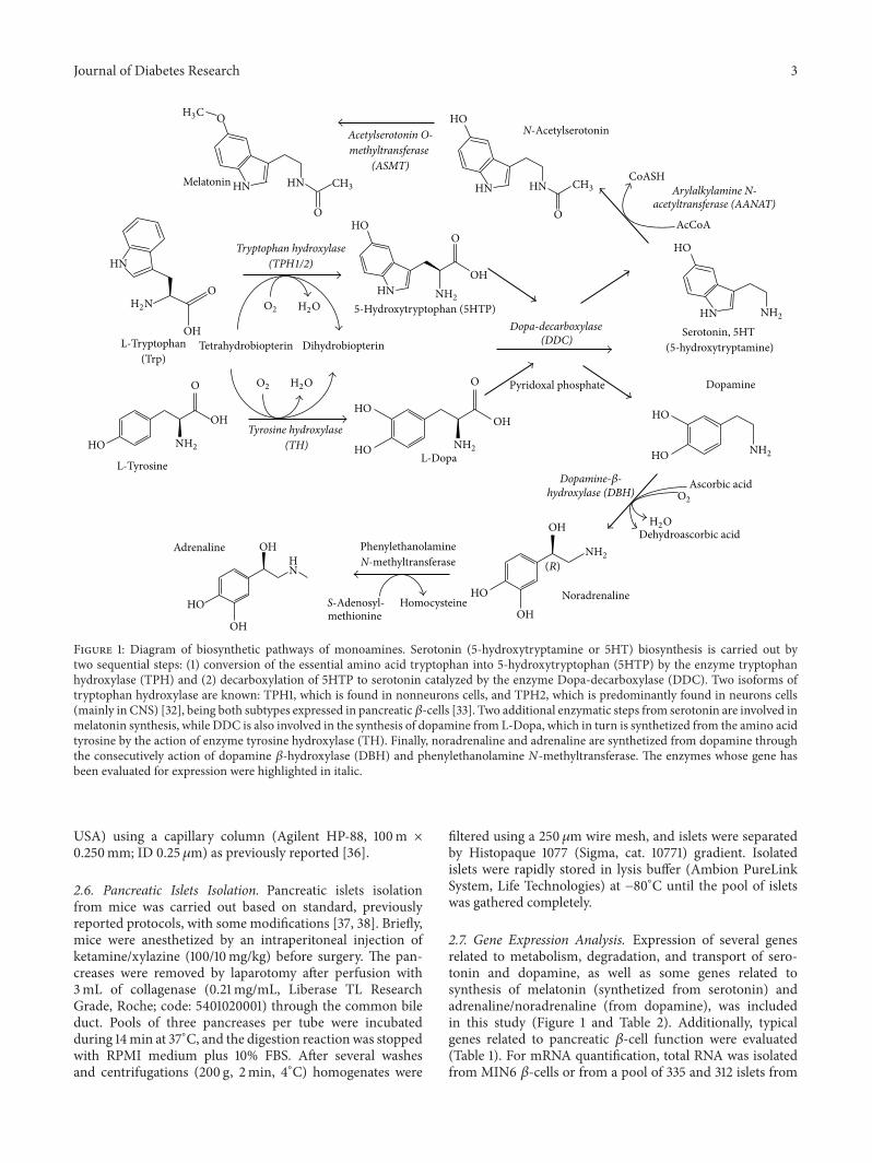
Figure 1: Diagram of biosynthetic pathways of monoamines. Serotonin (5-hydroxytryptamine or 5HT) biosynthesis is carried out bytwo sequential steps: (1) conversion of the essential amino acid tryptophan into 5-hydroxytryptophan (5HTP) by the enzyme tryptophanhydroxylase (TPH) and (2) decarboxylation of 5HTP to serotonin catalyzed by the enzyme Dopa-decarboxylase (DDC). Two isoforms oftryptophan hydroxylase are known: TPH1, which is found in nonneurons cells, and TPH2, which is predominantly found in neurons cells(mainly in CNS) [32], being both subtypes expressed in pancreatic 𝛽-cells [33]. Two additional enzymatic steps from serotonin are involved inmelatonin synthesis, while DDC is also involved in the synthesis of dopamine from L-Dopa, which in turn is synthetized from the amino acidtyrosine by the action of enzyme tyrosine hydroxylase (TH). Finally, noradrenaline and adrenaline are synthetized from dopamine throughthe consecutively action of dopamine 𝛽-hydroxylase (DBH) and phenylethanolamine N-methyltransferase. The enzymes whose gene hasbeen evaluated for expression were highlighted in italic.
USA) using a capillary column (Agilent HP-88, 100m ×0.250mm; ID 0.25 𝜇m) as previously reported [36].
2.6. Pancreatic Islets Isolation. Pancreatic islets isolationfrom mice was carried out based on standard, previouslyreported protocols, with some modifications [37, 38]. Briefly,mice were anesthetized by an intraperitoneal injection ofketamine/xylazine (100/10mg/kg) before surgery. The pan-creases were removed by laparotomy after perfusion with3mL of collagenase (0.21mg/mL, Liberase TL ResearchGrade, Roche; code: 5401020001) through the common bileduct. Pools of three pancreases per tube were incubatedduring 14min at 37∘C, and the digestion reactionwas stoppedwith RPMI medium plus 10% FBS. After several washesand centrifugations (200 g, 2min, 4∘C) homogenates were
filtered using a 250𝜇m wire mesh, and islets were separatedby Histopaque 1077 (Sigma, cat. 10771) gradient. Isolatedislets were rapidly stored in lysis buffer (Ambion PureLinkSystem, Life Technologies) at −80∘C until the pool of isletswas gathered completely.
2.7. Gene Expression Analysis. Expression of several genesrelated to metabolism, degradation, and transport of sero-tonin and dopamine, as well as some genes related tosynthesis of melatonin (synthetized from serotonin) andadrenaline/noradrenaline (from dopamine), was includedin this study (Figure 1 and Table 2). Additionally, typicalgenes related to pancreatic 𝛽-cell function were evaluated(Table 1). For mRNA quantification, total RNA was isolatedfrom MIN6 𝛽-cells or from a pool of 335 and 312 islets from
4 Journal of Diabetes Research
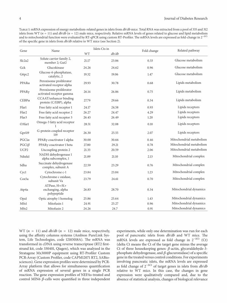
Table 1: mRNA expression of energymetabolism-related genes in islets from db/dbmice. Total RNAwas extracted from a pool of 335 and 312islets from WT (𝑛 = 11) and db/db (𝑛 = 12) male mice, respectively. Relative mRNA levels of genes related to glucose and lipid metabolismand to mitochondrial function were evaluated by RT-qPCR using custom RT-Profiler.ThemRNA levels are expressed as fold-change in 2−ΔCtof the specific gene in islets from db/db relative to WT mice (see Section 2).
Gene Name Islets Cts in Fold change Related pathwayWT db/db
Slc2a2 Solute carrier family 2,member 2; Gut2 21.17 23.06 0.33 Glucose metabolism
Gck Glucokinase 24.26 24.62 0.96 Glucose metabolism
G6pc2 Glucose-6-phosphatase,catalytic, 2 19.32 19.06 1.47 Glucose metabolism
PPAR𝛼 Peroxisome proliferatoractivated receptor alpha 29.93 30.78 0.68 Lipids metabolism
PPAR𝛾 Peroxisome proliferatoractivated receptor gamma 26.14 26.86 0.75 Lipids metabolism
CEBP𝛼 CCAAT/enhancer bindingprotein (C/EBP), alpha 27.79 29.66 0.34 Lipids metabolism
Ffar1 Free fatty acid receptor 1 24.17 24.58 0.93 Lipids receptorsFfar2 Free fatty acid receptor 2 26.27 24.47 4.29 Lipids receptorsFfar3 Free fatty acid receptor 3 26.45 26.49 1.20 Lipids receptors
O3far1 Omega-3 fatty acid receptor1 28.51 32.08 0.10 Lipids receptors
Gpr119 G-protein coupled receptor119 26.30 25.55 2.07 Lipids receptors
PGC1𝛼 PPAR𝛾 coactivator 1 alpha 30.00 30.66 0.46 Mitochondrial metabolismPGC1𝛽 PPAR𝛾 coactivator 1 beta 27.80 29.21 0.78 Mitochondrial metabolismUCP2 Uncoupling protein 2 21.33 20.59 2.06 Mitochondrial metabolism
Ndufa1 NADH dehydrogenase 1alpha subcomplex, 1 21.89 21.10 2.13 Mitochondrial complex
Sdha Succinate dehydrogenasecomplex, subunit A 22.59 23.29 0.76 Mitochondrial complex
Cyc1 Cytochrome c-1 23.84 23.84 1.23 Mitochondrial complex
Cox5a Cytochrome c oxidase,subunit Va 23.79 24.61 0.70 Mitochondrial complex
Atp4aATPase, H+/K+exchanging, alpha
polypeptide26.83 28.70 0.34 Mitochondrial dynamics
Opa1 Optic atrophy 1 homolog 25.86 25.64 1.43 Mitochondrial dynamicsMfn1 Mitofusin 1 24.91 25.27 0.96 Mitochondrial dynamicsMfn2 Mitofusin 2 24.26 24.7 0.91 Mitochondrial dynamics
WT (𝑛 = 11) and db/db (𝑛 = 12) male mice, respectively,using the affinity columns systems (Ambion PureLink Sys-tem, Life Technologies, code 12183018A). The mRNA wastransformed in cDNA using reverse transcriptase (RT2 first-strand kit, code 330401, Qiagen), which was analyzed in theStratagene Mx3000P equipment using RT-Profiler CustomPCR-Array (Custom Profiler, code CAPM12071 RT2, SABio-sciences). Gene expression profiles were determined by PCR-Array platform that allows for simultaneous quantificationof mRNA expression of several genes in a single PCRreaction. The gene expression profiles of NEFAs-treated andcontrol MIN6 𝛽-cells were quantified in three independent
experiments, while only one determination was run for eachpool of pancreatic islets from db/db and WT mice. ThemRNA levels are expressed as fold change in 2−ΔCt (Ct)(delta Ct means the Ct of the target gene minus the averageCt of three housekeeping genes: 𝛽-actin, glyceraldehyde-3-phosphate dehydrogenase, and 𝛽-glucuronidase) of a specificgene in the treated versus control conditions. For experimentsinvolving pancreatic islets, the mRNA levels are expressedas fold change of 2−ΔCt of target genes in islets from db/dbrelative to WT mice. In this case, the changes in geneexpression were qualitatively compared and, due to theabsence of statistical analysis, changes of biological relevance
Journal of Diabetes Research 5
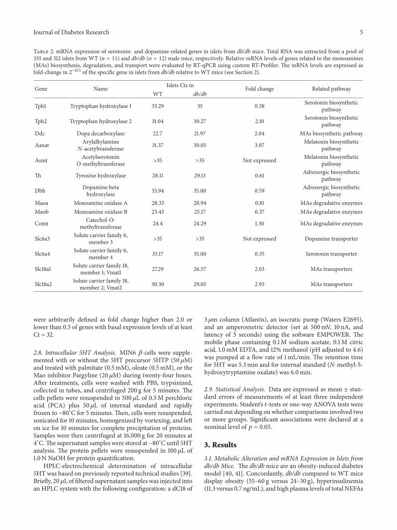
Table 2: mRNA expression of serotonin- and dopamine-related genes in islets from db/db mice. Total RNA was extracted from a pool of335 and 312 islets from WT (𝑛 = 11) and db/db (𝑛 = 12) male mice, respectively. Relative mRNA levels of genes related to the monoamines(MAs) biosynthesis, degradation, and transport were evaluated by RT-qPCR using custom RT-Profiler. The mRNA levels are expressed asfold-change in 2−ΔCt of the specific gene in islets from db/db relative to WT mice (see Section 2).
Gene Name Islets Cts in Fold change Related pathwayWT db/db
Tph1 Tryptophan hydroxylase 1 33.29 35 0.38 Serotonin biosyntheticpathway
Tph2 Tryptophan hydroxylase 2 31.04 30.27 2.10 Serotonin biosyntheticpathway
Ddc Dopa decarboxylase 22.7 21.97 2.04 MAs biosynthetic pathway
Aanat ArylalkylamineN-acetyltransferase 31.37 30.05 3.07 Melatonin biosynthetic
pathway
Asmt AcetylserotoninO-methyltransferase >35 >35 Not expressed Melatonin biosynthetic
pathway
Th Tyrosine hydroxylase 28.11 29.13 0.61 Adrenergic biosyntheticpathway
Dbh Dopamine betahydroxylase 33.94 35.00 0.59 Adrenergic biosynthetic
pathwayMaoa Monoamine oxidase A 28.33 28.94 0.81 MAs degradative enzymesMaob Monoamine oxidase B 23.43 25.17 0.37 MAs degradative enzymes
Comt Catechol-O-methyltransferase 24.4 24.29 1.30 MAs degradative enzymes
Slc6a3 Solute carrier family 6,member 3 >35 >35 Not expressed Dopamine transporter
Slc6a4 Solute carrier family 6,member 4 33.17 35.00 0.35 Serotonin transporter
Slc18a1 Solute carrier family 18,member 1; Vmat1 27.29 26.57 2.03 MAs transporters
Slc18a2 Solute carrier family 18,member 2; Vmat2 30.30 29.05 2.93 MAs transporters
were arbitrarily defined as fold change higher than 2.0 orlower than 0.5 of genes with basal expression levels of at leastCt = 32.
2.8. Intracellular 5HT Analysis. MIN6 𝛽-cells were supple-mented with or without the 5HT precursor 5HTP (50𝜇M)and treated with palmitate (0.5mM), oleate (0.5mM), or theMao inhibitor Pargyline (20 𝜇M) during twenty-four hours.After treatments, cells were washed with PBS, trypsinized,collected in tubes, and centrifuged 200 g for 5 minutes. Thecells pellets were resuspended in 500 𝜇L of 0.5M perchloricacid (PCA) plus 50𝜇L of internal standard and rapidlyfrozen to −80∘C for 5 minutes. Then, cells were resuspended,sonicated for 10 minutes, homogenized by vortexing, and lefton ice for 10 minutes for complete precipitation of proteins.Samples were then centrifuged at 16.000 g for 20 minutes at4∘C.The supernatant samples were stored at −80∘C until 5HTanalysis. The protein pellets were resuspended in 100 𝜇L of1.0N NaOH for protein quantification.
HPLC-electrochemical determination of intracellular5HT was based on previously reported technical studies [39].Briefly, 20𝜇L of filtered supernatant sampleswas injected intoan HPLC system with the following configuration: a dC18 of
3 𝜇m column (Atlantis), an isocratic pump (Waters E2695),and an amperometric detector (set at 500mV, 10 nA, andlatency of 5 seconds) using the software EMPOWER. Themobile phase containing 0.1M sodium acetate, 0.1M citricacid, 1.0mM EDTA, and 12% methanol (pH adjusted to 4.6)was pumped at a flow rate of 1mL/min. The retention timefor 5HT was 5.3min and for internal standard (N-methyl-5-hydroxytryptamine oxalate) was 6.0min.
2.9. Statistical Analysis. Data are expressed as mean ± stan-dard errors of measurements of at least three independentexperiments. Student’s 𝑡-tests or one-way ANOVA tests werecarried out depending on whether comparisons involved twoor more groups. Significant associations were declared at anominal level of 𝑝 = 0.05.
3. Results
3.1. Metabolic Alteration and mRNA Expression in Islets fromdb/db Mice. The db/dbmice are an obesity-induced diabetesmodel [40, 41]. Concordantly, db/db compared to WT micedisplay obesity (55–60 g versus 24–30 g), hyperinsulinemia(11.3 versus 0.7 ng/mL), and high plasma levels of total NEFAs
6 Journal of Diabetes Research
(0.93 versus 0.34mM). Furthermore, it is known that db/dbmice islets have functional defects and increased compen-satory 𝛽-cell mass [40–42]; in agreement we observed ahigher size in islets from db/db compared to WT mice (datanot shown).
To further study the metabolic disturbance, the mRNAexpression levels of some genes related to glucose, lipids,and mitochondrial metabolism were measured in islets fromdb/db and WT mice and they were compared qualitatively(Table 1). Islets from db/db versus WT mice showed lowerexpression of glucose transporter 2 gene Slc2a2 (Glut2) (0.33-fold; Table 1). In turn, lipid receptor genes such as Ffar2and Gpr119 had increased (4.29-fold and 2.07-fold), whileO3far1 decreased expression (0.10-fold) in islets from db/dbcompared with WT mice (Table 1). Mitochondrial-relatedgenes including PGC1𝛼 (0.46-fold) and ATP synthase alphasubunit (Atp4a) (0.34-fold) showed lower expression whilea higher expression was observed for uncoupled protein2 (UCP2) (2.06-fold) and subunit of the mitochondrialcomplex I (Ndufa1) (2.13-fold) in islets from db/db versusWTmice (Table 1).
3.2. mRNA Expression of Serotonin- and Dopamine-RelatedGenes in Islets from db/db Mice. Most of the genes involvedin monoamine biosynthesis (Tph1/2, Ddc, Th, and Dbh),vesicular transporters (Slc18a1/2-VMAT1/2), and degrada-tive enzymes (Maoa/b and Comt) were expressed in WTislets. The most highly expressed gene was Ddc (Ct = 22.7)that encode the metabolic crossroad enzyme of serotoninand dopamine synthesis (DDC). The second most highlyexpressed gene was the monoamine-oxidase type (Maob) (Ct= 23.4), encoding the enzyme that degrades monoamines(Maob) (Table 2).
When the expression of these genes was qualitativelycompared among islets from db/db and WT mice, Tph2(2.1-fold), Ddc (2.0-fold), Aanat (3.1-fold), Slc18a1 (2.0-fold),and Slc18a2 (2.9-fold) showed higher expression, whileMaob (0.37-fold) showed lower expression. The serotoninand dopamine plasma membrane transporter genes (slc6a4-SERT, slc6a3-DAT) were undetectable in islets from db/dbmice (Table 2).
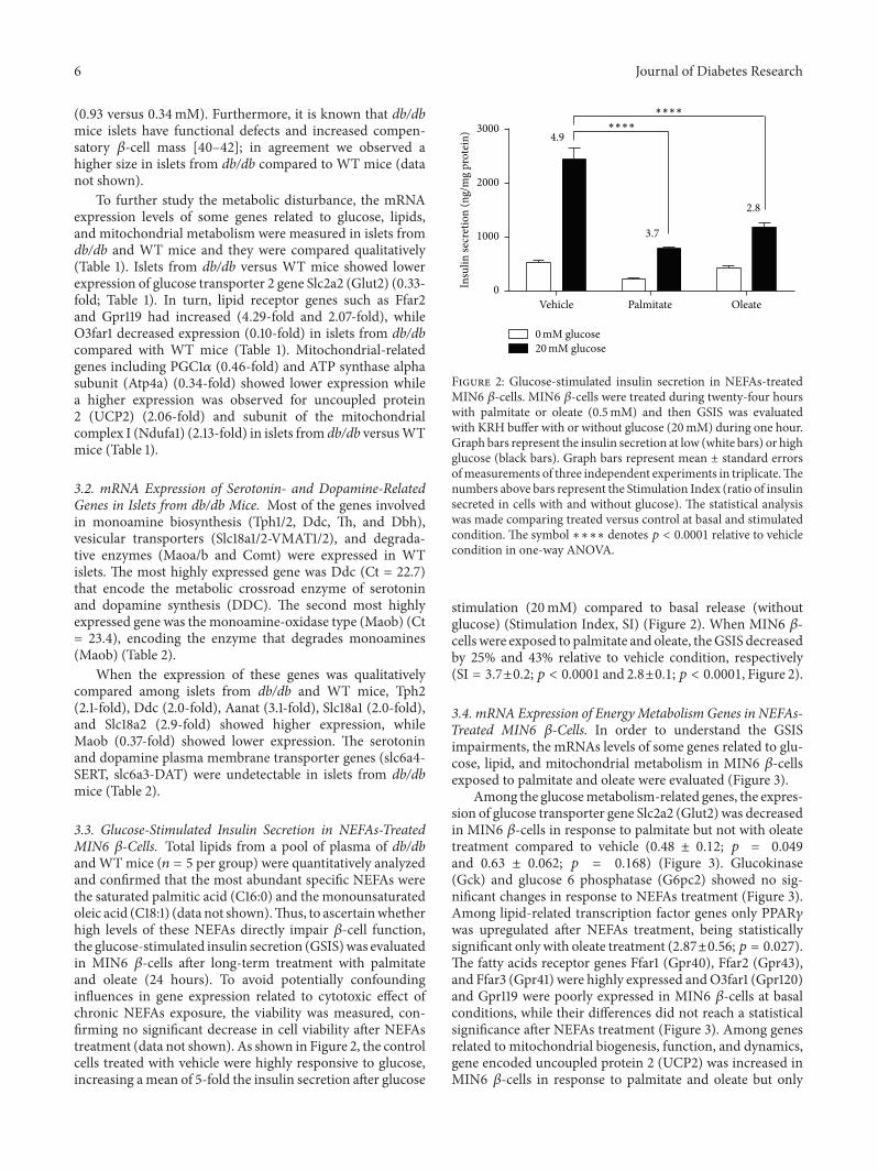
3.3. Glucose-Stimulated Insulin Secretion in NEFAs-TreatedMIN6 𝛽-Cells. Total lipids from a pool of plasma of db/dbandWTmice (𝑛 = 5 per group) were quantitatively analyzedand confirmed that the most abundant specific NEFAs werethe saturated palmitic acid (C16:0) and the monounsaturatedoleic acid (C18:1) (data not shown).Thus, to ascertainwhetherhigh levels of these NEFAs directly impair 𝛽-cell function,the glucose-stimulated insulin secretion (GSIS)was evaluatedin MIN6 𝛽-cells after long-term treatment with palmitateand oleate (24 hours). To avoid potentially confoundinginfluences in gene expression related to cytotoxic effect ofchronic NEFAs exposure, the viability was measured, con-firming no significant decrease in cell viability after NEFAstreatment (data not shown). As shown in Figure 2, the controlcells treated with vehicle were highly responsive to glucose,increasing amean of 5-fold the insulin secretion after glucose
Vehicle Palmitate Oleate
4.9
3.7
2.8
∗∗∗∗
∗∗∗∗
0mM glucose20mM glucose
0
1000
2000
3000
Insu
lin se
cret
ion
(ng/
mg
prot
ein)
Figure 2: Glucose-stimulated insulin secretion in NEFAs-treatedMIN6 𝛽-cells. MIN6 𝛽-cells were treated during twenty-four hourswith palmitate or oleate (0.5mM) and then GSIS was evaluatedwith KRH buffer with or without glucose (20mM) during one hour.Graph bars represent the insulin secretion at low (white bars) or highglucose (black bars). Graph bars represent mean ± standard errorsofmeasurements of three independent experiments in triplicate.Thenumbers above bars represent the Stimulation Index (ratio of insulinsecreted in cells with and without glucose). The statistical analysiswas made comparing treated versus control at basal and stimulatedcondition. The symbol ∗∗∗∗ denotes 𝑝 < 0.0001 relative to vehiclecondition in one-way ANOVA.
stimulation (20mM) compared to basal release (withoutglucose) (Stimulation Index, SI) (Figure 2). When MIN6 𝛽-cells were exposed to palmitate and oleate, theGSIS decreasedby 25% and 43% relative to vehicle condition, respectively(SI = 3.7±0.2;𝑝 < 0.0001 and 2.8±0.1;𝑝 < 0.0001, Figure 2).
3.4. mRNA Expression of Energy Metabolism Genes in NEFAs-Treated MIN6 𝛽-Cells. In order to understand the GSISimpairments, the mRNAs levels of some genes related to glu-cose, lipid, and mitochondrial metabolism in MIN6 𝛽-cellsexposed to palmitate and oleate were evaluated (Figure 3).
Among the glucosemetabolism-related genes, the expres-sion of glucose transporter gene Slc2a2 (Glut2) was decreasedin MIN6 𝛽-cells in response to palmitate but not with oleatetreatment compared to vehicle (0.48 ± 0.12; 𝑝 = 0.049and 0.63 ± 0.062; 𝑝 = 0.168) (Figure 3). Glucokinase(Gck) and glucose 6 phosphatase (G6pc2) showed no sig-nificant changes in response to NEFAs treatment (Figure 3).Among lipid-related transcription factor genes only PPAR𝛾was upregulated after NEFAs treatment, being statisticallysignificant only with oleate treatment (2.87±0.56; 𝑝 = 0.027).The fatty acids receptor genes Ffar1 (Gpr40), Ffar2 (Gpr43),and Ffar3 (Gpr41) were highly expressed andO3far1 (Gpr120)and Gpr119 were poorly expressed in MIN6 𝛽-cells at basalconditions, while their differences did not reach a statisticalsignificance after NEFAs treatment (Figure 3). Among genesrelated to mitochondrial biogenesis, function, and dynamics,gene encoded uncoupled protein 2 (UCP2) was increased inMIN6 𝛽-cells in response to palmitate and oleate but only

Journal of Diabetes Research 7
mRN
As e
xpre
ssio
n(fo
ld ch
ange
to co
ntro
l)m
RNA
s exp
ress
ion
(fold
chan
ge to
cont
rol)
∗
∗
∗∗
∗∗
0
1
2
3
4
5
0
1
2
3
VehiclePalmitate (0.5mM)Oleate (0.5mM)
Gck G6pc2Slc2a2Glucose and lipid metabolism
Ffar3 O3far1Ffar1 Ffar2 Gpr119Lipid receptors
Opa1 Mfn1 Mfn2Atp4aDynamics
Sdha Cyc1Ndufa1 Cox5aRespiratory chain
UCP2Biogenesis and function
Ct=
28.5
Ct=
26.7
Ct=
18.4
Ct=
29.8
Ct=
27.2
Ct>
35
Ct=
22.4
Ct=
26.9
Ct=
24.9
Ct=
32.2
Ct=
33.2
Ct=
26.9
Ct=
28.9
Ct=
24.0
Ct=
20.9
Ct=
22.5
Ct=
21.8
Ct=
21.7
Ct=
25.0
Ct=
24.4
Ct=
24.6
Ct=
23.7
PPAR𝛼 PPAR𝛾 CEBP𝛼
PGC1𝛼 PGC1𝛽
Figure 3: mRNA expression of energy metabolism-related genes in NEFAs-treated MIN6 𝛽-cells. MIN6 𝛽-cells were treated during twenty-four hours with palmitate or oleate (0.5mM). Then, total RNA was extracted and the mRNA levels of genes related to glucose and lipidmetabolism and with mitochondrial function were evaluated by RT-qPCR. The mRNA levels are expressed as fold change in 2−ΔCt of thespecific gene in the treated versus control conditions (see Section 2). Graph bars represent mean ± standard errors of measurements of threeindependent experiments in triplicate. The numbers above bars represent the mean Ct value in control cells. The symbol ∗ denotes 𝑝 < 0.05and ∗∗ denotes 𝑝 < 0.01 in one-way ANOVA.
reaching statistically significance with oleate (2.10 ± 0.34;𝑝 = 0.019).The gene of mitofusin 1 (Mfn1) was also increasedafter oleate treatment (1.31 ± 0.03; 𝑝 = 0.009) (Figure 3).
3.5. mRNA Expression of Monoamines-Related Genes inNEFAs-Treated MIN6 𝛽-Cells. The mRNA expression levelsof several genes related to biosynthesis and metabolism ofmonoamines were quantified in MIN6 𝛽-cells. As shown inFigure 4, MIN6 𝛽-cells at basal condition expressed almostall genes encoding enzymes needed for serotonin (Tph1/2 andDdc), dopamine (TH and Ddc), and noradrenalin (TH, Ddc,and Dbh) synthesis (Figure 1). The most highly expressedgene was Maob (Ct = 22.2), while the second most highlyexpressed was Ddc (Ct = 22.5). Similar to mice islets, noexpression of the main plasma membrane transporter genesof serotonin (slc6a4-SERT) and dopamine (slc6a3-DAT) (Ct> 35) was detected in MIN6 𝛽-cells.
Then, the expression changes of these genes in MIN6𝛽-cells in response to palmitate and oleate treatment wereevaluated. None of these genes showed significant expressionchanges in MIN6 𝛽-cells with exception of the Maob, whichsignificantly decreased in response to palmitate and oleatecompared to control (0.48 ± 0.03; 𝑝 = 0.002 and 0.64 ± 0.05;𝑝 = 0.014, resp.) (Figure 4).
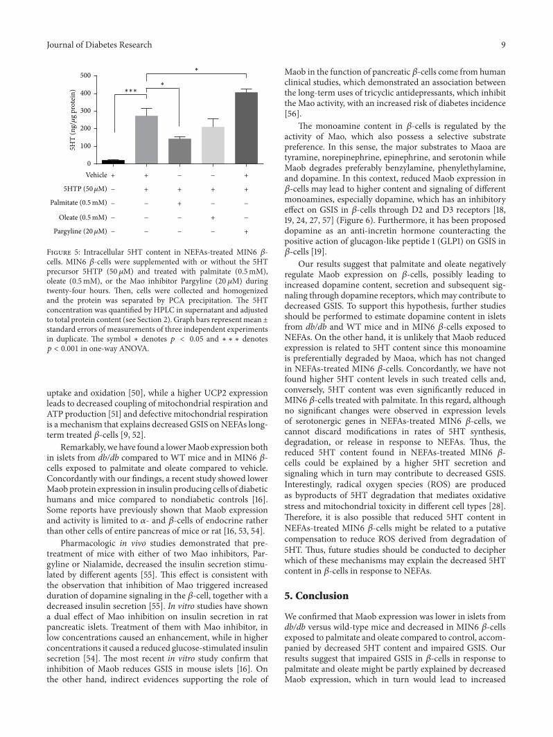
3.6. Intracellular 5HT Content in NEFAs-Treated MIN6 𝛽-Cells. It was assessed whether the treatment of MIN6 𝛽-cells with palmitate and oleate may change intracellular5HT content. In control conditions, the 5HT content inMIN6 𝛽-cells was detected in low levels (Figure 5). However,such level strongly increased after supplementation withthe 5HT precursor 5HTP, confirming the existence of afunctional microserotonergic system in 𝛽-cells. When the5HTP supplemented MIN6 𝛽-cells were additionally treatedwith palmitate, a significant reduction of intracellular 5HTcontent was observed (271.6 versus 141.5 ng of 5HT/𝜇g pro-tein; 𝑝 = 0.0479); similar trend was observed with oleate.As expected, in MIN6 𝛽-cells supplemented with 5HTP andsimultaneously treated with the Mao inhibitor Pargyline, the5HT content was further increased compared with cells onlyincubated with 5HTP (Figure 5).
4. Discussion
In this work, we have shown that islets from WT and db/dbmice expressedmRNAs ofmost genes related to the serotoninand dopamine metabolism, including enzymes involved inthe synthesis (Tph1/2, TH, and Ddc), vesicular transport(Slc18a1/2-VMAT1/2), and degradation (Maoa, Maob, andComt) of these monoamines. Conversely, the genes ofthe main cell membrane transporter of these monoamines

8 Journal of Diabetes Research
VehiclePalmitate (0.5mM)Oleate (0.5mM)
Monoamines biosynthetic enzymes
Dbh
∗∗∗
Maob ComtMaoaDegradative enzymes
Slc6a4 Slc18a2Slc18a1Slc6a3Transporters
ThDopamine and adrenalin
AsmtAanatMelatonin
DdcTph2Tph15HT and dopamine
0.0
0.5
1.0
1.5
2.0
mRN
As e
xpre
ssio
n(fo
ld ch
ange
to co
ntro
l)
0.0
0.5
1.0
1.5
2.0
mRN
As e
xpre
ssio
n(fo
ld ch
ange
to co
ntro
l)
Ct=
26.5
Ct=
27.8
Ct=
22.5
Ct=
29.8
Ct>
35
Ct=
25.3
Ct=
28.5
Ct=
26.6
Ct=
22.2
Ct=
23.1
Ct=
26.7
Ct>
35
Ct>
35
Ct=
28.3
Figure 4: mRNA expression of monoamines-related genes in NEFAs-treated MIN6 𝛽-cells. MIN6 𝛽-cells were treated during twenty-fourhours with palmitate or oleate (0.5mM). Then, total RNA was extracted and mRNA levels of genes related to the monoamines biosynthesis,degradation, and transport were evaluated by RT-qPCR.The mRNA levels are expressed as fold change between 2−ΔCt of the specific gene inthe treated versus control conditions (see Section 2). Graph bars represent mean ± standard errors of measurements of three independentexperiments in triplicate.The numbers above bars represent themeanCt value in control cells.The symbol∗ denotes𝑝 < 0.05 and∗∗ denotes𝑝 < 0.01 in one-way ANOVA.
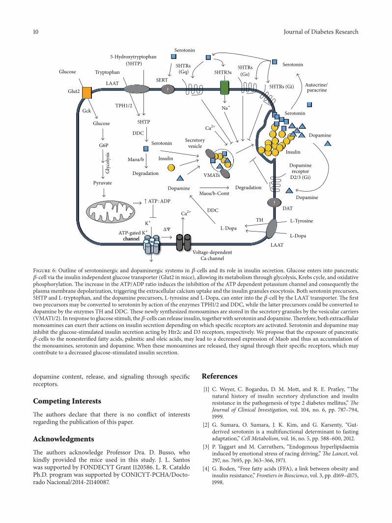
(slc6a4-SERT, slc6a3-DAT) were almost undetectable (Fig-ure 6). The db/db mice are an animal model of diabetescharacterized by high plasma levels of NEFAs and insulinsecretory defects of 𝛽-cells [40–42]. Remarkably, islets fromdb/db compared to WT mice had higher mRNA levels of theserotonin and dopamine common synthesis enzyme (Ddc)and vesicular transporters VMAT1/2 and lower mRNA levelof Maob, the degradative enzyme of the monoamines. Thesedifferences in gene expression suggest that islets from db/dbmice have an increased synthesis and storage and decreaseddegradation, which might enhance autocrine signaling ofserotonin and dopamine in 𝛽-cells (Figure 6). However,it is not possible to draw definitive conclusions of mRNAexpression changes obtained in our study because these werecarried out in pools of islets from db/db andWTmice ratherthan in independent experiments.This weakness is related tothe difficulty to obtain islets from the scarce db/dbmalemice.
Similarly to islets from WT mice, MIN6 𝛽-cells at basalconditions (vehicle) express most of the mRNAs of serotoninand dopamine metabolism genes mentioned above andcell membrane transporter genes (slc6a4-SERT, slc6a3-DAT)were undetectable. However, this does not rule out the pos-sibility that 𝛽-cells can take up monoamines because otherless known serotonin and dopamine transporters, such asthe plasmamembranemonoamines transporter (PMAT) andorganic cation transporter 3 (OCT3) [43], could be expressed.
In fact, gene expression databases indicate that PMAT andOCT3 are present in both human and mice islets and in dif-ferent𝛽-cell lines (https://www.t1dbase.org/page/AtlasView).
As expected, palmitic and oleic acids were the mostabundant, saturated, andmonounsaturated NEFAs in plasmaof mice, with higher circulating concentration found inplasma of db/db mice compared to WT mice. In agreementwith other studies [5, 6, 9, 44], here we show a decreasedGSIS in MIN6 𝛽-cells treated with palmitate and oleateversus vehicle. Considering previous reports of an inversecorrelation between the fetal bovine serumconcentration andcytotoxic action of NEFAs [45, 46], we used a mild protocoland discarded the influence of 𝛽-cell death on the observeddecreased GSIS.
The gene expression changes observed in MIN6 𝛽-cellsin response to NEFAs treatment were compatible with adecreased use of glucose as the energy source that explainsthe reduced GSIS. In this regard, we found a decrease inthe mRNA of glucose transporter Glut2 and an increase inUCP2 and PPAR𝛾 in MIN6 𝛽-cells treated with palmitateor oleate versus vehicle, respectively. Importantly, we haveobserved that both genes, Glut2 and UCP2, regulated byNEFAs in MIN6 𝛽-cells, are correspondingly different inislets from db/db versus WT mice. The expression changesof such genes have been associated with decreased GSIS [47–49]. A decreased Glut2 transporter implies a reduced glucose
Journal of Diabetes Research 9
+ + − − +
− + + + +
− − + − −
− − − + −
− − − − +
∗∗∗
∗
∗
Vehicle
5HTP (50𝜇M)
Palmitate (0.5mM)
Oleate (0.5mM)
Pargyline (20𝜇M)
0
100
200
300
400
5005
HT
(ng/𝜇
g pr
otei
n)
Figure 5: Intracellular 5HT content in NEFAs-treated MIN6 𝛽-cells. MIN6 𝛽-cells were supplemented with or without the 5HTprecursor 5HTP (50 𝜇M) and treated with palmitate (0.5mM),oleate (0.5mM), or the Mao inhibitor Pargyline (20 𝜇M) duringtwenty-four hours. Then, cells were collected and homogenizedand the protein was separated by PCA precipitation. The 5HTconcentration was quantified by HPLC in supernatant and adjustedto total protein content (see Section 2). Graph bars representmean±standard errors of measurements of three independent experimentsin duplicate. The symbol ∗ denotes 𝑝 < 0.05 and ∗ ∗ ∗ denotes𝑝 < 0.001 in one-way ANOVA.
uptake and oxidation [50], while a higher UCP2 expressionleads to decreased coupling of mitochondrial respiration andATP production [51] and defective mitochondrial respirationis a mechanism that explains decreased GSIS onNEFAs long-term treated 𝛽-cells [9, 52].
Remarkably, we have found a lowerMaob expression bothin islets from db/db compared to WT mice and in MIN6 𝛽-cells exposed to palmitate and oleate compared to vehicle.Concordantly with our findings, a recent study showed lowerMaobprotein expression in insulin producing cells of diabetichumans and mice compared to nondiabetic controls [16].Some reports have previously shown that Maob expressionand activity is limited to 𝛼- and 𝛽-cells of endocrine ratherthan other cells of entire pancreas of mice or rat [16, 53, 54].
Pharmacologic in vivo studies demonstrated that pre-treatment of mice with either of two Mao inhibitors, Par-gyline or Nialamide, decreased the insulin secretion stimu-lated by different agents [55]. This effect is consistent withthe observation that inhibition of Mao triggered increasedduration of dopamine signaling in the 𝛽-cell, together with adecreased insulin secretion [55]. In vitro studies have showna dual effect of Mao inhibition on insulin secretion in ratpancreatic islets. Treatment of them with Mao inhibitor, inlow concentrations caused an enhancement, while in higherconcentrations it caused a reduced glucose-stimulated insulinsecretion [54]. The most recent in vitro study confirm thatinhibition of Maob reduces GSIS in mouse islets [16]. Onthe other hand, indirect evidences supporting the role of
Maob in the function of pancreatic 𝛽-cells come from humanclinical studies, which demonstrated an association betweenthe long-term uses of tricyclic antidepressants, which inhibitthe Mao activity, with an increased risk of diabetes incidence[56].
The monoamine content in 𝛽-cells is regulated by theactivity of Mao, which also possess a selective substratepreference. In this sense, the major substrates to Maoa aretyramine, norepinephrine, epinephrine, and serotonin whileMaob degrades preferably benzylamine, phenylethylamine,and dopamine. In this context, reduced Maob expression in𝛽-cells may lead to higher content and signaling of differentmonoamines, especially dopamine, which has an inhibitoryeffect on GSIS in 𝛽-cells through D2 and D3 receptors [18,19, 24, 27, 57] (Figure 6). Furthermore, it has been proposeddopamine as an anti-incretin hormone counteracting thepositive action of glucagon-like peptide 1 (GLP1) on GSIS in𝛽-cells [19].
Our results suggest that palmitate and oleate negativelyregulate Maob expression on 𝛽-cells, possibly leading toincreased dopamine content, secretion and subsequent sig-naling through dopamine receptors, whichmay contribute todecreased GSIS. To support this hypothesis, further studiesshould be performed to estimate dopamine content in isletsfrom db/db and WT mice and in MIN6 𝛽-cells exposed toNEFAs. On the other hand, it is unlikely that Maob reducedexpression is related to 5HT content since this monoamineis preferentially degraded by Maoa, which has not changedin NEFAs-treated MIN6 𝛽-cells. Concordantly, we have notfound higher 5HT content levels in such treated cells and,conversely, 5HT content was even significantly reduced inMIN6 𝛽-cells treated with palmitate. In this regard, althoughno significant changes were observed in expression levelsof serotonergic genes in NEFAs-treated MIN6 𝛽-cells, wecannot discard modifications in rates of 5HT synthesis,degradation, or release in response to NEFAs. Thus, thereduced 5HT content found in NEFAs-treated MIN6 𝛽-cells could be explained by a higher 5HT secretion andsignaling which in turn may contribute to decreased GSIS.Interestingly, radical oxygen species (ROS) are producedas byproducts of 5HT degradation that mediates oxidativestress and mitochondrial toxicity in different cell types [28].Therefore, it is also possible that reduced 5HT content inNEFAs-treated MIN6 𝛽-cells might be related to a putativecompensation to reduce ROS derived from degradation of5HT. Thus, future studies should be conducted to decipherwhich of these mechanisms may explain the decreased 5HTcontent in 𝛽-cells in response to NEFAs.
5. Conclusion
We confirmed that Maob expression was lower in islets fromdb/db versus wild-type mice and decreased in MIN6 𝛽-cellsexposed to palmitate and oleate compared to control, accom-panied by decreased 5HT content and impaired GSIS. Ourresults suggest that impaired GSIS in 𝛽-cells in response topalmitate and oleate might be partly explained by decreasedMaob expression, which in turn would lead to increased
10 Journal of Diabetes Research
Glucose
Gck
G6P
Pyruvate
Insulin
Glucose
? 5HTRs (Gi)
Serotonin
SerotoninTPH1/2
Tryptophan
Glut2
5HTP
Gly
coly
sis
DDC
Serotonin
5HTRs (Gs)5HTR3s
5HTRs (Gq)
SERTAutocrine/ paracrine
5-Hydroxytryptophan (5HTP)
VMATs
Maoa/b
Degradation
LAAT
channel
Voltage-dependentCa channel
Dopamine
L-Tyrosine
LAAT
TH
Dopaminereceptor
D2/3 (Gi)
Maoa/b-ComtDopamine
DAT
Serotonin
Insulin
Dopamine
Secretoryvesicle
?
L-DopaL-Dopa
DDC
Degradation
Na+
Ca2+
Ca2+
K+
ATP-gated K+
channel
ΔΨ
↑ ATP : ADP
Figure 6: Outline of serotoninergic and dopaminergic systems in 𝛽-cells and its role in insulin secretion. Glucose enters into pancreatic𝛽-cell via the insulin independent glucose transporter (Glut2 in mice), allowing its metabolism through glycolysis, Krebs cycle, and oxidativephosphorylation. The increase in the ATP/ADP ratio induces the inhibition of the ATP dependent potassium channel and consequently theplasmamembrane depolarization, triggering the extracellular calcium uptake and the insulin granules exocytosis. Both serotonin precursors,5HTP and L-tryptophan, and the dopamine precursors, L-tyrosine and L-Dopa, can enter into the 𝛽-cell by the LAAT transporter. The firsttwo precursors may be converted to serotonin by action of the enzymes TPH1/2 and DDC, while the latter precursors could be converted todopamine by the enzymes TH and DDC.These newly synthesized monoamines are stored in the secretory granules by the vesicular carriers(VMAT1/2). In response to glucose stimuli, the𝛽-cells can release insulin, togetherwith serotonin and dopamine.Therefore, both extracellularmonoamines can exert their actions on insulin secretion depending on which specific receptors are activated. Serotonin and dopamine mayinhibit the glucose-stimulated insulin secretion acting by Htr2c and D3 receptors, respectively. We propose that the exposure of pancreatic𝛽-cells to the nonesterified fatty acids, palmitic and oleic acids, may lead to a decreased expression of Maob and thus an accumulation ofthe monoamines, serotonin and dopamine. When these monoamines are released, they signal through their specific receptors, which maycontribute to a decreased glucose-stimulated insulin secretion.
dopamine content, release, and signaling through specificreceptors.
Competing Interests
The authors declare that there is no conflict of interestsregarding the publication of this paper.
Acknowledgments
The authors acknowledge Professor Dra. D. Busso, whokindly provided the mice used in this study. J. L. Santoswas supported by FONDECYT Grant 1120586. L. R. CataldoPh.D. program was supported by CONICYT-PCHA/Docto-rado Nacional/2014-21140087.
References
[1] C. Weyer, C. Bogardus, D. M. Mott, and R. E. Pratley, “Thenatural history of insulin secretory dysfunction and insulinresistance in the pathogenesis of type 2 diabetes mellitus,” TheJournal of Clinical Investigation, vol. 104, no. 6, pp. 787–794,1999.
[2] G. Sumara, O. Sumara, J. K. Kim, and G. Karsenty, “Gut-derived serotonin is a multifunctional determinant to fastingadaptation,” Cell Metabolism, vol. 16, no. 5, pp. 588–600, 2012.
[3] P. Taggart and M. Carruthers, “Endogenous hyperlipidaemiainduced by emotional stress of racing driving,”The Lancet, vol.297, no. 7695, pp. 363–366, 1971.
[4] G. Boden, “Free fatty acids (FFA), a link between obesity andinsulin resistance,” Frontiers in Bioscience, vol. 3, pp. d169–d175,1998.
Journal of Diabetes Research 11
[5] J. D. McGarry and R. L. Dobbins, “Fatty acids, lipotoxicity andinsulin secretion,”Diabetologia, vol. 42, no. 2, pp. 128–138, 1999.
[6] J. P. H. Wilding, “The importance of free fatty acids in thedevelopment of Type 2 diabetes,” Diabetic Medicine, vol. 24, no.9, pp. 934–945, 2007.
[7] H. Kristinsson, P. Bergsten, and E. Sargsyan, “Free fatty acidreceptor 1 (FFAR1/GPR40) signaling affects insulin secretionby enhancingmitochondrial respiration during palmitate expo-sure,” Biochimica et Biophysica Acta: Molecular Cell Research,vol. 1853, no. 12, pp. 3248–3257, 2015.
[8] Y. Sako and V. E. Grill, “A 48-hour lipid infusion in the rat time-dependently inhibits glucose-induced insulin secretion and Bcell oxidation through a process likely coupled to fatty acidoxidation,” Endocrinology, vol. 127, no. 4, pp. 1580–1589, 1990.
[9] Y.-P. Zhou andV. E.Grill, “Long-term exposure of rat pancreaticislets to fatty acids inhibits glucose-induced insulin secretionand biosynthesis through a glucose fatty acid cycle,” Journal ofClinical Investigation, vol. 93, no. 2, pp. 870–876, 1994.
[10] Y.-P. Zhou and V. Grill, “Long term exposure to fatty acids andketones inhibits B-cell functions in human pancreatic islets ofLangerhans,” Journal of Clinical Endocrinology and Metabolism,vol. 80, no. 5, pp. 1584–1590, 1995.
[11] G. Patane, M. Anello, S. Piro, R. Vigneri, F. Purrello, and A. M.Rabuazzo, “Role of ATP production and uncoupling protein-2 in the insulin secretory defect induced by chronic exposureto high glucose or free fatty acids and effects of peroxisomeproliferator-activated receptor-𝛾 inhibition,” Diabetes, vol. 51,no. 9, pp. 2749–2756, 2002.
[12] M. A. Barradas, D. S. Gill, V. A. Fonseca, D. P. Mikhailidis, andP. Dandona, “Intraplatelet serotonin in patients with diabetesmellitus and peripheral vascular disease,” European Journal ofClinical Investigation, vol. 18, no. 4, pp. 399–404, 1988.
[13] K. Hara, Y. Hirowatari, Y. Shimura, and H. Takahashi, “Sero-tonin levels in platelet-poor plasma and whole blood in peoplewith type 2 diabetes with chronic kidney disease,” DiabetesResearch and Clinical Practice, vol. 94, no. 2, pp. 167–171, 2011.
[14] S. M. Innis and S. de la Presa Owens, “Dietary fatty acidcomposition in pregnancy alters neurite membrane fatty acidsand dopamine in newborn rat brain,” Journal of Nutrition, vol.131, no. 1, pp. 118–122, 2001.
[15] Q. Zhang, Y. Zhu, W. Zhou, L. Gao, L. Yuan, and X. Han,“Serotonin receptor 2C and insulin secretion,” PLoS ONE, vol.8, no. 1, Article ID e54250, 2013.
[16] E. Ganic, J. K. Johansson, H. Bennet, M. Fex, and I. Artner,“Islet-specific monoamine oxidase A and B expression dependson MafA transcriptional activity and is compromised in type2 diabetes,” Biochemical and Biophysical Research Communica-tions, vol. 468, no. 4, pp. 629–635, 2015.
[17] G. Eisenhofer, A. Aneman, P. Friberg et al., “Substantial produc-tion of dopamine in the human gastrointestinal tract,” Journal ofClinical Endocrinology andMetabolism, vol. 82, no. 11, pp. 3864–3871, 1997.
[18] A. Ustione and D. W. Piston, “Dopamine synthesis andD3 receptor activation in pancreatic 𝛽-cells regulates insulinsecretion and intracellular [Ca2+] oscillations,” MolecularEndocrinology, vol. 26, no. 11, pp. 1928–1940, 2012.
[19] A. Ustione, D. W. Piston, and P. E. Harris, “Minireview:dopaminergic regulation of insulin secretion from the pancre-atic islet,”Molecular Endocrinology, vol. 27, no. 8, pp. 1198–1207,2013.
[20] J. E. Richmond, A. Codignola, I. M. Cooke, and E. Sher,“Calcium and barium dependent exocytosis from the rat insuli-noma cell line RINm5F assayed using membrane capacitancemeasurements and serotonin release,” Pflugers Archiv, vol. 432,no. 2, pp. 258–269, 1996.
[21] R. Ekholm, L. E. Ericson, and I. Lundquist, “Monoamines inthe pancreatic islets of the mouse. Subcellular localization of 5-hydroxytryptamine by electron microscopic autoradiography,”Diabetologia, vol. 7, no. 5, pp. 339–348, 1971.
[22] E. Gylfe, “Association between 5-hydroxytryptamine releaseand insulin secretion,” Journal of Endocrinology, vol. 78, no. 2,pp. 239–248, 1978.
[23] I. Garcıa-Tornadu, A. M. Ornstein, A. Chamson-Reig et al.,“Disruption of the dopamine D2 receptor impairs insulinsecretion and causes glucose intolerance,” Endocrinology, vol.151, no. 4, pp. 1441–1450, 2010.
[24] L. R. Cataldo, V. A. Cortes, J. E. Galgani, P. R. Olmos, and J.L. Santos, “Role of peripheral serotonin in the insulin secretionand glucose homeostasis,” Nutricion Hospitalaria, vol. 30, no. 3,pp. 498–508, 2014.
[25] M. Ohara-Imaizumi, H. Kim, M. Yoshida et al., “Serotoninregulates glucose-stimulated insulin secretion from pancreatic𝛽 cells during pregnancy,” Proceedings of the National Academyof Sciences of the United States of America, vol. 110, no. 48, pp.19420–19425, 2013.
[26] K. Kim, C.M.Oh,M.Ohara-Imaizumi et al., “Functional role ofserotonin in insulin secretion in a diet-induced insulin-resistantstate,” Endocrinology, vol. 156, no. 2, pp. 444–452, 2015.
[27] N. Simpson, A. Maffei, M. Freeby et al., “Dopamine-mediatedautocrine inhibitory circuit regulating human insulin secretionin vitro,”Molecular Endocrinology, vol. 26, no. 10, pp. 1757–1772,2012.
[28] A. Nocito, F. Dahm, W. Jochum et al., “Serotonin mediatesoxidative stress and mitochondrial toxicity in a murine modelof nonalcoholic steatohepatitis,” Gastroenterology, vol. 133, no.2, pp. 608–618, 2007.
[29] C. J. Rosen, “Serotonin rising—the bone, brain, bowel connec-tion,”The New England Journal of Medicine, vol. 360, no. 10, pp.957–959, 2009.
[30] P. Amireault, D. Sibon, and F. Coıte, “Life without peripheralserotonin: insights from tryptophan hydroxylase 1 knockoutmice reveal the existence of paracrine/autocrine serotonergicnetworks,” ACS Chemical Neuroscience, vol. 4, no. 1, pp. 64–71,2013.
[31] N. Paulmann, M. Grohmann, J.-P. Voigt et al., “Intracellularserotonin modulates insulin secretion from pancreatic 𝛽-cellsby protein serotonylation,” PLoS Biology, vol. 7, no. 10, ArticleID e1000229, 2009.
[32] D. J. Walther, J.-U. Peter, S. Bashammakh et al., “Synthesisof serotonin by a second tryptophan hydroxylase isoform,”Science, vol. 299, no. 5603, article 76, 2003.
[33] Y. Ohta, Y. Kosaka, N. Kishimoto et al., “Convergence ofthe insulin and serotonin programs in the pancreatic 𝛽-cell,”Diabetes, vol. 60, no. 12, pp. 3208–3216, 2011.
[34] D. Sommerweiss, T. Gorski, S. Richter, A. Garten, andW. Kiess,“Oleate rescues INS-1E 𝛽-cells from palmitate-induced apopto-sis by preventing activation of the unfolded protein response,”Biochemical and Biophysical Research Communications, vol. 441,no. 4, pp. 770–776, 2013.
[35] E. G. Bligh and W. J. Dyer, “A rapid method of total lipidextraction and purification,” Canadian Journal of Biochemistryand Physiology, vol. 37, no. 8, pp. 911–917, 1959.
12 Journal of Diabetes Research
[36] R. Valenzuela, C. Barrera, A. Espinosa, P. Llanos, P. Orellana,and L. A. Videla, “Reduction in the desaturation capacity ofthe liver in mice subjected to high fat diet: relation to LCPUFAdepletion in liver and extrahepatic tissues,” ProstaglandinsLeukotrienes and Essential Fatty Acids, vol. 98, pp. 7–14, 2015.
[37] R. N. Kulkarni, J. N. Winnay, M. Daniels et al., “Alteredfunction of insulin receptor substrate-1-deficient mouse isletsand cultured 𝛽-cell lines,” The Journal of Clinical Investigation,vol. 104, no. 12, pp. R69–R75, 1999.
[38] D. S. Li, Y. Yuan, H. Tu, Q. Liang, and L. Dai, “A protocol for isletisolation from mouse pancreas,” Nature Protocols, vol. 4, no. 11,pp. 1649–1652, 2009.
[39] A. M. Kumar, M. Kumar, K. Deepika, J. B. Fernandez, and C.Eisdorfer, “A modified HPLC technique for simultaneous mea-surement of 5-hydroxytryptamine and 5-hydroxyindoleaceticacid in cerebrospinal fluid, platelet and plasma,” Life Sciences,vol. 47, no. 19, pp. 1751–1759, 1990.
[40] L. S. Dalbøge, D. L. Almholt, T. S. Neerup et al., “Characteri-sation of age-dependent Beta cell dynamics in the male db/dbmice,” PLoS ONE, vol. 8, no. 12, article e82813, 2013.
[41] O. Berglund, B. J. Frankel, and B. Hellman, “Development of theinsulin secretory defect in genetically diabetic (db/db) mouse,”Acta Endocrinologica, vol. 87, no. 3, pp. 543–551, 1978.
[42] O. H. Do, J. T. Low, H. Y. Gaisano, and P.Thorn, “The secretorydeficit in islets from db/db mice is mainly due to a loss ofresponding beta cells,”Diabetologia, vol. 57, no. 7, pp. 1400–1409,2014.
[43] H. Duan and J. Wang, “Selective transport of monoamineneurotransmitters by human plasma membrane monoaminetransporter and organic cation transporter 3,” Journal of Phar-macology and Experimental Therapeutics, vol. 335, no. 3, pp.743–753, 2010.
[44] Y. Itoh, Y. Kawamata, M. Harada et al., “Free fatty acids regulateinsulin secretion from pancreatic 𝛽 cells through GPR40,”Nature, vol. 422, no. 6928, pp. 173–176, 2003.
[45] I. Maestre, J. Jordan, S. Calvo et al., “Mitochondrial dysfunctionis involved in apoptosis induced by serum withdrawal and fattyacids in the 𝛽-cell line INS-1,” Endocrinology, vol. 144, no. 1, pp.335–345, 2003.
[46] T. Brun, P. Scarcia, N. Li et al., “Changes in mitochondrialcarriers exhibit stress-specific signatures in INS-1E 𝛽-cellsexposed to glucose versus fatty acids,” PLoS ONE, vol. 8, no. 12,Article ID e82364, 2013.
[47] C.-Y. Zhang, G. Baffy, P. Perret et al., “Uncoupling protein-2 negatively regulates insulin secretion and is a major linkbetween obesity, 𝛽 cell dysfunction, and type 2 diabetes,” Cell,vol. 105, no. 6, pp. 745–755, 2001.
[48] J. C. Yoon,G.Xu, J. T.Deeney et al., “Suppression of𝛽 cell energymetabolism and insulin release by PGC-1𝛼,”Developmental Cell,vol. 5, no. 1, pp. 73–83, 2003.
[49] M. C. Akerfeldt and D. R. Laybutt, “Inhibition of Id1 augmentsinsulin secretion and protects against high-fat diet-inducedglucose intolerance,” Diabetes, vol. 60, no. 10, pp. 2506–2514,2011.
[50] X. Quan, L. Zhang, Y. Li, and C. Liang, “TCF2 attenuates FFA-induced damage in islet 𝛽-cells by regulating production ofinsulin and ROS,” International Journal of Molecular Sciences,vol. 15, no. 8, pp. 13317–13332, 2014.
[51] C. Affourtit and M. D. Brand, “Uncoupling protein-2 con-tributes significantly to high mitochondrial proton leak in INS-1E insulinoma cells and attenuates glucose-stimulated insulin
secretion,” Biochemical Journal, vol. 409, no. 1, pp. 199–204,2008.
[52] J. Barlow and C. Affourtit, “Novel insights into pancreatic𝛽-cell glucolipotoxicity from real-time functional analysis ofmitochondrial energy metabolism in INS-1E insulinoma cells,”Biochemical Journal, vol. 456, no. 3, pp. 417–426, 2013.
[53] Y.-H. Huang, A. Ito, and R. Arai, “Immunohistochemicallocalization of monoamine oxidase type B in pancreatic isletsof the rat,” Journal of Histochemistry and Cytochemistry, vol. 53,no. 9, pp. 1149–1158, 2005.
[54] H. Aleyassine and R. J. Gardiner, “Dual action of antidepressantdrugs (MAO inhibitors) on insulin release,” Endocrinology, vol.96, no. 3, pp. 702–710, 1975.
[55] I. Lundquist, R. Ekholm, and L. E. Ericson, “Monoamines inthe pancreatic islets of the mouse-5-Hydroxytryptamine as anintracellular modifier of insulin secretion, and the hypogly-caemic action of monoamine oxidase inhibitors,” Diabetologia,vol. 7, no. 6, pp. 414–422, 1971.
[56] F. Andersohn, R. Schade, S. Suissa, and E. Garbe, “Long-termuse of antidepressants for depressive disorders and the risk ofdiabetes mellitus,” American Journal of Psychiatry, vol. 166, no.5, pp. 591–598, 2009.
[57] R. El-Merahbi, M. Loffler, A. Mayer, and G. Sumara, “The rolesof peripheral serotonin inmetabolic homeostasis,” FEBS Letters,vol. 589, no. 15, Article ID 37210, pp. 1728–1734, 2015.

Submit your manuscripts athttp://www.hindawi.com
Stem CellsInternational
Hindawi Publishing Corporationhttp://www.hindawi.com Volume 2014
Hindawi Publishing Corporationhttp://www.hindawi.com Volume 2014
MEDIATORSINFLAMMATION
of
Hindawi Publishing Corporationhttp://www.hindawi.com Volume 2014
Behavioural Neurology
EndocrinologyInternational Journal of
Hindawi Publishing Corporationhttp://www.hindawi.com Volume 2014
Hindawi Publishing Corporationhttp://www.hindawi.com Volume 2014
Disease Markers
Hindawi Publishing Corporationhttp://www.hindawi.com Volume 2014
BioMed Research International
OncologyJournal of
Hindawi Publishing Corporationhttp://www.hindawi.com Volume 2014
Hindawi Publishing Corporationhttp://www.hindawi.com Volume 2014
Oxidative Medicine and Cellular Longevity
Hindawi Publishing Corporationhttp://www.hindawi.com Volume 2014
PPAR Research
The Scientific World JournalHindawi Publishing Corporation http://www.hindawi.com Volume 2014
Immunology ResearchHindawi Publishing Corporationhttp://www.hindawi.com Volume 2014
Journal of
ObesityJournal of
Hindawi Publishing Corporationhttp://www.hindawi.com Volume 2014
Hindawi Publishing Corporationhttp://www.hindawi.com Volume 2014
Computational and Mathematical Methods in Medicine
OphthalmologyJournal of
Hindawi Publishing Corporationhttp://www.hindawi.com Volume 2014
Diabetes ResearchJournal of
Hindawi Publishing Corporationhttp://www.hindawi.com Volume 2014
Hindawi Publishing Corporationhttp://www.hindawi.com Volume 2014
Research and TreatmentAIDS
Hindawi Publishing Corporationhttp://www.hindawi.com Volume 2014
Gastroenterology Research and Practice
Hindawi Publishing Corporationhttp://www.hindawi.com Volume 2014
Parkinson’s Disease
Evidence-Based Complementary and Alternative Medicine
Volume 2014Hindawi Publishing Corporationhttp://www.hindawi.com
Related Documents