RESEARCH ARTICLE Open Access Clinical procedure for colon carcinoma tissue sampling directly affects the cancer marker-capacity of VEGF family members Sarah Pringels 1† , Nancy Van Damme 2† , Bram De Craene 1,3 , Piet Pattyn 2 , Wim Ceelen 2 , Marc Peeters 4 and Johan Grooten 1* Abstract Background: mRNA levels of members of the Vascular Endothelial Growth Factor family (VEGF-A, -B, -C, -D, Placental Growth Factor/PlGF) have been investigated as tissue-based markers of colon cancer. These studies, which used specimens obtained by surgical resection or colonoscopic biopsy, yielded contradictory results. We studied the effect of the sampling method on the marker accuracy of VEGF family members. Methods: Comparative RT-qPCR analysis was performed on healthy colon and colon carcinoma samples obtained by biopsy (n = 38) or resection (n = 39) to measure mRNA expression levels of individual VEGF family members. mRNA levels of genes encoding the eicosanoid enzymes cyclooxygenase 2 (COX2) and 5-lipoxygenase (5-LOX) and of genes encoding the hypoxia markers glucose transporter 1 (GLUT-1) and carbonic anhydrase IX (CAIX) were included as markers for cellular stress and hypoxia. Results: Expression levels of COX2, 5-LOX, GLUT-1 and CAIX revealed the occurrence in healthy colon resection samples of hypoxic cellular stress and a concurrent increment of basal expression levels of VEGF family members. This increment abolished differential expression of VEGF-B and VEGF-C in matched carcinoma resection samples and created a surgery-induced underexpression of VEGF-D. VEGF-A and PlGF showed strong overexpression in carcinoma samples regardless of the sampling method. Conclusions: Sampling-induced hypoxia in resection samples but not in biopsy samples affects the marker-reliability of VEGF family members. Therefore, biopsy samples provide a more accurate report on VEGF family mRNA levels. Furthermore, this limited expression analysis proposes VEGF-A and PlGF as reliable, sampling procedure insensitive mRNA-markers for molecular diagnosis of colon cancer. Keywords: VEGF family members, Colon cancer, Sampling procedure, Biomarker, Hypoxic stress Background Colorectal cancer is the second most commonly diag- nosed cancer in females and the third in males. It is the second leading cause of cancer-related death [1]. World- wide, it accounts for over 1.2 million new cases every year, and in 2008 it caused about 608,700 deaths. Colon carcinoma evolves from a premalignant adenoma pre- cursor stage or polyp. The progression from adenoma to carcinoma is a multistep process involving cumulative genetic and epigenetic alterations in proto-oncogenes, tumor suppressor genes and DNA repair genes [2-4]. Colon carcinoma tissue samples have been intensively studied in search for tissue-based diagnostic, prognostic and predictive markers. Samples are routinely obtained by two different clinical procedures. During colonos- copy, which is the gold standard for detection of colon carcinoma and adenoma, biopsies of polyp-like extru- sions are obtained for pathological examination, and these extrusions are removed whenever possible. In sur- gical resection, carcinoma-like outgrowths are removed by cutting out part of the colon containing the suspected * Correspondence: [email protected] † Equal contributors 1 Department of Biomedical Molecular Biology, Ghent University, Technologiepark 927, Zwijnaarde 9052, Belgium Full list of author information is available at the end of the article © 2012 Pringels et al.; licensee BioMed Central Ltd. This is an Open Access article distributed under the terms of the Creative Commons Attribution License (http://creativecommons.org/licenses/by/2.0), which permits unrestricted use, distribution, and reproduction in any medium, provided the original work is properly cited. Pringels et al. BMC Cancer 2012, 12:515 http://www.biomedcentral.com/1471-2407/12/515

Welcome message from author
This document is posted to help you gain knowledge. Please leave a comment to let me know what you think about it! Share it to your friends and learn new things together.
Transcript

Pringels et al. BMC Cancer 2012, 12:515http://www.biomedcentral.com/1471-2407/12/515
RESEARCH ARTICLE Open Access
Clinical procedure for colon carcinomatissue sampling directly affects the cancermarker-capacity of VEGF family membersSarah Pringels1†, Nancy Van Damme2†, Bram De Craene1,3, Piet Pattyn2, Wim Ceelen2, Marc Peeters4
and Johan Grooten1*
Abstract
Background: mRNA levels of members of the Vascular Endothelial Growth Factor family (VEGF-A, -B, -C, -D,Placental Growth Factor/PlGF) have been investigated as tissue-based markers of colon cancer. These studies, whichused specimens obtained by surgical resection or colonoscopic biopsy, yielded contradictory results. We studiedthe effect of the sampling method on the marker accuracy of VEGF family members.
Methods: Comparative RT-qPCR analysis was performed on healthy colon and colon carcinoma samples obtainedby biopsy (n = 38) or resection (n = 39) to measure mRNA expression levels of individual VEGF family members.mRNA levels of genes encoding the eicosanoid enzymes cyclooxygenase 2 (COX2) and 5-lipoxygenase (5-LOX) andof genes encoding the hypoxia markers glucose transporter 1 (GLUT-1) and carbonic anhydrase IX (CAIX) wereincluded as markers for cellular stress and hypoxia.
Results: Expression levels of COX2, 5-LOX, GLUT-1 and CAIX revealed the occurrence in healthy colon resectionsamples of hypoxic cellular stress and a concurrent increment of basal expression levels of VEGF family members.This increment abolished differential expression of VEGF-B and VEGF-C in matched carcinoma resection samples andcreated a surgery-induced underexpression of VEGF-D. VEGF-A and PlGF showed strong overexpression in carcinomasamples regardless of the sampling method.
Conclusions: Sampling-induced hypoxia in resection samples but not in biopsy samples affects themarker-reliability of VEGF family members. Therefore, biopsy samples provide a more accurate report on VEGFfamily mRNA levels. Furthermore, this limited expression analysis proposes VEGF-A and PlGF as reliable, samplingprocedure insensitive mRNA-markers for molecular diagnosis of colon cancer.
Keywords: VEGF family members, Colon cancer, Sampling procedure, Biomarker, Hypoxic stress
BackgroundColorectal cancer is the second most commonly diag-nosed cancer in females and the third in males. It is thesecond leading cause of cancer-related death [1]. World-wide, it accounts for over 1.2 million new cases everyyear, and in 2008 it caused about 608,700 deaths. Coloncarcinoma evolves from a premalignant adenoma pre-cursor stage or polyp. The progression from adenoma to
* Correspondence: [email protected]†Equal contributors1Department of Biomedical Molecular Biology, Ghent University,Technologiepark 927, Zwijnaarde 9052, BelgiumFull list of author information is available at the end of the article
© 2012 Pringels et al.; licensee BioMed CentraCommons Attribution License (http://creativecreproduction in any medium, provided the or
carcinoma is a multistep process involving cumulativegenetic and epigenetic alterations in proto-oncogenes,tumor suppressor genes and DNA repair genes [2-4].Colon carcinoma tissue samples have been intensively
studied in search for tissue-based diagnostic, prognosticand predictive markers. Samples are routinely obtainedby two different clinical procedures. During colonos-copy, which is the gold standard for detection of coloncarcinoma and adenoma, biopsies of polyp-like extru-sions are obtained for pathological examination, andthese extrusions are removed whenever possible. In sur-gical resection, carcinoma-like outgrowths are removedby cutting out part of the colon containing the suspected
l Ltd. This is an Open Access article distributed under the terms of the Creativeommons.org/licenses/by/2.0), which permits unrestricted use, distribution, andiginal work is properly cited.
Pringels et al. BMC Cancer 2012, 12:515 Page 2 of 9http://www.biomedcentral.com/1471-2407/12/515
outgrowth as well as some surrounding healthy tissue.However, little is known about the impact of the sam-pling method on the overall condition of the sampledtissue or the expression levels of potential cancer bio-marker genes.Vascular Endothelial Growth Factor (VEGF; VEGF-A)
has long been proposed as a biomarker for cancer as wellas a target for anti-angiogenic cancer therapy. Severalstudies consistently showed elevated VEGF-A expressionlevels in most solid tumors, including colon carcinoma[5-9]. Furthermore, these elevated expression levels havebeen correlated with tumor progression [10-12]. VEGF-A is an inflammation and hypoxia responsive gene, andits biomarker function is believed to be related to thehypoxic growth conditions characteristically associatedwith rapidly growing solid tumors and to its ability topromote the development of new vasculature [11,13].Fewer studies addressed the mRNA expression levels
in colon cancer of the other VEGF family members:VEGF-B, VEGF-C, VEGF-D and Placental Growth Factor(PlGF). Furthermore, some of these studies reportedcontradictory results. As such, similar expression levelsof VEGF-C in healthy and carcinoma tissue werereported in three studies [5,8,14]. However, other studiesreported higher levels [6,7] that were correlated withlymph node metastasis and poor prognosis [8].We believe that some of these controversial findings
might have resulted from the use of different types ofcolon tissue samples. Several studies performed expres-sion analysis on samples obtained during surgical resec-tion [5,7,8]. Others used biopsies obtained duringcolonoscopy [14] or did not specify the samplingmethod [6]. Yet, both sampling procedures differ strik-ingly; the acquirement of colon biopsies requires onlyminutes, whereas during surgical resection part of thecolon is clamped off for a considerable length of time.To examine to what extent the sampling procedure mayaffect VEGF gene expression, we analyzed mRNA ex-pression levels of all five VEGF family members in coloncarcinoma samples obtained by biopsy and in othersobtained by surgical resection. mRNA expression levelsin healthy colon tissue of the eicosanoid enzymes,cyclooxygenase 2 (COX2) and 5-lipoxygenase (5-LOX),were included as markers of cellular stress induced byinflammation, tissue damage and/or hypoxia [15-19]. Inaddition, mRNA expression levels of glucose transporter1 (GLUT-1) and carbonic anhydrase IX (CAIX) wereincluded as markers of hypoxia [20,21].
ResultsSurgical resection induces hypoxic cellular stress inhealthy colon tissueTo examine to what extent the sampling procedure(biopsy versus surgical resection) may affect the overall
condition of the sampled tissue, we analyzed the mRNAexpression of COX2 and 5-LOX in samples of healthycolon tissue. As shown in Figure 1A, expression levels ofCOX2, an inflammation and hypoxia responsive geneused here as a biomarker of cellular stress, were signifi-cantly higher in resections than in biopsies. Also the ex-pression levels of GLUT-1 and CAIX, two hypoxiamarkers, were significantly increased in resected samplescompared to biopsy samples (Figure 1C-D). Finally, theexpression levels of 5-LOX, included here as a controlgene induced by cellular stress but insensitive to hyp-oxia, were identical in the two groups of samples(Figure 1B). Combined, these results indicate the induc-tion by the surgical resection procedure of hypoxic cel-lular stress in the resected tissue.
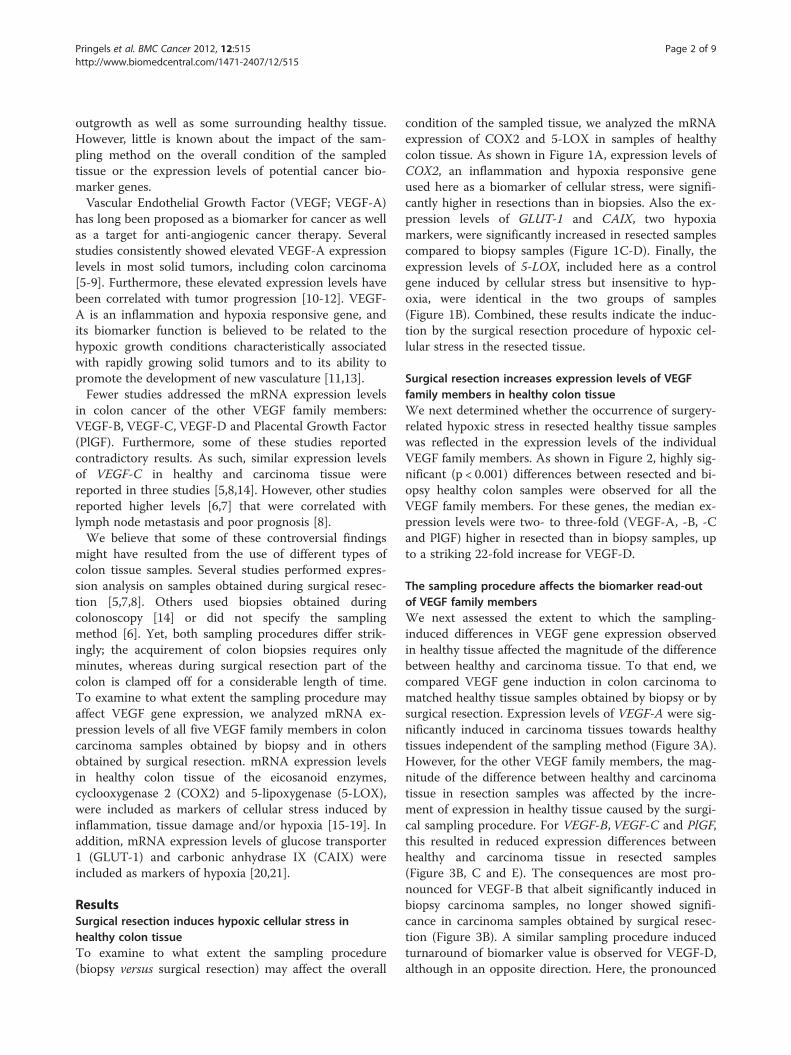
Surgical resection increases expression levels of VEGFfamily members in healthy colon tissueWe next determined whether the occurrence of surgery-related hypoxic stress in resected healthy tissue sampleswas reflected in the expression levels of the individualVEGF family members. As shown in Figure 2, highly sig-nificant (p < 0.001) differences between resected and bi-opsy healthy colon samples were observed for all theVEGF family members. For these genes, the median ex-pression levels were two- to three-fold (VEGF-A, -B, -Cand PlGF) higher in resected than in biopsy samples, upto a striking 22-fold increase for VEGF-D.
The sampling procedure affects the biomarker read-outof VEGF family membersWe next assessed the extent to which the sampling-induced differences in VEGF gene expression observedin healthy tissue affected the magnitude of the differencebetween healthy and carcinoma tissue. To that end, wecompared VEGF gene induction in colon carcinoma tomatched healthy tissue samples obtained by biopsy or bysurgical resection. Expression levels of VEGF-A were sig-nificantly induced in carcinoma tissues towards healthytissues independent of the sampling method (Figure 3A).However, for the other VEGF family members, the mag-nitude of the difference between healthy and carcinomatissue in resection samples was affected by the incre-ment of expression in healthy tissue caused by the surgi-cal sampling procedure. For VEGF-B,VEGF-C and PlGF,this resulted in reduced expression differences betweenhealthy and carcinoma tissue in resected samples(Figure 3B, C and E). The consequences are most pro-nounced for VEGF-B that albeit significantly induced inbiopsy carcinoma samples, no longer showed signifi-cance in carcinoma samples obtained by surgical resec-tion (Figure 3B). A similar sampling procedure inducedturnaround of biomarker value is observed for VEGF-D,although in an opposite direction. Here, the pronounced

A B
C D
Figure 1 Effect of sampling method on the expression of inflammatory and hypoxic stress responsive genes in healthy colon tissuesamples. Relative mRNA expression levels of the inflammatory eicosanoid enzymes COX2 (A) and 5-LOX (B) and of the hypoxia markers GLUT-1(C) and CAIX (D) are shown for healthy colon biopsies and healthy colon resection samples. Expression levels were normalized against referencegenes TBP and SDHA and were scaled against the median of the biopsy samples (median set to 1). Expression data are depicted as scatter plotsof the values obtained for each individual sample. The horizontal line represents the median; ns: not significant; *: p < 0.05; **: p < 0.01; ***: p <0.001 with Mann–Whitney U Test.
Pringels et al. BMC Cancer 2012, 12:515 Page 3 of 9http://www.biomedcentral.com/1471-2407/12/515
increase in the expression of VEGF-D in healthy resectedtissue as opposed to the near absence of such an in-crease in carcinoma tissue resulted in a highly significantunderexpression of VEGF-D in carcinoma resectionsamples (Figure 3D). On the contrary, in biopsy samplesno difference in VEGF-D expression between healthycolon and colon carcinoma samples was observed.
Cancer biomarker accuracy of VEGF family membersReceiver-operating characteristics (ROC) analysis iscommonly used to assess the reliability and accuracy ofpotential biomarkers. ROC-based assessment of the indi-vidual VEGF family members as biomarkers for coloncancer identified overexpression of PlGF (AUC 0.9342)as the most effective mRNA-marker for samplesobtained by biopsy with VEGF-A (AUC 0.8760) andVEGF-C AUC 0.8977) following as close seconds(Figure 4). This ranking however changes dramaticallywhen considering samples obtained by resection. Here,underexpression of VEGF-D emerges as the most potentbiomarker with an AUC of 0.9047 (p < 0.0001) and aROC-curve significantly different (p < 0.0001) from the
biopsy curve (Figure 4D). Overexpression of VEGF-A(AUC 0.8573) now precedes PlGF (AUC 0.8231), VEGF-C (AUC 0.6200) and especially VEGF-B (AUC 0.5621)shows strongly reduced accuracy as colon cancermRNA-marker (Figure 4).
DiscussionBiomarker expression profiles have become a valuabletool in diagnostic research, patient management andcancer therapy. We explored the influence of differentsampling methods on the expression of VEGF familybiomarkers in colon cancer. Samples obtained by eitherbiopsy or surgical resection were compared for thedifferential expression of VEGF-A, VEGF-B, VEGF-C,VEGF-D and PlGF. To examine the occurrence of cellu-lar stress caused by the sampling procedure, the expres-sion levels of the eicosanoid enzymes COX2 and 5-LOXwere quantified in healthy colon tissue. COX2 is a keyinflammatory enzyme, and its expression is stronglyinduced by NF-κB and HIF-1 transcription factors in re-sponse to inflammatory insults and hypoxic growth con-ditions, respectively [19,22-26]. In contrast, expression
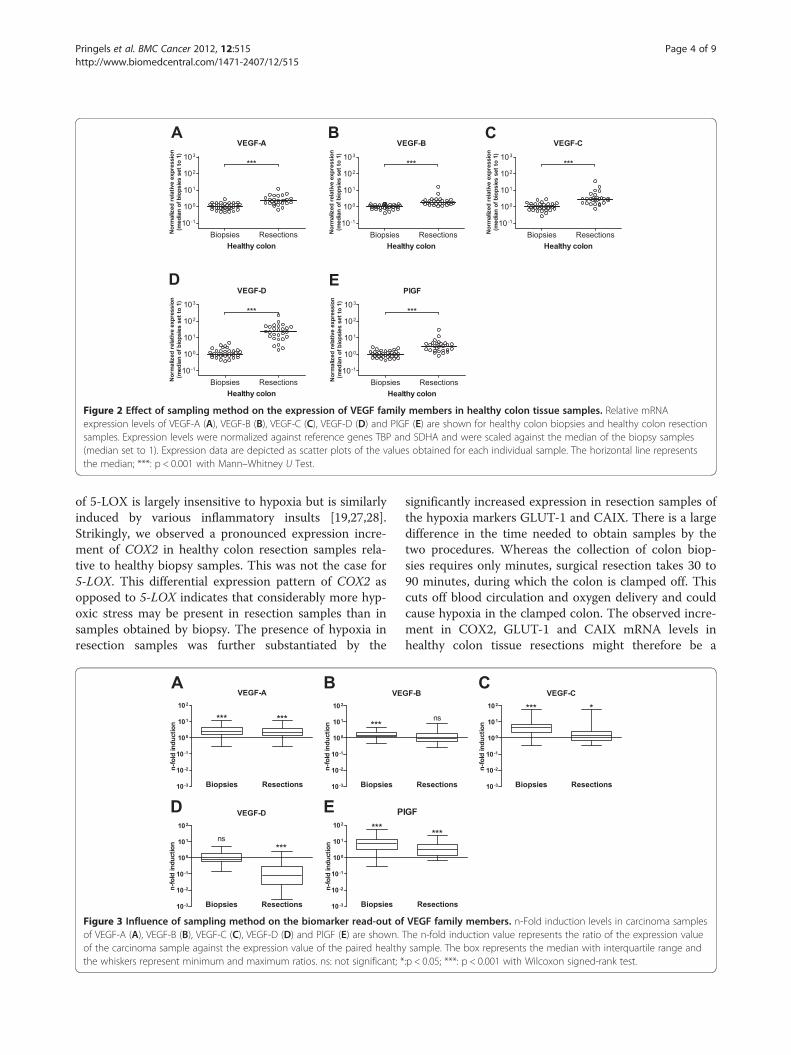
Figure 2 Effect of sampling method on the expression of VEGF family members in healthy colon tissue samples. Relative mRNAexpression levels of VEGF-A (A), VEGF-B (B), VEGF-C (C), VEGF-D (D) and PlGF (E) are shown for healthy colon biopsies and healthy colon resectionsamples. Expression levels were normalized against reference genes TBP and SDHA and were scaled against the median of the biopsy samples(median set to 1). Expression data are depicted as scatter plots of the values obtained for each individual sample. The horizontal line representsthe median; ***: p < 0.001 with Mann–Whitney U Test.
Pringels et al. BMC Cancer 2012, 12:515 Page 4 of 9http://www.biomedcentral.com/1471-2407/12/515
of 5-LOX is largely insensitive to hypoxia but is similarlyinduced by various inflammatory insults [19,27,28].Strikingly, we observed a pronounced expression incre-ment of COX2 in healthy colon resection samples rela-tive to healthy biopsy samples. This was not the case for5-LOX. This differential expression pattern of COX2 asopposed to 5-LOX indicates that considerably more hyp-oxic stress may be present in resection samples than insamples obtained by biopsy. The presence of hypoxia inresection samples was further substantiated by the
Figure 3 Influence of sampling method on the biomarker read-out ofof VEGF-A (A), VEGF-B (B), VEGF-C (C), VEGF-D (D) and PlGF (E) are shown.of the carcinoma sample against the expression value of the paired healththe whiskers represent minimum and maximum ratios. ns: not significant; *
significantly increased expression in resection samples ofthe hypoxia markers GLUT-1 and CAIX. There is a largedifference in the time needed to obtain samples by thetwo procedures. Whereas the collection of colon biop-sies requires only minutes, surgical resection takes 30 to90 minutes, during which the colon is clamped off. Thiscuts off blood circulation and oxygen delivery and couldcause hypoxia in the clamped colon. The observed incre-ment in COX2, GLUT-1 and CAIX mRNA levels inhealthy colon tissue resections might therefore be a
VEGF family members. n-Fold induction levels in carcinoma samplesThe n-fold induction value represents the ratio of the expression valuey sample. The box represents the median with interquartile range and:p < 0.05; ***: p < 0.001 with Wilcoxon signed-rank test.
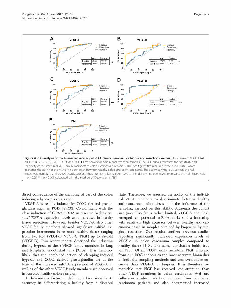
Figure 4 ROC-analysis of the biomarker accuracy of VEGF family members for biopsy and resection samples. ROC-curves of VEGF-A (A),VEGF-B (B), VEGF-C (C), VEGF-D (D) and PlGF (E) are shown for biopsy and resection samples. The ROC-curves represent the sensitivity andspecificity of the individual VEGF family members as colon carcinoma biomarkers. The insert gives the area under the curve (AUC), whichquantifies the ability of the marker to distinguish between healthy colon and colon carcinoma. The accompanying p-value tests the nullhypothesis, namely, that the AUC equals 0.50 and thus the biomarker is incompetent. The identity-line (Identity%) represents the null hypothesis.*: p < 0.05; ***: p < 0.001 calculated with the method of DeLong et al. [35].
Pringels et al. BMC Cancer 2012, 12:515 Page 5 of 9http://www.biomedcentral.com/1471-2407/12/515
direct consequence of the clamping of part of the coloninducing a hypoxic stress signal.VEGF-A is readily induced by COX2 derived prosta-
glandins such as PGE2 [29,30]. Concomitant with theclear induction of COX2 mRNA in resected healthy tis-sue, VEGF-A expression levels were increased in healthytissue resections. However, besides VEGF-A also otherVEGF family members showed significant mRNA ex-pression increments in resected healthy tissue rangingfrom 2–3 fold (VEGF-B, VEGF-C, PlGF) up to 22-fold(VEGF-D). Two recent reports described the inductionduring hypoxia of these VEGF family members in lungand lymphatic endothelial cells [31,32]. It is thereforelikely that the combined action of clamping-inducedhypoxia and COX2 derived prostaglandins are at thebasis of the increased mRNA expression of VEGF-A aswell as of the other VEGF family members we observedin resected healthy colon samples.A determining factor in defining a biomarker is its
accuracy in differentiating a healthy from a diseased
state. Therefore, we assessed the ability of the individ-ual VEGF members to discriminate between healthyand cancerous colon tissue and the influence of thesampling method on this ability. Although the cohortsize (n=77) so far is rather limited, VEGF-A and PlGFemerged as potential mRNA-markers discriminatingwith relatively high accuracy between healthy and car-cinoma tissue in samples obtained by biopsy or by sur-gical resection. Our results confirm previous studiesreporting significantly increased expression levels ofVEGF-A in colon carcinoma samples compared tohealthy tissue [5-9]. The same conclusion holds truefor PlGF. Of all VEGF family members, PlGF emergedfrom our ROC-analysis as the most accurate biomarkerin both the sampling methods and was even more ac-curate than VEGF-A in biopsies. It is therefore re-markable that PlGF has received less attention thanother VEGF members in colon carcinoma. Wei andcolleagues studied resection samples from colorectalcarcinoma patients and also documented increased
Pringels et al. BMC Cancer 2012, 12:515 Page 6 of 9http://www.biomedcentral.com/1471-2407/12/515
PlGF mRNA expression levels and their associationwith reduced survival [9]. A similar result wasobtained for both PlGF isoforms, PlGF-1 and PlGF-2,by Escudero-Esparza and colleagues [33]. Our observa-tions further confirm these findings.For VEGF-B, VEGF-C and VEGF-D we observed a sig-
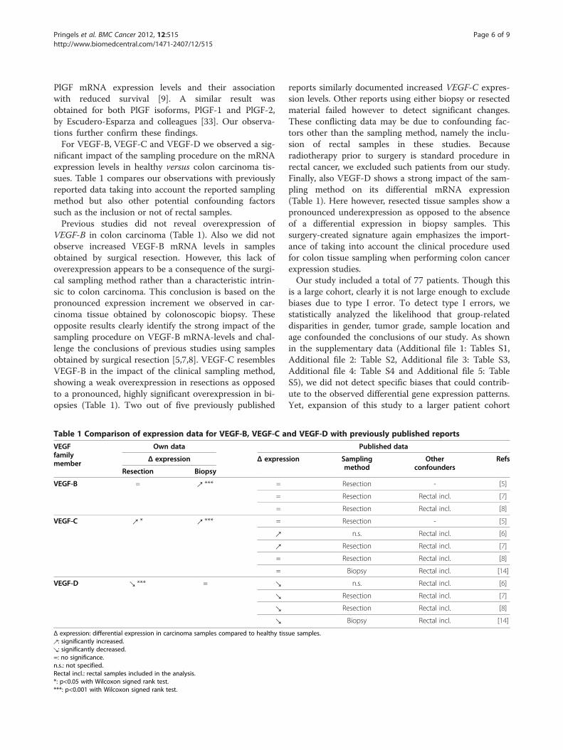
nificant impact of the sampling procedure on the mRNAexpression levels in healthy versus colon carcinoma tis-sues. Table 1 compares our observations with previouslyreported data taking into account the reported samplingmethod but also other potential confounding factorssuch as the inclusion or not of rectal samples.Previous studies did not reveal overexpression of
VEGF-B in colon carcinoma (Table 1). Also we did notobserve increased VEGF-B mRNA levels in samplesobtained by surgical resection. However, this lack ofoverexpression appears to be a consequence of the surgi-cal sampling method rather than a characteristic intrin-sic to colon carcinoma. This conclusion is based on thepronounced expression increment we observed in car-cinoma tissue obtained by colonoscopic biopsy. Theseopposite results clearly identify the strong impact of thesampling procedure on VEGF-B mRNA-levels and chal-lenge the conclusions of previous studies using samplesobtained by surgical resection [5,7,8]. VEGF-C resemblesVEGF-B in the impact of the clinical sampling method,showing a weak overexpression in resections as opposedto a pronounced, highly significant overexpression in bi-opsies (Table 1). Two out of five previously published
Table 1 Comparison of expression data for VEGF-B, VEGF-C a
VEGFfamilymember
Own data
Δ expression Δ expres
Resection Biopsy
VEGF-B = ↗ *** =
=
=
VEGF-C ↗ * ↗ *** =
↗
↗
=
=
VEGF-D ↘ *** = ↘
↘
↘
↘
Δ expression: differential expression in carcinoma samples compared to healthy tiss↗: significantly increased.↘: significantly decreased.=: no significance.n.s.: not specified.Rectal incl.: rectal samples included in the analysis.*: p<0.05 with Wilcoxon signed rank test.***: p<0.001 with Wilcoxon signed rank test.
reports similarly documented increased VEGF-C expres-sion levels. Other reports using either biopsy or resectedmaterial failed however to detect significant changes.These conflicting data may be due to confounding fac-tors other than the sampling method, namely the inclu-sion of rectal samples in these studies. Becauseradiotherapy prior to surgery is standard procedure inrectal cancer, we excluded such patients from our study.Finally, also VEGF-D shows a strong impact of the sam-pling method on its differential mRNA expression(Table 1). Here however, resected tissue samples show apronounced underexpression as opposed to the absenceof a differential expression in biopsy samples. Thissurgery-created signature again emphasizes the import-ance of taking into account the clinical procedure usedfor colon tissue sampling when performing colon cancerexpression studies.Our study included a total of 77 patients. Though this
is a large cohort, clearly it is not large enough to excludebiases due to type I error. To detect type I errors, westatistically analyzed the likelihood that group-relateddisparities in gender, tumor grade, sample location andage confounded the conclusions of our study. As shownin the supplementary data (Additional file 1: Tables S1,Additional file 2: Table S2, Additional file 3: Table S3,Additional file 4: Table S4 and Additional file 5: TableS5), we did not detect specific biases that could contrib-ute to the observed differential gene expression patterns.Yet, expansion of this study to a larger patient cohort
nd VEGF-D with previously published reports
Published data
sion Samplingmethod
Otherconfounders
Refs
Resection - [5]
Resection Rectal incl. [7]
Resection Rectal incl. [8]
Resection - [5]
n.s. Rectal incl. [6]
Resection Rectal incl. [7]
Resection Rectal incl. [8]
Biopsy Rectal incl. [14]
n.s. Rectal incl. [6]
Resection Rectal incl. [7]
Resection Rectal incl. [8]
Biopsy Rectal incl. [14]
ue samples.
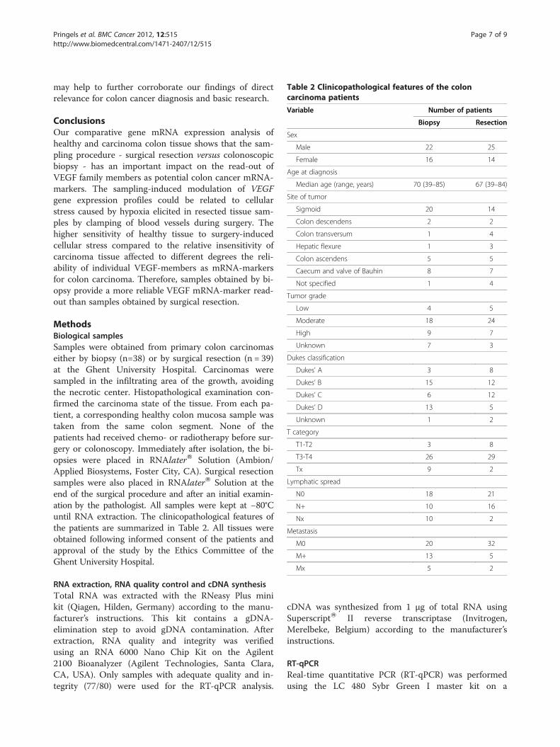
Table 2 Clinicopathological features of the coloncarcinoma patients
Variable Number of patients
Biopsy Resection
Sex
Male 22 25
Female 16 14
Age at diagnosis
Median age (range, years) 70 (39–85) 67 (39–84)
Site of tumor
Sigmoid 20 14
Colon descendens 2 2
Colon transversum 1 4
Hepatic flexure 1 3
Colon ascendens 5 5
Caecum and valve of Bauhin 8 7
Not specified 1 4
Tumor grade
Low 4 5
Moderate 18 24
High 9 7
Unknown 7 3
Dukes classification
Dukes’ A 3 8
Dukes’ B 15 12
Dukes’ C 6 12
Dukes’ D 13 5
Unknown 1 2
T category
T1-T2 3 8
T3-T4 26 29
Tx 9 2
Lymphatic spread
N0 18 21
N+ 10 16
Nx 10 2
Metastasis
M0 20 32
M+ 13 5
Mx 5 2
Pringels et al. BMC Cancer 2012, 12:515 Page 7 of 9http://www.biomedcentral.com/1471-2407/12/515
may help to further corroborate our findings of directrelevance for colon cancer diagnosis and basic research.
ConclusionsOur comparative gene mRNA expression analysis ofhealthy and carcinoma colon tissue shows that the sam-pling procedure - surgical resection versus colonoscopicbiopsy - has an important impact on the read-out ofVEGF family members as potential colon cancer mRNA-markers. The sampling-induced modulation of VEGFgene expression profiles could be related to cellularstress caused by hypoxia elicited in resected tissue sam-ples by clamping of blood vessels during surgery. Thehigher sensitivity of healthy tissue to surgery-inducedcellular stress compared to the relative insensitivity ofcarcinoma tissue affected to different degrees the reli-ability of individual VEGF-members as mRNA-markersfor colon carcinoma. Therefore, samples obtained by bi-opsy provide a more reliable VEGF mRNA-marker read-out than samples obtained by surgical resection.
MethodsBiological samplesSamples were obtained from primary colon carcinomaseither by biopsy (n=38) or by surgical resection (n = 39)at the Ghent University Hospital. Carcinomas weresampled in the infiltrating area of the growth, avoidingthe necrotic center. Histopathological examination con-firmed the carcinoma state of the tissue. From each pa-tient, a corresponding healthy colon mucosa sample wastaken from the same colon segment. None of thepatients had received chemo- or radiotherapy before sur-gery or colonoscopy. Immediately after isolation, the bi-opsies were placed in RNAlaterW Solution (Ambion/Applied Biosystems, Foster City, CA). Surgical resectionsamples were also placed in RNAlaterW Solution at theend of the surgical procedure and after an initial examin-ation by the pathologist. All samples were kept at −80°Cuntil RNA extraction. The clinicopathological features ofthe patients are summarized in Table 2. All tissues wereobtained following informed consent of the patients andapproval of the study by the Ethics Committee of theGhent University Hospital.
RNA extraction, RNA quality control and cDNA synthesisTotal RNA was extracted with the RNeasy Plus minikit (Qiagen, Hilden, Germany) according to the manu-facturer’s instructions. This kit contains a gDNA-elimination step to avoid gDNA contamination. Afterextraction, RNA quality and integrity was verifiedusing an RNA 6000 Nano Chip Kit on the Agilent2100 Bioanalyzer (Agilent Technologies, Santa Clara,CA, USA). Only samples with adequate quality and in-tegrity (77/80) were used for the RT-qPCR analysis.
cDNA was synthesized from 1 μg of total RNA usingSuperscriptW II reverse transcriptase (Invitrogen,Merelbeke, Belgium) according to the manufacturer’sinstructions.
RT-qPCRReal-time quantitative PCR (RT-qPCR) was performedusing the LC 480 Sybr Green I master kit on a
Pringels et al. BMC Cancer 2012, 12:515 Page 8 of 9http://www.biomedcentral.com/1471-2407/12/515
LightCyclerW 480 Real-Time PCR system (both fromRoche Applied Science, Penzberg, Germany). Primerswere designed using PrimerSelect (DNASTAR, Madison,USA) and purchased from Invitrogen. The sequences ofthe forward and reverse primers were as follows: VEGF-A 5’-TGAGTTGCCCAGGAGACCAC-3’ and 5’-GAAGGGGAGCAGGAAGAGGAT-3’; VEGF-B 5’-CCGGAAGCTGCGAAGGTGACA-3’ and 5’-GGGAGACAAGGGATGGCAGAAGAG-3’; VEGF-C 5’-CACGGCTTATGCAAGCAAAGA-3’ and 5’-TCCTTTCCTTAGCTGACACTTGT-3’; VEGF-D 5’-GCAGCCCTAGAGAAACGTG-3’ and 5’-AGGTGCTGGTGTTCATACAGAT-3’; PlGF 5’-TGCGGCGATGAGAATCTGC-3’ and 5’-AGCGAACGTGCTGAGAGAAC-3’; COX2 5’-TTGCTGGAACATGGAATTACC-3’ and 5’-TGCCTGCTCTGGTCAATG-3’; 5-LOX 5’-TGGCGCGGTGGATTCATAC-3’ and 5’-CAGGGGAACTCGATGTAGTCC-3’; GLUT-1 5’-CTTGTGTGGCCTTCTTTGAAGT-3’ and 5’-CCACACAGTTGCTCCACAT-3’; CAIX 5’-GGAAGGCTCAGAGACTCA-3’ and 5’-CTTAGCACTCAGCATCAC-3’. All samples were assayedin triplicate. Relative expression values were calculatedusing the 2(−delta delta C(T)) method and were normal-ized against reference genes: tata-binding protein (TBP)and succinate dehydrogenase complex subunit A (SDHA)(primers: TBP 5’-CGGCTGTTTAACTTCGCTTC-3’ and5'-CACACGCCAAGAAACAGTGA-3’; SDHA 5’-TGGGAACAAGAGGGCATCTG-3’ and 5’-CCACCACTGCATCAAATTCATG-3’). In these calculations we took into ac-count the PCR efficiency of the individual PCR reactions,calculated on the basis of linear regression as described inRuijter et al. [34]. For the comparison between healthycolon biopsies and resections, the normalized relative ex-pression values were scaled against the median of thehealthy biopsies (median of biopsies set to 1). The specifi-city of amplification was confirmed by evaluation of themelting curves.
Statistical analysisStatistical analysis was performed using the GraphPadPrismW software (GraphPad Software Inc., La Jolla,California, USA). Statistical significance of compari-sons between two independent groups was determinedwith the two-tailed Mann–Whitney U test. The com-parison between paired samples was performed withthe Wilcoxon signed-rank test. The accuracy of themarkers was determined with receiver operator char-acteristic curves (ROC). The statistical significance ofthe difference between two areas under the ROCcurves was calculated by the method of DeLong et al.and performed with MedCalcW software (MedCalcSoftware, Mariakerke, Belgium) [35]. Significant p-values were ranked as p < 0.05 (*), p < 0.01 (**) andp < 0.001 (***).
Additional files
Additional file 1: Table S1. Comparison of expression levels in maleversus female patients with Mann-Whitney test. *: p < 0.05.
Additional file 2: Table S2. Comparison of expression levels in coloncarcinoma with tumor grade low versus moderate versus high withKruskal Wallis test. *: p < 0.05.
Additional file 3: Table S3. Comparison of expression levels in coloncarcinoma with Dukes classification A versus B versus C versus D withKruskal Wallis test. *: p < 0.05.
Additional file 4: Table S4. Comparison of expression levels in healthycolon and colon carcinoma samples from different tumor sites (caecumand Valve of Bauhin versus colon ascendens, transversum, descendensand hepatic flexure versus sigmoid) with Kruskal Wallis test. *: p < 0.05;**:p < 0.01. n/a: not applicable.
Additional file 5: Table S5. Comparison of expression levels in patientsyounger than 70 years or of 70 years versus patients older than 70 withMann-Whitney test.
Abbreviations5-LOX: 5-lipoxygenase; AUC: Area under the curve; CAIX: Carbonic anhydraseIX; COX2: Cyclooxygenase 2; GLUT-1: Glucose transporter 1; HIF-1: Hypoxiainducible factor 1; NFκB: Nuclear factor kappa B; PlGF: Placental growthfactor; ROC: Receiver operator characteristic; RT-qPCR: Real-time quantitativePCR; SDHA: Succinate dehydrogenase complex subunit A; TBP: Tata-bindingprotein; VEGF: Vascular endothelial growth factor.
Competing interestsThe authors declare that they have no competing interests.
Authors’ contributionsSP conceived and initiated this project, performed RNA extractions, RT-qPCRassays and data analysis, and wrote the manuscript. NVD participated in thedesign and coordination of the study, coordinated the acquisition of patientdata and clinical samples, and revised the manuscript. BDC contributed tothe experimental design and the data analysis and revised the manuscript.PP and WC provided clinical samples and clinicopathological data andrevised the manuscript. MP conceived the study, participated in the design,and revised the manuscript. JG conceived the study, and participated in itsdesign and coordination and helped to draft the manuscript. All authorsread and approved the final manuscript.
AcknowledgmentsThis work was supported by the Research Foundation-Flanders GrantG.0043.08N. Additional financial support to J.G. was provided by the IAP6/18,funded by the Interuniversity Attraction Poles Program initiated by theBelgian State Science Policy Office. S.P. is supported by a Ph.D. grant fromthe Institute for the Promotion of Innovation through Science andTechnology in Flanders (IWT-Vlaanderen). B.D.C. is supported by theGeconcerteerde Onderzoeksacties of Ghent University. M.P. is Senior ClinicalInvestigator of the Research Foundation-Flanders, Belgium.
Author details1Department of Biomedical Molecular Biology, Ghent University,Technologiepark 927, Zwijnaarde 9052, Belgium. 2Department of Surgery,Ghent University Hospital, De Pintelaan 185, Gent 9000, Belgium.3Department for Molecular Biomedical Research VIB, Zwijnaarde 9052,Belgium. 4Department of Oncology, Antwerp University Hospital, Wilrijkstraat10, Edegem 2650, Belgium.
Received: 9 March 2012 Accepted: 25 October 2012Published: 13 November 2012
References1. Jemal A, Bray F, Center MM, Ferlay J, Ward E, Forman D: Global cancer
statistics. CA Cancer J Clin 2011, 61(2):69–90.2. Arnold CN, Goel A, Blum HE, Boland CR: Molecular pathogenesis of
colorectal cancer: implications for molecular diagnosis. Cancer 2005,104(10):2035–2047.

Pringels et al. BMC Cancer 2012, 12:515 Page 9 of 9http://www.biomedcentral.com/1471-2407/12/515
3. Sillars-Hardebol AH, Carvalho B, de Wit M, Postma C, Delis-van Diemen PM,Mongera S, Ylstra B, van de Wiel MA, Meijer GA, Fijneman RJ: Identificationof key genes for carcinogenic pathways associated with colorectaladenoma-to-carcinoma progression. Tumour Biol 2010, 31(2):89–96.
4. Cappell MS: Pathophysiology, clinical presentation, and management ofcolon cancer. Gastroenterol Clin North Am 2008, 37(1):1–24. v.
5. Andre T, Kotelevets L, Vaillant JC, Coudray AM, Weber L, Prevot S, Parc R,Gespach C, Chastre E: Vegf, Vegf-B, Vegf-C and their receptors KDR, FLT-1and FLT-4 during the neoplastic progression of human colonic mucosa.Int J Cancer 2000, 86(2):174–181.
6. George ML, Tutton MG, Janssen F, Arnaout A, Abulafi AM, Eccles SA, SwiftRI: VEGF-A, VEGF-C, and VEGF-D in colorectal cancer progression.Neoplasia 2001, 3(5):420–427.
7. Hanrahan V, Currie MJ, Gunningham SP, Morrin HR, Scott PA, Robinson BA,Fox SB: The angiogenic switch for vascular endothelial growth factor(VEGF)-A, VEGF-B, VEGF-C, and VEGF-D in the adenoma-carcinomasequence during colorectal cancer progression. J Pathol 2003,200(2):183–194.
8. Kawakami M, Furuhata T, Kimura Y, Yamaguchi K, Hata F, Sasaki K, Hirata K:Expression analysis of vascular endothelial growth factors and theirrelationships to lymph node metastasis in human colorectal cancer.J Exp Clin Cancer Res 2003, 22(2):229–237.
9. Wei SC, Tsao PN, Yu SC, Shun CT, Tsai-Wu JJ, Wu CH, Su YN, Hsieh FJ, WongJM: Placenta growth factor expression is correlated with survival ofpatients with colorectal cancer. Gut 2005, 54(5):666–672.
10. Otrock ZK, Makarem JA, Shamseddine AI: Vascular endothelial growthfactor family of ligands and receptors: review. Blood Cells Mol Dis 2007,38(3):258–268.
11. Roskoski R Jr: Vascular endothelial growth factor (VEGF) signaling intumor progression. Crit Rev Oncol Hematol 2007, 62(3):179–213.
12. Ferrara N, Davis-Smyth T: The biology of vascular endothelial growthfactor. Endocr Rev 1997, 18(1):4–25.
13. Ferrara N, Gerber HP, LeCouter J: The biology of VEGF and its receptors.Nat Med 2003, 9(6):669–676.
14. Onogawa S, Kitadai Y, Tanaka S, Kuwai T, Kimura S, Chayama K: Expressionof VEGF-C and VEGF-D at the invasive edge correlates with lymph nodemetastasis and prognosis of patients with colorectal carcinoma. CancerSci 2004, 95(1):32–39.
15. Colotta F, Allavena P, Sica A, Garlanda C, Mantovani A: Cancer-relatedinflammation, the seventh hallmark of cancer: links to genetic instability.Carcinogenesis 2009, 30(7):1073–1081.
16. Hyde CA, Missailidis S: Inhibition of arachidonic acid metabolism and itsimplication on cell proliferation and tumour-angiogenesis. IntImmunopharmacol 2009, 9(6):701–715.
17. Aggarwal BB, Shishodia S, Sandur SK, Pandey MK, Sethi G: Inflammationand cancer: how hot is the link? Biochem Pharmacol 2006,72(11):1605–1621.
18. Maynard MA, Ohh M: The role of hypoxia-inducible factors in cancer. CellMol Life Sci 2007, 64(16):2170–2180.
19. Harris AL: Hypoxia–a key regulatory factor in tumour growth. Nat RevCancer 2002, 2(1):38–47.
20. Wood IS, Wang B, Lorente-Cebrian S, Trayhurn P: Hypoxia increasesexpression of selective facilitative glucose transporters (GLUT) and2-deoxy-D-glucose uptake in human adipocytes. Biochem Biophys ResCommun 2007, 361(2):468–473.
21. Vordermark D, Kaffer A, Riedl S, Katzer A, Flentje M: Characterization ofcarbonic anhydrase IX (CA IX) as an endogenous marker of chronichypoxia in live human tumor cells. Int J Radiat Oncol Biol Phys 2005,61(4):1197–1207.
22. Chiarugi V, Magnelli L, Chiarugi A, Gallo O: Hypoxia induces pivotal tumorangiogenesis control factors including p53, vascular endothelial growthfactor and the NFkappaB-dependent inducible nitric oxide synthase andcyclooxygenase-2. J Cancer Res Clin Oncol 1999, 125(8–9):525–528.
23. Gately S, Li WW: Multiple roles of COX-2 in tumor angiogenesis: a targetfor antiangiogenic therapy. Semin Oncol 2004, 31(2 Suppl 7):2–11.
24. Heirman I, Ginneberge D, Brigelius-Flohe R, Hendrickx N, Agostinis P,Brouckaert P, Rottiers P, Grooten J: Blocking tumor cell eicosanoidsynthesis by GP x 4 impedes tumor growth and malignancy. Free RadicBiol Med 2006, 40(2):285–294.
25. Kaidi A, Qualtrough D, Williams AC, Paraskeva C: Direct transcriptionalup-regulation of cyclooxygenase-2 by hypoxia-inducible factor (HIF)-1
promotes colorectal tumor cell survival and enhances HIF-1transcriptional activity during hypoxia. Cancer Res 2006, 66(13):6683–6691.
26. Schmedtje JF Jr, Ji YS, Liu WL, DuBois RN, Runge MS: Hypoxia inducescyclooxygenase-2 via the NF-kappaB p65 transcription factor in humanvascular endothelial cells. J Biol Chem 1997, 272(1):601–608.
27. Romano M, Claria J: Cyclooxygenase-2 and 5-lipoxygenase convergingfunctions on cell proliferation and tumor angiogenesis: implications forcancer therapy. FASEB J 2003, 17(14):1986–1995.
28. Radmark O, Werz O, Steinhilber D, Samuelsson B: 5-Lipoxygenase:regulation of expression and enzyme activity. Trends Biochem Sci 2007,32(7):332–341.
29. Liu XH, Kirschenbaum A, Lu M, Yao S, Dosoretz A, Holland JF, Levine AC:Prostaglandin E2 induces hypoxia-inducible factor-1alpha stabilizationand nuclear localization in a human prostate cancer cell line. J Biol Chem2002, 277(51):50081–50086.
30. Fukuda R, Kelly B, Semenza GL: Vascular endothelial growth factor geneexpression in colon cancer cells exposed to prostaglandin E2 ismediated by hypoxia-inducible factor 1. Cancer Res 2003, 63(9):2330–2334.
31. Sands M, Howell K, Costello CM, McLoughlin P: Placenta growth factor andvascular endothelial growth factor B expression in the hypoxic lung.Respir Res 2011, 12:17.
32. Garrafa E, Caprara V, Di Castro V, Rosano L, Bagnato A, Spinella F:Endothelin-1 cooperates with hypoxia to induce vascular-like structuresthrough vascular endothelial growth factor-C, -D and -A in lymphaticendothelial cells. Life Sci 2012, 91(13-14):638–643.
33. Escudero-Esparza A, Martin TA, Davies ML, Jiang WG: PGF isoforms, PLGF-1and PGF-2, in colorectal cancer and the prognostic significance. CancerGenomics Proteomics 2009, 6(4):239–246.
34. Ruijter JM, Ramakers C, Hoogaars WM, Karlen Y, Bakker O, van den Hoff MJ,Moorman AF: Amplification efficiency: linking baseline and bias in theanalysis of quantitative PCR data. Nucleic Acids Res 2009, 37(6):e45.
35. DeLong ER, DeLong DM, Clarke-Pearson DL: Comparing the areas undertwo or more correlated receiver operating characteristic curves: anonparametric approach. Biometrics 1988, 44(3):837–845.
doi:10.1186/1471-2407-12-515Cite this article as: Pringels et al.: Clinical procedure for colon carcinomatissue sampling directly affects the cancer marker-capacity of VEGFfamily members. BMC Cancer 2012 12:515.
Submit your next manuscript to BioMed Centraland take full advantage of:
• Convenient online submission
• Thorough peer review
• No space constraints or color figure charges
• Immediate publication on acceptance
• Inclusion in PubMed, CAS, Scopus and Google Scholar
• Research which is freely available for redistribution
Submit your manuscript at www.biomedcentral.com/submit
Related Documents