Research Article Formulation, Optimization, and Characterization of Repaglinide Loaded Nanocrystal for Diabetes Therapy Gajanan Shinde, 1 Mitesh Patel, 1 Manan Mehta, 1 Rajesh Kesarla, 1 and Ganesh Bangale 2 1 Parul Institute of Pharmacy, Limda, Vadodara, Gujarat, India 2 Government College of Pharmacy, Amravati, Maharashtra, India Correspondence should be addressed to Gajanan Shinde; [email protected] Received 19 August 2014; Revised 3 December 2014; Accepted 3 December 2014 Academic Editor: Changyang Gong Copyright © 2015 Gajanan Shinde et al. is is an open access article distributed under the Creative Commons Attribution License, which permits unrestricted use, distribution, and reproduction in any medium, provided the original work is properly cited. e aim of the present investigation was to formulate and characterize nanocrystal formulation of Repaglinide for diabetes therapy. Formulation was done by high pressure homogenization. HPH pressure and cycles range were screened by preliminary batches (T1 and T2). 5, 8, and 10 cycles and 500 to 1500 bar pressure range had kept for further investigation. Taguchi design was used to optimize type of polymer, % polymer concentration, number of cycles, and HPH pressure for nanocrystal formulation. Formulations were characterized for particle size, zeta potential, and in vitro drug release. Optimized formulation (NC 3) showed particle size of 187 nm, zeta potential of −29.4mv, and % drug release of 80.58% and it was used for further study. Data analysis proved significant effects of factors on responses. Polydispersity index (PDI) Analysis of optimized formulation were found to be 0.248. SEM showed nanocrystal aggregation of drug, may be due to water removal process. DSC showed slight change in crystallinity, may be due to the presence of PEG 4000. Stability study was carried out for 3 months. It indicated no significant change in particle size and zeta potential. However, further studies in higher animals and human being need to be performed before this formulation can be commercially exploited. 1. Introduction [1, 2] Diabetes is one of the major diseases. Diabetes is a chronic condition that is caused by the body’s incapability to produce or effectively use its own insulin. Insulin is a hormone necessary to convert food into glucose, which the body needs for the energy. Without production of enough insulin, glucose level becomes so high. As a result, permanent damage of blood vessel can occur. ere are two types of diabetes [3]: (1) diabetes insipidus (DI) and (2) diabetes mellitus (DM); it is a disorder that is characterized by the excretion of large amounts of severely diluted urine and excessive thirst with reduction of fluid intake having no effect on the concentration of the urine. ere are several types of diabetes mellitus. Diabetes mellitus (DM) is a group of metabolic disorder in that the person has high blood sugar, either because cells do not respond to the insulin that is produced by the body or body does not produce enough insulinIt has also several types of various symptoms of diabetes which are as follows: polyphagia, blurred vision, loss of weight, and polyuria. Repaglinide [4, 5] is antidiabetic drug in the class of medications known as meglitinides [6]. Repaglinide is used to treat type 2 diabetes. Repaglinide depresses blood glucose concentrations by stimulating the release of insulin from beta cells of pancreatic islet tissue. is is done by a selective ion channel mechanism. Repaglinide prevents adenosine triphosphate (ATP) potassium chan- nels on the beta cell membrane and potassium efflux. e resultant depolarization and calcium influx induce insulin secretion. Repaglinide is oral antihyperglycemic agent in type II diabetes. Repaglinide is BCS class II drug and its oral bioavailability is 56%. Repaglinide is incompletely absorbed from the gastrointestinal tract and has low oral bioavailability because of the poor solubility. So, an attempt was made to increase the solubility of Repaglinide while formulating nanocrystal. If one could be increase the solubility then may be the rapid absorption and enhance the bioavailability. Decreasing the particle size by formulating nanocrystal may increase the surface area. Hindawi Publishing Corporation Advances in Pharmaceutics Volume 2015, Article ID 363061, 7 pages http://dx.doi.org/10.1155/2015/363061

Welcome message from author
This document is posted to help you gain knowledge. Please leave a comment to let me know what you think about it! Share it to your friends and learn new things together.
Transcript
Research ArticleFormulation, Optimization, and Characterization ofRepaglinide Loaded Nanocrystal for Diabetes Therapy
Gajanan Shinde,1 Mitesh Patel,1 Manan Mehta,1 Rajesh Kesarla,1 and Ganesh Bangale2
1Parul Institute of Pharmacy, Limda, Vadodara, Gujarat, India2Government College of Pharmacy, Amravati, Maharashtra, India
Correspondence should be addressed to Gajanan Shinde; [email protected]
Received 19 August 2014; Revised 3 December 2014; Accepted 3 December 2014
Academic Editor: Changyang Gong
Copyright © 2015 Gajanan Shinde et al.This is an open access article distributed under the Creative CommonsAttribution License,which permits unrestricted use, distribution, and reproduction in any medium, provided the original work is properly cited.
The aim of the present investigation was to formulate and characterize nanocrystal formulation of Repaglinide for diabetes therapy.Formulation was done by high pressure homogenization. HPH pressure and cycles range were screened by preliminary batches (T1andT2). 5, 8, and 10 cycles and 500 to 1500 bar pressure range had kept for further investigation. Taguchi designwas used to optimizetype of polymer, % polymer concentration, number of cycles, and HPH pressure for nanocrystal formulation. Formulations werecharacterized for particle size, zeta potential, and in vitro drug release. Optimized formulation (NC 3) showed particle size of 187nm, zeta potential of −29.4mv, and % drug release of 80.58% and it was used for further study. Data analysis proved significanteffects of factors on responses. Polydispersity index (PDI) Analysis of optimized formulation were found to be 0.248. SEM showednanocrystal aggregation of drug, may be due to water removal process. DSC showed slight change in crystallinity, may be dueto the presence of PEG 4000. Stability study was carried out for 3 months. It indicated no significant change in particle size andzeta potential. However, further studies in higher animals and human being need to be performed before this formulation can becommercially exploited.
1. Introduction [1, 2]
Diabetes is one of the major diseases. Diabetes is a chroniccondition that is caused by the body’s incapability to produceor effectively use its own insulin. Insulin is a hormonenecessary to convert food into glucose, which the bodyneeds for the energy. Without production of enough insulin,glucose level becomes so high. As a result, permanent damageof blood vessel can occur.
There are two types of diabetes [3]: (1) diabetes insipidus(DI) and (2) diabetes mellitus (DM); it is a disorder that ischaracterized by the excretion of large amounts of severelydiluted urine and excessive thirst with reduction of fluidintake having no effect on the concentration of the urine.There are several types of diabetes mellitus. Diabetes mellitus(DM) is a group of metabolic disorder in that the person hashigh blood sugar, either because cells do not respond to theinsulin that is produced by the body or body does not produceenough insulinIt has also several types of various symptomsof diabetes which are as follows: polyphagia, blurred vision,
loss of weight, and polyuria. Repaglinide [4, 5] is antidiabeticdrug in the class of medications known as meglitinides [6].Repaglinide is used to treat type 2 diabetes. Repaglinidedepresses blood glucose concentrations by stimulating therelease of insulin from beta cells of pancreatic islet tissue.Thisis done by a selective ion channel mechanism. Repaglinideprevents adenosine triphosphate (ATP) potassium chan-nels on the beta cell membrane and potassium efflux. Theresultant depolarization and calcium influx induce insulinsecretion. Repaglinide is oral antihyperglycemic agent in typeII diabetes. Repaglinide is BCS class II drug and its oralbioavailability is 56%. Repaglinide is incompletely absorbedfrom the gastrointestinal tract and has low oral bioavailabilitybecause of the poor solubility. So, an attempt was madeto increase the solubility of Repaglinide while formulatingnanocrystal. If one could be increase the solubility thenmay be the rapid absorption and enhance the bioavailability.Decreasing the particle size by formulating nanocrystal mayincrease the surface area.
Hindawi Publishing CorporationAdvances in PharmaceuticsVolume 2015, Article ID 363061, 7 pageshttp://dx.doi.org/10.1155/2015/363061
2 Advances in Pharmaceutics
The aim here is to make a nanocrystal formulation usingTaguchi design with variable process in the face of variationover have little or no control. The Taguchi method involvesreducing the variation in a process through robust designof experiments. The overall objective of the method is toproduce high quality product at low cost to themanufacturer.
Nanocrystals are crystals having size less than 1 𝜇m [7, 8].As the particle size of a crystal is decreased to about 100 nm,there is a drastic change in the properties of the material. Thedecreased size increases the surface area and solubility of drugand there is proportionate increase in the bioavailability ofpoorly soluble drugs.
Drug nanocrystals consist of a polymeric matrix and anincorporated drug. Drug nanocrystals do not consist of anymatrixmaterial. Nanocrystal formulations are particles madefromdrug. Typically, they are stabilized by surfactant or poly-meric steric stabilizers. Hence, these particles possess a betterdrug loading in contrast to matrix nanoparticle consistingof polymeric matrix (polymeric nanoparticle) or lipid matrix(nanoemulsions, liposomes, and lipid nanoparticles [9]).
The high loading makes them very efficient in trans-porting drug to or into cells, reaching a sufficiently hightherapeutic concentration for the pharmacological effect.Thenanocrystals are prepared in a liquid dispersion medium;that is, the nanocrystals are suspended in liquid. There arevarious types of advantages [8, 10, 11]. If Increase the rate ofabsorption and bioavailability may be Reduction in requireddose. There are many types [9–17] of prepared nanocrystals;here, we prepare nanocrystals by high pressure homogeniza-tion microfluidizer technology (IDD-P technology), piston-gap homogenization in water (DissoCubes), and piston-gaphomogenization in nonaqueous medium (nanopure technol-ogy).
2. Materials and Method
2.1. Materials Used in the Present Work. The Repaglinidedrug was obtained as a gift sample from Torrent ResearchCenter, Ahmedabad, Poloxamer 188 was obtained as a giftsample from Sigma Aldrich, Peg 4000 was obtained as a giftsample from Sisco Research Lab., Mumbai, and polyvinylalcohol, acetone, concentration HCL, and water used were ofanalytical grade. Dialysis bag was used from HiMedia Labs,Mumbai.
2.2. Method
2.2.1. Experimental Methods
(1) Solubility Study [16]. An appropriate amount of Repaglin-ide was dissolved in a beaker by continuously adding thesuitable solvents. The solvents screening was conducted inwater, methanol, chloroform, and acetone. The solvents wereadded in a pipette in aliquots of 0.2mL applying magneticstirring until complete dissolution of drug.The solubility wascalculated in mg/mL. Once an approximate solubility wasfound, the saturation solubility was determined, after whichthe mixture was stirred on a magnetic stirrer at 80 rpm for
24 hr and then filtered, and the content of dissolved drug wasanalyzed spectrophotometrically at 243 nm.
(2) Production of Nanocrystal Formulations. Nanocrystal for-mulations were prepared by high pressure homogenization.The optimum combination of four independent variables,such as type of polymer, % polymer concentration, number ofcycles, and pressure of HPH, were at three levels by Taguchiorthogonal experimental design to achieve optimum particlesize, zeta potential, and in vitro drug release.
(3) High Pressure Homogenization Method. Repaglinideloaded nanocrystal was prepared by high pressure homoge-nizationmethod. Stabilizer was dissolved in 50mLof distilledwater to obtain aqueous surfactant solution and drug isseparately dissolved in acetone. Then, drug solution is addedinto aqueous surfactant solution under high speed homoge-nizer at 10,000 rpm for 15min. to obtain coarse suspension.Then, this coarse suspension was subjected to high pressurehomogenizer at varying pressures and cycles. Samples werewithdrawn after the size reduction step for size distributionanalysis. Then, this nanocrystal dispersion was lyophilized toobtain the nanocrystal.
2.2.2. Evaluation Method
(1) Drug Excipients Compatibility Study [11]. FTIR spec-troscopy was carried out to further elucidate the interactionWith Repaglinide polymer. Repaglinide and polymers weremixed with KBr and pressed (1 tone) into the pellets to carryout spectra by applying sufficient pressure with precaution toavoid moisture.
(2) Measurement of Particle Size [18–26]. Particle Size andsize distribution of the particles in the formulation weredetermined with a zetasizer nanoseries ZS. The samplefor particle size analysis was added to a small dispersionunit called a cuvette. Average values were calculated fromthree batches of each sample. The diameters reported werecalculated using volume distribution.
(3) Zeta Potential [20]. Zeta potential of the formulation wasdetermined with a zetasizer nanoseries ZS. The sample forparticle size analysis was added to the small dispersion unitcalled a cuvette. Average values were calculated from threebatches of each sample.
(4) In Vitro Drug Release [26]. Dialysis bag diffusion tech-nique was used to study in vitro release of drug from theprepared nanocrystal formulation. The 5mL of formulationwas placed in the dialysis bag HiMedia, molecular weightcut off 110 Dalton, sealed, and immersed into a 250mLbeaker containing 200mL of the release media 0.1 N HClwhich was maintained at 37 ± 0.5∘C (stirred at 500 rpm) onmagnetic stirrer (Remi instruments, India). Aliquots of 5mLwere withdrawn at predetermined time intervals (5, 15, 30,45, 60, 90, 120, 150, and 180min) and immediately restoredwith the same volume of fresh media maintained at the sametemperature. The drug was analyzed spectrophotometricallyusing 0.1 N HCl as a blank.
Advances in Pharmaceutics 3
(5) Polydispersity Index [20–22]. PDI analysis of the formu-lation were determined with a zetasizer nanoseries ZS. Thesamples for PDI analysis were added to the small sampledispersion unit called as a cuvette. Average values werecalculated from three batches of each sample.
(6) Scanning Electron Microscopy [11]. Scanning electronmicroscopy was used to check the morphological evaluationof drug nanocrystals. For SEM, the sample were glued andmounted on metal sample plate. The sample plate was goldplated with sputter coater using electrical potential of 2 kv at25mv for 10min.The sampleswere examined under scanningelectron microscope.
(7) Stability Study of the Formulation. The stability studyof the formulation was carried out using three differenttemperature conditions according to ICH stability guidelineQ1A R2: 5∘C ± 3∘C (refrigerator), 30∘C ± 2∘C/65% ± 5%RH (Ambient conditions), and 40∘C ± 2∘C/75% ± 5% RH(Stability Chamber).The nanocrystal formulation was storedin sealed vials and physical stability of the nanocrystalformulation was evaluated after 3 months. The particle sizeand zeta potential were measured by the Malvern Zetasizer.
3. Result and Discussion
3.1. Solubility Study. Methanol gave the highest solubility forRepaglinide, but it is toxic for oral use so it is not used.Repaglinide was also soluble in acetone and chloroform sothat acetone was selected as a vehicle acetone 70±1.5mg/mL.
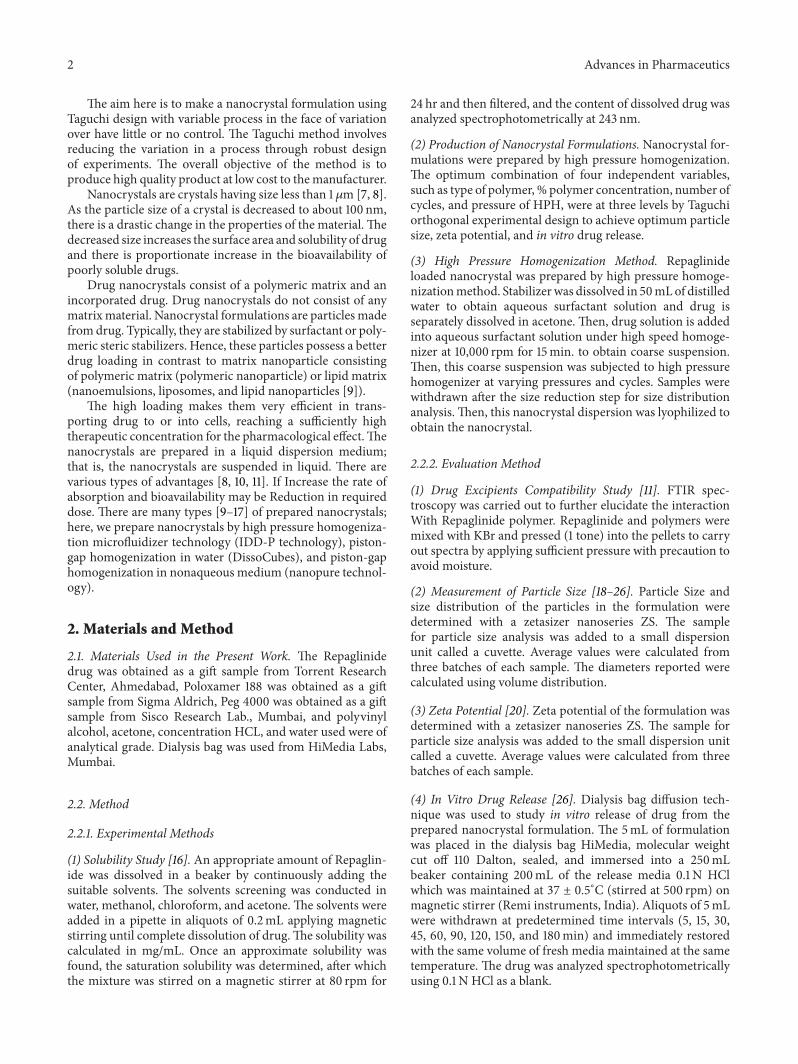
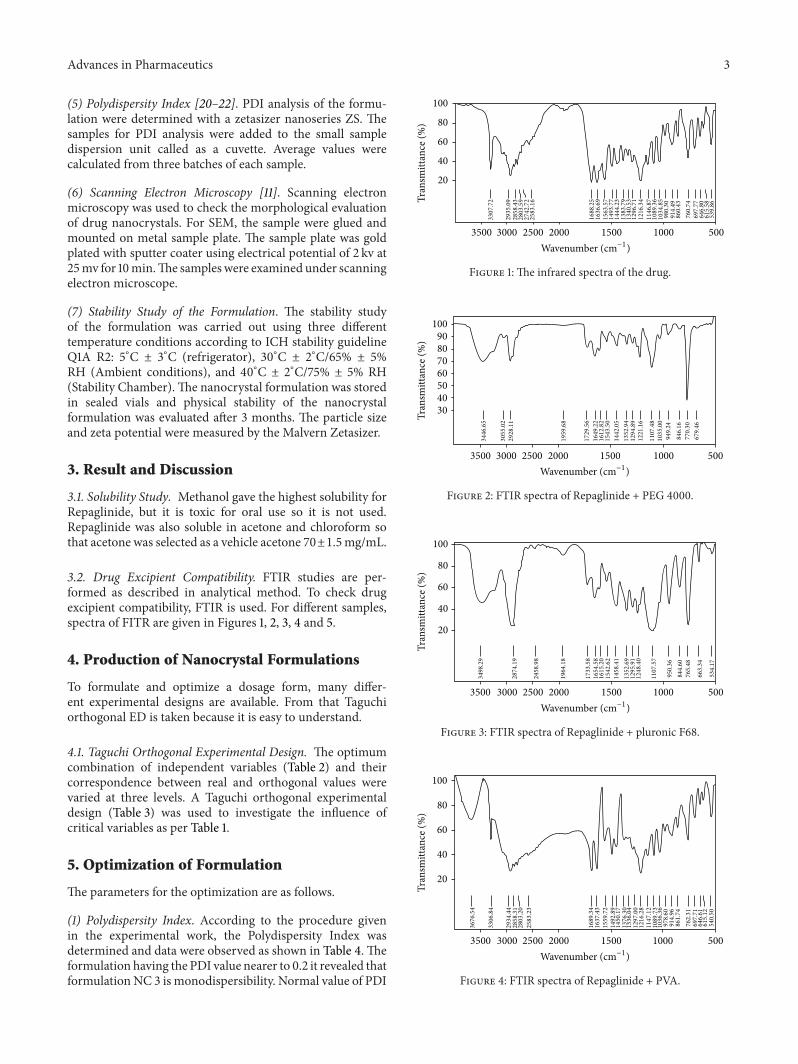
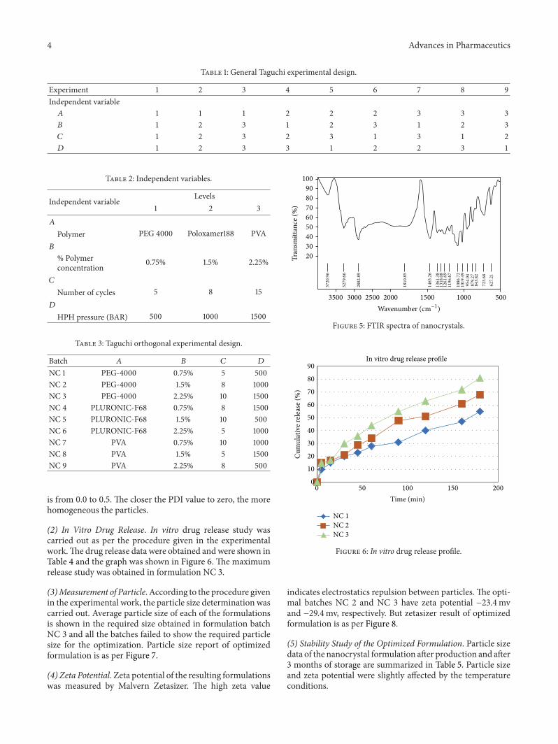
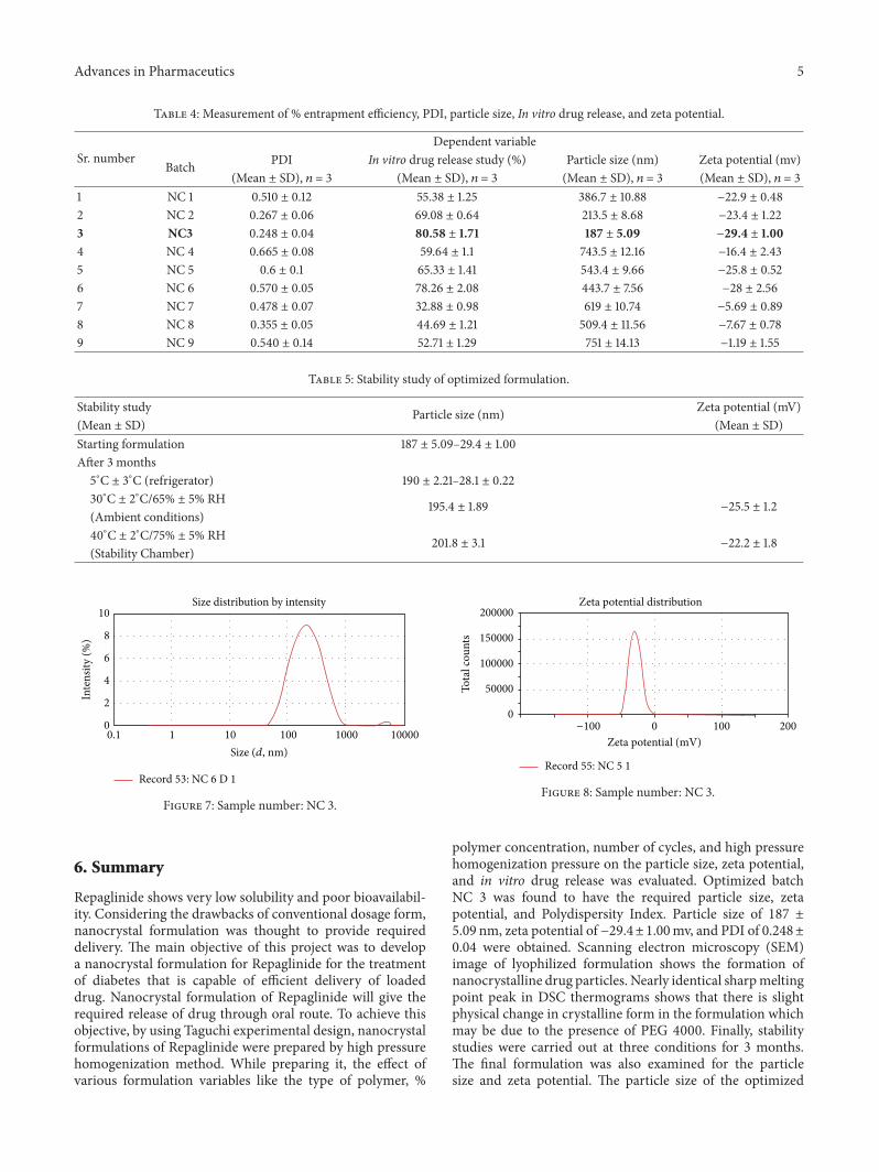
3.2. Drug Excipient Compatibility. FTIR studies are per-formed as described in analytical method. To check drugexcipient compatibility, FTIR is used. For different samples,spectra of FITR are given in Figures 1, 2, 3, 4 and 5.
4. Production of Nanocrystal Formulations
To formulate and optimize a dosage form, many differ-ent experimental designs are available. From that Taguchiorthogonal ED is taken because it is easy to understand.
4.1. Taguchi Orthogonal Experimental Design. The optimumcombination of independent variables (Table 2) and theircorrespondence between real and orthogonal values werevaried at three levels. A Taguchi orthogonal experimentaldesign (Table 3) was used to investigate the influence ofcritical variables as per Table 1.
5. Optimization of Formulation
The parameters for the optimization are as follows.
(1) Polydispersity Index. According to the procedure givenin the experimental work, the Polydispersity Index wasdetermined and data were observed as shown in Table 4. Theformulation having the PDI value nearer to 0.2 it revealed thatformulation NC 3 is monodispersibility. Normal value of PDI
100
80
60
40
20
3500 3000 2500 2000 1500 1000 500
Wavenumber (cm−1)
Tran
smitt
ance
(%)
3307.72
2935.09
2858.43
2803.59
2742.72
2583.16
1688.25
1636.69
1563.57
1493.77
1444.23
1383.79
1340.53
1296.71
1216.34
1146.87
1089.36
1034.85
980.30
914.49
860.43
760.74
697.77
646.80
615.58
539.86
Figure 1: The infrared spectra of the drug.
100
90
80
70
60
50
40
30
3446.65
3055.02
2928.11
1959.68
1729.56
1649.22
1612.82
1543.50
1442.05
1352.94
1294.89
1221.16
1107.48
1035.00
949.24
846.16
770.30
679.46
3500 3000 2500 2000 1500 1000 500
Wavenumber (cm−1)
Tran
smitt
ance
(%)
Figure 2: FTIR spectra of Repaglinide + PEG 4000.
100
80
60
40
20
3498.29
2874.19
2458.98
1964.18
1733.58
1654.58
1615.20
1542.62
1458.41
1352.69
1295.91
1248.40
1107.57
950.36
844.60
765.48
663.34
534.17
3500 3000 2500 2000 1500 1000 500
Wavenumber (cm−1)
Tran
smitt
ance
(%)
Figure 3: FTIR spectra of Repaglinide + pluronic F68.
100
80
60
40
20
3676.54
3306.84
2934.44
2858.31
2803.20
2583.23
1689.34
1637.43
1559.72
1492.89
1450.17
1376.30
1336.04
1297.00
1216.28
1147.12
1089.73
1036.36
978.60
914.96
861.74
762.31
697.71
646.61
615.12
540.50
3500 3000 2500 2000 1500 1000 500
Wavenumber (cm−1)
Tran
smitt
ance
(%)
Figure 4: FTIR spectra of Repaglinide + PVA.
4 Advances in Pharmaceutics
Table 1: General Taguchi experimental design.
Experiment 1 2 3 4 5 6 7 8 9Independent variable𝐴 1 1 1 2 2 2 3 3 3𝐵 1 2 3 1 2 3 1 2 3𝐶 1 2 3 2 3 1 3 1 2𝐷 1 2 3 3 1 2 2 3 1
Table 2: Independent variables.
Independent variable Levels1 2 3
𝐴
Polymer PEG 4000 Poloxamer188 PVA𝐵
% Polymerconcentration 0.75% 1.5% 2.25%
𝐶
Number of cycles 5 8 15𝐷
HPH pressure (BAR) 500 1000 1500
Table 3: Taguchi orthogonal experimental design.
Batch 𝐴 𝐵 𝐶 𝐷
NC 1 PEG-4000 0.75% 5 500NC 2 PEG-4000 1.5% 8 1000NC 3 PEG-4000 2.25% 10 1500NC 4 PLURONIC-F68 0.75% 8 1500NC 5 PLURONIC-F68 1.5% 10 500NC 6 PLURONIC-F68 2.25% 5 1000NC 7 PVA 0.75% 10 1000NC 8 PVA 1.5% 5 1500NC 9 PVA 2.25% 8 500
is from 0.0 to 0.5. The closer the PDI value to zero, the morehomogeneous the particles.
(2) In Vitro Drug Release. In vitro drug release study wascarried out as per the procedure given in the experimentalwork.The drug release data were obtained and were shown inTable 4 and the graph was shown in Figure 6. The maximumrelease study was obtained in formulation NC 3.
(3)Measurement of Particle. According to the procedure givenin the experimental work, the particle size determination wascarried out. Average particle size of each of the formulationsis shown in the required size obtained in formulation batchNC 3 and all the batches failed to show the required particlesize for the optimization. Particle size report of optimizedformulation is as per Figure 7.
(4) Zeta Potential. Zeta potential of the resulting formulationswas measured by Malvern Zetasizer. The high zeta value
100
80
70
50
30
90
60
40
20
3720.96
3279.66
2882.89
1810.85
1465.26
1361.38
1318.08
1261.69
1196.67
1086.72
1019.49
954.04
879.27
843.82
723.68
627.21
3500 3000 2500 2000 1500 1000 500
Wavenumber (cm−1)
Tran
smitt
ance
(%)
Figure 5: FTIR spectra of nanocrystals.
0
10
20
30
40
50
60
70
80
90
0 50 100 150 200
Cum
ulat
ive r
elea
se (%
)
Time (min)
In vitro drug release profile
NC 1
NC 2
NC 3
Figure 6: In vitro drug release profile.
indicates electrostatics repulsion between particles. The opti-mal batches NC 2 and NC 3 have zeta potential −23.4mvand −29.4mv, respectively. But zetasizer result of optimizedformulation is as per Figure 8.
(5) Stability Study of the Optimized Formulation. Particle sizedata of the nanocrystal formulation after production and after3 months of storage are summarized in Table 5. Particle sizeand zeta potential were slightly affected by the temperatureconditions.
Advances in Pharmaceutics 5
Table 4: Measurement of % entrapment efficiency, PDI, particle size, In vitro drug release, and zeta potential.
Sr. numberDependent variable
Batch PDI In vitro drug release study (%) Particle size (nm) Zeta potential (mv)(Mean ± SD), 𝑛 = 3 (Mean ± SD), 𝑛 = 3 (Mean ± SD), 𝑛 = 3 (Mean ± SD), 𝑛 = 3
1 NC 1 0.510 ± 0.12 55.38 ± 1.25 386.7 ± 10.88 −22.9 ± 0.482 NC 2 0.267 ± 0.06 69.08 ± 0.64 213.5 ± 8.68 −23.4 ± 1.223 NC3 0.248 ± 0.04 80.58 ± 1.71 187 ± 5.09 −29.4 ± 1.004 NC 4 0.665 ± 0.08 59.64 ± 1.1 743.5 ± 12.16 −16.4 ± 2.435 NC 5 0.6 ± 0.1 65.33 ± 1.41 543.4 ± 9.66 −25.8 ± 0.526 NC 6 0.570 ± 0.05 78.26 ± 2.08 443.7 ± 7.56 −28 ± 2.567 NC 7 0.478 ± 0.07 32.88 ± 0.98 619 ± 10.74 −5.69 ± 0.898 NC 8 0.355 ± 0.05 44.69 ± 1.21 509.4 ± 11.56 −7.67 ± 0.789 NC 9 0.540 ± 0.14 52.71 ± 1.29 751 ± 14.13 −1.19 ± 1.55
Table 5: Stability study of optimized formulation.
Stability study Particle size (nm) Zeta potential (mV)(Mean ± SD) (Mean ± SD)Starting formulation 187 ± 5.09–29.4 ± 1.00After 3 months
5∘C ± 3∘C (refrigerator) 190 ± 2.21–28.1 ± 0.2230∘C ± 2∘C/65% ± 5% RH 195.4 ± 1.89 −25.5 ± 1.2(Ambient conditions)40∘C ± 2∘C/75% ± 5% RH 201.8 ± 3.1 −22.2 ± 1.8(Stability Chamber)
10
8
6
4
2
00.1 1 10 100 1000 10000
Size distribution by intensity
Inte
nsity
(%)
Size (d, nm)
Record 53: NC 6 D 1
Figure 7: Sample number: NC 3.
6. Summary
Repaglinide shows very low solubility and poor bioavailabil-ity. Considering the drawbacks of conventional dosage form,nanocrystal formulation was thought to provide requireddelivery. The main objective of this project was to developa nanocrystal formulation for Repaglinide for the treatmentof diabetes that is capable of efficient delivery of loadeddrug. Nanocrystal formulation of Repaglinide will give therequired release of drug through oral route. To achieve thisobjective, by using Taguchi experimental design, nanocrystalformulations of Repaglinide were prepared by high pressurehomogenization method. While preparing it, the effect ofvarious formulation variables like the type of polymer, %
200000
150000
100000
50000
0
−100 0 100 200
Zeta potential (mV)
Tota
l cou
nts
Zeta potential distribution
Record 55: NC 5 1
Figure 8: Sample number: NC 3.
polymer concentration, number of cycles, and high pressurehomogenization pressure on the particle size, zeta potential,and in vitro drug release was evaluated. Optimized batchNC 3 was found to have the required particle size, zetapotential, and Polydispersity Index. Particle size of 187 ±5.09 nm, zeta potential of −29.4±1.00mv, and PDI of 0.248±0.04 were obtained. Scanning electron microscopy (SEM)image of lyophilized formulation shows the formation ofnanocrystalline drug particles. Nearly identical sharpmeltingpoint peak in DSC thermograms shows that there is slightphysical change in crystalline form in the formulation whichmay be due to the presence of PEG 4000. Finally, stabilitystudies were carried out at three conditions for 3 months.The final formulation was also examined for the particlesize and zeta potential. The particle size of the optimized
6 Advances in Pharmaceutics
formulation was tested after 3 months and also zeta potentialwas determined in three different conditions. Results showthat there is no more variation in particle size and zetapotential so the formulation is stable.
7. Conclusion
The present study was an attempt to formulate nanocrys-tals of poorly water soluble drug, Repaglinide, having lowbioavailability.Theobjectivewas to increase the dissolution ofdrug and to improve the patient compliance.The nanocrystalformulation of Repaglinide with the smaller particle size canbe effectively produced by the high pressure homogenizationmethod. The nanosizing approach enhanced the in vitrodrug release of the Repaglinide. Repaglinide was success-fully entrapped within the polymer with high efficiency.Thus, nanocrystal approach may be a promising carrier forRepaglinide and other class II drugs.
Conflict of Interests
The authors declare that there is no conflict of interestsregarding the publication of this paper.
References
[1] K. G. M. M. Alberti and P. Z. Zimmet, “Definition, diagnosisand classification of diabetesmellitus and its complications. Part1: diagnosis and classification of diabetes mellitus. Provisionalreport of a WHO Consultation,” Diabetic Medicine, vol. 15, no.7, pp. 539–553, 1998.
[2] WHOExpertCommittee onDiabetesMellitus, “Second report,”Technical Report Series 646, WHO, Geneva, Switzerland, 1980.
[3] American Diabetes Association, “Economic consequences ofdiabetes mellitus in the U.S. in 1997,” Diabetes Care, vol. 21, no.2, pp. 296–309, 1998.
[4] T. Abhinov, A. S. A. Khan, Ashrafa, S. Parveen, and K. P.Samapth Kumar, “Diabetes epidemic in India: risk factors,symptoms and treatment,” Indian Journal of Research in Phar-macy and Biotechnology, vol. 1, no. 2, pp. 234–243, 2013.
[5] National Diabetes Data Group, “Classification and diagnosis ofdiabetes mellitus and other categories of glucose intolerance,”Diabetes, vol. 28, no. 12, pp. 1039–1057, 1979.
[6] D. W. Cooke and L. Plotnick, “Type 1 diabetes mellitus inpediatrics,” Pediatrics in Review, vol. 29, no. 11, pp. 374–385,2008.
[7] R. H. Muller, S. Runge, V. Ravelli, W. Mehnert, A. F. Thune-mann, and E. B. Souto, “Oral bioavailability of cyclosporine:solid lipid nanoparticles (SLN) versus drug nanocrystals,”International Journal of Pharmaceutics, vol. 317, no. 1, pp. 82–89,2006.
[8] E. Merisko-Liversidge, G. G. Liversidge, and E. R. Cooper,“Nanosizing: a formulation approach for poorly-water-solublecompounds,” European Journal of Pharmaceutical Sciences, vol.18, no. 2, pp. 113–120, 2003.
[9] G. Shinde, K. S. Rajesh, B. Devang, G. Bangale, D. Umalkar,and G. Virag, “Current status of colloidal system (nano range),”International Journal of Drug Formulation and Research, vol. 2,no. 6, pp. 39–54, 2011.
[10] J. Salazar, A. Ghanem, R. H. Muller, and J. P. Moschwitzer,“Nanocrystals: comparison of the size reduction effectivenessof a novel combinative method with conventional top-downapproaches,” European Journal of Pharmaceutics and Biophar-maceutics, vol. 81, no. 1, pp. 82–90, 2012.
[11] W. Sun, W. Tian, Y. Zhang, J. He, S. Mao, and L. Fang, “Effectof novel stabilizers—cationic polymers on the particle sizeand physical stability of poorly soluble drug nanocrystals,”Nanomedicine: Nanotechnology, Biology, and Medicine, vol. 8,no. 4, pp. 460–467, 2012.
[12] J.-U. A. H. Junghanns and R. H. Muller, “Nanocrystal tech-nology, drug delivery and clinical applications,” InternationalJournal of Nanomedicine, vol. 3, no. 3, pp. 295–309, 2008.
[13] E. Che, X. Zheng, C. Sun, D. Chang, T. Jiang, and S. Wang,“Drug nanocrystals: a state of the art formulation strategy forpreparing the poorly water-soluble drugs,” Asian Journal ofPharmaceutical Sciences, vol. 7, no. 2, pp. 85–95, 2012.
[14] H. Banavath, K. S. Raju, M. T. Ansari, M. S. Ali, and G. Pattnaik,“Nanosuspension: an attempt to enhance bioavailability ofpoorly soluble drugs,” International Journal of PharmaceuticalSciences and Research, vol. 1, no. 9, pp. 1–11, 2010.
[15] R. H. Muller and K. Peters, “Nanosuspensions for the formula-tion of poorly soluble drugs. I. Preparation by a size-reductiontechnique,” International Journal of Pharmaceutics, vol. 160, no.2, pp. 229–237, 1998.
[16] S. Katteboinaa, “Drug nanocrystals: a novel formulationapproach for poorly solubledrugs,” International Journal ofPharmTech Research, vol. 1, no. 3, pp. 682–694, 2009.
[17] B. K. Nanjwade, G. K. Derkar, H. Bechra, and F. V. Manvi,“Nanosized technological approaches for the delivery of poorlywater soluble drugs,” Iranian Journal of Pharmaceutical Sciences,vol. 6, no. 3, pp. 149–162, 2010.
[18] J.-Y. Choi, J. Y. Yoo, H.-S. Kwak, B. U. Nam, and J. Lee, “Role ofpolymeric stabilizers for drug nanocrystal dispersions,”CurrentApplied Physics, vol. 5, no. 5, pp. 472–474, 2005.
[19] Q. Fu, L. Kou, C. Gong et al., “Relationship between dissolutionand bioavailability for nimodipine colloidal dispersions: thecritical size in improving bioavailability,” International Journalof Pharmaceutics, vol. 427, no. 2, pp. 358–364, 2012.
[20] J. Hecq, M. Deleers, D. Fanara, H. Vranckx, and K. Amighi,“Preparation and characterization of nanocrystals for solubilityand dissolution rate enhancement of nifedipine,” InternationalJournal of Pharmaceutics, vol. 299, no. 1-2, pp. 167–177, 2005.
[21] R. Mauludin, R. H. Muller, and C. M. Keck, “Kinetic solubilityand dissolution velocity of rutin nanocrystals,” European Jour-nal of Pharmaceutical Sciences, vol. 36, no. 4-5, pp. 502–510,2009.
[22] P. Quan, K. Shi, H. Piao, N. Liang, D. Xia, and F. Cui, “A novelsurface modified nitrendipine nanocrystals with enhancementof bioavailability and stability,” International Journal of Pharma-ceutics, vol. 430, no. 1-2, pp. 366–371, 2012.
[23] D. K. Krishna and H. P. Rajesh, “Dissolution enhancementof albendazole through nanocrystal formulation,” Journal ofPharmacy and Bioallied Sciences, vol. 4, no. 5, pp. 62–63, 2012.
[24] F. Lai, E. Pini, G. Angioni et al., “Nanocrystals as tool to improvepiroxicamdissolution rate in novel orally disintegrating tablets,”European Journal of Pharmaceutics and Biopharmaceutics, vol.79, no. 3, pp. 552–558, 2011.
[25] R. Mauludin, R. H. Muller, and C. M. Keck, “Development ofan oral rutin nanocrystal formulation,” International Journal ofPharmaceutics, vol. 370, no. 1-2, pp. 202–209, 2009.
Advances in Pharmaceutics 7
[26] N. G. Sahoo, M. Kakran, L. A. Shaal et al., “Preparation andcharacterization of quercetin nanocrystals,” Journal of Pharma-ceutical Sciences, vol. 100, no. 6, pp. 2379–2390, 2011.

Submit your manuscripts athttp://www.hindawi.com
PainResearch and TreatmentHindawi Publishing Corporationhttp://www.hindawi.com Volume 2014
The Scientific World JournalHindawi Publishing Corporation http://www.hindawi.com Volume 2014
Hindawi Publishing Corporationhttp://www.hindawi.com
Volume 2014
ToxinsJournal of
VaccinesJournal of
Hindawi Publishing Corporation http://www.hindawi.com Volume 2014
Hindawi Publishing Corporationhttp://www.hindawi.com Volume 2014
AntibioticsInternational Journal of
ToxicologyJournal of
Hindawi Publishing Corporationhttp://www.hindawi.com Volume 2014
StrokeResearch and TreatmentHindawi Publishing Corporationhttp://www.hindawi.com Volume 2014
Drug DeliveryJournal of
Hindawi Publishing Corporationhttp://www.hindawi.com Volume 2014
Hindawi Publishing Corporationhttp://www.hindawi.com Volume 2014
Advances in Pharmacological Sciences
Tropical MedicineJournal of
Hindawi Publishing Corporationhttp://www.hindawi.com Volume 2014
Medicinal ChemistryInternational Journal of
Hindawi Publishing Corporationhttp://www.hindawi.com Volume 2014
AddictionJournal of
Hindawi Publishing Corporationhttp://www.hindawi.com Volume 2014
Hindawi Publishing Corporationhttp://www.hindawi.com Volume 2014
BioMed Research International
Emergency Medicine InternationalHindawi Publishing Corporationhttp://www.hindawi.com Volume 2014
Hindawi Publishing Corporationhttp://www.hindawi.com Volume 2014
Autoimmune Diseases
Hindawi Publishing Corporationhttp://www.hindawi.com Volume 2014
Anesthesiology Research and Practice
ScientificaHindawi Publishing Corporationhttp://www.hindawi.com Volume 2014
Journal of
Hindawi Publishing Corporationhttp://www.hindawi.com Volume 2014
Pharmaceutics
Hindawi Publishing Corporationhttp://www.hindawi.com Volume 2014
MEDIATORSINFLAMMATION
of
Related Documents