Research Article Evaluation of Light-Activated Provisional Resin Materials for Periodontal Soft Tissue Management Soo-Kyung Jun, 1,2 Hae-Hyoung Lee, 1,2 and Jung-Hwan Lee 2 1 Department of Biomaterials Science, College of Dentistry, Dankook University, Cheonan, Republic of Korea 2 Institute of Tissue Regeneration Engineering, Dankook University, Cheonan, Republic of Korea Correspondence should be addressed to Jung-Hwan Lee; [email protected] Received 3 June 2016; Accepted 17 August 2016 Academic Editor: Hom-Lay Wang Copyright © 2016 Soo-Kyung Jun et al. is is an open access article distributed under the Creative Commons Attribution License, which permits unrestricted use, distribution, and reproduction in any medium, provided the original work is properly cited. e purpose of this study was to determine mechanical properties using a compressive test with cylinder specimen (ℎ=6 mm and =4 mm) as well as cytotoxicity using elutes from disk specimen ( = 10 mm and ℎ=2 mm) against human gingival fibroblasts and oral keratinocytes with light-activated provisional resin materials (Revotek LC and Luxatemp Solar) compared to chemically activated counterpart (Snap, Trim II, and Jet). Significantly increased compressive strength (210∼280 MPa) was detected in light- activated products compared to chemically activated ones (20∼65 MPa, < 0.05) and similar compressive modulus was detected in both types (0.8∼1.5 and 0.5∼1.3 GPa). Simultaneously, the light-activated products showed less adverse effects on the periodontal soſt tissue cells in any polymerization stage compared to the chemically activated products. Particularly, chemically activated products had significantly greater adverse effects during the “polymerizing” phase compared to those that were “already set” ( < 0.05), as shown in confocal microscopic images of live and dead cells. In conclusion, light-activated provisional resin materials have better mechanical properties as well as biocompatibility against two tested types of oral cells compared to the chemically activated counterpart, which are considered as more beneficial choice for periodontal soſt tissue management. 1. Introduction Many new biomaterials and technologies have been intro- duced both to replace missing or damaged dental tissues and to promote dental tissue regeneration [1–5]. Among these tissues, periodontal soſt tissue has been highlighted for its aesthetic characteristics and inflammation resistance to the oral environment [6, 7]. erefore, periodontal soſt tissue management during and aſter teeth/implant restoration is essential for esthetic soſt tissue contour along with dental restoration, particularly for anterior tooth region. Provisional restoration is a key step in providing healthy and aesthetically pleasing periodontal soſt tissue as well as in maintaining the marginal integrity of prepared teeth/implants and ensuring occlusal function until final prosthodontics are adjusted [8]. Provisional resin materials are widely used for interim restorations due to their ease of handling, suitable mechanical properties, time saving ability, and low cost [9]. Light-curable products have recently been introduced and have beneficial qualities, such as decreas- ing shrinkage and the time-consuming setting time and increased favorability to patients because of less toxic odor [10]. e clinical use of provisional resin materials is classified into two categories based on how solidly the resins are polymerized from the monomer: (1) traditional, chemically activated materials and (2) recently developed, light-activated materials, including products that are dually activated by chemicals and light. Depending on the how these materials are solidified, their biocompatibility to periodontal tissue and their mechanical properties can differ. Both are crucial for maintaining a natural periodontal tissue appearance in accordance with the tooth/implant contour. When interim restoration using provisional resin mate- rials is accomplished and positioned around periodontal soſt tissue, including the teeth/implant margin and gingival sulcus, toxic components of these materials can adversely affect the tissue via saliva and induce soſt tissue management Hindawi Publishing Corporation BioMed Research International Volume 2016, Article ID 1209705, 10 pages http://dx.doi.org/10.1155/2016/1209705

Welcome message from author
This document is posted to help you gain knowledge. Please leave a comment to let me know what you think about it! Share it to your friends and learn new things together.
Transcript
Research ArticleEvaluation of Light-Activated Provisional Resin Materials forPeriodontal Soft Tissue Management
Soo-Kyung Jun,1,2 Hae-Hyoung Lee,1,2 and Jung-Hwan Lee2
1Department of Biomaterials Science, College of Dentistry, Dankook University, Cheonan, Republic of Korea2Institute of Tissue Regeneration Engineering, Dankook University, Cheonan, Republic of Korea
Correspondence should be addressed to Jung-Hwan Lee; [email protected]
Received 3 June 2016; Accepted 17 August 2016
Academic Editor: Hom-Lay Wang
Copyright © 2016 Soo-Kyung Jun et al.This is an open access article distributed under the Creative Commons Attribution License,which permits unrestricted use, distribution, and reproduction in any medium, provided the original work is properly cited.
The purpose of this study was to determine mechanical properties using a compressive test with cylinder specimen (ℎ = 6mm and𝜙 = 4mm) as well as cytotoxicity using elutes from disk specimen (𝜙 = 10mm and ℎ = 2mm) against human gingival fibroblastsand oral keratinocytes with light-activated provisional resin materials (Revotek LC and Luxatemp Solar) compared to chemicallyactivated counterpart (Snap, Trim II, and Jet). Significantly increased compressive strength (210∼280 MPa) was detected in light-activated products compared to chemically activated ones (20∼65MPa,𝑃 < 0.05) and similar compressivemodulus was detected inboth types (0.8∼1.5 and 0.5∼1.3 GPa). Simultaneously, the light-activated products showed less adverse effects on the periodontal softtissue cells in any polymerization stage compared to the chemically activated products. Particularly, chemically activated productshad significantly greater adverse effects during the “polymerizing” phase compared to those that were “already set” (𝑃 < 0.05),as shown in confocal microscopic images of live and dead cells. In conclusion, light-activated provisional resin materials havebetter mechanical properties as well as biocompatibility against two tested types of oral cells compared to the chemically activatedcounterpart, which are considered as more beneficial choice for periodontal soft tissue management.
1. Introduction
Many new biomaterials and technologies have been intro-duced both to replace missing or damaged dental tissues andto promote dental tissue regeneration [1–5]. Among thesetissues, periodontal soft tissue has been highlighted for itsaesthetic characteristics and inflammation resistance to theoral environment [6, 7]. Therefore, periodontal soft tissuemanagement during and after teeth/implant restoration isessential for esthetic soft tissue contour along with dentalrestoration, particularly for anterior tooth region.
Provisional restoration is a key step in providinghealthy and aesthetically pleasing periodontal soft tissue aswell as in maintaining the marginal integrity of preparedteeth/implants and ensuring occlusal function until finalprosthodontics are adjusted [8]. Provisional resin materialsare widely used for interim restorations due to their ease ofhandling, suitable mechanical properties, time saving ability,and low cost [9]. Light-curable products have recently been
introduced and have beneficial qualities, such as decreas-ing shrinkage and the time-consuming setting time andincreased favorability to patients because of less toxic odor[10].
The clinical use of provisional resin materials is classifiedinto two categories based on how solidly the resins arepolymerized from the monomer: (1) traditional, chemicallyactivatedmaterials and (2) recently developed, light-activatedmaterials, including products that are dually activated bychemicals and light. Depending on the how these materialsare solidified, their biocompatibility to periodontal tissueand their mechanical properties can differ. Both are crucialfor maintaining a natural periodontal tissue appearance inaccordance with the tooth/implant contour.
When interim restoration using provisional resin mate-rials is accomplished and positioned around periodontalsoft tissue, including the teeth/implant margin and gingivalsulcus, toxic components of these materials can adverselyaffect the tissue via saliva and induce soft tissue management
Hindawi Publishing CorporationBioMed Research InternationalVolume 2016, Article ID 1209705, 10 pageshttp://dx.doi.org/10.1155/2016/1209705
2 BioMed Research International
Table 1: Materials tested in this study.
Product Code Manufacturer Lot number Composition Polymerizing typeSnap SN Parkell Inc., USA 01707 Polyethyl methacrylate (PEMA) ChemicalTrim II TR Bosworth, USA 1409-442 Polyethyl methacrylate (PEMA) ChemicalJet JE Lang Dental, USA 40142 Polymethyl methacrylate (PMMA) ChemicalRevotek LC RL GC America Inc., USA 1409191 Urethane dimethacrylate (UDMA) LightLuxatemp Solar LS DMG, Germany 726443 Bis-acryl composites Light (dual cure)
Table 2: Experimental conditions of provisional resin materials.
Materials Extraction starting time after the start of mixing Polymerizing methodInitial Intermediate After polymerization
Snap 2min, 30 sec (SN1) 5min (SN2) 10min (SN3) ChemicalTrim Plus� 2min (TR1) 4min (TR2) 8min (TR3) ChemicalJet 2min, 30 sec (JE1) 5min (JE2) 10min (JE3) ChemicalRevotek LC Unpolymerized (RL1) 10 s (RL2) 20 s (RL3) LightLuxatemp Solar 1min (LS1) 4min (LS2) 4min, curing 20 s (LS3) Dual cure
failure [11, 12]. Even worse, particularly in chemically acti-vated materials, “polymerizing” provisional resin materialsaccelerate adverse effects to periodontal soft tissue because“polymerizing” provisional resin materials increase adverseeffects from unreacted monomers or increased concentra-tions of eluates in the polymerizing phase when they areextracted in the oral cavity [13–15]. Light-activated materialscan release fewer cytotoxic components due to their quick set-ting time (less than 20 s) in the oral cavity and are consideredmore biocompatible than long-setting, chemically activatedmaterials. In addition, high mechanical properties are pos-itively assumed in light-activated materials due to polymermaterials’ (UDMA and bis-acryl) natural characteristics,which have better mechanical properties than do PEMA andPMMA [16]. When provision resin materials are broken dueto biting force in the oral cavity, the broken pieces can damageperiodontal soft tissue and periodontal soft managementcannot be successfully performed because of the loss of theprovisional restoration [17].
Therefore, in this experiment, we performed cytotoxicitytests to determine the effects on periodontal soft tissue con-sisting of human oral keratinocytes and gingival fibroblastsduring the initial, intermediate, and final polymerizing stagesof the provisional resin materials. In addition, as repre-sentative mechanical properties, compressive strength andmodulus were measured. The first null hypothesis was thatthe mechanical properties of light-activated provisional resinmaterials would not differ from those of their chemically acti-vated counterparts. In terms of the biocompatibility, the sec-ond null hypothesis was that the cytotoxic effects from light-activated provisional resin materials of extracts on humanoral cells would not differ from those of their chemicallyactivated counterparts. The third null hypothesis was thatcytotoxic effects to human oral cells would not differ between“polymerizing” and “already set” provisional resin materials.
2. Materials and Methods
2.1. Provisional Resin Materials. The most commonly usedprovisional resin materials were chosen for this experimentand included chemically activated polyethyl methacrylate(PEMA) (SN: Snap, Parkell Inc., and TR: Trim, Bosworth),chemically activated polymethyl methacrylate (PMMA) (JE:Jet, Lang Dental), light-activated urethane dimethacrylate(UDMA) and bis-acryl (RL; Revotek LC, GC America), andLS (Luxatemp Solar, DMG). The study involved testing thematerials, which are summarized in Table 1.
2.2. Preparation of Materials. Three types of chemicallyactivated provisional resin materials (SN, TR, and JE) andtwo types of light-activated materials (RL and LS) werehandled according to the manufacturers’ instructions. Forin vitro testing, when the chemically activated provisionalresin materials reached the early “dough” stage, they werepacked into a Teflon mold (𝜙 = 10mm, ℎ = 2mm) andcovered with a glass plate. Specimens were removed fromthe molds at three different setting times (25% set, 50%set, and 100% set), as recommended in the manufacturers’instructions, and were immediately placed in distilled water(DW). After the light-activated materials (RL and LS) hadbeen placed on the Teflon mold, the RL specimen was eitherleft unpolymerized or polymerized by means of an LEDcuring light gun (Litex 695, Dentamerica Industry) for 10or 20 s. LS specimens were mixed using an automixing gunand placed in the mold for 1 minute or 4 minutes beforebeing placed in DW. For fully polymerized specimens, 10 slight curing using an LED curing light gun (Litex 695) wasperformed after 4 minutes of chemical activation accordingto the manufacturer’s recommendations. The experimentalconditions are detailed in Table 2.
BioMed Research International 3
Shear
Tensile
4mm
6mm
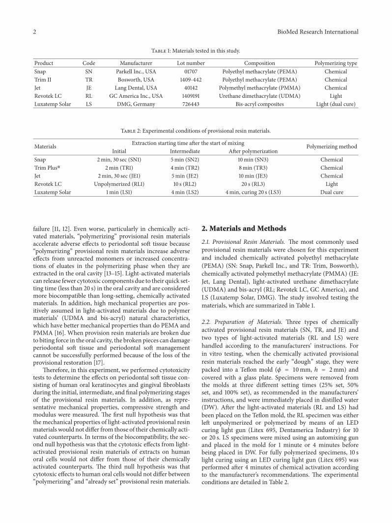
Figure 1: Photograph and schematic diagram of specimens for compressive testing.
For compressive testing, specimens were produced usinga stainless steel mold (ℎ = 6mm and 𝜙 = 4mm, Figure 1).When the chemically activated materials reached the doughstage, they were packed into the mold and covered bycelluloid strip and a glass plate, over which an equal amountof pressure was applied during complete polymerization.The RL specimens were compacted into the mold using aresin instrument. Celluloid strips were placed on top of themold, pressed flat using a glass plate, and allowed to curefor 20 s from both the bottom and the top according tothe manufacturer’s specifications. The LS specimens weremixed using an automixing tip, placed into the mold, initiallycured for 4 minutes, and then light-cured for completepolymerization for 20 s from both the bottom and the top.All materials were further allowed to set even after the finalsetting time in a static loading device to which 5 kg of loadwas applied for 3 minutes for complete polymerization. Themold was carefully split, and the specimens were removedand examined to exclude those with porosity or defects. Theexcess was removed using a diamond bur and specimenswere wet polished up to 1200-grit SiC paper. Specimens wereimmediately immersed in DW and stored in an incubator for24 hours at 37∘C before mechanical testing.
2.3. Collection of Resin Extract. Extracts were obtained at aratio of 3 cm2/mL following the recommendations of ISO10993-12 [18]. Because the surface of the specimens was2.2 cm2, they were incubated in 0.73mL of medium. Extractswere collected for 24 hours at 37∘C in a shaking incubator(120 rpm). All samples were checked for expiration datesand stored according to the manufacturers’ recommendedconditions throughout the experiment. All procedures wereperformed on a clean bench to prevent contamination of thespecimens.
2.4. Culture of Oral Cells. Immortalized human oral ker-atinocytes (IHOK) and immortalized humannormal gingival
fibroblasts (hNOF) were used to mimic cytotoxicity to oralmucosal tissue [19, 20]. IHOK and hNOF were generouslyprovided by Professor Dolphine Oda from the Departmentof Oral Biology from the University of Washington (Seattle,WA) and Professor Jin Kim from the Department of OralPathology and Oral Cancer Research Institute from YonseiUniversity College of Dentistry (Seoul, Korea), respectively.The IHOK and hNOF were cultured in a DMEM/F-12 3 : 1mixture (Welgene, Daegu, Korea) supplemented with 10%fetal bovine serum (Gibco) and 1% penicillin/streptomycin(Invitrogen) in a humidified atmosphere of 5% CO
2at 37∘C
[20, 21].
2.5. Cytotoxicity Tests and Cell Viability. Cytotoxicity testswere performed according to ISO 10093-5 [22]. Briefly, 1× 10⁴ cells/well were cultured in a 96-well plate (SPL LifeSciences) with 100 𝜇L of supplemented medium for 24 hours.After being washed with phosphate buffered saline (PBS), thecells were cocultured with 50 𝜇L of 2x supplementedmediumand 50 𝜇L of extract or a serially diluted extract by DW foranother 24 hours.The percentages of the final concentrationsof extract in the culture media were 50% and 25%; 50 𝜇L ofDW with 50𝜇L 2x supplemented medium was used as thecontrol.
Cell viability wasmeasured using anMTS assay (CellTiter96 Aqueous One Solution Cell Proliferation Assay, Promega)according to the manufacturer’s protocol, and the resultswere expressed as the percentage of optical density of eachtest group compared with each control group (𝑛 = 6).Optical absorbance was read at 490 nm using a microplatereader (SpectraMax M2e, Molecular Devices). Confocalmicroscopic images of live and dead cells were obtainedto confirm the cytotoxicity results. After the cells werewashed with PBS and stained with calcein AM (0.5 𝜇M) andethidium homodimer-1 (4 𝜇M) (Molecular Probes, Eugene)for 30 minutes, they were examined under a confocal lasermicroscope (LSM 700, Carl Zeiss). Green fluorescence was
4 BioMed Research International
Com
pres
sive s
treng
th (M
Pa)
Product SN TR JE LSRLPolymerizing type Ch Ch Ch DuLT
0
50
100
150
200
250
300
A A
B
C
D
(a)
Com
pres
sive m
odul
us (G
Pa)
0.0
0.4
0.8
1.2
1.6
2.0
AA
B
A, C
B
Product SN TR JE LSRLPolymerizing type Ch Ch Ch DuLT
(b)
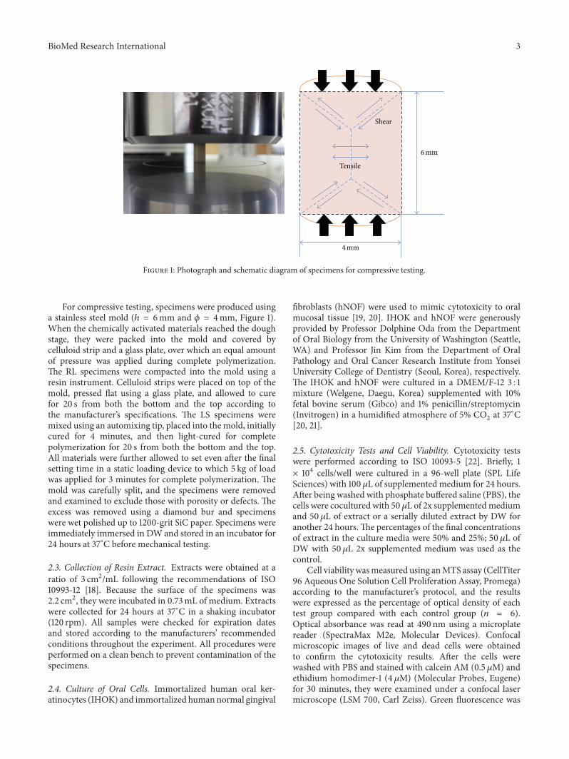
Figure 2: Compressive strength (a) and compressive modulus (b) of provisional restoration materials. Different letters indicate significantdifferences among the extract conditions (𝑃 < 0.05).
observed from the live cells and bright red fluorescence fromthe dead cells. All analyses were independently performed intriplicate, and the representativemeans± standard deviationsor images were shown.
2.6. Measurement of Compressive Strength and Modulus. Thespecimens (ℎ = 6mm and 𝜙 = 4mm), which were fully poly-merized, were positioned on the universal testing machine(Instron 5966, MA, USA) for compressive testing. The cross-head speed of this machine was set at 1.0mm/min usingthe ±10 kN of load cell. Compressive strength was calculatedby dividing the maximum load, expressed in kN, by theoriginal cross section area, expressed inm2 of a specimen in acompression test (N/m2). The compressive modulus was cal-culated according to the equation described in other studies[23].
2.7. Statistical Analysis. Data were analyzed by a one-wayANOVA with a Duncan post hoc test. Statistical significancewas set at 0.05. All statistical analyses were performed usingthe SPSS version 21.0 software program (SPSS Inc., IL,Chicago, USA).
3. Results
3.1. Compressive Test. Figure 2 shows the results of thecompressive strength and modulus testing. As seen by themean compressive strength values of the light-cured groups,LS showed the highest value (280.0MPa), and RL showedthe second highest value (197.4MPa). TR showed the lowestvalue (22.6MPa) among the tested products. SN and JEshowed significantly lower compressive strength (27.4 and62.1MPa, resp.) compared to the light-activated products(RL and LS). In terms of the compressive modulus, twochemically activated products (SN and TR) and one light-activated product (RL) showed lower values less than 0.8GPa,
while JE and LS had higher compressivemodulus values (1.3∼1.5 GPa) than did the other specimens (𝑃 < 0.05).
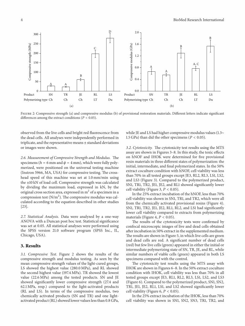
3.2. Cytotoxicity. The cytotoxicity test results using the MTSassay are shown in Figures 3–8. In this study, the toxic effectson hNOF and IHOK were determined for five provisionalresin materials in three different states of polymerization: theinitial, intermediate, and final polymerized states. In the 50%extract coculture condition with hNOF, cell viability was lessthan 70% in all tested groups except JE3, RL2, RL3, LS1, LS2,and LS3 (Figure 3). Compared to the polymerized product,SN1, TR1, TR2, JE1, JE2, and RL1 showed significantly lowercell viability (Figure 3, 𝑃 < 0.05).
In the 25% extract incubation of the hNOF, less than 70%cell viability was shown in SN1, TR1, and TR2, which were allfrom the chemically activated provisional resins (Figure 4).SN1, TR1, TR2, JE1, JE2, RL1, RL2, and LS1 had significantlylower cell viability compared to extracts from polymerizingmaterials (Figure 4, 𝑃 < 0.05).
The results of the cytotoxicity tests were confirmed byconfocal microscopic images of live and dead cells obtainedafter incubation in 50% extract in the supplementedmedium.The results are shown in Figure 5, in which live cells are greenand dead cells are red. A significant number of dead cells(red) but few live cells (green) appeared in either the initial orintermediate polymerizing state of SN, TR, JE, and RL, whilesimilar numbers of viable cells (green) appeared in both LSspecimens compared with the control.
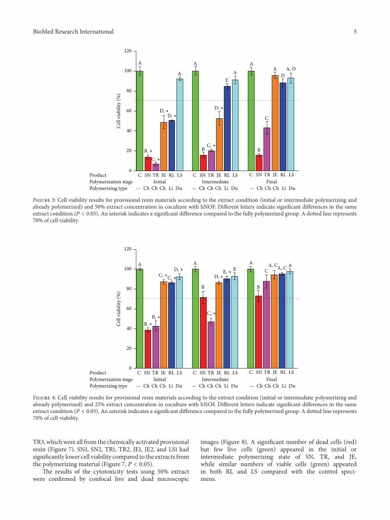
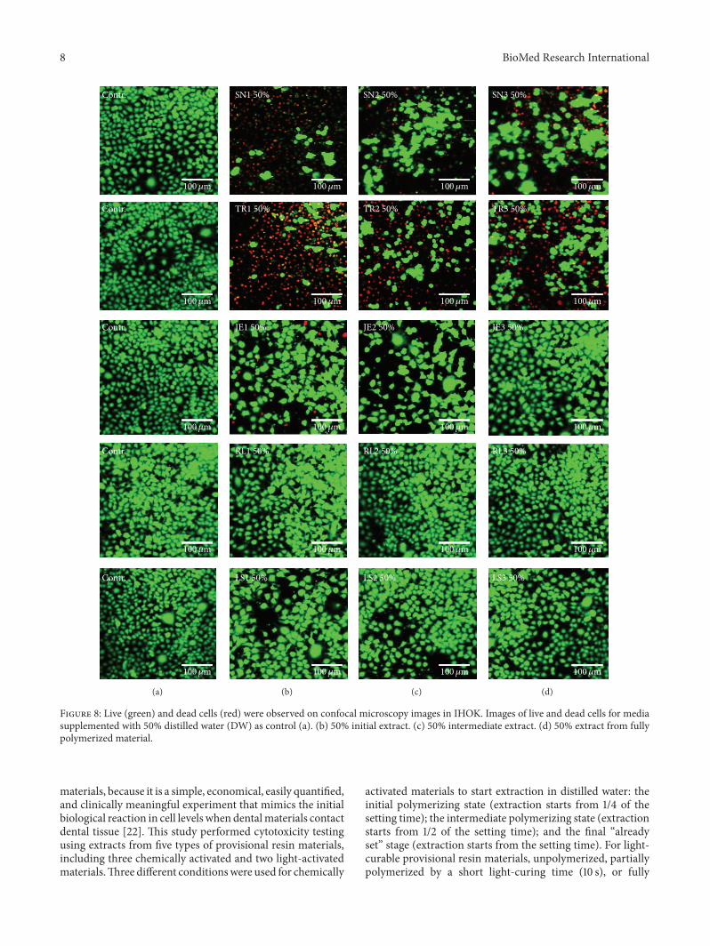
The cytotoxicity test results using the MTS assay withIHOK are shown in Figures 6–8. In the 50% extract coculturecondition with IHOK, cell viability was less than 70% in alltested groups except JE3, RL1, RL2, RL3, LS1, LS2, and LS3(Figure 6). Compared to the polymerized product, SN1, SN2,TR1, JE1, JE2, RL1, LS1, and LS2 showed significantly lowercell viability (Figure 6, 𝑃 < 0.05).
In the 25% extract incubation of the IHOK, less than 70%cell viability was shown in SN1, SN2, SN3, TR1, TR2, and
BioMed Research International 5
Initial Intermediate FinalPolymerization stagePolymerizing type
Product JEC SN TR RL LSJEC SN TR RL LSJEC SN TR RL LS0
20
40
60
80
100
120
A
A
A
EA
A
C
AD
A, D
B B
Cel
l via
bilit
y (%
)
Ch Ch Ch DuLi—Ch Ch Ch DuLi—Ch Ch Ch DuLi—
B, ∗C, ∗
C, ∗
D, ∗D, ∗D, ∗
Figure 3: Cell viability results for provisional resin materials according to the extract condition (initial or intermediate polymerizing andalready polymerized) and 50% extract concentration in coculture with hNOF. Different letters indicate significant differences in the sameextract condition (𝑃 < 0.05). An asterisk indicates a significant difference compared to the fully polymerized group. A dotted line represents70% of cell viability.
Initial Intermediate FinalPolymerization stagePolymerizing type
Product JEC SN TR RL LSJEC SN TR RL LSJEC SN TR RL LS0
20
40
60
80
100
120
Cel
l via
bilit
y (%
)
Ch Ch Ch DuLi—Ch Ch Ch DuLi—Ch Ch Ch DuLi—
B, ∗B, ∗
C, ∗C, ∗
C, ∗
D, ∗D, ∗
A A
B
EA
B
CA, CA, C A
E, ∗
Figure 4: Cell viability results for provisional resin materials according to the extract condition (initial or intermediate polymerizing andalready polymerized) and 25% extract concentration in coculture with hNOF. Different letters indicate significant differences in the sameextract condition (𝑃 < 0.05). An asterisk indicates a significant difference compared to the fully polymerized group. A dotted line represents70% of cell viability.
TR3, whichwere all from the chemically activated provisionalresin (Figure 7). SN1, SN2, TR1, TR2, JE1, JE2, and LS1 hadsignificantly lower cell viability compared to the extracts fromthe polymerizing material (Figure 7, 𝑃 < 0.05).
The results of the cytotoxicity tests using 50% extractwere confirmed by confocal live and dead microscopic
images (Figure 8). A significant number of dead cells (red)but few live cells (green) appeared in the initial orintermediate polymerizing state of SN, TR, and JE,while similar numbers of viable cells (green) appearedin both RL and LS compared with the control speci-mens.

6 BioMed Research International
100𝜇m
100𝜇m
100𝜇m
100𝜇m
100𝜇m
Contr.
Contr.
Contr.
Contr.
Contr.
(a)
100𝜇m
100𝜇m
100𝜇m
100𝜇m
100𝜇m
SN1 50%
TR1 50%
JE1 50%
RL1 50%
LS1 50%
(b)
100𝜇m
100𝜇m
100𝜇m
100𝜇m
100𝜇m
SN2 50%
TR2 50%
JE2 50%
RL2 50%
LS2 50%
(c)
100𝜇m
100𝜇m
100𝜇m
100𝜇m
100𝜇m
SN3 50%
TR3 50%
JE3 50%
RL3 50%
LS3 50%
(d)
Figure 5: Live (green) and dead cells (red) were observed on confocal microscopy images in hNOF. Images of live and dead cells for mediasupplemented with 50% distilled water (DW) as control (a). (b) 50% initial extract. (c) 50% intermediate extract. (d) 50% extract from fullypolymerized material.
4. Discussion
Periodontal soft tissue management during treatment isimportant for fixed prosthodontic or complex implantrestorations for maintaining aesthetically pleasing andhealthy periodontal soft tissue. Before a permanent prost-hodontic tooth is applied on a prepared tooth or implant
abutment, an interim restoration using provisional resinmaterials is usually performed to enhance the aestheticoutcomes of periodontal tissue by restoring the gingivalcontour. During the fabrication process or restoration period,provisional resin materials can adversely affect periodontaltissue by extract or direct contact, which induces periodontaltissue management failure.

BioMed Research International 7
Initial Intermediate FinalPolymerization stagePolymerizing type
Product JEC SN TR RL LSJEC SN TR RL LSJEC SN TR RL LS0
20
40
60
80
100
120
Cel
l via
bilit
y (%
)
Ch Ch Ch DuLi—Ch Ch Ch DuLi—Ch Ch Ch DuLi—
A A
B
A A
B B
C
A A
B, ∗B, ∗ B, ∗
C, ∗
C, ∗
D, ∗D, ∗ A, ∗
Figure 6: Cell viability results for provisional resin materials according to the extract condition (initial or intermediate polymerizing andalready polymerized) and 50% extract concentration in coculture with IHOK. Different letters indicate significant differences in the sameextract condition (𝑃 < 0.05). An asterisk indicates a significant difference compared to the fully polymerized group. A dotted line represents70% of cell viability.
B, ∗B, ∗
B, ∗
C, ∗
C, ∗
D, ∗
A, ∗
Initial Intermediate FinalPolymerization stagePolymerizing type
Product JEC SN TR RL LSJEC SN TR RL LSJEC SN TR RL LS0
20
40
60
80
100
120
Cel
l via
bilit
y (%
)
Ch Ch Ch DuLi—Ch Ch Ch DuLi—Ch Ch Ch DuLi—
A A AA
A A
BB
A A A
Figure 7: Cell viability results for provisional resin materials according to the extract condition (initial or intermediate polymerizing andalready polymerized) and 25% extract concentration in coculture with IHOK. Different letters indicate significant differences in the sameextract condition (𝑃 < 0.05). An asterisk indicates a significant difference compared to the fully polymerized group. A dotted line represents70% of cell viability.
The mechanical properties of provisional resin materialsare important for resisting severe biting force. If mechan-ical properties, including compressive strength, are lower,the material will break from biting force and damage theperiodontal tissue. It is known that bis-acryl and UDMA,which are basic components of light-curable provisionalresins, have better mechanical properties than do monomers
of chemically activated provisional resins (MMA and EMA),which has been confirmedby the compressive strength resultsin this study. Therefore, from a mechanical point of view, itis better to use light-curable provisional resin materials forperiodontal tissue management.
Cytotoxicity testing is a basic step in evaluating the bio-compatibility of dental materials, including provisional resin
8 BioMed Research International
100𝜇m
100𝜇m
100𝜇m
100𝜇m
100𝜇m
Contr.
Contr.
Contr.
Contr.
Contr.
(a)
100𝜇m
100𝜇m
100𝜇m
100𝜇m
100𝜇m
SN1 50%
TR1 50%
JE1 50%
RL1 50%
LS1 50%
(b)
100𝜇m
100𝜇m
100𝜇m
100𝜇m
100𝜇m
SN2 50%
TR2 50%
JE2 50%
RL2 50%
LS2 50%
(c)
100𝜇m
100𝜇m
100𝜇m
100𝜇m
100𝜇m
SN3 50%
TR3 50%
JE3 50%
RL3 50%
LS3 50%
(d)
Figure 8: Live (green) and dead cells (red) were observed on confocal microscopy images in IHOK. Images of live and dead cells for mediasupplemented with 50% distilled water (DW) as control (a). (b) 50% initial extract. (c) 50% intermediate extract. (d) 50% extract from fullypolymerized material.
materials, because it is a simple, economical, easily quantified,and clinically meaningful experiment that mimics the initialbiological reaction in cell levels when dentalmaterials contactdental tissue [22]. This study performed cytotoxicity testingusing extracts from five types of provisional resin materials,including three chemically activated and two light-activatedmaterials.Three different conditionswere used for chemically
activated materials to start extraction in distilled water: theinitial polymerizing state (extraction starts from 1/4 of thesetting time); the intermediate polymerizing state (extractionstarts from 1/2 of the setting time); and the final “alreadyset” stage (extraction starts from the setting time). For light-curable provisional resin materials, unpolymerized, partiallypolymerized by a short light-curing time (10 s), or fully
BioMed Research International 9
polymerized by a long light-curing time (20 s) specimenswere used for extraction. In terms of the dual curing systemproduct (LS), chemical curing for 1 or 4 minutes or 4-minute chemical curing followed by 20 s of light curing wasperformed. The reason for using various extracting condi-tions is to mimic the conditions under which these materialsare applied clinically around the periodontal soft tissue.Setting (or polymerizing) accelerates leaching or dissolvedsubstances from the dental materials, which adversely affectsthe surrounding periodontal tissues. A low degree of resinpolymerization tends to result in increased cytotoxicity dueto an increase of released toxic components, particularlymonomers, in all polymerizing products [24, 25].
Human oral mucosa consists of human oral keratinocytesand gingival fibroblasts, which play amajor role in the healingand regeneration of periodontal tissue. When the above-mentioned cells are adversely affected by toxic chemicalsor extracts, they lose their potency to maintain a healthystate and thereby induce inflammation or necrosis. Accordingto the cytotoxicity test that used two types of oral cells,chemically active products have greater cytotoxicity than dolight-curable products under all conditions (initial, interme-diate, and “already set” stages), indicating that light-curableprovisional resin materials are suitable choice for betterperiodontal tissue management outcomes. The monomersor eluates released from chemically activated acrylic resinsmight be toxic to various types of oral cells, such as ker-atinocytes, gingival fibroblasts, and dental pulp cells [26–28].For most products, cytotoxicity from polymerizing chemi-cally activated provisional resin materials was significantlyhigher than from fully polymerized materials [8]. To ourknowledge, this study is the first to examine the cytotoxicityof chemically activated provisional resin materials againstoral mucosa cells while they are polymerizing, and we foundthat these materials were severely cytotoxic to both oralkeratinocytes and gingival fibroblasts.
Recent studies have reported that the substances thatinduce cytotoxic effects of provisional resin materials aremostly resin monomers, such as MMA, EMA, UDMA, andbis-acryl [16, 27]. These resin monomers can be releasedinto the oral cavity and interfere with intracellular metabolicenzymes as well as with the production of fundamentalproteins for regenerating periodontal tissue or maintainingits homeostasis [29, 30]. Among the materials we tested,EMA, MMA, UDMA, and bis-acryl were able to be involvedin extraction and induced cytotoxicity. Along with previouscytotoxicity tests using various acrylic resin monomers, SNand TR polymerized from EMA were more cytotoxic thanwas JE polymerized from MMA [31]. In addition, LS and RLpolymerized frombis-acryl andUDMA, respectively, showedlower levels of cytotoxicity than did chemically activatedmaterials, primarily because of the hydrophobic characteris-tics of bis-acryl and UDMA.These different cytotoxic effectsof monomers on oral cells can be explained by differences inlipophilicity because cell membrane lipids have the probabil-ity of being solubilized by the monomers, and cell membraneintegrity can be damaged when monomers merge with thesurface of the membrane’s lipid bilayers [31, 32]. Accordingto a previous study, MMA and EMA consist of traditional
“chemically activated provisional resins” and belong to morelipophilic monomers than do bis-acryl and UDMA, whichleads to a lack of biocompatibility as well as time-efficientcharacteristics, marginal adaptation, mechanical properties,and aesthetic performance [33, 34]. Therefore, to enhancethe above-mentioned drawbacks, light-curable resins withbis-acryl or UDMA were introduced into provisional resinmaterials. Further quantitative and qualitative analyses usingchromatography are needed to determine the major cause ofcytotoxicity.
5. Conclusions
Our three null hypotheses were rejected. In terms ofmechan-ical properties using compressive testing, chemically acti-vated provisional resin materials had compromised prop-erties compared to light-activated materials. In terms ofcytotoxicity test, the light-activated products showed lessadverse effects on the periodontal soft tissue cells in anypolymerization stage compared to the chemically activatedproducts. In addition, chemically activated products hadsignificantly greater adverse effects during the “polymerizing”phase compared to those that were “already set.” In conclu-sion, theweakness of compressive strength and the possibilityof cytotoxicity from chemically activated provisional resinmaterials, particularly during polymerization, should betaken into account when provisional resin materials are to beused around periodontal soft tissue after implant surgery andprosthodontic rehabilitation to avoid possible adverse effectsfrom released toxic components on periodontal soft tissue.
Competing Interests
The authors declare that there are no competing interestsregarding the publication of this paper.
Acknowledgments
This research was supported by the Basic Science ResearchProgram through theNational Research Foundation of Korea(NRF) and funded by the Ministry of Science, ICT & FuturePlanning (NRF-2015R1C1A1A01052127).
References
[1] F. Chen and X. Liu, “Advancing biomaterials of human originfor tissue engineering,” Progress in Polymer Science, vol. 53, pp.86–168, 2016.
[2] J.-H. Lee, M.-S. Kang, C. Mahapatra, and H.-W. Kim, “Effectof aminated mesoporous bioactive glass nanoparticles on thedifferentiation of dental pulp stem cells,” PLoS ONE, vol. 11, no.3, Article ID e0150727, 2016.
[3] J.-H. Lee and S.-J. Seo, “Biomedical application of dental tissue-derived induced pluripotent stem cells,” Stem Cells Interna-tional, vol. 2016, Article ID 9762465, 7 pages, 2016.
[4] J.-H. Lee,H.Kim, and S.-J. Seo, “Polymer-ceramic bionanocom-posites for dental application,” Journal of Nanomaterials, vol.2016, Article ID 3795976, 8 pages, 2016.
10 BioMed Research International
[5] S.-J. Seo, H.-W. Kim, and J.-H. Lee, “Electrospun nanofibersapplications in dentistry,” Journal of Nanomaterials, vol. 2016,Article ID 5931946, 7 pages, 2016.
[6] M. Esposito, H. Maghaireh, M. G. Grusovin, I. Ziounas, and H.V. Worthington, “Soft tissue management for dental implants:what are the most effective techniques? A Cochrane systematicreview,” European Journal of Oral Implantology, vol. 5, no. 3, pp.221–238, 2012.
[7] J.-H. Lee, E.-H. Choi, K.-M. Kim, and K.-N. Kim, “Effect ofnon-thermal air atmospheric pressure plasma jet treatment ongingival wound healing,” Journal of Physics D: Applied Physics,vol. 49, no. 7, Article ID 075402, 2016.
[8] M. Ulker, H. E. Ulker, M. Zortuk, M. Bulbul, A. R. Tuncdemir,and M. S. Bilgin, “Effects of current provisional restorationmaterials on the viability of fibroblasts,” European Journal ofDentistry, vol. 3, no. 2, pp. 114–119, 2009.
[9] J. Guth, J. A. e Silva, and D. Edelhoff, “Enhancing the pre-dictability of complex rehabilitation with a removable CAD/CAM-fabricated long-term provisional prosthesis: a clinicalreport,” The Journal of Prosthetic Dentistry, vol. 107, no. 1, pp.1–6, 2012.
[10] M. Patras, O. Naka, S. Doukoudakis, and A. Pissiotis, “Man-agement of provisional restorations’ deficiencies: a literaturereview,” Journal of Esthetic and Restorative Dentistry, vol. 24, no.1, pp. 26–38, 2012.
[11] N. Labban, F. Song, N. Al-Shibani, and L. J. Windsor, “Effects ofprovisional acrylic resins on gingival fibroblast cytokine/growthfactor expression,” The Journal of Prosthetic Dentistry, vol. 100,no. 5, pp. 390–397, 2008.
[12] S. D. Schulz, T. Laquai, K. Kummerer, R. Bolek, V. Mersch-Sundermann, and O. Polydorou, “Elution of monomers fromprovisional composite materials,” International Journal of Poly-mer Science, vol. 2015, Article ID 617407, 7 pages, 2015.
[13] J.-S. Kwon, S.-B. Lee, C.-K. Kim, and K.-N. Kim, “Modifiedcytotoxicity evaluation of elastomeric impression materialswhile polymerizing with reduced exposure time,” Acta Odon-tologica Scandinavica, vol. 70, no. 6, pp. 597–602, 2012.
[14] J. Lee, H. Lee, K. Kim, and K. Kim, “Cytotoxicity and anti-inflammatory effects of zinc ions and eugenol during setting ofZOE in immortalized human oral keratinocytes grown as three-dimensional spheroids,”DentalMaterials, vol. 32, no. 5, pp. e93–e104, 2016.
[15] O. Sahin, A. K. Ozdemir, M. Turgut, A. Boztug, and Z. Sumer,“Investigation of flexural strength and cytotoxicity of acrylicresin copolymers by using different polymerization methods,”The Journal of Advanced Prosthodontics, vol. 7, no. 2, pp. 98–107,2015.
[16] W. Geurtsen, F. Lehmann, W. Spahl, and G. Leyhausen, “Cyto-toxicity of 35 dental resin composite monomers/additives inpermanent 3T3 and three human primary fibroblast cultures,”Journal of Biomedical Materials Research, vol. 41, no. 3, pp. 474–480, 1998.
[17] J.-H. Fu, C.-Y. Su, andH.-L.Wang, “Esthetic soft tissuemanage-ment for teeth and implants,” Journal of Evidence-Based DentalPractice, vol. 12, no. 3, pp. 129–142, 2012.
[18] ISO, “Biological evaluation of medical devices—part 12: samplepreparation and reference materials,” ISO 10993-12, 2012.
[19] D. Oda, L. Bigler, P. Lee, and R. Blanton, “HPV immortalizationof human oral epithelial cells: a model for carcinogenesis,”Experimental Cell Research, vol. 226, no. 1, pp. 164–169, 1996.
[20] R. P. Illeperuma, Y. J. Park, J. Kim et al., “Immortalized gingivalfibroblasts as a cytotoxicity test model for dental materials,”
Journal of Materials Science: Materials in Medicine, vol. 23, no.3, pp. 753–762, 2012.
[21] J.-H. Lee, S.-H. Seo, S.-B. Lee, J.-Y. Om, K.-M. Kim, and K.-N. Kim, “Cytotoxicity and terminal differentiation of humanoral keratinocyte by indium ions from a silver-palladium-gold-indium dental alloy,”DentalMaterials, vol. 31, no. 2, pp. 123–133,2015.
[22] ISO, “Biological evaluation of medical devices—part 5: tests forin vitro cytotoxicity,” ISO 10993-5, 2009.
[23] S.-K. Jun, D.-A. Kim, H.-J. Goo, and H.-H. Lee, “Investigationof the correlation between the different mechanical propertiesof resin composites,” Dental Materials Journal, vol. 32, no. 1, pp.48–57, 2013.
[24] J. H. Jorge, E. T. Giampaolo, A. L. Machado, and C. E. Vergani,“Cytotoxicity of denture base acrylic resins: a literature review,”Journal of Prosthetic Dentistry, vol. 90, no. 2, pp. 190–193, 2003.
[25] M. M. Pithon, R. L. D. Santos, F. O. Martins, M. T. V. Romanos,and M. T. D. S. Araujo, “Evaluation of cytotoxicity and degreeof conversion of orthodontic adhesives over different timeperiods,”Materials Research, vol. 13, no. 2, pp. 165–169, 2010.
[26] Y.-L. Lai, Y.-T. Chen, S.-Y. Lee, T.-M. Shieh, and S.-L. Hung,“Cytotoxic effects of dental resin liquids on primary gingivalfibroblasts and periodontal ligament cells in vitro,” Journal ofOral Rehabilitation, vol. 31, no. 12, pp. 1165–1172, 2004.
[27] H. Schweikl, G. Spagnuolo, and G. Schmalz, “Genetic andcellular toxicology of dental resin monomers,” Journal of DentalResearch, vol. 85, no. 10, pp. 870–877, 2006.
[28] O. Trubiani, S. Caputi, D. Di Iorio et al., “The cytotoxic effectsof resin-based sealers on dental pulp stem cells,” InternationalEndodontic Journal, vol. 43, no. 8, pp. 646–653, 2010.
[29] A. M. Fletcher, S. Purnaveja, W. M. Amin, G. M. Ritchie, S.Moradians, and A.W. Dodd, “The level of residual monomer inself-curing denture-base materials,” Journal of Dental Research,vol. 62, no. 2, pp. 118–120, 1983.
[30] P. K. Vallittu, V. Miettinen, and P. Alakuijala, “Residualmonomer content and its release into water from denture basematerials,” Dental Materials, vol. 11, no. 5-6, pp. 338–342, 1995.
[31] E. Yoshii, “Cytotoxic effects of acrylates and methacrylates:relationships of monomer structures and cytotoxicity,” Journalof BiomedicalMaterials Research, vol. 37, no. 4, pp. 517–524, 1997.
[32] S. Fujisawa, Y. Kadoma, and Y. Komoda, “1H and 13C NMRstudies of the interaction of eugenol, phenol, and triethyleneg-lycol dimethacrylate with phospholipid liposomes as a modelsystem for odontoblast membranes,” Journal of Dental Research,vol. 67, no. 11, pp. 1438–1441, 1988.
[33] K. Moharamzadeh, I. M. Brooki, and R. Van Noortr, “Biocom-patibility of resin-based dental materials,” Materials, vol. 2, no.2, pp. 514–548, 2009.
[34] D. R. Burns, D. A. Beck, and S. K. Nelson, “A review ofselected dental literature on contemporary provisional fixedprosthodontic treatment: report of the Committee on Researchin Fixed Prosthodontics of the Academy of Fixed Prosthodon-tics,” Journal of Prosthetic Dentistry, vol. 90, no. 5, pp. 474–497,2003.

Submit your manuscripts athttp://www.hindawi.com
ScientificaHindawi Publishing Corporationhttp://www.hindawi.com Volume 2014
CorrosionInternational Journal of
Hindawi Publishing Corporationhttp://www.hindawi.com Volume 2014
Polymer ScienceInternational Journal of
Hindawi Publishing Corporationhttp://www.hindawi.com Volume 2014
Hindawi Publishing Corporationhttp://www.hindawi.com Volume 2014
CeramicsJournal of
Hindawi Publishing Corporationhttp://www.hindawi.com Volume 2014
CompositesJournal of
NanoparticlesJournal of
Hindawi Publishing Corporationhttp://www.hindawi.com Volume 2014
Hindawi Publishing Corporationhttp://www.hindawi.com Volume 2014
International Journal of
Biomaterials
Hindawi Publishing Corporationhttp://www.hindawi.com Volume 2014
NanoscienceJournal of
TextilesHindawi Publishing Corporation http://www.hindawi.com Volume 2014
Journal of
NanotechnologyHindawi Publishing Corporationhttp://www.hindawi.com Volume 2014
Journal of
CrystallographyJournal of
Hindawi Publishing Corporationhttp://www.hindawi.com Volume 2014
The Scientific World JournalHindawi Publishing Corporation http://www.hindawi.com Volume 2014
Hindawi Publishing Corporationhttp://www.hindawi.com Volume 2014
CoatingsJournal of
Advances in
Materials Science and EngineeringHindawi Publishing Corporationhttp://www.hindawi.com Volume 2014
Smart Materials Research
Hindawi Publishing Corporationhttp://www.hindawi.com Volume 2014
Hindawi Publishing Corporationhttp://www.hindawi.com Volume 2014
MetallurgyJournal of
Hindawi Publishing Corporationhttp://www.hindawi.com Volume 2014
BioMed Research International
MaterialsJournal of
Hindawi Publishing Corporationhttp://www.hindawi.com Volume 2014
Nano
materials
Hindawi Publishing Corporationhttp://www.hindawi.com Volume 2014
Journal ofNanomaterials
Related Documents