Hindawi Publishing Corporation Evidence-Based Complementary and Alternative Medicine Volume 2013, Article ID 967986, 10 pages http://dx.doi.org/10.1155/2013/967986 Research Article Effect of Wenxin Granule on Ventricular Remodeling and Myocardial Apoptosis in Rats with Myocardial Infarction Aiming Wu, 1 Jianying Zhai, 2 Dongmei Zhang, 1 Lixia Lou, 1 Haiyan Zhu, 3 Yonghong Gao, 1 Limin Chai, 1 Yanwei Xing, 4 Xiying Lv, 1 Lingqun Zhu, 1 Mingjing Zhao, 1 and Shuoren Wang 1 1 Key Laboratory of Chinese Internal Medicine of Ministry of Education and Beijing, Dongzhimen Hospital Affiliated to Beijing University of Chinese Medicine, Beijing 100700, China 2 National Engineering Research Center for R&D of TCM Multi-Ingredient Drugs, Beijing 100079, China 3 Beijing University of Chinese Medicine Institute for Cardiovascular Disease, Dongzhimen Hospital Affiliated to Beijing University of Chinese Medicine, Beijing 100700, China 4 Guang’An Men Hospital, Chinese Academy of Chinese Medical Sciences, Beijing 100053, China Correspondence should be addressed to Mingjing Zhao; [email protected] and Shuoren Wang; doctor [email protected] Received 1 April 2013; Revised 1 July 2013; Accepted 12 July 2013 Academic Editor: Keji Chen Copyright © 2013 Aiming Wu et al. is is an open access article distributed under the Creative Commons Attribution License, which permits unrestricted use, distribution, and reproduction in any medium, provided the original work is properly cited. Aim. To determine the effect of a Chinese herbal compound named Wenxin Granule on ventricular remodeling and myocardial apoptosis in rats with myocardial infarction (MI). Methods. Male Sprague-Dawley (SD) rats were randomly divided into four groups: the control group, the model group, the metoprolol group, and the Wenxin Granule group (WXKL group) with sample size () of 7 rats in each group. An MI model was established in all rats by occlusion of the leſt anterior descending coronary artery (the control group was without occlusion). Wenxin Granule (1.35 g/kg/day), metoprolol (12 mg/kg/day), and distilled water (5 mL/kg/day for the control and model groups) were administered orally for 4 weeks. Ultrasonic echocardiography was used to examine cardiac structural and functional parameters. Myocardial histopathological changes were observed using haematoxylin and eosin (H&E) dyeing. Myocardial apoptosis was detected by terminal deoxynucleotidyl transferase mediated dUTP nick end labeling (TUNEL) staining. Serum angiotensin II (Ang II) concentration was measured using the enzyme-linked immunosorbent assay (ELISA). Results. It was found that Wenxin Granule could partially reverse ventricular remodeling, improve heart function, alleviate the histopathological damage, inhibit myocardial apoptosis, and reduce Ang II concentration in rats with MI. Conclusions. e results of the current study suggest that Wenxin Granule may be a potential alternative and complementary medicine for the treatment of MI. 1. Introduction Myocardial infarction (MI) is an acute and critical disease of the cardiovascular system endangering human health [1]. e prevalence of MI continues to increase in a Japanese popula- tion [2]. With the impact of the aging Chinese population, the accelerated pace of modern life, changes in eating habits, and social, psychological, and other factors, the incidence of MI in China shows an increasing tendency [3]. e negative impact on family and society is serious due to the economic and death burdens resulting from MI. Prevention and control of this increased occurrence of MI in the Chinese population is currently unsatisfactory [4]. Currently, the reperfusion therapy such as percutaneous coronary intervention (PCI) has been widely carried out [5], with the early mortality in patients with MI significantly reduced [6]. Whilst a large number of patients with MI survive into the recovery phase, patients are still confronted with the risks of recurrent acute cardiovascular events, readmission to hospital, and unfavor- able quality of life [7, 8]. In addition, patients also need to manage difficulties such as severe leſt ventricular dysfunction and potentially the development of heart failure [9]. Ventricular remodeling and myocardial apoptosis are the primary causes of heart failure following MI and the major pathological factors affecting prognosis of heart fail- ure following MI [10, 11]. Neither ventricular remodeling

Welcome message from author
This document is posted to help you gain knowledge. Please leave a comment to let me know what you think about it! Share it to your friends and learn new things together.
Transcript

Hindawi Publishing CorporationEvidence-Based Complementary and Alternative MedicineVolume 2013, Article ID 967986, 10 pageshttp://dx.doi.org/10.1155/2013/967986
Research ArticleEffect of Wenxin Granule on Ventricular Remodeling andMyocardial Apoptosis in Rats with Myocardial Infarction
Aiming Wu,1 Jianying Zhai,2 Dongmei Zhang,1 Lixia Lou,1 Haiyan Zhu,3
Yonghong Gao,1 Limin Chai,1 Yanwei Xing,4 Xiying Lv,1 Lingqun Zhu,1
Mingjing Zhao,1 and Shuoren Wang1
1 Key Laboratory of Chinese Internal Medicine of Ministry of Education and Beijing,Dongzhimen Hospital Affiliated to Beijing University of Chinese Medicine, Beijing 100700, China
2National Engineering Research Center for R&D of TCMMulti-Ingredient Drugs, Beijing 100079, China3 Beijing University of Chinese Medicine Institute for Cardiovascular Disease,Dongzhimen Hospital Affiliated to Beijing University of Chinese Medicine, Beijing 100700, China
4Guang’An Men Hospital, Chinese Academy of Chinese Medical Sciences, Beijing 100053, China
Correspondence should be addressed to Mingjing Zhao; [email protected] and Shuoren Wang; doctor [email protected]
Received 1 April 2013; Revised 1 July 2013; Accepted 12 July 2013
Academic Editor: Keji Chen
Copyright © 2013 Aiming Wu et al. This is an open access article distributed under the Creative Commons Attribution License,which permits unrestricted use, distribution, and reproduction in any medium, provided the original work is properly cited.
Aim. To determine the effect of a Chinese herbal compound named Wenxin Granule on ventricular remodeling and myocardialapoptosis in ratswithmyocardial infarction (MI).Methods.Male Sprague-Dawley (SD) ratswere randomly divided into four groups:the control group, the model group, the metoprolol group, and theWenxin Granule group (WXKL group) with sample size (𝑛) of 7rats in each group. AnMI model was established in all rats by occlusion of the left anterior descending coronary artery (the controlgroup was without occlusion). Wenxin Granule (1.35 g/kg/day), metoprolol (12mg/kg/day), and distilled water (5mL/kg/day forthe control and model groups) were administered orally for 4 weeks. Ultrasonic echocardiography was used to examine cardiacstructural and functional parameters. Myocardial histopathological changes were observed using haematoxylin and eosin (H&E)dyeing. Myocardial apoptosis was detected by terminal deoxynucleotidyl transferase mediated dUTP nick end labeling (TUNEL)staining. Serum angiotensin II (Ang II) concentration was measured using the enzyme-linked immunosorbent assay (ELISA).Results. It was found that Wenxin Granule could partially reverse ventricular remodeling, improve heart function, alleviate thehistopathological damage, inhibitmyocardial apoptosis, and reduceAng II concentration in rats withMI.Conclusions.The results ofthe current study suggest thatWenxin Granulemay be a potential alternative and complementarymedicine for the treatment ofMI.
1. Introduction
Myocardial infarction (MI) is an acute and critical disease ofthe cardiovascular system endangering human health [1].Theprevalence of MI continues to increase in a Japanese popula-tion [2]. With the impact of the aging Chinese population,the accelerated pace of modern life, changes in eating habits,and social, psychological, and other factors, the incidence ofMI in China shows an increasing tendency [3]. The negativeimpact on family and society is serious due to the economicand death burdens resulting fromMI. Prevention and controlof this increased occurrence of MI in the Chinese populationis currently unsatisfactory [4]. Currently, the reperfusion
therapy such as percutaneous coronary intervention (PCI)has been widely carried out [5], with the early mortality inpatients with MI significantly reduced [6]. Whilst a largenumber of patients with MI survive into the recovery phase,patients are still confronted with the risks of recurrent acutecardiovascular events, readmission to hospital, and unfavor-able quality of life [7, 8]. In addition, patients also need tomanage difficulties such as severe left ventricular dysfunctionand potentially the development of heart failure [9].
Ventricular remodeling and myocardial apoptosis arethe primary causes of heart failure following MI and themajor pathological factors affecting prognosis of heart fail-ure following MI [10, 11]. Neither ventricular remodeling
2 Evidence-Based Complementary and Alternative Medicine
nor myocardial apoptosis, however, is independent disease.Both diseases are the secondary pathophysiological responseprocess following MI. Ventricular remodeling is the resultof overall ventricular compensation represented by a seriesof changes in heart size, shape, wall thickness, cardiac tissuestructure, and aggravation of heart function [12]. Apoptosis,also known as programmed cell death, is a physiologicalphenomenon.The increase inmyocardial apoptosis followingMI is one of many mechanisms involved in aggravatedcardiac tissue injury [13]. The ventricular remodeling andmyocardial apoptosis following MI are inextricably linkedwith each other [14, 15]. Excessive apoptosis may result intwo different events.Whilst excessive apoptosis can acceleratethe loss of myocardial cells, deteriorate heart function, andpromote the development of ventricular remodeling [10],the ventricular remodeling can also aggravate myocardialischemia and the excessive apoptosis result in hypoxic injury[16]. Regardless in both cases, ventricular remodeling andmyocardial apoptosis are the primary reasons leading toheart failure following MI and ultimately death [17]. There-fore, both reverse ventricular remodeling and inhibition ofmyocardial apoptosis are beneficial to delay the incidence ofheart failure afterMI and reducemortality in patients [18, 19].
Wenxin Granule is a Chinese herbal compound devel-oped by the China Academy of Chinese Medical Sciencesand funded by Chinese national “85” science and technol-ogy research project. It contains Radix Codonopsis Pilosu-lae, Rhizoma Polygonati, Radix Notoginseng, Succinum andRadix et Rhizoma Nardostachyos. In recent years, more andmore clinicians have successfully applied Wenxin Granule incardiovascular disease prevention and treatment, and havereceived a satisfactory clinical outcome [20–22]. Neverthe-less, the role of Wenxin Granule in cardiovascular diseasesrequires further clinical evidence and definitive mechanismsof action. The current study focused on ventricular remod-eling and myocardial apoptosis after MI, in an attempt toprovide experimental evidence of the cardioprotective effectof Wenxin Granule in a rat model of MI.
2. Materials and Methods
2.1. Animals. Male Sprague-Dawley (SD) rats (190–210 g)were purchased from the animal laboratory of the AcademyofMedical Sciences, Beijing, China (certificate number SCXK(Beijing) 2009-0007).
2.2. Drugs and Reagents. Wenxin Granule was produced byShandong Buchang pharmaceutical Co., Ltd., Xi’An, China(Med-drug permit number Z10950026, China). Accordingto the Chinese National Pharmacopoeia (National Phar-macopoeia Committee, 2010), the total amount of noto-ginseng saponin R1 (C47H80O18), ginseng saponin Rg1(C42H72O15), and ginseng saponin Rb1 (C54H92O23)should not be less than 17.0mg per bag (5 g). Metoprololtartrate tablets were produced by AstraZeneca Pharmaceu-tical Co., Ltd., Jiangsu, China (Med-drug permit numberH32025391, China). The terminal deoxynucleotidyl trans-ferase mediated dUTP nick end labeling (TUNEL) apoptosis
assay kit was purchased fromWuhan Boster Bio-Engineeringlimited company (product number MK1020, China). Ratangiotensin II (Ang II) enzyme-linked immunosorbent assay(ELISA) Kit (batch number 201211) was provided by BeijingUBIO Biotechnology Co., Ltd., China.
2.3. Establishment of theMyocardial Infarction (MI) RatModel[23]. Male SD rats were anaesthetised by intraperitoneal(i.p) injection of a 1% solution of sodium pentobarbital(50mg/kg). The procedures performed consisted of endotra-cheal intubation; positive pressure ventilation; preoperativerecording by twelve-lead electrocardiogram (ECG); one-leadmonitoring; local skin disinfection; chest opening; thoraco-tomy device setup and opening of the pericardium; occlusionof the left anterior descending coronary artery at the locationbetween the pulmonary cone and the left atrial appendageunder its origin 2-3mm. In the control group, the left anteriordescending artery was not occluded. Additional twelve-leadECG recordings were made postoperatively. Successful liga-tion was confirmed by ST segment elevation in postoperativeECG, compared with preoperative ones. After the coronaryartery occlusion surgery, all animals were given penicillinby i.p injection for three days to prevent infection. One ratdied due to surgical bleeding during the operation. Within24 h after surgery, three rats died of ventricular fibrillationfollowing acute MI. The rat mortality rate was 12.5%.
2.4. Design and Allocation of Rats. All rats used in thisstudy received humane care in compliance with the NationalInstitutes of Health Guide for the Care and Use of Labo-ratory Animals. Rats were randomly divided into 4 groups:control, model, metoprolol, and Wenxin Granule (WXKL)groups with each group consisting of 7 rats. The rats inthe WXKL group were administrated oral doses of 1.35 g/kgof Wenxin Granule per day. Rats in the metoprolol groupwere treated with 12mg/kg of metoprolol tartrate tablets perday. The Wenxin Granule and metoprolol tartrate tabletswere grinded and then mixed with distilled water prior toadministration. Rats in the control and the model groupswere administrated equivalent amounts of distilled waterorally each day. The day after the coronary artery occlusion,all rats were administrated treatment orally for 4 weeks. After4 weeks of treatment, rats were injected with 1% solution ofsodium pentobarbital (40mg/kg), and an echocardiographywas performed. Blood samples were taken from the abdomi-nal aorta. After separation of serum, Ang II was determinedusing an ELISA assay. The heart was excised and weighedfor calculation of the heart weight/body weight ratio. Theheart samples were fixed in 4% paraformaldehyde for furtherpathological experiments.
2.5. Echocardiography. At 4 weeks after the coronary arteryocclusion surgery, a noninvasive transthoracic echocardiog-raphymethodwas used to evaluate the structure and functionof the left ventricle in each group. Under anesthesia byi.p injection of 40mg/kg pentobarbital sodium, rats werefixed on their backs with their fur shaved and skin cleaned.The parasternal long axis view was selected by using a
Evidence-Based Complementary and Alternative Medicine 3
high-frequency linear-array transducer.Then, the parametersof heart structure and function were checked in the two-dimensional ultrasound-guided M-curve. The parameterswere automatically recorded and consisted of left ventric-ular posterior wall end-diastolic thickness (LVPWTd); leftventricular posterior wall end-systolic thickness (LVPWTs);interventricular septum end-diastolic thickness (IVSTd);interventricular septum end-systolic thickness (IVSTs); leftventricular end-diastolic inner diameter (LViDd); left ven-tricular end-systolic inner diameter (LViDs); end-diastolicvolume (EDV); end-systolic volume (ESV); stroke volume(SV); ejection fraction (EF); and fractional shortening (FS).The instrument used was a Sino-Japanese joint AloCa5000color ultrasound diagnostic apparatus. Echocardiographywas operated by a technician who was blind to the groupingallocation.
2.6. Myocardial Histopathology. Rat heart samples were fixedin 4% paraformaldehyde and embedded in paraffin. Thetissue slices (4 𝜇m) of the heart underwent haematoxylin andeosin (H&E) staining. Histopathological changes were exam-ined and photographed under a lightmicroscope (×400).Theexperiment of histopathology has been previously described[24].
2.7. Apoptosis Detection. Myocardial apoptosis was detectedby the method of TUNEL. All procedures were performedas per the manufacturers’ instructions. Diaminobenzidine(DAB) was used to label the nucleus. Samples were coun-terstained with hematoxilin. The nuclei were defined asapoptotic if the whole nuclear area of the cell was labeled pos-itively. The apoptotic cells were counted manually in 5 high-power fields (×400 magnification) by the Image Pro Plus 6.0program. The apoptosis rate was calculated manually as thepercentage of positively staining cells: apoptosis rate = num-ber of apoptotic cells/total number of nucleated cells [25].
2.8. Detection of Ang II. Blood samples were taken fromthe abdominal aorta at 4 weeks after the coronary arteryocclusion surgery.The serumwas separated by centrifugationat 3,000 rpm for 10min. The serum was kept at −70∘C. Theserum was examined using the ELISA method to detect thelevels of Ang II. The serum was further analyzed to quantifythe concentration of Ang II in strict accordance with themanufacturers’ protocols. The main assay procedures are asfollows: (1) dilute standard; (2) inject samples and standardwells; (3) add both Ang II-antibody and Streptavidin-HRP,seal the sealing membrane, gently shake, and incubate for60min at 37∘C; (4) remove the membrane carefully, drainthe liquid, and shake away the remaining water; (5) addchromogen solution A and then chromogen solution B toeach well. Gently mixed, incubate for 10min at 37∘C in thedark; (6) add Stop Solution into eachwell to stop the reaction;(7) take blank well as zero, measure the optical density (OD)under the wavelength of 450 nm by using Thermo scientificMultiskan MK3 microplate reader.
2.9. Statistical Analysis. Data were analyzed using StatisticalPackage for Social Sciences (SPSS) forwindows (version 13.0).
The measurement data were expressed as mean ± standarddeviation (SD). The data generated from multiple sampleswere statistically analyzed by one-way analysis of variance(ANOVA) and Fisher’s least significant difference (LSD) test.The count data are expressed as frequency (%).The data fromthe multiple samples for grade materials were analyzed bythe Kruskal-Wallis test. A value of 𝑃 < 0.05 was consideredstatistically significant.
3. Results
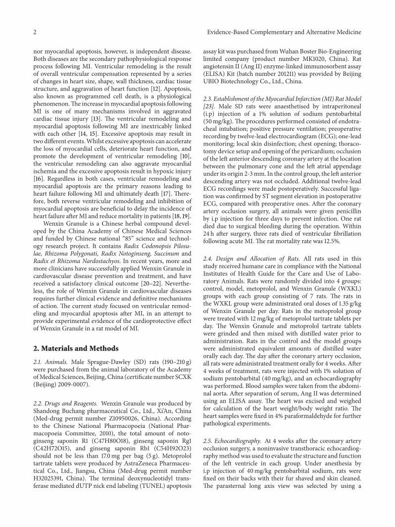
3.1. The Heart Weight/Body Weight (HW/BW) Ratio andVentricular Wall Thickness. As shown in Figure 1(a), theHW/BW ratio in themodel group was significantly increasedcompared with the control group (0.383 ± 0.071 versus0.312 ± 0.008mg/g, resp., 𝑃 < 0.01). The HW/BW ratioin the metoprolol group (0.325 ± 0.024mg/g, 𝑃 < 0.05)and the WXKL group (0.333 ± 0.017mg/g, 𝑃 < 0.05)was significantly decreased compared with the model group.As shown in Figure 1(b), there was no significant differencebetween LVPWTd, LVPWTs, and IVSTd among the fourgroups. The IVSTs in the control group was 0.32 ± 0.03 cm,and those of themodel (0.19±0.01 cm, 𝑃 < 0.01), metoprolol(0.23 ± 0.08 cm, 𝑃 < 0.01), and the WXKL groups (0.25 ±0.05 cm,𝑃 < 0.01)were significantly lower in various degrees.Compared with the model group, the IVSTs in the WXKLgroup were significantly increased (𝑃 < 0.05).
3.2. Left Ventricular Contraction Movement, Internal Diam-eter, and Volume. At 4 weeks after the coronary arteryocclusion surgery, echocardiography was performed, and thetypical echocardiography images were taken among differentgroups. As shown in Figure 2(a), the image of contractionmovement in the control group is shaped like waves (redarrow). As shown in Figure 2(b), the waves in the modelgroup weakened and even straightened (red arrow), indi-cating diminished and even disappearance of contractionmovement. The left ventricle expanded significantly in themodel group compared with the control group. As shownin Figures 2(c) and 2(d), the changes of left ventricular sizeand contraction weakness were alleviated in the metoprololand the WXKL groups. Figures 2(e) and 2(f) show thequantitative analysis of the internal diameter and volume ofthe left ventricle. The LViDd, LViDs, and EDV in the otherthree groups were significantly increased to various extentscompared with the control group (𝑃 < 0.01, 𝑃 < 0.05). TheESV in the model and the metoprolol groups also increasedsignificantly (𝑃 < 0.01). The SV did not show any significantdifferences between the four groups. Compared with themodel group, Wenxin Granule administration decreasedLViDs (0.66 ± 0.09 versus 0.49 ± 0.12 cm, 𝑃 < 0.05) and ESV(0.67±0.24 versus 0.30±0.23mL,𝑃 < 0.05) but had no effectson LViDd (0.81 ± 0.09 versus 0.72 ± 0.08 cm, 𝑃 > 0.05) andEDV (1.14±0.35 versus 0.84±0.26mL,𝑃 > 0.05). Metoprololhad no effects on the left ventricular internal diameter andvolume, neither in the systolic nor diastolic phase.
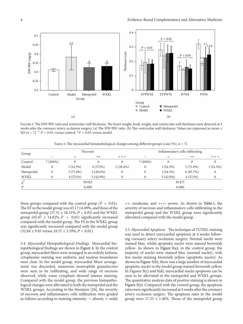
3.3. Cardiac Function. As shown in Figure 3, the EF and FSwere significantly decreased to various extents in the other
4 Evidence-Based Complementary and Alternative Medicine
GroupControl Model Metoprolol WXKL
Δ Δ
∗∗
0.5
0.45
0.4
0.35
0.3
0.25
0.2
HW
/BW
(mg/
g)
(a)
GroupControlModel
MetoprololWXKL
P > 0.05
P > 0.05
P > 0.05
∗∗
∗∗
0.4
0.3
0.2
0.1
0
Thic
knes
s (cm
)
LVPWTd LVPWTs IVSTd IVSTs
∗∗Δ
(b)
Figure 1: The HW/BW ratio and ventricular wall thickness. The heart weight, body weight, and ventricular wall thickness were detected at 4weeks after the coronary artery occlusion surgery. (a) The HW/BW ratio. (b) The ventricular wall thickness. Values are expressed as mean ±SD (𝑛 = 7). ∗∗𝑃 < 0.01, versus control. Δ𝑃 < 0.05, versus model.
Table 1: The myocardial histopathological changes among different groups (case (%), 𝑛 = 7).
Group Necrosis Inflammatory cells infiltrating− + ++ + + + − + ++ + + +
Control 7 (100%) 0 0 0 7 (100%) 0 0 0Model 0 1 (14.3%) 4 (57.1%) 2 (28.6%) 0 1 (14.3%) 5 (71.4%) 1 (14.3%)Metoprolol 0 5 (71.4%) 2 (28.6%) 0 0 1 (14.3%) 6 (85.7%) 0WXKL 0 4 (57.1%) 3 (42.9%) 0 0 3 (42.9%) 4 (57.1%) 0𝜒2 19.923 19.475𝑃 0.000 0.000
three groups compared with the control group (𝑃 < 0.01).TheEF in themodel groupwas 43.17±6.89%, and those of themetoprolol group (57.51 ± 18.31%, 𝑃 < 0.05) and the WXKLgroup (65.67 ± 14.82%, 𝑃 < 0.01) significantly increasedcompared with the model group.The FS in the WXKL groupwas significantly increased compared with the model group(32.84 ± 9.85 versus 18.57 ± 3.59%, 𝑃 < 0.01).
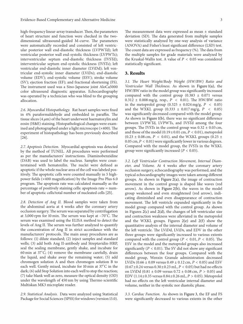
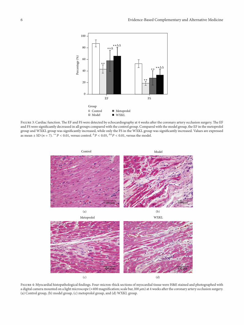
3.4. Myocardial Histopathological Findings. Myocardial his-topathological findings are shown in Figure 4. In the controlgroup, myocardial fibers were arranged in an orderly fashion,cytoplasmic staining was uniform, and nucleus boundarieswere clear. In the model group, myocardial fibers arrange-ment was discorded, numerous neutrophile granulocyteswere seen to be infiltrating, and wide range of necrosisobserved, while some cytoplasts showed intense staining.Compared with the model group, the previous histopatho-logical changes were alleviated in both themetoprolol and theWXKL groups. According to the literature [24], the severityof necrosis and inflammatory cells infiltration were gradedas follows according to staining intensity: −: absent; +: mild;
++: moderate; and +++: severe. As shown in Table 1, theseverity of necrosis and inflammatory cells infiltrating in themetoprolol group and the WXKL group were significantlyalleviated compared with the model group.
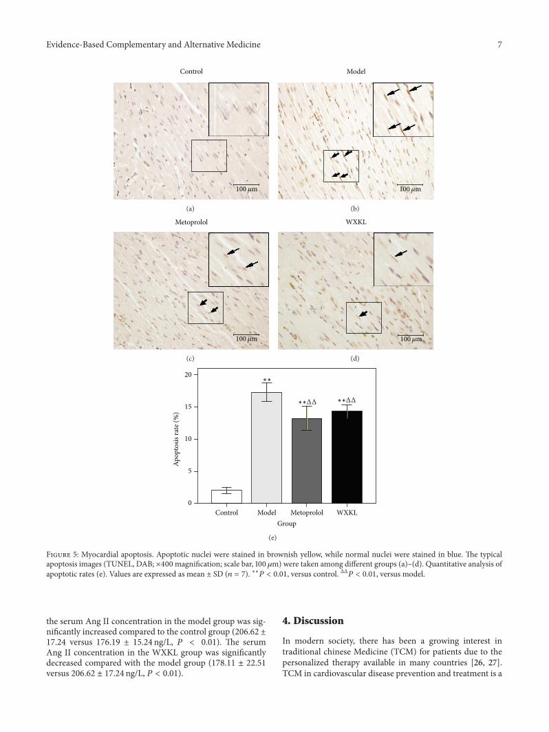
3.5. Myocardial Apoptosis. The technique of TUNEL stainingwas used to detect myocardial apoptosis at 4 weeks follow-ing coronary artery occlusion surgery. Normal nuclei werestained blue, whilst apoptotic nuclei were stained brownishyellow. As shown in Figure 5(a), in the control group, themajority of nuclei were stained blue (normal nuclei), withfew nuclei staining brownish yellow (apoptotic nuclei). Asshown in Figure 5(b), there was a large number ofmyocardialapoptotic nuclei in the model group stained brownish yellow.In Figures 5(c) and 5(d), myocardial nuclei apoptosis can beseen to be alleviated in the metoprolol and WXKL groups.The quantitative analysis data of positive staining is shown inFigure 5(e). Compared with the control group, the apoptosisrateswere significantly increased at 4weeks after the coronaryartery occlusion surgery. The apoptosis rates in the modelgroup were 17.33 ± 1.46%. Those of the metoprolol group

Evidence-Based Complementary and Alternative Medicine 5
Control
(a)
Model
(b)
Metoprolol
(c)
WXKL
(d)
GroupControlModel
MetoprololWXKL
1
0.8
0.6
0.4
0.2
0
Inne
r dia
met
er (c
m)
LViDd LViDs
∗∗∗∗
∗∗∗∗ ∗∗
∗∗Δ
(e)
GroupControlModel
MetoprololWXKL
Volu
me (
mL)
1.5
1.25
1
0.75
0.5
0.25
0EDV ESV SV
P > 0.05
∗∗
∗∗∗∗
∗
∗∗
Δ
(f)
Figure 2: Left ventricular contraction movement, internal diameter, and volume. At 4 weeks after the coronary artery occlusion surgery,echocardiography was performed, and the typical echocardiography images were taken among different groups ((a)–(d)). Internal diameterand volume of the left ventricle show the quantitative analysis data ((e)-(f)). Values are expressed asmean± SD (𝑛 = 7). ∗𝑃 < 0.05, ∗∗𝑃 < 0.01,versus control. Δ𝑃 < 0.05, versus model.
(13.23 ± 1.91%, 𝑃 < 0.01) and the WXKL group (14.36 ±0.98%, 𝑃 < 0.01) were significantly lower compared with themodel group.
3.6. Serum Ang II Concentration. At 4 weeks after thecoronary artery occlusion surgery, the serum Ang II concen-tration was detected by ELISA assay. As shown in Figure 6,
6 Evidence-Based Complementary and Alternative Medicine
∗∗
∗∗
∗∗∗∗ΔΔ
∗∗ΔΔ∗∗Δ
Perc
enta
ge (%
)
100
80
60
40
20
0FSEF
GroupControlModel
MetoprololWXKL
Figure 3: Cardiac function.The EF and FS were detected by echocardiography at 4 weeks after the coronary artery occlusion surgery.The EFand FS were significantly decreased in all groups compared with the control group. Compared with themodel group, the EF in themetoprololgroup and WXKL group was significantly increased, while only the FS in the WXKL group was significantly increased. Values are expressedas mean ± SD (𝑛 = 7). ∗∗𝑃 < 0.01, versus control. Δ𝑃 < 0.05, ΔΔ𝑃 < 0.01, versus the model.
Control
100𝜇m
(a)
Model
100𝜇m
(b)
Metoprolol
100𝜇m
(c)
WXKL
100𝜇m
(d)
Figure 4: Myocardial histopathological findings. Four-micron-thick sections of myocardial tissue were H&E stained and photographed witha digital cameramounted on a lightmicroscope (×400magnification; scale bar, 100 𝜇m) at 4 weeks after the coronary artery occlusion surgery.(a) Control group, (b) model group, (c) metoprolol group, and (d) WXKL group.
Evidence-Based Complementary and Alternative Medicine 7
Control
100𝜇m
(a)
Model
100𝜇m
(b)
Metoprolol
100𝜇m
(c)
WXKL
100𝜇m
(d)
∗∗
∗∗ΔΔ ∗∗ΔΔ
Apop
tosis
rate
(%)
20
15
10
5
0
GroupControl Model Metoprolol WXKL
(e)
Figure 5: Myocardial apoptosis. Apoptotic nuclei were stained in brownish yellow, while normal nuclei were stained in blue. The typicalapoptosis images (TUNEL, DAB; ×400 magnification; scale bar, 100 𝜇m) were taken among different groups (a)–(d). Quantitative analysis ofapoptotic rates (e). Values are expressed as mean ± SD (𝑛 = 7). ∗∗𝑃 < 0.01, versus control. ΔΔ𝑃 < 0.01, versus model.
the serum Ang II concentration in the model group was sig-nificantly increased compared to the control group (206.62 ±17.24 versus 176.19 ± 15.24 ng/L, 𝑃 < 0.01). The serumAng II concentration in the WXKL group was significantlydecreased compared with the model group (178.11 ± 22.51versus 206.62 ± 17.24 ng/L, 𝑃 < 0.01).
4. Discussion
In modern society, there has been a growing interest intraditional chinese Medicine (TCM) for patients due to thepersonalized therapy available in many countries [26, 27].TCM in cardiovascular disease prevention and treatment is a

8 Evidence-Based Complementary and Alternative Medicine
∗∗
ΔΔ
Ang
II (n
g/L)
240
220
200
180
160
140
120
GroupControl Model Metoprolol WXKL
Figure 6: SerumAng II concentration in the 4 experimental groups.The serum Ang II concentration was detected by ELISA assay at4 weeks after the coronary artery occlusion surgery. Ang II in themodel group was significantly increased compared with the controlgroup. Ang II in the WXKL group was significantly decreasedcompared with the model group. Values are expressed as mean ±SD (𝑛 = 7). ∗∗𝑃 < 0.01, versus control. ΔΔ𝑃 < 0.01, versus model.
valuable and promising prospect. TCM has a history of morethan 2500 years with a unique theory of diagnosis and treat-ment. Whilst there is no such term as MI in TCM, its symp-toms can be classified into the categories of true heart pain,palpitation, thoracic obstruction, and heart-energy stagna-tion syndrome. In recent years, an increasing number of clini-cians have successfully appliedTCMdrugs for supplementingQi, nourishing Yin, and activating blood circulation in thetreatment ofMI [28, 29].Nevertheless, the role of TCM in car-diovascular diseases requires further experimental evidence.
Wenxin Granule is a Chinese medicinal granule, whichhas effects on supplementing Qi and nourishing Yin,promoting blood circulation for removing blood stasis. Sev-eral studies have shown that Wenxin Granule can increasecoronary blood flow, reduce myocardial oxygen consump-tion, enhance myocardial compliance, improve myocardialhypoxia tolerance, relieve anterior and posterior cardiac load-ing, reduce myocardial tissue damage in patients with highblood pressure, and reduce the occurrence of arrhythmia[30]. To further support previous studies, the present studyaimed to provide experimental evidence of the cardioprotec-tive effect of Wenxin Granule in the MI rat model.
The results of the current study showed that administra-tion ofWenxinGranule could partially reverse left ventricularremodeling and improve left ventricular function to anextent. It should be noted that whilst the Wenxin Granuledecreased LViDs and ESV, Wenxin Granule administrationhad no effects on LViDd and EDV. It is possible that4 weeks after modeling, fibrotic scaring or a ventricularaneurysm occurred in the left ventricular infarct wall, whichmay be responsible for the increase in LViDd and EDV.Wenxin Granule did not completely reverse fibrotic scar-ing and ventricular aneurysm after MI. Consequently, theLViDd and EDV did not change significantly in the diastolic
phase. During the systolic phase, the contractile functionof non-infarct myocardium in the Wenxin Granule groupwas stronger than that in the model group. Therefore, theWenxin Granule could decrease LViDs and ESV markedly.This was the main difference between the metoprolol groupand the WXKL groups whereby metoprolol only showed adecreasing trend on the left ventricular internal diameterand volume, but without significant difference, in either thesystolic or the diastolic phase. The possible reasons for thisare that the cardioprotective mechanisms of metoprolol areachievedmainly by blocking cardiac𝛽1-receptors and therebyslowing heart rate and reduction in myocardial contractilityand myocardial oxygen consumption. Compared with otherbeta-Blockers and angiotensin converting enzyme inhibitors(ACEI), the effect ofmetoprolol on preventing left ventricularremodeling is relatively weak [31, 32].
In addition, the present study observed the improvementof histopathological injury and the inhibition of myocardialapoptosis after MI in the WXKL group and the metoprololgroup. Radix Notoginseng, one of the main components ofWenxin Granule, can repair ischemia myocardial and reducemyocardial ischemic injury by decreasing oxidative stressand repressing the inflammatory cascade [33]. This may bethe means by which treatment withWenxin Granule reducedhistopathologic injury afterMI. Meanwhile, it was also foundthatWenxin Granule was capable of reducing Ang II concen-trations in the MI rat model. Myocardial ischemia inducesactivation of various components of the renin-angio-tensin system (RAS), including angiotensinogen, renin,angiotensin-converting enzyme, angiotensins, and angio-tensin receptors, in the acute phase of MI and in the postin-farction remodeling process [34]. In the RAS, Ang II is abiologically active substance, which is closely correlated withmyocardial apoptosis. Several studies have investigated therelationship between Ang II and myocardial apoptosis. Astudy byKajstura et al. (1997) showed that in primary culturesof adult rat ventricular myocytes exposed to 10−9Mof Ang IIfor 24 h, presented with a fivefold increase in apoptosis docu-mented by the terminal deoxynucleotidyl transferase assay,and confirmed by DNA agarose gel electrophoresis [35]. Astudy conducted by Leri et al. (1998) confirmed that AngII could increase the susceptibility of myocytes to undergoapoptosis [36]. Ang II stimulation was associated withtranslocation of the epsilon and delta isoforms of proteinkinase C.This was coupled with an increase in cytosolic Ca2+in the cells which can induce apoptosis [35]. Several studieshave confirmed that Radix Codonopsis Pilosulae, one ofthe main components of Wenxin Granule, can reverse Ca2+influx and the increase in apoptosis.This is achieved by atten-uating Ang II and the cardiac-impaired insulin-like growthfactor II (IGF II) receptor pathway in myocardial cells [37].The current study found that treatment withWenxin Granuledecreased Ang II concentrations. Consequently, Ang II couldbe the underlying mechanism of Wenxin Granule inhibitionof apoptosis.
Based on the previous findings, the authors draw the con-clusion thatWenxin Granule can partially reverse ventricularremodeling, improve heart function, alleviate the histopatho-logical damage, inhibit myocardial apoptosis, and reduce

Evidence-Based Complementary and Alternative Medicine 9
AngII concentrations in rats with MI. These results suggestthat Wenxin Granule might be a potentially alternative andcomplementary medicine for the treatment of MI.
Authors’ Contribution
Aiming Wu, Jianying Zhai, Dongmei Zhang, Lixia Lou, andHaiyan Zhu contributed equally to this work.
Conflict of Interests
The authors declare that they have no conflict of interests.
Acknowledgments
This paper was partially supported by the National NaturalScience Foundation Project of China (no. 81202685) andBeijingMunicipal Commission of Education Build Project ofChina (2012 Beijing University of Chinese Medicine).
References
[1] D. D.McManus, S. M. Piacentine, D. Lessard et al., “Thirty-year(1975 to 2005) trends in the incidence rates, clinical features,treatment practices, and short-term outcomes of patients <55years of age hospitalized with an initial acute myocardialinfarction,”The American Journal of Cardiology, vol. 108, no. 4,pp. 477–482, 2011.
[2] N. Rumana, Y. Kita, T. C. Turin et al., “Trend of increase in theincidence of acute myocardial infarction in a Japanese popu-lation: TakashimaAMI registry, 1990–2001,”TheAmerican Jour-nal of Epidemiology, vol. 167, no. 11, pp. 1358–1364, 2008.
[3] S. Yi, W. Lei, and Z. Minzhou, “Progress in the epidemiologicalstudy of acutemyocardial infarction,”Chinese Journal of Integra-tive Medicine on Cardio-/Cerebrovascular Disease, vol. 10, no. 4,pp. 467–469, 2012.
[4] L. Lacey and M. Tabberer, “Economic burden of post-acutemyocardial infarction heart failure in the United Kingdom,”European Journal of Heart Failure, vol. 7, no. 4, pp. 677–683,2005.
[5] D. V. Baklanov, L. A. Kaltenbach, S. P. Marso et al., “The preva-lence and outcomes of transradial percutaneous coronary inter-vention for ST-segment elevation myocardial infarction: analy-sis from thenational cardiovascular data registry (2007 to 2011),”Journal of the American College of Cardiology, vol. 61, no. 4, pp.420–426.
[6] G. Falsini, F. Liistro, K. Ducci et al., “Shifting from pharmaco-logical to systematic mechanical reperfusion therapy for acutemyocardial infarction via a cooperating network: impact onreperfusion rate and in-hospital mortality,” Journal of Cardio-vascular Medicine, vol. 9, no. 3, pp. 245–250, 2008.
[7] S. Gupta, S. Das, R. Sahewalla et al., “A study on quality of lifein patients following myocardial infarction,” Indian Journal ofPhysiology and Pharmacology, vol. 56, no. 1, pp. 28–35, 2012.
[8] W.Wang,D. R.Thompson, C. F. Ski, andM. Liu, “Health-relatedquality of life and its associated factors in Chinese myocardialinfarction patients,” European Journal of Preventive Cardiology.In press.
[9] S. Mangiapane and R. Busse, “Prescription prevalence and con-tinuing medication use for secondary prevention after myocar-dial infarction: the reality of care revealed by claims data
analysis,” Deutsches Arzteblatt, vol. 108, no. 50, pp. 856–862,2011.
[10] A. Abbate, G. G. L. Biondi-Zoccai, R. Bussani et al., “Increasedmyocardial apoptosis in patients with unfavorable left ventric-ular remodeling and early symptomatic post-infarction heartfailure,” Journal of the American College of Cardiology, vol. 41,no. 5, pp. 753–760, 2003.
[11] J. N. Kirkpatrick and M. S. J. Sutton, “Assessment of ventricularremodeling in heart failure clinical trials,”Current Heart FailureReports, vol. 9, no. 4, pp. 328–336, 2012.
[12] J. J. Gajarsa and R. A. Kloner, “Left ventricular remodeling inthe post-infarction heart: a review of cellular, molecular mech-anisms, and therapeutic modalities,”Heart Failure Reviews, vol.16, no. 1, pp. 13–21, 2011.
[13] P. M. Kang and S. Izumo, “Apoptosis and heart failure: a criticalreview of the literature,” Circulation Research, vol. 86, no. 11, pp.1107–1113, 2000.
[14] Y. Hojo, T. Saito, and H. Kondo, “Role of apoptosis in left ven-tricular remodeling after acute myocardial infarction,” Journalof Cardiology, vol. 60, no. 2, pp. 91–92, 2012.
[15] F. Sam, D. B. Sawyer, D. L. Chang et al., “Progressive left ventric-ular remodeling and apoptosis late after myocardial infarctioninmouse heart,”TheAmerican Journal of Physiology—Heart andCirculatory Physiology, vol. 279, no. 1, pp. H422–H428, 2000.
[16] A. Abbate, G. G. L. Biondi-Zoccai, and A. Baldi, “Patho-physiologic role of myocardial apoptosis in post-infarction leftventricular remodeling,” Journal of Cellular Physiology, vol. 193,no. 2, pp. 145–153, 2002.
[17] A. M. Shah, C. L. Hung, S. H. Shin et al., “Cardiac structure andfunction, remodeling, and clinical outcomes among patientswith diabetes after myocardial infarction complicated by leftventricular systolic dysfunction, heart failure, or both,” TheAmerican Heart Journal, vol. 162, no. 4, pp. 685–691, 2011.
[18] L. Spinelli, C. Morisco, E. A. di Panzillo, R. Izzo, and B.Trimarco, “Reverse left ventricular remodeling after acutemyocardial infarction: the prognostic impact of left ventricularglobal torsion,” International Journal of Cardiovascular Imaging,vol. 29, no. 4, pp. 787–795, 2013.
[19] V. Jayasankar, Y. J. Woo, T. J. Pirolli et al., “Induction of angio-genesis and inhibition of apoptosis by hepatocyte growth factoreffectively treats postischemic heart failure,” Journal of CardiacSurgery, vol. 20, no. 1, pp. 93–101, 2005.
[20] W. Liu, R. Jiang, S. Ding et al., “Quality assessment of random-ized controlled trials on Wenxin granule for treatment of atrialfibrillation,” Zhongguo Zhong Yao Za Zhi, vol. 37, no. 1, pp. 109–114, 2012.
[21] X. Wang, Y. Gu, T. Wang, and C. Huang, “Wenxin Keli atten-uates ischemia-induced ventricular arrhythmias in rats: in-volvement of Ltype calcium and transient outward potassiumcurrents,”Molecular Medicine Reports, vol. 7, no. 2, pp. 519–524,2013.
[22] M. Wang, Y. B. Yu, and S. E. Huang, “Clinical observationon effect and safety of combined use of wenxin granule andamiodarone for conversion of auricular fibrillation,” ZhongguoZhong Xi Yi Jie He Za Zhi, vol. 26, no. 5, pp. 445–448, 2006.
[23] Y. Xing, Y. Gao, J. Chen et al., “Wenxin-Keli regulates thecalcium/calmodulin-dependent protein kinase II signal trans-duction pathway and inhibits cardiac arrhythmia in rats withmyocardial infarction,” Evidence-Based Complementary andAlternative Medicine, vol. 2013, Article ID 464508, 15 pages,2013.
10 Evidence-Based Complementary and Alternative Medicine
[24] M. C. Fishbein, D. Maclean, and P. R. Maroko, “Experimentalmyocardial infarction in the rat. Qualitative and quantitativechanges during pathologic evolution,” The American Journal ofPathology, vol. 90, no. 1, pp. 57–70, 1978.
[25] Y. C. Zhou, B. Liu, Y. J. Li et al., “Effects of Buyang Huanwudecoction on ventricular remodeling and differential proteinprofile in a rat model of myocardial infarction,” Evidence-BasedComplementary and Alternative Medicine, vol. 2012, Article ID385247, 11 pages, 2012.
[26] J. Wang and X. Xiong, “Current situation and perspectives ofclinical study in integrative medicine in China,” Evidence-BasedComplementary and Alternative Medicine, vol. 2012, Article ID268542, 11 pages, 2012.
[27] J. J. Park, S. Beckman-Harned, G. Cho, D. Kim, and H.Kim, “The current acceptance, accessibility and recognition ofChinese and Ayurvedic medicine in the United States in thepublic, governmental, and industrial sectors,” Chinese Journalof Integrative Medicine, vol. 18, no. 6, pp. 405–408, 2012.
[28] S. L. Qiu, M. Jin, J. H. Yi, T. G. Zhu, X. Quan, and Y. Liang,“Therapy for replenishing qi, nourishing yin and promotingblood circulation in patients with acute myocardial infarctionundergoing percutaneous coronary intervention: a randomizedcontrolled trial,”ZhongXi Yi JieHeXue Bao, vol. 7, no. 7, pp. 616–621, 2009.
[29] Y. Q. Li, M. Jin, S. L. Qiu et al., “Effect of Chinese drugs forsupplementing Qi, nourishing yin and activating blood circu-lation on myocardial perfusion in patients with acute myocar-dial infarction after revascularization,” Chinese Journal of Inte-grative Medicine, vol. 15, no. 1, pp. 19–25, 2009.
[30] N. Su, T. Xu, Y. Tang, and Z. Zhou, “Efficacy and safety ofWenxin granules in the treatment of congestive heart failure: asystematic review,” China Pharmacy, vol. 21, no. 7, pp. 637–640,2010.
[31] G. Cimmino, B. Ibanez, C. Giannarelli et al., “Carvedilol admin-istration in acute myocardial infarction results in strongerinhibition of early markers of left ventricular remodeling thanmetoprolol,” International Journal of Cardiology, vol. 153, no. 3,pp. 256–261, 2011.
[32] R. Ricci, C. Coletta, V. Ceci et al., “Effect of early treatment withcaptopril and metoprolol singly and together on postinfarctionleft ventricular remodeling,” The American Heart Journal, vol.142, no. 4, article E5, 2001.
[33] S. Y. Han, H. X. Li, X. Ma et al., “Evaluation of the anti-myo-cardial ischemia effect of individual and combined extracts ofPanax notoginseng and Carthamus tinctorius in rats,” Journal ofEthnopharmacology, vol. 145, no. 3, pp. 722–727, 2013.
[34] W. Dai and R. A. Kloner, “Potential role of renin-angioten-sin system blockade for preventing myocardial ischemia/reper-fusion injury and remodeling after myocardial infarction,”Postgraduate Medicine, vol. 123, no. 2, pp. 49–55, 2011.
[35] J. Kajstura, E. Cigola, A.Malhotra et al., “Angiotensin II inducesapoptosis of adult ventricular myocytes in vitro,” Journal ofMolecular and Cellular Cardiology, vol. 29, no. 3, pp. 859–870,1997.
[36] A. Leri, P. P. Claudio, Q. Li et al., “Stretch-mediated release ofangiotensin II induces myocyte apoptosis by activating p53 thatenhances the local renin-angiotensin system and decreases theBcl-2-to-Bax protein ratio in the cell,” Journal of Clinical Investi-gation, vol. 101, no. 7, pp. 1326–1342, 1998.
[37] K. H. Tsai, N. H. Lee, G. Y. Chen et al., “Dung-shen (Codonopsispilosula) attenuated the cardiac-impaired insulin-like growth
factor II receptor pathway onmyocardial cells,” Food Chemistry,vol. 138, no. 2-3, pp. 1856–1867, 2013.

Submit your manuscripts athttp://www.hindawi.com
Stem CellsInternational
Hindawi Publishing Corporationhttp://www.hindawi.com Volume 2014
Hindawi Publishing Corporationhttp://www.hindawi.com Volume 2014
MEDIATORSINFLAMMATION
of
Hindawi Publishing Corporationhttp://www.hindawi.com Volume 2014
Behavioural Neurology
EndocrinologyInternational Journal of
Hindawi Publishing Corporationhttp://www.hindawi.com Volume 2014
Hindawi Publishing Corporationhttp://www.hindawi.com Volume 2014
Disease Markers
Hindawi Publishing Corporationhttp://www.hindawi.com Volume 2014
BioMed Research International
OncologyJournal of
Hindawi Publishing Corporationhttp://www.hindawi.com Volume 2014
Hindawi Publishing Corporationhttp://www.hindawi.com Volume 2014
Oxidative Medicine and Cellular Longevity
Hindawi Publishing Corporationhttp://www.hindawi.com Volume 2014
PPAR Research
The Scientific World JournalHindawi Publishing Corporation http://www.hindawi.com Volume 2014
Immunology ResearchHindawi Publishing Corporationhttp://www.hindawi.com Volume 2014
Journal of
ObesityJournal of
Hindawi Publishing Corporationhttp://www.hindawi.com Volume 2014
Hindawi Publishing Corporationhttp://www.hindawi.com Volume 2014
Computational and Mathematical Methods in Medicine
OphthalmologyJournal of
Hindawi Publishing Corporationhttp://www.hindawi.com Volume 2014
Diabetes ResearchJournal of
Hindawi Publishing Corporationhttp://www.hindawi.com Volume 2014
Hindawi Publishing Corporationhttp://www.hindawi.com Volume 2014
Research and TreatmentAIDS
Hindawi Publishing Corporationhttp://www.hindawi.com Volume 2014
Gastroenterology Research and Practice
Hindawi Publishing Corporationhttp://www.hindawi.com Volume 2014
Parkinson’s Disease
Evidence-Based Complementary and Alternative Medicine
Volume 2014Hindawi Publishing Corporationhttp://www.hindawi.com
Related Documents