Gene Function in Cell Growth, Differentiation & Development Director Univ.-Prof. Dr. rer. nat. Martin Zenke RWTH Aachen University Hospital Pauwelsstrasse 30, 52074 Aachen Helmholtz Institute for Biomedical Engineering Pauwelsstrasse 20, 52074 Aachen Phone: +49-241-80 80760 (Office) +49-241-80 80759 (Secretary) Fax: +49-241-80 82008 Email: [email protected] Web: http://www.molcell.de Staff Offergeld, Andrea, Administrative Assistant Becker, Christiane, Scientific Assistant Baden, Sabrina, MSc, PhD Student Baek, Jea-Hyun, MSc, PhD Student Brosig, Stephanie, Technician Bross, Daniela, Student Bundscherer, Lena, Student Chauvistré, Heike, MSc, PhD Student Christ, Anette, MSc, PhD Student Ding, Xiaolei, MSc, PhD Student Döring, Yvonne, MSc, PhD Student Elbers, Bärbel, Technician Gamper, Ivonne, MSc, PhD Student Guhe, Zita, Student Hieronymus, Thomas, PhD, Group Leader Jäntti, Piritta, MSc, PhD Student Lin, Qiong, MSc, PhD Student Lüneberger, Sigrid, Technician Mitzka, Saskia, Technician Ober-Blöbaum, Julia, MSc, PhD Student Pabich, Julia, Student Ruau, David, MSc, PhD Student Schneider-Kramann, Rebekka, MD, Postdoc Schwarz, Sebastian, MSc, PhD Student Sechi, Antonio, PhD, Group Leader Seré, Kristin, PhD, Postdoc Shi, Nian, MSc, PhD Student Shokouhi, Behnaz, MSc, PhD Student Siegler, Heike, Student Simons, Nadine, Technician Thönes, Stephan, Student Wanek, Paul, Technician Wang, Mengxi, Student Stem Cell Biology and Cellular Engineering Univ.-Prof. Dr. med., Dr. rer. nat. Wolfgang Wagner Helmholtz Institute for Biomedical Engineering Pauwelsstrasse 20, 52074 Aachen Phone: +49-241-80 88611 (Office) Fax: +49-241-80 82008 Email: [email protected] Bokermann, Gudrun, PhD Student Cholewa, Dominik, PhD Student Joussen, Sylvia, Technician Koch, Carmen, Postdoc Walenda, Thomas, PhD Student

Research Annual Report 2009
Mar 28, 2016
Research Annual Report 2009
Welcome message from author
This document is posted to help you gain knowledge. Please leave a comment to let me know what you think about it! Share it to your friends and learn new things together.
Transcript
5
2009Helmholtz-Institute for Biomedical EngineeringRWTH Aachen University
Gene Function in Cell Growth, Differentiation & DevelopmentDirectorUniv.-Prof. Dr. rer. nat.Martin Zenke
RWTH Aachen University HospitalPauwelsstrasse 30, 52074 Aachen
Helmholtz Institute for Biomedical EngineeringPauwelsstrasse 20, 52074 Aachen
Phone: +49-241-80 80760 (Office) +49-241-80 80759 (Secretary)Fax: +49-241-80 82008Email: [email protected]: http://www.molcell.de
StaffOffergeld, Andrea, Administrative AssistantBecker, Christiane, Scientific Assistant
Baden, Sabrina, MSc, PhD StudentBaek, Jea-Hyun, MSc, PhD StudentBrosig, Stephanie, TechnicianBross, Daniela, StudentBundscherer, Lena, StudentChauvistré, Heike, MSc, PhD StudentChrist, Anette, MSc, PhD StudentDing, Xiaolei, MSc, PhD StudentDöring, Yvonne, MSc, PhD StudentElbers, Bärbel, TechnicianGamper, Ivonne, MSc, PhD StudentGuhe, Zita, StudentHieronymus, Thomas, PhD, Group LeaderJäntti, Piritta, MSc, PhD StudentLin, Qiong, MSc, PhD StudentLüneberger, Sigrid, TechnicianMitzka, Saskia, TechnicianOber-Blöbaum, Julia, MSc, PhD StudentPabich, Julia, StudentRuau, David, MSc, PhD StudentSchneider-Kramann, Rebekka, MD, PostdocSchwarz, Sebastian, MSc, PhD StudentSechi, Antonio, PhD, Group LeaderSeré, Kristin, PhD, PostdocShi, Nian, MSc, PhD StudentShokouhi, Behnaz, MSc, PhD StudentSiegler, Heike, StudentSimons, Nadine, TechnicianThönes, Stephan, StudentWanek, Paul, TechnicianWang, Mengxi, Student
Stem Cell Biology and Cellular EngineeringUniv.-Prof. Dr. med., Dr. rer. nat.Wolfgang Wagner
Helmholtz Institute for Biomedical EngineeringPauwelsstrasse 20, 52074 Aachen
Phone: +49-241-80 88611 (Office)Fax: +49-241-80 82008Email: [email protected]
Bokermann, Gudrun, PhD StudentCholewa, Dominik, PhD StudentJoussen, Sylvia, TechnicianKoch, Carmen, PostdocWalenda, Thomas, PhD Student
6
2009Helmholtz-Institute for Biomedical EngineeringRWTH Aachen University
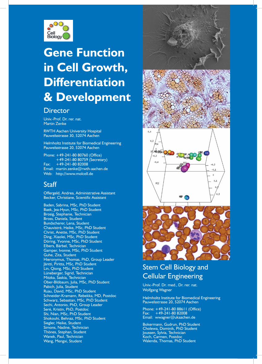
Introduction
Genetic programs determine cell identity and function and thus cells are now being engineered to acquire novel and wanted identities and functions. The laboratory studies the developmental potential of stem cells, including hematopoi-etic stem cells (HSC), mesenchymal stem cells (MSC) and embryonic stem cells (ES cells), and their differentiated progeny. In addition, efforts are directed towards enlarging the potential of somatic cells by employing various repro-gramming strategies, such as induced pluripotent stem (iPS) cell technology. HSC and MSC reside in bone marrow in a highly specialized area, referred to as stem cell niche, and in this context we study HSC/MSC interactions in homeosta-sis, pathology and aging.
Biomedical engineering involves the development of biohy-brid systems comprising cells and engineered synthetic materials. Thus, the laboratory investigates the impact of natural and synthetic materials and of engineered nanopar-ticles on cell differentiation and function. This also includes studies on unwanted immune responses elicted by antigen presenting dendritic cells (DC). Nanoparticles are used for monitoring cell migration and function by magnetic reso-nance imaging (MRI).
In 2009 the institute welcomed Wolfgang Wagner, MD, PhD from the Ruprechts-Karls-University Heidelberg, Germany who took up his position as a Professor for Stem Cell Biology and Cellular Engineering. The group of Wolfgang Wagner receives core funding from the Stem Cell Network North Rhine Westphalia (NRW), Ministry of Innovation, Science, Research and Technology NRW, Düsseldorf, Germany.
Antigen Presenting Dendritic Cells
Antigen presenting dendritic cells (DC) represent highly specialized immune cells with a central role in immunity and tolerance induction. DC sense antigens, which are taken-up, processed and presented in the context of MHC mole-cules to elicit antigen specific T cell responses (Zenke and Hieronymus, 2006). Specific DC subsets exists that differ in surface phenotype, function, activation state and anatom-ical localization. The main DC subsets are (i) tissue/inter-stitial DC in organs, now referred to as conventional DC (cDC); (ii) plasmacytoid DC (pDC) in blood that represent the major producers of type 1 interferon (iii) CD8α DC in lymphoid tissue and (iv) Langerhans cells (LC), the cutane-ous contingent of DC in epidermis.
All DC subsets develop from hematopoietic stem cells via Flt3 expressing progenitors through consecutive steps of lineage commitment and differentiation (Fig. 2). Surprisingly, DC development shows remarkable plasticity and DC can develop from both lymphoid and myeloid compartments. Additionally, a clonogenic DC progenitor for cDC and pDC was identified, referred to as Flt3+M-CSFR+c-kitlow common DC progenitor (CDP). The laboratory studies DC development by employing in vitro culture systems and knockout mouse models (Hacker et al., 2003; Hieronymus et al., 2005; Ju et al., 2007).
Cell-based therapies, including immunotherapy with DC, re-quire accurate delivery of cells to target tissue and monitor-ing cell position over extended periods of time. Therefore, DC were labelled with nano-sized magnetic iron oxide nanoparticles (MNP) for monitoring DC migration and po-sition by MRI (Schwarz et al., 2009; in collaboration with M. Hoehn, MPI for Neurological Research, Cologne, Germany; U. Himmelreich, Catholic University of Leuven, Belgium; S. Aime, University of Torino, Italy; D. Schueler, Department of Microbiology, Ludwig-Maximilians-University, Munich, Germany; C. Bergemann, Chemicell, Berlin, Germany; W. Richtering, Institute for Physical Chemistry, M. Hodenius
Cell Biology
Fig. 1: Embryonic stem cells (ES cells) are pluripotent and give rise to cells of all three germ layers, including hemat-opoietic cells and mesenchymal cells. Hematopoietic stem cells (HSC) and mesenchymal stem cells (MSC) develop into all cells in blood and connective tissue, respectively. Dif-ferentiated cells can be reprogrammed to acquire pluripo-tency and properties of ES cells, referred to as induced pluripotent stem cells (iPS cells).
Fig. 2: Hematopoietic stem cells (HSC) give rise to all ma-ture cells in blood and to blood-borne cell in peripheral lymphoid organs. DC subsets develop from HSC through consecutive steps of lineage commitment and differentia-tion.

7
2009Helmholtz-Institute for Biomedical EngineeringRWTH Aachen University
Reprogramming and Induction of Pluripotency
Recent progress in stem cell biology demonstrates that so-matic cells can be readily reprogrammed and induced to acquire pluripotency by a defined set of transcription fac-tors, including Oct4, Sox2, c-Myc and Klf4 (referred to as induced pluripotent stem cells, iPS cells; Fig. 1).
We have found that mouse neural stem cells (NSC) en-dogenously express the two reprogramming factors Sox2 and c-Myc (Ruau et al., 2008; Kim et al., 2008). Thus, iPS cells were derived from adult NSC by the expression of Oct4 together with either Klf4 or c-Myc (Kim et al., 2008). Additionally, subsequent work demonstrated that Oct4 alone is sufficient for reprogramming of NSC (Kim
and T. Schmitz-Rode, Applied Medical Engineering, Helmholtz Institute for Biomedical Engineering, RWTH Aachen University, Aachen, Germany). First, we investigat-ed the uptake of synthetic MNP or bacterial magnetosome MNP into DC. Second, we determined detection limits of MNP-loaded DC in agarose phantoms (Fig. 3). It was found that DC readily engulf MNP and MNP-labeled DC were de-tected by MRI. Detection limit was 100 MNP-labeled DC (Fig. 3). Prussian Blue staining confirmed the presence of iron oxide in DC (Fig. 4).
Cel
l Bio
logy
Fig.3: Agarose phantoms of MNP-loaded DC detected by 11.7 T MRI (Schwarz et al., 2009).
Fig. 4: Lipid-shell MNP and bacterial magnetosome MNP are taken-up by DC. Iron concentration per cell and iron locali-zation in DC by histological staining are shown (A and B, respectively).
Fig. 5: Hierarchical cluster analysis of genome-wide gene expression by DNA microarrays. MEF, mouse embryo fibroblasts; 1 factor and 2 factor NSC-derived iPS cells, 1FNSCiPS and 2FNSCiPS, respectively; ES cells, ESC (Ko et al., 2009). Pluripotent cells (ESC, iPS cells and gPS cells) cluster together. GSC form a cluster, which is distinct from somatic cells (MEF and NSC).
Fig.6: Global gene expression of GSC, ESC and gPS cells is depicted by scatter plot analysis (Ko et al., 2009). A panel of pluripotency-associated genes are indicated. Please note the high similarity of gPS cells and ES cells (ESC).

8
2009Helmholtz-Institute for Biomedical EngineeringRWTH Aachen University
therapeutic cell preparations (Wagner et al., 2009; 2010). For example, culture-expansion of MSC over long periods of time impacts on cell proliferation and differentiation po-tential. Microarray analysis revealed that this is accompa-nied by continuous and organized gene expression changes. Following cell passaging genes involved in cell division and DNA repair are down regulated whereas others are high-er expressed (Bork et al., 2010; Teschendorff et al., 2010; Fig. 8). These long-term culture associated gene expression changes are also observed in independent donor samples and even by cross-validation between different laborato-ries (Schallmoser et al., 2010). Therefore, it is conceivable to use a specific panel of gene expression markers for qual-ity control of MSC.
Acknowledgements
This work was supported by
• GermanResearchFoundation(DFG) • GermanFederalMinistryof Education andResearch
(BMBF) • Interdisciplinary Centre for Clinical Research on
Biomaterials and Implants (IZKF BioMAT) • Stem Cell Network NRW, Ministry of Innovation,
Science, Research and Technology of the State Nordrhein-Westfalen
• ErasmusProgramoftheEuropeanCommunity • DonationbyU.L.
Selected references [1] Bork, S., Pfister, S., Witt, H., Horn, P., Korn, B., Ho, A. D. and Wag-
ner, W. (2010). DNA methylation pattern changes upon long-term culture and aging of human mesenchymal stromal cells. Aging Cell 9, 54-63.
[2] Cao, X., Pettit, M. E., Conlan, S. L, Wagner, W., Ho, A. D., Clare, A. S,. Callow, J. A., Callow, M. E., Grunze, M. and Rosenhahn, A. (2009). Resistance of polysaccharide coatings to proteins, hematopoietic cells, and marine organisms. Biomacromolecules 10, 907-915.
et al., 2009). Germline-derived stem cells (GSC) express Oct4 and thus GSC can be induced to acquire pluripoten-cy without exogenous transcription factors by employing specific culture conditions (referred to as germline-de-rived pluripotent cells, gPS cells; Ko et al., 2009). gPS cells exhibit a gene expression repertoire which is very similar to ES cells (Figs. 5 and 6; in collaboration with H. R. Schöler, MPI for Molecular Biomedicine, Münster, Germany). Pluripotency of gPS cells was confirmed by in vitro and in vivo differentiation, including germ cell contri-bution and transmission (Ko et al., 2009).
Molecular Analysis of Adult Stem Cells
Throughout life regeneration of tissues is facilitated by stem cells, referred to as adult/somatic/tissue specific stem cells. Stem cells have the dual capacity (i) of differentiating into specific cell types and (ii) of self-renewing to maintain the stem cell pool. These functions have to be tightly reg-ulated according to the physiologic needs of the organism. There is a growing perception that direct cell-cell con-tacts, referred to as “stem cell niche”, plays a central role for stem cell maintenance and regulation. Our bone mar-row for example harbours two types of adult stem cells: (i) HSC that give rise to all cell types in blood and blood-borne lymphoid organs and (ii) MSC that resemble pro-genitors for bone, fat and cartilage (Figs. 1 and 7). Our group investigates molecular properties of HSC and MSC and mechanisms that govern the balance between self-renewal and differentiation. Additionally, we study the HSC/MSC interplay and the adhesion molecules involved (Walenda et al., 2009; Wein et al., 2010; Fig. 7).
MSC are currently being tested in more than hundred clini-cal trials. Hope in regenerative medicine has been fueled by novel insights from stem cell biology, new molecular tools and promising preclinical models. At the same time, there is a growing perception that standardized culture conditions and reliable methods for quality control of MSC are urgent-ly needed.
Our group gained insight into how culture media, bioma-terials and cell culture techniques affect the composition of
Cell Biology
Fig. 8: Human MSC were expanded for 11 passages (up to three months) and subjected to gene expression profiling with DNA microarrays. Hierarchical cluster analysis of gene expression profiles revealed continuous changes with higher passages (red and blue, higher and lower gene expression than median, respectively).
Fig. 7: The stem cell niche in bone marrow and molecules involved in HSC/MSC interactions.

9
2009Helmholtz-Institute for Biomedical EngineeringRWTH Aachen University
[3] Dickhut, A., Pelttari, K., Janicki, P., Wagner, W., Eckstein, V., Eger-mann, M. and Richter, W. (2009). Calcification or dedifferentiation: requirement to lock mesenchymal stem cells in a desired differen-tiation stage. J. Cellular Physiol. 219, 219-226.
[4] Gamper, I., Koh, K.-R., Ruau, D., Ullrich, K., Bartunkova, J., Piroth, D., Hacker, C., Bartunek, P. and Zenke, M. (2009). GAR22: A novel target gene of thyroid hormone receptor causes growth inhibition in human erythropoid cells. Exp. Hematol. 37, 539-548.
[5] Kim, J. B., Sebastiano, V., Wu, G., Araúzo-Bravo, M. J., Sasse, P., Gentile, L., Ko, K., Ruau, D., Ehrich, M., van den Boom, D., Meyer, J., Hübner, K., Bernemann, C., Ortmeier, C., Zenke, M., Fleischmann, B. K., Zaehres, H. and Schöler, H. R. (2009). Oct4-in-duced pluripotency in adult neural stem cells. Cell 136, 411-419.
[6] Ko, K., Tapia, N., Wu, G., Kim, J. B., Araúzo Bravo, M. J., Sasse, P., Glaser, T., Ruau, D., Han. D. W., Greber, B., Hausdörfer, K., Sebastiano, V., Stehling, M., Fleischmann, B. K., Brüstle, O., Zenke, M. and Schöler, H. R. (2009). Induction of pluripotency in adult unipotent germline stem cells. Cell Stem Cell 5, 87-96.
[7] Marciniak-Czochra, A., Stiehl, T., Ho, A. D., Jäger, W. and Wagner, W. (2009). Modelling of asymmetric cell division in hematopoietic stem cells - regulation of self-renewal is most essential for efficient repopulation. Stem Cells Dev. 18, 377-385.
[8] Marciniak-Czochra, A., Stiehl, T. and Wagner, W. (2009). Modeling of replicative senescence in hematopoietic development. Aging 1, 723-732.
[9] Ong, L., Li, W., Oldigs, J. K., Kaminski, A., Gerstmayer, B., Piechac-zek, C., Wagner, W., Li, R. K., Ma, N. and Steinhoff, G. (2010). Hypoxic/normoxic preconditioning increases endothelial differen-tiation potential of human bone marrow CD133+ cells. Tissue Eng Part C Methods, doi:10.1089/ten.TEC.2009.0641.
[10] Pan, Y., Leifert, A., Ruau, D., Neuss, S., Bornemann, J., Schmid, G., Brandau, W., Simon, U. and Jahnen-Dechent, W. (2009). Gold na-noparticles of diameter 1.4 nm trigger necrosis by oxidative stress and mitochondrial damage. Small 5, 2067-2076.
[11] Pfannkuche, K., Neuss, S., Pillekamp, F., Frenzel, L. P., Attia, W., Hannes, T., Hoss, M., Salber, J., Zenke, M., Fleischmann, B. K., Hescheler, J. and Šaric, T. (2010). Fibroblasts facilitate the engraft-ment of embryonic stem cell-derived cardiomyocytes on three-dimensional collagen matrices and aggregation in hanging drops. Stem Cells Dev., doi: .10.1089/scd.2009.0255.
[12] Pruessmeyer, J., Martin, C., Hess, F. M., Schwarz, N., Schmidt, S., Kogel, T., Hoettecke, N., Schmidt, B., Sechi, A., Uhlig, S. and Lud-wig, A. (2009). A disintegrin and metalloproteinase 17 (ADAM17) mediates inflammation-induced shedding of syndecan-1 and -4 by lung epithelial cells. J. Biol. Chem. 285, 555-564.
[13] Saleh, H. S., Merkel, U., Geißler, K. J., Sperka, T., Sechi, A., Breith-aupt, C. and Morrison, H. (2009). Properties of an ezrin mutant defective in F-actin binding. J. Mol. Biol. 385, 1015-1031.
[14] Schallmoser, K., Bartmann, C., Rohde, E., Bork, S., Guelly, C., Obenauf, A. C., Reinisch, A., Horn, P., Ho, A. D., Strunk, D. and Wagner, W. (2010). Replicative senescence-associated gene ex-pression changes in mesenchymal stromal cells are similar under different culture conditions. Haematologica, doi:10.3324/haema-tol.2009.011692.
[15] Schwarz, S., Fernandes, F., Sanroman, L., Hodenius, M., Lang, C., Himmelreich, U., Schmitz-Rode, T., Schueler, D., Hoehn, M., Zenke, M. and Hieronymus, T. (2009). Synthetic and biogenic mag-netite nanoparticles for tracking of stem cells and dendritic cells. J. Magn. Magn. Mater. 321, 1533-1538.
[16] Teschendorff, A. E., Menon, U., Gentry-Maharaj, A., Ramus, S. J., Weisenberger, D. J., Shen, H., Campan, M., Noushmehr, H., Bell, C. G., Maxwell, A. P., Savage, D. A., Mueller-Holzner, E., Marth, C.,
Cel
l Bio
logy
Kocjan, G., Gayther, S. A., Jones, A., Beck, S., Wagner, W., Laird, P. W., Jacobs, I. J. and Widschwendter, M. (2010). Age-dependent DNA methylation of genes that are suppressed in stem cells is a hallmark of cancer. Genome Res. doi:10.1101/gr.103606.109.
[17] Wagner, W., Bork, S., Horn, P., Krunic, D., Walenda, T., Diehlmann, A., Benes, V., Blake, J., Huber, F. X., Eckstein, V., Boukamp, P. and Ho, A. D. (2009). Aging and replicative senescence have related effects on human stem and progenitor cells. PLoS One 4, e5846.
[18] Wagner, W., Ho, A. D. and Zenke, M. (2010). The many facets of cellular aging in mesenchymal stromal cells. Tissue Eng Part B Rev., doi:10.1089/ten.TEB.2009.0825.
[19] Walenda, T., Bork, S., Horn, P., Wein, F., Saffrich, R., Diehlmann, A., Eckstein, V., Ho, A. D. and Wagner, W. (2009). Co-culture with mesenchymal stromal cells increases proliferation and mainte-nance of hematopoietic progenitor cells. J. Cell. Mol. Med., doi: 10.1111/j.1582-4934.2009.00776.
[20] Wein, F., Pietsch, L., Saffrich, R., Wuchter, P., Walenda, T., Bork, S., Horn, P., Diehlmann, A., Eckstein, V., Ho, A. D. and Wagner, W. (2010). N-Cadherin is expressed on human hematopoietic pro-genitor cells and mediates interaction with human mesenchymal stromal cells. Stem Cell Res. 4, 129-139.
[21] Zhang, B., Liu, R., Shi, D., Liu, X., Chen, Y., Dou, X., Zhu, X., Lu, C., Liang, W., Liao, L., Zenke, M. and Zhao, R. C. (2009). Mes-enchymal stem cells induce mature dendritic cells into a novel Jagged-2-dependent regulatory dendritic cell population. Blood 113, 46-57.
Further reading [1] Hacker, C., Kirsch, R. D., Ju, X.-S., Hieronymus, T., Gust, T. C.,
Kuhl, C., Jorgas, T., Kurz, S. M., Rose-John, S., Yokota, Y. and Zenke, M. (2003). Transcriptional profiling identifies Id2 function in dendritic cell development. Nature Immunol. 4, 380-386.
[2] Hieronymus, T., Gust, T. C., Kirsch, R. D., Jorgas, T., Blendinger, G., Goncharenko, M., Supplitt, K., Rose-John, S., Müller, A. M. and Zenke, M. (2005). Progressive and controlled development of mouse dendritic cells from Flt3+CD11b+ progenitors in vitro. J. Immunol. 174, 2552-2562.
[3] Ju, X.-S., Ruau, D., Jäntti, P., Sere, K., Becker, C., Wiercinska, E., Bartz, C., Erdmann, B., Dooley, S. and Zenke, M. (2007). Trans-forming growth factor β1 (TGF-β1) up regulates interferon regu-latory factor 8 (IRF-8) during dendritic cell development. Eur. J. Immunol., 37, 1174-1183.
[4] Kim, J. B., Zaehres, H., Wu, G., Gentile, L., Sebastiano, V., Ko, K., Araúzo-Bravo, M. J., Han, D. W., Ruau, D., Zenke, M. and Schöler, H. R. (2008). Pluripotent stem cells induced from adult neural stem cells by reprogramming with two factors. Nature 454, 646-650.
[5] Ruau, D., Ensenat-Waser, C., Dinger, T. C., Vallabhapurapu, D. S., Rolletschek, A., Hacker, C., Hieronymus, T., Wobus, A. M., Müller, A. M. and Zenke, M. (2008). Pluripotency associated genes are re-activated by chromatin modifying agents in neurophere cells. Stem Cells 26, 920-926.
[6] Wagner, W., Horn, P., Castoldi, M., Diehlmann, A., Bork, S., Saf-frich, R., Benes, V., Blake, J., Pfister, S., Eckstein, V. and Ho, A. D. (2008). Cellular aging of mesenchymal stem cells - a continuous and organized process. PLoS ONE 3, e2213.
[7] Zenke, M. and Hieronymus, T. (2006). Towards understanding the transcription factor network of dendritic cell development. Trends Immunol., 27, 140-145.

10
2009Helmholtz-Institute for Biomedical EngineeringRWTH Aachen University
Team
Cell Biology
Fig 9: Stem Cell Biology and Cellular Engineering lab.
Related Documents