Cell Stem Cell Perspective Reprogramming the Methylome: Erasing Memory and Creating Diversity Heather J. Lee, 1,2,4 Timothy A. Hore, 1,4 and Wolf Reik 1,2,3, * 1 Epigenetics Programme, The Babraham Institute, Cambridge, CB22 3AT, UK 2 Wellcome Trust Sanger Institute, Hinxton CB10 1SA, UK 3 Centre for Trophoblast Research, University of Cambridge, Cambridge CB2 3EG, UK 4 Co-first author *Correspondence: [email protected] http://dx.doi.org/10.1016/j.stem.2014.05.008 This is an open access article under the CC BY license (http://creativecommons.org/licenses/by/3.0/). The inheritance of epigenetic marks, in particular DNA methylation, provides a molecular memory that en- sures faithful commitment to transcriptional programs during mammalian development. Epigenetic reprog- ramming results in global hypomethylation of the genome together with a profound loss of memory, which underlies naive pluripotency. Such global reprogramming occurs in primordial germ cells, early embryos, and embryonic stem cells where reciprocal molecular links connect the methylation machinery to pluripo- tency. Priming for differentiation is initiated upon exit from pluripotency, and we propose that epigenetic mechanisms create diversity of transcriptional states, which help with symmetry breaking during cell fate de- cisions and lineage commitment. Introduction Cellular identity is maintained by epigenetic memory, which in part reflects the developmental history of a cell (Reik, 2007; Sa- saki and Matsui, 2008; Surani et al., 2007). Epigenetic memory relies upon the faithful inheritance of epigenetic marks (such as DNA methylation or histone modifications) during cell division. This heritability is a hallmark of epigenetic regulation (Russo et al., 1996) and, in the case of DNA methylation, is brought about by a well-understood mechanism. In mammals, cytosine methylation occurs mostly in the context of palindromic CpG dinucleotides, where methyl- ation occurs on both DNA strands in an antiparallel fashion. During DNA replication, the CpG methylation pattern can be copied from the template strand to the newly synthe- sized DNA strand. The UHRF1 protein recognizes hemi- methylated sites and recruits the maintenance DNA methyltransferase, DNMT1, to methylate the newly synthesized strand. In contrast, the de novo methyltransferases DNMT3A/B, and their cofactor DNMT3L, do not depend on a hemi- methylated template and can establish new patterns of DNA methylation. As well as methylating CpG dinucleotides, these enzymes are thought to be responsible for the low, but consis- tent, levels of non-CpG methylation observed in oocytes, pros- permatogonia, embryonic stem cells (ESCs), and neural cells (Hon et al., 2013; Lister et al., 2009; Shirane et al., 2013). There is no apparent epigenetic memory of non-CpG methylation, as there is no mechanism to maintain these marks following repli- cation. Epigenetic memory is thought to be robust in somatic tis- sues, where levels of CpG methylation are consistently high (70%–85%) (Hon et al., 2013; Ziller et al., 2013)(Figure 1). Methylation is mainly located in transposons, gene bodies, and intergenic regions, while regions of high CpG density (known as CpG islands, CGIs), often located at gene pro- moters, are generally kept free of methylation. A minority of CGIs (approximately 10% of a total 23,000 in mouse) are differ- entially methylated between tissues. In addition, distal regu- latory elements, such as enhancers (defined by DNase I hypersensitivity, transcription factor binding, and chromatin modifications), show differential methylation across tissues (Hon et al., 2013; Ziller et al., 2013). Thus, tissues and develop- mental lineages can be robustly delineated by the extent and pattern of DNA methylation at relatively few genic and nongenic CGIs and enhancers. This epigenetic patterning probably pro- vides a framework for the stability of the 10,000–13,000 genes that are differentially expressed between somatic tissues (Ram- sko ¨ ld et al., 2009). These patterns first arise during early post- implantation development and are dependent on the de novo methyltransferases DNMT3A and B, but the precise mecha- nisms by which they are generated are not known (Borgel et al., 2010; Smallwood et al., 2011). In marked contrast to somatic tissues, mammalian primordial germ cells (PGCs), early embryos, and naive embryonic stem cells (ESCs) have methylation levels between 5% and 30% (Figure 1), meaning that in the extreme case they have lost up to 15 million methylated CpGs per haploid genome. Since these hypomethylated cell types are pluri- potent, or in the case of PGCs can give rise to cells with pluri- potent ability (that is, the capability to differentiate into all cell types of the embryo), it raises the question of whether and how epigenetic reprogramming is connected with develop- mental capacity. Here we compare the synergistic mechanisms that result in epigenetic memory loss in PGCs, the early embryo, and naive ESCs, and we explore the link between pluripotency and loss of epigenetic memory. Importantly, as cells exit pluripotency and begin cell fate commitment, they re-establish epigenetic marks. We propose a model whereby heterogeneous patterns of DNA methylation are generated at this time to allow cell line- age priming prior to commitment. 710 Cell Stem Cell 14, June 5, 2014 ª2014 The Authors

Welcome message from author
This document is posted to help you gain knowledge. Please leave a comment to let me know what you think about it! Share it to your friends and learn new things together.
Transcript
Cell Stem Cell
Perspective
Reprogramming the Methylome:Erasing Memory and Creating Diversity
Heather J. Lee,1,2,4 Timothy A. Hore,1,4 and Wolf Reik1,2,3,*1Epigenetics Programme, The Babraham Institute, Cambridge, CB22 3AT, UK2Wellcome Trust Sanger Institute, Hinxton CB10 1SA, UK3Centre for Trophoblast Research, University of Cambridge, Cambridge CB2 3EG, UK4Co-first author*Correspondence: [email protected]://dx.doi.org/10.1016/j.stem.2014.05.008This is an open access article under the CC BY license (http://creativecommons.org/licenses/by/3.0/).
The inheritance of epigenetic marks, in particular DNA methylation, provides a molecular memory that en-sures faithful commitment to transcriptional programs during mammalian development. Epigenetic reprog-ramming results in global hypomethylation of the genome together with a profound loss of memory, whichunderlies naive pluripotency. Such global reprogramming occurs in primordial germ cells, early embryos,and embryonic stem cells where reciprocal molecular links connect the methylation machinery to pluripo-tency. Priming for differentiation is initiated upon exit from pluripotency, and we propose that epigeneticmechanisms create diversity of transcriptional states, which help with symmetry breaking during cell fate de-cisions and lineage commitment.
IntroductionCellular identity is maintained by epigenetic memory, which in
part reflects the developmental history of a cell (Reik, 2007; Sa-
saki and Matsui, 2008; Surani et al., 2007). Epigenetic memory
relies upon the faithful inheritance of epigenetic marks (such as
DNA methylation or histone modifications) during cell division.
This heritability is a hallmark of epigenetic regulation (Russo
et al., 1996) and, in the case of DNA methylation, is brought
about by a well-understood mechanism.
In mammals, cytosine methylation occurs mostly in
the context of palindromic CpG dinucleotides, where methyl-
ation occurs on both DNA strands in an antiparallel fashion.
During DNA replication, the CpG methylation pattern can
be copied from the template strand to the newly synthe-
sized DNA strand. The UHRF1 protein recognizes hemi-
methylated sites and recruits the maintenance DNA
methyltransferase, DNMT1, to methylate the newly synthesized
strand. In contrast, the de novo methyltransferases DNMT3A/B,
and their cofactor DNMT3L, do not depend on a hemi-
methylated template and can establish new patterns of DNA
methylation. As well as methylating CpG dinucleotides, these
enzymes are thought to be responsible for the low, but consis-
tent, levels of non-CpG methylation observed in oocytes, pros-
permatogonia, embryonic stem cells (ESCs), and neural cells
(Hon et al., 2013; Lister et al., 2009; Shirane et al., 2013). There
is no apparent epigenetic memory of non-CpG methylation, as
there is no mechanism to maintain these marks following repli-
cation.
Epigenetic memory is thought to be robust in somatic tis-
sues, where levels of CpG methylation are consistently high
(70%–85%) (Hon et al., 2013; Ziller et al., 2013) (Figure 1).
Methylation is mainly located in transposons, gene bodies,
and intergenic regions, while regions of high CpG density
(known as CpG islands, CGIs), often located at gene pro-
moters, are generally kept free of methylation. A minority of
710 Cell Stem Cell 14, June 5, 2014 ª2014 The Authors
CGIs (approximately 10% of a total 23,000 in mouse) are differ-
entially methylated between tissues. In addition, distal regu-
latory elements, such as enhancers (defined by DNase I
hypersensitivity, transcription factor binding, and chromatin
modifications), show differential methylation across tissues
(Hon et al., 2013; Ziller et al., 2013). Thus, tissues and develop-
mental lineages can be robustly delineated by the extent and
pattern of DNA methylation at relatively few genic and nongenic
CGIs and enhancers. This epigenetic patterning probably pro-
vides a framework for the stability of the 10,000–13,000 genes
that are differentially expressed between somatic tissues (Ram-
skold et al., 2009). These patterns first arise during early post-
implantation development and are dependent on the de novo
methyltransferases DNMT3A and B, but the precise mecha-
nisms by which they are generated are not known (Borgel
et al., 2010; Smallwood et al., 2011).
In marked contrast to somatic tissues, mammalian primordial
germ cells (PGCs), early embryos, and naive embryonic
stem cells (ESCs) have methylation levels between 5% and
30% (Figure 1), meaning that in the extreme case they
have lost up to 15 million methylated CpGs per haploid
genome. Since these hypomethylated cell types are pluri-
potent, or in the case of PGCs can give rise to cells with pluri-
potent ability (that is, the capability to differentiate into all cell
types of the embryo), it raises the question of whether and
how epigenetic reprogramming is connected with develop-
mental capacity.
Here we compare the synergistic mechanisms that result in
epigenetic memory loss in PGCs, the early embryo, and naive
ESCs, and we explore the link between pluripotency and loss
of epigenetic memory. Importantly, as cells exit pluripotency
and begin cell fate commitment, they re-establish epigenetic
marks. We propose a model whereby heterogeneous patterns
of DNA methylation are generated at this time to allow cell line-
age priming prior to commitment.

0
20
40
60
80
100
CpG
methylation(%)
Serum/LIF
2i/LIF
Somatic tissues
0 0.5 3.5 6.5 9.5 10.5 11.5 13.5 16.5 NGOocyte
Mouse germline(days post coitum)
GametesGametes
Zygote BlastocystICM/ Epiblast PGCs
18.5Birth
Somatic/sperm/primed pluripotent
Naivepluripotent
Erased
Figure 1. Global CpG Methylation Levels in the Mouse Germline, Somatic Tissues, and ESCsThemouse germline undergoes twomajor waves of demethylation, the first in the early embryo where the paternal genome (blue) is actively demethylated prior toand during replication. Both the paternal and maternal (red) genomes passively lose methylation after this until the blastocyst stage (E3.5). The second wave ofdemethylation occurs in the primordial germ cells between E6.5 and E13.5 as they emerge from the epiblast. Methylation is then re-established in a sex-specificmanner after E13.5 and the nongrowing (NG) oocyte stage, in males and females, respectively, eventually giving rise to mature gametic patterns. Naive andprimed ESCs can be cultured from the ICM or be interchanged with each other (dashed line), by growth in either serum or 2i media, respectively. Only naive ESCsdisplay low methylation (�30%) that corresponds to in vivo pluripotent tissues (shaded boxes on the far right). Erased cells display less than 10% methylation,whereas somatic tissues (derived from the E6.5 epiblast) show consistently high methylation around 70%–85%. The placenta is relatively demethylatedcompared to somatic tissues and is derived from the blastocyst trophectoderm (E3.5). In order to compare between genome-wide (Ficz et al., 2013; Hon et al.,2013; Kobayashi et al., 2012; Seisenberger et al., 2012; Shirane et al., 2013) and reduced representation bisulfite sequencing data sets (Smallwood et al., 2011;Smith et al., 2012), 100 kb probes not overlapping CpG islands were analyzed as previously (Ficz et al., 2013).
Cell Stem Cell
Perspective
Synergistic Mechanisms of Epigenetic ReprogrammingPrimordial Germ Cells
Primordial germ cell (PGC) specification results in the single
largest change of DNA methylation in the mammalian life cycle.
PGCs differentiate from precursor cells in the epiblast, which
at E6.5 is as highly methylated as somatic cells, and over the
following 7 days or so lose 90% of their global methylation
(Figure 1). Significantly, CGI methylation patterns in early PGCs
correlate highly with the epiblast and other somatic tissues but
not with the oocyte, the ICM, or any other cells of the preimplan-
tation embryo (Ficz et al., 2013; Seisenberger et al., 2012).
Although quantitative single-cell analysis will undoubtedly
further illuminate this phenomenon, the presence of an
epiblast-derived methylation signature in PGCs almost certainly
demonstrates that they are reprogrammed from differentiated
tissues rather than being rare remnants of the ICM that have
escaped de novo methylation.
Demethylation in PGCs occurs in two phases (Figure 2), the
first during their migration to the gonad anlagen, beginning at
around E8.0 (Seki et al., 2005). Careful analysis of migratory
PGC demethylation over consecutive cell divisions and hairpin
bisulfite analysis has revealed that this process mainly occurs
by inactivation of the maintenance methylation system (Kagi-
wada et al., 2013; Kobayashi et al., 2013; Seisenberger et al.,
2012; Ohno et al., 2013). The underlying mechanism presumably
involves transcriptional downregulation ofUhrf1 and exclusion of
UHRF1 protein from the nucleus (Kagiwada et al., 2013; Mag-
nusdottir et al., 2013; Seisenberger et al., 2012), as well as loss
of H3K9me2, which in other systems causes DNMT1 to be mis-
targeted at replication foci (Esteve et al., 2006; Liu et al., 2013).
As such, relatively indiscriminate passive demethylation reduces
global methylation levels to 30% by the time PGCs first arrive in
the gonad anlagen. In addition, downregulation of the de novo
methyltransferases DNMT3A, -3B, and -3L (Kurimoto et al.,
2008) precludes restoration of methylation patterns following
replication, contributing to rapid loss during the first wave of
demethylation. Despite this widespread reduction, specific
sequences remain methylated until the second wave of deme-
thylation occurs around E11.5, including differentially methyl-
ated regions (DMRs) in imprinted loci, CGIs on the X chromo-
some, and germline-specific genes. Demethylation of these
sequences appears to require the TET1 and TET2 proteins,
which oxidize 5-methylcytosine (5mC) to 5-hydroxymethylcyto-
sine (5hmC) (Hackett et al., 2013b; Okashita et al., 2014; Vincent
et al., 2013; Yamaguchi et al., 2013a). How the TET enzymes are
targeted and the ultimate fate of hydroxymethylated DNA is the
subject of debate—5hmC may be passively lost by replication
(currently there is no known mechanism to maintain 5hmC), or
it could be subjected to further oxidation to 5-formyl- and 5-
carboxyl-cytosine (5fC and 5caC, respectively), which can be
actively removed by decarboxylation or base excision repair
(Branco et al., 2012). Interestingly, base excision repair (BER)
Cell Stem Cell 14, June 5, 2014 ª2014 The Authors 711
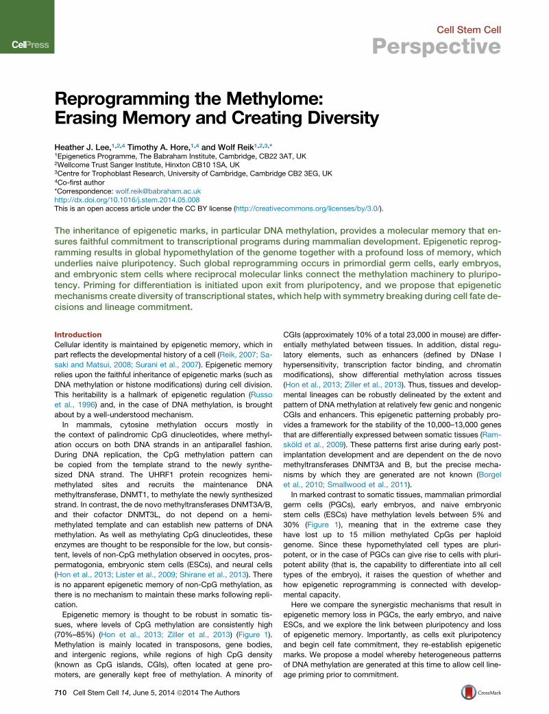
Figure 2. Kinetics of Genome-wideDemethylation in Early Embryos, PGCs, andESCsImpairment of methylation establishment and mainte-nance contributes to genome-wide demethylationin vivo (early embryo and PGCs) and in culture (naiveESCs), as do the oxidation and base excision repairpathways. Approximate levels of 5-methylcytosine(5mC) and 5-hydroxymethylcytosine (5hmC) are repre-sented by blue and red lines, respectively. Dashed bluelines indicate the expected level of 5mC if demethylationwas caused solely by complete inactivation of mainte-nance methylation.
Cell Stem Cell
Perspective
is also implicated in PGC demethylation (Hajkova et al., 2010),
and deficiency of the deaminase AID results in 2- to 3-fold-higher
levels of methylation in PGCs at E13.5 (Popp et al., 2010). How-
ever, the time and substrate of action, as well as the genomic
targets of AID, are unknown. The glycosylase TDG, which can
excise 5fC and 5caC as well as deaminated 5mC, is not ex-
pressed during the second window of demethylation (Kagiwada
et al., 2013). Thus, while the BER pathway has been implicated in
PGC demethylation, it is not clear precisely which components
are involved and to what extent. Elucidating the combinatorial
action of various factors during the demethylation process will
need detailed studies analyzing where these factors are targeted
in the genome andwhich downstream pathways resolve specific
base modifications.
Despite the magnitude of PGC reprogramming, there are
some sequences in the genome that are resistant to demethyla-
tion. These recalcitrant sequences include members of the intra-
cisternal A particle (IAP) retrotransposon family and adjacent
CGIs, as well as approximately 250 CGIs in the genome, which
are variably erased (VECs) (Guibert et al., 2012; Hackett et al.,
2013b; Seisenberger et al., 2012). This variable erasure may be
significant because it provides a potential mechanism for trans-
mission of epigenetic traits acquired in one generation to the
next, including metabolic phenotypes recently observed in ani-
mal models (Ferguson-Smith and Patti, 2011). The large-scale
erasure of epigenetic marks in the germline may explain why
transgenerational epigenetic inheritance in mammals is rare.
Indeed, it is more common in plants that do not undergo
genome-wide epigenetic reprogramming in germ cells (Bond
and Baulcombe, 2014).
Early Embryo
Another major wave of demethylation in themammalian germline
occurs in the early embryo, and while the magnitude of
methylation loss might not be as large as in PGCs, it is perhaps
more dramatic due to the precipitous loss of DNA methylation
specifically in the male pronucleus (Kobayashi et al., 2012;
712 Cell Stem Cell 14, June 5, 2014 ª2014 The Authors
Reik et al., 2001; Smith et al., 2012; Wang
et al., 2014). Here, highly methylated sperm-
derived DNA is rapidly and actively demethy-
lated before and during the first S phase in
the zygote, making it a definitive example of
large-scale active demethylation (Figure 2). A
number of factors have been implicated in
this rapid demethylation, most prominently
hydroxylation by TET3. Unlike TET1 and
TET2, TET3 is highly expressed in the zygote (Gu et al., 2011; Iq-
bal et al., 2011; Wossidlo et al., 2011) and is required for most of
the 5hmC that appears in the paternal genome upon fertilization,
and for a proportion of its demethylation. Until recently, it was
thought that only the paternal genome undergoes active deme-
thylation in the early embryo; however, the discovery of consid-
erable amounts of 5hmC and 5fC in both pronuclei suggests that
it may also affect the maternal genome to some extent (Wang
et al., 2014). As TDG is apparently not involved in removing these
modifications, it is thought that they are lost during DNA replica-
tion (Inoue et al., 2011; Santos et al., 2013). Recent work has
shown that TET3-mediated hydroxylation and demethylation is
confined to S phase chromatin, while surprisingly the bulk of
the demethylation occurs prior to DNA synthesis (Santos et al.,
2013) (Figure 2, stages 1 and 2). AID was also shown to be
required for paternal demethylation, and initial deamination
may be resolved by the UNG2 glycosylase rather than TDG,
opening up the intriguing possibility that cytosine deamination
and long-patch BER are involved in demethylation (Santos
et al., 2013). Unravelling the targets of these active demethyla-
tion pathways in the zygote and to what extent they synergize
or operate in parallel will require detailed genome-wide profiling
coupled with disruption of specific pathways.
As in PGCs, inactivation of themaintenancemethyltransferase
and de novo methylation system is thought to play a role in de-
methylation in preimplantation embryos following the zygote
stage (Carlson et al., 1992; Hirasawa and Sasaki, 2009; Oda
et al., 2013). UHRF1 protein is highly abundant in the zygote
but is excluded from the nucleus together with DNMT1, resulting
in passive demethylation of the maternal (and paternal) genome
in the early embryo such that by E3.5 methylation in the blasto-
cyst reaches a global methylation level of �25% (Figure 1) (Ko-
bayashi et al., 2012; Smallwood et al., 2011). From basal levels
in the early blastocyst, methylation slightly increases in the
ICM (E3.5) and the placenta (Hon et al., 2013; Smith et al.,
2012). As in migratory PGCs, some regions such as IAPs, and
Cell Stem Cell
Perspective
DMRs in imprinted genes, are protected from both active and
passive demethylation in the early embryo. The mechanism by
which DMRs are protected is thought to involve binding by pro-
teins such as STELLA, ZFP57, and KAP1 and recruitment of the
low level of nuclear DNMTs and UHRF1 (Li et al., 2008b; Naka-
mura et al., 2007, 2012; Quenneville et al., 2011).
An outstanding question in the field is the extent to which DNA
demethylation contributes to the activation of the early embryo
transcriptional network and acquisition of totipotency (Ishiuchi
and Torres-Padilla, 2013). A large number of retrotransposon
sequences are expressed in the early embryo (Evsikov et al.,
2004; Kigami et al., 2003; Peaston et al., 2004). Expression of
some of these repetitive sequences may be required for zygotic
genome activation—inhibition of both endogenous retroviral ele-
ments (ERVs) and LINE1 elements can impair developmental
competence of embryos (Beraldi et al., 2006; Kigami et al.,
2003). Moreover, around 300 early embryo genes are activated
by the LTRs of the MERV-L class of endogenous retroviruses
through the production of chimeric transcripts (Kigami et al.,
2003; Macfarlan et al., 2012), directly implicating activation of
ERVs and establishment of the early embryo transcriptional
network. Along with the rest of the genome, these elements
are subjected to rapid DNA demethylation in the early embryo
that coincides with their activation (Smith et al., 2012). Recently,
a rare population of ESCs that express markers of the early em-
bryo was identified, and as these cells can contribute to the
extraembryonic lineages, they are considered to have totipotent
features (Macfarlan et al., 2012; Morgani et al., 2013). Relative to
other ESCs, this subpopulation has reduced methylation at
MERV-L repetitive sequences, as in the early embryo. Despite
this, complete removal of DNA methylation from mouse ESCs
apparently does not stimulate expression of ERV elements or
the transcriptional network of the early embryo (Karimi et al.,
2011). Thus, the extent to which the totipotent state is dependent
upon DNA demethylation, either in the context of ERVs or the
transcriptional network of the early embryo in general, is
currently unclear.
Naive ESCs
While the link between pluripotent cell types and genome-wide
methylation is apparent in vivo (as witnessed by strikingly similar
methylation levels of E9.5 PGCs and ICM), it was initially puzzling
that ESCs conventionally grown in serum containing media ex-
hibited high CpGmethylation levels (�80%) characteristic of so-
matic cells, in contrast to the hypomethylated ICM from which
they are derived (Figure 1). This paradox was recently resolved;
by blocking the prodifferentiation signal that serum-grown ESCs
receive (by inhibiting ERK1/2 and GSK3b signaling with two
small molecule inhibitors [Ying et al., 2008]), genome-wide de-
methylation was induced to the same extent as in preimplanta-
tion embryos (�30%) (Ficz et al., 2013; Habibi et al., 2013). In
addition to achieving striking demethylation, use of these two in-
hibitors (commonly known as ‘‘2i’’) enabled derivation of bona
fide naive ESCs from previously recalcitrant mouse strains (Kiyo-
nari et al., 2010; Nichols et al., 2009), rats (Buehr et al., 2008; Li
et al., 2008a), and even humans (Chan et al., 2013; Gafni et al.,
2013). Accordingly, 2i ESCs are now considered the best cell
culture representation of in vivo pluripotent stem cells.
In contrast to serum-grown ESCs, naive ESCs grown in 2i have
low levels of DNMT3A/B and their targeting factor DNMT3L,
suggesting a mechanistic explanation for their low methylation
levels (Ficz et al., 2013; Habibi et al., 2013; Leitch et al., 2013).
While knockdown of the de novo methyltransferases in serum-
cultured ESCs does not immediately affect many regions thus
far analyzed, LINE1 elements undergo demethylationwith similar
kinetics to 2i treatment following DNMT3A/B ablation (Ficz et al.,
2013). These elements (and some single-copy loci) were previ-
ously shown to require continuous de novo methylation in order
to maintain high levels of methylation in serum-grown ESCs
(Arand et al., 2012), indicating that DNA methylation mainte-
nance is inefficient at these loci. Thus, DNMT3A/B repression
in 2i is sufficient to cause demethylation of certain genomic re-
gions in these cells.
Demethylation in 2i occurs with similar kinetics to that of PGCs
and early embryos (Figure 2), but surprisingly without global
alteration of UHRF1 and DNMT1 expression (Ficz et al., 2013;
Habibi et al., 2013). TET2 is upregulated by 2i treatment and
hydroxylation is induced. TET1 is targeted preferentially to CGIs
while TET2 is enriched at gene bodies (Huang et al., 2014), where
more substantial demethylation is observed in 2i. The extent of
demethylation can be enhanced by the addition of Vitamin C to
2i culture medium, which is a cofactor for activity of the TET en-
zymes (Blaschke et al., 2013). Interestingly, this demethylation
is focused on specific sequences including germline-specific
genes. Hence a significant mode of demethylation may be
erosion of methylation patterns by hydroxylation and failure to
repair this erosion by de novo methylation after replication.
The similarity of demethylation mechanisms between migra-
tory PGCs, preimplantation embryos, and 2i ESCs extends to
resistance to methylation erasure of IAP retrotransposons and
DMRs in imprinted genes. However, with prolonged culture of
ESCs in 2i, some erosion of DMR methylation does occur (Ficz
et al., 2013; Hackett et al., 2013a), perhaps in part because of
the consistently elevated levels of PRDM14. In that respect, pro-
longed 2i treatment may mimic, to some extent, the transition of
migratory to gonadal PGCs.
In summary, genome-wide demethylation is achieved in naive
ESCs by a combination of disabling the de novo methylation
machinery and increased hydroxylation, which may occur in
the context of a partially impaired maintenance methylation sys-
tem (Figure 2). Further study is required to elucidate the exact
contribution of these different mechanisms at distinct genomic
sites and how this relates to what is known about the removal
of epigenetic memory in in vivo systems.
Reciprocal Links between Loss of Epigenetic Memoryand PluripotencyLack of epigenetic memory is a common characteristic of plurip-
otent cell types and their precursors, including PGCs, ESCs, and
induced pluripotent stem cells (iPSCs). This is thought to ensure
that future differentiation decisions are not affected by events in
the past. In support of this idea, inefficient methylation erasure at
imprinted loci has been associated with a spectrum of develop-
mental abnormalities in the progeny of Tet1-deleted male mice
(Yamaguchi et al., 2013b). Insights into the mechanistic links
between reprogramming and pluripotency are now beginning
to emerge.
All experimental reprogramming techniques, including so-
matic cell nuclear transfer (SCNT), cell fusion, and iPSC
Cell Stem Cell 14, June 5, 2014 ª2014 The Authors 713

Cell Stem Cell
Perspective
reprogramming, involve demethylation of the genome that ap-
pears to be crucial for successfully achieving pluripotency
(Apostolou and Hochedlinger, 2013; Pasque et al., 2011; Theu-
nissen and Jaenisch, 2014 this issue of Cell Stem Cell). Further-
more, ESCs that lack all three methyltransferases (and are
devoid of virtually all methylation) are highly resistant to differen-
tiation, spontaneously reverting to pluripotency (Schmidt et al.,
2012). Full expression of the pluripotency network is observed
in both migratory PGCs and ICM cells, showing that a genomic
methylation level of 30% or less is characteristically associated
with the naive pluripotent state (Figure 1). This appears to be
true for naive ESCs derived from mice (Ficz et al., 2013) and
may be useful as a diagnostic marker for naive cells from other
species (Chan et al., 2013; Gafni et al., 2013). Expression of
key pluripotency factors is associated with demethylation of
these loci in PGCs, in early embryos, and during experimental re-
programming (Apostolou and Hochedlinger, 2013; Hochedlinger
and Plath, 2009). However, global demethylation in PGCs is not
associated with promiscuous transcription (Seisenberger et al.,
2012), nor is promoter demethylation in 2i associated with tran-
scriptional activation of demethylated genes (Ficz et al., 2013;
Habibi et al., 2013). Thus, while global DNA demethylation is
required for activation of the pluripotency network, pluripotent
cells exhibit an uncoupling of DNA methylation and transcrip-
tional regulation. Interestingly, expression of the pluripotency
network declines in gonadal PGCs (E13.5), which show further
demethylation to less than 10% (Seisenberger et al., 2012).
Perhaps extreme demethylation is detrimental since it may
lead to genome instability especially during cell division (methyl-
ation of major satellites, for example, appears to be needed for
proper chromosome segregation) (Smith and Meissner, 2013;
Xu et al., 1999).
Just as DNA methylation levels potentially influence the
expression of the pluripotency network, the pluripotency
network can direct the machinery of epigenetic reprogramming.
TET1 and TET2 are directly linked to the pluripotency network by
physical interactions with NANOG (Costa et al., 2013) and
PRDM14 (Okashita et al., 2014). TET1 andNANOGhave overlap-
ping patterns of DNA binding, and NANOG is required to recruit
TET1 to a subset of these common loci (Costa et al., 2013).
Furthermore, NANOG and TET1 act synergistically during the
reprogramming of neural stem cells to iPSCs (Costa et al.,
2013), and TET1 can replace OCT4 in conventional fibroblast
reprogramming (Gao et al., 2013). Similarly, PRDM14 recruits
TET1/2 to target loci and TET1/2 enhance PRDM14-induced
DNA demethylation in ESCs (Okashita et al., 2014). DNMT3A/
B/L are negatively regulated by PRDM14 (Hackett et al.,
2013a; Nakaki et al., 2013; Okashita et al., 2014; Yamaji et al.,
2013), in part via binding of PRDM14 to an upstream enhancer
of Dnmt3b (Ficz et al., 2013). NANOG also binds this locus and
may have similar effects on Dnmt3b expression. As well as the
de novo methyltransferases, UHRF1 appears to be suppressed
by PRDM14 in PGCs (Grabole et al., 2013; Magnusdottir et al.,
2013). These molecular interactions conspire to ensure that
DNA methylation is maintained at low levels in pluripotent cells.
In summary, the pluripotent ground state is maintained by
direct interactions between the transcriptional network and the
DNA methylation machinery. Cross-regulatory mechanisms
ensure that robust expression of pluripotency factors is accom-
714 Cell Stem Cell 14, June 5, 2014 ª2014 The Authors
panied by stable hypomethylation of the genome, yielding a
pluripotent ground state with little epigenetic memory.
Creating Epigenetic Diversity at the Exit fromPluripotencyAs cells exit pluripotency and begin to differentiate, new epige-
netic memories are formed to define cellular identity and restrict
lineage choices. The exit from pluripotency is characterized by
a steep decline in TET1/2 levels and an increase in DNMT3A/B
enzymes together with re-engagement of DNMT1 at replication
foci (Oda et al., 2013). During this transition, cells appear to
pass through an intermediate state of epigenetic priming that is
characterized by high levels of both de novo methyltransferases
and TET enzymes. For example, cells expressing both DNMT3B
and TET1 are present in the ICM of late blastocysts (Ficz et al.,
2013), and the E6.5 epiblast displays high expression of
DNMT3A/B and of TET1 (Seisenberger et al., 2012). Interestingly,
serum ESCs are similar to the E6.5 epiblast in the pattern and
extent of DNA methylation (Ficz et al., 2013; Seisenberger et al.,
2012) (Figure 1), and ESCs grown in serum conditions also ex-
press high levels of DNMT3A/B/L and of TET1/2, making them
a useful model for the relatively transient primed cell population
in vivo. These cells also display remarkable methylome plasticity
that is highly sensitive to growth conditions. For example, the
methylation status of CGIs even varies with cell passage number
(Booth et al., 2012). We speculate that rapid turnover of DNA
methylation may generate epigenetic heterogeneity in primed
ESCs and in the epiblast and that this heterogeneity may
contribute to cell fate decisions by allowing diversification prior
to lineage commitment. Consistent with this hypothesis, the
number of low-methylated regions (LMRs, an indirect indicator
of heterogeneous methylation across a cell population) de-
creases as mouse ESCs differentiate to neural progenitor cells
(Stadler et al., 2011). However, a decrease in the number of
LMRs was not observed in a similar study of human ESC dif-
ferentiation (Xie et al., 2013). Further studies utilizing single-cell
approaches are necessary to resolve these discrepancies by
directly analyzing cell-to-cell variation in DNA methylation.
Heterogeneity at the level of transcription has already been
linked to the propensity of serum ESCs to differentiate. Reporter
cell lines have shown that key pluripotency factors such as
NANOG, STELLA, and REX1 are heterogeneously and dynami-
cally expressed in these cells (Chambers et al., 2007; Hayashi
et al., 2008; Toyooka et al., 2008). For example, while NANOG-
low and -high cells are interchangeable, the NANOG-low popu-
lation has an increased propensity to differentiate (Chambers
et al., 2007) and elevated expression of differentiation markers
(Kalmar et al., 2009; Singh et al., 2007). More recent studies
have employed computational approaches, single-cell analyses,
and live-cell imaging to demonstrate that serum ESCs are a
metastable population and that cells switch between transcrip-
tional states in a stochastic manner (Abranches et al., 2013;
Chia and Ng, 2012; Kalmar et al., 2009; MacArthur et al., 2012;
Mallanna and Rizzino, 2012).
Emerging evidence indicates that epigenetic mechanisms
may contribute to this metastable state. One study has found
that STELLA-low and -high cells have different chromatin modi-
fications at the Stella promoter, and a slight increase in DNA
methylation was observed in a subset of STELLA-low cells

A B D F
C E G
Figure 3. Potential Sources of DNA Methylation Heterogeneity(A) Differential expression of TET or DNMT3 enzymes between cells would lead to global changes in DNA methylation levels.(B andC) Differential recruitment of DNMT3 (B) or TET (C) enzymes at certain loci could generate cells with distinct patterns of DNAmethylation. In the case of TET(C) hemi-5hmC would be an intermediate to loss of DNA methylation since DNMT1 does not maintain this mark.(D and E) Strand-specific effects of DNMT3 (D) or TET (E) enzymes could also produce daughter cells with distinct methylation patterns. In each case, hemi-modified DNA would be a transitional state.(F) Erasure of inherited methylation patterns (e.g., removal of oocyte derived methylation in the ICM) could also be inefficient and stochastic, generating sistercells with distinct patterns of inherited DNA methylation.(G) Inefficient maintenance of DNA methylation could also produce DNA methylation heterogeneity via hemimethylated intermediaries.
Cell Stem Cell
Perspective
(Hayashi et al., 2008). We have also reported differences in 5mC
and 5hmC between NANOG-low and -high cells, with increased
expression of TET1/2 being associated with increased 5hmC in
NANOG-high cells (Ficz et al., 2013). In line with these findings,
it has recently been shown that transcription patterns between
daughter cells differ depending on the methylation state. The
hypomethylated state of ESCs grown in 2i is associated with
greater transcriptional similarity between daughter cells than
for ESCs grown in serum. Furthermore, cells that are deficient
for themethyltransferases (TKO cells) have daughters with highly
similar transcriptional patterns despite being in serum culture
conditions (Jasnos et al., 2013). Generally, DNA methylation is
thought to act downstream of transcriptional changes during
cell fate decisions, but this fascinating result suggests that
DNA methylation heterogeneity may also be able to generate
transcriptional diversity in certain contexts. In support of this
idea, a study of DNMT3A null hematopoietic stem cells (HSCs)
has suggested that DNA methylation dynamics can direct tran-
scriptional changes and lineage choice in response to differenti-
ation stimuli (Challen et al., 2012). Ultimately, transcriptional and
epigenetic heterogeneity are likely to be tightly linked and cross-
regulatory, such that stochastic differences at either level can
generate cell diversity.
DNA methylation heterogeneity may be generated by several
mechanisms (Figure 3). Stochastic fluctuations in the expression
of TET or DNMT3 enzymes could generate differences in DNA
methylation between sister cells. Differential targeting of these
enzymes could also produce cells with variable epigeneticmarks
at the same sequence. Alternatively, strand-specific effects of
DNMT3 or TET enzymes could introduce asymmetric DNA
modifications that would yield daughter cells with distinct
methylation patterns. Differences in the efficiency of methylation
maintenance may also lead to DNA methylation heterogeneity
within a cell population, and incomplete erasure of inherited
methylation patterns (e.g., oocyte-derived methylated regions)
might be an additional source of epigenetic heterogeneity.
The precise patterns and kinetics of such events across cell pop-
ulations will depend on the balance between expression of
DNMT3 and TET enzymes, as these genes are themselves het-
erogeneously expressed in ESCs (Ficz et al., 2013). Rapid turn-
over of DNAmethylation through 5hmC is also likely to contribute
to epigenetic heterogeneity as CGIs with variable methylation in
ESCs are also enriched for 5hmC (Booth et al., 2012). Loss of
DNA methylation through 5hmC will occur via hemi-methylated
intermediates since there is no known mechanism for 5hmC
maintenance (Figure 3). Consistently, 5hmC has been linked to
Cell Stem Cell 14, June 5, 2014 ª2014 The Authors 715
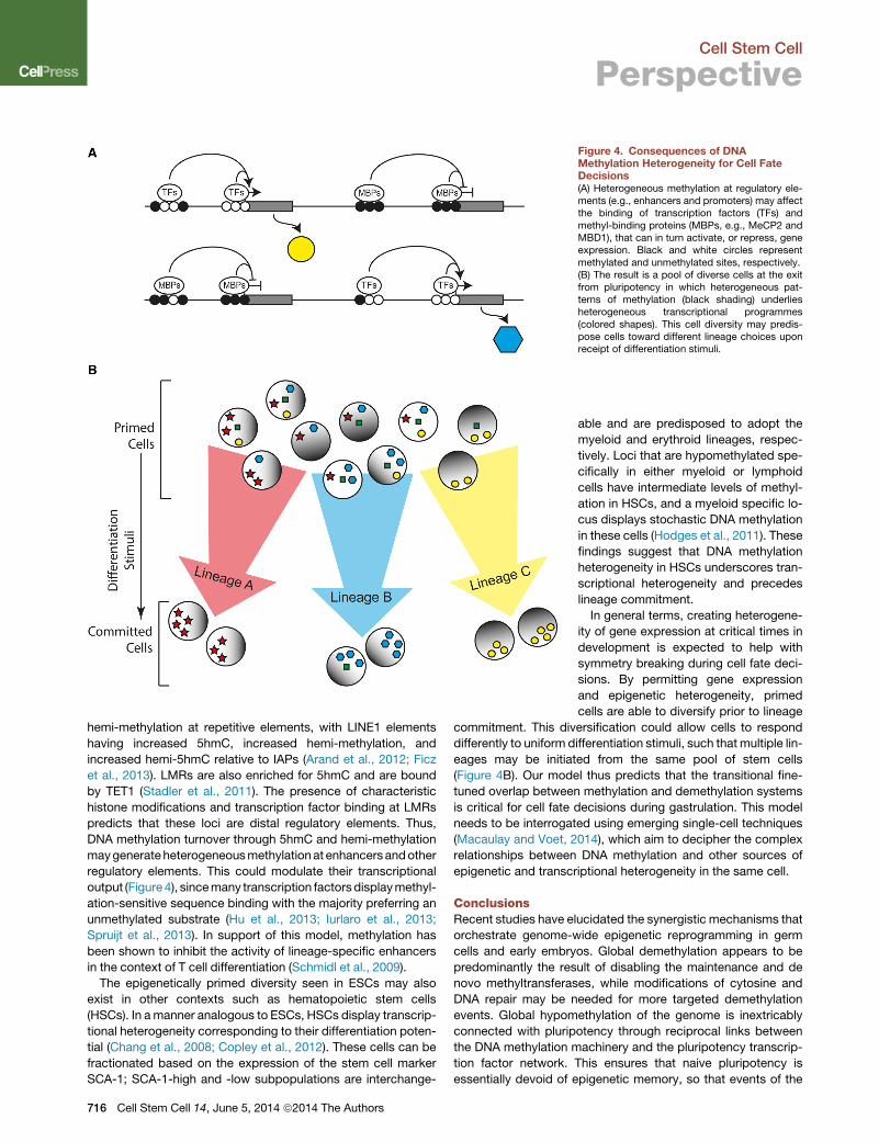
Figure 4. Consequences of DNAMethylation Heterogeneity for Cell FateDecisions(A) Heterogeneous methylation at regulatory ele-ments (e.g., enhancers and promoters) may affectthe binding of transcription factors (TFs) andmethyl-binding proteins (MBPs, e.g., MeCP2 andMBD1), that can in turn activate, or repress, geneexpression. Black and white circles representmethylated and unmethylated sites, respectively.(B) The result is a pool of diverse cells at the exitfrom pluripotency in which heterogeneous pat-terns of methylation (black shading) underliesheterogeneous transcriptional programmes(colored shapes). This cell diversity may predis-pose cells toward different lineage choices uponreceipt of differentiation stimuli.
Cell Stem Cell
Perspective
hemi-methylation at repetitive elements, with LINE1 elements
having increased 5hmC, increased hemi-methylation, and
increased hemi-5hmC relative to IAPs (Arand et al., 2012; Ficz
et al., 2013). LMRs are also enriched for 5hmC and are bound
by TET1 (Stadler et al., 2011). The presence of characteristic
histone modifications and transcription factor binding at LMRs
predicts that these loci are distal regulatory elements. Thus,
DNA methylation turnover through 5hmC and hemi-methylation
maygenerateheterogeneousmethylationat enhancersandother
regulatory elements. This could modulate their transcriptional
output (Figure4), sincemany transcription factorsdisplaymethyl-
ation-sensitive sequence binding with the majority preferring an
unmethylated substrate (Hu et al., 2013; Iurlaro et al., 2013;
Spruijt et al., 2013). In support of this model, methylation has
been shown to inhibit the activity of lineage-specific enhancers
in the context of T cell differentiation (Schmidl et al., 2009).
The epigenetically primed diversity seen in ESCs may also
exist in other contexts such as hematopoietic stem cells
(HSCs). In amanner analogous to ESCs, HSCs display transcrip-
tional heterogeneity corresponding to their differentiation poten-
tial (Chang et al., 2008; Copley et al., 2012). These cells can be
fractionated based on the expression of the stem cell marker
SCA-1; SCA-1-high and -low subpopulations are interchange-
716 Cell Stem Cell 14, June 5, 2014 ª2014 The Authors
able and are predisposed to adopt the
myeloid and erythroid lineages, respec-
tively. Loci that are hypomethylated spe-
cifically in either myeloid or lymphoid
cells have intermediate levels of methyl-
ation in HSCs, and a myeloid specific lo-
cus displays stochastic DNA methylation
in these cells (Hodges et al., 2011). These
findings suggest that DNA methylation
heterogeneity in HSCs underscores tran-
scriptional heterogeneity and precedes
lineage commitment.
In general terms, creating heterogene-
ity of gene expression at critical times in
development is expected to help with
symmetry breaking during cell fate deci-
sions. By permitting gene expression
and epigenetic heterogeneity, primed
cells are able to diversify prior to lineage
commitment. This diversification could allow cells to respond
differently to uniform differentiation stimuli, such that multiple lin-
eages may be initiated from the same pool of stem cells
(Figure 4B). Our model thus predicts that the transitional fine-
tuned overlap between methylation and demethylation systems
is critical for cell fate decisions during gastrulation. This model
needs to be interrogated using emerging single-cell techniques
(Macaulay and Voet, 2014), which aim to decipher the complex
relationships between DNA methylation and other sources of
epigenetic and transcriptional heterogeneity in the same cell.
ConclusionsRecent studies have elucidated the synergistic mechanisms that
orchestrate genome-wide epigenetic reprogramming in germ
cells and early embryos. Global demethylation appears to be
predominantly the result of disabling the maintenance and de
novo methyltransferases, while modifications of cytosine and
DNA repair may be needed for more targeted demethylation
events. Global hypomethylation of the genome is inextricably
connected with pluripotency through reciprocal links between
the DNA methylation machinery and the pluripotency transcrip-
tion factor network. This ensures that naive pluripotency is
essentially devoid of epigenetic memory, so that events of the

Cell Stem Cell
Perspective
past do not influence future differentiation decisions. Epigenetic
memory is re-engaged at the exit from pluripotency with robust
expression of the de novo and maintenance methylation sys-
tems. We propose that differentiating cells pass through a tran-
sient epigenetically primed state characterized by high levels of
TET and DNMT enzymes, and heterogeneous patterns of DNA
methylation. Notably, primed ESCs (grown in serum) express
high levels of methyltransferases and of TET1 and 2, and similar
cells appear to exist in the ICM in vivo. Epigenetic heterogeneity
in the ICM and epiblast may aid cell fate decisions by allowing
diversification prior to lineage commitment.
AUTHOR CONTRIBUTIONS
H.J.L., T.A.H., and W.R. wrote the manuscript and prepared figures.
ACKNOWLEDGMENTS
The authors would like to thank all members of the Reik lab for helpful discus-sions, particularly Julian Peat andMelanie Eckersley-Maslin for critical readingof the manuscript. We also thank Sebastien Smallwood for assistance withcompiling data for Figure 1. Funding from the following organizations is greatlyappreciated; the EU Blueprint Epigenome Consortium (H.L. andW.R.), the Hu-man Frontiers Science Program (T.H.), the EU EpiGeneSys Network (T.H. andW.R.), The Wellcome Trust (W.R.), and the BBSRC (W.R.). W.R. acts as aconsultant for Cambridge Epigenetix Limited.
REFERENCES
Abranches, E., Bekman, E., and Henrique, D. (2013). Generation and charac-terization of a novel mouse embryonic stem cell line with a dynamic reporter ofNanog expression. PLoS ONE 8, e59928.
Apostolou, E., and Hochedlinger, K. (2013). Chromatin dynamics duringcellular reprogramming. Nature 502, 462–471.
Arand, J., Spieler, D., Karius, T., Branco, M.R., Meilinger, D., Meissner, A.,Jenuwein, T., Xu, G., Leonhardt, H., Wolf, V., and Walter, J. (2012). In vivocontrol of CpG and non-CpG DNA methylation by DNA methyltransferases.PLoS Genet. 8, e1002750.
Beraldi, R., Pittoggi, C., Sciamanna, I., Mattei, E., and Spadafora, C. (2006).Expression of LINE-1 retroposons is essential for murine preimplantationdevelopment. Mol. Reprod. Dev. 73, 279–287.
Blaschke, K., Ebata, K.T., Karimi, M.M., Zepeda-Martınez, J.A., Goyal, P.,Mahapatra, S., Tam, A., Laird, D.J., Hirst, M., Rao, A., et al. (2013). VitaminC induces Tet-dependent DNA demethylation and a blastocyst-like state inES cells. Nature 500, 222–226.
Bond, D.M., and Baulcombe, D.C. (2014). Small RNAs and heritable epigeneticvariation in plants. Trends Cell Biol. 24, 100–107.
Booth, M.J., Branco, M.R., Ficz, G., Oxley, D., Krueger, F., Reik, W., andBalasubramanian, S. (2012). Quantitative sequencing of 5-methylcytosineand 5-hydroxymethylcytosine at single-base resolution. Science 336,934–937.
Borgel, J., Guibert, S., Li, Y., Chiba, H., Schubeler, D., Sasaki, H., Forne, T.,and Weber, M. (2010). Targets and dynamics of promoter DNA methylationduring early mouse development. Nat. Genet. 42, 1093–1100.
Branco, M.R., Ficz, G., and Reik, W. (2012). Uncovering the role of 5-hydrox-ymethylcytosine in the epigenome. Nat. Rev. Genet. 13, 7–13.
Buehr, M., Meek, S., Blair, K., Yang, J., Ure, J., Silva, J., McLay, R., Hall, J.,Ying, Q.L., and Smith, A. (2008). Capture of authentic embryonic stem cellsfrom rat blastocysts. Cell 135, 1287–1298.
Carlson, L.L., Page, A.W., and Bestor, T.H. (1992). Properties and localizationof DNA methyltransferase in preimplantation mouse embryos: implications forgenomic imprinting. Genes Dev. 6 (12B), 2536–2541.
Challen, G.A., Sun, D., Jeong, M., Luo, M., Jelinek, J., Berg, J.S., Bock, C.,Vasanthakumar, A., Gu, H., Xi, Y., et al. (2012). Dnmt3a is essential for hemato-poietic stem cell differentiation. Nat. Genet. 44, 23–31.
Chambers, I., Silva, J., Colby, D., Nichols, J., Nijmeijer, B., Robertson, M.,Vrana, J., Jones, K., Grotewold, L., and Smith, A. (2007). Nanog safeguardspluripotency and mediates germline development. Nature 450, 1230–1234.
Chan, Y.S., Goke, J., Ng, J.H., Lu, X., Gonzales, K.A., Tan, C.P., Tng, W.Q.,Hong, Z.Z., Lim, Y.S., and Ng, H.H. (2013). Induction of a human pluripotentstate with distinct regulatory circuitry that resembles preimplantation epiblast.Cell Stem Cell 13, 663–675.
Chang, H.H., Hemberg, M., Barahona, M., Ingber, D.E., and Huang, S. (2008).Transcriptome-wide noise controls lineage choice in mammalian progenitorcells. Nature 453, 544–547.
Chia, N.-Y., and Ng, H.-H. (2012). Stem cell genome-to-systems biology.WileyInterdiscip Rev Syst Biol Med 4, 39–49.
Copley, M.R., Beer, P.A., and Eaves, C.J. (2012). Hematopoietic stem cellheterogeneity takes center stage. Cell Stem Cell 10, 690–697.
Costa, Y., Ding, J., Theunissen, T.W., Faiola, F., Hore, T.A., Shliaha, P.V.,Fidalgo, M., Saunders, A., Lawrence, M., Dietmann, S., et al. (2013).NANOG-dependent function of TET1 and TET2 in establishment of pluripo-tency. Nature 495, 370–374.
Esteve, P.O., Chin, H.G., Smallwood, A., Feehery, G.R., Gangisetty, O., Karpf,A.R., Carey, M.F., and Pradhan, S. (2006). Direct interaction between DNMT1and G9a coordinates DNA and histone methylation during replication. GenesDev. 20, 3089–3103.
Evsikov, A.V., de Vries, W.N., Peaston, A.E., Radford, E.E., Fancher, K.S.,Chen, F.H., Blake, J.A., Bult, C.J., Latham, K.E., Solter, D., and Knowles,B.B. (2004). Systems biology of the 2-cell mouse embryo. Cytogenet. GenomeRes. 105, 240–250.
Ferguson-Smith, A.C., and Patti, M.E. (2011). You are what your dad ate. CellMetab. 13, 115–117.
Ficz, G., Hore, T.A., Santos, F., Lee, H.J., Dean, W., Arand, J., Krueger, F.,Oxley, D., Paul, Y.L., Walter, J., et al. (2013). FGF signaling inhibition inESCs drives rapid genome-wide demethylation to the epigenetic ground stateof pluripotency. Cell Stem Cell 13, 351–359.
Gafni, O., Weinberger, L., Mansour, A.A., Manor, Y.S., Chomsky, E., Ben-Yosef, D., Kalma, Y., Viukov, S., Maza, I., Zviran, A., et al. (2013). Derivationof novel human ground state naive pluripotent stem cells. Nature 504,282–286.
Gao, Y., Chen, J., Li, K., Wu, T., Huang, B., Liu, W., Kou, X., Zhang, Y., Huang,H., Jiang, Y., et al. (2013). Replacement of Oct4 by Tet1 during iPSC inductionreveals an important role of DNA methylation and hydroxymethylation inreprogramming. Cell Stem Cell 12, 453–469.
Grabole, N., Tischler, J., Hackett, J.A., Kim, S., Tang, F., Leitch, H.G., Magnus-dottir, E., and Surani, M.A. (2013). Prdm14 promotes germline fate and naivepluripotency by repressing FGF signalling and DNA methylation. EMBO Rep.14, 629–637.
Gu, T.-P., Guo, F., Yang, H.,Wu, H.-P., Xu, G.-F., Liu,W., Xie, Z.-G., Shi, L., He,X., Jin, S.G., et al. (2011). The role of Tet3 DNA dioxygenase in epigeneticreprogramming by oocytes. Nature 477, 606–610.
Guibert, S., Forne, T., and Weber, M. (2012). Global profiling of DNA methyl-ation erasure in mouse primordial germ cells. Genome Res. 22, 633–641.
Habibi, E., Brinkman, A.B., Arand, J., Kroeze, L.I., Kerstens, H.H., Matarese,F., Lepikhov, K., Gut, M., Brun-Heath, I., Hubner, N.C., et al. (2013). Whole-genome bisulfite sequencing of two distinct interconvertible DNA methylomesof mouse embryonic stem cells. Cell Stem Cell 13, 360–369.
Hackett, J.A., Dietmann, S., Murakami, K., Down, T.A., Leitch, H.G., andSurani, M.A. (2013a). Synergistic mechanisms of DNA demethylation duringtransition to ground-state pluripotency. Stem Cell Rev. 1, 518–531.
Hackett, J.A., Sengupta, R., Zylicz, J.J., Murakami, K., Lee, C., Down, T.A.,and Surani, M.A. (2013b). Germline DNA demethylation dynamics and imprinterasure through 5-hydroxymethylcytosine. Science 339, 448–452.
Cell Stem Cell 14, June 5, 2014 ª2014 The Authors 717

Cell Stem Cell
Perspective
Hajkova, P., Jeffries, S.J., Lee, C., Miller, N., Jackson, S.P., and Surani, M.A.(2010). Genome-wide reprogramming in the mouse germ line entails thebase excision repair pathway. Science 329, 78–82.
Hayashi, K., Lopes, S.M.C.S., Tang, F., and Surani, M.A. (2008). Dynamic equi-librium and heterogeneity of mouse pluripotent stem cells with distinct func-tional and epigenetic states. Cell Stem Cell 3, 391–401.
Hirasawa, R., and Sasaki, H. (2009). Dynamic transition of Dnmt3b expressionin mouse pre- and early post-implantation embryos. Gene Expr. Patterns 9,27–30.
Hochedlinger, K., and Plath, K. (2009). Epigenetic reprogramming and inducedpluripotency. Development 136, 509–523.
Hodges, E., Molaro, A., Dos Santos, C.O., Thekkat, P., Song, Q., Uren, P.J.,Park, J., Butler, J., Rafii, S., McCombie, W.R., et al. (2011). Directional DNAmethylation changes and complex intermediate states accompany lineagespecificity in the adult hematopoietic compartment. Mol. Cell 44, 17–28.
Hon, G.C., Rajagopal, N., Shen, Y., McCleary, D.F., Yue, F., Dang, M.D., andRen, B. (2013). Epigenetic memory at embryonic enhancers identified in DNAmethylation maps from adult mouse tissues. Nat. Genet. 45, 1198–1206.
Hu, S.,Wan, J., Su, Y., Song, Q., Zeng, Y., Nguyen, H.N., Shin, J., Cox, E., Rho,H.S., Woodard, C., et al. (2013). DNA methylation presents distinct bindingsites for human transcription factors. Elife 2, e00726.
Huang, Y., Chavez, L., Chang, X., Wang, X., Pastor, W.A., Kang, J., Zepeda-Martınez, J.A., Pape, U.J., Jacobsen, S.E., Peters, B., and Rao, A. (2014).Distinct roles of the methylcytosine oxidases Tet1 and Tet2 in mouse embry-onic stem cells. Proc. Natl. Acad. Sci. USA 111, 1361–1366.
Inoue, A., Shen, L., Dai, Q., He, C., and Zhang, Y. (2011). Generation and repli-cation-dependent dilution of 5fC and 5caC during mouse preimplantationdevelopment. Cell Res. 21, 1670–1676.
Iqbal, K., Jin, S.-G., Pfeifer, G.P., and Szabo, P.E. (2011). Reprogramming ofthe paternal genome upon fertilization involves genome-wide oxidation of 5-methylcytosine. Proc. Natl. Acad. Sci. USA 108, 3642–3647.
Ishiuchi, T., and Torres-Padilla, M.-E. (2013). Towards an understanding of theregulatory mechanisms of totipotency. Curr. Opin. Genet. Dev. 23, 512–518.
Iurlaro, M., Ficz, G., Oxley, D., Raiber, E.A., Bachman, M., Booth, M.J.,Andrews, S., Balasubramanian, S., and Reik, W. (2013). A screen for hydrox-ymethylcytosine and formylcytosine binding proteins suggests functions intranscription and chromatin regulation. Genome Biol. 14, R119.
Jasnos, L., Aksoy, F.B., Hersi, H.M.,Wantuch, S., and Sawado, T. (2013). Iden-tifying division symmetry of mouse embryonic stem cells: negative impact ofDNA methyltransferases on symmetric self-renewal. Stem Cell Rev. 1,360–369.
Kagiwada, S., Kurimoto, K., Hirota, T., Yamaji, M., and Saitou, M. (2013). Repli-cation-coupled passive DNA demethylation for the erasure of genome imprintsin mice. EMBO J. 32, 340–353.
Kalmar, T., Lim, C., Hayward, P., Munoz-Descalzo, S., Nichols, J., Garcia-Ojalvo, J., and Martinez Arias, A. (2009). Regulated fluctuations in Nanogexpression mediate cell fate decisions in embryonic stem cells. PLoS Biol. 7,e1000149.
Karimi, M.M., Goyal, P., Maksakova, I.A., Bilenky, M., Leung, D., Tang, J.X.,Shinkai, Y., Mager, D.L., Jones, S., Hirst, M., and Lorincz, M.C. (2011). DNAmethylation and SETDB1/H3K9me3 regulate predominantly distinct sets ofgenes, retroelements, and chimeric transcripts in mESCs. Cell Stem Cell 8,676–687.
Kigami, D., Minami, N., Takayama, H., and Imai, H. (2003). MuERV-L is one ofthe earliest transcribed genes in mouse one-cell embryos. Biol. Reprod. 68,651–654.
Kiyonari, H., Kaneko, M., Abe, S., and Aizawa, S. (2010). Three inhibitors ofFGF receptor, ERK, and GSK3 establishes germline-competent embryonicstem cells of C57BL/6Nmouse strain with high efficiency and stability. Genesis48, 317–327.
Kobayashi, H., Sakurai, T., Imai, M., Takahashi, N., Fukuda, A., Yayoi, O., Sato,S., Nakabayashi, K., Hata, K., Sotomaru, Y., et al. (2012). Contribution of intra-genic DNA methylation in mouse gametic DNA methylomes to establishoocyte-specific heritable marks. PLoS Genet. 8, e1002440.
718 Cell Stem Cell 14, June 5, 2014 ª2014 The Authors
Kobayashi, H., Sakurai, T., Miura, F., Imai, M., Mochiduki, K., Yanagisawa, E.,Sakashita, A., Wakai, T., Suzuki, Y., Ito, T., et al. (2013). High-resolution DNAmethylome analysis of primordial germ cells identifies gender-specific reprog-ramming in mice. Genome Res. 23, 616–627.
Kurimoto, K., Yabuta, Y., Ohinata, Y., Shigeta, M., Yamanaka, K., and Saitou,M. (2008). Complex genome-wide transcription dynamics orchestrated byBlimp1 for the specification of the germ cell lineage in mice. Genes Dev. 22,1617–1635.
Leitch, H.G., McEwen, K.R., Turp, A., Encheva, V., Carroll, T., Grabole, N.,Mansfield, W., Nashun, B., Knezovich, J.G., Smith, A., et al. (2013). Naivepluripotency is associated with global DNA hypomethylation. Nat. Struct.Mol. Biol. 20, 311–316.
Li, P., Tong, C., Mehrian-Shai, R., Jia, L., Wu, N., Yan, Y., Maxson, R.E.,Schulze, E.N., Song, H., Hsieh, C.L., et al. (2008a). Germline competentembryonic stem cells derived from rat blastocysts. Cell 135, 1299–1310.
Li, X., Ito, M., Zhou, F., Youngson, N., Zuo, X., Leder, P., and Ferguson-Smith,A.C. (2008b). A maternal-zygotic effect gene, Zfp57, maintains both maternaland paternal imprints. Dev. Cell 15, 547–557.
Lister, R., Pelizzola, M., Dowen, R.H., Hawkins, R.D., Hon, G., Tonti-Filippini,J., Nery, J.R., Lee, L., Ye, Z., Ngo, Q.M., et al. (2009). Human DNAmethylomesat base resolution show widespread epigenomic differences. Nature 462,315–322.
Liu, X., Gao, Q., Li, P., Zhao, Q., Zhang, J., Li, J., Koseki, H., and Wong, J.(2013). UHRF1 targets DNMT1 for DNAmethylation through cooperative bind-ing of hemi-methylated DNA and methylated H3K9. Nat Commun 4, 1563.
MacArthur, B.D., Sevilla, A., Lenz, M., Muller, F.-J., Schuldt, B.M., Schuppert,A.A., Ridden, S.J., Stumpf, P.S., Fidalgo, M., Ma’ayan, A., et al. (2012). Nanog-dependent feedback loops regulate murine embryonic stem cell hetero-geneity. Nat. Cell Biol. 14, 1139–1147.
Macaulay, I.C., and Voet, T. (2014). Single cell genomics: advances and futureperspectives. PLoS Genet. 10, e1004126.
Macfarlan, T.S., Gifford, W.D., Driscoll, S., Lettieri, K., Rowe, H.M., Bonanomi,D., Firth, A., Singer, O., Trono, D., and Pfaff, S.L. (2012). Embryonic stem cellpotency fluctuates with endogenous retrovirus activity. Nature 487, 57–63.
Magnusdottir, E., Dietmann, S., Murakami, K., Gunesdogan, U., Tang, F., Bao,S., Diamanti, E., Lao, K., Gottgens, B., and Azim Surani, M. (2013). A tripartitetranscription factor network regulates primordial germ cell specification inmice. Nat. Cell Biol. 15, 905–915.
Mallanna, S.K., and Rizzino, A. (2012). Systems biology provides new insightsinto the molecular mechanisms that control the fate of embryonic stem cells.J. Cell. Physiol. 227, 27–34.
Morgani, S.M., Canham, M.A., Nichols, J., Sharov, A.A., Migueles, R.P., Ko,M.S., and Brickman, J.M. (2013). Totipotent embryonic stem cells arise inground-state culture conditions. Cell Rep 3, 1945–1957.
Nakaki, F., Hayashi, K., Ohta, H., Kurimoto, K., Yabuta, Y., and Saitou, M.(2013). Induction of mouse germ-cell fate by transcription factors in vitro.Nature 501, 222–226.
Nakamura, T., Arai, Y., Umehara, H., Masuhara, M., Kimura, T., Taniguchi, H.,Sekimoto, T., Ikawa, M., Yoneda, Y., Okabe, M., et al. (2007). PGC7/Stellaprotects against DNA demethylation in early embryogenesis. Nat. Cell Biol.9, 64–71.
Nakamura, T., Liu, Y.J., Nakashima, H., Umehara, H., Inoue, K., Matoba, S.,Tachibana, M., Ogura, A., Shinkai, Y., and Nakano, T. (2012). PGC7 bindshistone H3K9me2 to protect against conversion of 5mC to 5hmC in earlyembryos. Nature 486, 415–419.
Nichols, J., Jones, K., Phillips, J.M., Newland, S.A., Roode, M., Mansfield, W.,Smith, A., and Cooke, A. (2009). Validated germline-competent embryonicstem cell lines from nonobese diabetic mice. Nat. Med. 15, 814–818.
Oda, M., Oxley, D., Dean, W., and Reik, W. (2013). Regulation of lineagespecific DNA hypomethylation inmouse trophectoderm. PLoSONE 8, e68846.
Ohno, R., Nakayama, M., Naruse, C., Okashita, N., Takano, O., Tachibana, M.,Asano, M., Saitou, M., and Seki, Y. (2013). A replication-dependent passivemechanism modulates DNA demethylation in mouse primordial germ cells.Development 140, 2892–2903.

Cell Stem Cell
Perspective
Okashita, N., Kumaki, Y., Ebi, K., Nishi, M., Okamoto, Y., Nakayama, M.,Hashimoto, S., Nakamura, T., Sugasawa, K., Kojima, N., et al. (2014).PRDM14 promotes active DNA demethylation through the ten-eleven translo-cation (TET)-mediated base excision repair pathway in embryonic stem cells.Development 141, 269–280.
Pasque, V., Jullien, J., Miyamoto, K., Halley-Stott, R.P., and Gurdon, J.B.(2011). Epigenetic factors influencing resistance to nuclear reprogramming.Trends Genet. 27, 516–525.
Peaston, A.E., Evsikov, A.V., Graber, J.H., de Vries, W.N., Holbrook, A.E.,Solter, D., and Knowles, B.B. (2004). Retrotransposons regulate host genesin mouse oocytes and preimplantation embryos. Dev. Cell 7, 597–606.
Popp, C., Dean, W., Feng, S., Cokus, S.J., Andrews, S., Pellegrini, M., Jacob-sen, S.E., and Reik, W. (2010). Genome-wide erasure of DNA methylation inmouse primordial germ cells is affected by AID deficiency. Nature 463,1101–1105.
Quenneville, S., Verde, G., Corsinotti, A., Kapopoulou, A., Jakobsson, J.,Offner, S., Baglivo, I., Pedone, P.V., Grimaldi, G., Riccio, A., and Trono, D.(2011). In embryonic stem cells, ZFP57/KAP1 recognize a methylated hexanu-cleotide to affect chromatin and DNAmethylation of imprinting control regions.Mol. Cell 44, 361–372.
Ramskold, D., Wang, E.T., Burge, C.B., and Sandberg, R. (2009). An abun-dance of ubiquitously expressed genes revealed by tissue transcriptomesequence data. PLoS Comput. Biol. 5, e1000598.
Reik, W. (2007). Stability and flexibility of epigenetic gene regulation inmammalian development. Nature 447, 425–432.
Reik, W., Dean, W., and Walter, J. (2001). Epigenetic reprogramming inmammalian development. Science 293, 1089–1093.
Russo, V.E., Martienssen, R.A., and Riggs, A.D. (1996). Epigenetic Mecha-nisms of Gene Regulation. (Cold Spring Harbor, NY: Cold Spring HarborLaboratory Press).
Santos, F., Peat, J., Burgess, H., Rada, C., Reik, W., and Dean, W. (2013).Active demethylation in mouse zygotes involves cytosine deamination andbase excision repair. Epigenetics Chromatin 6, 39.
Sasaki, H., and Matsui, Y. (2008). Epigenetic events in mammalian germ-celldevelopment: reprogramming and beyond. Nat. Rev. Genet. 9, 129–140.
Schmidl, C., Klug, M., Boeld, T.J., Andreesen, R., Hoffmann, P., Edinger, M.,and Rehli, M. (2009). Lineage-specific DNA methylation in T cells correlateswith histone methylation and enhancer activity. Genome Res. 19, 1165–1174.
Schmidt, C.S., Bultmann, S., Meilinger, D., Zacher, B., Tresch, A., Maier, K.C.,Peter, C., Martin, D.E., Leonhardt, H., and Spada, F. (2012). Global DNA hypo-methylation prevents consolidation of differentiation programs and allowsreversion to the embryonic stem cell state. PLoS ONE 7, e52629.
Seisenberger, S., Andrews, S., Krueger, F., Arand, J., Walter, J., Santos, F.,Popp, C., Thienpont, B., Dean, W., and Reik, W. (2012). The dynamics ofgenome-wide DNA methylation reprogramming in mouse primordial germcells. Mol. Cell 48, 849–862.
Seki, Y., Hayashi, K., Itoh, K., Mizugaki, M., Saitou, M., and Matsui, Y. (2005).Extensive and orderly reprogramming of genome-wide chromatin modifica-tions associated with specification and early development of germ cells inmice. Dev. Biol. 278, 440–458.
Shirane, K., Toh, H., Kobayashi, H., Miura, F., Chiba, H., Ito, T., Kono, T., andSasaki, H. (2013). Mouse oocyte methylomes at base resolution revealgenome-wide accumulation of non-CpG methylation and role of DNA methyl-transferases. PLoS Genet. 9, e1003439.
Singh, A.M., Hamazaki, T., Hankowski, K.E., and Terada, N. (2007). A hetero-geneous expression pattern for Nanog in embryonic stem cells. Stem Cells 25,2534–2542.
Smallwood, S.A., Tomizawa, S., Krueger, F., Ruf, N., Carli, N., Segonds-Pichon, A., Sato, S., Hata, K., Andrews, S.R., and Kelsey, G. (2011). Dynamic
CpG island methylation landscape in oocytes and preimplantation embryos.Nat. Genet. 43, 811–814.
Smith, Z.D., and Meissner, A. (2013). DNA methylation: roles in mammaliandevelopment. Nat. Rev. Genet. 14, 204–220.
Smith, Z.D., Chan, M.M., Mikkelsen, T.S., Gu, H., Gnirke, A., Regev, A., andMeissner, A. (2012). A unique regulatory phase of DNA methylation in the earlymammalian embryo. Nature 484, 339–344.
Spruijt, C.G., Gnerlich, F., Smits, A.H., Pfaffeneder, T., Jansen, P.W.T.C.,Bauer, C., Munzel, M., Wagner, M., Muller, M., Khan, F., et al. (2013). Dynamicreaders for 5-(hydroxy)methylcytosine and its oxidized derivatives. Cell 152,1146–1159.
Stadler, M.B., Murr, R., Burger, L., Ivanek, R., Lienert, F., Scholer, A., vanNimwegen, E., Wirbelauer, C., Oakeley, E.J., Gaidatzis, D., et al. (2011).DNA-binding factors shape the mouse methylome at distal regulatory regions.Nature 480, 490–495.
Surani, M.A., Hayashi, K., and Hajkova, P. (2007). Genetic and epigeneticregulators of pluripotency. Cell 128, 747–762.
Theunissen, T.W., and Jaenisch, R. (2014). Molecular control of inducedpluripotency. Cell Stem Cell 14this issue, 720–734.
Toyooka, Y., Shimosato, D., Murakami, K., Takahashi, K., and Niwa, H. (2008).Identification and characterization of subpopulations in undifferentiated EScell culture. Development 135, 909–918.
Vincent, J.J., Huang, Y., Chen, P.Y., Feng, S., Calvopina, J.H., Nee, K., Lee,S.A., Le, T., Yoon, A.J., Faull, K., et al. (2013). Stage-specific roles for tet1and tet2 in DNA demethylation in primordial germ cells. Cell Stem Cell 12,470–478.
Wang, L., Zhang, J., Duan, J., Gao, X., Zhu, W., Lu, X., Yang, L., Zhang, J., Li,G., Ci, W., et al. (2014). Programming and inheritance of parental DNA methyl-omes in mammals. Cell 157, 979–991.
Wossidlo, M., Nakamura, T., Lepikhov, K., Marques, C.J., Zakhartchenko, V.,Boiani, M., Arand, J., Nakano, T., Reik, W., and Walter, J. (2011). 5-Hydroxy-methylcytosine in the mammalian zygote is linked with epigenetic reprogram-ming. Nat Commun 2, 241.
Xie, W., Schultz, M.D., Lister, R., Hou, Z., Rajagopal, N., Ray, P., Whitaker,J.W., Tian, S., Hawkins, R.D., Leung, D., et al. (2013). Epigenomic analysisof multilineage differentiation of human embryonic stem cells. Cell 153,1134–1148.
Xu, G.-L., Bestor, T.H., Bourc’his, D., Hsieh, C.-L., Tommerup, N., Bugge, M.,Hulten, M., Qu, X., Russo, J.J., and Viegas-Pequignot, E. (1999). Chromosomeinstability and immunodeficiency syndrome caused by mutations in a DNAmethyltransferase gene. Nature 402, 187–191.
Yamaguchi, S., Hong, K., Liu, R., Inoue, A., Shen, L., Zhang, K., and Zhang, Y.(2013a). Dynamics of 5-methylcytosine and 5-hydroxymethylcytosine duringgerm cell reprogramming. Cell Res. 23, 329–339.
Yamaguchi, S., Shen, L., Liu, Y., Sendler, D., and Zhang, Y. (2013b). Role ofTet1 in erasure of genomic imprinting. Nature 504, 460–464.
Yamaji, M., Ueda, J., Hayashi, K., Ohta, H., Yabuta, Y., Kurimoto, K., Nakato,R., Yamada, Y., Shirahige, K., and Saitou, M. (2013). PRDM14 ensures naivepluripotency through dual regulation of signaling and epigenetic pathways inmouse embryonic stem cells. Cell Stem Cell 12, 368–382.
Ying, Q.L., Wray, J., Nichols, J., Batlle-Morera, L., Doble, B., Woodgett, J.,Cohen, P., and Smith, A. (2008). The ground state of embryonic stem cellself-renewal. Nature 453, 519–523.
Ziller, M.J., Gu, H., Muller, F., Donaghey, J., Tsai, L.T.Y., Kohlbacher, O., DeJager, P.L., Rosen, E.D., Bennett, D.A., Bernstein, B.E., et al. (2013). Chartinga dynamic DNA methylation landscape of the human genome. Nature 500,477–481.
Cell Stem Cell 14, June 5, 2014 ª2014 The Authors 719
Related Documents