REPORT DOCUMENTATION PAGE KLtMSCUUNUI HtlUMW TUUK rUMIVI IU I Mt MBUVt MUUrttSS. ntrum u«it 05 11/2010 t. KtKum i rrt Final Technical j. UMitstuvtntu 12/01/07-11/30/09 « . Ill LC MIXU aUIJIIILt Structural and Functional Mechanisms of Adaptations of WrbA in 1 •Atremophilic Organisms oa. UUNIUHOI rvuiviöt« DO. (JMMIMI rautvmtn FA9550-08-1-0013 DC. rmjurtMM ttcivitix i fMuiviotn D. «uinumai James G. Ferry DO. KHUJtUI NUMötM DC I H» tVUIVIOC« or. wunrvunni rautvmtr« /. rLm-umvumu urtuAniiz.Ai turn niAivitiDi AIMU Muutitaaitai Pennsylvania State University Ö. rtttrUMIVIIIMU UMUMIMIiMI IUIM ntrum rourvrntn D. aruixsurMMu iviuiviiunnvo MUCIMLT IMMMCIDI MNU AUUtttDDttDi IU. arUWDUMMVIunjIIU« D AUKUNTIVIIDI AFOSR/RSL 875 N.Randolph St. Suite 325 Arlington. VA 22203-1768 I. DfUNDUrVIVIUNII UM D KtKUHl njuiviBtmui Pffe\ raSß<\)/}4%7ö\in)ol', I Z. UID I MIBU I IUIV'A V AILABILI IT D I A I tlVItni I A (approved for public release; distribution is unlimited) IJ. DUrrLtlVICN I AMT NU I CD IH. MODI KAU I Properties of FMN-containing NADH:quinone oxidoreductascs (WrbA) from the hypcrthermophile Archaeoglobus fulgidus and the mesophile Escherichia coli were investigated to gain an understanding of the mechanisms by which proteins in extremophilic organisms adapt to high temperature. A model of the thermophilic enzyme was constructed based on the crystal Structure of the mesophilie counterpart to guide experiments. An electrochemical cell was designed and constructed to probe the redox properties of FMN. Only subtle differences in the midpoint potential were recorded for the two enzymes consistent with congruent physiological functions. Contrary to redox potentials, the two proteins showed differences in FMN binding. Strong coopcrativity in flavin binding for the mesophilie enzyme was dependent on the concentration of FMN whereas binding for the thermophilic enzyme was independent of ligand concentration. Comparison of enzyme activities between the two proteins with a variety of quinones suggested differences in the active site architecture accommodating electron acceptors. Properties of the mesophilie wild-type versus single r\t\* ...ii, ID. DUBJtUI I tMIVID thermophilic. flavoprotein. Archaeoglobus fulgidus, biosensor, biofucl cell ID. Dtuunn T ^LMDDIMUMI lum ur: D. ADD I KAU I C. I MID KAUfc i /. Liivn i M i luiM ur ADD I MAUI ID. NUtVIÖtM ur KAUtD i Da. NMtvit ur htaruixaiQLt rcuDui* James G. Ferry IDD. I tLtfMUNt NUMBCM 814 404 7740 3toUOVS(|o(r

Welcome message from author
This document is posted to help you gain knowledge. Please leave a comment to let me know what you think about it! Share it to your friends and learn new things together.
Transcript
REPORT DOCUMENTATION PAGE
KLtMSCUUNUI HtlUMW TUUK rUMIVI IU I Mt MBUVt MUUrttSS.
ntrum u«it
05 11/2010 t. KtKum i rrt
Final Technical j. UMitstuvtntu
12/01/07-11/30/09 «♦. Ill LC MIXU aUIJIIILt
Structural and Functional Mechanisms of Adaptations of WrbA in 1 •Atremophilic Organisms
oa. UUNIUHOI rvuiviöt«
DO. (JMMIMI rautvmtn
FA9550-08-1-0013
DC. rmjurtMM ttcivitix i fMuiviotn
D. «uinumai
James G. Ferry
DO. KHUJtUI NUMötM
DC I H» tVUIVIOC«
or. wunrvunni rautvmtr«
/. rLm-umvumu urtuAniiz.Ai turn niAivitiDi AIMU Muutitaaitai
Pennsylvania State University
Ö. rtttrUMIVIIIMU UMUMIMIiMI IUIM ntrum rourvrntn
D. aruixsurMMu iviuiviiunnvo MUCIMLT IMMMCIDI MNU AUUtttDDttDi IU. arUWDUMMVIunjIIU« D AUKUNTIVIIDI
AFOSR/RSL 875 N.Randolph St. Suite 325 Arlington. VA 22203-1768
I. DfUNDUrVIVIUNII UM D KtKUHl njuiviBtmui
Pffe\ raSß<\)/}4%7ö\in)ol', I Z. UID I MIBU I IUIV'A V AILABILI IT D I A I tlVItni I
A (approved for public release; distribution is unlimited)
IJ. DUrrLtlVICN I AMT NU I CD
IH. MODI KAU I
Properties of FMN-containing NADH:quinone oxidoreductascs (WrbA) from the hypcrthermophile Archaeoglobus fulgidus and the mesophile Escherichia coli were investigated to gain an understanding of the mechanisms by which proteins in extremophilic organisms adapt to high temperature. A model of the thermophilic enzyme was constructed based on the crystal Structure of the mesophilie counterpart to guide experiments. An electrochemical cell was designed and constructed to probe the redox properties of FMN. Only subtle differences in the midpoint potential were recorded for the two enzymes consistent with congruent physiological functions. Contrary to redox potentials, the two proteins showed differences in FMN binding. Strong coopcrativity in flavin binding for the mesophilie enzyme was dependent on the concentration of FMN whereas binding for the thermophilic enzyme was independent of ligand concentration. Comparison of enzyme activities between the two proteins with a variety of quinones suggested differences in the active site architecture accommodating electron acceptors. Properties of the mesophilie wild-type versus single
r\t\* ...ii, ID. DUBJtUI I tMIVID
thermophilic. flavoprotein. Archaeoglobus fulgidus, biosensor, biofucl cell
ID. Dtuunn T ^LMDDIMUMI lum ur:
D. ADD I KAU I C. I MID KAUfc
i /. Liivn i M i luiM ur ADD I MAUI
ID. NUtVIÖtM ur KAUtD
i Da. NMtvit ur htaruixaiQLt rcuDui*
James G. Ferry IDD. I tLtfMUNt NUMBCM
814 404 7740
3toUOVS(|o(r
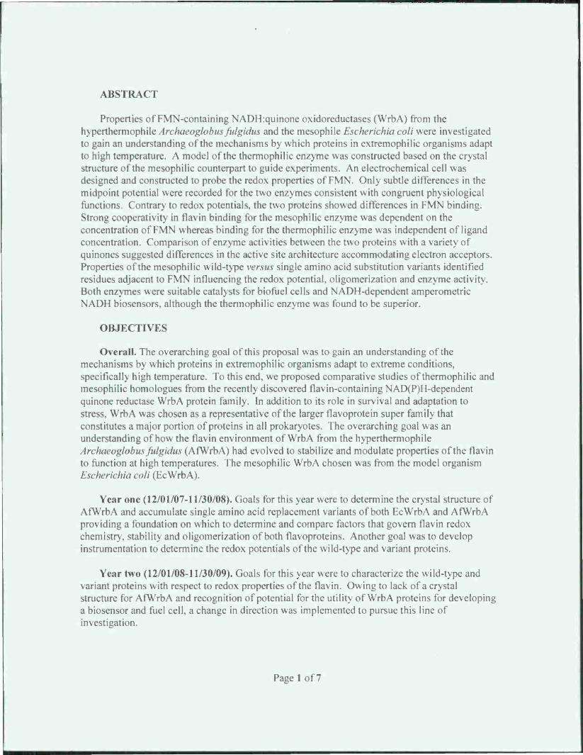
ABSTRACT
Properties of FMN-containing NADH:quinone oxidoreductases (WrbA) from the hypcrthcrmoph'tieArchaeoglohns fidgidus and the mesophile EMiwruhia coli were investigated to gain an understanding of the mechanisms by which proteins in extremophilic organisms adapt to high temperature. A model of the thermophilic enzyme was constructed based on the crystal structure of the mesophilic counterpart to guide experiments. An electrochemical cell was designed and constructed to probe the redox properties of FMN. Only subtle differences in the midpoint potential were recorded for the two enzymes consistent with congruent physiological functions. Contrary to redox potentials, the two proteins showed differences in FMN binding. Strong cooperativity in flavin binding for the mesophilic enzyme was dependent on the concentration of FMN whereas binding for the thermophilic enzyme was independent of ligand concentration. Comparison of enzyme activities between the two proteins with a variety of quinones suggested differences in the active site architecture accommodating electron acceptors. Properties of the mesophilic wild-type versus single amino acid substitution variants identified residues adjacent to FMN influencing the redox potential, oligomerization and enzyme activity. Both enzymes were suitable catalysts for biofuel cells and NADH-dependent amperometric NADH biosensors, although the thermophilic enzyme was found to be superior.
OBJECTIVES
Overall. The overarching goal of this proposal was to gain an understanding of the mechanisms by which proteins in extremophilic organisms adapt to extreme conditions, specifically high temperature. To this end. we proposed comparative studies of thermophilic and mesophilic homologues from the recently discovered flavin-containing NAD(P)H-dependent quinone reductase WrbA protein family. In addition to its role in survival and adaptation to stress. WrbA was chosen as a representative of the larger flavoprotein super family that constitutes a major portion of proteins in all prokaryotes. The overarching goal was an understanding of how the flavin environment of WrbA from the hyperthermophile Archaeoglobus fitlgidus (AfWrbA) had evolved to stabilize and modulate properties of the flavin to function at high temperatures. The mesophilic WrbA chosen was from the model organism Escherichia coli (EcWrbA).
Year one (12/01/07-11/30/08). Goals for this year were to determine the crystal structure of AfWrbA and accumulate single amino acid replacement variants of both EcWrbA and AfWrbA providing a foundation on which to determine and compare factors that govern flavin redox chemistry, stability and oligomerization of both flavoproteins. Another goal was to develop instrumentation to determine the redox potentials of the wild-type and variant proteins.
Year two (12/01/08-11/30/09). Goals for this year were to characterize the wild-type and variant proteins with respect to redox properties of the flavin. Owing to lack of a crystal structure for AfWrbA and recognition of potential for the utility of WrbA proteins for developing a biosensor and fuel cell, a change in direction was implemented to pursue this line of investigation.
Page 1 of7
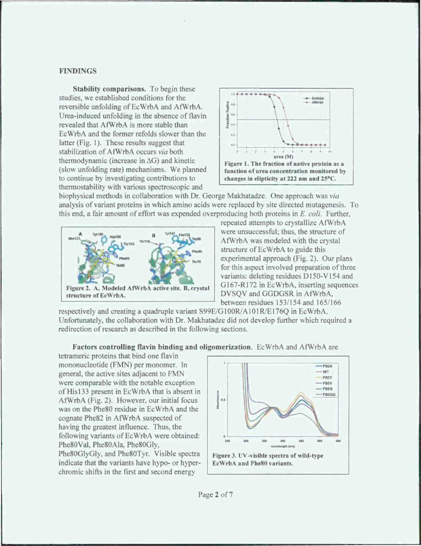
FINDINGS
UITtt(M)
Figure 1. The fraction of native protein as a function of urea concentration monitored by changes in elipticity at 222 nm and 25°C.
stability comparisons. To begin these studies, we established conditions for the reversible unfolding of EcWrbA and AfWrbA. Urea-induced unfolding in the absence of flavin revealed that AfWrbA is more stable than EcWrbA and the former refolds slower than the latter (Fig. 1). These results suggest that stabilization of AfWrbA occurs via both thermodynamic (increase in AG) and kinetic (slow unfolding rate) mechanisms. We planned to continue by investigating contributions to thermostability with various spectroscopic and biophysical methods in collaboration with Dr. George Makhatadze. One approach was via analysis of variant proteins in which amino acids were replaced by site directed mutagenesis. To this end, a fair amount of effort was expended overproducing both proteins in E. coli. Further.
repeated attempts to crystallize AfWrbA were unsuccessful: thus, the structure of AfWrbA was modeled with the crystal structure of EcWrbA to guide this experimental approach (Fig. 2). Our plans for this aspect involved preparation of three variants: deleting residues D150-V154 and GI67-R172 in EcWrbA, inserting sequences DVSQV and GGDGSR in AfWrbA, between residues 153/154 and 165/166
respectively and creating a quadruple variant S99E/G100R/A101R/E176Q in EcWrbA. Unfortunately, the collaboration with Dr. Makhatadze did not develop further which required a redirection of research as described in the following sections.
A Ty»1« B ""*« »*i
TiplM
Figure 2. A, Modeled AfYVrbA active site. B, crystal structure of EcWrbA.
Factors controlling flavin binding and oligomerization. EcWrbA and AfWrbA are tetrameric proteins that bind one flavin mononucleotide (FMN) per monomer. In general, the active sites adjacent to FMN were comparable with the notable exception of His 133 present in EcWrbA that is absent in AfWrbA (Fig. 2). However, our initial focus was on the Phe80 residue in EcWrbA and the cognate Phe82 in AfWrbA suspected of having the greatest influence. Thus, the following variants of EcWrbA were obtained: Phe80Val. Phe80Ala, Phe80Gly, Phe80Gl\(il>. and Phc80Tyr. Visible spectra indicate that the variants have hypo- or hyper- chromic shifts in the first and second energy
| Of
III
II
Figur EcWi
e 3. UV-visible spectra of wild-type •bA and Phe80 variants.
Page 2 of7

transitions, and batho- or hypso-chromic shifts in the second energy transitions (Fig 3). In addition, each variant had different vibrational fine structure, which suggests that interactions w ith FMN are variant-dependent. In addition, analyses of these variants suggests Phe80 is important for oligomerization as dynamic light scattering showed Phe80GlyGly and Phe80VaI do not participate in tetramerization whereas Phe80Gly retains the ability to form tetramers. These data suggest Phe80 is important for flavin binding and oligomerization.
1 —m T11SF T11«Y T1«A T1UW T116M
Comparison of structures identifies Metl 18 of AfWrbA in the equivalent position of Thrl 16 in EcWrbA. To determine if this position is important in FMN binding and oligomerization. we cloned, over expressed, purified and analyzed five variants wherein Thrl 16 was replaced: Thrl 16Ala. Thrl 16Met, Thrl 16Trp, Thrl 16Phe, and Thrl 16Tyr. The UV-visible spectra indicate that several of the Thrl 16 variants displayed hypo- or hyper-chromic shifts in the first and second energy transitions, and batho- or hypso-chromic shifts in the second energy transitions (Fig 4). In addition, these variants also showed different vibrational
fine structure. Variants with a significantly altered spectrum also showed decreased affinity for FMN suggesting a binding role for Thrl 16. Dynamic light scattering showed the Thrl 16Met and Thrl 16Trp variants retain the ability to form tetramers suggesting that Thrl 16 does not play a significant role in oligomerization.
The UV-visible spectra of oxidized and electrochemical ly-reduced wild-type EcWrbA and AfWrbA are shown in Figure 5. Although a spectral shift between the two proteins suggested possible variation in molecular orbital configuration, the results are consistent with flavoproleins that do not accumulate the one-electron reduced flavin semiquinone. The redox potentials of FMN in EcWrbA and AfWrbA were determined to further compare cofactor binding sites. This
Figure 4. UV-visible spectra of wild-t\pe I t \N rbA and Tl 16 variants.
380
Wavelength (nm)
Figure 5. I V -visible spectra of oxidized and reduced EcWrbA and AfWrbA.
Em.6 5 AG AA<3 FMN n
F80A -lsi r : 17 5 -1.4 1.19± 0.13
EcWrbA -172± 5 16.6 -:.: 1.01 ± 0.13
T116A -159 . 153 0.96 ± 0.07
Y143A -156 ± 4 15.1 -3.8 0.97 ± 0.18
T78A -155 ± 4 14 0 -3.9 0.96 ± 0.07
W98A -15: . 14.7 -4.1 0.84 ± 0.02
HI 33 A -148 ± 3 14.3 -4.« 2.30 ± .
Table 1. Reduction potentials for wild-type EcWrbA and several alanine variants.
approach required construction of a specialized electrochemical cell with a small volume that limits diffusion distances from the electrode while retaining optical sensitivity via incorporating a longer (and variable) optical path length. The cell afforded reasonable speed and accuracy in
Page 3 of7

data collection, and the water-tight construction provided stability of the system longer than 24 hours. Voltage across the electrochemical cell was controlled with an EG&G PAR 273 potentiostat for data collection. The cell is essentially a clamp that ensures a water-tight seal to a gold-capillary electrode. An exploded view of the cell illustrates the basic construction (Figure 6). Detailed schematics are available to the scientific community sufficient for reproduction.
I rt&(£ 7 J;
Figure 6. Views of the spectroelectrochemical cell. Left, top view of connections with the potentiostat, light source, and spectrometer. Abbreviations: counter electrode (C.F.), working electrode (VV.E.) and reference electrode (Ref). Right, side view showing sample injection ports indicated by arrows.
Potentiometric titrations of wild-type Ec WrbA (-172 mV ± 5.0) and AfWrbA (-158 mV ± 5.4) yielded similar redox potentials consistent with similar FMN-protein environments influencing the redox potential. The protein environment influencing the redox potential of FMN in EcWrbA was further investigated by replacing adjacent amino acids with alanine (Table 1). Redox potentials for the Tl 16A, HI 33 A and T78A variants of EcWrbA were significantly less negative compared to the wild-type suggesting a role for these residues in modulating the redox potential. Nernst plots of the H133 A variant data indicated a two-electron reduction, while all EcWrbA variants and the wild-type EcWrbA and AfWrbA proteins (not
shown) appeared to participate in sequential one-electron reductions suggesting a role for this residue in stabilizing the two-electron reduced hydroquinone form in both proteins.
Contrary to redox properties. FMN binding assays indicated differences between EcWrbA and AfWrbA. Assays with saturating (150 uM) and limiting (5 uM) FMN are shown
in Figure 7. Binding with apo-EcWrbA was dependent on tlavin concentration and showed tighter affinity with saturated flavin. A single binding event was observed with either saturated or limited flavin, and the event was protein-dependent and appeared to correlate with
0.1 I Protein (|iM) Protein (MM)
Figure 7. FMN binding assayed by fluorescence quenching. Left panel, saturating FMN. Symbols: (▼) EcWrbA, (■) AfWrbA. Right panel, limiting FMN. Symbols: (T) wild type EcWrbA, (■) F80A variant, (A)W98A variant, (O) T78A variant.
Page 4 of7
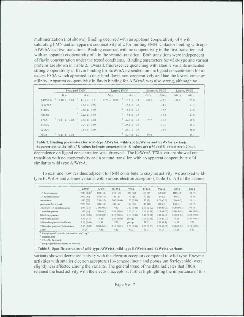
multimerization (not shown). Binding occurred with an apparent cooperativity of 4 with saturating FMN and an apparent cooperativity of 2 for limiting FMN. Cofactor binding with apo- AfWrbA had two transitions. Binding occurred with no cooperativity in the first transition and \\ ith an apparent cooperativity of 4 in the second transition. Both transitions were independent of flavin concentration under the tested conditions. Binding parameters for wild-type and variant proteins are shown in Table 2. Overall, fluorescence quenching with alanine variants indicated strong cooperativity in flavin binding for EcWrbA dependent on the ligand concentration for all except F80A which appeared to only bind flavin non-cooperatively and had the lowest cofactor affinity. Apparent cooperativity in flavin binding for AfWrbA was also strong, although no
AfWrbA
Saturated FMN United FMN Saturated FMN United n
*SI *s: *u Ku AGSI
-40.0
AGs; AGL1
-34.9 -27.8 1 0.10 ± 0.03 4 13.3 ± 0.6 ■ 0.76 ± 0.08 4 13.6 ± 0.1
EcWrbA 4 0.82 ± 0.09 16.8 ± 0.4 -34.7
T116A 4 0.66 ± 0.08 2 14.8 ± 0.3
H133A 4 0.62 ± 0.08 1 18.6 ± 0.5 -35.4 -27.0
T78A 1 0.11 ± 0.03 4 0.43 ± 0.04 2 0.4 -36.3 -26.6
Y143A 0.05 : 0.5 -26.2
W98A 2 0.44 ± 0.05 2 28.0 ± 0.4 -36.3 -260
i - A 1 0.13 r 0.03 :
39.4 ± 1.0 -39.3 -25 1
Table 2. Bin Superscript
ding parameters for wild-type AfWrbA, wild-type EcWrbA and EcWrbA variants. * to the left of K values indicate cooperate it>. K \ alues are u.M and G values are kJ/mol.
dependence on ligand concentration was observed. The EcWrbA T78A variant showed one transition with no cooperativity and a second transition with an apparent cooperativity of 4 similar to wild type AfWrbA.
To examine how residues adjacent to FMN contribute to enzyme activity, we assayed \\ iId- type EcWrbA and alanine variants with various electron acceptors (Table 3). All of the alanine
1.4-bcmnyMM'oc
Af\\r EcWT HI 33 A T"8A T116A Ni4l\ mt v F80A 4940(270)* 660(10) 650 (10) 390 (10) 150(6) 270(10) 180(10) 50 (2)
1.4-n»phdKMf nnuur 3000(30) 510(10) 80(2) 70(1) 70(3) 60(2) 60(1) 10(0 11 muMKsonr 630(20) 320(10) 200(004) 20(001) 80(3) 8 50(0 2) 7 90(01) 10(1) ;.v:.i>t>nm MMCJWMI 3970(60) 180(10) 180(4) 150(10) 140 (10) 140(7) 110(4) 59(2) 2-hydVoKy-14- ngpbibocjunonr 2 90 (02) 3 80 (0 03) ND 0 60 (0.01) 120(002) 0 10 (0 01) 0 20 (0 02) 1 40 (0 1) 1.4-aabnqmtw 380(10) 7.90 (0 5) 0 80 (001) 0 70(01) 0 50 (001) 0 70(0.01) 0.80 (0 01) 1.00 (0.01) 8-hy*aKyqunokoc 0.30 (0.03) 0.10(001) 010(001) 010(0 01) o io (0 on 010 (0.01) 0 20 (0.02) 0.20 (0.02) 9.10-AXIOM MflMUUD? 3 30 (0.4) ND 0 20 (002) prccv' 020(001) 020(005) ND 020 (001) 9JOHUMfareqoBioar-2-tuBontr 0 20 (0.01) ND ND pn*«P ND 0.80(0 1i ND ND. 910-anairaquaiaor-.? 6-dnudfanntr 0 60 (0 03) 0 60(0 01) 010(001) 020(001) 1 60 (0 03) 0 20 (0.01) 040(0 02) 010(002) FMN ND' ND ND ND ND ND ND ND 'Avenge «pacific «cttnaes (—r tonnte« as'1 ms') * StandJrtl Etroi 'ND-NotDrtecttbte ' precs - ptec^tatma ttemA on reduction
Table 3. Specific activities of wild-type AfWr b \. wild- type EcWrbA and EcWrbA variants.
variants showed decreased activity with the electron acceptors compared to wild-type. Enzyme activities with smaller electron acceptors (1,4-benzoquinone and potassium ferricyanide) were slightly less affected among the variants. The general trend of the data indicates that F80A retained the least activity with the electron acceptors, further highlighting the importance of this
Page 5 of7
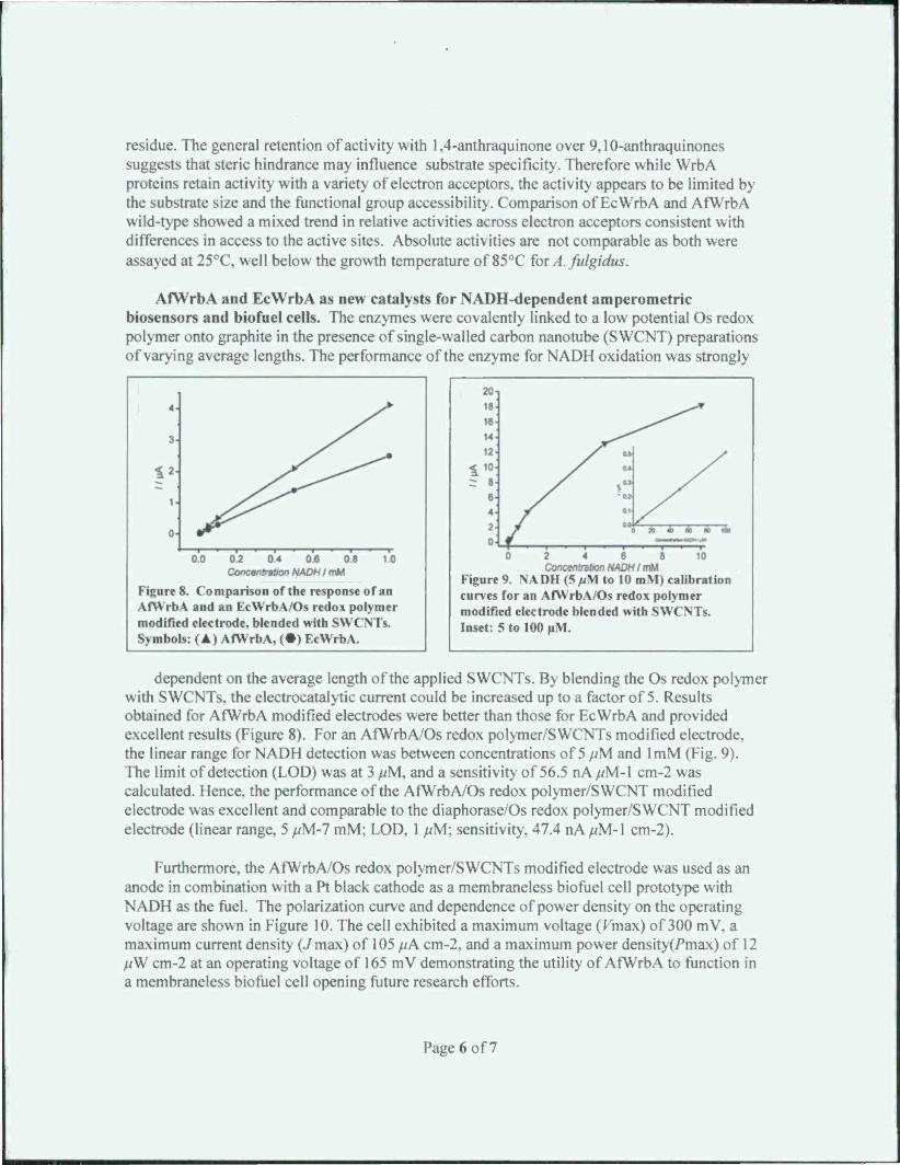
residue. The general retention of activity with 1,4-anthraquinone over 9,10-anthraquinones suggests that steric hindrance may influence substrate specificity. Therefore while WrbA proteins retain activity with a variety of electron acceptors, the activity appears to be limited by the substrate size and the functional group accessibility. Comparison of EcWrbA and AfWrbA \\ iId-type showed a mixed trend in relative activities across electron acceptors consistent with differences in access to the active sites. Absolute activities are not comparable as both were assayed at 25°C, well below the growth temperature of 85°C for A. fulgidus.
AfWrbA and EcWrbA as new catalysts for NADH-dependent amperometric biosensors and biofuel cells. The enzymes were covalently linked to a low potential Os redox polymer onto graphite in the presence of single-walled carbon nanotube (S WCNT) preparations of varying average lengths. The performance of the enzyme for NADH oxidation was strongly
0.2 04 0.6 0.8 Concentration NADH I mM
Figure 8. Comparison of the response of an AfWrbA and an EcWrbA/Os redox polymer modified electrode, blended with SWCNTs. Symbols: (A) AfWrbA, (•) EcWrbA.
2 4 6 8 Concentration NADH I mM
Figure 9. NA DU (5 /At to 10 mM) calibration curves for an AfWrbA/Os redox polymer modified electrode blended with SWCNTs. Inset: 5 to 100 uM.
dependent on the average length of the applied SWCNTs. By blending the Os redox polymer \\ ith SWCNTs. the electrocatalytic current could be increased up to a factor of 5. Results obtained for AfWrbA modified electrodes were better than those for EcWrbA and provided excellent results (Figure 8). For an AfWrbA/Os redox polymer/SWCNTs modified electrode, the linear range for NADH detection was between concentrations of 5 /vM and lmM (Fig. 9). The limit of detection (LOD) was at 3 //M, and a sensitivity of 56.5 nA //M-l cm-2 was calculated. Hence, the performance of the AfWrbA/Os redox polymer/SWCNT modified electrode was excellent and comparable to the diaphorase/Os redox polymer/SWCNT modified electrode (linear range, 5 /vM-7 mM; LOD. 1 /^M: sensitivity, 47.4 nA //M-l cm-2).
Furthermore, the AfWrbA/Os redox polymer/SWCNTs modified electrode was used as an anode in combination with a Pt black cathode as a membraneless biofuel cell prototype with NADH as the fuel. The polarization curve and dependence of power density on the operating voltage are shown in Figure 10. The cell exhibited a maximum voltage (Kmax) of 300 mV, a maximum current dcnsit\ (./ max) of 105 //A cm-2. and a maximum power density^max) of 12 //W cm-2 at an operating voltage of 165 mV demonstrating the utility of AfWrbA to function in a membraneless biofuel cell opening future research efforts.
Page 6 of7

-300 -2» -3Ö0 -150 -100 •«> Ö 300 -250 -200 -150 -100 40
Figure 10. Panel \. polarization curve measured with linear sweep (0 to -350 mV ) voltammetry. Panel B, dependence of the po wer density on the operating voltage.
COLLABORATIONS
Collaborators on the suitability of WrbA's as biosensors and biofuel cells were M. N. Zafar. F. Tasca and L. Gorton from Lund University, Sweden and G. Noll from the University of Siegen, Germany.
PUBLICATIONS
Zafar, M. N., F. Tasca, L. Gorton, E. V. Patridge, J. G. Ferry, and G. Noll. 2009. Tryptophan repressor-binding proteins from Eschertehia coli and Archaeoglobus fulgidus as new catalysts for 1,4-dihydronicotinamide adenine dinucleotide-dependent amperometric biosensors and biofuel cells. Anal. Chem. 81:4082-4088.
INTERACTIONS/TRANSITIONS
Our interaction with Dr. Uwe Slyter at the first AFOSR contractor meeting developed into a collaboration investigating the use of a carbonic anhydrase we discovered for immobilization onto S-layer proteins by Dr. Slyter as a means to capture carbon dioxide from point sources for mitigation of atmospheric levels.
Page 7 of7

Related Documents