Report Second Research Coordination Meeting of the IAEA Coordinate Research Project /F23032/RC-2 on “Developing Radiation Treatment Methodologies and New Resin Formulations for Consolidation and Preservation of Archived Materials and Cultural Heritage Artefacts” 25 – 30 September 2017. Bucharest, Romania

Welcome message from author
This document is posted to help you gain knowledge. Please leave a comment to let me know what you think about it! Share it to your friends and learn new things together.
Transcript

Report
Second Research Coordination Meeting of the
IAEA Coordinate Research Project /F23032/RC-2 on
“Developing Radiation Treatment Methodologies and New Resin Formulations for
Consolidation and Preservation of Archived Materials and Cultural Heritage Artefacts”
25 – 30 September 2017.
Bucharest, Romania

F23032/RC-2/ “Coordinating Meeting”, Bucharest, Romania, September, 2017
2
Table of Contents
1. INTRODUCTION ............................................................................................................................................ 3
2. CRP OVERALL OBJECTIVES ...................................................................................................................... 3
3. CURRENT STATUS OF R&D WORK IN INDIVIDUAL INSTITUTIONS ................................................. 4
4. FUTURE R&D WORK PLANS ..................................................................................................................... 16
5. SUMMARY OF COUNTRY REPORTS ...................................................................................................... 22
6. CONCLUSIONS ........................................................................................................................................... 181
7. RECOMMENDATIONS .............................................................................................................................. 182
ANNEX 1: LIST OF PARTICIPANTS............................................................................................................ 183
ANNEX 2: AGENDA ....................................................................................................................................... 188
OUTPUT RELATED RESULTS ..................................................................................................................... 193

3
1. INTRODUCTION
The preservation of world heritage is a key issue for maintaining national identity, and understanding
the exchanges among civilizations throughout history. Cultural heritage artefacts are made up of
materials varying from simple mono-components to complex structures integrating inorganic and
organic materials. Many of artefacts such as easel and panel paintings, wooden sculptures, library
materials, prints, textiles are based on natural organic materials which are prone to biological attack
under improper environmental conditions. Degradation by insects and microorganisms such as fungi
and bacteria constitute a major threat against the long-term preservation of World-CH (WCH). The
success and consolidation of the application of ionizing radiation in sterilization of medical, surgical
products and food irradiation presents a powerful technique for the disinfection of cultural artefacts
as paper, textile and wood. This environmentally friendly technology ensures the integrity and the
preservation of the objects. In recent years, collaboration of radiation processing facilities with
appropriate configuration for the treatment of cultural heritage with institutions such as museums,
archives, libraries, archaeological institutions and conservation workshops, has allowed the use of
this technology for treating large quantities of deteriorated products that required emergency
intervention or had a complex structure that limited the use of conventional techniques. The wider
use of this technique will depend upon effectively demonstrating that irradiation does not lead to
unacceptable changes in the functional or decorative properties of the artefact as well does not
compromise with the authenticity of the artefact. This CRP has been initiated to focus on evaluating
the effect of irradiation on functional properties of base materials of artefact, minor constituents, post
irradiation effects and developing appropriate irradiation procedures enabling wider use of the
technology.
2. CRP OVERALL OBJECTIVES
Wider acceptance and use of radiation processing techniques for conservation and consolidation of
Cultural Heritage Artefacts.
2.1. SPECIFIC RESEARCH OBJECTIVES
1. Understand the effect of specific irradiation conditions on the functional properties of base
materials present in cultural artefacts to establish appropriate irradiation conditions for treating CH
artefacts.
2. Develop new radiation curable resins with enhanced compatibility with cultural heritage artefacts
and at low radiation doses
3. Establish appropriate procedures for irradiation of artefacts, including dose mapping, dose limit
ratio and simulation techniques to predict dose uniformity during the irradiation process.
2.3. OUTCOMES
Improved conservation methods using radiation processing techniques for conservation and
consolidation of cultural heritage artefacts.

F23032/RC-2/ “Coordinating Meeting”, Bucharest, Romania, September, 2017
4
2.3 EXPECTED RESEARCH OUTPUTS
1. Scientific data on the effect on various materials typically used in artefacts under simulated
conditions of irradiation,
2. Appropriate procedures and technical requirements including optimal radiation doses for the
radiation treatment of CH objects including archived materials.
3. Formulations of new resins and methodologies for consolidation of CH artefacts, 4. Availability of improved dosimetry methods including simulation techniques.
3. CURRENT STATUS OF R&D WORK IN INDIVIDUAL
INSTITUTIONS
3.1 PARTICIPATING MEMBER STATES
Brazil, Bulgaria, Croatia, Cuba, Egypt, France, Iran, Italy, Poland, Portugal, Romania, Serbia,
Turkey, and Ukraine.
Brazil
Brazilian weather conditions affect directly tangible materials causing deterioration getting
worse by insects and fungi attack. In this sense, ionising radiation is an excellent alternative tool
to the traditional preservation process mainly because of its biocidal action. Several national
museums, conservation institutions, conservators-restorers, curators, etc. have been benefited for
this technique and currently most of them maintain institutional partnerships. Constitutive
materials including paper, paintings, photographs, films, parchments, leather, textiles, wood,
bones, etc. have been disinfected by gamma radiation with excellent results at the Multipurpose
Gamma Irradiation Facility in the Nuclear and Energy Research Institute -IPEN. In this work,
dose rate mappings were obtained before and after the loading of fresh cobalt-60 and the results
were compared using statistical tools. Depending on the size of the objects, variation of the
distributions dose in the irradiated product is unavoidable. Most of the cultural heritage objects
need to be irradiated using a stationary method to control the dose distribution and usually
manipulated by hand. A two-side irradiation method was developed and validated to process
large objects when it is not possible to obtain dosimetry measurements inside the material. Some
conservators and restorers frequently get worried about possible long time or post-effects in
irradiated materials. Contemporary paper samples were irradiated using gamma radiation from
Co-60 with different absorbed doses and the kinetics of decay of the cellulose free radicals
induced by irradiation were analysed using Electron Paramagnetic Resonance. X-ray, scanning
electron microscopy and Scanning Electron Microscopy Energy Dispersive Spectrometry were
performed to analyse structure modifications by ionizing radiation. Results have shown that for

F23032/RC-2/ “Coordinating Meeting”, Bucharest, Romania, September, 2017
5
disinfection dose, 80% of the cellulose free radicals induced by ionizing radiation disappear in
almost 8 days. Adequate storage of photographic and cinematographic materials is a challenge
for conservators from preservation institutions. This work presents the preliminary results of
effects of ionizing radiation in photographic and cinematographic films. Selected film samples
made on cellulose acetate were disinfected by gamma radiation and characterized by FTIR-ATR
spectroscopy, UV-VIS spectroscopy and electron microscopy techniques. Results have shown
that disinfection by gamma radiation can be achieved safely applying the disinfection dose
between 6 kGy to 15 kGy with no significant change or modification of main properties of the
constitutive materials.
Bulgaria
According to the work plan for the first year of the project samples of leather were
gamma-irradiated by doses up to 15 kGy, using low (0.006 - 0.06 Gy/s) and standard dose rates
(0.6 - 6 Gy/s) and side-effects on their structure and morphology were studied. Calf leather, calf
suede and pig skin patterns were selected and analysed by: FT-IR, SEM/EDX, EPR, DSC and
TG/DTG before and after the gamma-irradiation treatment with 5 kGy, 10 kGy and 15 kGy
absorbed doses at dose rates of 0.037 Gy/s and 1 Gy/s. The irradiation of the leather patterns was
performed in the gamma-irradiation facility BULGAMMA based on JS-850 60
Co type gamma
irradiator at Sopharma. The results of SEM-EDX analysis revealed that the calf suede and the pig
skin samples were chrome tanned and contained 4.56 % Cr (suede) and 6.74 % Cr (pig skin),
while the calf leather was vegetable tanned.
The results of the performed study can be summarized as follows:
- No changes in the morphology and molecular structure of the studied leather samples
were observed, according to the data from the SEM and FT-IR analysis of the internal and
external sides of the leather samples before and after gamma-irradiation treatment.
- EPR analysis showed increased number of radiation-induced radicals in the calf leather
samples, compared to the calf suede and pig skin patterns. This can be explained by the presence
of the non-isolated Cr3+
ions in the chrome tanned leather patterns (calf suede and pig skin),
which interact with the oxygen radicals, formed during the gamma-irradiation. The spin
concentrations in the samples from the three leather patterns is not influenced by the dose rate of
the gamma-irradiation, except the signal in the calf leather, where higher spin concentrations
were found in the samples, irradiated at low dose rate.
- According to DSC analysis, gamma-irradiation with fungicide doses led to increase of the
enthalpy of the calf leather melting, attributed to cross linking of the biopolymer chains and
leading to expected strengthening of the material. Gamma-irradiation of calf suede at standard
dose rate and pig skin samples at both dose rates led to decrease with 20 to 25 % of the
temperature at the minimum heat flow (Tm, oC), which implies for molecular destabilization.

F23032/RC-2/ “Coordinating Meeting”, Bucharest, Romania, September, 2017
6
- TG analysis showed no significant changes in the thermal decomposition of the three
leather patterns, irradiated at 15 kGy at low and standard dose rates.
The obtained results showed that gamma-irradiation treatment of calf leather, calf suede and pig
skin with insecticide and fungicide doses can be successfully applied for disinfestation and
preservation without causing significant changes in their structure and morphology, even at low
dose rate, where higher radiation-induced side effects are to be expected.
Croatia
There are large needs for systematic approach to data concerning radiation treatment of
CH artefacts because of the complex situation of cultural heritage (CH) objects constituting of
different materials in relation to various bioburdens. The goal of this work is to address those
needs by studying the radiation sensitivity of selected microorganisms commonly occurring on
CH artefacts and of the model materials used on paintings. A common carrier for paintings is
glue-coated linen that is vulnerable to fungal biodeterioration. This study aimed to assess
antifungal effect of gamma-irradiation doses and dose rates against naturally occurring
mycobiota and artificially inoculated primary (Aspergillus jensenii), secondary (Cladosporium
spaherospermum) and tertiary (Trichoderma harzianum) fungal colonizers common for cellulose
materials like linen.
The model systems were irradiated with 2, 7, 20 and 50 kGy at two dose rates that differ by two
orders of magnitude, 0.1 Gy/s and 9.8 Gy/s. After irradiation, the number of viable fungi was
determined by the plate count method in order to determine the proper irradiation dose for
eradication of a particular fungi type. Results indicated that species of Cladosporium spp. and
yeasts seem to be the most resistant to gamma irradiation. In parallel, preliminary assessment of
gamma radiation impact on selected physico-chemical properties of textile were determined.
Cellulose is a main ingredient of base material – linen. It is known that cellulose is radiation
sensitive but the sensitivity of overall material depends on various additives present. Preliminary
FTIR-ATR measurements on coated linen did not indicate degradation in the dose range applied
in this study. The protective effect of the glue coating cannot be excluded although it is relatively
difficult to assess. Further research on coated and non-coated samples, using FTIR and other
complementary experimental techniques (SEM, TGA) is planned.
Cuba
The better procedure to stop the fungal growth on books and archive documents is
keeping low humidity and temperature levels, together with a good air circulation on archive and
museum rooms. Actually, in Cuba many heritage depository institutions do not have the means
to achieve the ideal air conditions. For this reason, the disinfection of paper against fungi should
be a priority in our country and the gamma irradiation could be the best approach to solve this
serious problem.

F23032/RC-2/ “Coordinating Meeting”, Bucharest, Romania, September, 2017
7
As a result, after discussion with cultural heritage specialists we agreed to applying gamma
irradiation for mass treatment of deteriorated books and documents stored at Cuban National
Library (Dose ≈ 3-5 kGy). In addition, in the case of emergency due to flood or any water related
event the combination of Freeze-drying and gamma irradiation methods (Dose ≈ 3 kGy) can be
used.
Practically all chemical agents are banned or strong limited for employing in CH conservation in
our country because of their toxicity and long-term effects.
The majority of CH conservation groups are suspicious about the use of artificial polymers or
hybrid materials for consolidation and protection of artworks made of textiles, wood and paper.
The most important biodeterioration agents in our museums and archives were identified as the
first step for the creation of dose D10 database and the development of conservation procedures
applying gamma irradiation. This task should be concluded with the elaboration of reference
guide for action depending on the material composition and contamination level.
The importance of this CRP is to disseminate the benefits of the gamma irradiation technique for
Culture Heritage consolidation and preservation.
Absorbed dose distribution was calculated by Monte Carlo simulation method for laboratory
scale gamma irradiator. The results were compared to experimental measurements using Fricke
and Alanine dosimetric systems.
Dose rate uniformity (DUR) and its dependence on material density was simulated for semi-
industrial gamma plant using MCNPX code for calculation of gamma dose deposition in 9 tissue
equivalent spheres fixed in different positions inside the irradiation container during its traslation
around the source rack.
In this work revealed the importance of irradiation dose planning by Monte Carlo modelling for
limiting the side effects on CH artefacts and archived documents due to overdose or non-
homogenous irradiation. In case of voluminous and non-symmetric objects a detailed Monte
Carlo simulation should always be guaranteed.
Egypt
Gamma irradiation was used as an effective and “eco-friendly” method for production of
graphene sheets. Graphene oxide prepared from graphite was reduced to graphene upon exposure
to gamma irradiation. The corrosion behaviour of iron coated with gamma irradiated graphene
and its composites with chitosan or an organic inhibitor was evaluated in 3.5% NaCl and in 0.5
M sulfuric acid solution respectively. The resulting gamma irradiated graphene and graphene-
composites covering iron were characterized using UV-Vis, XRD spectroscopies and FE-SEM.
The protection efficiencies of graphene and graphene/chitosan coatings were 82.2% and 89%,
respectively. Graphene sheets/chitosan films over steel showed higher corrosion activation

F23032/RC-2/ “Coordinating Meeting”, Bucharest, Romania, September, 2017
8
energies compared to graphene sheets. EIS proved the stability of graphene and
graphene/chitosan coatings after different immersion times in 3.5% NaCl solution. Coated
surfaces were free from pits on the scale of magnification as demonstrated from SEM images.
Potentiodynamic polarization indicated the protection efficiency offered by graphene coatings
which was enhanced upon using the inhibitor with graphene sheets. The protection efficiency
increased (up to 95.8%) with increasing the inhibitor concentration with an optimum
concentration of 2 mM. Electrochemical impedance spectroscopy measurements (EIS) confirmed
the stability of the graphene/coatings after prolonged immersion time in 0.5 M sulfuric acid
solution.
On the other hand, graphene oxide sheets were used to improve the mechanical properties of
some naturally occurring materials and to enhance their radiation resistance properties against
degradation. Textile linen, papyrus and cellulosic paper, which were selected as natural
materials, were immersed in an aqueous graphene oxide solution. Exposure of selected natural
polymeric materials to 25 kGy gamma radiation results in the formation of free radicals detected
by electron paramagnetic resonance spectroscopy at room temperature. The peak intensity of
irradiated graphene oxide treated materials is lower than that of the un-treated ones. Upon
storage at room temperature, the radicals decayed and the decay rate depended on the nature of
the materials. Of particular interest is the very fast decay rate of the radical trapped in papyrus
and cellulosic paper. The half-life of graphene oxide treated Papyrus and cellulosic paper is
much lower than that of treated linen textile. Incorporation of graphene oxide into papyrus sheets
reinforces their tensile strength mechanical properties. 25 kGy gamma ray irradiation has no
significant effect on mechanical properties of graphene oxide papyrus, as well as their
morphological properties and colour stability
France
The aim of this research is to study the feasibility of using styrene-free unsaturated resins
and acrylic monomers for the consolidation of degraded wooden artefacts from cultural heritage
by in-situ radiation-curing. Indeed, styrene unsaturated polyester resin is implemented during
decades for this purpose, but the safety regulation concerning the styrene monomer is more and
more severe, and the request from museum conservators for styrene-free, acrylic consolidants in
the type of thermoplastics are the main motivations of this work. Acrylic monomers such as
hydroxy-propyl methacrylates, alkyl methacrylates and available styrene-free resins are tested
for the consolidation of sound wooden samples, as well as degraded samples taken from ancient
artefacts. In order to increase the viscosities of the monomers (very low initially), addition of
polymers such as well-known Paraloid® are implemented. Partial consolidation of the wood was
realized by formulating the monomers in various solvents at different concentrations; such
formulations can avoid the inhibition of oxygen on the polymerization of acrylic compounds
which are especially sensitive to this effect. Besides spectroscopic analysis for the radiation-
cured polymers (FTIR, NMR), dimensional changes, mechanical testing, colorimetry, SEM
F23032/RC-2/ “Coordinating Meeting”, Bucharest, Romania, September, 2017
9
observations are carried out to characterize the wood-polymer composites. Very promising
results were obtained after this study: styrene-free resin tested could be an alternative to the
actual styrene-unsaturated polyester one, and the formulations based on the tested monomers
gave interesting results in terms of surface appearance, mechanical improvement and
dimensional stabilization.
Iran
Preserving historic heritage is the duty of a nation that cares about its history. Canvas
based paintings are mainly subject to fungal infestation under improper conservation conditions.
The aim of this study was to evaluate the optimal gamma ray dose for fungal decontamination of
a historical oil painting from the 19th
century stored in Saad-abad Palace.
Sampling for fungal determination was done from 31 points of discoloured points and surface
cultured. Classification was based on overall morphological properties.
Colours used were identified with infrared spectroscopy (mid and far regions). Type of support
was identified by checking the fibre, type and burning behaviour, burning smell and ash type.
Canvas type was identified with FTIR spectroscopy. The wooden framework was not old and not
analysed.
Due to age and the value of the painting sampling from the ancient painting was not possible, so
it was decided to have simulated samples.
Fungi resistance to irradiation was investigated on strips made of similar canvas and colours.
Light-thermal aging of strips was done in a QUV/Spray device.
Due to the fact that the painting could not be moved from the museum, the irradiation time was
calculated by a virtual simulation process. To determine the time required to receive a minimum
absorbed dose of 5 kGy, the irradiation room of the facility was simulated using MCNP4C code.
Aged strips were inoculated with equal amount of Penicillium crysogenum ATCC12690 and
Aspergillus niger CBS 104.57 spore suspensions then exposed to 0.2-2 kGy of gamma rays from 60
Co in GC220, with a dose rate of 2.08Gy/Sec calibrated with fricke dosimeters. D10 was
determined by graphing survival populations after a series of radiation doses. Irradiated strips
with 5-25 kGy were subjected to the sterility test. The minimum dose in which fungal growth is
detected after 14 days of incubation was the sterilization dose.
The results showed that Penicillium and Aspergillus were common fungi of the front, back and
between canvas and the frame.
Infrared spectroscopy revealed the presence of linseed oil in both spectra by its characteristic
bands. The comparison of far-IR spectra with literature data confirmed that the green pigment is
similar to vagone green earth, while the brown pigment is similar to burnt umber. Infrared
spectroscopy revealed that the canvas type was jute.

F23032/RC-2/ “Coordinating Meeting”, Bucharest, Romania, September, 2017
10
The mean D10 value of used fungi on aged coloured linen strips was 0.8-1 kGy.
There were differences between D10 values on culture medium treated with 0.41 and 0.34 kGy
and on canvas treated with 0.8-1 and 0.8 kGy for Penicillium and Aspergillus respectively, while
there were no significant differences on aged and not aged strips. Aged strips showed better
capacity for spore recovery (1.7%) comparing to the not aged (0.15%). This may be due to
production of nutrition elements in the ageing process.
Sterility test indicated that the minimal dose of 5 kGy was sufficient for sterilization of strips
with 2.7 x 10⁶ cfu of each fungus.
The colour measurements of irradiated aged samples will be studied later.
In conclusion and according to fungal contamination of the painting, the dose of 5kGy was
suitable for decontamination. According to the absorbed dose values and the source activity
equal to 3134 Ci on 2017/07/07, while the density of the irradiating products in the carriers was
0.001293 and 0.1 g/cm3, the required time to absorb the minimal dose of 5kGy in the painting
was determined to be about 13.35 and 23.06 hours respectively.
Italy
The activities performed at the ENEA Calliope gamma irradiation facility (Casaccia R.C.,
Rome, Italy) in the framework of the IAEA Coordinated Research Project ‘F23032’- Research
Agreement No. 18922/R0 (first year) are related to the following tasks:
-Task 1: microbiological investigations to study the effect of dose rate and ambient irradiation
conditions on the typical microbes present on archived materials.
Experimental results provide confirmation that chewing insects are potentially harmful bio-
deterioration agents due to their ability to feed on papery materials. It is evident that within a
relative short time they have the potential to completely destroy the samples, as well staining
them with their excretions as causing visible erosions and modifications. The different
vulnerability of irradiated papers varies according to the applied absorbed dose, dose rate and
environmental atmosphere, as was highlighted. The doses used in this experiment, high enough
for disinfesting and reducing considerably microbial load, do not cause appreciable negative
effects since all erosion percentages are negligible and extremely acceptable.
-Task 2: study of the instantaneous and post-irradiation effect on paper irradiated by gamma
radiation using chemical and spectroscopic techniques.
Several characterization techniques were used for the evaluation of the gamma induced side-
effects on cellulose-based materials. In particular, at low doses no differences in term of
cellulose oxidation (ATR-FTIR C=O peak formation) are shown as a function of the irradiation
atmosphere, while with the increase of the absorbed dose (10 kGy) the oxidation in air becomes

F23032/RC-2/ “Coordinating Meeting”, Bucharest, Romania, September, 2017
11
more severe, as confirmed by the decrease of the viscosity (proportional to the cellulose
polymerization degree). No significant dose rate effect was evident.
The investigation of the paramagnetic species induced by radiation (ESR) indicates that the
process is more effective in case of irradiation under inert atmosphere and this kind of radicals
are also more stable during time.
-Task 3: study of gamma radiation induced co-polymerization of acrylic polymers to achieve
formulations suitable and compatible with CH artefacts.
The irradiation tests performed for the formation of EMA/MA co-polymers, allowed to study the
radiation absorbed dose dependence of the radiation induced processes. Different results were
obtained as a function of the environmental atmosphere and dose rate values: i) in presence of
oxygen, higher radiation absorbed dose was required to obtain a solid co-polymer with specific
characteristics since a partial amount of energy released to the samples was involved in
competitive processes; ii) irrespectively to the environmental atmosphere, the formation of more
homogeneous samples in term of polymerization degree dispersion was achieved at lower dose
rates; iii) on samples irradiated at radiation absorbed doses higher than the co-polymerization
dose, while in case of samples irradiated in air heavy depolymerization was verified, a sensible
increase of the samples stability was attained if the irradiation was performed under nitrogen
atmosphere.
Poland
The scope of this research is to investigate the influence of electron beam irradiation used
for the microbiological decontamination process on cultural heritage paper-based objects.
Changes in colour, chemical, mechanical and thermal properties of different types of paper
treated with electron beam before and after the radiation process were studied with SEM, EDS,
EPR and TGA methods. This complex analysis together with the study of the effect of different
irradiation doses on bacteria and fungi for their elimination allowed to determine proper process
conditions for different types of materials. It also gave the possibility to evaluate the possible
effect of radiation on the materials. Observation of colour change in time after irradiation will
give an opportunity to evaluate influence of natural aging on irradiated papers. The following
years the project will concentrate on implementation of the elaborated procedure.
Implementation of the project will help develop new procedure for mass decontamination of
cultural heritage paper-based objects which is still unknown in Poland. Advanced study on the
influence of electron beam irradiation on the properties of different paper-based objects and
elaboration of irradiation procedures suitable for their treatment will help implement this
technology in Poland. The project will be expected to surpass conventionally and commonly
applied EtO fumigation. Determination of process parameters in relation to different sorts of
paper and different types of bioburden will ensure high efficiency of the applied procedure.

F23032/RC-2/ “Coordinating Meeting”, Bucharest, Romania, September, 2017
12
Characterization of different material properties before and after radiation treatment will help to
determine the impact of e-beam irradiation on integrity of paper and help minimalize or avoid
paper changes caused by radiation. Appropriate procedures and technical requirements including
optimal radiation doses for the radiation treatment of CH objects will be developed. The final
outcome of the project will be development of improved conservation methods using radiation
processing techniques for conservation and hygienization of cultural heritage incunables,
manuscripts, papers and books.
Portugal
The development in this period of the research contract was in accordance with the plan
proposed, attending that no additional recommendations were given during the 1st RCM at IAEA
Headquarters (28 Sept - 2 Oct 2015). Nevertheless, small adjustments were introduced in order
to overcome unexpected problems, among which the most restricting were associated with
equipment’s maintenance (60Co recharging process), breakdowns and delays on getting official
authorization to access the archaeological site for inspection and sampling (Conimbriga is an
archaeological site open to general public for visits with a huge flow of tourists). Apart from
these, hard meteorological conditions of last winter and summer also complicated and delayed
the fieldwork.
Objectives achieved:
1. Physico-chemical characterization of Roman mosaics revealed the limestone nature of
tesserae, the respective group of colours and also the structure and elemental composition
of mortars;
2. Bioburden evaluation of Roman mosaics by phenotypic methods conducted to the
isolation of 17 different bacteria corresponding to 12 genera and 13 species, including
PGPB (plant growth promoting bacteria), gram-positive rods and cocci and gram-
negative rods. Fungus Aspergillius fumigates was also detected;
3. Six different batches of PDMS-TEOS-ZrPO hybrid materials were prepared by the
gamma irradiation method developed. Materials revealed to be compact and
homogeneous with a smooth surface, transparent, monolithic and amorphous with a
thermal resistance up to 350 °C. All hybrid materials proved to be hydrophobic with the
CA varying from 95 to 115 °;
4. Biocide activity of HMs’ were tested with the most dangerous and resistant
microorganisms found in Roman mosaics: HMs’ with high content in ZrPO (20 % wt.)
showed biostatic activity against Staphylococcus capitis (Gram+), sporulated bacilli
(Gram+) and fungi Aspergellius fumigatos (Gram+), without evidence of ionic migration
(Zr) to the surrounding medium;

F23032/RC-2/ “Coordinating Meeting”, Bucharest, Romania, September, 2017
13
5. Replicas of Roman mosaics were produced for future HMs’ application assays.
Romania
This talk summarizes the results obtained in the first two years of the project “Improving
the Gamma Radiation Treatment Methodology for Disinfestation of Artefacts” in the frame of
the IAEA CRP F23032 project “Developing Radiation Treatment Methodologies and New Resin
Formulations for Consolidation and Preservation of Archived Materials and Cultural Heritage
Artefacts”.
The main research objectives of the project for the first two years are:
a) Study the post-irradiation effects of free radicals on sensitive materials such as paper
b) Determine Dmax for reference materials not previously investigated
The stability of free radicals induced by gamma irradiation was investigated over two years on
four types of paper: reference Whatman (pure cellulose paper), permanent paper, newspaper, and
an old book printed in 1898. Upon gamma irradiation at 10 kGy, all samples exhibited EPR
signals which are typical for irradiated cellulose. Preliminary results show that the stability of
free radicals is correlated with the degree of crystallinity of contained cellulose as well as with
the age/state of degradation. Significant colour changes between non-irradiated and irradiated
samples were recorded for Whatman and permanent paper, but just perceptible (dE*2000 < 2).
Mechanical tests show significant differences on tensile strength of non-irradiated and irradiated
newspaper samples at five months after irradiation, for both machine and cross directions.
Colorimetry and vibrational spectroscopy (FT-IR/Raman) show no structural or colour changes
for three shades of irgazine pigments as well as for titanium white gamma irradiated at 30 kGy.
As a painting model, a contemporary art work (2013) - oil on canvas, was used. Ten zones of
irradiated painting (18 kGy) were monitored for two years by means of colourimetry. No trend in
the colours of the painting can be observed.
Serbia
Written documents are a very important part of our cultural heritage (CH), and should
therefore be well preserved. Nowadays, increased concerns regarding the safeguarding of
patrimony result in constant evolution of conservation and restoration methods. Biodeterioration
is one of the most challenging issues that curators should deal with. The organic matter materials
(paper, textile, wood, leather, etc.) are susceptible to degradation by insects, fungi, moulds,
bacteria, which inhabit and feed on these materials. In addition, these microorganisms may
present serious health hazards for people dealing with CH artefacts.

F23032/RC-2/ “Coordinating Meeting”, Bucharest, Romania, September, 2017
14
High energy radiation is a powerful tool for disinfection, effectively used in many fields,
especially for sterilization of medical products. Gamma irradiation was used since the 1960s for
the disinfection in archives, however it has not been widely used for that purpose. The wider use
of this technique requires conclusively establishing that irradiation does not lead to unacceptable
changes in the functional or decorative properties of the CH artefacts.
Gamma rays kill microorganisms by direct modification (e.g. crosslinking) of proteins and
nucleic acids, and by free-radical effects in free water. Sterilization and disinfection by gamma-
irradiation are highly efficient methods, and, consequently, widely used throughout the world.
The recovery of several types of materials infected by living organisms through gamma ionizing
radiation has a great potential due to inherent advantages of gamma radiation processing over
other methods of sterilization or disinfection.
Biodeterioration is one of the most serious problems in preservation of cultural heritage artefacts
(CHA). The organic matter in CHA is susceptible to degradation by insects, fungi, moulds,
bacteria, which inhabit and feed on these materials. The first step in conservation of CHA is to
stop bio-deterioration by removing the cause of degradation, e.g. to perform disinfection. The
use of gamma radiation for CHA disinfection requires firmly established conclusions that
irradiation does not lead to unacceptable changes in the functional or decorative properties of
CHA artefacts.
For the application of paper over a longer period it is very important to predict the extent to
which the paper will change the optical characteristics i.e. yellowing under the influence of
gamma irradiation on which it is exposed. Samples of 11 different types of paper were irradiated,
using doses ranging from 1 to 300 kGy. Optical properties were determined on the samples, such
as ISO brightness, whiteness and CIELab color space parameters (L*, a*, b*), after the
application of different dose rates. Colour differences dE 2000 were analysed for all samples.
Optical properties of non-irradiated papers were compared and represented.
Microorganisms, fungi, and insects are often present in many types of cultural goods, from the
immovable objects, through archival and library materials, cellulose type of film materials,
studio paintings, articles of wood, textiles, leather, and other museum objects. The biocide effect
of the ionizing radiation can be effectively implemented for their removal. The objective of this
work is the conservation by gamma irradiation of objects from the Museum of Nikola Tesla,
which is of outstanding value. In this case, infected leather gloves are irradiated with a dose of 5
kGy to eliminate mould. The amount of mould was evaluated by microbiological analysis before
and after gamma irradiation treatment. After successful treatment, Tesla’s leather gloves are
preserved from further deterioration and are now displayed in the museum.
High energy radiation is a powerful tool for disinfection, effectively used in many fields,
especially for sterilization of medical products. Irradiation techniques are being used to protect
and preserve works of art around the world. The techniques are supported by the International
Atomic Energy Agency (IAEA), which operates projects to preserve cultural heritage artefacts

F23032/RC-2/ “Coordinating Meeting”, Bucharest, Romania, September, 2017
15
using radiation. Wooden items, film archives, documents, textiles, leather, parchment even
mummies can be attacked and destroyed by bacteria, fungi, mould and insects. The effectiveness
of ionizing radiation on bio-deteriorating organisms has been further confirmed as well as the
need to further study on the radio-induced effects. Based on the physical and microbiological
obtained results, a dose of treatment ranging from 5 to 7kGy has been recommended to obtain
significant reduction of the microbial load with minimum negative effect on the paper substrate.
The gamma sterilization process in “Vinča” Institute of Nuclear Sciences uses cobalt 60
radiation for research and industrial irradiation, for radiation sterilization of medical devices,
pharmaceuticals, as well as for microbial decontamination of herbs and spices and a variety of
other products. Processing with gamma rays yields quick turnaround time, easily penetrating the
packaging and product and it is cost-effective. Dosimetry measurements were performed using
the ECB dosimetry system which provides reliable means of measuring the absorbed dose in
materials.
The microbiological parameters evaluated: total counts, moulds and yeasts counts detection. The
evaluation of the microbiological parameters was performed before and after irradiation
treatments based on the validated methodologies.
Turkey
Museums, libraries and archives are preserving documents that are slowly degrading due
to the inherent ageing of the cellulose substrate or to the technological errors of the past (acid
paper, iron gall ink). Beside this, large quantities of paper are rapidly damaged by biological
attacks, following natural disasters and improper storage conditions. Cellulose is the major
structural component of wood and plant fibres and is the most abundant polymer synthesized by
nature. Despite this great abundance, cellulosic biomass has seen limited application outside of
the paper industry. Its use as a feedstock for fuels and chemicals has been limited because of its
highly crystalline structure, inaccessible morphology and limited solubility. Electron beams
(EB), X-rays or gamma rays produce ions in a material which can then initiate chemical
reactions and cleavage of chemical bonds. Such ionizing radiation predominantly scissions and
degrades or depolymerizes cellulose. The gamma radiation will also be used for decontamination
and conservation purposes. Important advantages can be mentioned in its favour: no toxic or
radioactive residues remained in the treated item; large amounts of objects can be treated
quickly; excellent reliability; attractive cost. There is also a potential side-effect. Interaction of
gamma rays with any substance may change its chemical and physical properties. The change is
proportional with the irradiation dose. For a successful treatment, an optimal absorbed dose has
to be established. An excessive dose may damage papers and an insufficient one will not reduce
bioburden to the desired level.
In this work, N-butyl acrylate (NBA) and methyl methacrylate (MMA) were chosen as the
monomers for grafting on cellulose surface. The grafted polymer, PBA, is hydrophobic and
F23032/RC-2/ “Coordinating Meeting”, Bucharest, Romania, September, 2017
16
expected to enhance the interface adhesion. Furthermore, PBA has a low glass-transition
temperature (Tg) and a soft chain. The effect of gamma-irradiation process was investigated for
grafting of NBA and MMA/NBA mixtures on Whatman No.1 paper. NBA and MMA/NBA
mixtures were grafted on Whatman No.1 paper. The NBA and MMA/NBA grafted Whatman
No.1 papers were characterized by gravimetric measurements and also by chemical analysis,
such as Fourier Transform Infrared (FTIR) spectroscopy. In this report, thermal and mechanical
properties of Cellulose (Whatman No.1 paper)-g-poly(NBA) (Cell-g-poly(NBA) and Cellulose
(Whatman No.1 paper)-g-poly(MMA-co-NBA) (Cell-g-poly(MMA-co-NBA) were investigated
and we determined also the hydrophilicity properties of grafted and control samples.
Ukraine
Radiation processing techniques are in wide use for disinfection and consolidation of
archived materials and cultural heritage artefacts. The maximum dose (Dmax), which can be
absorbed by the product without changing its properties, is known from the research phase. So,
the minimal absorbed dose (Dmin) should be transferred to the product to achieve disinfection and
this dose shouldn’t be higher than the maximum dose. The location and magnitude of the dose
minimum and maximum is critical for process control, optimized irradiation configurations and
it affects both disinfection and product properties. Reliable product dose-maps are necessary for
identification of these critical process parameters and may involve time consuming and laborious
dosimetry. In some cases, determination of dose-maps is difficult to produce by experiments.
Such cases very often occur during cultural heritage artefacts radiation treatment. In such
situations, the numerical simulation can be used. After consideration of all possible software
toolkits for passage of ionization radiation through the matter GEANT4 was chosen. The
CADMesh library was implemented in developed code to input complicated geometry. The
radiation sources (plaque and cylindrical) were input into the code. Their activities, loading date
into operation can be loaded from .csv file. The comparison between measurements and
simulated results were made. The simulated results have shown a good agreement with the
measured ones. The cloud computing was used for numerical simulation.
4. FUTURE R&D WORK PLANS
Brazil
1. EPR Free radical kinetics research on paper (different types) disinfected by gamma
radiation and electron beam (high dose rate).
2. Studies on cinematographic and photographic films disinfected by gamma and electron
beam.
3. Studies on cinematographic and photographic films deteriorated by the “vinegar
syndrome” irradiated by electron beam.

F23032/RC-2/ “Coordinating Meeting”, Bucharest, Romania, September, 2017
17
4. Studies of the effect of gamma radiation on modern pigments used for restoration by
colorimetry, XRD, XRF and microscopic techniques: restoration pigments, tempera, oil
painting and acrylic paint.
5. Wood consolidation by gamma radiation: basic experiments, small scale applications.
Bulgaria
Studying side-effects of gamma irradiation treatment of leather samples at bactericide dose,
ebony and paper samples at insecticide, fungicide and bactericide doses.
1. Select materials.
2. Characterize the initial samples before irradiation by non-destructive (FT-IR or ATR) and
destructive methods (SEM, EPR, DSC, TG/DTG).
3. Irradiate leather samples at bactericide doses, ebony wood and paper samples at
insecticide, fungicide and bactericide doses, using low (0.006 - 0.06 Gy/s) and standard
(0.6 - 6 Gy/s) dose rates.
4. Determination of the exact absorbed doses and dose rates of the irradiated samples.
5. Evaluation of the radiation induced changes, identified in the irradiated items by using
the same non-destructive and destructive methods.
6. Conclusions on the effects of the different dose rates on the identified radiation induced
changes in the leather, ebony wood and paper samples.
7. Conclusions on the applicability of gamma irradiation treatment of leather with
bactericide dose and ebony wood artefacts and paper with insecticide, fungicide and
bactericide doses.
Croatia
1. The study of the radiation sensitivity of common mycobiota and radiation effects on
inoculated animal glue-impregnated linen canvas will be completed.
2. Preliminary results showed that Alternaria spp., Aspergillus (section Flavi),
Cladosporium spp., Fusarium spp. and Penicillium spp. survived even 20 kGy at the dose
rate of 0.1 Gy/s. Because of this, dose rate effects will be further studied.
3. Model systems based on paper used in paintings will also be prepared, inoculated,
radiation-treated and analysed in the same manner as canvas, including dose rate effects.
Possible differences in development and radiation sensitivity of mycobiota will be
assessed.
4. A selection of binders will be tested, some historic and some modern. The effect of
irradiation on properties of treated binders including their appearance will be
investigated.
5. Influence of common binders for canvas and paper including synthetic ones on radiation
sensitivity of mycobiota will be assessed. The dose rate effects and state (age) of each
coating will be assessed.
6. The effect of the irradiation dose and dose rate on properties of treated materials
including changes in visual appearance will be assessed. We will continue with the model
system investigated until now. The research will be expanded on paper carriers and
results compared.

F23032/RC-2/ “Coordinating Meeting”, Bucharest, Romania, September, 2017
18
7. Influence of (canvas and paper) carrier coating type and state (age) on irradiated carrier
properties including appearance immediately after irradiation and during post-irradiation
period will be studied.
According to the results further plans may be modified.
Cuba
1. To create a database of D10 doses for most important biodeterioration agents in our
museums and archives.
2. To develop a CH conservation procedure applying gamma irradiation and the elaboration
of reference guide for action depending on the material composition and contamination
level.
3. To realize the dosimetric characterization of 100 kCi semi-industrial gamma plant
employing different high dosimetry methods.
4. To use the Monte-Carlo simulation for planning the bulk gamma treatment of books and
archive documents at the IIA semi-industrial gamma plant.
5. To organize periodically national workshops in contact to the CH specialists in order to
disseminate the results of CRP participants on the study of gamma irradiation effects on
paper properties and on application of hybrid materials for protection of artworks.
6. To simulate with MCNPX code the Multipurpose Gamma Irradiation Facility – IPEN-
CNEN (1000 kCi of Cobalt-60) as a contribution for artwork irradiation planning in
Brazil.
Egypt
1. Consolidation and Protection
Continuing the research on modified consolidants for ancient artefacts containing cellulose
compounds such as wood, textiles and manuscripts will be continued. The use of some nano-
scale materials with resistive properties against radiation to minimize damages caused by
ionizing radiation will be considered. Nano-scale materials (like silicon oxide and graphene
oxide) will be also used for cellulosic materials consolidation to improve their mechanical
properties. The work will consider how to rescue ancient carbonized materials such as papyri
and textile wrappings, as they could retain their lost cellulosic formation and thus their
durability to survive using new organic and inorganic materials prepared by radiation. The
research on radiation synthesis of some nano-particles of antimicrobial and antioxidant
properties for protection and de-acidification of artwork will be done.
Study in depth the properties of artefact’s base materials, minor constituents and post
irradiation effects using different tools such as ESR, XRD, TEM, FE-EM, UV, FTIR, DSC,
TGA and mechanical behaviour.
2. Cleaning
The applicative potentialities of appropriate gels based on semi-interpenetrating (IPN)
polymer networks prepared by radiation for cleaning of artwork’s surfaces will be studied.

F23032/RC-2/ “Coordinating Meeting”, Bucharest, Romania, September, 2017
19
Several water-based polymers incorporated surfactants, chelating agents and nano-structured
fluids will be tested as cleaning systems for real Egyptian artefacts.
France
1. Consolidation of wood by radiation curing of monomers
- Choice of monomers to synthetize Parraloid B72 by gamma irradiation
- Test of polymerization, with neutral atmosphere or not, with additive or not (to avoid
oxygen inhibition)
- Test of polymerization on monomers + Parraloid B72 (in order to adjust the viscosity),
with neutral atmosphere or not, with additive or not (to avoid oxygen inhibition)
- Test of reversibility (dissolution)
- Characterization of such synthetized polymers (FTIR, NMR, DSC, ATG, SANS…)
- UV aging tests on the synthetized polymers
- Impregnation tests with wooden samples (new ones and degraded ones on retained
wood species): vacuum/pressure impregnation
- In situ polymerization of the samples (in air or with nitrogen atmosphere)
- Test of reversibility (with respect to the traditional B72 used)
- Characterization of the consolidated samples (dimension variation, colour
measurements, mechanical tests, SEM, Radiography, Tomography, Neutron
tomography…, behaviour with humidity and dry (dimension))
2. Secondary Effect on Irradiated Materials
Ongoing collaboration with Mines d’Alès on textile and dyed textile behaviour under
irradiation
- Assessment analyses of first results on undyed, Garance dyed and chlorophile dyed
irradiated samples of linen: colorimetry and mechanical tests
- New tests with new dye (henna, Prussian blue, indigo, …)
Possible cooperation with CEA Grenoble team working on Free radical characterization by
EPR (on irradiated linen and paper, and maybe on binder).
Binder adhesion behaviour after irradiation: observation after dry and humid cycles.
Iran
Study the post irradiation changes in samples of the painting
1. D10 determination of standard fungi in wood samples
2. Accelerating tests and evaluation of the age effect in post irradiation changes in chemical
structure of substances in samples, if possible.
3. Performing the practical irradiation on the model painting
4. Principals of ancient paintings irradiation should be discussed to develop the appropriate
protocol

F23032/RC-2/ “Coordinating Meeting”, Bucharest, Romania, September, 2017
20
Italy
1. Evaluate the side-effects and post-irradiation effects of the developed formulations by
means of sophisticated analytical techniques like FTIR, ESR, 13C-NMR, 1H-NMR and
mechanical tests.
2. Studies on environment friendly and non-toxic consolidants and development of new
compositions for use as consolidating agents to improve the efficiency and safety of the
CH artefact:
a) Study of gamma radiation induced co-polymerization of acrylic polymers to achieve
formulations suitable and compatible with CH artefacts.
b) A new approach using the lignin-like structures has been developed as a new green
system for consolidation. Lignin-like oligomers or polymers provide maximum
compatibility with the wood structure, improved mechanical properties, strong
hydrogen bonds and cross-linking with existing lignin and antimicrobial activity
versus Staphylococcus aureus. Chemical production of lignin-like structures occurs in
presence of catalysts, but gamma radiation induced polymerization allows to obtain
the compound without chemical (no additives) and physical (no increase of
temperature) modification. Moreover, it is possible to control the polymer’s features
by modifying the irradiation parameters such as irradiation dose, irradiation dose rate,
atmosphere, etc.
Poland
1. Application of other characterization methods for paper properties evaluation after EB
irradiation (XRD, SEC, GC, chemical parameters- pH of paper extract, copper number,
tearing resistance with Elmendorf method)
2. Observation of natural aging effect on paper colour - continuation
3. Determination of the accelerated aging influence
4. Comparative study – ethylene oxide fumigation and γ irradiation
5. Study of the effect of EB irradiation on „real” paper-based objects
Portugal
1. Finish the physico-chemical characterization of native materials.
Essential for the adequate definition of hybrids composition (guarantee of materials
compatibility) and final form of application.

F23032/RC-2/ “Coordinating Meeting”, Bucharest, Romania, September, 2017
21
2. Improvement of hybrid material’s biocide activity, enlarging its effectiveness to a larger
range of potentially dangerous microorganism, namely to Pseudomonas spp., a plant
growth-promoting bacteria.
3. Complement mosaics bioburden evaluation through genotypic techniques.
Romania
1. Study the kinetics of free radicals induced by ionizing radiation in cellulose based
materials and the related side-effects through accelerated ageing (thermal annealing,
controlled atmosphere/humidity, UV irradiation)
2. Irradiation side-effects on CH materials at high dose rates (e-beam and X-ray)
3. Dosimetry data for MC simulations for CH artefacts (dummy and real artefacts)
Serbia
We will take participation in the next three tasks: consolidation, free radicals and simulation.
1. Consolidation: We need some time for preparation and after that we can start with work
on it and cooperation with other countries. The start of this task should be in the end of
March.
2. Free radicals: We can start immediately.
3. Simulation: Our data and blue prints will be available in one month. So, at the beginning
of November we will give all necessary documents to Mr. Volodymyr Morgunov.
Turkey
1. In addition to the used monomers, the effect of the different amounts of microcrystallline
cellulose will be investigated.
2. The grafting of cellulose papers with monomer solutions of the above mentioned
monomers in the presence of microcrystalline cellulose and different concentrations will
be tested.
3. Irradiation of impregnated papers with gamma rays and if possible by electron beams.
4. Chemical, mechanical, and surface property testing of (co)polymer loaded papers will be
performed.
5. Microbiological tests will be performed.
Ukraine

F23032/RC-2/ “Coordinating Meeting”, Bucharest, Romania, September, 2017
22
1. Carrying out the numerical experiments on gamma and X-Rays treatment of cultural
heritage artefacts.
2. Simulation of X-Ray conversion of accelerated electrons and electron beam treatment and
X-Ray treatment of cultural heritage artefacts
3. Development of graphical user interface for the developing code
4. Carrying out the numerical experiments via grid computing.
4.1. Receiving certificate for grid computing.
4.2. Installing proper software and libraries on grid.
4.3. Carrying out of numerical experiments based on data given by participants of the
current CRP.
5. Numerical simulation of dose maps for participants of the current CRP
5. SUMMARY OF COUNTRY REPORTS
The participants presented their reports in accordance with the guidelines issued by the IAEA.
Lively and informative discussions occurred during and following the presentations and were
frequently continued during the coffee breaks.
Each country was asked to provide a short text summarizing its major achievements, major
constraints/problems encountered and lessons learned.
Brazil
RADIATION PROCESSING FOR PRESERVATION OF CULTURAL HERITAGE
OBJECTS: ABSORVED DOSE DISTRIBUTION MAPPINGS, TWO-FACES
IRRADIATION METHOD, CINEMATOGRAPHIC-PHOTOGRAPHIC FILMS
PRESERVATION AND FREE RADICALS DECAY IN PAPER STUDIES

F23032/RC-2/ “Coordinating Meeting”, Bucharest, Romania, September, 2017
23
P.A.S. VASQUEZ1, P. S. SANTOS
1, Y. KODAMA
1, R.H.L. GARCIA
1, L. OTUBO
1,
M.L.E. NAGAI2, A. PIRES
3
1 Nuclear and Energy Research Institute -IPEN/CNEN, Sao Paulo, Brazil
2 University of Sao Paulo - USP, Sao Paulo, Brazil
3 Bandeirantes Palace, Sao Paulo, Brazil
Abstract
Brazilian weather conditions affect directly tangible materials causing deterioration getting worse by insects and
fungi attack. In this sense, ionising radiation is an excellent alternative tool to the traditional preservation
process mainly because its biocidal action. Several national museums, conservations institutions,
conservators-restorers, curators, etc. have been benefited for this technique and currently most of them
maintain institutional partnerships. Constitutive materials including paper, paintings, photographs, films,
parchments, leather, textiles, wood, bones, etc. have been disinfected by gamma radiation with excellent
results at the Multipurpose Gamma Irradiation Facility in the Nuclear and Energy Research Institute -
IPEN. The distribution of the cobalt-60 radioactive sources in the racks is a very important procedure to
guarantee the homogeneity of the irradiation process. In this work, dose rate mappings were obtained
before and after the loading of fresh cobalt-60 and the results were compared using statistical tools.
Depending of the size of the objects, variation of the distributions dose in the irradiated product is
unavoidable. For this reason, optimizations and simulations of the irradiation process are necessary
always validated by a dosimetry system. Most of the cultural heritage objects need to be irradiated using a
stationary method to control the dose distribution and usually manipulated by hand. A two-side irradiation
method was developed and validated to process large objects when is not possible to obtain dosimetry
measurements inside the material. Some conservators and restorers frequently get worried about possible
long time or post-effects in irradiated materials. During irradiation process, some energetic and unstable
chemical species called free radicals appear in the treated matter. Contemporary paper samples were
irradiated using gamma radiation from Co-60 with different absorbed doses and the kinetics of decay of
the cellulose free radicals induced by irradiation was analyzed using Electron Paramagnetic Resonance.
De-noising treatment of the original obtained spectra signals were performed using wavelets. X-ray
diffraction was carried out to identify crystalline phases and the effect of ionizing radiation on the
crystalline structure of cellulose in paper. Scanning electron microscopy and Scanning Electron
Microscopy Energy Dispersive Spectrometry were performed to analyze structure modifications by
ionizing radiation. Results shown that for sterilization dose, 80% of the cellulose free radicals induced by
ionizing radiation disappear in almost 40 days and for disinfection dose in 8 days. It can be concluded that
if no significant modifications or side-effects appear in the irradiated material after the radical decay time,
the material will stay stable for the remaining lifetime. Adequate storage of photographic and
cinematographic materials is a challenge for conservators from preservation institutions. Contamination
by fungi is one of leading causes of problem in photographic and cinematographic collections. In this
work are presented preliminary results of effects of the ionizing radiation in photographic and
cinematographic films. Selected film samples made on cellulose acetate were prepared and characterized
F23032/RC-2/ “Coordinating Meeting”, Bucharest, Romania, September, 2017
24
by FTIR-ATR spectroscopy. Samples were irradiated by gamma rays with absorbed dose between 2 kGy
and 50 kGy. Irradiated samples were analyzed by UV-VIS spectroscopy and electron microscopy
techniques. Results shown that disinfection by gamma radiation can be achieved safely applying the
disinfection dose between 6 kGy to 15 kGy with no significant change or modification of main properties
of the constitutive materials.
1. OBJECTIVE OF THE RESEARCH
Develop an irradiation method performing dose mapping and dose distribution studies in cultural heritage
objects using PMMA dosimetry systems.
Evaluate the effects of the radiation processing using gamma rays and electron beam for disinfection in
functional properties of tangible base materials of cultural heritage artefacts such as textiles, varnishes,
paper, films, wood and pigments under specific irradiation conditions using electron paramagnetic
resonance spectroscopy (EPR), colorimetric techniques, field-emission gun scanning electron microscopy
(FEGSEM) / energy dispersive spectroscopy (EDS) and X-ray transmission and diffraction techniques.
2. INTRODUCTION
Brazil is a multicultural South American country has had the influence of the pre-Columbian native
civilizations, the Portuguese and African colonization and the European colonization especially from
Germany and Italy, not to mention that Brazil is home to the largest Japanese population outside Japan.
Besides other factors, this situation makes the country own several collections of historic value objects.
Brazilian weather conditions have been affected directly tangible materials causing deterioration getting
worse by insects and fungi attack. Natural disasters particularly floods also have been affected many
collections inside the country. Within this scenario, the radiation processing specially by gamma arises as
an alternative to traditional methods to the disinfection of cultural heritage artefacts and archived
materials. Gamma irradiation has several advantages when compared with conventional preservation
methods mainly related to the safety, efficiency, reliability, capacity, process time and safe for
environment. Over the last years, the Nuclear and Energy Research Institute–IPEN mainly through the
Multipurpose Gamma Irradiation Facility located inside the University of São Paulo campus started a
strong interaction program with conservation and preservation institutions and the conservation
community to disclose the irradiation technique. Currently, this facility has been irradiated for
disinfection purposes successfully several works of art, museum collections artefacts, books, manuscripts,
drawings, archive documents, musical instruments, ethnographic objects, archaeological findings, natural
history collections among others from various regions of the country.
However, more research is still required to study undesirable effects (secondary or side effects) which
may appear in sensitive materials as a function of the delivered dose. Some conservators and restorers
frequently get worried about possible long time effects in irradiated materials (post-effects). Especially
pieces of modern art present themselves as a challenge for disinfection by ionizing radiation by the
variety of materials used in their structures. Another interesting aspect are the modern pigments that the
F23032/RC-2/ “Coordinating Meeting”, Bucharest, Romania, September, 2017
25
restorers and conservators have been using to restore ancient art works as well paintings and their possible
changes of mains properties (e.g. colour) after the irradiation process.
The IAEA have been supporting many regional projects and research contracts related to the nuclear
techniques applied to cultural heritage preservation and research helping to understand that the cultural
heritage is the legacy of physical artefacts and intangible attributes of a group or society that are inherited
from past generations, maintained in the present and restored for the benefit of future generations.
In this report are presented the results of the first two years of the IAEA Coordinated Research Project
F23032, related to:
-Absorbed dose rate distribution mappings: after and before the loading of fresh Co-60 in the IPEN
facility.
-Two-faces stationary irradiation method for tangible materials.
-Kinetics of free radicals decay reactions in cellulosic based heritage materials disinfected by gamma
radiation
-Characterization of cinematographic and photographic films disinfected by gamma radiation.
3. MATERIAL AND METHODS
3.1 Gamma irradiation at the Multipurpose Gamma Irradiation Facility – IPEN-CNEN
The Multipurpose Gamma Irradiation Facility at the Nuclear and Energy Research Institute – IPEN is
located inside the campus of the University of São Paulo –USP.

F23032/RC-2/ “Coordinating Meeting”, Bucharest, Romania, September, 2017
26
This facility can operate with 1000 kCi of cobalt-60 classified as Category IV (IAEA-SSG-8). The
relevant facility characteristics shown in Fig.1.
The cobalt-60 source pencils are loaded into predetermined positions into source modules and distributed
these modules over two source racks of the gamma irradiator [20]. The sources are contained in a water-
filled storage pool when the irradiator is not operating. A pneumatic system controls the rack movements.
A sliding door (120 ton) allows entering to the irradiation chamber if necessary. The facility can be
operated in continuous mode or in stationary mode. In the continuous mode, the product containers
(aluminium tote boxes) are moved around the sources using a mechanical transport system. A rotating
door releases simultaneously input and output product. In the stationary mode, the radiation sources
(racks) are moved into the irradiation chamber after the product has been arranged in fixed positions.
Most of materials are irradiated using dose rates 5-6 kGy/h.
3.2 Dosimetry system
Measurements of the absorbed dose at the Multipurpose Gamma Irradiation Facility were performed
using an industrial routine dosimetry system known as dyed polymethyl methacrylate (PMMA). This
dosimetry system is based on the measurement of the radiation induced absorbance change in dyed
PMMA, creating a broad absorption band in a special region of the UV-VIS spectrum. Harwell
Dosimeters-UK has been providing commercial dosimeters for over 40 years for the entire world [9].
FIG. 1 - Multipurpose Gamma Irradiation Facility – IPEN-CNEN
F23032/RC-2/ “Coordinating Meeting”, Bucharest, Romania, September, 2017
27
Absorbed dose can be easily calculated using a normalized ratio between the measurement of the specific
absorbance obtained by the UV-VIS spectrophotometer and the thickness of each dosimeter. Then the
obtained value is replaced into a calibration curve (fourth-order polynomial regression previously founded
by the least squares method) and the absorbed dose value is calculated directly. This procedure is possible
because when analyzed the absorbance curves spectrum in function of the wave length after irradiation
with doses over 1 kGy, new specific defined absorption optical bands are created (e.g. 530 nm, 651nm,
640 nm, etc.) in function of the dyed of the dosimeter and just is this property allows create a correlation
with the absorbed dose.
Dosimeters are positioned in front, in back and if possible in the middle of the objects to be irradiated,
special considerations are taken to samples for research objectives. For routine irradiations of large
batches, several dosimeters are distributed in many positions to ensure the delivered absorbed required
dose [10], [11], [12], [18]. Harwell also provides an individual calibration curve for each commercialized
dosimeter batch however the calibration procedure is performed again inside the laboratories of IPEN
only to confirm the results.
3.3 Absorbed dose rate mappings
3.3.1 Absorbed dose and dose rate
The success of radiation processing depends largely on the ability of the processor to measure the
absorbed dose delivered to the product (reliable dosimetry), determining the dose distribution patterns in
the product package (process qualification procedures) and to control the routine radiation process
(process control procedures). Since the radiation, absorbed dose is the quantity, which relates directly to
the desired effect in a specific material, the need for suitable and accurate dose measurement techniques
must not be underestimated. This is best appreciated by realizing the consequences of using inadequate
techniques, causing under or overexposure of the product and the resulting failure to administer an
effective treatment. The consequences to the processor can be both legal and economic, while the
consumer may not only suffer an economic loss by having to discard an inadequately treated product but
also lose confidence in the irradiation process.
In the processing of products by radiation, reliance is placed on the radiation quantity absorbed dose to
obtain accurate and expressive information about the relevant radiation effects. The regulatory authority
or any other group responsible for the acceptance of the specific application requires information that
demonstrates that every part of the process load under consideration has been treated within the range of
acceptable absorbed dose limits.
The absorbed dose, D, is the amount of energy absorbed per unit mass of irradiated matter at a point in the
region of interest. It is defined as the mean energy, , imparted by ionizing radiation to the matter in a
volume element divided by the mass, dm, of that volume element [8],[17]:

F23032/RC-2/ “Coordinating Meeting”, Bucharest, Romania, September, 2017
28
(1)
The SI derived unit of absorbed dose is the gray (Gy), which replaced the earlier unit of absorbed dose,
the rad, 1 Gy = 1 J/kg = 100 rad.
The absorbed dose rate, , is defined as the rate of change of the absorbed dose with time:
(2)
In practical situations, D and are measurable only as average values in a larger volume than is specified
in the definitions, since it is generally not possible to measure these quantities precisely in a very small
volume in the material. In this work, the absorbed dose is considered to be an average value, either as
measured in the sensitive volume of the dosimeter used if it is of appreciable size or existing in its
immediate vicinity if the dosimeter is very small or thin, where cavity theory is applicable. For any given
irradiation conditions, it is necessary to specify the absorbed dose in the particular material of interest
because different materials have different radiation absorption properties.
3.3.2 Dose rate distribution mappings
The absorbed dose mappings were performed using Amber PMMA dosimeters arranged in a matrix
geometry 19x22 (19 rows and 22 columns) separated 10 cm each points. The total mapping area was 4.18
m2 as shown in FIG. 2. Absorbed dose mapping were performed before and after the loading of fresh Co-
60. The dose rates were calculated
FIG. 2 – PMMA dosimeters placed in a matrix geometry 19x22 before the gamma irradiation
F23032/RC-2/ “Coordinating Meeting”, Bucharest, Romania, September, 2017
29
3.4 Two-faces stationary irradiation method for tangible materials
3.4.1 Distribution of the Absorbed Dose
The dose needed to achieve a desired effect in the product or process dose is determined through several
research stages. In general, it involves determine the relationship between different values of absorbed
dose with some parameters of interest which can be modified by the irradiation process, for example the
sterility level versus dose or a mechanical propriety of the product versus dose. Therefore, the goal of
such research is the identification of two dose limits: a) the lower dose limit to achieve a desired effect
(e.g. sterility level) and b) the upper dose limit to be sure that radiation will not adversely affect the
quality of the product (e.g. degradation of polymers of health care products). Usually, each product or
process has a pair of these limits, and these values define an acceptable dose window, such that every part
of the product should receive a dose within that range. The ratio of the upper dose limit to the lower dose
limit may be referred to as dose limit ratio (DLR) [8], [17].
During a radiation process, gamma radiation interacts with the product through several types of atomic
interactions, such as Compton scattering, photoelectric effect and pair production. Through these and
subsequent interactions, it imparts energy and consequently radiation dose to the product. As radiation
proceeds through the product, its intensity decreases as a result the absorbed dose also decreases with
depth. This phenomenon is referred to as depth–dose distribution. The rate of decrease depends on the
composition and density of the product and the energy of the gamma radiation. Besides the variation of
dose with depth, there is also dose variation in the lateral direction. This variation depends on the
geometry of irradiation. Both types of dose variation contribute to the non-uniformity of the dose
delivered to the product. Then, variation in dose in the irradiated product is unavoidable. One accepted
method of describing this non-uniformity of dose is the concept of dose uniformity ratio (DUR), which is
the ratio of the maximum dose in a product (container or package) to the minimum dose. DUR increases
with the density of the product as well as with the size of the container or package.
DUR should be close to unity (e.g. less than 1.05) for irradiation of research samples, where the research
objective is to correlate radiation effect in the sample to the dose. This is generally achieved by reducing
the size of the sample. For commercial operation (industrial scale), this is not possible for economic
reasons. A typical product container size can be 60cm×50cm×50cm, and some irradiators are designed to
irradiate entire pallets of product, for example, of 120cm×100cm×150cm. DUR would be significantly
larger than unity for such large containers. Fortunately, for a large majority of products, there is a wide
window of dose that is acceptable to achieve the desired level of sterility without detrimentally affecting
the quality of the product. For such products, the dose limit ratio is between 1.5 and 3, and sometimes
even larger.
Therefore, the guiding principle to processing by radiation is that the measured dose uniformity ratio
should be smaller than the dose limit ratio prescribed for the product (DUR<DLR).
There are different ways to reduce the DUR or increasing the dose uniformity in a product container. The
variation along the depth is easily reduced by irradiating the product from more than one side. This can be
accomplished either by rotating the product container during irradiation or for the product container to
travel around a radiation source. All gamma irradiators use one of these techniques for the purpose. The
lateral dose variation may be reduced in several ways, including placing the higher activity source pencils

F23032/RC-2/ “Coordinating Meeting”, Bucharest, Romania, September, 2017
30
near the periphery of the source rack (source augmentation), and relative arrangement of the product
containers and the source (source overlap or product overlap). Different irradiators apply different
methods to improve dose uniformity.
3.4.2 Two-Faces Stationary Irradiation Method
Cultural heritage objects need to be loaded in the irradiation chamber usually by hand, then the stationary
method is the more suitable to take care mainly parameters related to the distribution dose (DUR). When
DUR is close to the unity, this means small research samples or another objects; the irradiation process is
performed placing the materials in specific locations inside the irradiation chamber where the dose rate is
known. The processed materials are not rotated or moved while the irradiation is happening.
The two-faces stationary method is executed in two stages, first the samples (boxes or packages) are fixed in specific positions inside the
irradiation chamber where the dose rate is known, after having passed 50% of the total planned irradiation time, the samples are rotated 180
degrees in a way that the face of the sample was totally opposite (back) to the radioactive sources now is in front of them and then start the
second stage of the irradiation process to complete the 50% of time remaining. FIG. 3 shows the depth-dose distribution using this method.
[17].
When the sample is irradiated in the first time stage (50% of the total), the maximal value of the absorbed
dose DF-1 is located on the near side to the radioactive sources (front) and the minimal value on the
FIG. 3 – Depth-dose distribution using the Two-Faces Stationary Irradiation Method.

F23032/RC-2/ “Coordinating Meeting”, Bucharest, Romania, September, 2017
31
opposite side, DB-1 (back) as shown in Fig. 1 on the dose distribution curve D1(x). Then the sample is
rotated and is completed the 50% of the remaining irradiation time, so that the face of the sample now is in
front of the sources will receive an absorbed dose of DF-2 and the far side DB-2 as shown on the dose
distribution curve D2(x). For cumulative proprieties of the absorbed dose, the total effect of the irradiation
will be represented by the distribution curve D1+2(x) where D1+2 min represent the minimal processing dose.
Therefore, D1+2 max represent the maximal value achieved by an external the dosimeter. DUR can be
calculated as shown in Equation 3.
(3)
The variation of the absorbed dose (D) in function of the thickness of the sample (x) can be represented by
the Equation 4. Solving the differential equation (Equation 5) and integrating between the corresponding
limits can be obtained the dose distribution curves D1(x) and D2(x) described by the Equation 6 and 7
respectively.
(4)
(5)
(6)
(7)
Where, µ is a proportionality constant representing the effective global attenuation coefficient of the
irradiated material. It was assumed that DF =DF-1=DF-2 and DB = DB-1= DB-2 because the partial irradiation
intervals of time were the same and thus the absorbed dose is proportional to the time.
This pseudo-exponential behavior of the distribution dose curves is particularly useful when is not
possible to place a dosimeter inside the sample, allowing the calculation of the delivered minimal dose in
function of the external dosimeters placed in front and back of the irradiated object (maximal dose).
By adding the Equations 6 and 7 is obtained Equation 8 that represents the total distribution of the dose
D1+2(x), in both irradiation stages, then differentiating and equaling to zero can be obtained the thickness
x1+2 min that can be replaced in Equation 8 to find the minimal processing dose D1+2 min as showed in
Equation 11.

F23032/RC-2/ “Coordinating Meeting”, Bucharest, Romania, September, 2017
32
(8)
(9)
(10)
(11)
Another way to obtain the same result can be dividing Equations 6 and 7 to get the minimal dose in each
irradiation stage (Equation 12) but this can be deducted from Equation 11 because each irradiation stage
is performed using the same time.
(12)
Through Equations 6,7 and 8 also is be possible to calculate de global effective attenuation coefficient
that can be useful to standardize the irradiation method for the same material.
The Multipurpose Gamma Irradiation Facility to make easy the understanding of these methods usually
works with DUR values in sense of a factor f as showed in Equation 13. This factor is useful to calculate
the minimal processing dose in function of the value registered in the dosimeter (maximal dose).
(13)
3.4.3 Irradiation Time Calculations
The irradiation time t is calculates performing partial dosimetric measurements for the dose rate in
desired positions (e.g. front, back, middle, etc.). Stipulating a partial irradiation time lower than the
total expected to reach the processing desired dose ( << t ) and making measurements of the partial
absorbed dose , dose rates can be calculated as shown in Equation 14.
(14)

F23032/RC-2/ “Coordinating Meeting”, Bucharest, Romania, September, 2017
33
Then, knowing the dose rates in specifically locations, irradiation time can be obtained. For research
samples (DUR≈1) and for the one-face stationary irradiation method, the irradiation time is calculated
using Equation 15.
(15)
Where, Dmin is the minimal processing dose required (e.g. 5, 15, 25 kGy, etc.)
The two-faces irradiation method is used for large volumes and when is not possible to locate a dosimeter
inside the samples, partial dose rate measurements are necessary for dosimeter located in front and back
of the volume. The methodology to determine de frontal dose rate and the back dose rate is the
same used to the one-face method.
Then, analogously to the deductions developed early and using the expression of the Equation 10, the
dose rate can be calculated inside the material in the position where is expected the minimal processing
dose or as showed in Equation 16.
(16)
Next is calculated the irradiation time in the first irradiation stage as shown in Equation 17.
(17)
However, exist another situations more complicated that no were described in this work and more detail
and additional calculations are required.
3.4.4 Validation of the Two-faces Stationary Irradiation Method
The two-faces stationary method was validated irradiation different type of materials used in medical
products (plastic, rubber, glass and metal), animal products (feed and shavings) and cultural heritage
products (paper).
Several dosimeters were placed in front ( ), back ( ) and in the middle ( ) of the objects, this the
last one was used to compare with the calculated absorbed dose value . All experimental data were
obtained using five samples (n=5). Next, the relative standard deviation (%RSD) was calculated for each
measurement as shown in Equation 18.
(18)
F23032/RC-2/ “Coordinating Meeting”, Bucharest, Romania, September, 2017
34
Where s is the standard deviation for the sample and is equal to the mean (absorbed dose values).
3.5 Kinetics of free radicals decay reactions in cellulosic based heritage materials disinfected by
gamma radiation
3.5.1 Sample preparation
A contemporary paper was used (made in 2010), manufactured with bleached chemical pulp, mineral load
above 10%, pH 4.6 and 100% short fibers. Paper samples were prepared and cut into several 2 mm × 25
mm pieces. Samples were irradiated inside EPR quartz tubes, room temperature (~25 oC) with radiation
absorbed doses of 1, 6, 10, 12 and 15 kGy and dose rate of 5 kGy/h.
3.5.2 Electron Paramagnetic Resonance, EPR
Electron Paramagnetic Resonance spectra were obtained at room temperature using Bruker EMX plus
model, X band, interval from 337.6 to 367.5 mT, field modulation amplitude 0.2 mT, field modulation
frequency 100 kHz, microwave power 2 mW. Measurements were performed at room temperature, at
different time intervals after irradiation, from 1.5 to 670 h to understand the decay free radical
mechanism. De-noising treatment of the original signals obtained by EPR was performed using the
method suggested by Antoniadis et al. (1995). By integrating the EPR curves it is possible to obtain area
values that can be correlated to concentration, in our case, it is equivalent to spin concentration.
3.5.3 Kinetic Proposed Model
The kinetics of cellulose free radicals decaying was studied by normalizing and integrating the EPR
spectra, where the calculated area corresponds to cellulose radicals spin concentration (Weil and Bolton,
2007). The integrated spin concentration was compared to the time after irradiation and a kinetic model
was proposed as shown in Eq.1. In order to find the rate equation for each absorbed dose, a differential
method of data analysis was applied. [19], [21].
(1)
Where, is the reaction rate of the cellulose radicals, is the cellulose radicals spin concentration or
equivalent measure of the cellulose radicals, is time, the reaction constant and is the reaction order.
Additionally, the half life of the decying time was calculated using Eq.2, where is the initial
concentration of the cellulose free radicals.
(2)
3.5.4 Scanning Electron Microscopy, SEM and Scanning Electron Microscopy Energy Dispersive
Spectrometry (SEM–EDS)
Scanning electron microscopy (SEM) images (in backscattered electron mode) and energy dispersive
spectroscopy (EDS) elemental mapping analysis were obtained using a Hitachi TM-3000, operating at an
accelerating voltage of 15 kV. SEM images were also obtained using a JEOL JSM-6701F, operating at 1
kV. The samples were prepared using a carbon tape to fix them over the sample holder. For quantification
of elements, it was chosen calcium, carbon and oxygen.

F23032/RC-2/ “Coordinating Meeting”, Bucharest, Romania, September, 2017
35
3.6 Characterization of cinematographic and photographic films disinfected by gamma radiation
3.6.1 Films samples selection and preparation
For this study, two black-and-white negative -photographic and cinematographic films samples were
selected from USP libraries. Several samples were cut into small pieces in order to fit in different
equipment samplers.
3.6.2 Attenuated total reflection Fourier-transform infrared spectroscopy (FTIR-ATR)
Samples were analyzed by FTIR-ATR to characterization organic compounds of the materials. Spectra
were collected using a Thermo Scientific Nicolet FTIR-ATR 6700 with range from 4000 to 400
wavenumber (cm-1
).
3.6.3 Ultraviolet-Visible Spectroscopy (UV-VIS)
Changes in color of the gamma irradiated materials can occur due to the excitation of the electrons
depending of the absorbed dose. For this reason, the UV-visible spectroscopy was chosen to ensure that
the effective disinfection dose do not induce secondary effects in the constitutive materials also because
the films have transparent characteristics. Absorption spectra were carried out using Shimadzu UV-
1601PC model UV-Visible spectrophotometer with scan range of 190 to 1100 nm. The measured
absorbance was normalized to the thickness of the films samples to find specific absorbance. Samples
thickness were measured three times with a digital micrometer Mitotuyo code nº 156-101, Mexico, and
was calculated the median values of each sample.
3.6.4 Field-emission Gun Scanning Electron Microscopy (FEGSEM) and Energy Dispersive
Spectroscopy (EDS)
Scanning electron microscopy was used to analyze and characterize the non-irradiated (0kGy) and the
effective disinfected (10kGy) films samples. Preliminary observations have shown two regions inside the
cinematographic sample, a darkest area that received the most light exposure and a clearest area that
received no light. In the darkest area the silver halide molecules was converted to metallic silver and on
clearest area the silver halide it was not converted. Surface topography and elemental analysis of the films
were analyzed by scanning electron microscopy (FEGSEM), using a Jeol JSM-6701F electron
microscope with a field emission gun operating at 1kV and 6kV with a coupled Thermo EDS detector. A
piece of each sample was cut and fixed with a double sided conducting carbon tape. The images were
taken with the “raw” samples at an accelerating voltage of 1kV, but for EDS analysis, the samples were
previously coated with carbon to avoid damage using 6kV of accelerating voltage. For semi-
quantification of elements, it was chosen a general scan for the elements distributions of the samples and
a single point individual analyses, which means selecting many points to reach the composition of a
selected region of the micrographs [18].
F23032/RC-2/ “Coordinating Meeting”, Bucharest, Romania, September, 2017
36
4. RESULTS AND DISCUSSION
4.1 Absorbed dose rate mappings
The results of the rate dose mapping before and after Co-60 were obtained and analysed. The new array
applied at the distribution for the fresh Co-60 increased the uniformity of the absorbed dose rate
consequently improved the total irradiation area from 4 to 6 m2. To analyse the distribution of the dose
rates, data was analysed using histograms and applied the Kolmogorov-Smirnov statistical test to verify if
the normal distribution.
4.2 Two-faces stationary irradiation method for large tangible materials
The results for the two-faces stationary method were validated for the Amber and Red Harwell PMMA
dosimeter. The relative standard deviation for the absorbed dose value at the internal point was not greater
than 3 %. [17].
4.3 Kinetics of free radicals decay reactions in cellulosic based heritage materials disinfected by
gamma radiation
4.3.1 EPR and free radicals cellulose kinetics
The EPR spectrum of cellulose radical of paper irradiated with radiation absorbed doses of 1, 6, 10, 12
and 15 kGy was obtained and measured at different periods of time after irradiation. It is possible to
observe different intensities of cellulose radical due to different radiation doses imparted to paper, higher
radiation dose increases concentration of cellulose radicals spin. It was possible to observe the cellulose
radicals decay of the irradiated contemporary paper through time after irradiation for all doses of
radiation. Can be attributed the observed EPR lines to cellulose free radicals formed by the ionizing
radiation. Also in our previous study, EPR signal was attributed to radical at C(5) position of the glucose
unit, yielding a triplet line shape because of hyperfine interactions between unpaired electron of a carbon
atom and two protons [19], as observed in this study for all range of radiation absorbed doses. Other
researchers reported that local magnetic field can be induced, by orbital motion of the unpaired electron
when submitted to a strong magnetic field [22]. When magnetic field, B, and frequency, ν, varies but the
other parameters are constant, g-value can be estimated by dividing frequency by field value, enabling to
evaluate radicals modification by further reaction of recombination, if it occurs. In this study, it was not
possible to observe g-value significant variation, meaning that radicals did not suffer modification during
its decay. In addition, it is possible to predict how long the radicals will last. We considered 80% of
reacted radicals as the end of reaction. Results shown that for sterilization dose, 80% of the cellulose free
radicals induced by ionizing radiation disappear in almost 40 days.
The integrated spin concentration (CA) of cellulose radical decrease with the variation of time after
irradiation of contemporary paper irradiated with radiation absorbed doses of 1, 6, 10, 12 and 15 kGy.
When applied the differential method to analyze the equation rate it is possible to observe that linear
tendency decay of cellulose radicals changes with radiation absorbed dose. It can be observed that rate
equation changes with radiation absorbed doses, order of reaction decreases with decreasing absorbed
dose up to 10 kGy, and half-life also decreases with decreasing radiation absorbed doses up to 1 kGy.[21]
4.3.2 Structure modifications and morphology

F23032/RC-2/ “Coordinating Meeting”, Bucharest, Romania, September, 2017
37
In this study it was not possible to observe by SEM cellulose fibers damage, with radiation absorbed
doses up to 15 kGy. Neither it was possible to observe any morphology modification by SEM-FEG. It can
been observed that no significant modification could be seen below 15 kGy being difficult to quantify the
irradiation effects. For most of the tested mechanical properties and for absorbed doses lower than 15kGy,
the uncertainty of the measurement appeared to be higher than the degradation induced by gamma
irradiation. On the other hand, researchers had cited that gamma radiation can cause severe
depolymerization of cellulose with radiation absorbed doses up to 20 kGy, being contradictory with our
SEM images results. This effects cannot been noticed in macroscale conditions.
Also, it is possible to observe in our SEM images regularity of cellulose fibers. According to other
research, cellulose is a polymer consisting of linear β (1-4) D-glucopyranosyl units [23]. The authors had
cited that due to regularity of polymeric chains, cellulose fibers have been characterized by a relatively
high degree of crystallinity. The degree of polymerization of cellulose chains and the degree of
crystallinity of cellulose micro-fibrils depend upon the nature and origin of the vegetable. In previous
study was observed that non-irradiated contemporary paper presented high crystallinity index, 79.9%,
followed by the sample irradiated with 24 kGy radiation absorbed dose, 78.8%. Practically no difference
in crystalline index was detected as irradiation effect on cellulose based paper[21].
It was possible to observe in this work, paper samples some white particles, which could be attributed to
paper processing. Cellulose is not inert, because many books and documents have become brittle [23].
The library, archive, and museum communities had emphasized the necessity of having specific paper
composition standards for paper requiring permanence. It was established standards that require paper to
be alkaline, to contain an alkaline reserve (amount of buffer component for neutralizing acids along time)
such as calcium carbonate, and to have no more than 1% lignin content. Also, besides permanence,
alkaline technology of paper manufacturing was very significant for improving paper properties.
Furthermore, foreign organic materials have been detected, such as fungal spores, hyphae, and bacteria on
the entire surface of studied paper samples by SEM. In this paper sample it was not possible to observe
those species. On the other hand, it was observed presence in both leaves of inorganic fillers (talc,
calcium carbonate, gypsum) and sodium chloride, similar fillers was detected in paper sample studied
here, and identified as Ca, by SEM-EDS.
4.4 Characterization of cinematographic and photographic films disinfected by gamma radiation
4.4.1 FTIR-ATR spectroscopy analysis
The infrared spectra of two samples showed coincident peaks of cellulose acetate, gelatin and triphenyl
phosphate. Spectra were accented by the C–H stretching region between 3000 cm-1
and 2800 cm-1
, carbon
double bond region between 1800 cm-1
and 1500 cm-1
and the fingerprint region (incorporating the amide
region and C–H deformation region) between 1500 cm-1
and 500 cm-1
. The C–H stretching region has two
prominent peaks at approximately 2969 cm-1
(CH3 asymmetric stretch) and 2865 cm-1
(CH2 asymmetric
stretch). Carbonyl stretching (C=O) region has one peak at approximately 1726 cm-1
. The amide II region
has one peak at approx. 1531 cm-1
. The C–H deformation region has one peak at approx. 1373 cm-1
(asymmetric deformation of CH3). Fingerprint region has strong peaks between approximately 1074 cm-1
and 600 cm-1
. The exact peak positions and probably assignments for two spectra are given in

F23032/RC-2/ “Coordinating Meeting”, Bucharest, Romania, September, 2017
38
4.4.2 UV-VIS spectroscopy analysis
The UV-visible absorption spectra of irradiated film samples were obtained. Samples were irradiated with
different gamma rays irradiation doses. Both samples were colorless and opaque films. UV-visible
absorption spectra of negative and cinematographic film samples did not present detectable modifications
produced by gamma rays doses from 0kGy until 50kGy. The changes of the spectra of irradiated and non-
irradiated samples were statistically no significant.
4.4.3 FEGSEM microscopy analyses
No effect of the irradiation on the structure of the samples can be observed on the FEGSEM images of
non-irradiated and gamma irradiated samples. Results shown different morphologies for the samples
films: the latter is covered by very small particles observed as bright dots on the surface of the films, both
white and black areas. In addition, elemental analysis were carried out with EDS coupled to the SEM.
4.4.4 FEGSEM-EDS spectroscopy analyses
a. Elemental distribution analyses
FEGSEM-EDS was used to study the homogeneity and the elemental compositions of the samples. The
mapping of elemental distribution on the sample surface, according to each element was obtained. The
EDS spectra also were obtained.. All samples show carbon and oxygen as majority elements due to the
organic compounds of film materials such as gelatin and cellulose acetate. Furthermore, coating samples
with carbon (during preparation for microscopy analysis) enhances the carbon peak of the EDS spectrum.
Silver are present in all samples with a good distribution in the materials. Phosphorus from triphenyl
phosphate plasticizer can be observed in sample of photographic films. Presence of sulfur may occur
because of the thiosulfate from fixing solution in the reversal process black and white films. Aluminum
can be attributed to the sample-holder material. Molybdenum and silicon can be attributed to external
contaminants. The presence of chloride in sample of cinematographic film may be derived from the flame
retardant additive used in the films. The composition of these elements in both of samples was similar.
b. Selected points individual semi-quantitative analyses
When the micrographs were analyzed, different kind of intensities and variations of white and black
contrast can be associated with specific elements or with impurities and superficial contamination. For
further investigation, different spots were analyzed to clarify their composition and the results of
photographic film sample. White spots in the image are directly associated with silver. For
cinematographic film samples was found dust particles around the white spots on the films (silicon),
however silver (Ag) is also present because in most of the films, Ag is located inside the polymer labels,
as expected from the layered structure of the film.
F23032/RC-2/ “Coordinating Meeting”, Bucharest, Romania, September, 2017
39
5. CONCLUSIONS
Dose rate mapping and optimization of the irradiation process is very important in relation to the dose
distribution with large materials that have high sensibility to radiation as well cultural heritage objects.
The Two-Faces irradiation method was proposed and validated for several irradiated materials with good
results. It can be concluded that if no significant modifications (side-effects) appear in the irradiated paper
after the radical decay time, the material will stay stable for the remaining lifetime. Proposed method
using electron paramagnetic resonance results showed suitably to study the behavior of radicals on
cellulosic based cultural heritage materials. FTIR-ATR spectroscopy analysis characterized photographic
and cinematographic samples with cellulose acetate base, gelatin emulsion and triphenyl phosphate. The
results obtained corroborate the studies of the application of gamma radiation to preserve materials of
cellulosic origin. Hence, the present study demonstrated that the gamma radiation applied to photographic
and cinematographic film samples on cellulose acetate support for fungi disinfection can be achieved
safety applying dose between 6 kGy to 15kGy with no significant change or modification of main
properties of the constitutive materials.
All results obtained in this facility related to radiation processing are fully reproducible in industrial scale,
that because this equipment takes into account industry real situations. If necessary, to obtain the dose
distribution in non-homogenous materials or difficult to monitoring, could be used Monte Carlo
simulations or other tools.
ACKNOWLEDGMENTS
The authors would like to thank the financial support provided by the IAEA -Coordinated Research
Project F23032 entitled “Developing Radiation Treatment Methodologies and New Resin Formulations
for Consolidation And Preservation of Archived Materials and Cultural Heritage Artefacts”. (research
contract No.18942).
REFERENCES
[1] CENTRO DE GESTÃO E ESTUDOS ESTRATÉGICOS, Estudo da Cadeia do Suprimento do
Programa Nuclear Brasileiro – Irradiadores e Aplicações, Brasília, Brasil, 2010.
[2] CALVO, W.A.P., RELA, P., SILVA, L.G.A., A small size continuous run industrial gamma
irradiator, Rad. Phys. Chem., 71(2004), 561-563.
[3] INTERNATIONAL ATOMIC ENERGY AGENCY, Trends in radiation sterilization of health care
products, IAEA, Vienna (2008)
[4] GLOVER, K. M., PLESTED M. E., WATTS, M. F., WHITTAKER, B., A Study of Some Parameters
Relevant to the Response of Harwell PMMA Dosimeters to Gamma and Electron Irradiation. Rad.
Phys. Chem., Vol 42, 1993, pp. 739-742
[5] COEURE, P., Preservation and Restoration of Cultural Property Using Nuclear techniques –Nuclear
Science and Preservation of Cultural Heritage. RGN International Edition, Vol A – July 1999

F23032/RC-2/ “Coordinating Meeting”, Bucharest, Romania, September, 2017
40
[6] HAVERMANS, J, KATARZYNA, Z., THOMASZ L. New insights on disinfection of archival and
library materials using gamma radiation. AmericanInstitute for Conservation of Historic and Artistic
Works, 35th Annual Meeting 16–20 April 2007, Abstracts.
http://aic.stanford.edu/meetings/abstracts/bpg_abstracts.html
[7] TOMAZELLO, M.G.C. The applicability of gamma radiation on the fungi affecting papers. D.Sc.
Thesis, IPEN, São Paulo University, São Paulo, Brazil. 1994.
[8] TOMAZELLO, M.G. C., WIENDL, F.M., The applicability of gamma radiation to the control of
fungi in naturally contaminated paper. Restaurator 16 (2), 93–99 (Review in Abbey Newslett. 20(2)).
1996.
[9] D’ALMEIDA, M.L.O.; BARBOSA, P.S.M.; BOARATTI, M.F.G.; BORRELY, S.I. Radiation effects
on the integrity of paper. Radiation Physics and Chemistry 78 (2009) 489–492
[10] SEVERIANO, L.C.; LAHR, A.F.R.; BARDI, M.A.G.; MACHADO, L.D.B. Influence of gamma
radiation on mechanical and thermal properties of Cedrella fissilis and Ocotea porosa used in works
of art. 2009 International Nuclear Atlantic Conference - INAC 2009Rio de Janeiro, RJ, Brazil,
September 27 to October 2, 2009 ASSOCIAÇÃO BRASILEIRA DE ENERGIA NUCLEAR – ABEN.
ISBN: 978-85-99141-03-8
[11] SEVERIANO, L.C. Influence of gamma radiation on some physical-mechanical properties of Wood
used in Brazilian Cultural Heritage. M.Sc. Dissertation. IPEN, Sao Paulo University, São Paulo,
Brazil. 2010.
[12] SEVERIANO, L.C.;. LAHR, F.R.; BARDI, A.G.; MACHADO, L.D.B. Evaluation of the effects of
gamma radiation on thermal properties of wood species used in Brazilian artistic and cultural
heritage. J Therm. Anal. Calorim. (2011) 106:783–786. DOI 10.1007/s10973-011-1840-y
[13] RIZZO, M.M.; MACHADO, L.D.B.; BORRELY, S.I., SAMPA, M.H.O.; RELA, P.R.; FARAH,
J.P.S.; Schumacher, R.I. Effects of gamma rays on a restored painting from the XVIIth century.
Radiation Physics and Chemistry 63 (2002) 259–262
[14] RIZZO M.M.; MACHADO L.D.B.; RELA P.R.; KODAMA, Y. Gamma rays irradiation process on
a restored painting from the XVIIth century. 2009 International Nuclear Atlantic Conference - INAC
2009 Rio de Janeiro, RJ, Brazil, September 27 to October 2, 2009 ASSOCIAÇÃO BRASILEIRA DE
ENERGIA NUCLEAR – ABEN. ISBN: 978-85-99141-03-8
[15] SANTOS, P.; NAPOLITANO, C. Dose rate mapping near sources rack. 2009 International Nuclear
Atlantic Conference - INAC 2011, Belo Horizonte , MG, Brazil, October 24 to October 28, 2011
ASSOCIAÇÃO BRASILEIRA DE ENERGIA NUCLEAR – ABEN. ISBN: 978-85-99141-04-5
[16] SAMPA, M.H.O.; RELA, P.; MACHADO L.D.B; THOME, L. SANTOS P.S., VASQUEZ PA.S;
Preservation of paper and cultural objects: the use of ionizing radiation. ICOM e-jornal 2014
https://elp.northumbria.ac.uk/bbcswebdav/courses/DEV_VA0740/conservationEd/story_content/exte
rnal_files/e%20journal%20Africa%20Asia%20%20Latin%20America%2011.pdf#page=2&zoom=a
uto,-107,665
[17] SANTOS, P. S.; VÁSQUEZ, S. P. A. Two-Faces Stationary Irradiation Method and Dosimetric
Considerations for Radiation Processing at the Multipurpose Gamma Irradiation Facility/ IPEN-
CNEN. International Nuclear Atlantic Conference - INAC 2015, São Paulo, 2015
[18] SANTOS, P. S.; VÁSQUEZ, S. P. A. Effects of the Interruption of the Irradiation Process on PMMA
Harwell Industry Dosimetry System. International Nuclear Atlantic Conference - INAC 2015. São
Paulo. 2015
[19] KODAMA, Y.; RODRIGUES, O. JR.; GARCIA, R. H. L.; SANTOS, P. S.; VASQUEZ, P. A. S.
Study of Free Radicals in Gamma Irradiated Cellulose of Cultural Heritage Materials Using Electron
Paramagnectic Resonance. Radiation Physics and Chemistry, 124, 2016. 169-173
[20] SANTOS, P. S. Study and optimization of parameters of gamma ray processing in industrial scale
considering operational factors. M.Sc. Thesis, University of São Paulo, 2017.
[21] KODAMA, Y.; RODRIGUES, O. JR.; GARCIA, R. H. L.; OTUBO, L.; SANTOS, P. S.;
VASQUEZ, P. A. S.. Kinetics of Free Radicals Decay Reactions in Cellulosic Based Heritage
F23032/RC-2/ “Coordinating Meeting”, Bucharest, Romania, September, 2017
41
Materials Disinfected by Gamma Radiation International Conference on Applications of Radiation
Science and Technology (ICARST 2017) 24 to 28 April 2017, Vienna, Austria
[22] WEIL, J. A. AND BOLTON, J. R. Subject Index, in Electron Paramagnetic Resonance: Elementary
Theory and Practical Applications, Second Edition, John Wiley & Sons, Inc., Hoboken, NJ, 2006,
USA. doi: 10.1002/9780470084984.indsub
[23] AREA, M.C., CALVO A.M., FELISSIA, F.E., DOCTERS, A., MIRANDA, M.V., 2014. Influence
of dose and dose rate on the physical properties of commercial papers commonly used in libraries and
archives, Radiation Physics and Chemistry, 96, 217–222.
Bulgaria
STUDYING SIDE-EFFECTS OF GAMMA-IRRADIATION TREATMENT OF
LEATHER ITEMS AT LOW AND STANDARD DOSE RATES
P. KOVACHEVA1)
, N. BOSHNAKOVA2)
, D. ZHEKOV1)
1) University of Sofia “St. Kliment Ohridski”, Faculty of Chemistry and Pharmacy, Sofia, Bulgaria
2) BULGAMMA, Sopharma JSC, Sofia, Bulgaria
Abstract
The report describes the results of the first year of the IAEA Coordinated Research Project F23032,
Contract № 20567 on “Studying Side-Effects of Gamma Irradiation Treatment for Disinfestation of
Cultural Heritage Artefacts”. Calf leather, calf suede and pig skin patterns were selected and analysed by:
FT-IR, SEM/EDX, EPR, DSC and TG/DTG before and after the gamma-irradiation treatment with 5
kGy, 10 kGy and 15 kGy absorbed doses at dose rates of 0.037 Gy/s and 1 Gy/s. The effects of gamma-
irradiation treatment on the structure, morphology and thermal properties of the leather items were
studied. No significant changes in the leather morphology, molecular structure or thermal decomposition
were observed as a result of the gamma-irradiation treatment. The applicability of gamma-irradiation
treatment for preservation of leather items with insecticide and fungicide doses was demonstrated.
1. INTRODUCTION
Preservation of cultural heritage artefacts is one of the major objectives of archaeologists,
restorers and museum workers. Biological attack of insects, larves, fungi and bacteria is a serious
problem in the preservation and long-term keeping of natural materials (wood, paper, leather,
textiles, relogious icons, etc.) when stored in improper conditions. Successful application of

F23032/RC-2/ “Coordinating Meeting”, Bucharest, Romania, September, 2017
42
nuclear techniques (gamma irradiation and electron beam treatment) for disinfestation of
archives and cultural heritage artefacts has been demonstrated in the last decades. There are
several advantages of radiation disinfestation, compared to the traditional chemical treatment,
including higher effectiveness, reliabilty, lack of toxic residues, applicability on large amount of
objects etc [1-7]. However there are not enough data on the side-effects of gamma irradiation on
leather items, especially at fungicide radiation doses. This impedes the development of
methodology for gamma irradiation treatment for their disinfestation and preservation. Cultural
heritage artefacts are often unique and their structure can not be simulated easily. Studies of the
effects on irradiated items require the different extent of aging to be considered. Investigations of
the side-effects on leather samples will contribute to clarify the structural and morphological
changes and select appropriate doses for treatment and allow widening the preservation of
leather-containing items by gamma- irradiation. Gamma-irradiation at low radiation dose rate is
found to cause accelerating aging of the items, due to radical formations [2, 8]. The radiation
induced oxidative degradation is observed to increase at low dose rate values due to increased
time for oxygen diffusion [8]. Thus the application of low dose rate gamma-irradiation might
contribute to determine the effects of gamma irradiation on artefacts by using model samples.
This paper presents results, obtained during the first year of Contract № 20567 “Studying Side-
Effects of Gamma Irradiation Treatment for Disinfestation of Cultural Heritage Artefacts”. Side
effects of gamma-irradiation treatment of leather items: calf leather, calf suede and pigskin, with
5 kGy, 10 kGy and 15 kGy at low and standard dose rate are presented. The radiation induced
changes in the structure and morphology of the samples were studied by using Scanning electron
microscopy (SEM), Differential Scanning Calorimetry (DSC), Fourier Transformation Infrared
(FT-IR) Spectroscopy, Electron Paramagnetic Resonance (EPR) Spectroscopy and Thermal
gravimetric analysis (TGA).
2. MATERIALS AND METHODS
2.1. Samples description
Three natural leather patterns were chosen for this study: calf leather, calf suede and pig skin.
Pictures of their both sides are presented in Figure 1. No chemical treatment of the leather
samples was performed before and after the gamma-irradiation.

F23032/RC-2/ “Coordinating Meeting”, Bucharest, Romania, September, 2017
43
FIG. 1. Physical observation of the selected leather patterns.
2.2. Gamma irradiation
The irradiation of the leather patterns was performed in the gamma-irradiation facility
BULGAMMA based on JS-850 60
Co type gamma irradiator at Sopharma. JS-850 60
Co gamma
irradiator is a wet storage, tote-box irradiator, produced by MDS Nordian, Canada. JS-850 is an
elevator type irradiator. It was replenished in 2007 with total irradiator activity 98.484 Ci after
source reloading.
The absorbed dose was measured with ethanol–chlorobenzene by oscillometric method.
Irradiator BULGAMMA is certified by the Quality Management System ISO 9001: 2008,
applicable to Processing, decontamination and sterilization of products by gamma-irradiation for
industrial, medical and scientific purposes.
The samples (calf leather, calf suede and pig skin) were packed in plastic bags separately, closed
in paper envelopes and irradiated by: 5 kGy, 10 kGy and 15 kGy absorbed doses at low dose rate
(0.037 Gy/s) and standard dose rate (1 Gy/s).
2.3. Methods of investigations
The general morphology of the non-irradiated and gamma-irradiated leather samples was studied
by SEM. A scanning electron microscope Lyra 3 XMU (Tescan with Quantax EDS detector -

F23032/RC-2/ “Coordinating Meeting”, Bucharest, Romania, September, 2017
44
Bruker) was employed. Prior to the measurements, the samples were covered with a thin film of
carbon. Analysis of the non-irradiated leathers was performed by SEM-EDX in order to obtain
information on the elemental composition of the samples and the tanning methods.
FT-IR spectra were taken with Nicolet 6700 (ThermoScientific) FT-IR spectrometer in the
interval 400–4000 cm−1
. The leather samples before and after gamma-irradiation treatment were
scratched by scalpel and sterile blade resulting in the leather powder. Separate powder samples
were taken from both sides (external and internal) of the calf leather and pig skin. The powders,
scratched from both sides of the calf suede patterns were combined in a single samples since no
difference between the internal and external sides was observed. The leather powders were
mixed and compressed with potassium bromide resulting in a transparent thin pellet for analysis
by FTIR spectroscopy.
The EPR analysis of the samples was performed with the system Bruker EMX premium X. The
calculation of the spin concentrations was performed by using EPR Xenon software with
application Absolute Number of Spins. The measurements were carried out at room temperature,
with 9.4 GHz frequency of radiation of the samples in X band of the instrument. All the
measurements were carried out at modulation of the magnetic field 100 kHz. The calf leather and
suede were measured at magnetic field strength 0.6523 mW and amplitude 0.05 mT, while the
pig skin samples were measured at magnetic field strength 0.6523 mW and amplitude 0.2 mT.
Differential scanning calorimeter Q200, TA, USA was used for analysis under inert atmosphere
(50 mL min-1
pure nitrogen). A quantity of about 5 mg was tested between -10 and 550 oC at 10
K min-1
in aluminum crucibles with pierced caps.
The thermal properties of the samples were studied by thermogravimetry (TG/DTG) in pure
argon, using Perkin-Elmer TGS-2.
3. RESULTS AND DISCUSSION
The morphology of carbon-coated leather samples before and after gamma-irradiation at low and
standard dose rates with 5, 10 and 15 kGy was observed in several SEM images, at three
different magnification ranges: x 200, x 500 and x 2000.
The SEM images of the external and internal surfaces of studied leather samples did not show
changes of the morphology as a result of the gamma-irradiation treatment.
Selected SEM images of the leather patterns before and after gamma-irradiation with 15 kGy at
low dose rate are presented on Figure 2.
The results of SEM-EDX analysis, revealed that the calf suede and the pig skin samples were
chrome tanned and contained 4.56 % Cr (suede) and 6.74 % Cr (pig skin). The calf leather did
not show elements, untypical for natural leather content and considering that it is light in color,
harder and less flexible than the suede and pig skin, we suppose that it has been vegetable
tanned.

F23032/RC-2/ “Coordinating Meeting”, Bucharest, Romania, September, 2017
45
The spectra of the FT-IR analysis of calf leather, calf suede and pig skin samples before and after
gamma-irradiation with 5, 10 and 15 kGy at low and standard dose rates did not show radiation
induced structural changes.
Fig. 3 presents the selected FT-IR spectra of the external sides of non-irradiated and irradiated
calf leather samples at dose rate of 1 Gy/s. No changes in the positions (wavenumber) of the
absorption peaks of the irradiated and non irradiated leather samples were observed.
FIG.2. SEM images of calf leather, pig skin and calf suede before and after gamma- irradiation with 15
kGy dose at 0.037 Gy/s.

F23032/RC-2/ “Coordinating Meeting”, Bucharest, Romania, September, 2017
46
8.80E+02 1.36E+03 1.84E+03 2.33E+03 2.81E+03 3.29E+03 3.77E+03
0
20
40
60
80
100
120
140
Tra
nsm
itta
nce
, a
. u
.
Wavenumber, cm-1
calf leather, non-irradiated
calf leather 5 kGy, 1 Gy/s
calf leather 10 kGy, 1 Gy/s
calf leather 15 kGy,1 Gy/s
FIG.3. FT-IR spectra of the external sides of non-irradiated and irradiated calf leather at standard dose
rate.
Figure 4 illustrates the EPR signals of the non-irradiated and irradiated calf leather samples at the
two dose rates.
The results from the EPR analysis of the studied non-irradiated and gamma-irradiated leather
samples can be summarized as follows:
The spin concentration in the samples from the three leather patterns is not influenced by the
dose rate of the gamma-irradiation, except the signal with g=2.0046 in the calf leather, where
higher spin concentrations were found in the samples, irradiated at low dose rate.
All the observed signals in the irradiated samples are present in the initial non-irradiated
samples. The signal in the non-irradiated calf leather is about 10 times more intensive,
compared to the signal from radical in the initial pig skin; 100 times more intensive than the
signal from radical with g=2.0046 in the non-irraidated calf suede and about 400 times more
intensive than the signal in calf suede with g=1.98, caused by presence of Cr3+
. These
differences might be due to the lack of paramagnetic ions in the calf leather. In the pig skin
and calf suede samples there is a strong signal from non-isolated Cr3+
ions, which interact
with the oxygen radicals, formed during the irradiation process, and thus decrease their
destructive effects on the peptide chains.

F23032/RC-2/ “Coordinating Meeting”, Bucharest, Romania, September, 2017
47
332 334 336 338
-40
-20
0
20
40
332 334 336 338-30
-20
-10
0
10
20
dP
/dB
, a
.u.
B, mT
1N
5 kGy-L
10 kGy-L
15 kGy-L
B, mT
1N
5 kGy-S
10 kGy-S
15 kGy-S
FIG. 4. EPR spectra of calf leather: non-irradiated (1N) and irradiated with 5 kGy - L, 10 kGy -L and 15
kGy-L with low dose rate (left) and with 5 kGy - S, 10 kGy - S and 15 kGy - S with standard dose rate
(right).
The DSC diagrams of the non-treated and irradiated leather samples showed two main
endothermic effects. The first effect can be ascribed to humidity loss and the second - to
collagen softening (melting), followed by polymer decomposition. Due to the non uniform
shapes of the second effects, observed in the temperature range 220 – 400 oC, quantitative
analysis of the enthalpy -∆ H (J/g) was performed only for the first process. Integration of the
peak and determination of the temperature at the minimum heat flow Tm, oC was done as shown
on Fig. 5.
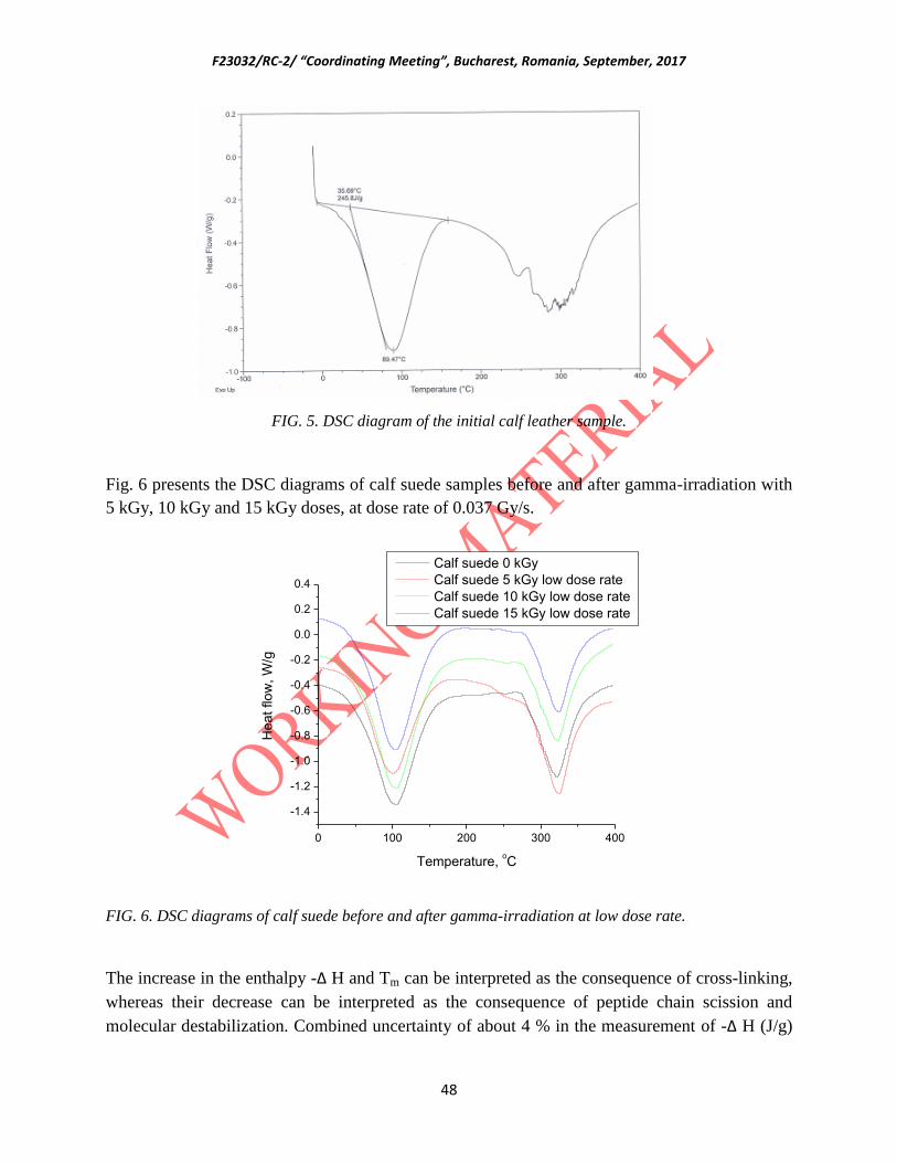
F23032/RC-2/ “Coordinating Meeting”, Bucharest, Romania, September, 2017
48
FIG. 5. DSC diagram of the initial calf leather sample.
Fig. 6 presents the DSC diagrams of calf suede samples before and after gamma-irradiation with
5 kGy, 10 kGy and 15 kGy doses, at dose rate of 0.037 Gy/s.
0 100 200 300 400
-1.4
-1.2
-1.0
-0.8
-0.6
-0.4
-0.2
0.0
0.2
0.4
He
at
flo
w,
W/g
Temperature, oC
Calf suede 0 kGy
Calf suede 5 kGy low dose rate
Calf suede 10 kGy low dose rate
Calf suede 15 kGy low dose rate
FIG. 6. DSC diagrams of calf suede before and after gamma-irradiation at low dose rate.
The increase in the enthalpy -∆ H and Tm can be interpreted as the consequence of cross-linking,
whereas their decrease can be interpreted as the consequence of peptide chain scission and
molecular destabilization. Combined uncertainty of about 4 % in the measurement of -∆ H (J/g)
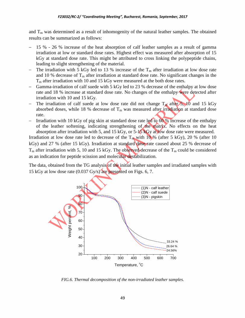
F23032/RC-2/ “Coordinating Meeting”, Bucharest, Romania, September, 2017
49
and Tm was determined as a result of inhomogenity of the natural leather samples. The obtained
results can be summarized as follows:
15 % - 26 % increase of the heat absorption of calf leather samples as a result of gamma
irradiation at low or standard dose rates. Highest effect was measured after absorption of 15
kGy at standard dose rate. This might be attributed to cross linking the polypeptide chains,
leading to slight strengthening of the material.
The irradiation with 5 kGy led to 13 % increase of the Tm after irradiation at low dose rate
and 10 % decrease of Tm after irradiation at standard dose rate. No significant changes in the
Tm after irradiation with 10 and 15 kGy were measured at the both dose rates.
Gamma-irradiation of calf suede with 5 kGy led to 23 % decrease of the enthalpy at low dose
rate and 18 % increase at standard dose rate. No changes of the enthalpy were detected after
irradiation with 10 and 15 kGy.
The irradiation of calf suede at low dose rate did not change Tm after 5, 10 and 15 kGy
absorbed doses, while 18 % decrease of Tm was measured after irradiation at standard dose
rate.
Irradiation with 10 kGy of pig skin at standard dose rate led to 60 % increase of the enthalpy
of the leather softening, indicating strengthening of the matrix. No effects on the heat
absorption after irradiation with 5, and 15 kGy, or 5-15 kGy at low dose rate were measured.
Irradiation at low dose rate led to decrease of the Tm with 10 % (after 5 kGy), 20 % (after 10
kGy) and 27 % (after 15 kGy). Irradiation at standard dose rate caused about 25 % decrease of
Tm after irradiation with 5, 10 and 15 kGy. The observed decrease of the Tm could be considered
as an indication for peptide scission and molecular destabilization.
The data, obtained from the TG analysis of the initial leather samples and irradiated samples with
15 kGy at low dose rate (0.037 Gy/s) are presented on Figs. 6, 7.
100 200 300 400 500 600 70020
30
40
50
60
70
80
90
100
26.64 %
24.56%
33.24 %
We
igh
t p
erc
en
t, %
Temperature, oC
(1)N - calf leather
(2)N - calf suede
(3)N - pigskin
FIG.6. Thermal decomposition of the non-irradiated leather samples.

F23032/RC-2/ “Coordinating Meeting”, Bucharest, Romania, September, 2017
50
The TG curves of the three leather patterns have similar shapes. Highest weight percent
remained in the calf leather after heating up to 650 oC (33.24 %), followed by pig skin (26.64 %)
and calf suede (24.56 %). The irradiated samples of calf suede showed slight increase of the
weight percent remained after heating up to 650 oC, as compared to the non-irradiated sample
(from 24.56 % to 27.06 %). This effect can be due radiation induced crosslinking of the collagen.
100 200 300 400 500 600 70020
30
40
50
60
70
80
90
100
27.06 %
33.43 %
We
igh
t p
erc
en
t, %
Temperature, oC
15 kGy calf leather
15 kGy calf suede
15 kGy pigskin
FIG. 7. Thermal decomposition of the leather samples, irradiated with 15 kGy at low dose rate.
The TG analysis of the leather samples with absorbed does of 15 kGy at standard dose rate (1
Gy/s) showed slight increase of the thermal decomposition (1.5 - 2 %), which might be related to
gamma-irradiation induced destruction changes of their matrixes..
4. CONCLUSIONS
The studies on the effects of gamma-irradiation treatment on the structure and morphology of
calf leather, calf suede and pig skin with 5 kGy, 10 kGy and 15 kGy at low and standard dose
rates can be summarized as follows:
No significant changes in the morphology and molecular structure of the studied leather
samples were observed, as revealed by SEM and FT-IR analysis of the internal and external
sides of the leather samples before and after gamma-irradiation treatment.
EPR analysis showed increased number of radiation-induced radicals in the calf leather
samples, compared to the calf suede and pig skin patterns. This can be explained by the
F23032/RC-2/ “Coordinating Meeting”, Bucharest, Romania, September, 2017
51
presence of the non-isolated Cr3+
ions in the chrome tanned leather patterns (calf suede and
pig skin), which interact with the oxygen radicals, formed during the gamma-irradiation. The
spin concentration in the samples from the three leather patterns is not influenced by the dose
rate of the gamma-irradiation, except the signal in the calf leather, where higher spin
concentrations were found in the samples, irradiated at low dose rate.
According to DSC analysis, gamma-irradiation with fungicide doses led to increase of the
enthalpy of the calf leather melting, attributed to cross linking of the biopolymer chains and
leading to expected strengthening of the material. Gamma-irradiation of calf suede at
standard dose rate and pig skin samples at both dose rates led to decrease with 20 to 25 % of
the temperature at the minimum heat flow (Tm, oC), which implies for molecular
destabilization.
TG analysis showed no significant changes in the thermal decomposition of the three leather
patterns, irradiated at 15 kGy.
The obtained results showed that gamma-irradiation treatment of calf leather, calf suede and pig
skin with insecticide and fungicide doses can be successfully applied for disinfestation and
preservation without causing significant changes in their structure and morphology, even at low
dose rate, where higher radiation-induced side effects are to be expected.
REFERENCES
[1] Cortella, L., Tran, K.Q., Głuszewski, W.J., Moise, I.V., Ponta, C.C., 2011. Nuclear
Techniques for Preservation of Cultural Heritage Artefacts. IAEA Technical Cooperation Project
– RER 8015: Using Nuclear Techniques for the Characterization and Preservation of Cultural
Heritage Artefacts in the European Region.
[2] Adamo, M., Baccaro, S., Cemmi, A., 2015. Radiation processing for bio-deteriorated
archived materials and for consolidation of porous artefacts, ENEA Technical Report
RT/2015/5/ENEA.
[3] Katušin-Ražem, B., Ražem, D., Braun, M., 2009. Irradiation treatment for the protection and
conservation of cultural heritage artefacts in Croatia. Rad. Phys. Chem. 78, 729–731.
[4] Magaudda,G., 2004. The recovery of biodeteriorated books and archive documents through
gamma irradiation: some considerations on the results achieved. J. Cultural Heritage 5(1),113–
118.
[5] Moise, I. V., Virgolici, M., Negut, C. D., Manea, M., Alexandru, M., Trandafir, L., Zorila,
F.L., Talasman, C. M., Manea, D. ,Nisipeanu, S., Haiducu, M., Balan, Z., 2012. Establishing the
irradiation dose for paper decontamination. Radiat. Phys. Chem. 81(8),1045–1050.
[6] Da Silva, M., Moraes, A.M.L., Nishikawa, M.M., Gatti, M.J.A., Vallim de Alencar, M.A.,
Brandão, L.E., Nobrega, A., 2006. Inactivation of fungi from deteriorated paper materials by
radiation, Int. Biodet. Biodegr., 57, 163-167.
[7] Baccaro, S., Cemmi, A., Ferrara, G., Fiore, S., 2015. Calliope gamma irradiation facility at
ENEA-Casaccia R.C. (Rome), ENEA Technical Report RT/2015/13/ENEA.

F23032/RC-2/ “Coordinating Meeting”, Bucharest, Romania, September, 2017
52
[8] Baccaro, S, Caccia, B., Onori, S., Pantaloni, M., 1995. The influence of dose rate and oxygen
on the irradiation induced degradation in ethylene-propylene rubber, Nucl. Insrr. and Meth. in
Phys. Res. B 105(1-4), 97-99.
Croatia
Assessment of optimal irradiation conditions for gamma-radiation treatment
of common fungi on selected cultural heritage objects
B. Mihaljevic, I. Pucic, Katarina Marušic, M. Šegvic Klaric*
Radiation Chemistry and Dosimetry Laboratory (RCDL), Ruđer Boškovic Institute, Zagreb,
Croatia
*Department of Microbiology, Faculty of Pharmacy and Biochemsitry, University of Zagreb,
Croatia
Abstract
There are large needs for systematic approach to data concerning radiation treatment of CH artefacts because of the complex
situation of cultural heritage (CH) objects constituting of different materials in relation to various bioburdens. The goal of this
work is to address those needs by studying the radiation sensitivity of selected microrganisms commonly occurring on CH
artefacts and of the model materials used on paintings. A common carrier for paintings is glue-coated linen that is vulnerable to
fungal biodeterioration. This study aimed to assess antifungal effect of gamma-irradiation doses and dose rates against naturally
occurring mycobiota and artificially inoculated primary (Aspergillus jensenii), secondary (Cladosporium spaherospermum) and
tertiary (Trichoderma harzianum) fungal colonizers common for cellulose materials like linen.
The model systems were irradiated with 2, 7, 20 and 50 kGy at two dose rates that differ by two orders of magnitude, 0.1 and 0.9
Gy/s. After irradiation the number of viable fungi was determined by plate count method in order to determine the proper
irradiation dose for eradication of a particular fungi type. Results indicated that species of Cladosporium and yeasts seem to be
the most resistant fungi to gamma irradiation. In parallel, preliminary assessment of gamma radiation impact on selected physico-
chemical properties of textile was determined.
1. RCM project’s aims
The objective of this CRP is to investigate radiation effects on components of cultural heritage
objects (paintings), with the special consideration directed to the functional properties under

F23032/RC-2/ “Coordinating Meeting”, Bucharest, Romania, September, 2017
53
specific irradiation conditions driven by specific radiation resistance of the microorganisms
typically found on cultural heritage artefacts. The project has two aims:
1. To investigate the gamma irradiation resistance of fungal species that are usually
encountered on paintings;
2. To investigate the impact of gamma irradiation on physico-chemical properties of
selected base materials common in paintings.
In the first year the research on CRP project was focused on gamma radiation resistance of
fungal species that are commonly encountered on CH artefacts and assessment of the doses
necessary for reduction of the contamination to an acceptable level: the effect of the dose rate.
2. Work plan of the present work
The work was planned as follows:
Preparation of model systems
Linen that is the most common base material in paintings was selected as model material. The
base material was coated with size (rabbit-skin glue) that is typical for CH artefacts. The samples
were inoculated with a single fungal species that are primary (Aspergillus spp.), secondary
(Cladosporium spp.) or tertiary colonizers (Trichoderma spp.).
Irradiation of the model systems.
The model systems were irradiated to several doses at two dose rates that differ by two orders of
magnitude. The dosimetry was performed with the ECB dosimeter. After the 60Co source was
replenished new dose mapping is being performed by ECB and verified by ionization chamber
measurements.
Impact of gamma radiation on fungi
After irradiation, the number of viable fungi was determined by plate count method in order to
determine proper irradiation dose for eradication of a particular fungi type.
Preliminary assessment of gamma radiation impact on selected physico-chemical properties of
base materials was determined.
3. Determination of natural mycobiota and inoculation of the model samples

F23032/RC-2/ “Coordinating Meeting”, Bucharest, Romania, September, 2017
54
Firstly, the natural mycobiota of the model samples was determined. Secondly, model samples
were inoculated with a particular fungal species including primary (Aspergillus jensenii),
secondary (Cladosporium spaherospermum) and tertiary colonizers (Trichoderma harzianum) at
concentration 50 000 CFU per mL of sterile water. Plates with inoculated linen as well as
controls (without inoculum) were incubated at 25°C and 70-80% of relative humidity (Rv) for 7
days. Figure 1. represents linen model samples with Cladosporium spp.
FIG. 3.1. Linen model samples with Cladosporium spp.
Fig. 3.2 presents the obtained results of the naturally occurring mycobiota after 7 days of
incubation at 25°C and 70-80% of Rh.
FIG. 3.2. Mean concentration of naturally occurring mycobiota and its growth after 7 days of
incubation at 25°C and 70-80% of Rh
Fungi Mean fungal concentration
(CFU/g)
Growth of fungal concentration after
7 days (CFU/g)
Aleternaria spp. 2∙103 -
Aspergillus spp. 103 9∙10
7
Cladosporium spp. 103 5∙10
5
Fusarium spp. 103 -
Penicillium spp. 103 4∙10
7
Yeast - 9∙107

F23032/RC-2/ “Coordinating Meeting”, Bucharest, Romania, September, 2017
55
Other fungi 2∙103 -
Alternaria spp., Aspergillus spp., Cladosporium spp., Fusarium spp. and Penicillium spp.
comprised in naturally occurring mycobiota, in initial concentrations of 103 and 2∙10
3 CFU/g
(yeast). These fungi were non-homogeneously dispersed on glue-coated linen which resulted in
uneven development of microbiological populations.
Upon 7 days of incubation in humid atmosphere the concentration of mycobiota increased for
few orders of magnitude. Yeast, which did not appear in the initial state grew for almost 8 orders
of magnitude after a week, indicating that for the development of yeast humidity and time are
needed.
4. Impact of gamma radiation on selected fungi
The model textile systems were exposed to a range of radiation doses:
- 2 kGy and 7 kGy that are common for CH artefacts treatment against fungi,
- 20 kGy and 50 kGy as greater doses for identification of radiation effects on materials.
The dose rates were also varied: 0.1 Gy/s and 9.8 Gy/s. The temperature in gamma chamber was
about 18°C.
Alternaria spp., Aspergillus (section Flavi), Cladosporium spp., Fusarium spp., Penicillium spp.
and Rhizopus spp. comprised naturally occurring mycobiota on glue-impregnated linen. These
fungi were inhomogeneously dispersed on the linen. After irradiation treatment of the samples
from each plate (controls with natural mycobiota and samples inoculated with primary,
secondary or tertiary colonizers) were serially diluted (up to 10-3
) and inoculated in culture
medium in duplicate. Plates were incubated for 7, 14 and 28 days at 25°C. After the incubation
period, plate count method was applied to determine the number of viable fungi and proper
irradiation doses that eliminate fungi, respectively. Results after irradiation of the naturally
occurring mycobiota are presented in Tables 4.1. and 4.2.
TABLE 4.1. Levels of naturally occurring mycobiota after irradiation at 2 kGy and dose rates of
0.1 and 9.8 Gy/s.

F23032/RC-2/ “Coordinating Meeting”, Bucharest, Romania, September, 2017
56
Fungi
Mean fungal concentration (CFU/g)
0th
day 7th
day 14th
day 28th
day
Dose rate (Gy/s)
0.1 9.8 0.1 9.8 0.1 9.8 0.1 9.8
Aletrnaria spp. - -
Aspergillus
(Flavi) - - - - -
A. fumigatus - - - - - - -
Aspergillus
(Nigri) - - - - - - -
Aspergillus spp. - - - - - - -
Cladosporium
spp. -
Epicocccum spp. - - - - - - -
Fusarium spp. - - - - -
Mucor spp. - - - - - - -
Penicillium spp. - - - - - -
Phoma spp. - - - - - - -
Stemphylium spp. - - - - - - -
Yeast - - - -
Other fungi - - - - -

F23032/RC-2/ “Coordinating Meeting”, Bucharest, Romania, September, 2017
57
TABLE 4.2. Mean fungal concentration of naturally occurring mycobiota (CFU/g) after
irradiation at 7 kGy and 20 kGy, both applied at dose rates of 0.1 and 9.8 Gy/s.
Fungi
CFU/g at
7 kGy 20 kGy
0th
day 7th
day 14th
day 28th
day 28th
day
Dose rate (Gy/s)
0.1 9.8 0.1 9.8 0.1 9.8 0.1 9.8 0.1 9.8
Aletrn.spp. - - - -
-
-
-
Clados.spp. - - -
- -
-
Fusar.spp. - - - - - -
- - 6
Penicil.spp. - - - - - - - - -
Yeast - - - -
Other fungi - - - - -
Dose of 2 kGy was ineffective in reduction of linen mycobiota to the initial level. After 28 days
of incubation fungi were recovered in concentrations up to 106 CFU/g. Yeast, Alternaria spp.
and Cladosporium spp. showed the highest resistance towards irradiation since recovery was
observed already on the 0th
day. Dose of 7 kGy (0.1 Gy/s) was ineffective in reduction of linen
mycobiota to the initial level; after 28 days of incubation fungi were recovered in concentrations
up to 106 and 10
5 CFU/g, respectively. Dose of 20 kGy (0.1 Gy/s) reduced Cladosporium spp.,
and Alternaria spp. to 104 CFU/g. Penicillium spp. was reduced to the initial level while yeasts,
Aspergillus spp. and Fusarium spp. recovered in concentrations below initial. Upon exposure to
50 kGy sterile white mycelia was recovered on few plates after incubation periods. Higher dose
rate (9.8 Gy/s) was more effective in fungal elimination than lower 0.1 Gy/s.
Table. 4.3. presents the mean concentration of inoculated mycobiota and its growth after 7, 14
and 28 days of incubation at 25°C and 70-80% of Rh after irradiation with 20 kGy. Fig. 4.1.

F23032/RC-2/ “Coordinating Meeting”, Bucharest, Romania, September, 2017
58
presents the gamma-irradiation treatment survival of secondary colonizer Cladosporium
sphaerospermum grown on glue-coated linen.
TABLE 4.3. Mean concentration of inoculated mycobiota and its growth after 7, 14 and 28 days
of incubation at 25°C and 70-80% of Rh after irradiation with 20 kGy
Fungi
Mean fungal concentration (CFU/g)
0th day 7
th day 14
th day 28
th day
Dose rate (Gy/s)
0.1 9.8 0.1 9.8 0.1 9.8 0.1 9.8
Aspergillus
jensenii
- - - - - - - -
Cladosporium
spaherospermum
- - - - - -
Trichoderma
harzianum
- - - - - - - -
Mixed cultures - - - - - -
FIG. 4.1. Gamma-irradiation treatment survival of secondary colonizer Cladosporium
sphaerospermum grown on glue-coated linen
All applied doses and dose rates were effective against artificially inoculated primary
(Aspergillus jensenii) and tertiary colonizers (Trichoderma harzianum). Secondary colonizer,
F23032/RC-2/ “Coordinating Meeting”, Bucharest, Romania, September, 2017
59
Cladosporium sphaerospermum survived radiation treatment with 2 and 7 kGy, but the number
of viable Cladosporia was significantly reduced as compared to the initial inoculum (50 000
CFU/mL); from the samples treated with 2 kGy, at both dose rates (0.1 and 9.8 Gy/s), 2.5 %, 5%
and 20% of initial inoculum was recovered after 7, 14 and 28 days of incubation, respectively.
From the samples exposed to 7 kGy at 0.1 Gy/s, 0.1%, 3 and 4% of initial inoculum was
recovered after 7, 14 and 28 days of incubation, respectively, while 7 kGy at 9.8Gy/s suppressed
growth of Cladosporia. Doses of 20 and 50 kGy suppressed the growth of all artificially
inoculated fungi.
5. Preliminary assessment of gamma radiation impact on selected physico-chemical
properties of textile
Preliminary FTIR-ATR measurements on coated linen did not indicate degradation in the dose
range applied in this study.
ATR-FTIR on linen used in contemporary paintings has shown it to be a mixture of linen and
polyamide. Polyamides are relatively radioresistent, far more than cellulose. In FTIR spectra,
polyamide band covers most absorption of cellulose and it is not possible to determine any
changes in cellulose.
Comparison of paper consisting of pure cellulose: the differences between the ATR-FTIR spectra
of paper samples (Fig. 5.1) do not correlate with the irradiation dose in the examined range of
doses (0-50 kGy). The protective effect of the glue coating cannot be excluded although is
relatively difficult to assess.

F23032/RC-2/ “Coordinating Meeting”, Bucharest, Romania, September, 2017
60
FIG. 5.1. ATR-FTIR spectra of paper samples
Further research on coated and non-coated samples, using FTIR and other complementary
experimental techniques (SEM, TGA) is planned while mycelia was recovered on few plates
after 7, 14 and 28 days of incubation at 25°C.
6. Work Program for the next period
1. The study of the radiation sensitivity of common mycobiota and radiation effects on
inoculated animal glue-impregnated linen canvas will be completed. Preliminary results showed
that Alternaria spp., Aspergillus (section Flavi), Cladosporium spp., Fusarium spp. and
Penicillium spp. survived even 20 kGy at the dose rate of 0.1 Gy/s. Because of these dose rate
effects will be further studied.
2. Model systems based on paper used in paintings will also be prepared, inoculated, radiation-
treated and analysed in the same manner as canvas, including dose rate effects. Possible
differences in development and radiation sensitivity of mycobiota will be assessed.
3. The study of the radiation sensitivity of common mycobiota and radiation effects on
inoculated varnishes. A selection of varnishes will be tested, some of historic varnishes (like
shellac, dammar, mastic, Gum Arabic and the like) and some modern varnishes. The effect of
irradiation on properties of treated varnishes including appearance.

F23032/RC-2/ “Coordinating Meeting”, Bucharest, Romania, September, 2017
61
4. Influence of bioburden concentration at inoculation on radiation survival of common fungi and
molds on canvas and paper, uncoated and coated with selected coatings will be assessed. The
dose and dose rate will be varied.
5. Influence of common coatings for canvas and paper including synthetic ones on radiation
sensitivity of mycobiota will be assessed. The dose rate effects and state (age) of each coating
will be assessed.
6. The effect of the irradiation dose and dose rate on properties of treated materials including
changes in visual appearance will be assessed. We will continue with the model system
investigated until now. The research will be expanded on paper carriers and results compared.
7. We will investigate the materials common on cultural heritage separately, but also combined
(carriers: paper, canvas and different binders).
8. Influence of (canvas and paper) carrier coating type and state (age) on irradiated carrier
properties including appearance immediately after irradiation and during post-period will be
studied.
According to the results further planes may be modified.
Cuba
PROGRESS REPORT – Research Contract 18924
E. PRIETO MIRANDA, I. PADRON DIAZ
Center of Technological Application and Nuclear Development (CEADEN), La Habana, Cuba
Abstract
The better procedure to stop the fungal growth on books and archive documents is keeping a low
humidity rate and temperature levels, together with a good air circulation on archive and museum
rooms. Actually, in Cuba many heritage depository institutions do not have the means to achieve the
ideal air conditions. For this reason, the disinfection of paper against fungi should be a priority in our
country and the gamma irradiation could be the best approach to solve this serious problem.
A database of D10 doses for most important biodeteriorations agents in our museums and archives
should be created as first step for the development of conservation procedures applying gamma

F23032/RC-2/ “Coordinating Meeting”, Bucharest, Romania, September, 2017
62
irradiation and the elaboration of reference guide for action depending on the material composition
and contamination level.
The importance of this CRP to disseminate the benefits of gamma irradiation technique for Culture
Heritage consolidation and preservation is argumented.
Absorved dose distribution is calculated by Monte Carlo simulation method for laboratory scale
gamma irradiator. The results are compared to experimental measurements using Fricke and Alanine
dosimetric systems.
Dose rate uniformity (DUR) and its dependence of material density was calculated for semi-industrial
gamma plant using MCNPX code. The importance of Monte Carlo simulation for irradiation planning
in the case of voluminous and non-symmetric artifacts is revealed.
1. The use of gamma irradiation as antifungal method on paper conservation.
Since ancient times, paper has become the main carrier of cultural, political, scientific and
historical information. Its preservation against physical, chemical and biological deteriogenesis a
matter of great interest. Due to the inappropriate conservation conditions throughout history,
fungi are the major responsible of documentation damages and losses. The main goal of this
work, is the discussion of advantages and disadvantages of gamma treatment as antifungal
method on paper conservation.
Why the disinfection of paper against fungi should be a priority in Cuba?
The bioreceptivity of paper is high due to its higroscopicity and composition (cellulose,
ligning, adhesives), which represent an abundant carbon source to the heterotrophic
organisms.
Fungi require less moisture than bacteria to develop.
Keeping a low humidity rate and temperature levels, together with a good air circulation
on archive and museum rooms is the better procedure to stop the fungal growth on
papers. Actually, some heritage depository institutions do not have the means to achieve
the ideal air conditions.
At least in Cuba, the environmental conditions normally existent in museums, archives
and libraries frecuently possibilitate the growth of fungi
A good antimicrobial method should have chemical stability, low cost, broad activity spectrum,
and should have no adverse effects on treated materials or toxicity for humans.
Gamma radiation has a great microbicidal activity destroying cellular DNA due to its ability to
generate radicals capable of cleaving carbon-carbon bonds and does not leave chemical residues
[1]. As advantage of gamma irradiation as antifungal method, there are not toxic residues in the
case of cultural heritage preservation. Also, the PH and mechanical resistance of treated papers
remains practically unaffected.
Different authors [2, 3, 4] indicate the range between 10 and 20 kGy as the lethal dose of
radiation for all fungal species, but also some deterioration effects on paper should be
considered. In order to estimate the minimal radiation dosage and to avoid the cumulative
degrading effects on archived documents is important to determine the fungal specie to be
treated.

F23032/RC-2/ “Coordinating Meeting”, Bucharest, Romania, September, 2017
63
The formation of radicals, the way as gamma radiation acts killing the microorganisms, is the
same mechanism that deteriorates the organic matter like cellulose, the main component of
paper. Besides, a fall in the degree of polymerization, gamma irradiation at dose rate about 10
kGy, can also become cellulose more susceptible to further fungal contaminations [5].
Gamma treatment at dose rate 17 kGy can induce fungi to produce more colored metabolic
residues. This increment on pigmentation represents a serious problem for museum curators,
when valuable heritage documents are treated by gamma method.
2. Scientific collaboration with museum and archive conservators
Points of view in common with conservation specialists:
The use of gamma irradiation for mass treatment of deteriorated book and documents
stored at Cuban National Library (Dose ≈ 3-5 KGy)
In case of Emergency due to flood or any water related event can be used the
combination of Freezing-drying and gamma irradiation methods (Dose ≈ 3 KGy)
The importance of irradiation dose planning by Monte Carlo modeling for limiting the
side effects on CH artifacts and archived documents due to overdose or non-homogeneus
irradiation. In case of voluminous and non-symmetric objects a detailed Monte Carlo
simulation should be always guaranteed.
A database of D10 doses for most important biodeteriorations agents in our museums and
archives should be created.
To work together on the development of conservation procedures applying gamma
irradiation and the elaboration of reference guide for action depending on the material
composition and contamination level.
To disseminate the benefits of gamma irradiation technique for Culture Heritage
consolidation and preservation.
Disagreements related to CH conservation methods:
Practically all chemical agents are banned or strong limited for employing in CH
conservation because of its toxicity and long term effects.
The majority of CH conservation groups are suspicius about the use of artificial polymers
or hybrid materials for consolidation and protection of artworks made of textiles, wood
and paper.
Importance of CRP experiences dissemination for the promotion of gamma irradiation technique:
The use of gamma irradiation for obtaining nanocomposities and its aplication in CH
preservation due to its biocide effect is a novedous topic that should be studied.
To follow the results on aplication of hybrid materials developed by some CRP
participants.
The study of gamma irradiation effects on paper properties is important for optimal dose
selection and preservation procedure development.
The aplication of Monte Carlo Simulation Method to the dosimetric characterization of
gamma irradiation devices as a tool for gamma treatment planning.

F23032/RC-2/ “Coordinating Meeting”, Bucharest, Romania, September, 2017
64
The project of various participants in this CRP includes the developments of hybrid materials
for consolidation and protection of CH artifacts. It is important for us the dissemination of
their positive experience to convince our conservation colaborators.
Other participants are reporting the colateral effects on paper properties. That is quite
important for selection of gamma dose to eliminate specific fungal species and to develop the
conservation procedure and the reference guide employing gamma irradiation technique.
3. Monte Carlo modeling and dosimetric characterization of ISOGAMMA
LLCoGamma Irradiator at the CEADEN
Fig. 1 Geometrical setup ofISOGAMMA LLCoGamma Irradiator installed at the CEADEN,
including the source rack and 5 liters sample holder.
The irradiator dose rate was calculated using MCNPX code considering the 60
Co gamma source
distribution supplied by the fabricant. The geometry setup showed in figure 1 was simulated in
details according its design and real dimensions according to the design showed in figure 2.
The dose required to achieve a desired effect was established through research comprising
determining the dose-effect relation, for example sterilization or reduction of pathogens. These
works are conducted to determine the minimum required to achieve the desired effect and the
maximum tolerated dose irradiated product without modifying the physical and chemical
properties.
The ISOGAMMA LLCo radiation facility of the CEADEN not ordered by their manufacturers
commissioning dosimetry, or a procedure to be followed for such purposes, for such reason it is
necessary to develop the dosimetry to characterize dose distribution of gamma laboratory
irradiator, which will contribute to the correct operation of the facility, ensuring the quality of
future radiation process.

F23032/RC-2/ “Coordinating Meeting”, Bucharest, Romania, September, 2017
65
Figure 2: CEADEN´s Gamma irradiator design for MCNPX simulations.
Selection of experimental measurement points. The selection of the experimental measurement points was performed taking into account the
geometry of the irradiation chamber of the facility. In this case the dosimeters were distributed in
areas where the maximum and minimum dose values are expected within the irradiation
chamber, as shown in Figure 3 below.
Low dose zone
High dose zone
Figure 3. Fricke Dosimeter distribution inside the sample holder
The cylindrical irradiation chamber was divided into three study zones, Figure 4. In each of these
zones 9 Fricke dosimeters were placed. At the bottom zone corresponds the positions 1 to 9, in
the central zone the positions 10 to 18 and the upper zone the positions 19 to 27.

F23032/RC-2/ “Coordinating Meeting”, Bucharest, Romania, September, 2017
66
Figure 4 Zone distribution for dosimetric study of the irradiation chamber.
The results of dose distribution in the vertical direction of the irradiation chamber, obtained with
the Fricke dosimeters, are reflected in the figure 5.
Figure 5. Dose Rate Distribution measured with Fricke Dosimeters inside the sample holder
Figure 6. Dose distribution values in the vertical direction of the irradiation chamber
Upper zone
Central zone
Bottom zone

F23032/RC-2/ “Coordinating Meeting”, Bucharest, Romania, September, 2017
67
Analyzing the obtained absorbed dose values for each study zone in the vertical direction, it was
observed that the maximum dose values in the irradiation camera corresponds at the central zone,
while the minimum dose values are in the high and low zones. These results, showed in the
figure 6 can be attributed to the distribution geometry of the radioactive sources.
Later, the alanine-ESR dosimetry was also employed to determine the dose distribution in the
vertical direction of the irradiation chamber. The highest dose values are observed in the central
part of the radiation chamber and the lower values in the upper and bottom parts (see figure 7).
Figure 7. Dose Rate Distribution measured with Alanine Dosimeters inside the sample holder
The dose rate value obtained by the Fricke dosimeter was of 6,024 kGy/h in march/2013 and for
alanine dosimeter of 4,529 kGy/h in august/2015, which corresponding with the of estimated
dose rate value of 4,44kGy/h, with a relative error of 1,9% between both dosimetric systems.
It was expected that Dose Uniformity Ratio (DUR) should be approximately equal to unity in all
positions inside the 5 liters’ sample holder for material densities corresponding to paper, wood
and textiles. Meaning that, for small documents and CH objects, gamma treatment for plague
disinfection could be guaranteed at CEADEN with good dose uniformity.
4. Gamma treatment for Cultural heritage conservation extended to the
(100kCi) 60Co gamma plant at the IIIA.
Due to its penetrability and microbicidal activity, specifically for fungi, gamma rays can be used
for bulk treatment of books and voluminous documents. The adequate facility for treatment at
that scale is the semi-industrial (100 kCi) Gamma Irradiator at IIIA, a panoramic (Category II)
product overlap type of irradiation. The irradiator has a tote box-type product transportation
system with tote box dimensions (50x50x90cm) as is represented in the figure 8. The unique
source frame is droved by the source hoisting system based on electrical motor, reduction and
transmission mechanisms.

F23032/RC-2/ “Coordinating Meeting”, Bucharest, Romania, September, 2017
68
Gamma irradiation dose for plague disinfection of archive documents can be easily estimated,
due to the low density and homogeneity of these materials. Radiation treatment of cultural
heritage objects demands more detailed dose rate modeling, especially voluminous non-
symmetric artifacts.
We have estimated the internal book density about 1.209 g/cm3 with a variation (± 20%)
depending on moisture content and other materials used for book fabrication. Moisture content
will have a marked influence, and should be considered for dose rate homogeneity modeling.
For different species the wood density oscillates from 0.36 to 1.35 gr/cm3. The most common art
material between densest woods is African Blackwood (1.27 gr/cm3), the legendary “Ebony”.
Figure 8. Block diagram of the Tote Box Transport System (TBTS)
Monte Carlo simulations of Dose Uniformity Ratio (DUR) were carried out using MCNPX for
the tote box diagram showed in the figure 2. In order to calculate the gamma dose deposition,
inside each (50x 50 x 90 cm) box 9 similar tissue equivalent spheres (5 cm dia.) were fixed for
gamma flux monitoring. One sphere is fixed on each tote box corner and one in the middle for all
the irradiation positions around the source rack.
The DUR calculation was carried out comparing the gamma flux accumulated on each nominal
sphere No.1, No.2, ..., No.9 inside the box at the 64 irradiation positions.The sphere projections
for bottom and central edges are represented in the figure 9.
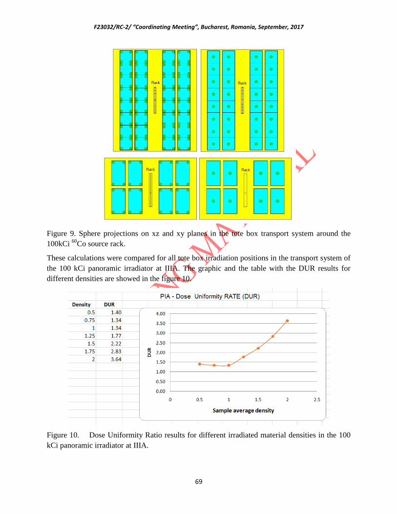
F23032/RC-2/ “Coordinating Meeting”, Bucharest, Romania, September, 2017
69
Figure 9. Sphere projections on xz and xy planes in the tote box transport system around the
100kCi 60
Co source rack.
These calculations were compared for all tote box irradiation positions in the transport system of
the 100 kCi panoramic irradiator at IIIA. The graphic and the table with the DUR results for
different densities are showed in the figure 10.
Figure 10. Dose Uniformity Ratio results for different irradiated material densities in the 100
kCi panoramic irradiator at IIIA.

F23032/RC-2/ “Coordinating Meeting”, Bucharest, Romania, September, 2017
70
Proposed Work Plan:
To create a database of D10 doses for most important biodeteriorations agents in our
museums and archives.
To develop a CH conservation procedure applying gamma irradiation and the elaboration
of reference guide for action depending on the material composition and contamination
level.
To realize the dosimetric characterization of 100kCi semi-industrial gamma plant
employing different high dosimetry methods.
To use the Monte-Carlo simulation for planning the bulk gamma treatment of books and
archive documents at the IIA semi-industrial gamma plant.
To organize periodically national workshops in contact to the CH specialists in order to
disseminate the results of CRP participants on the study of gamma irradiation effects on
paper properties and on aplication of hybrid materials for protection of artworks.
To simulate with MCNPX code the Multipurpose Gamma Irradiation Facility – IPEN-
CNEN (1000 kCi of Cobalt-60) as a contribution for artwork irradiation planning in
Brazil.
References
[1] S. Sequeira, E.J. Cabrita, M.F. Macedo in the J. International Biodeterioration &
Biodegradation 74 (2012) 67-86.
[2] S.C. Pavon Flores in the Bulletin of American Institute for Conservation of Historic and
Artistic Works, 16, (1976), 15-44.
[3] F. Butterfield in the J. Studies in Conservation 32, (1987), 181-191.
[4] G.C. Tomazello, M. Wiendl, M. Maximiliano in the J. Restaurator, 16, (1995), 93-99.
[5] M. Adamo, G. Magaudda, P.T. Nisini, G. Tronelli in the J. Restaurator, 24, (2003), 145-151.
Egypt
Nano- Carbon Based Materials Serving as Corrosion protection in Cultural Heritage
Metals and Free Radical Scavenger in Artefacts made from Cellulosic Materials
H.A. ABD EL-REHIM*, K.M. AMIN, N.M. DEGHIEDY Radiation Research of Polymer Chemistry Department, National Centre for Radiation Research and Technology
(NCRRT),
Atomic Energy Authority, Cairo,Egypt

F23032/RC-2/ “Coordinating Meeting”, Bucharest, Romania, September, 2017
71
Abstract
Gamma irradiation was used as an effective and “eco-friendly” method for production of nano-
carbon based material graphene sheets. Graphene oxide, prepared from graphite, was reduced to graphene
upon exposure to gamma radiation. The corrosion behavior of iron alloys coated with gamma irradiated
graphene and its composites with chitosan and inhibitor were evaluated in 3.5% NaCl and 0.5M sulfuric
acid solution respectively. The resulting gamma irradiated graphene and graphene- composites covered
iron alloys were characterized using UV-Vis spectroscopy, X-ray Diffraction (XRD)and Field Emission
Scanning Electron Microscopy (FE-SEM). The protection efficiencies for graphene and
graphene/chitosan were 82.2% and 89%, respectively. Graphene/chitosan films over iron alloys showed
higher corrosion activation energies compared to graphene sheets. Electrochemical
ImpedanceSpectroscopy(EIS) proved the stability of graphene and graphene/chitosan coatings after
different immersion times in 3.5% NaCl solution. Coated surfaces were free from pits on the scale of
magnification as demonstrated from SEM images. Potentiodynamic polarization indicated the protection
efficiency offered by graphene coatings which was enhanced upon using the inhibitor with graphene
sheets. The protection efficiency increased (up to 95.8%) with increasing the inhibitor concentration with
an optimum concentration of 2 mM. Electrochemical impedance spectroscopy measurements EIS
confirmed the stability of the graphene/inhibitor coatings after prolonged immersion time in 0.5 M
sulfuric acid solution.
On the other hand, Papyrus Linen textile and cellulosic paper were selected as examples of
natural occurring materials. In order to improve the mechanical properties of these natural materials and
their radiation resistance properties against degradation, they were immersed in graphene oxide solution
which was used as a reinforcement material.
The exposure of cellulosic samples and graphene oxide reinforced ones to 25kGy irradiation
dose resulted in the formation of free radicals detected by electron paramagnetic resonance spectroscopy
at room temperature. The peak intensity of the irradiated reinforced materials was lower than that of
graphene oxide -free ones. Upon storage at room temperature, the radicals decayed and the decay rate
depended on the nature of the materials. The results revealed that the fate of radicals trapped inPapyrus
and Cellulosic paper is very fast and the half-life for Papyrus and Cellulosic papers incorporated with
graphene oxide is much lower than that of reinforced Linen textile. This means that the incorporation of
all cellulosic samples with graphene oxide enhanced their resistance toward gamma radiation. Also, it
was found that the incorporation of graphene oxide into papyrus sheets improved and reinforce their
tensile strength mechanical properties. No noticeable changes on the mechanical and morphological

F23032/RC-2/ “Coordinating Meeting”, Bucharest, Romania, September, 2017
72
properties was detected as well as colour stability when graphene oxide reinforced natural materials
irradiated with to 25kGy as well as colour stability.
INTRODUCTION
Metallic artifacts are an important part of the cultural heritage and must be protected.
During their life and after their abandonment and sometime their burial, metallic artefacts of
cultural heritage are submitted to long term corrosion processes, associated with the progressive
modification of their surfaces and their covering of corrosion products [1]. The nature of these
corrosion products depends on one hand on the kind of metal that is corroded and, on the other
hand, on the kind of environment (soil, atmosphere, marine water…) presenting a specific
chemicalcomposition. Classical metal protection used for example in industry anticorrosion
methods can in most of cases not be used in the context of the conservation of metal cultural
heritage because artefacts must not be aesthetically modified and any protection treatment must
be potentially removable without any damage to the artefact [2]. Therefore, the metallic
heritage cannot be protected by usual industrial anticorrosion methods as painting or inhibiting
agents that will cause changes of the external appearance of the artefacts. The coating should
have no or very little effect on the surface appearance; it should be as reversible as possible so it
can be removed to return the object to its original state; it should not modify the material of the
original artefact; it must have long-term efficiency because heritage artefacts are intended to be
preserved for as long as possible; and finally, it should be easy to maintain. These considerations
impose important limitations on the selection of the heritage artefacts protective materials [3].
Non negligible part of artefacts of the cultural heritage made of iron or iron alloy
submitted to atmospheric corrosion. In museum exhibition showcases, it is possible to control the
exposure conditions and to keep the artefacts at a low relative humidity. Nevertheless, in most of
cases, it is not possible to control these conditions [1]. Most of the treatments that are applied
today as waxes or carboxylate deal with layers with thickness of several micron or tenth of
microns, susceptible to modify the external aspect after applying [4]. Moreover, the durability of
the efficiency of these treatments is questioned.
The revolution of materials science allowed the development of smart and effective
methods for corrosion protection. Graphene is considered one of the most important and
extensively investigated materials for the last decade [5]. Graphene, a two dimensional material,
is a one atom thick structure consisting of hexagonal units of sp2 -bonded carbon atoms.

F23032/RC-2/ “Coordinating Meeting”, Bucharest, Romania, September, 2017
73
Nowadays, graphene has been introduced as an excellentanticorrosion material because of its
unique characteristics such as excellent thermal and chemical stability, chemical inertness, high
flexibility, impermeability to molecules even as small as helium, and remarkable mechanical
properties [6].
Most of the previous studies used expensive Chemical Vapor deposition (CVD) for
deposition of graphene on metals [7]. Therefore, a novel route which combines the high purity of
physical routes with convenience of chemical synthesis and economic benefits is economically
and environmentally advantageous. Gamma radiation (γ-radiation), electromagnetic radiation of
high frequency, is one of the well-known ionizing radiation. Recently, reduction of graphene
oxide using gamma irradiation offers a promising “green” method for large scale and low-cost
production of graphene. Furthermore, no chemicals or catalyst precursors are used which leads to
a process with no chemical requirements or waste streams [8].
2. METHODOLOGY
2.1. PREPARATION OF GRAPHENE OXIDE (GO) SUSPENSION
In brief, GO was prepared according to Hummers' method throughoxidation of graphite
powder (<50 mm, purchased from Merck) [10]. Graphite powder was added to a mixture of 0.5 g
of NaNO3 and4.0 g of KMnO4 in concentrated H2SO4 (70 mL) under vigorousstirring at 0oC.
Then, the temperature of the suspension was brought to 35oC with stirring for 2h 50 mL of water
was slowly added and the temperature was maintained at 98oC for 15 min. Thesuspension was
then further diluted with approximately 160 mL ofwater. Shortly thereafter, the suspension was
treated with 30%hydrogen peroxide. The suspension turned into bright yellow color. GO was
then filtrated and washed with 5% HCl to remove residual metal ions. Finally dissolution and
centrifugation were performed in DI water, repeatedly to remove the unwanted acid and then
leftto dry to obtain pure graphene oxide.
2.2. IRRADIATION REDUCTION OF GO INTO GAMMA IRRADIATED GRAPHENE (G)
G was prepared by radiation induced reduction of GO. The aqueous suspension of GO
atconcentration of 1 mg/mL was prepared. The as-prepared samplewas sealed and irradiated by
a60
Co γ-ray source constructed by National Center for Radiation Research and Technology
(NCRRT), Egyptian Atomic Energy Authority (EAEA) at a dose rate of 2.5 kGy/hand exposed

F23032/RC-2/ “Coordinating Meeting”, Bucharest, Romania, September, 2017
74
to a total dose 50 kGy. After the irradiation, the samplewas purified bywashing with ethanol and
plenty of DIwater. Finally,a black powder of graphenewas obtained after the sample was
driedindicating the reduction of GO. Graphene powderwas dissolved in DMF toobtain a
suspension of concentration 1.5 mg/ml. Elemental analysiswas conducted on graphene oxide
(GO) and GIG; the C to O atomicratios is found to be 3.8:1 for GO, and 8.1:1 for G.
FIG. 1. Schematic illustration of GO and G formation.
2.3. PREPARATION OF GRAPHENE/CHITOSAN COMPOSITE (G/CS)
To obtain G/CS hybrid, a 0.5% (mass ratio) chitosan solution was prepared by dissolving
chitosan in 1.0% acetic acid solution then 10 mL of CS solution was added to the above G
suspension.
2.4. PREPARATION OF GRAPHENE/INHIBITOR COMPOSITE (G/INH)
In the first step, 1.5 mg/mL G suspension was prepared bydissolving graphene powder in
DMF. Then inhibitor powder wasadded to the prepared G suspension to obtain three
differentinhibitor concentrations (0.1, 1 and 2 mM). The GIG/IN compositeswere sonicated
using ultrasonic probe for 1 h. Aliquots of 10 mL ofinvestigated coating were added onto the
surface of iron alloy electrode. The electrode was left to dry at 60 oC in air and finally exposed to
the test solution. Based on the inhibitor content in thecomposite, the samples were designated as
GIG/0.1 mMInh, GIG/1 mMInh, and G/2 mMInh. G, Inh and the numbers representgamma
irradiated graphene, inhibitor and the concentration ofinhibitor in the composite, respectively.
2.5. PHYSICOCHEMICAL CHARACTERIZATION
Infrared spectrophotometry was carried out by a Jasco FT/IR-6600 spectrophotometer
(Japan). Spectra were recorded using 32repeated scans per spectrum at 0.4 cm-1 resolution with
S/N ratio:45000:1 in the region of 4000-400 cm-1 to obtain the FTIR spectrumbased on the

F23032/RC-2/ “Coordinating Meeting”, Bucharest, Romania, September, 2017
75
transmission characteristics of the sample. The scanning electron microscopy (SEM)
measurements were performed on the surface of coatedsubstrates to characterize the surface
morphology using Field emission scanning electron microscope (FE-SEM) by a Quanta250 FEG
Field Emission Scanning Electron Microscope.
2.6. SUBSTRATE PREPARATION AND COATING DEPOSITION
The substrate material used for the present investigation was iron alloy (AISI 316).
Specimens were in the form of sheets (1.0 mm thick) androds. The sheets were used for the
surface measurements whilethe rods were employed for electrochemical experiments. Thetest
electrode which has cross-sectional area of 1.13 cm2 wasmechanically polished by emery papers
to ensure the samesurface roughness. This was followed by degreasing in acetone,rinsing with
ethanol and drying in air. Aliquots of 10 mL of Gsuspension or G/CS mixture were added onto
the surface of iron alloy electrode. The electrode was left to dry at 60 oC (for10 minutes) and
finally exposed to the test solution.
2.7. ELECTROCHEMICAL CELLS AND EQUIPMENTS
The cell used for electrochemical measurements was a typicalthree-electrode/one
compartment glass cell. The working electrodewas a 316 stainless steel alloy, reference electrode
wasAg/AgCl (4.0 M KCl) and a Pt wire (5 cm long; diameter: 2 mm)as auxiliary
electrode.Electrochemical impedance spectroscopy measurements(EIS) were employed to
monitor the corrosion performance ofthe coated iron alloy substrates in a 3.5% NaCl
solution.EIS measurements were carried out at the open circuitpotential (OCP), using a Gamry-
750 instrument and a lock-inamplifier that are connected to a personal computer. The
dataanalysis was provided with the instrument and applied nonlinearleast square fitting with
Levenberg–Marquardt algorithm.All impedance experiments were recorded between0.1 Hz and
100 kHz with an excitation signal of 10 mVamplitude. The potentiodynamic polarization test
was used to determinethe overall corrosion behavior of the specimen. Thepotential of the
electrode was swept at a rate of 1 mV s-1 fromthe initial potential (Ei) of ~250 mV versus open
circuit potential(OCP) to the final potential (Ef) of +250 mV versus OCP. Beforethe test, the
electrode was left under open-circuit conditionsuntil a steady corrosion potential value was
reached.Cyclic polarization experiments were used to determine theprotection and pitting

F23032/RC-2/ “Coordinating Meeting”, Bucharest, Romania, September, 2017
76
corrosion potentials. Experimental setupwas as described for the potentiodynamic polarization
text withan initial potential (Ei) of +0.5 V, maximum potential (Emax) of+0.5 V and a final
potential (Ef) of _0.5 V (vs. EOC). All experimentswere performed in controlled room
temperature conditions at 25oC.
3. RESULTS AND DISSCUSSION
The aim of the this part of the present work is to use gamma irradiation as a safe and
useful route to reduce graphene oxide to graphene and its application for protection of metallic
heritage iron alloy (austenitic stainless steel 316) against corrosion in 3.5% sodium chloride
electrolyte. The resulting graphene was also mixed with chitosan and inhibitor to form a
composites that were also evaluated as effective coatings against corrosion for iron alloy. On one
hand it was important to evaluate the extent of matrix holding of G to CS and inhibitor and on
the other to evaluate its performance as coating protection against corrosion.
3.1.STRUCTURAL AND SURFACE STUDIES OF GRAPHENE
In order to follow the successful steps of the G synthesis, UV-Vis spectra and X-ray
diffraction measurements have been used during different preparation stages.The UV–Vis
spectra of graphene oxide and graphene are shown in Fig. 2. Both materials were dispersed in
double distilled water until homogeneously distributed materials were obtained. It is observed
that the spectra shows a shoulder around 299 nm that corresponds to n→π* transition of C=O
bondsfor GO.This shoulder practically disappeared and the absorption peak around 239 nm
corresponding to π →π* transitions of aromatic C–C bondsis red-shifted to 276 nm after
irradiation for G. This is the first indication of the conversion of GO to G.
FIG. 2.UV-Vis spectra of GO and GIG.FIG. 3.XRD patterns of GO and GIG.
XRD patterns of the graphene oxide and graphene are shown in Fig.3.

F23032/RC-2/ “Coordinating Meeting”, Bucharest, Romania, September, 2017
77
The XRD pattern of GO shows a characteristic peak at 2θ value of 11.3. After γ-
irradiation, the intensity of this peak decreases significantly upon formation of GIG and two new
broad peaks appear at 2θ values of about 26 and 42.3, respectively. This indicates that the
oxygen functional groups in the interlayer spacing of the graphene oxide had been removed
during reduction process.
The qualitative analysis for GO and GIG samples using FTIR is shown in Fig.4. FTIR
spectrum of GO confirmed the presence of different types of oxygen functionalities. It shows a
broad band at 3430 cm-1
which is attributed to stretching of the OH groups. This band
significantly weakened in the GIG spectrum due to reduction and removal of hydroxyl groups.
GO spectrum shows strong peaks at around 1716 and 1060 cm-1
correspond to C=O and C-O
stretching vibrations, respectively. The intensity of these peaks dramatically decreased indicating
the decarboxylation effect of g-ray irradiation on GO sheets and the peak at 1060 cm1 became
sharper which is attributed to the remaining carboxyl groups even after reduction. The spectrum
of GO also shows peaks at 1628 cm-1
(aromatic C=C skeletal vibration of unoxidized graphitic
carbons) and at 1384 cm1 may be ascribed to the presence of C-C bonds. Both spectra of GO and
GIG show peaks between 2800 and 2980 cm-1
, assigned to stretching vibration of C-H,
indicating the presence of some alkyl groups attached to the sheets.
FIG. 4.FTIR spectra of GO and GIG.FIG. 5.FTIR spectra of GO and G.
Raman spectroscopy is a powerful nondestructive tool to characterize carbonaceous
materials, especially for distinguishing ordered and disordered crystal structures of carbon. The
Raman spectra of GO and GIG in Fig. 5show documented D band peak at 1346 cm-1
, which
represents a breathing mode of k-point phonons of A1g symmetry due to the sp3defects and G
band peak at 1585 cm-1
, which can be attributed to the inplane vibrations of sp2carbon atoms and

F23032/RC-2/ “Coordinating Meeting”, Bucharest, Romania, September, 2017
78
a doubly degenerated phonon mode (E2gsymmetry) at the Brillouin zone center. The overtone of
the D line, the 2D line, which is related to the stacking nature of graphene layers, was observed
at 2686 cm-1
and the 2G line appears at 2930 cm-1
. In the Raman spectrum of graphene, the
intensity of the D band increased markedly due to the significant decrease of the size of the in-
plane sp2 domains and the partially ordered graphite crystal structure of graphene nanosheets.
The appearance of D peak requires a defect for its activation, so the D band intensity can be
usedas a measure for the degree of disorder. After the irradiation, the ID/ IG ratio increased
substantially from 0.96 to 1.22. Although the decarboxylation is evident from the FT-IR
measurements, however the increase in the ID/IG ratio accounts more for the defects created in
the structure.
FIG. 6.FE-SEM of G .FIG. 7.TEM of G.
The field-emission scanning electron microscope (FE-SEM) image in Fig.6 shows the
morphology of the as-prepared G. The G film showed a rippling structure with compact stacking
over the substrate. The as-prepared G nanosheets were fully analyzed by TEM observations.
Fig.7. shows TEM image of graphene indicating the general view of G sheets and clearly
illustrating the flake-like shapes of graphene. Large graphene sheets corrugated together with
sizes in the range of tens to several hundreds of square nanometers where they resemble
crumpled silk veil waves. Wrinkled regions and a scrolled edge are observed which is the
intrinsic microscopic roughening of graphene sheets. This is attributed to the thermodynamic
stability of the 2D membrane comes from microscopic crumpling via bending. G sheets were
rippled and entangled with each other. They seem transparent in some regions and exhibit a very
stable nature under the electron beam. The most transparent and featureless regions seem to be
few layers of graphene sheets.
3.2. PROTECTIVE ABILITY OF GRAPHENE PREPARED BY γ- IRRADIATION
AND IMPREGNATED WITH CHITOSAN AND ORGANIC INHIBITOR
AGAINST CORROSION

F23032/RC-2/ “Coordinating Meeting”, Bucharest, Romania, September, 2017
79
3.2.1. Electrochemical measurements
3.2.1.1. Potentiodynamic polarization measurements
Fig. 8 shows the polarization curves of bare AISI 316, AISI 316 coated with GIG and
AISI 316 coated with GIG/CS in 3.5% NaClat room temperature (25oC). The corrosion current
densities were determined by extrapolating the linear part of the anodic and cathodic components
of the curves. The results of the potentiodynamic polarization including corrosion potential
(Ecorr), corrosion current (icorr), anodic and cathodic Tafelconstants (βa, βc) and the protection
efficiencies (PE) of GIG and GIG/CS are presented in Table 1.
FIG. 8.Tafel polarization curves of bare iron alloy, G and G/CS in 3.5% NaCl at 298 K.
The corrosion potential of G-coated stainless steel is -57.1 mV while that measured at
iron alloy bare is -183.1 mV. As indicated by the values of Ecorr, a shift is observed to more
positive potential in the anodic direction indicating more protection as the value mounts to more
noble direction. The icorrvalue (0.0745 μA cm-2
) for the G-coated alloy is remarkably lower than
the corresponding value recorded for bare iron alloy surface, ca. 0.4183 mA cm-2
. It is therefore
concluded that the presence of GIG coating resulted in corrosion protection to the surface of
steel. It is also important to relate the corrosion protection ability of graphene coating over iron
alloy to its largespecific surface area, excellent mechanical properties and two dimensional
geometry of the graphene sheet. While graphene is expected to allow electronic conduction to
the surface, but its hydrophobic nature and chemical inertness contributes to the isolation of the
surface from electrolytic interactions. G/CS shows the lowest icorr value (0.0462 μA cm-2
)
indicating enhanced corrosion protection efficiency of the G/CS composite for the steel surface.
It is therefore concluded that a synergistic effect with respect to corrosion protection of iron alloy
surface is expected when mixing CS to G to form the corresponding composite.
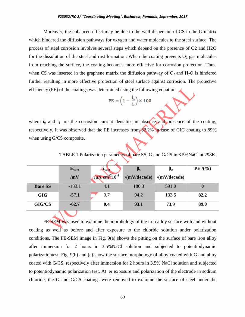
F23032/RC-2/ “Coordinating Meeting”, Bucharest, Romania, September, 2017
80
Moreover, the enhanced effect may be due to the well dispersion of CS in the G matrix
which hindered the diffusion pathways for oxygen and water molecules to the steel surface. The
process of steel corrosion involves several steps which depend on the presence of O2 and H2O
for the dissolution of the steel and rust formation. When the coating prevents O2 gas molecules
from reaching the surface, the coating becomes more effective for corrosion protection. Thus,
when CS was inserted in the graphene matrix the diffusion pathway of O2 and H2O is hindered
further resulting in more effective protection of steel surface against corrosion. The protective
efficiency (PE) of the coatings was determined using the following equation
where i0 and i1 are the corrosion current densities in absence and presence of the coating,
respectively. It was observed that the PE increases from 82.2% in case of GIG coating to 89%
when using G/CS composite.
TABLE 1.Polarization parameters of bare SS, G and G/CS in 3.5%NaCl at 298K.
FE-SEM was used to examine the morphology of the iron alloy surface with and without
coating as well as before and after exposure to the chloride solution under polarization
conditions. The FE-SEM image in Fig. 9(a) shows the pitting on the surface of bare iron alloy
after immersion for 2 hours in 3.5%NaCl solution and subjected to potentiodynamic
polarizationtest. Fig. 9(b) and (c) show the surface morphology of alloy coated with G and alloy
coated with G/CS, respectively after immersion for 2 hours in 3.5% NaCl solution and subjected
to potentiodynamic polarization test. A er exposure and polarization of the electrode in sodium
chloride, the G and G/CS coatings were removed to examine the surface of steel under the
Ecorr
/mV
icorr
/μA cm-2
10-1
βc
/(mV/decade)
βa
/(mV/decade)
PE /(%)
Bare SS -183.1 4.1 180.3 591.0 0
GIG -57.1 0.7 94.2 133.5 82.2
GIG/CS -62.7 0.4 93.1 73.9 89.0

F23032/RC-2/ “Coordinating Meeting”, Bucharest, Romania, September, 2017
81
coatings. Fig. 9(d) and (e) show the morphology of the steel surface after removing the G and
G/CS coatings, respectively. It is observed that the GIG and G/CS offer effective protection to
the steel surface and no pitting spots were observed on the surface. It is important to mention that
some “black” spots appeared in the images of the SEM that mainlycorresponded to the most
adherent parts of graphene after peeling the layer off the surface of iron alloy.
FIG.9. FE-SEM of (a) bare iron alloy after immersion in 3.5% NaCl and subjected to Tafel test
(250x & 2500x), (b) iron alloy /G before immersion in 3.5% NaCl (20 000x), (c) iron alloy
/(G/CS) before immersion in 3.5% NaCl (20 000x), (d) and (e) the iron alloy surfaces with G
and G/CS films removed, respectively, and after immersion in 3.5% NaCl for 2 hours and
subjected to Tafel test (2500x).
Fig. 10 represents the potentiodynamic polarization curves of “bare” stainless steel,
stainless steel coated with graphene prepared by gamma irradiation (G), and stainless steel
coated with G containing different concentrations of the organic inhibitor (1-aryl-3-
phenylcarbamoyl-8,9-dimethoxy-10-b-methyl-[1,2,4]-triazolo-[3,4-a]-1,5,6,10-b-tetrahydro-
isoquinoline-4-methoxy) (PDTI). Prior to the potentiodynamic experiment, all electrodeswere
left in solution for a period of 30 min until a steady state value of potential is attained (open
circuit potential, OCP). Anodic polarizationwas then scanned (5mVs-1
) from -250 mV (vs.
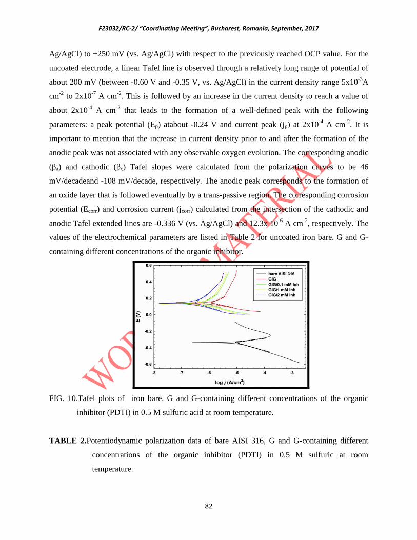
F23032/RC-2/ “Coordinating Meeting”, Bucharest, Romania, September, 2017
82
Ag/AgCl) to +250 mV (vs. Ag/AgCl) with respect to the previously reached OCP value. For the
uncoated electrode, a linear Tafel line is observed through a relatively long range of potential of
about 200 mV (between -0.60 V and -0.35 V, vs. Ag/AgCl) in the current density range 5x10⁻³
A
cm-2
to 2x10⁻⁷
A cm-2
. This is followed by an increase in the current density to reach a value of
about 2x10⁻⁴
A cm-2
that leads to the formation of a well-defined peak with the following
parameters: a peak potential (Ep) atabout -0.24 V and current peak (jp) at 2x10⁻⁴
A cm-2
. It is
important to mention that the increase in current density prior to and after the formation of the
anodic peak was not associated with any observable oxygen evolution. The corresponding anodic
(βa) and cathodic (βc) Tafel slopes were calculated from the polarization curves to be 46
mV/decadeand -108 mV/decade, respectively. The anodic peak corresponds to the formation of
an oxide layer that is followed eventually by a trans-passive region. The corresponding corrosion
potential (Ecorr) and corrosion current (jcorr) calculated from the intersection of the cathodic and
anodic Tafel extended lines are -0.336 V (vs. Ag/AgCl) and 12.3x 10⁻⁶
A cm-2
, respectively. The
values of the electrochemical parameters are listed in Table 2 for uncoated iron bare, G and G-
containing different concentrations of the organic inhibitor.
FIG. 10.Tafel plots of iron bare, G and G-containing different concentrations of the organic
inhibitor (PDTI) in 0.5 M sulfuric acid at room temperature.
TABLE 2.Potentiodynamic polarization data of bare AISI 316, G and G-containing different
concentrations of the organic inhibitor (PDTI) in 0.5 M sulfuric at room
temperature.

F23032/RC-2/ “Coordinating Meeting”, Bucharest, Romania, September, 2017
83
The scanning electron microscopy pictures are given in Fig. 11 for a cross-section of
coating over iron alloy of G containing the inhibitor. In general, the film show rough structure
that is not apparently porous and no voids are identified according to the magnification used
(x150000). From surface water contact angle testing, the contact angle was found to be 84.15o.
Thus, partial wettability is expected that shows relatively low surface hydrophobicity. Thus, the
hydrophobic character and structure of the film will not be accounted for as an effective barrier
against water and oxygen penetration. From the elevations noticed in the SEM images for both
films, surface roughness is highly represented and results in a distorted double layer capacitance
at the interface.
FIG. 11. FE-SEM of a cross section of G/2 mMInh coating over iron alloy.
3.3. ENHANCEMENT OF MECHANICAL PROPERTIES AND RADIATION RESISTANCE
OF SAME MATURAL MATERIALS USING GRAPHENE OXIDE
Ecorr
/mV
jcorr x 10-6
A cm-2
βc
(mV/decade)
βa
(mV/decade)
PE /(%)
Bare AISI 316 -336 12.3 108 46.2 0
GIG 141 2.94 103 62.3 76.0
GIG/0.1
mMInh
139 1.34 129 62.4 89.2
GIG/1 mMInh 137 1.14 130 602 90.8
GIG/2 mMInh 140 0.520 133 408 95.8
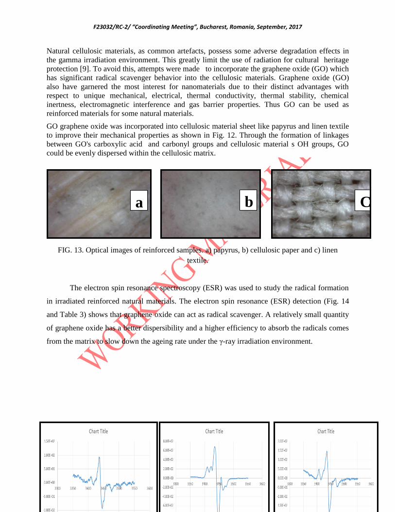
F23032/RC-2/ “Coordinating Meeting”, Bucharest, Romania, September, 2017
84
Natural cellulosic materials, as common artefacts, possess some adverse degradation effects in
the gamma irradiation environment. This greatly limit the use of radiation for cultural heritage
protection [9]. To avoid this, attempts were made to incorporate the graphene oxide (GO) which
has significant radical scavenger behavior into the cellulosic materials. Graphene oxide (GO)
also have garnered the most interest for nanomaterials due to their distinct advantages with
respect to unique mechanical, electrical, thermal conductivity, thermal stability, chemical
inertness, electromagnetic interference and gas barrier properties. Thus GO can be used as
reinforced materials for some natural materials.
GO graphene oxide was incorporated into cellulosic material sheet like papyrus and linen textile
to improve their mechanical properties as shown in Fig. 12. Through the formation of linkages
between GO's carboxylic acid and carbonyl groups and cellulosic material s OH groups, GO
could be evenly dispersed within the cellulosic matrix.
FIG. 13. Optical images of reinforced samples. a) papyrus, b) cellulosic paper and c) linen
textile.
The electron spin resonance spectroscopy (ESR) was used to study the radical formation
in irradiated reinforced natural materials. The electron spin resonance (ESR) detection (Fig. 14
and Table 3) shows that graphene oxide can act as radical scavenger. A relatively small quantity
of graphene oxide has a better dispersibility and a higher efficiency to absorb the radicals comes
from the matrix to slow down the ageing rate under the γ-ray irradiation environment.
a b C

F23032/RC-2/ “Coordinating Meeting”, Bucharest, Romania, September, 2017
85
FIG. 14.ESR spectra of a) papyrus, b) linen textile and c) cellulosic paper.
TABLE 3.Peak intensity and decay percent of different natural materials
Papyrus Textile White Paper
hour PI Decay % PI Decay % PI Decay %
4 299 0.00 1388 0.00 477 0.00
52 204 31.77 715 48.49 372 22.01
148 168 43.81 846 39.05 275 42.35
4. COCLUSION
Gamma irradiation we successfully used for the reduction of graphene oxide into
graphene (G). The G and its composites with chitosan and organic inhibitor offered high
protection efficiency for the iron alloy .Potentiodynamic polarization exhibited remarkable
decrease in jcorr for G-coated iron alloy compared to the iron alloy bare. Natural materials
(papyrus, linen textile and cellulosic papers) were successfully reinforced by using GO nano-
sheets. ESR study of the γ-ray irradiation materials showed that the graphene oxide can act as
radical scavenger, since the amount of radicals in the matrix formed by the γ-irradiation
decreased.
REFRENCES
[1] DILLMANN, P., Nanoscale Aspects of Corrosion on Cultural Heritage Metals, Atlantis
Press and the Author, (2016).
a b c

F23032/RC-2/ “Coordinating Meeting”, Bucharest, Romania, September, 2017
86
[2] CANO, E., LAFUENTE, D.,BASTIDAS, D.M., Use of EIS for the evaluation of the
protective properties of coatings for metallic cultural heritage: a review, J. Solid State
Electrochem. 14 (2010) 381.
[3] MIRAMBET, F., REGUER,S., ROCCA,E., HOLLNER,S., TESTEMALE, D., A
complementary set of electrochemical and X-ray synchrotron techniques to determine the
passivation mechanism of iron treated in a new corrosion inhibitor solutionspecifically
developed for the preservation of metallic artefacts, Appl. Phys. A Mater. Sci. Process. 99
(2010) 341.
[4] HOLLNER, S., MIRAMBET, F.,ROCCA, E., REGUER, S., Evaluation of new non-toxic
corrosion inhibitors for conservation of iron artefacts, Corros. Eng. Sci. Technol., 45 (2010)
362.
[5] ZHANG, J., CAO, Y., FENG, J., WU, P., Graphene-oxide-sheet-induced gelation of
cellulose and promoted mechanical properties of composite aerogels, J. Phys. Chem. C, 116
(2012) 8063.
[6] OKAFOR,P.A., SINGH-BEEMAT,J., IROH,J.O., Thermomechanical and corrosion
inhibition properties of graphene/epoxy estersiloxan urea hybrid polymer nanocomposites,
Prog. Org. Coat.,88 (2015) 237.
[7] BONACCORSOL, F., LOMBARDO, A., HASAN, T., SUN, Z., COLOMBO, L., FERRARI,
A.,Production and processing of graphene and 2d crystals, Mater. Today, 15 (2012) 564.
[8] ZHANG,B., LI,L., WANG,Z., XIE,S., ZHANG,Y., SHEN,Y., YU,M., DENG,B.,
HUANG,Q., FAN,C., LI,J., Radiation induced reduction: an effective and clean route to
synthesize fuctionalized graphene, J. Mater. Chem.,22 (2012) 7775.
[9] SUN, Y., CHMIELEWSKI, A.G., Applications of Ionizing Radiation in Materials
Processing, Institute of Nuclear Chemistry and Technology, Poland (2017).
[10] HUMMERS,W.S., OFFEMAN, R.E., Preparation of graphitic oxide, J. Am. Chem. Soc.,80
(1958) 1339.
France
Study of styrene-free unsaturated resins and acrylic monomers for the consolidation of
wooden cultural heritage artefacts by radiation-curing

F23032/RC-2/ “Coordinating Meeting”, Bucharest, Romania, September, 2017
87
K.TRAN, C.SALVAN, M.POLONI
ARC-Nucléart, CEA-Grenoble, France
Abstract
The aim of this research is to study the feasibility of using styrene-free unsaturated resins and acrylic
monomers for the consolidation of degraded wooden artefacts from cultural heritage by in-situ radiation-
curing. Indeed, styrene unsaturated polyester resin is implemented during decades for this purpose, but
the safety regulation concerning the styrene monomer is more and more severe, and the request from
museum conservators for styrene-free, acrylic consolidants in the type of thermoplastics are the main
motivations of this work. Acrylic monomers such as hydroxy-propyl methacrylates, alkyl methacrylates
and available styrene-free resins are tested for the consolidation of sound wooden samples, as well as
degraded samples taken from ancient artefacts. In order to increase the viscosities of the monomers (very
low initially), addition of polymers such as well-known Paraloid® are implemented. Partial consolidation
of the wood was realized by formulating the monomers in various solvents at different concentrations;
such formulations can avoid the inhibition of oxygen on the polymerization of acrylic compounds which
are especially sensitive to this effect. Besides spectroscopic analysis for the radiation-cured polymers
(FTIR, NMR), dimensional changes, mechanical testing, colorimetry, SEM observations are carried out to
characterize the wood-polymer composites. Very promising results were obtained after this study:
styrene-free resin tested could be an alternative to the actual styrene-unsaturated polyester one, and the
formulations based on the tested monomers gave interesting results in terms of surface appearance,
mechanical improvement and dimensional stabilization.
Introduction
Since the seventies, ARC-Nucléart is using a styrene unsaturated polyester resin for the
consolidation of degraded wooden artefacts from cultural heritage, following a process called
“Nucléart” by resin impregnation under vacuum/pressure, and then in-situ polymerization of the
resin under gamma irradiation [1]. However, this method is irreversible due to the crosslinked
solid state resin which is insoluble in any solvent.
Moreover, the consolidation of wood by such 100 % reactive resin fills almost completely the
wooden pore structure, creating in fact a wood-plastic composite which density is much higher
than untreated wood one. These features constitute some drawbacks of this method going against
two well-known criteria of the conservation of artefacts: the reversibility of the treatment and the
minima intervention in order to avoid the denaturation of the original and unique object.
Furthermore, even if this technique had proved during many decades its effectiveness for saving
from destruction numerous highly degraded artefacts [2,3,4], another drawback of the current
process is the toxicity and the relatively high vapour pressure of the styrene monomer.
Moreover, since the safety regulation is becoming more severe, our research is focused on the
improvement of our radiation method in two directions: the use of already available styrene-free
resins, and the development of hydroxyl-acrylic and alkyl monomers, whose polymers are in
principle reversible.

F23032/RC-2/ “Coordinating Meeting”, Bucharest, Romania, September, 2017
88
1. Materials and methods
1.1 Material
The reference resin is the styrene-unsaturated polyester resin (CookCompositesP, France)
implemented so far for wood consolidation. The acrylic polymers used worldwide for
conservation-restoration are the Paraloid® B72, B82, B67, B66, acrylic-methacrylic copolymer
(Rohm & Hass, USA) soluble in current solvents [5].
The styrene-free resins are based on Advalite 35065® with a polar monomer (hydroxyl-propyl
methacrylate) and were delivered for testing by Reichhold Company (USA). The Advalite 35065
is a vinyl hybrid resin. The hydroxy-propyl methacrylate (HPMA) was purchased to TCI Europe,
Belgium. The alkyl methacrylates were purchased at Sigma-Aldrich. Two types of wood are
tested: sound wood (fir and beech) and degraded historical wood by insects dating from the 50’s.
Figure 4: Samples of sound wood and degraded historical wood
Sample preparation
Modern fir and beech were cut to a size of 7*2*1 cm3 blocks and treated by the pure monomer
and its solutions as well as by the resins in liquid state. The wood impregnation is carried out in a
small tank by a vacuum/pressure process: the vacuum was made during 3 hours and the resin or
the monomer was introduced in the tank and a 6 bar nitrogen pressure was maintained for 18
hours. The same protocol is applied for the historical wood. All the samples are irradiated using a 60
Co gamma ray source at a dose rate of 1 kGy/h. Dosimetry by Red Perspex dosimeters
(Harwell, GB). The irradiation facility at Grenoble is a water pool storage one presenting an
activity of 1500 TBq (2017), and an irradiation chamber of 4x4x2.30 m3.
1.2 Color measurement
A Minolta spectrophotometer CM-508i was used to measure wood sample color. A mark was
made on the wood sample to measure CIE Lab value at the exact same place before and after
treatment.
1.3 FTIR, solid state NMR and DSC
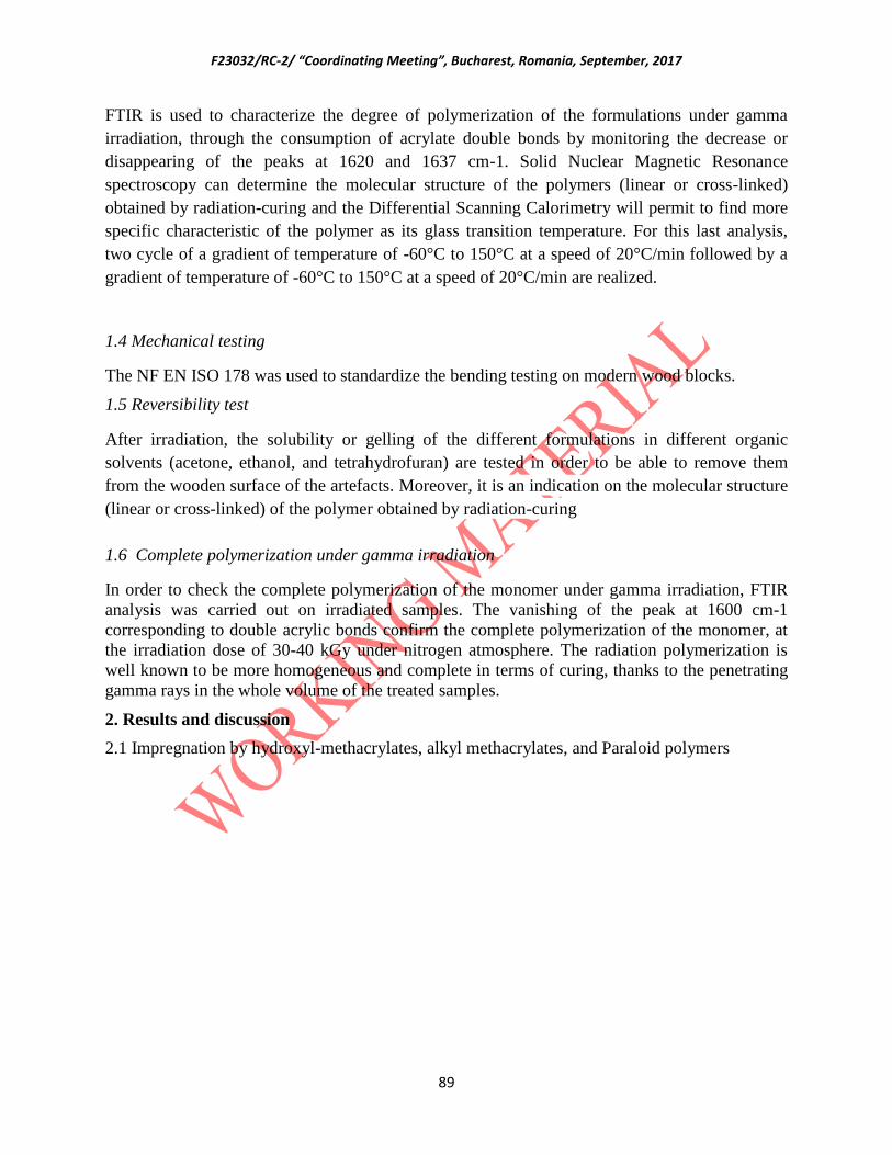
F23032/RC-2/ “Coordinating Meeting”, Bucharest, Romania, September, 2017
89
FTIR is used to characterize the degree of polymerization of the formulations under gamma
irradiation, through the consumption of acrylate double bonds by monitoring the decrease or
disappearing of the peaks at 1620 and 1637 cm-1. Solid Nuclear Magnetic Resonance
spectroscopy can determine the molecular structure of the polymers (linear or cross-linked)
obtained by radiation-curing and the Differential Scanning Calorimetry will permit to find more
specific characteristic of the polymer as its glass transition temperature. For this last analysis,
two cycle of a gradient of temperature of -60°C to 150°C at a speed of 20°C/min followed by a
gradient of temperature of -60°C to 150°C at a speed of 20°C/min are realized.
1.4 Mechanical testing
The NF EN ISO 178 was used to standardize the bending testing on modern wood blocks.
1.5 Reversibility test
After irradiation, the solubility or gelling of the different formulations in different organic
solvents (acetone, ethanol, and tetrahydrofuran) are tested in order to be able to remove them
from the wooden surface of the artefacts. Moreover, it is an indication on the molecular structure
(linear or cross-linked) of the polymer obtained by radiation-curing
1.6 Complete polymerization under gamma irradiation
In order to check the complete polymerization of the monomer under gamma irradiation, FTIR
analysis was carried out on irradiated samples. The vanishing of the peak at 1600 cm-1
corresponding to double acrylic bonds confirm the complete polymerization of the monomer, at
the irradiation dose of 30-40 kGy under nitrogen atmosphere. The radiation polymerization is
well known to be more homogeneous and complete in terms of curing, thanks to the penetrating
gamma rays in the whole volume of the treated samples.
2. Results and discussion
2.1 Impregnation by hydroxyl-methacrylates, alkyl methacrylates, and Paraloid polymers

F23032/RC-2/ “Coordinating Meeting”, Bucharest, Romania, September, 2017
90
Figure 2: Resin or polymer content contain in the treated wood
The monomer and the resin penetrate efficiently into the wood. Fir is a soft wood, and beech is a
hardwood with lower porosity. It is the reason why the resin contents in fir wood are, in the most
cases, higher than the ones with beech wood. Wood impregnated by formulations containing
solvent up to 60 % present obviously lower resin content in wood, due to the solvent loss by
evaporation. Indeed, the lowest resin content was obtained by impregnation with the polymer
Paraloïd B72 at 20 % in ethyl acetate (80%). The aims of using solvent in formulations were to
impregnate the wood partially, avoiding thus the total filling of the wood porosity.
2.2 Colour appearance
The difference of colour between the wood before and after treatment is shown on the Figure 6
below.

F23032/RC-2/ “Coordinating Meeting”, Bucharest, Romania, September, 2017
91
The smallest change in colour (close to natural appearance of non-treated wood) is obtained with
the HPMA 40 IPA 60 (volume %) formulation, even better than with conventional Paraloïd B72
resin application). In general, the monomer consolidation presents a ΔE lower than the ones
obtained with the resins Advalite or styrene-polyester.
Figure 3: ΔE from CIE Lab 2000 norm between the wood before and after treatment.

F23032/RC-2/ “Coordinating Meeting”, Bucharest, Romania, September, 2017
92
2.3 Mechanical bending testing
Flexural testing was carried out on untreated wood and wood consolidated by the resins and
acrylic polymers.
Figure 4 : Diagram of the elastic modulus in bending (MPa) for the wood treated with the
different resins and monomers.
As a result, all the treatments increase the elastic modulus in bending.
- The monomers diluted in solvent do not increase significantly the mechanical properties,
even if the percentage of monomer is high (80%)
- The acrylic formulations offer the best results in increasing the mechanical properties of
the wood, even better than the styrene polyester, especially for beech wood.
The impregnation of the wood with Advalite increases also the mechanical properties, even if
they are lower than those obtained with the styrene polyester resin.
2.4 Volume change
The dimensional changes of the wooden material induced by the treatment are among the most
important features of the process. One should avoid the cracking, the shrinkage, or in the
opposite way, the swelling of the wood structure following the operation. Without solvent, the
resins or monomers present generally shrinkage during the polymerization, inducing decreasing
shrinkage values in the tangential, then radial and finally longitudinal directions of the wood
structure. In presence of solvent in the formulations, the phenomena could be different since the
a) b)
d)
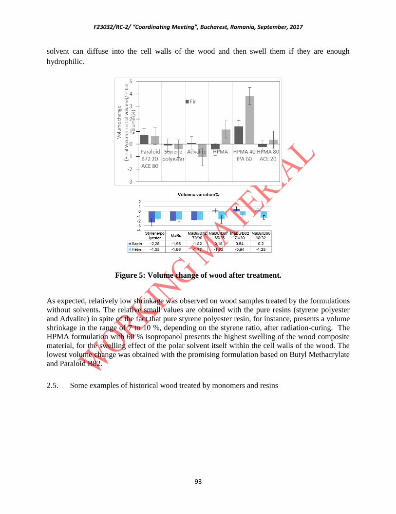
F23032/RC-2/ “Coordinating Meeting”, Bucharest, Romania, September, 2017
93
solvent can diffuse into the cell walls of the wood and then swell them if they are enough
hydrophilic.
Figure 5: Volume change of wood after treatment.
As expected, relatively low shrinkage was observed on wood samples treated by the formulations
without solvents. The relative small values are obtained with the pure resins (styrene polyester
and Advalite) in spite of the fact that pure styrene polyester resin, for instance, presents a volume
shrinkage in the range of 7 to 10 %, depending on the styrene ratio, after radiation-curing. The
HPMA formulation with 60 % isopropanol presents the highest swelling of the wood composite
material, for the swelling effect of the polar solvent itself within the cell walls of the wood. The
lowest volume change was obtained with the promising formulation based on Butyl Methacrylate
and Paraloid B82.
2.5. Some examples of historical wood treated by monomers and resins
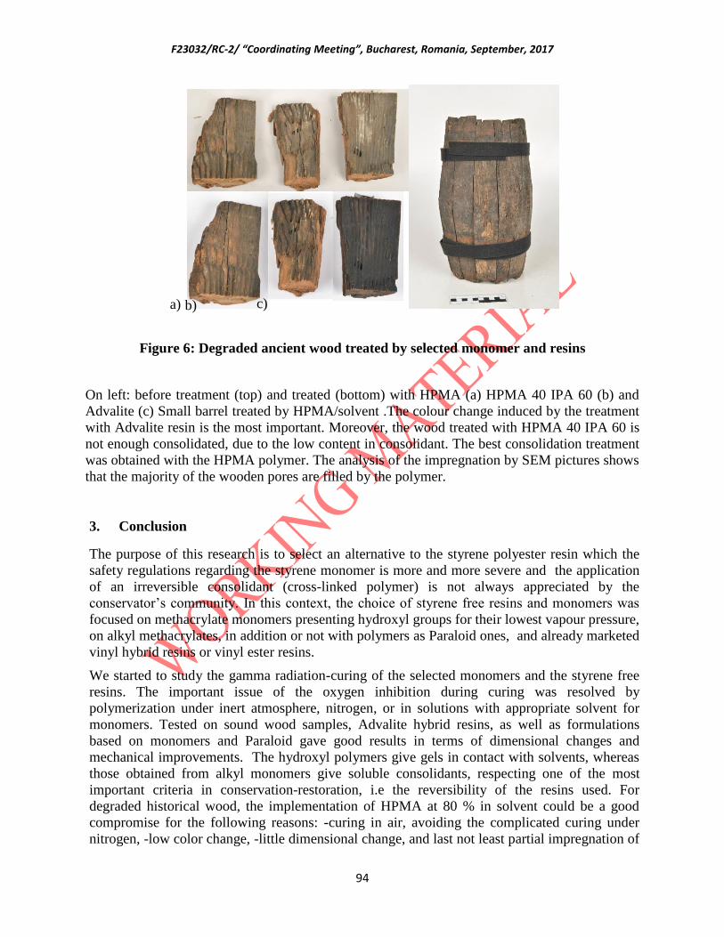
F23032/RC-2/ “Coordinating Meeting”, Bucharest, Romania, September, 2017
94
Figure 6: Degraded ancient wood treated by selected monomer and resins
On left: before treatment (top) and treated (bottom) with HPMA (a) HPMA 40 IPA 60 (b) and
Advalite (c) Small barrel treated by HPMA/solvent .The colour change induced by the treatment
with Advalite resin is the most important. Moreover, the wood treated with HPMA 40 IPA 60 is
not enough consolidated, due to the low content in consolidant. The best consolidation treatment
was obtained with the HPMA polymer. The analysis of the impregnation by SEM pictures shows
that the majority of the wooden pores are filled by the polymer.
3. Conclusion
The purpose of this research is to select an alternative to the styrene polyester resin which the
safety regulations regarding the styrene monomer is more and more severe and the application
of an irreversible consolidant (cross-linked polymer) is not always appreciated by the
conservator’s community. In this context, the choice of styrene free resins and monomers was
focused on methacrylate monomers presenting hydroxyl groups for their lowest vapour pressure,
on alkyl methacrylates, in addition or not with polymers as Paraloid ones, and already marketed
vinyl hybrid resins or vinyl ester resins.
We started to study the gamma radiation-curing of the selected monomers and the styrene free
resins. The important issue of the oxygen inhibition during curing was resolved by
polymerization under inert atmosphere, nitrogen, or in solutions with appropriate solvent for
monomers. Tested on sound wood samples, Advalite hybrid resins, as well as formulations
based on monomers and Paraloid gave good results in terms of dimensional changes and
mechanical improvements. The hydroxyl polymers give gels in contact with solvents, whereas
those obtained from alkyl monomers give soluble consolidants, respecting one of the most
important criteria in conservation-restoration, i.e the reversibility of the resins used. For
degraded historical wood, the implementation of HPMA at 80 % in solvent could be a good
compromise for the following reasons: -curing in air, avoiding the complicated curing under
nitrogen, -low color change, -little dimensional change, and last not least partial impregnation of
a) b) c)

F23032/RC-2/ “Coordinating Meeting”, Bucharest, Romania, September, 2017
95
the wood porosity, satisfying the deontological criteria of the conservation. However, the very
low viscosity, as well as the evaporation or monomer loss at the wood surface could be a
drawback when we have to consolidate artefacts presenting much degraded surface areas. In
order to increase the viscosity of the monomer-solvent formulations, incorporation of the well-
known acrylic polymers as Paraloid B72, B66, B68 , B82, in the radiation-curing system are
tested. The most promising formulations are so far the butyl methacrylate/ Paraloid B82, and the
isobutyl methacrylate/B72.
The air curing protocol could be more satisfactory by addition of phosphite compound in the
acrylic formulations, and the feasibility of them will be implemented on numerous degraded
artefacts selected for testing.
References
[1] Tran Q.K., Ramière R., Ginier-Gillet A., Impregnation with radiation-curing monomers and
resins, in Archaeological Wood, Properties, Chemistry and Preservation, Advances in Chemistry
Series N°225
[2] Tran, K., and M. Guinard M. 2009. Stabilisation of dry archaeological wood having sulphur
compounds by impregnation of radiation-curing unsaturated polyester resin. In Proceedings of
the 10th ICOM Group on Wet Archaeological Materials Conference, Amsterdam 2007, eds. K.
Straetkvern and D.J. Huisman. Amersfoort.
[3] Alejandra Alonso-Olvera and Khôi Tran. Conservation of a pre-Columbian wooden
sculpture: a Mexican French collaboration using gamma radiation technology for consolidation
.Proceedings ICOM-Committee for Conservation 15th
International Conference in New Delhi,
22-26 September 2008,Vol II, pp 724-730
[4] Tran, K. and al. Characterization and Conservation of a gun carriage excavated from the 17th
century HMS Stirling Castle shipwreck. Proceedings of the ICOM-Committee for Conservation
16th
International Conference in Lisbon, 19-23 September 2011, pp 1-9
[5] Crisci G.M., La Russa M.F., Malagodi M., Ruffolo S.A. Consolidating properties of Regalrez
1126 and Paraloid B72 applied to wood, J. of Cultural Heritage 11 (2010), pp 304-308
Iran
Fungal Decontamination of Historical Oil Painting by Using Gamma Ray
R.BETESHO BABRUD1*
, N. SHEIKH1, F.KHATAMIFAR
2, M.E. MOGHADAM
2
1. Radiation Application Research School, Nuclear Science and Technology Research Institute (NSTRI), Atomic
Energy Organization of Iran Tehran-Iran, 2. Conservation Group, Art and Architecture Department, Central Tehran
Branch, Islamic Azad University. Tehran-Iran
Abstract: To evaluate the optimal gamma ray dose for fungal decontamination of a historical oil
painting of 19th century, its fungal contamination were counted and identified with direct sampling and
culture methods. Colors used were identified with infrared spectroscopy (mid and far regions). Fungi D10
F23032/RC-2/ “Coordinating Meeting”, Bucharest, Romania, September, 2017
96
value was investigated on light-thermal aged strips made of similar canvas and colors. The time to
achieve required dose of gamma was calculated by a virtual simulation process using MCNP4C code.
D10 value for dominant fungi (Aspergillus and Penicillium) on aged strips was 0.6 and 1kGy
respectively. Dominant colors were identified as vagon green and burnt amber according to FTIR
spectra. Based on sterility tests and radiation resistance of fungi, 5kGy was sufficient to decontaminate
the selected painting. Time required to irradiation the virtual painting in IR-136 facility according to
results obtained from MCNP4Ccode were 13.35 and 23.06 hours in products density of 0.001293 and 0.1
g/cm3 respectively.
Introduction: Preserving historic heritage is the duty of every nation that cares about its
history. Canvas based paintings are mainly subject to fungal infestation under improper
conservation conditions. The aim of this project was a case study for evaluating the optimal
gamma ray dose for fungal decontamination of a historical oil painting in Iran trough a
coordinate research project with IAEA. The steps of planned program for first two years of the
project and the results achieved are described.
1. Study the fungi bioburden of a selected ancient oil painting and evaluation the dose
required according obtained biological data(1st year)
Selection of painting: Old oil paintings are not only objects of fine art but also
certificates of human history. In order to evaluate t h e optimal gamma ray dose for fungal
decontamination of the paintings, an ancient one over canvas belonging to 19th century with the
dimentions of 3.03m 𝗑1.06m stored in a stock in Museum of Fine Arts of White Palace (Melal
museum) of Sa d-abad Palace was selected.
Characterization and quantification of fungal contamination: Sampling was done from 31
points of 2cm x 2cm of the suspected and discolored points of the painting including front, back
and the areas between canvas and substrate. Sampling was in duplicate directly by wet sterile
swab then surface cultured on Sabouraud Dextrose Agar (SDA) medium and indirectly in sterile
saline which was then tube shaked and cultured on the same medium. Growth of fungi was
determined after 8-12 days incubation in 29°C. Total fungi contamination per cm2 of the painting
was calculated according to the total sampling area and total fungi count on the plates.
Classification was based on cell wall and overall morphological properties like hyphae or
pseudohyphae, septa, spore size and shape.
Investigation of fungal resistance to gamma ray on culture media: To determine the
radiation resistance of native fungi on cultured media (D10 value), duplicate plates of 15 ml SDA
containing equal number of each fungus colonies on surface, were exposed to 1,3,5,7 kGy
gamma rays in Gamma cell GC220 with dose rate of 3.52 Gy/s calibrated with fricke dosimeters.
Whole irradiated agars containing fungi were suspended in 85ml distilled water. One ml from the
serial dilution was pour plated in SDA with pH 6.8. The number of grown fungi was counted
after 21 days incubation in 25°C. D10 value (decimal reduction value) was determined by
graphing fungi populations after the series of radiation dose applied.
F23032/RC-2/ “Coordinating Meeting”, Bucharest, Romania, September, 2017
97
2. Sampling from different materials of the painting and study the effect of gamma
irradiation on the infected samples of the painting after irradiation to varying dose
of radiation
It was necessary to check fungi resistance to irradiation on parts of painting board as the native
situation. Due to age and the value of the painting, sampling from the ancient painting was not
possible so it was decided to have simulated samples. For this reason, the following steps were
performed:
I. Identification colors of the painting with infrared spectroscopy
The analytical identification of pigments used in the painting can provide data and insights of art
historical or archaeological relevance. Infrared spectroscopy is a useful method which can be
applied to examine paintings for the identification of pigments and other materials. Usually the
mid-IR region (4000-400 cm-1
) is used. But some of inorganic pigments will often have no
vibrations in the mid-IR range, and their lattice vibrations occur in the far-IR region. For this
reason colors used were identified with infrared spectroscopy (mid and far regions). Two main
colors of the oil painting were analyzed by FTIR in mid-IR region. FTIR spectra were recorded
in KBr pellets and transmittance mode using Tensor 27- Bruker spectrometer. All the spectra
were manipulated and analyzed with the OPUS software. Atmospheric compensation,
normalization and baseline correction were applied to the spectra.
Whereas the pigments used in the historical oil paintings may be mineral pigments, those were
also analyzed in far-IR region. For this purpose, the spectra were recorded in CsI pellets and
transmittance mode by using spectrum 400-Perkin Elmer spectrometer. In each case, small
samples of the original painting pigments were taken from the painting border. Also the ATR-
FTIR spectrum of canvas was obtained in mid-IR region by using Tensor 27- Bruker
spectrometer. Since, in the old days this painting was rolled up and without framework so, the
wood of its framework was not old and it was not analyzed.
II. Preparation a simulated painting
Type of canvas was identified by checking the fiber, type of burning, burning behavior when
away from the flame, burning smell and ash type. A linen board was painted with the same
detected dominant colors.
III. Ageing the simulated painting was done as follows
Light-thermal aging of 12×8 cm strips was done in a QUV/Spray device powered with
fluorescent lamp with the emission maximum of 0.71 W/m2 for UVB 313nm, in 60 °C for UVB
313nm for 100 hours and then 4 hours in 100%relative humidity in 50°C.
Aged strips were then inoculated with equal amount of standard fungi spores. Due to the loss of
native isolates, standard strains of dominant isolates it est. Penicillium crysogenum ATCC12690
and Aspergillus niger CBS 104.57 were used. Inoculated strips were exposed to 0.2-25 kGy of
gamma rays from 60
Co in GC220 with dose rate of 2.08Gy/Sec calibrated with fricke dosimeters.
In order to investigate the resistance of fungi to radiation mentioned strips were submerged in
ringer ¼ and after 15 minutes shaking with 125 rpm/min, 100µl was surface cultured on SDA
and Malt extract agar. Residual spores were counted after 72 hours incubation in 26°C. Radiation
survival spores were cultured and counted after 5 days. D10 was determined by graphing survival
populations after a series of radiation dose. Irradiated strips with 5-25 kGy were subjected to

F23032/RC-2/ “Coordinating Meeting”, Bucharest, Romania, September, 2017
98
sterility test by submerging method in 20ml of Soya bean casein digest broth medium. The
minimum dose in which fungal growth is detected after 14 days of incubation in 26°C was
reported as the sterilization dose.
3. Determination the kind and type of colors used in the painting (2nd
Year)
The same as part 4 of first year
4. Choosing a similar painting in case of age, the type of color and size
5. Irradiation with optimal dose, conduct dosimetry for uniformity irradiation
Due to the fact that the painting could not be moved from the museum, the irradiation time was
calculated by a virtual simulation process. Irradiation of a painting with dimension of 1.6 m×3.03
m and a minimum absorbed dose of 5 kGy IR-136 irradiation facility was considered.
Due to the specifications of the conveyor system and the system of product movement in the
irradiation room, the infrastructure of the irradiation of the painting has not been provided. Thus
the perfect solution for the irradiation of the painting was outside the conveyor system and beside
the northern wall in the irradiation room. To determine the time required to receive a minimum
absorbed dose of 5 kGy, the irradiation room of the facility was simulated using MCNP4C code.
The minimum absorbed dose rate was calculated at the location of the minimum absorbed dose
on the painting. In order to determine the dose uniformity ratio, the absorbed dose ratio was also
calculated at the location of the maximum dose on the painting.
Introducing IR-136 irradiation facility
IR-136 irradiator has been installed by NORDION Canadian Company in the Institute of
Radiation Applications Research School, in 1985. In this facility, disposable medical products,
food, spices and medicinal plants are irradiated to sterilize or reduce the microbial pollution. The
facility consists of warehouses of irradiated and non-irradiated goods, the irradiation room and
the control room. The irradiation room of IR-136 includes the source, cables for holding and
moving the source rack, the pool, carriers of the product boxes and multi-bent maze with
concrete walls with a thickness of 180 cm. The source consists of a number of cobalt-60 source
pencils of type C-188(fig.1). The source may go pneumatically up by steel cables and when not
be used, it goes down in a pool of water with a depth of 5 meters with the help of its own weight.
The product handling system is a carrier type with the motion of Shuffle & Dwell. There are 69
aluminum carriers around the source that each contains four boxes with the dimension of 44cm×
44cm × 44cm to carry products in 6 rail paths. So 276 boxes are irradiated in various locations of
the irradiation room at a moment (fig.1). Each box of product at each position of irradiation
remains at rest (dwell) and then in a short time moves to the next position (shuffle time), so in the
direction of carrier movement it places all irradiation positions in 6 rows and 4 floors.
The time of Dwell and shuffle (usually a few minutes) is set and controlled by timer device of
control panel for delivery of the desired dose. The irradiator is able to irradiate products with a
density range of 0.05 - 0.23 g/cm3. Due to specifications of the conveyor system and product
carriers, the weight of each box should not exceed 20 kilograms [1].
Dosimetry calculations using MCNP code

F23032/RC-2/ “Coordinating Meeting”, Bucharest, Romania, September, 2017
99
To calculate the maximum and minimum dose rate values in the painting, a simulation of IR-136
irradiator was performed based on Monte Carlo calculations using the MCNP4C computer code
[2].The irradiation room was simulated cubically with internal dimension of
962cm×594cm×335cm with walls made of concrete with 60 cm thickness (about 8 free paths) for
considering the effect of back-scattering and with the density of 2.35 g/cm3. The cobalt-60 sealed
source was simulated according to the model of C-188 pencil [3,7] forming a cobalt-60
cylindrical capsule with 0.403cm radius and a height of 41.968cm doubly encapsulated in
Zircaloy4[4] with 0.419cm radius and a height of 42cm and stainless Steel 316L [5] with a radius
of 0.4825cm and a height of 43.580cm[8,9]. An example of the source pencil of model C-188 is
shown in fig.1. There are two stainless steel caps at two ends of the mentioned collection with a
radius of 0.555cm and a height of 0.79cm. The density of Cobalt, Zircaloy, stainless steel and air
were considered in the simulation as 8.92 g/cm3, 6.56 g/cm
3, 7.99 g/cm
3 and 0.001293 g/cm
3
respectively. Source Pencils were placed in Source Modules with external dimension of
49.5cm×0.555cm×47.5cm. The number and the activity of each pencil was brought in the
simulation program according to the arrangement of pencils in 8th Source loading of the IR-136
irradiator.
Source Rack was simulated with ten modules in the two containing 186 pencils. Fig.1 shows the
module and the Source rack. Product irradiation positions are shown in fig.1. 69 aluminum
carriers were simulated with a density of 2.18 g/cm3 in totally 6 rows on both sides of the source
and 10, 11, 13 and 14 carriers parallel with the Source according to Fig1. Each carrier was
simulated containing four product boxes, each with the dimension of 44cm×44cm×44 cm in four
floors. The material of product boxes was considered to be water. The simulation program for
two densities of 0.1 and 0.001293 g/cm3 was run separately.
The painting material was considered as linen and in order to be protected from secondary
infections, it was considered to be covered by a glass box. The painting with the dimension of
3.03×0.5×1.6m was placed virtually alongside the northern wall so that the center of the painting
is in the direction of the center of the source rack. 5 dosimeter cells with the dimension of
5m×5m×5m were defined in the center and four corners of the painting board. Dose values for 5
cells were calculated using the F4 tally and flux to dose conversion factors obtained from mass
absorption coefficients [6, 10]. The run time of the program to achieve the error of less than 1%
for the calculated absorbed dose was about 16 hours using an Intel(R)-3.06 GHz-504 MB
computer. After running the program, the dose values of 5dosimeters were calculated as the
adsorbed dose rates per activity of the source. By calculating the average of the minimum
absorbed doses (which is considered as an average of 4 values of doses absorbed in 4 cells of
dosimeters in corners of the painting) and the maximum absorbed dose (which corresponds to the
dose of a cell located in the middle of the board), the dose uniformity ratio was calculated as the
ratio of the maximum to the minimum dose values [11].

F23032/RC-2/ “Coordinating Meeting”, Bucharest, Romania, September, 2017
100
Fig 1: Left: The schematic dimensions of different parts of the source pencil of model C-188 used in IR-
136 irradiation facility. Middle: Source pencils loading in the source rack of IR-136 irradiation facility.
Right: A schematic diagram of irradiation positions of product boxes in the irradiation room of IR-136
irradiator. The numbers show the moving order of boxes[3].
Results
1. Study the fungi bioburden of a selected ancient oil painting and evaluation the dose
required according obtained biological data
An ancient oil painting over canvas stored in a stock in Museum of Fine Arts of White Palace
(Melal museum) of Sa d-abad Palace was selected entitled Ashura events by Shah-Abdollah
Abdolrahman Ramezan in Ghahvekhanei style belonging to 1848 A.C, Ghajar period, in
dimensions of 3.03m 𝗑 1.60 m. The artwork was arrived at the museum without framework,
rolled up, torn in some points, with a significant loss of the support (canvas) and of the painting
layer. It had some improper patches and it was repainted in some areas. This painting was
restored according to the appropriated procedures and materials.
Fig.2: The selected artwork of 19th century
Characterization and quantification of fungal contamination: Common fungi of the front
were consisted of Penicillium and of the back and between canvas and the frame were
consisted of Aspergillus and Beahveria bassiana(Table 1).
As revealed from the table 1 total amounts of contamination on the front and back of the painting
were calculated as 28 and 53 2CFU/cm
2 for Penicillium and Aspergillus respectively.
2 Colony Forming Unit

F23032/RC-2/ “Coordinating Meeting”, Bucharest, Romania, September, 2017
101
TABLE 1. ISOLATED FUNGI BY PLACE OF SAMPLING AND GAMMA RADIATION
RESISTANCE
Fungi Front of the
painting (cfu)
Back and between
canvas and frame
(cfu)
Total (front and
back) (cfu)
D10(kGy)
Aspergillus 6 20 26 0.35
Penicillium 15 1 16 0.41
Absidia 5 12 17 0.30
Aureobasidium 2 - 2 -
Beahveria bassiana - 20 20 0.51
Total 28 53 81
Investigation of native fungal resistance to gamma ray on culture media: To determine the
radiation resistance (D10 value) of isolated fungi on cultured media, duplicate petridishes
containing equal number of each fungus were exposed to 1,3,5,7 kGy gamma rays. D10 value
was determined by graphing populations of grown fungi after the series of radiation dose
applied (Fig.3).
Fig.3: Survival curve of four common fungal contamination of ancient oil painting to gamma rays on SDA
According to equation D10 =-1 (1/slope), the negative inverse of the slope was equivalent to the
D10 value which indicates the organism's resistance to radiation. The calculated D10 value of
identified fungi is indicated in table 1.
The dose required to reduce the population of fungi to Sterility Assurance Level (SAL) was
calculated by the equation: D10=d/ (log10 N0 - log10 N1).
where D stands for radiation dose applied, log10 N0 stands for fungi population prior to
irradiation, log10 N1 which means fungi population after irradiation and N1 stands for SAL.

F23032/RC-2/ “Coordinating Meeting”, Bucharest, Romania, September, 2017
102
With regarding to total area of sampling (124 cm2) and enumerated fungal contamination (81
cfu), fungal contamination in surface unit (N0) was estimated (0.65 CFU/cm2).
2. Sampling from different materials of the painting and study the effect of gamma
irradiation on the infected samples of the painting after irradiation to varying dose of
radiation.
Identification colors of the painting with infrared spectroscopy
IR spectra of two pigments, green & brown, in mid-IR region are shown in Figs. 4 and 5.The
presence of linseed oil is evident in both spectra by its characteristic bands in 2850, 2920, 1730
cm-1
related to CH3, CH2 and C=O [1]. In paint samples the pigments are never pure, but mixed
with binder material. Linseed oil is one of the drying oils most widely used as a binder in
paintings. Also peaks at 3400 & 1027 cm-1
are characteristics for the adherent dust to the paint
layer [1]. The spectra were compared with electronic database of infrared spectra and showed
similarities with inorganic pigments [2].
Whereas inorganic pigments have vibrations in the far-IR region so, these colors were analyzed
by FTIR in 700-150 cm-1
. The IR spectra of green and brown pigments, in the low wave number
region are illustrated in Figs. 4 and 5. It is observed that both pigments have characteristic
absorption bands in this region as follows: Green: 465, 315, 278, 246 cm-1
and Brown: 464, 426,
307, 246 cm-1
. The comparison of these spectra with literature data confirmed that green pigment
is similar to vagone green earth and brown pigment is similar to burnt umber
(Fe2O3+MnO2+clay). It must be noted that in the spectra of pigments especially iron containing
pigments the wave numbers of bands may shift somewhat depending on the pigment origin and
additives [3].
In Fig.6 the spectrum of canvas is shown. The comparison of this spectrum with literature data
confirmed that the canvas was jute type.
Fig.4: FTIR spectra of green pigment in two wave number regions. Left: mid-IR, Middle: far-IR,
Right: vagone green earth (literature)
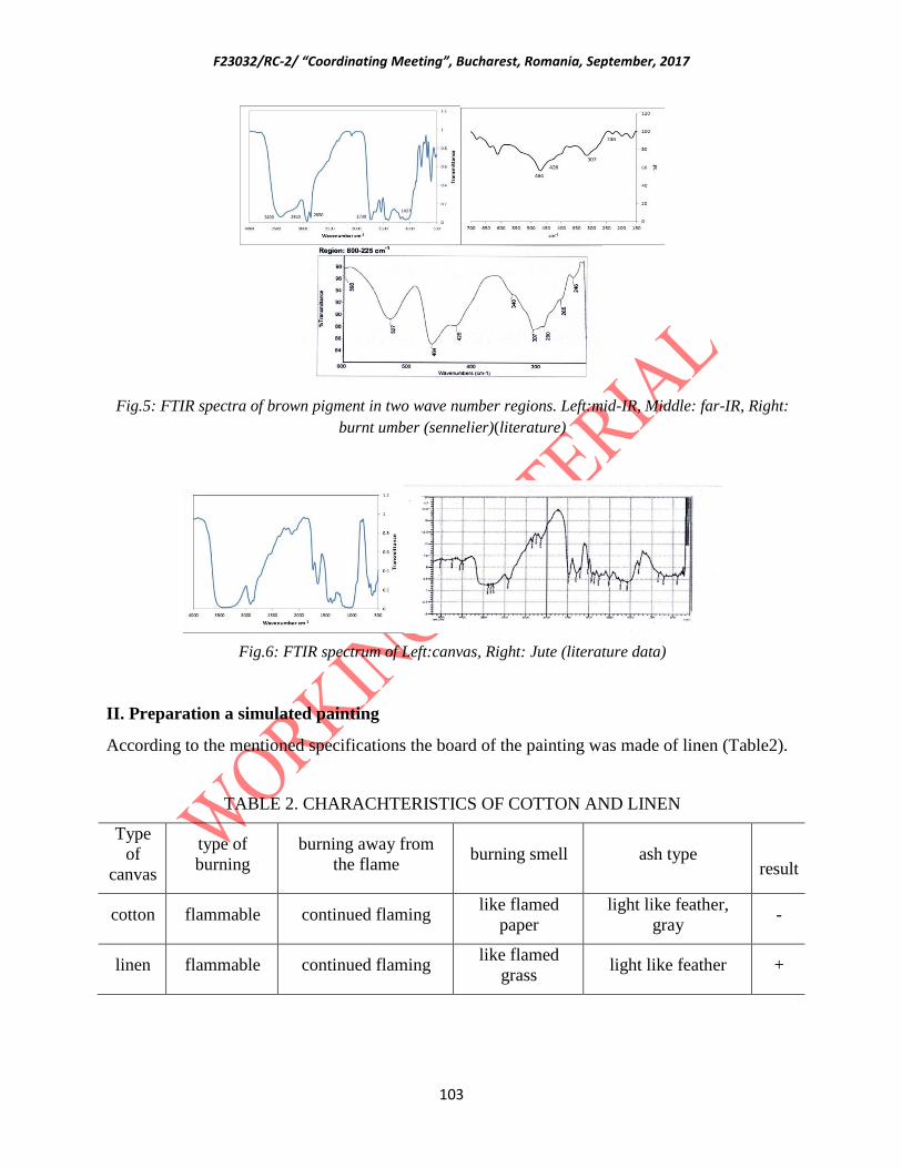
F23032/RC-2/ “Coordinating Meeting”, Bucharest, Romania, September, 2017
103
Fig.5: FTIR spectra of brown pigment in two wave number regions. Left:mid-IR, Middle: far-IR, Right:
burnt umber (sennelier)(literature)
Fig.6: FTIR spectrum of Left:canvas, Right: Jute (literature data)
II. Preparation a simulated painting
According to the mentioned specifications the board of the painting was made of linen (Table2).
TABLE 2. CHARACHTERISTICS OF COTTON AND LINEN
Type
of
canvas
type of
burning
burning away from
the flame burning smell ash type
result
cotton flammable continued flaming like flamed
paper
light like feather,
gray -
linen flammable continued flaming like flamed
grass light like feather +
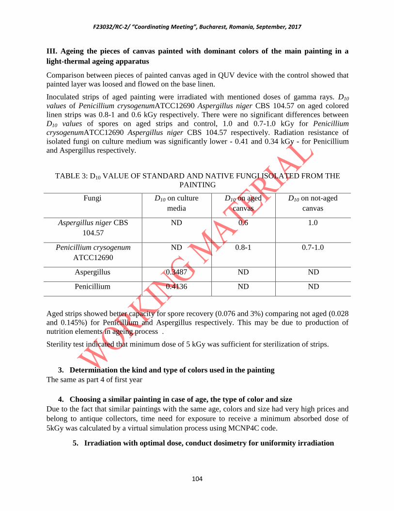
F23032/RC-2/ “Coordinating Meeting”, Bucharest, Romania, September, 2017
104
III. Ageing the pieces of canvas painted with dominant colors of the main painting in a
light-thermal ageing apparatus
Comparison between pieces of painted canvas aged in QUV device with the control showed that
painted layer was loosed and flowed on the base linen.
Inoculated strips of aged painting were irradiated with mentioned doses of gamma rays. D10
values of Penicillium crysogenumATCC12690 Aspergillus niger CBS 104.57 on aged colored
linen strips was 0.8-1 and 0.6 kGy respectively. There were no significant differences between
D10 values of spores on aged strips and control, 1.0 and 0.7-1.0 kGy for Penicillium
crysogenumATCC12690 Aspergillus niger CBS 104.57 respectively. Radiation resistance of
isolated fungi on culture medium was significantly lower - 0.41 and 0.34 kGy - for Penicillium
and Aspergillus respectively.
TABLE 3: D10 VALUE OF STANDARD AND NATIVE FUNGI ISOLATED FROM THE
PAINTING
D10 on not-aged
canvas
D10 on aged
canvas
D10 on culture
media
Fungi
1.0 0.6 ND Aspergillus niger CBS
104.57
0.7-1.0 0.8-1 ND Penicillium crysogenum
ATCC12690
ND ND 0.3487 Aspergillus
ND ND 0.4136 Penicillium
Aged strips showed better capacity for spore recovery (0.076 and 3%) comparing not aged (0.028
and 0.145%) for Penicillium and Aspergillus respectively. This may be due to production of
nutrition elements in ageing process .
Sterility test indicated that minimum dose of 5 kGy was sufficient for sterilization of strips.
3. Determination the kind and type of colors used in the painting
The same as part 4 of first year
4. Choosing a similar painting in case of age, the type of color and size
Due to the fact that similar paintings with the same age, colors and size had very high prices and
belong to antique collectors, time need for exposure to receive a minimum absorbed dose of
5kGy was calculated by a virtual simulation process using MCNP4C code.
5. Irradiation with optimal dose, conduct dosimetry for uniformity irradiation

F23032/RC-2/ “Coordinating Meeting”, Bucharest, Romania, September, 2017
105
Due to the fact that the painting could not be moved from the museum, the irradiation time was
calculated by a virtual simulation process. After running the simulation program written by
MCNP4C computer code for IR-136 irradiation facility, while the painting was virtually placed
alongside the northern wall of the room, the dose values of 5 defined dosimeter cells were
calculated as the absorbed dose rate per activity of the source. By calculating the average of
minimum absorbed doses (the average of absorbed doses in 4 dosimeter cells in corners of the
painting) and the maximum absorbed dose (the dose of the cell located in the middle of the
painting), the dose uniformity ratio was calculated as the ratio of the maximum to the minimum
dose values. Table 4 represents the values of minimum and maximum dose rate and the dose
uniformity ratio of the painting in two situation of irradiation in which density of products was
considered to be 0.001293 and 0.1 g/cm3.
TABLE 4. THE VALUES OF THE MINIMUM AND MAXIMUM DOSE RATE AND THE DOSE
UNIFORMITY RATIO OF THE PAINTING, IN TWO SITUATIONS WHERE THE DENSITY OF
PRODUCTS WAS CONSIDERED TO BE 0.001293 AND 0.1 g/cm3
Uniformity Dose
Ratio
Dmin- canvas
(Gy/100 kCi-sec)
Dmax- canvas
(Gy/100 kCi-sec)
Density of products in the
carriers
1.66 0.03 0.05 0.001293
1.75 0.02 0.035 0.1
According to absorbed dose values represented in table4 and the source activity equal to 3134[2]
Ci at 2017/07/07, while the density of the irradiating products in the carriers are 0.001293 g/cm3,
the required time to absorb the minimum dose of 5kGy in the painting was determined about
13.35 and when irradiating products with the density of 0.1 g/cm3, the time needed to achieve
5kGy, is calculated about 23.06 hours.
References
1. R. A. Cristache, I. Sandu, V. Vasilache, O. Cristache, ACTA CHEMICA, 21 (2), 71-82, 2013.
2. http://www.tera.chem.ut.ee/IR_spectra of paint and coating materials; (06/2009) Created by Signe Vahur.
3. B. H. Berrie (Ed.), Artists’ pigments, A Handbook of their History and Characteristics, Vol. 4, National
Gallery of Art, Washington, 2007.
4. Raisali,Gh., Ataeinia,V., Gorjifard,R., Rezaeian,P., Zarrin,E. “The Calculating and Experimental
Evaluation of Date Irradiation in Semi-Industrial Scale without Using the Product Handling System”, Iranian
Physics Conference, (2011).
5. Raisali,Gh., Ataeinia,V., Faramarzi, A. “Evaluating of Performance Characters of IR-136 Irradiator after
Converting the Product Handling System from 4 levels to 2 Virtual levels”, 16th
Iranian Nuclear Conference, (2009).
6. Briesmeister, Editor J. F., “MCNP-4C, A General Monte Carlo N Particle Transport Code System Version
4C”, Los Alamos National Laboratory LA-13709-M (2000).
7. Atomic Energy of Canada Limited (AECL), "Cobalt-60 Irradiator Model IR-136 for IAEA-Iran", Operator's
Manual, Document No. IN- IR136-84-04 (1985).

F23032/RC-2/ “Coordinating Meeting”, Bucharest, Romania, September, 2017
106
8. Chirigos J.N., Kass S., Kirk W. W. and Salvaggio G. J., “Development of Zircaloy-4 in fuel Element
Fabrication with Special Emphasis on Cladding Materials”, Proceedings of a Symposium Held in Vienna, May 10-
13, 1960, Vol.
9. Lenntech Water Treatment and Air Purification Holding B. V., Stainless Steel 316L, Brochure,
Rotterdamseweg 402M, The Netherlands (2007).
10. Seltzer, S. M., “Calculation of Photon Mass Energy-Transfer and Mass Energy Absorption Coefficients”,
Radiat. Res., 136, 147-179 (1993).
11. International Atomic Energy Agency, "Manual of Food Irradiation Dosimetry", Technical Reports Series
No. 178, VIENNA, PP: 84-85, (1977).
Italy
Radiation processing for bio-deteriorated archived materials and consolidation of porous
artefacts
S.Baccaro, O. Bal, A.Cemmi, I. Di Sarcina
ENEA, Department for Fusion and Nuclear Safety Technologies,
Via Anguillarese 301, 00123 Rome (Italy)
Abstract:
The present report is related to the activities performed at the ENEA Calliope gamma irradiation facility
(Casaccia R.C., Rome, Italy) in the framework of the IAEA Coordinated Research Project ‘F23032’-
Research Agreement No. 18922/R0 (first year). In particular, the researches were focused on: i)
microbiological investigations to study the effect of dose rate and ambient irradiation conditions on the
typical microbes present on archived materials; ii) study of the instantaneous and post-irradiation effect
on paper irradiated by gamma radiation using chemical and spectroscopic techniques; iii) study of gamma
radiation induced co-polymerization of acrylic polymers to achieve formulations suitable and compatible
with CH artefacts. Finally, the work plan regarding the new activities that will be performed in the second
year of the Project is reported.
1. Microbiological investigations to study the effect of dose rate and ambient irradiation
conditions on the typical microbes present on archived materials.
In order to investigate the microbiological effects on gamma treated papery artefacts, Whatman
paper No. 1 (thickness = 0.20 mm, Carlo Erba, Italy), exclusively consisting of cellulose, was
used as feeding materials for Blaptica dubia (Serville, Blaberidae family, order Blattodea)
insects. Blattodea are very widely distributed and considered particularly harmful for library and
archival heritage, since they can erode and leave stains, deriving from their bodily excretions, on
the materials coming in contact with them. Moreover they can provoke public health problems
such as allergies, asthma and sometimes even transmit infective diseases.

F23032/RC-2/ “Coordinating Meeting”, Bucharest, Romania, September, 2017
107
Irradiation tests were carried out at the Calliope plant (ENEA Casaccia R. C., Rome, Italy). The
Calliope plant is a pool-type irradiation facility equipped with a 60
Co source licensed for a
maximum nominal activity of 3,7 x 10¹⁵
Bq (100 kCi). Fricke dosimetry was used to determine
the dose rate values at the different irradiation positions [1].
The samples were irradiated at 2 kGy and 3 kGy absorbed doses, since it was demonstrated to
be highly effective in eradicating all species of noxious insects, reducing considerably
microfungi population to a level lower than that associated to usual environmental conditions
and not inducing negative effects on paper [2]. To investigate the dose rate effects on the
investigated materials, the irradiation tests were performed at high (1.5 kGy/h) and low (211
Gy/h) dose rate. Finally, the irradiation was carried out in air and under argon atmosphere to
study the degradation processes induced by radiation in presence of oxygen. The gamma
radiation effect was evaluated by measuring, under controlled environmental conditions, the
weight of samples in contact with the insects over eight weeks after the end of irradiation and
comparing the obtained results with those related to a not irradiated control sample.
Visual examination revealed that all irradiated samples showed evidence of damage caused by
stains due to regurgitated food and physiological excretions of the cockroaches and that the main
areas of erosion were found to start at the sample edges and move inwards and the same damage
is presented by the not irradiated control sample.
The weigh values put in evidence the effects of the environment atmosphere on the paper
damage caused by erodent insects on irradiated samples: the presence of oxygen in air
atmosphere induces higher damage and weight loss, even if extremely acceptable (maximum -
1.5%). This process is more pronounced at lower dose rate, due to the oxidative degradation
occurring during long lasting irradiation at the same final absorbed dose. Moreover, the damage
up to 3 kGy is practically the same. On the contrary, in the irradiation tests carried out under
inert atmosphere, the damages were drastically reduced and no significant modifications occur
during irradiation.
2. Study of the instantaneous and post-irradiation effect on paper irradiated by
gamma radiation using chemical and spectroscopic techniques.
The same materials described in the previous section were investigated, paying particular
attention on the side-effects evaluation due to the irradiation tests above described.
Moreover, to achieve a more complete evaluation, the samples were irradiated at absorbed dose
higher than 3 kGy (up to 10 kGy or 64 kGy for the viscosity measurements). Specific interest
was paid on the evaluation of the radiation-induced processes (cross-linking and degradation) on
cellulose and on the formation of C=O bonding.
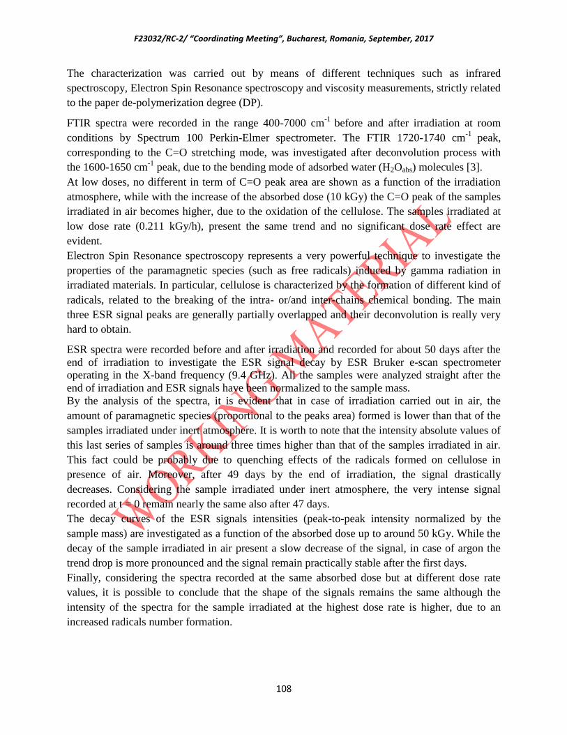
F23032/RC-2/ “Coordinating Meeting”, Bucharest, Romania, September, 2017
108
The characterization was carried out by means of different techniques such as infrared
spectroscopy, Electron Spin Resonance spectroscopy and viscosity measurements, strictly related
to the paper de-polymerization degree (DP).
FTIR spectra were recorded in the range 400-7000 cm-1
before and after irradiation at room
conditions by Spectrum 100 Perkin-Elmer spectrometer. The FTIR 1720-1740 cm-1
peak,
corresponding to the C=O stretching mode, was investigated after deconvolution process with
the 1600-1650 cm-1
peak, due to the bending mode of adsorbed water (H2Oabs) molecules [3].
At low doses, no different in term of C=O peak area are shown as a function of the irradiation
atmosphere, while with the increase of the absorbed dose (10 kGy) the C=O peak of the samples
irradiated in air becomes higher, due to the oxidation of the cellulose. The samples irradiated at
low dose rate (0.211 kGy/h), present the same trend and no significant dose rate effect are
evident.
Electron Spin Resonance spectroscopy represents a very powerful technique to investigate the
properties of the paramagnetic species (such as free radicals) induced by gamma radiation in
irradiated materials. In particular, cellulose is characterized by the formation of different kind of
radicals, related to the breaking of the intra- or/and inter-chains chemical bonding. The main
three ESR signal peaks are generally partially overlapped and their deconvolution is really very
hard to obtain.
ESR spectra were recorded before and after irradiation and recorded for about 50 days after the
end of irradiation to investigate the ESR signal decay by ESR Bruker e-scan spectrometer
operating in the X-band frequency (9.4 GHz). All the samples were analyzed straight after the
end of irradiation and ESR signals have been normalized to the sample mass.
By the analysis of the spectra, it is evident that in case of irradiation carried out in air, the
amount of paramagnetic species (proportional to the peaks area) formed is lower than that of the
samples irradiated under inert atmosphere. It is worth to note that the intensity absolute values of
this last series of samples is around three times higher than that of the samples irradiated in air.
This fact could be probably due to quenching effects of the radicals formed on cellulose in
presence of air. Moreover, after 49 days by the end of irradiation, the signal drastically
decreases. Considering the sample irradiated under inert atmosphere, the very intense signal
recorded at t = 0 remain nearly the same also after 47 days.
The decay curves of the ESR signals intensities (peak-to-peak intensity normalized by the
sample mass) are investigated as a function of the absorbed dose up to around 50 kGy. While the
decay of the sample irradiated in air present a slow decrease of the signal, in case of argon the
trend drop is more pronounced and the signal remain practically stable after the first days.
Finally, considering the spectra recorded at the same absorbed dose but at different dose rate
values, it is possible to conclude that the shape of the signals remains the same although the
intensity of the spectra for the sample irradiated at the highest dose rate is higher, due to an
increased radicals number formation.

F23032/RC-2/ “Coordinating Meeting”, Bucharest, Romania, September, 2017
109
The evaluation of the depolymerization degree of cellulose after gamma irradiation was obtain
by viscosimetric measurements carried out by TA Instrument AR 2000 EX at 20°C, following
the procedure described in the ISO: 5351:2012 standard regulation.
Considering the results obtained at 3 kGy at different dose rates in air and under inert
atmosphere, the stabilizing effect of argon gas is clear because of the limited variation of the
viscosity in comparison with the not irradiated sample (around 10% higher). Finally, the data
confirm that if the irradiation is carried out at lower dose rate, the damage is heavier [4].
3. Study of gamma radiation induced co-polymerization of acrylic polymers to achieve
formulations suitable and compatible with CH artefacts.
Acrylic-based polymeric systems present specific physical and chemical characteristics desirable
for many different fields: among them, the application of these compounds as consolidant and
protection for Cultural Heritage porous artefacts is the focus of the present work. In particular,
methyl acrylate (MA) and ethyl methacrylate (EMA) solvent free monomers mixture is used as
starting materials to investigate the effectiveness of γ radiation to obtain a co-polymer similar to
one of the traditionally most used commercial product Paraloid B72 (PB72, Röhm and Haas,
EMA:MA 70:30 molar ratio) [5].
The influence of preparation procedure (monomers ratio) and of irradiation parameters such as
radiation absorbed dose, dose rate and environmental atmosphere (air, nitrogen) was
investigated by Attenuated total reflectance - Fourier transform infrared spectroscopy
(ATR-FTIR), nuclear magnetic resonance high-resolution spectra of hydrogen and carbon
nuclei (1H- and 13C-NMR) and diffusion-ordered NMR (DOSY-NMR) experiments. Herein, the
EMA and MA starting monomers solutions mixture is firstly characterized before irradiation.
The modifications induced by gamma irradiation, at different irradiation and environmental
conditions, and the co-polymerization process are followed to define the optimized irradiation
parameters needed to obtain a product with requested features. All the irradiation tests were
performed at the ENEA Calliope Plant, previously described. Finally, more information
regarding the present research is available on our published paper [5].
3.1 Materials and Methods
Ethyl methacrylate (EMA, molar mass 114.14 g/mol, density 0.917 g/ml) and methyl acrylate
(MA, molar mass 86.09 g/mol, density 0.956 g/ml) monomers solutions were purchased by
Sigma – Aldrich Chemie GmbH, Schnelldorf, Germany and used as starting materials.
ATR-FTIR analyses were performed exclusively on solid single-phase samples. ATR-FTIR
spectra were collected at room temperature by a Perkin Elmer Spectrum One Spectrometer,

F23032/RC-2/ “Coordinating Meeting”, Bucharest, Romania, September, 2017
110
equipped with a horizontal attenuated total reflection accessory (ZnSe crystal, 45° Flat-Plate), in
the range 4000 - 650 cm-1
.
High-resolution 1H- and
13C-NMR data were recorded by Bruker Avance III spectrometer at
room temperature, at work frequencies 400.13 and 100.6 MHz for 1H- and
13C-NMR spectra,
respectively, on about 10 mg of the solid portion of single- or multi-phase irradiated samples
dissolved in 600 μL of deuterated chloroform. Chemical shifts (δ) are relative to
tetramethylsilane (TMS, δ = 0.00 ppm) for 1H-spectra and to the residual solvent (CDCl3, δ =
77.0 ppm) for 13
C-spectra. Diffusion-ordered spectroscopy (DOSY-NMR) experiments were
realized with the same spectrometer and carried out by stimulated echo sequence only on the
highly diluted solid phase products in deuterated chloroform, as previously described [5].
Proper amounts of EMA and MA monomers solutions were mixed before the irradiation tests. A
first series of monomers solutions (labeled with “A”, Table 1) were prepared in air by mixing 2.3
ml of EMA and 0.9 ml of MA in 5 ml cylindrical glassy vials (diameter = 1.5 cm), obtaining a
molar ratio of 65:35. Finally, the vials were kept closed by a plastic lid before and after γ-
irradiation. To investigate the influence of oxygen on the polymerization process, a second
series of glassy vials (labeled with “N”, Table 2) containing the same solution, were prepared
and sealed under nitrogen atmosphere. All the starting solutions and the samples, before and after
γ-irradiation, were stored at 4°C in the dark to avoid undesired modification [6]. The irradiation
parameters (dose rate and radiation absorbed doses) used in the gamma induced polymerization,
the physical state of the samples after irradiation and the solubility in CDCl3 of the solid single-
phase co-polymers are reported in Tables 1 and 2.
TABLE 1: SAMPLES IN AIR (LABELED WITH “A”): IRRADIATION DOSE RATE AND
RADIATION ABSORBED DOSE, PHYSICAL STATE OF SAMPLES AND SOLID SAMPLES
SOLUBILITY IN CDCL3, QUALITATIVELY EVALUATED AFTER ABOUT FIVE MINUTES.
Dose rate
(kGy/h)
Radiation absorbed dose
(kGy) Physical state
Solubility in
CDCl3
A1
1.50
11 Liquid, gel/solid -
A2 15 Liquid, gel/solid -
A3 20 Liquid, solid -
A4 24 Liquid, solid -
A5 26 Liquid, solid -
A6 28 Solid Partial
A7 30 Solid Partial

F23032/RC-2/ “Coordinating Meeting”, Bucharest, Romania, September, 2017
111
A8 38 Gel, solid -
A9 45 Gel, solid -
A10 0.40 30 Solid Complete
A11 0.04 30 Solid Complete
TABLE 2: SAMPLES UNDER NITROGEN ATMOSPHERE (LABELED WITH “N”): IRRADIATION
DOSE RATE AND RADIATION ABSORBED DOSE, PHYSICAL STATE OF SAMPLES AND
SOLID SAMPLES SOLUBILITY IN CDCL3, QUALITATIVELY EVALUATED AFTER ABOUT
FIVE MINUTES.
Dose rate
(kGy/h)
Radiation absorbed
dose
(kGy)
Physical state Solubility in
CDCl3
N1
1.50
10 Gel, solid -
N2 15 Solid Partial
N3 20 Solid Partial
N4 30 Solid Partial
N5
0.40
5 Gel -
N6 10 Solid Partial
N7 15 Solid Complete
N8 30 Solid Complete
3.2 Experimental results and Discussion
Characterization of samples before irradiation
Liquid starting EMA and MA monomers solutions were also characterized before irradiation by
NMR analyses (data here not shown) and the spectra were compared with those of reference co-
polymer PB72 [5], verifying that the co-polymerization of monomers induces significant
modifications in term of 1H-NMR spectra peaks position and broadening. The effective co-
polymer chemical composition, expressed in term of EMA:MA molar ratio was calculated by the
ratio of the 1H-NMR EMA methylene protons (4.02 - 4.09 ppm) and of MA methoxy protons
(3.54 - 3.69 ppm) signals area, present in the spectra of the solid products.

F23032/RC-2/ “Coordinating Meeting”, Bucharest, Romania, September, 2017
112
Characterization of samples irradiated in air
A series of samples were irradiated at 1.50 kGy/h up to 45 kGy radiation absorbed dose and the 1H-NMR spectra of A1, A2, A6, A7 and A8 samples are reported in Fig. 1.
FIG. 1: 1H-NMR spectra of samples prepared and irradiated in air at 1.50 kGy/h at different radiation
absorbed doses: 11 kGy (A1), 15 kGy (A2), 28 kGy (A6), 30 kGy (A7) and 38 kGy (A8). Spectrum of PB72
is given as reference.
The high-resolution NMR spectra show, increasing the radiation absorbed dose up to 30 kGy
(A1 - A7 samples), the gradual disappearance of the monomers peaks (above 5.0 ppm) and the
increase of those relative to the co-polymer structure. Considering the broadening of the peaks in
the range 3.3 - 4.5 ppm and 0.7 - 2.7 ppm and the complete disappearance of residual monomers,
a complete polymerization can be assigned to the A7 sample (30 kGy), as monomers signals are
still present in the A6 sample. Competitive bonding formation (polymerization, cross-linking)
and disruption (degradation processes) occur in polymeric materials during irradiation. The data
above discussed are consistent with the prevalent formation, under γ-irradiation up to 30 kGy, of
a bonded structure, i.e. co-polymer, evident also by more viscous phases formed with the
increase of the radiation absorbed dose (Table 1). The evaluation of the physical state of the
irradiated samples (Table 1) shows that, starting from a liquid solution, the increase of the
radiation absorbed dose induces the creation of even more viscous single- or multi-phases
(liquid/gel/solid), before the final formation of an homogeneous and solid single-phase for the
co-polymers irradiated at 28 - 30 kGy. On the contrary, if the irradiation was carried on reaching
radiation absorbed doses of 45 kGy (samples A8 and A9), the formation of a multi-phase
system, i.e. gel/solid, again occurs. Further validation of the optimized radiation absorbed dose
of 30 kGy to obtain a co-polymer (A7) with the same chemical and structural characteristics of
PB72, came from the comparison of their 13
C-NMR and ATR-FTIR spectra (here not reported)
[5].

F23032/RC-2/ “Coordinating Meeting”, Bucharest, Romania, September, 2017
113
A more complete description of the co-polymer formation reaction can be achieved by the
DOSY-NMR method that allowed to determine, through the Stokes-Einstein equation, the mean
values of the hydrodynamic radius-size distributions of the polymeric clusters, inversely
proportional to the diffusion coefficient ( D-1
). Moreover, it was indirectly possible to evaluate
the polydispersity and the sample polymerization degree [5]. In Fig. 2a, the inverse values of the
mean diffusion coefficients of A3 - A9 co-polymers are reported as a function of the radiation
absorbed doses: after a slight increase at the lowest doses, sharply reaches a maximum at 30
kGy, typical of Trommsdorf effect, and decreases again if the irradiation is carried on up to 45
kGy.
FIG. 2: (a) Inverse of diffusion coefficient of A3 - A9 samples (1.50 kGy/h). (b) Inverse of diffusion
coefficient of samples irradiated at 30 kGy radiation absorbed dose at different dose rates: 1.50 kGy/h
(A7), 0.40 kGy/h (A10) and 0.04 kGy/h (A11). Inset: 1H-NMR spectra of A7, A10 and A11 samples.
In case of A7 sample, the lowest diffusion coefficient (D = 2.0 E-12 m2/s) suggests that a
polymer characterized by molecular weight higher than reference PB72 (D = 2.0 E-11 m2/s) and
more branched and highly polydispersed structures, not completely soluble in deuterated
chloroform (Table 1), was obtained. Considering the A8 and A9 samples irradiated up to 45 kGy,
at radiation absorbed doses higher than that corresponding to the complete co-polymerization,
as a result of the depolymerization process not only the gel-phase is formed again, but the solid
co-polymer structures underwent to bonds breaking and to smaller polymeric clusters formation
(Fig. 2a) [4].
Additional studies were performed to investigate the dose rate effect on the polymerization
process in presence of oxygen. For this purpose, irradiation tests were performed at 30 kGy
radiation absorbed dose, correspondent to the formation of A7 co-polymer, at medium (0.40
kGy/h, A10) and low (0.04 kGy/h, A11) dose rates. As reported in Table 1, the A7, A10 and A11
samples exhibit a solid single-phase and the co-polymers irradiated at medium and low dose
rates, differently from A7, are completely soluble in CDCl3. Interesting differences among the
three samples are shown by DOSY-NMR data reported in Fig. 2b: the higher the dose rate, the
higher the polymerization degree. The A7, A10 and A11 co-polymers present different

F23032/RC-2/ “Coordinating Meeting”, Bucharest, Romania, September, 2017
114
polymerization degree dependent on the logarithm of the dose rate (Fig. 2b), due to the oxidative
degradation that prevents the formation of bigger polymeric clusters. Besides, DOSY pattern
indicates a sharp diffusion coefficient distribution for the samples irradiated at low and medium
dose rates, indicating lower branching and more homogeneous molecular weight and
polymerization degree, consistent with the solubility data reported in Table 1 [4, 5].
Characterization of samples irradiated under nitrogen atmosphere
The irradiation tests were carried out under nitrogen atmosphere at high (samples N1 - N4) and
medium (samples N5 - N8) dose rate up to 30 kGy radiation absorbed dose (Table 2).
The D-1
DOSY-NMR data of samples irradiated under nitrogen are reported in Fig. 3 and
compared with those of samples irradiated in air at 1.50 kGy/h up to 30 kGy radiation absorbed
dose. The data related to the co-polymers irradiated under nitrogen atmosphere at 1.50 kGy/h
indicate that a dose of 15 kGy, corresponding to N2 sample, resulted enough to obtain a solid
single-phase co-polymer, showing that the radiation absorbed dose value needed to produce a
solid co-polymer is lower in nitrogen than in air. Moreover, the polymerization degree of N2
sample (D-1
= 1.0 E+12 m2/s) is higher than that of the sample irradiated in air at the same dose
rate and at 30 kGy radiation absorbed dose (D-1
= 0.5 E+12 m2/s), due to the competitive
processes, such as the formation of oxygen-containing free radicals (less reactive than those of
monomeric and polymeric species) and their following recombination, occurring when oxygen is
present [5].
FIG. 3: Inverse of diffusion coefficient of samples irradiated under nitrogen atmosphere at dose rates of
1.50 kGy/h (N2 - N4, open circles) and of 0.40 kGy/h (N6 - N8, open triangles), and in air at 1.50 kGy/h
(A3 - A7, filled circles). Inset: 1H-NMR spectra of A7, N2 and N6 samples.
Solid single-phase co-polymers with the same features of A7 sample were obtained at radiation
absorbed dose of 15 kGy (N2, 1.50 kGy/h) and at 10 kGy (N6, 0.40 kGy/h). Their 1H-NMR
spectra are compared with that of A7 in the inset of Fig. 3.

F23032/RC-2/ “Coordinating Meeting”, Bucharest, Romania, September, 2017
115
If the samples irradiated under nitrogen are compared in term of D-1
(Fig. 3), the polymerization
process seems to be more efficient at the lowest dose rate (0.40 kGy) since N6 solid co-polymer,
with the required features and structure, is obtained with radiation absorbed dose of 10 kGy.
This result is obtained because in acrylate systems, particularly at higher dose rates and whatever
the environmental atmosphere, primary radical’s recombination process takes place, bringing on
a partial subtracting of the energy involved in polymerization [8, 28, 31].
The dose rate effect verified on the co-polymers irradiated in air was also confirmed for the
samples prepared under nitrogen at the same radiation absorbed dose (15 kGy): by lowering the
dose rate, greater cluster-sizes homogeneity can be achieved, resulting in an increased solubility
of the sample irradiated at lower dose rate (N7). Finally, as demonstrated by DOSY-NMR
analyses (Fig. 3) and by physical state data reported in Table 2,the presence of nitrogen
atmosphere markedly improves the sample stability, allowing to maintain the same cluster sizes
and solid single-phase also if the irradiation dose is doubled or tripled (up to 30 kGy) [33].
3.3 Conclusions
The irradiation tests performed for the formation of EMA/MA co-polymers, allowed to study
the radiation absorbed dose dependence of the radiation induced processes (bonds formation and
breaking): different results were obtained as a function of the environmental atmosphere and
dose rate values: i) in presence of oxygen, higher radiation absorbed dose was required to obtain
a solid co-polymer with specific characteristics since a partial amount of energy released to the
samples was involved in competitive processes, i.e. oxygen-containing free radicals formation
and primary radicals recombination in case of high dose rates; ii) performing the irradiation at
different dose rates, irrespectively to the environmental atmosphere, the formation of more
homogeneous samples in term of polymerization degree dispersion was achieved at lower dose
rates, with clear effects on the co-polymers solubility properties; iii) on samples irradiated at
radiation absorbed doses higher than those needed for the formation of the co-polymer, while in
case of samples irradiated in air heavy depolymerization was verified, as confirmed by the
formation of a gel-phase and by the disruption of polymeric clusters, a sensible increase of the
samples stability was attained if the irradiation was performed under nitrogen atmosphere.
4. Work plan of the second year of the IAEA Coordinated Research Project ‘F23032’-
Research Agreement No. 18922/R0
Task 1 - Evaluate the side-effects and post-irradiation effects of the developed formulations by
means of sophisticated analytical techniques like FTIR, ESR, 13C-NMR, 1H-NMR and
mechanical tests.
Task 2 - Environment friendly and non-toxic consolidants and development of new composition
for use as consolidating agents to improve the efficacy and safety of the CH artifact:

F23032/RC-2/ “Coordinating Meeting”, Bucharest, Romania, September, 2017
116
a) Study of gamma radiation induced co-polymerization of acrylic polymers to achieve
formulations suitable and compatible with CH artefacts.
b) A new approach using the lignin-like structures has been developed as a new green
system for consolidation [6, 7]. Lignin-like oligomers or polymers provide maximum
compatibility with wood structure, improved mechanical properties, strong hydrogen
bonds and cross-linking with existing lignin and antimicrobial activity versus
staphylococcus aureus. Chemical production of lignin-like structures occurs in presence
catalysts, but gamma radiation induced polymerization allows to obtain the compound
without chemical (no additives) and physical (no increase of temperature) modification
[8]. Moreover, it is possible to control the polymers features by modifing the irradiation
parameters such as irradiation dose, irradiation dose rate, atmosphere etc. [5, 9].
References
[1] Baccaro, S., Cemmi, A., Ferrara, G., Fiore, S., 2015. Calliope gamma irradiation facility at
ENEA-Casaccia R. C. (Rome), ENEA Technical Report RT/2015/13/ENEA, ISSN/0393-3016.
[2] Adamo, M., Brizzi, M., Magaudda, G., Martinelli, G., Plossi-Zappalà, M., Rocchetti, F.,
Savagnone, F., 2001.Gamma radiation of paper in different environmental conditions: chemical,
physical and microbiological analysis, Restaurator, 22, 107-131.
[3] Baccaro, S., Carewska, M., Casieri, C., Cemmi, A., Lepore, A., 2013. Structural
modifications and interaction with moisture in γ –irradiated pure cellulose by thermal analysis
and infrared spectroscopy, Polym. Degrad. Stab., 98 2005-2010
[4] Charlesby, A., 1960. Atomic radiation and polymers, in: International Series of Monographs
on Radiation Effects in Materials, Vol.1, Pergamon Press LTD, Oxford.
[5] Baccaro, S., Casieri, C., Cemmi, Chiarini, M., D’Aiuto, V., Tortora, M., 2017.
Characterization of γ-radiation induced polymerization in ethyl methacrylate and methyl acrylate
monomers solutions, Rad. Phys. Chem., 141 131-137,
https://doi.org/10.1016/j.radphyschem.2017.06.017.
[6] McHale, E., Steindal, C.C., Kutzke, H., Benneche T., Harding, S.E., 2017. In situ
polymerisation of isoeugenol as a green consolidation method for waterlogged archeological
wood. Scientific reports, DOI:10.1038/srep46481.
[7] Vanholme, R., Demedts, B., Morreel, K., Ralph, J., Boerjam, W., 2017. Lignin biosynthesis
and structure,. Plant Phys. 153, 895-905.
[8] Fournand, D., Cathala, B., Lapierre, C., 2003. Initial steps of the peroxidase-catalyzed
polymerization of coniferyl alcohol and/or sinapyl aldehyde: capillary zone electrophoresis study
of pH effect. Phytochem. 62, 139-146.

F23032/RC-2/ “Coordinating Meeting”, Bucharest, Romania, September, 2017
117
[9] Adamo, M., Baccaro, S., Cemmi, A., 2015. Radiation processing for bio-deteriorated
archived materials and consolidation of porous artefacts. ENEA – Casaccia R.C., Rome, Italy.
(ENEA Report RT/2015/5/ENEA).
Poland
Electron beam for preservation of biodeteriorated cultural heritage paper-
based objects
D. CHMIELEWSKA-ŚMIETANKO1, M. WÓJCIK
1, W. MIGDAŁ
1, U. GRYCZKA
1, J. SADŁO
1, K. KOPEĆ
2
1 Institute of Nuclear Chemistry and Technology, Poland
2 Faculty of Chemical and Process Engineering, Warsaw University of Technology, Poland
Abstract
Unsuitable storage conditions or accidents such as floods can present a serious threat for large quantities
of book making them prone to attack by harmful microorganisms. The microbiological degradation of
archives and book collections can be efficiently inhibited with irradiation processing. Application of EB
irradiation to book and archive collections can also be a very effective alternative to the commonly used
ethylene oxide treatment, which is toxic to the human and natural environment. In this study was
evaluated the influence of EB irradiation used for microbiological decontamination process on paper-
based objects. Three different kinds of paper (Whatman CHR 1, office paper and newsprint paper) were
treated with 0.4, 1, 2, 5, 10 and 25 kGy electron beam irradiation. Optical and mechanical properties of
different sorts of paper treated with e-beam, before and after the radiation process were studied. These
results, which correlated with absorbed radiation doses effective for the elimination of Aspergillus niger
(A. niger) allowed to determine that EB irradiation with absorbed radiation dose of 5 kGy ensures safe
decontamination of different sorts of paper-based objects.
Introduction
Paper has been the most important and popular information storage medium for two thousand
years. The technology of paper production has changed over time. Because of their complex
structure and varied nature, paper physicochemical properties, mechanical characteristics,
stability and resistance to degradation are inseparably linked with the composition of the
material, manufacture process, and interactions of the paper components [1]. The main paper
component is cellulose fibres forming a three-dimensional structure. Paper contains also
hemicelluloses, lignin and additives (binding materials, inorganic fillers, dyes, pigments, metal
ions, etc.) in different amounts, depending on the source of cellulose and the potential use of the
material, respectively [2]. It is the reason why investigation of the degradation of such a multi-
component material should involve a complex experimental procedure.
Protecting books and archives from destruction by harmful microorganisms and inappropriate
storing conditions is one of the main factors in the preservation of objects of cultural heritage.
Moreover, the microbiological burden is also harmful to the health of librarians, archivists and
the public. Libraries, museums and archives are still looking for optimal decontamination
methods because the commonly used fumigation with ethylene oxide has many disadvantages.
Fumigation with ethylene oxide is a time-consuming process. Moreover the toxicity of ethylene

F23032/RC-2/ “Coordinating Meeting”, Bucharest, Romania, September, 2017
118
oxide to environment and humans leads to gradual withdrawal of this technology by more and
more countries. The successful recovery of big amount of library and archival materials depends
on timely response to a disaster. Therefore fast and effective treatment method that enables
decontamination of thousand volumes in short time is desired.
Radiation technologies are successfully applied for the sterilization of medical devices, for the
hygienization and preservation of agri-food products, for the modification of materials and in the
protection of the environment. The groundbreaking study concerning the application of γ-
radiation on the disinfection of paper-based objects was carried out in early 1960s [3]. Since
then, γ irradiation has been applied in the treatment of large quantities of books and archives
[4-6] However, the most prevalent literature on applications of ionizing radiation to book and
archives decontamination was related to treatment with γ radiation, because of its high
penetration. Nevertheless EB the decontamination of microbiologically infected paper is also an
area for the new application of EB irradiation, but this process needs to be investigated
in detail. Preliminary investigation carried out in this project confirmed that electron beam
decontamination of archives can be promising alternative to ethylene oxide fumigation, because
high-energy electron beam is very effective and, simultaneously, safe for the paper object
method of paper decontamination.
Materials and methods
Three different kinds of paper, which varied in their manufacture process and composition, were
investigated:
• Whatman paper CHR 1 (pure cellulose paper)
• Office paper
• Newsprint paper (produced from 100% recycled fibres)
Papers have different structure therefore their grammage and thickness differ significantly
(parameters summed up in the Table 1).
Table 1. THE GRAMMAGE AND THICKNESS OF THE PAPER SAMPLES
Paper Grammage (m/g2) Thickness (m)
Whatman CHR1 88 180
Office paper 80 106
Newsprint paper 48.8 103
Investigation of the samples with Skanning Electron Mocrocopy (SEM) revealed that the
samples morphology differs as well (Fig. 1).

F23032/RC-2/ “Coordinating Meeting”, Bucharest, Romania, September, 2017
119
Fig. 1. SEM images of the different papers surface: Whatman CHR1 (a), office (b), newsprint
(c). Magnification 500 ×.
SEM images together with Energy Dispersive Spectrometry (EDS) results (Fig. 2) gave very
valuable information on samples composition and manufacturing process.
Fig. 2. EDS analysis of the elemental composition of the paper samples: Whatman CHR1 (a),
office (b), newsprint (c).
The Whatman CHR1 paper morphology shows a fibrous structure (Fig. 1a). EDS analysis (Fig.
2a) confirmed the presence of only carbon and oxygen, which are the components of the
cellulose, therefore the analysis proved that Whatman paper does not contain any additives.
In Fig. 1b, fibre length reduction as a result of the beating process can be observed for office
paper. Moreover, presence of fillers that bind the fibres together is visible in the SEM image as
well. EDS analysis (Fig. 2b), besides the presence of C and O, indicated smaller amounts of Ca
and Cl. Ca constitutes the calcium carbonate CaCO3, which is filler in the alkaline papermaking
process. Chloride is a residue from the bleaching process that uses chlorine dioxide for the
bleaching of wood pulp. EDS analysis of the newsprint paper showed that besides C and O, some
amounts of Si, Al and Ca are present (Fig. 2c). Therefore we can conclude that recycled fibres in
the newsprint paper are coated with a layer of kaolin (Fig. 1c).
While Ca again indicates the presence of CaCO3, Si and Al constituting kaolin Al2Si2O5(OH)4,
which is used in the coating process to enhance gloss and opacity.
Electron beam irradiation
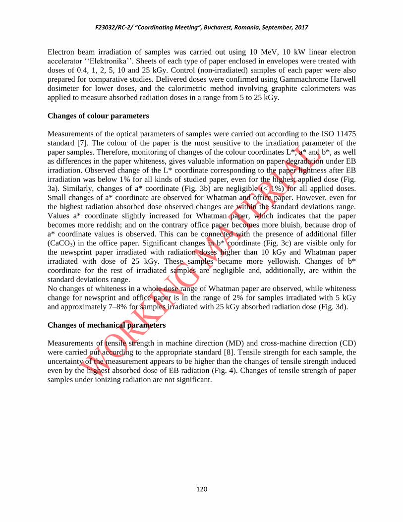
F23032/RC-2/ “Coordinating Meeting”, Bucharest, Romania, September, 2017
120
Electron beam irradiation of samples was carried out using 10 MeV, 10 kW linear electron
accelerator ‘‘Elektronika’’. Sheets of each type of paper enclosed in envelopes were treated with
doses of 0.4, 1, 2, 5, 10 and 25 kGy. Control (non-irradiated) samples of each paper were also
prepared for comparative studies. Delivered doses were confirmed using Gammachrome Harwell
dosimeter for lower doses, and the calorimetric method involving graphite calorimeters was
applied to measure absorbed radiation doses in a range from 5 to 25 kGy.
Changes of colour parameters
Measurements of the optical parameters of samples were carried out according to the ISO 11475
standard [7]. The colour of the paper is the most sensitive to the irradiation parameter of the
paper samples. Therefore, monitoring of changes of the colour coordinates L*, a* and b*, as well
as differences in the paper whiteness, gives valuable information on paper degradation under EB
irradiation. Observed change of the L* coordinate corresponding to the paper lightness after EB
irradiation was below 1% for all kinds of studied paper, even for the highest applied dose (Fig.
3a). Similarly, changes of a* coordinate (Fig. 3b) are negligible (< 1%) for all applied doses.
Small changes of a* coordinate are observed for Whatman and office paper. However, even for
the highest radiation absorbed dose observed changes are within the standard deviations range.
Values a* coordinate slightly increased for Whatman paper, which indicates that the paper
becomes more reddish; and on the contrary office paper becomes more bluish, because drop of
a* coordinate values is observed. This can be connected with the presence of additional filler
(CaCO3) in the office paper. Significant changes in b* coordinate (Fig. 3c) are visible only for
the newsprint paper irradiated with radiation doses higher than 10 kGy and Whatman paper
irradiated with dose of 25 kGy. These samples became more yellowish. Changes of b*
coordinate for the rest of irradiated samples are negligible and, additionally, are within the
standard deviations range.
No changes of whiteness in a whole dose range of Whatman paper are observed, while whiteness
change for newsprint and office paper is in the range of 2% for samples irradiated with 5 kGy
and approximately 7–8% for samples irradiated with 25 kGy absorbed radiation dose (Fig. 3d).
Changes of mechanical parameters
Measurements of tensile strength in machine direction (MD) and cross-machine direction (CD)
were carried out according to the appropriate standard [8]. Tensile strength for each sample, the
uncertainty of the measurement appears to be higher than the changes of tensile strength induced
even by the highest absorbed dose of EB radiation (Fig. 4). Changes of tensile strength of paper
samples under ionizing radiation are not significant.

F23032/RC-2/ “Coordinating Meeting”, Bucharest, Romania, September, 2017
121
Fig. 3. Changes of colour parameters of the different kinds of paper under electron beam
irradiation: coordinate L* (a), coordinate a* (b), coordinate b* (c), whiteness CIE (d).
Fig. 4. Changes of tensile strength of the different kinds of paper under electron beam
irradiation: cross machine direction (a), cross direction (b).
Antimicrobial effectiveness of EB irradiation
The influence of the EB radiation dose on the effectiveness of the microbiological
decontamination was evaluated against the fungi Aspergillus niger. To prepare the spore
suspension of A. niger, the fungal culture was grown on Sebouraud Agar for 72 h in 25 °C and
suspended in Ringer's solution. Square paper samples were artificially contaminated with a spore
suspension of A. niger by immersion in the spore suspension for 5 min. Next, inoculated samples
were placed on the Petri plates and incubated for 24 h at 25 °C. Three inoculated samples of each
kind of paper were irradiated with doses: 0.4, 1, 2, 5, and 10 kGy. Irradiated samples were
individually homogenized in 100 mL Ringer's solution. Logarithmic dilution (up to 10−3
dilution)
of the homogenized samples were prepared and then 0.1 mL of each homogenate was inoculated
(in triplicate) on Czapka Agar on Petri plates. Inoculated Perti plates were incubated for 72 h at
25 °C. Amount of bioburden was evaluated by counting the colonies and determining the number

F23032/RC-2/ “Coordinating Meeting”, Bucharest, Romania, September, 2017
122
of Colony Forming Units per cm2 (CFU/cm
2) for each paper sample. For all paper the A. niger
population decrease was observed with the EB radiation dose increase (Fig. 5). A dose of 5 kGy
was sufficient for the elimination of A. niger from all kinds of paper. However usual level of
microbial contamination in libraries and archives is significantly lower (102–10
3 CFU/cm
2)
therefore applied doses in such cases will be lower as well [9].
Fig. 5. A. niger population on the different kinds of paper as a function of EB radiation dose.
Conclusions and the programme of future works
Investigation of EB irradiation effects on the different kinds of paper confirmed safety of its
application to the decontaminated papers. EB irradiation of different kinds of paper with doses
up to 10 kGy doesn’t influence their colour noticeably and only for the samples of office paper
irradiated with a dose of 25 kGy changes of the paper colour can be noticed by the experienced
observer.
Mechanical parameter like tensile strength of paper samples is not sufficiently sensitive to
electron beam irradiation in applied dose ranges to indicate paper degradation. Investigation
carried out in this work enabled the optimisation of irradiation procedure and estimation a dose
of 5 kGy as sufficient for the complete elimination of A. niger from all types of paper and did not
influence the studied optical and mechanical parameters of different papers.
The programme of work to be performed:
• Application of other characterization methods for paper properties evaluation after EB
irradiation (XRD, SEC, GC, chemical parameters- pH of paper extract, copper number,
tearing resistance with Elmendorf method
• Observation of natural aging effect on paper colour - continuation
• Determination of the accelerated aging influence
• Comparative study – ethylene oxide fumigation and g irradiation
• Study of the effect of EB irradiation on „real” paper-based objects
References

F23032/RC-2/ “Coordinating Meeting”, Bucharest, Romania, September, 2017
123
[1] Area, M.C., Cheradame, H., 2011. Paper aging and degradation: recent findings and research
methods. Bioresources 6, 5307–5337.
[2] Ion, R.M., Doncea, S.M., Ion, M.L., Raditoiu, V., Amariutei, V., 2013. Surface investigations
of old book paper treated with hydroxyapatite nanoparticles. Appl. Surf. Sci. 285, 27–32.
[3] Belyakova, L.A., 1961. Gamma-radiation as a disinfecting agent for books infected with
mould spores. Microbiology 29, 548–550.
[4] Area, M.C., Calvo, A.M., Felissia, F.E., Docters, A., M.F, M., 2014. Influence of dose and
dose rate on the physical properties of commercial papers commonly used in libraries
and archives. Radiat. Phys. Chem. 96, 217–222.
[5] Bratu, E., Moise, I.V., Cutrubinis, M., Negut, D.C., M, V., 2009. Archives decontamination
by gamma irradiation. Nukleonika 54, 77–84.
[6] Sinco, P., 2000. The use of gamma rays in book conservation. Abbey Newsl. 24, 38–40.
[7] ISO 11475, 2004. Paper and board. Determination of CIE whiteness, D65/10 degrees
(outdoor daylight).
[8] ISO 1924-2, 2008. Paper and board. Determination of tensile properties. Part 2: Constant rate
of elongation method (20 mm/min).
[9] Moise, I.V., Virgolici, M., Negut, C.D., Manea, M., Alexandru, M., Trandafir, L., Zorila,
F.L., Talasman, C.M., Manea, D., Nisipeanu, S., Haiducu, M., Balan, Z., 2012.
Establishing the irradiation dose for paper decontamination. Radiat. Phys. Chem. 81, 1045–1050.
Portugal
HYBRID MATERIALS PREPARED BY IONIZING RADIATION FOR
CONSOLIDATION AND PRESERVATION OF ROMAN MOSAICS
IAEA Research Contract No. 18982
LUÍS M. FERREIRA
1
M.F. Araújo1, M.H. Casimiro
1, S. Cabo Verde
1, A.N. Falcão
1, L.C. Alves
1, Alexandra P.
Rodrigues1,2
1Centro de Ciências e Tecnologias Nucleares (C
2TN), Campus Tecnológico e Nuclear, Instituto Superior Técnico,
Universidade de Lisboa, Estrada Nacional 10 (km 139,7), 2695-066 Bobadela LRS, Portugal 2Departamento de Conservação e Restauro, Faculdade de Ciências e Tecnologia, Universidade Nova de Lisboa
Abstract
The aim of this research project is to prepare and functionalize new PDMS-based Ormosils (Organically Modified
Silicates) by ionizing radiation techniques, which could be used alone or as additives to the existent composite
materials used in Roman mosaics conservation. This report resumes the development of the project during the first
period of the research contract, which was mayority in accordance with the plan proposed.

F23032/RC-2/ “Coordinating Meeting”, Bucharest, Romania, September, 2017
124
The exaushitive characterization of the Roman mosaics (mortars and tesserae) confirmed their mineral nature giving
important information for the definition of the composition of the new hybrid materials prepared by gamma
irradiation. From the six batches of hybrid materials prepared, two of them revealed biostactic activity against three
of the most dangerous and resistant microorganisms found in the Roman mosaics from Conimbriga archeological
site. It was confirmed that basides the good structural and physico-chemical properties the biocide activity of the
hybrids is a function of the respective content in zirconium propoxide (ZrPO).
1. Introduction
The development of the project in this period of the research contract was in accordance with the
plan initially proposed. No additional recommendations were given during the 1st RCM at IAEA
Headquarters (28 Sept - 2 Oct 2015) and the planification showed to be adequated. Nevertheless,
small adjustments were introduced in order to overcome unexpected problems, which the most
restricting were associated with equipment’s maintenance (60
Co recharging process), breakdowns
and delays on getting official authorization to access the archeological site for inspection and
sampling (Conimbriga is an archeological site open to general public for visits with a huge diary
amount of tourists). Apart from these, the severe meteorological conditions of last winters and
summers also difficult and delayed fieldwork.
The observation and sampling in situ confirmed that the most important and costly effective
natural causes of mosaics degradation are weathering and bioactive effects. Regarding
bioactivity effects, fungi, bacteria and plants are of major concern.
The state of degradation and diversity of situations found on field demanded a special attention
to hybrid materials surface properties (morpholgy and wettability) and to their biocide activity.
The influence of composition, precursors ratios, irradiation conditions and synthesis procedure
on these properties was studied. The hybrids biocide activity was evidenced to be Zr content
dependent. However, the most promissing hybrids prepared, with a ZrPO content of 20 wt%,
according to previous studies [1], are almost in the limit of percolation process (≈ 25 wt%), with
the consequent loose of molecular structure and stability.
2. Results and Discussion
Characterization of Roman mosaics – mortars
Mortars characterization revealed that they are apparently doble layered with hetrogeneous
aggregates (with different colors which suggests the existance of different minerals) and rough
surfaces. The upper stratum binder show a reddish hue and lower startum a grey hue. The
presence of organic deposits at the external surface was also detected. Even showing a good level
of cohesion the mortars between tesserae present a high level of degradation.

F23032/RC-2/ “Coordinating Meeting”, Bucharest, Romania, September, 2017
125
FIG. 1: Mortar and tesserea collected at Conimbriga (Portugal); Detail of the degraded binding
mortars between tesserae.
Elemental characterization of mortars by PIXE (Proton Induced X-ray Emission) technique
showed a major concentration of SiO2, followed by CaCO3, Al2O3, MgO, Fe2O3 and P2O5.
Residual concentrations of S, Cl, TiO2, Cr2O3, MnO, CuO ZnO and PbO were also found.
FTIR analysis of the different sections of mortar showed that the content of water decrease from
top to bottom layer. In the surface layer was detected the presence of organic compounds, which
were associated to the mortar surface exposition to ambient conditions and living beings.
Data from thermal analysis (TGA and DSC) were in agreament with data obtained by FTIR.
Characterization of Roman mosaics – tesserae
One thousand unpaired tesserae were collected in archeological site for characterization, from
which fourteen tesserae were selected based on the color, texture and brightness.
After a deeply cleaning process the tesserae were analyse by FORS technique revealing the
existance of 6 different colours (white, light brown, grey, red, pink and yellow) with more than
12 hues.
This group of 14 tesserae were also characterized by XRF, XRD and RAMAN. The elemental
composition was identified as being: Na, Mg, Al, Si, P, K, K, Ti and V. The minerals detected
were dolomite, quartz, goethite, haematite and calcite as the majority mineralogical phase in all
tesserae. These data confirmed the limestone nature of original rocks.
Characterization of Roman mosaics – bioburden evaluation
From the collect in situ, the bioburden evaluation of Roman mosaics by phenotypic methods
conducted to the isolation of 17 different bacteria corresponding to 12 genera and 13 species,

F23032/RC-2/ “Coordinating Meeting”, Bucharest, Romania, September, 2017
126
including PGPB (plant growth-promoting bacteria), gram positive rods and cocci and gram-
negative rods. Fungus Aspergilus fumigates was also detected.
The characterization by genotypic techniques was not yet possible due to difficults in obtaining
time of use and technical support in the Clean Areas Room.
From the microbiota already identified, three bacteria (Salmonela 3, Staphilococcus capitis
capitis and a sporulated bacilli) and one fungus (Aspergellius fumigatus), considered the most
danger and resistant already found, were considered for the biocide studies with the hybrid
materials.
Preparation of hybrid materials by gamma irradiation
Six batches of hybrid materials of the system PDMS-TEOS-ZrPO were prepared by the gamma
irradiation method developed (FIG. 2).
FIG. 2: Diagram of the experimental protocol for the preparation of the hybrid materials through
gamma irradiation.
The compositions, using polydimethylsiloxane (PDMS), tetraethylorthosylicate (TEOS) and
zirconium propoxide (ZrPO) as precursors, were optimized based in previous studies regarding
homogenity and molecular stability [1, 2, 3, 4].
The mixture of precursors were irradiated at the same dose rate (10 kGy.h-1
) with an absorbed
dose ranging from 450-700 kGy. Table I resumes the composition of the different batches.
Table I. Composition of the batches of hybrid materials of the system PDMS-TEOS-ZrPO
prepared.
Sample name Composition
(wt%)
H1 39PDMS-54TEOS-7ZrPO
H2 33PDMS-64TEOS-3ZrPO
H3 33PDMS-62TEOS-5ZrPO
H4 33PDMS-61TEOS6ZrPO

F23032/RC-2/ “Coordinating Meeting”, Bucharest, Romania, September, 2017
127
H5 33PDMS-47TEOS-20Zrpo
H6 20PDMS-60TEOS-20ZrPO
The inorganic precursor ZrPO, apart from other properties and the role in the microstructure of
the hybrids [4, 5], was used in the preparation of these hybrids due its natural biocide activity
against Gram-, Gram+ bacteria and yeast. For this reason Zr(IV) has been increasingly used in
the form of phosphates and aluminosilicates for dental applications.
The materials prepared are homogeneous, transparent, monolithic and amourphous. The
increment of ZrPO content has lead to a decrease in the flexibility of the samples.
Characterization
Thermal resistance
Up to a [ZrPO] = 6-7 wt% the thermal resistance and stability of hybrids increase (concentration
threshold), above which the hybrids departs too much their behavior from to the pure PDMS,
responsible for the interconnection between the oxide clusters in the hybrid network (the material
increasingly turns more brittle). Even so, hybrids with a ZrPO content of 20 wt% show good
thermal stability for the intended use, presenting a temperature of thermal rupture (Ttr) near 350
°C (see Fig. 4).
FIG. 3: Evolution of the Temperature of Weight Loss (Twl) and of Thermal Rupture (Ttr) with the
[ZrPO] on the hybrids prepared.
Contact angle
Even that the contact angle of the hybrid decreases with the increase in ZrPO content, all of them
proves to be hydrophobic with the respective contact angle (CA) varying from 95 to 115 °.

F23032/RC-2/ “Coordinating Meeting”, Bucharest, Romania, September, 2017
128
SEM
SEM micrographs of this group of hybrids revealed that they are very homogeneous materials
with compact structures, with a smooth surface and do not evidence a porous structure (see Fig.
5).
FIG. 4. SEM micrographs of pure PDMS and some of the PDMS-TEOS-ZrPO hybrid prepared:
surface and cross-section takes.
Biocide activity tests
Biocide activity of the six batches of hybrid materials as so of the pure PDMS were evaluated
using isolated cultures of some of the most danger and resistant microorganisms found in the
Roman mosaics (collected in situ) from Conimbriga. The assays were performed by direct
contact of the hybrids with the culture of the selected microorganisms and their behavior were
observed at day 1, 2 and 7. Two of the hybrid compositions, both with a ZrPO content of 20 wt%
(H5: 33%PDMS-47%TEOS-20%ZrPO; H6: 20%PDMS-60%TEOS-20%ZrPO), showed
biostatic activity against Staphylococcus capitis capitis (Gram+), sporulated bacilli (Gram+) and
fungi Aspergellius fumigatus (Gram+). It was not detected evidences of ionic migration of Zr to
the surrounding medium, which is a good indicator for preventing possible future environmental
contaminations. These assays also showed that the biocide activity of the prepared hybrid
materials is a function of the ZrPO content.
Replicas of Roman mosaics
Replicas of Roman mosaics were prepared at the Mosaic Studio of the Monographic Museum
and Ruins of Conimbriga, using the techniques and materials commonly used in conservation
and restoration work performed in the studio.
These replicas will be used in three main studies:
- simulation of the growth of the microbiota identified in the Roman mosaics;
- tests of application of the hybrid materials in consolidation processes (form of application
and method);
- aging studies.

F23032/RC-2/ “Coordinating Meeting”, Bucharest, Romania, September, 2017
129
FIG. 5. Replicas of the Roman mosaics prepared at the MOSAIC studio of the Monographic
Museum and Ruins of Conimbriga.
3. Conclusions
- The gamma irradiation technique developed proved to be efficient for the preparation of these
PDMS based hybrid materials;
- All the hubrid materils prepared showed to be stable and with a good homogenity;
- Even the hybrid materials with the highest ZrPO content and lower content of PDMS showed
to be stable and with the temperature of thermal rupture improved;
- The biocide/biostatic activity of this group of hybrids is a function of the ZrPO content;
- Biocide assays did not evidence the ionic migration of Zr to the surrounding medium,
preventing possible future environmental contaminations);
- Biocide/biostatic activity of the hybrid materials must be improved and enlarged to a group of
other potential danger microorganisms (algae, cyanobacterias and to plant growth-promoting
bacterias).
4. Adjustments and future work
The team will focus the coming period on two main issues:
1. To finish the deeply characterization of the native materials (mortars and tesserae), once new
problems were addressed from Conimbriga official authorities mainly because of the hard last
winters and summer. 2. Improvement of hybrid materials biocide/biostatic activity and enlargement to a group of other
potential danger microorganisms.
In this way, the following tasks must be performed:

F23032/RC-2/ “Coordinating Meeting”, Bucharest, Romania, September, 2017
130
Task 1- Further and more exhaustive chemical, mineralogical and biological characterization of
the Roman mosaics (special attention must be given to the diversity and nature of tesserae as so
to the type and ratio between binder and aggregate in mortars). To finish the bioburden
evaluation is also included in this task; Task 2- Introduction of other more effective biocide agents in the hybrids composition, keeping
the good properties already revealed in the first batches prepared; Task 3- Evaluation of biocide activity of the new formulated hybrids through culture-based
techniques, using microbial cultures obtained from the in situ identified bioburden; Task 4- Construction of Roman mosaics replicas and beginning of the hybrids application form
and method (optimization of material characteristics/form and application procedures);
Accelerated aging studies are also considered.
Acknowledgments Gintarė Plečkaitytė, ERASMUS student from the University of Vilnius, Lithuania
João Mendes, MsC student from the Faculty of Science and Technology, New University of
Lisbon, Portugal
Pedro Valério, Graduated Technician (PhD), C2TN/CTN/IST, University of Lisbon,
Portugal
Alexandra P. Rodrigues, PhD student
PhD Research Grant PD/BD/114410/2016 RP F23032
(in the framework of PhD Course in the Conservation and Restoration of Cultural Heritage,
CORES, FCT-UNL)
Joana J.H. Lancastre, Research Fellowship (MSc), C2TN/CTN/IST, University of Lisbon,
Portugal
IAEA Research Contract No 18982 (CRP F23032) – Hybrid Materials Prepared by Ionizing
Radiation for Consolidation and Preservation of Roman Mosaics
4. References
[1] S.R. Gomes, F.MA. Margaça, L.M. Ferreira, I.M. Salvado, A.N. Falcão, Preparation of
silica-based hybrid materials by gamma irradiation, Nucl. Instrum. And Meth. B248
(2006) 291-296.
[2] S.R. Gomes, F.M.A. Margaça, L.M. Ferreira, I.M. Miranda Salvado, A.N. Falcão,
Hybrid PDMS-Silica_Zirconia materials prepared by -irradiation, Nuc. Instr. And
Meth. B 265 (2007) 114-117.
[3] Joana J.H. Lancastre, António N. Falcão, Fernanda M.A. Margaça, Luis M. Ferreira,
Isabel M. Miranda Salvado, Maria H. Casimiro, László Almásy, Anikó Meiszterics,
Influence of the polymer molecular weigh on the microstructure of hybrid materials
prepared by -irradiation, Radiat. Phys. Chem. 106 (2015) 126-129.
[4] Joana J.H. Lancastre, António N. Falcão, Fernanda M.A. Margaça, Luis M. Ferreira,
F23032/RC-2/ “Coordinating Meeting”, Bucharest, Romania, September, 2017
131
Isabel M. Miranda Salvado, László Almásy, Maria H. Casimiro, Anikó Meiszterics,
Nanostructure of PDMS-TEOS-PrZr hybrids prepared by direct deposition of gamma
radiation energy, App. Surf. Sci. 352 (2015) 91-94.
[5] Gomes, S.R., Margaça, F.MA, Ferreira, L.M., Miranda Salvado, I.M., Falcão, A.N.
Almásy, L., Teixeira, J., SANS investigation of PDMS hybrid materials prepared by
gamma-irradiation, Nuc. Instrum. and Meth. B 266 (2008) 5166-5170.
Romania
Improving the Gamma Radiation Treatment Methodology for Disinfestation
of Artefacts
C.D. NEGUT, I.B. LUNGU, M.M. MANEA, I.V. MOISE, M. ENE, M. CUTRUBINIS
HORIA HULUBEI National Institute of Physics and Nuclear Engineering,
IRASM Radiation Processing Department
Magurele – Ilfov, ROMANIA
Abstract
The main research objectives of this project are: a) study the post-irradiation effects of free radicals on
sensitive materials such as paper and b) determine maximum acceptable dose (Dmax) for CH materials not
previously investigated.
The stability of free radicals induced by gamma irradiation was investigated over two years on four types
of paper: reference Whatman (pure cellulose) paper, permanent paper, newsprint, and an old book printed
in 1898. Upon irradiation at 10 kGy, all samples show EPR signals which are typical for irradiated
cellulose. Preliminary results show that the stability of free radicals is correlated with the degree of
crystallinity of contained cellulose as well as with the age / state of degradation. Significant colour
changes between non-irradiated and irradiated samples were recorded for Whatman and permanent paper,
but just perceptible (dE*2000 < 2). Mechanical tests show significant differences on tensile strength of
non-irradiated and irradiated newspaper samples at five months after irradiation, for both machine and
cross directions..
Colorimetry and vibrational spectroscopy (FT-IR/Raman) show no structural or colour changes for three
shades of irgazine pigments as well as for titanium white gamma irradiated at 30 kGy. As a painting
model, a contemporary art work (2013) - oil on canvas, was used. Ten zones of gamma irradiated (18
kGy) painting were monitored for two years by means of colorimetry. No trend in the colours of painting
can be observed.

F23032/RC-2/ “Coordinating Meeting”, Bucharest, Romania, September, 2017
132
1. Introduction
Absorbed doses used in the disinfestation process of CH artefacts range from 500 Gy (enough to
kill larvae) to 10 kGy at which fungi are inactivated [1]. At certain doses the radiation can
produce unwanted effects on the irradiated object. Although there are some discrepancies among
published works on the effects of ionizing radiation on the properties of most common materials
(wood, paper, mineral pigments) encountered in CH artefacts, 10 kGy can be regarded as a
threshold dose below which the macroscopic or functional properties (mechanical, aesthetical)
of these materials are not significantly affected [2 - 4].
Still there are few papers describing the effects of irradiation on the base materials such as
leather, parchment, textile fibres or decorative materials such as modern pigments or binders
(especially organic synthetic pigments and binders developed from the end of 19th century)
which can be originally constituents of the artefacts or added in the restoration process. Thus,
reliable data can be added in order to widen the acceptance of disinfestations by gamma radiation
treatment. Ideally, these effects are investigated by non-destructive techniques.
The overall objective of this project is to widen the acceptability of the radiation disinfestation
method. For this purpose it is necessary to obtain reliable data regarding the effect of ionizing
radiation on the CH materials or mixture of materials not previously investigated. The improved
methodology should be based on the irradiation parameters (Dmin, Dmax, and DUR), on non-
destructive physical-chemical tests that can highlight changes induced by gamma irradiation in
the functional and decorative properties of artefacts and on microbiological tests that assess the
efficiency of the treatment.
Dmax is related to the radiation sensitivity of materials constituting the artefacts. We propose
non-destructive (Colorimetry, FT-IR/Raman) and destructive (mechanical, EPR) tests on
reference materials not/less investigated (base materials such as leather, textile fibres, modern
pigments and binders) at doses higher than 10 kGy to establish Dmax. We will try to correlate
data obtained by destructive tests with those obtained by non-destructive tests, in order to set out
the appropriateness of some non-destructive techniques for the investigation of defects induced
by gamma radiation in CH artefacts.
Among base materials used in CH artefacts, paper is the most sensitive to gamma irradiation.
There are great concerns regarding post-irradiation effects of free radicals. We propose a
combined EPR and Colorimetry/FT-IR/Mechanical study on different types of paper: pure
cellulose paper, permanent paper (that contains fillers and brightening agents), newsprint paper,
and naturally aged paper. Considering yellowing an index for degradation (b* coordinate of CIE
L*a*b* colour space), we will compare its change over time with that of free radicals
concentration in non-irradiated and irradiated paper at doses up to 10 kGy.

F23032/RC-2/ “Coordinating Meeting”, Bucharest, Romania, September, 2017
133
Related to the optimization of irradiation geometry, we propose to participate in validation of
MC simulations by dose mapping in dummy wooden artefacts of irregular shapes. By MC
simulations, zones of high dose gradient will be identified and characterized in terms of dose
distribution. Having MC simulations validated, gamma irradiation treatment of CH artefacts can
be controlled by few dosimeters placed in accessible zones of the artefacts.
The main research objectives of this project for the first two years are:
a) Study the post-irradiation effects of free radicals on sensitive materials such as paper
b) Determine maximum acceptable dose (Dmax) for CH materials not previously investigated.
3. Materials and methods
Paper
Four types of paper were used in this study: reference Whatman (pure cellulose) paper,
permanent paper (manufactured by Xerox), newsprint, and an old book - “Électricité - 1ère partie
: théorie et production” by Édouard Dacremont published by Dunod (Paris) in 1898. Samples
have been gamma irradiated at 10 kGy using a dose rate of 5 kGy/h. The irradiation was
performed in air; irradiation temperature in the gamma chamber, as measured in air, reached a
maximum at 32 ⁰C. A conventional x-band EPR spectrometer was used to study the free radicals
induced by gamma irradiation. Colorimetry was performed in diffuse geometry with the specular
component included. EPR and Colorimetry measurements were performed for both non-
irradiated and irradiated samples over two years. For mechanical tests we used a constant rate of
elongation of 20 mm/min. Mechanical tests for determine the tensile strength were performed,
for both non-irradiated and irradiated samples, in the machine direction and cross machine
direction, at one day and at five months after irradiation.
Modern/contemporary synthetic pigments
Three shades of irgazine pigments and titanium white (manufactured by Kremer Pigmente) have
been irradiated in air at 30 kGy using a dose rate of about 5 kGy/h. Additionally, yellow and
orange irgazine samples were irradiated in the range 5 – 30 kGy. Colorimetry measurements
were performed immediately and at one year after irradiation.
Contemporary painting
A contemporary painting (2013) – oil on canvas (oil colours manufactured by Royal Talens) was
irradiated at an average dose of 18 kGy using a dose rate lower than 1 kGy. Dose uniformity
ratio was better than 1.5. Colour measurements were performed before and after irradiation (up
to two years) on 10 fairly uniform areas.
3. Results and discussion

F23032/RC-2/ “Coordinating Meeting”, Bucharest, Romania, September, 2017
134
Paper
For all the types of paper, there is no significant change in their colour or tensile strength
immediately after irradiation. EPR signals induced by irradiation are typical for irradiated
cellulose [5].
Whatman
Non-irradiated sample is EPR silent. Upon irradiation, complex EPR signal is detected. It is
composed of three overlapping signals, at least: a central broad signal, a triplet and an unresolved
doublet. The kinetic of free radicals can be described by a summation of three exponential decay
functions with very different time constant varying from few hours to hundreds of days.
However, after five months about 90 % of the free radicals are depleted. Only b* colour
coordinate show significant different evolution over time, L* and a* fluctuating around their
initial values. For both non-irradiated and irradiated samples, there is a clear yellowing effect,
but at different paces, irradiated sample yellowing at a pace three times higher than non-
irradiated one. No significant shift in the tensile strength of the irradiated sample was put into
evidence at five months after irradiation.
Old book
Non-irradiated sample shows a central, strong, asymmetric resonance line with an apparent g-
factor value higher than that of free electron, as well as two week resonance which can be
attributed to Mn2+
taking into account their splitting. The central signal suggests the presence of
degraded cellulose. EPR signal of the irradiated sample show the triplet overlapped at the second
line by the central asymmetric line. The stability of free radicals is low, their total concentration
having a life-time of about 10 days. No colour differences between non-irradiated and irradiated
samples were observed.
Newsprint
Non-irradiated sample exhibits a strong, slightly asymmetrical signal that suggests a low quality
of paper because sample was fresh. Irradiated sample has a very similar signal to the old book,
but of a higher stability, the left satellite of the triplet having a component with a life-time higher
than 50 days. Regarding the colour, non-irradiated and irradiated samples shows a clear increase
in a* and b* values, they becoming less green and yellower over time. Interestingly enough,
yellowing of irradiated sample is lower and at a slower pace. Mechanical tests performed five
months after irradiation revealed small but significant differences between non-irradiated and
irradiated samples: upon irradiation tensile strength decreases in the machine direction but
increases in the cross machine direction.
Permanent paper
Non-irradiated sample shows a similar signal to that of the old book, but very week. Upon
irradiation, a very complex signal is developed. It is dominated by a sharp line with a g factor

F23032/RC-2/ “Coordinating Meeting”, Bucharest, Romania, September, 2017
135
lower than 2.005. This resonance can be attributed to a radical of carbonate as calcium carbonate
is common filler for permanent paper. It is very stable and can be observed two years after
irradiation. The signal of cellulose radicals has two components, at least, as it comes out from the
decay curve. The highest life-time is about 40 days. Non-irradiated and irradiated samples show
a decrease in a* value and an increase in b* value, becoming less red and less blue over time.
The life times for both of them are very close. Regarding tensile strength, irradiated sample
shows a decrease for cross direction after five months.
Modern/contemporary pigments
Colorimetry and vibrational spectroscopy (FT-IR/Raman) show no structural or colour changes
for three shades of irgazine pigments as well as for titanium white gamma irradiated at 30 kGy.
Immediately after irradiation, yellow and orange irgazine pigments showed small colour changes
(dE*2000 < 2 units), but this change is reversible as the measurements after one year showed.
Contemporary painting
As a painting model, a contemporary art work (2013) - oil on canvas, was used. Ten zones of
irradiated painting (18 kGy) were monitored for two years by means of colorimetry. No trend in
the colours of painting can be observed, values of colour coordinates fluctuating around their
references.
4. Future work
- continue the study of free radicals depletion and colour changes on paper
- assess the stability of free radicals in paper by thermal annealing
- determine Dmax for modern/contemporary pigments not previously investigated
References
[1] P. Tiano (2002). Biodegradation of cultural heritage: decay mechanism and control methods,
In: Proc. Ninth ARIADNE Workshop “Historic Material and their Diagnostic”, ARCCHIP,
Prague, 22–28 April 2002. http://www.arcchip.cz/w09/w09_tiano.pdf (last accessed 22.09.2017).
[2] C.D. Negut, V. Bercu, O.G. Duliu (2012). Defects induced by gamma irradiation in historical
pigments, Journal of Cultural Heritage 13, Pages 397 – 403.
[3] I.V. Moise, M. Virgolici, C.D. Negut, M. Manea, M. Alexandru, L. Trandafir, F.L. Zorila,
C.M. Talasman, D. Manea, S. Nisipeanu, M. Haiducu, Z. Balan (2012). Establishing the
irradiation dose for paper decontamination, Radiation Physics and Chemistry 81, Pages 1045 –
1050.

F23032/RC-2/ “Coordinating Meeting”, Bucharest, Romania, September, 2017
136
[4] M.M. Manea, C.D. Negut, I.R. Stanculescu, C.C. Ponta (2012). Irradiation effects on canvas
oil painting: spectroscopic observations, Radiation Physics and Chemistry 81, Pages 1595 –
1599.
[5] M. Wencka, K. Wichlacz, H. Kasprzyk, S. Lijewski, S. Hoffmann (2006). Free radicals and
their electron spin relaxation in cellobiose. X-band and W-band ESR and electron spin echo
studies, Cellulose 14, Pages 183–194.
Serbia
Gamma Irradiation Methods for Consolidation and Preservation of Archived
Materials and Cultural Heritage Artefact in Serbia
S. Masic, I. Vujcic, M. Medic, M.D. Dramicanin,
Vinca Institute of Nuclear Sciences, Belgrade, Serbia
Abstract
The Radiation unit of the Vinca Institute of Nuclear Sciences, as a leader in the region, has almost 40
years of experience in providing reliable sterilization and product conservation. In addition to the services
of industrial sterilization of medical products and food conservation, more and more attention has been
paid to preserving cultural heritage. This report provides an overview of projects dealing with the
preservation of cultural heritage in the Radiation unit. The first project is studying the effects of gamma
irradiation on functional properties of paper, and the other one deals with establishing gamma irradiation
method of preservation of leather gloves.
1. Effect of gamma-irradiation on functional properties of paper of cultural heritage
documents
Written documents are a very important part of our cultural heritage (CH), and should therefore
be well preserved. Nowadays, increased concerns regarding the safeguarding of patrimony result
in constant evolution of conservation and restoration methods. Biodeterioration is one of the
most challenging issues that curators should deal with. The materials organic matter (paper,
textile, wood, leather, etc.) is susceptible to degradation by insects, fungi, molds, bacteria, which
inhabit and feed on these materials. In addition, these microorganisms may present serious health
hazards for people dealing with CH artefacts.
F23032/RC-2/ “Coordinating Meeting”, Bucharest, Romania, September, 2017
137
High energy radiation is a powerful tool for disinfection, effectively used in many fields,
especially for sterilization of medical products. Gamma irradiation was used since the 1960s for
the disinfection in archives, however it has not been widely used for that purpose. The wider use
of this technique requires conclusively establishing that irradiation does not lead to unacceptable
changes in the functional or decorative properties of the CH artefacts.
Gamma rays kill microorganisms by direct modification (e.g. crosslinking) of proteins and
nucleic acids, and by free-radical effects in free water. Sterilization and disinfection by gamma-
irradiation are highly efficient methods, and, consequently, widely used throughout the world.
The recovery of several types of materials infected by living organisms through gamma ionizing
radiation has a great potential due to inherent advantages of gamma radiation processing over
other methods of sterilization or disinfection. After the discovery of very serious harmful effects
caused by ethylene oxide gas on human health and banning sterilization process with ethylene
oxide in many countries, the ionizing radiation disinfection of materials has gained additional
interest. Biodeterioration is one of the most serious problems in preservation of cultural heritage
artefacts (CHA). The organic matter in CHA is susceptible to degradation by insects, fungi,
molds, bacteria, which inhabit and feed on these materials. The first step in conservation of CHA
is to stop biodeterioration by removing the cause of degradation, e.g. to perform disinfection.
The use of gamma radiation for CHA disinfection requires firmly established conclusions that
irradiation does not lead to unacceptable changes in the functional or decorative properties of
CHA artefacts.
The project provided a scientific and technological basis to realize activities related to the
disinfection of CH documents made from paper, parchment and textile, and other minor
constituents (like inks, pigments, etc.) [1]. The project focused on the effect of gamma-
irradiation on functional properties of materials (structure, mechanical and optical properties,
texture). We have analyzed the overall effect of absorbed dose and dose rate on materials
properties, as well as synergistic effects of irradiation dose and environmental conditions
(temperature, humidity). Besides the apparent changes of materials properties after irradiation,
the project dealt with the materials properties after accelerated aging, since the gamma radiation
can initiate degradation reactions which can occur later during storage of the documents.
The aim of this project was mostly to evaluate the effects of dose of gamma irradiation on the
optical properties of commercial papers commonly used in libraries and archives to optimize the
irradiation conditions. For the application of paper over a longer period it is very important to
predict the extent to which the paper will change the optical characteristics i.e. yellowing under
the influence of gamma irradiation on which it is exposed. Samples of papers that are most often
used to document were analyzed, and those are: kunstdruck (115, 200, 215, 250, 300 g/cm2),
offset (80, 100, 160, 200 g/cm2), GCT (250 g/cm
2) and thermal paper.
Figure 1 shows the types of paper that are used in this study.
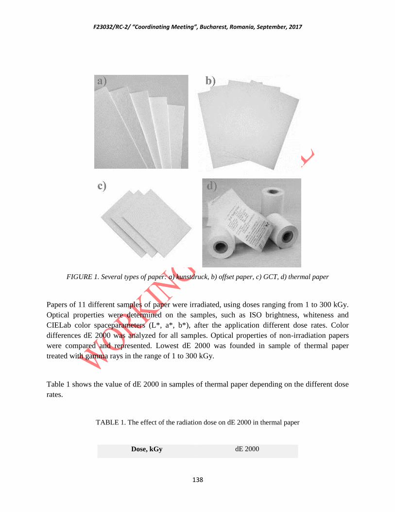
F23032/RC-2/ “Coordinating Meeting”, Bucharest, Romania, September, 2017
138
FIGURE 1. Several types of paper: a) kunstdruck, b) offset paper, c) GCT, d) thermal paper
Papers of 11 different samples of paper were irradiated, using doses ranging from 1 to 300 kGy.
Optical properties were determined on the samples, such as ISO brightness, whiteness and
CIELab color spaceparameters (L*, a*, b*), after the application different dose rates. Color
differences dE 2000 was analyzed for all samples. Optical properties of non-irradiation papers
were compared and represented. Lowest dE 2000 was founded in sample of thermal paper
treated with gamma rays in the range of 1 to 300 kGy.
Table 1 shows the value of dE 2000 in samples of thermal paper depending on the different dose
rates.
TABLE 1. The effect of the radiation dose on dE 2000 in thermal paper
Dose, kGy dE 2000

F23032/RC-2/ “Coordinating Meeting”, Bucharest, Romania, September, 2017
139
0 0
1 4.624763
3 0.249431
6 0.483687
9 0.205267
12 0.259483
15 0.317893
25 0.439133
35 0.619702
50 0.77271
75 1.431264
100 1.46334
150 1.573838
200 2.083946
300 2.29583
For samples of kunstdruck papers have been noticed significant color differences on samples
with highest weight which is explained by the highest absorption of radiation energy.
Table 2 shows the value of dE 2000 in samples of kunstdruck 115 g/cm2 and 300 g/cm
2
depending on the different dose rates.
TABLE 2. The effect of the radiation dose on dE 2000 in kunstdruck
Dose, kGy dE 2000 (115g/cm2) dE 2000 (300 g/cm
2)
0 0 0
1 0.079034 0.048439
3 0.344323 0.417948
6 0.567765 0.507746
9 0.898488 0.485173
12 0.663135 0.665831
15 0.915191 0.904626

F23032/RC-2/ “Coordinating Meeting”, Bucharest, Romania, September, 2017
140
25 1.146779 1.113864
35 1.088203 1.086172
50 1.812608 1.571964
75 1.762916 1.829547
100 2.353179 2.543139
150 3.333387 3.058238
200 2.976191 3.925216
300 3.10848 4.827202
Figure 2 shows a color change depending on the dose in samples kunstdruck 115 g/cm2 and 300
g/cm2.
FIGURE 2. The effect of the dose on dE 2000 valuse in different samples of kunstdruck
2. Gamma irradiation of leather gloves in terms of cultural heritage preservation

F23032/RC-2/ “Coordinating Meeting”, Bucharest, Romania, September, 2017
141
Microorganisms, fungi, and insects are often present in many types of cultural goods, from the
immovable objects, through archival and library materials, cellulose tape of film materials,
studio paintings, articles of wood, textiles, leather, and other museum objects. The biocide effect
of the ionizing radiation can be effectively implemented for their removal. The objective of this
work is the conservation by gamma irradiation of objects from the Museum of Nikola Tesla,
which is of outstanding value. In this case, infected leather gloves are irradiated with a dose of 5
kGy to eliminate mold. The amount of mold was evaluated by microbiological analysis before
and after gamma irradiation treatment. After successful treatment, Tesla’s leather gloves are
preserved from further deterioration and are now displayed in the museum [2].
Nowadays, increased concerns regarding the safeguarding of patrimony result in the constant
evolution of conservation and restoration methods. Biodeterioration is one of the most
challenging issues that curators have to deal with. The materials organic matter (paper, textile,
wood, leather, etc) is susceptible to degradation by insects, fungi, molds, bacteria, which inhabit
and feed on these materials. In recent years the storage of objects of cultural heritage has become
a major problem all over the world due to changes in the microclimate, especially due to the
change of climatic conditions, increased the content of moisture in the air as well as the
temperature, which is susceptible to the development of said pests. Improper storage conditions
lead to massive biological attacks in museums, archives or libraries, which are difficult to stop or
eliminate because of the large quantities of items involved. In addition, these microorganisms
may present serious health hazards for people dealing with CH artefacts [3, 4].
High energy radiation is a powerful tool for disinfection, effectively used in many fields,
especially for sterilization of medical products. Irradiation techniques are being used to protect
and preserve works of art around the world. The techniques are supported by the International
Atomic Energy Agency (IAEA) [5], which operates projects to preserve cultural heritage
artefacts using radiation. Wooden items, film archives, documents, textiles, leather, parchment
even mummies can be attacked and destroyed by bacteria, fungi, mould and insects [6,7,8].
These items can be brought from churches, museums and conservation centres to irradiation
facilities where it could be treated with gamma rays, however, it has not been widely used for
that purpose, although there is a great need for this kind of technique. Compared to other
techniques, gamma irradiation treatment is very high-level effectiveness and reliability and has a
major advantage: the biocide effect is ensured by the radiation’s high penetration power. Also,
removing bio-deteriorating agents, stopping the ongoing destructive process, restoring the object
of cultural value. The wider use of this technique requires conclusively establishing that
irradiation does not lead to unacceptable changes in the functional or decorative properties of the
CH artefacts. Regarding requirements of effectiveness within ethical guidelines such as minimal
intervention, non-contact irradiation techniques is very attractive. Besides, it enhanced by
penetration power of gamma rays that provides an interesting way to reach the inside of 3-
dimension items.

F23032/RC-2/ “Coordinating Meeting”, Bucharest, Romania, September, 2017
142
The effectiveness of ionizing radiation on bio-deteriogen organisms has been further confirmed
as well as the need to further study on the radio-induced effects. On the basis of physical and
microbiological obtained results, a dose of treatment ranging from 5 to 7kGy has been
recommended to obtain a significant reduction of the microbial load with minimum negative
effect on the paper substrate [9].
Nikola Tesla Museum in Belgrade is conceived as the establishment of a complex institution
with cultural, educational, scientific and memorial character. This item is largely determined by
his original vision and mission. As a cultural institution, the Museum dedicated to the protection
and presentation of Tesla's legacy and this cultural heritage is an invaluable [10].
The objective of this work is the conservation and preservation of objects from the Museum of
Nikola Tesla, in this case, the leather gloves. It is important to emphasize that these high
contaminated objects represent a potential threat to nearby objects or a collection depot where
they are located. On the surface analysed gloves present dispute mass (millions) 2-5 m size.
These spores are extremely light, dry quickly and effectively transmitted through the air and
colonize the surrounding objects. Transmission is multiplied during the manipulation of infested
items. These items were subjected to gamma irradiation in order to disinfection. The radiation
dose that is delivered is determined based on the comprehensive analysis of the results of
microbiological laboratories, material composition, as well as the condition of subjects and
literature data based [11]. After the performed treatment, it confirmed that it has successfully
carried out disinfection of infected objects, and they are again exposed in the museum.
The subjects of the leather gloves were brought from the Nikola Tesla Museum (Figure 3).
FIGURE 3. Samples of Nikola Tesla leather gloves highly contaminated with mold
The gamma sterilization process in “Vinča” Institute of Nuclear Sciences uses cobalt 60
radiation for research and industrial irradiation, for radiation sterilization of medical devices,
pharmaceutical and as well as for microbial decontamination of herbs and spices and the variety
of different products. Processing with gamma rays yields quick turnaround time, easily
penetrating packaging and product and is cost-effective. Dosimetry measurements were
performed using the ECB dosimetry system which provides a reliable means of measuring
absorbed dose in materials [12].
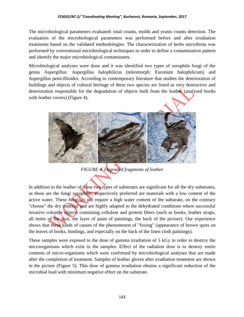
F23032/RC-2/ “Coordinating Meeting”, Bucharest, Romania, September, 2017
143
The microbiological parameters evaluated: total counts, molds and yeasts counts detection. The
evaluation of the microbiological parameters was performed before and after irradiation
treatments based on the validated methodologies. The characterization of herbs microbiota was
performed by conventional microbiological techniques in order to define a contamination pattern
and identify the major microbiological contaminants.
Microbiological analyses were done and it was identified two types of xerophile fungi of the
genus Aspergillus: Aspergillus halophilicus (teleomorph: Eurotium halophilicum) and
Aspergillus penicillioides. According to contemporary literature that studies the deterioration of
buildings and objects of cultural heritage of these two species are listed as very destructive and
deterioration responsible for the degradation of objects built from the leather (analysed books
with leather covers) (Figure 4).
FIGURE 4. Degraded fragments of leather
In addition to the leather of these two types of substrates are significant for all the dry substrates,
as these are the fungi xerophilic, respectively preferred are materials with a low content of the
active water. These fungi do not require a high water content of the substrate, on the contrary
"choose" the dry material and are highly adapted to the dehydrated conditions where successful
invasive colonize objects containing cellulose and protein fibers (such as books, leather straps,
all items of the skin, the layer of paint of paintings, the back of the picture). Our experience
shows that these kinds of causes of the phenomenon of "foxing" (appearance of brown spots on
the leaves of books, bindings, and especially on the back of the linen cloth paintings).
These samples were exposed to the dose of gamma irradiation of 5 kGy in order to destroy the
microorganisms which exist in the samples. Effect of the radiation dose is to destroy entire
contents of micro-organisms which were confirmed by microbiological analyses that are made
after the completion of treatment. Samples of leather gloves after irradiation treatment are shown
in the picture (Figure 5). This dose of gamma irradiation obtains a significant reduction of the
microbial load with minimum negative effect on the substrate.
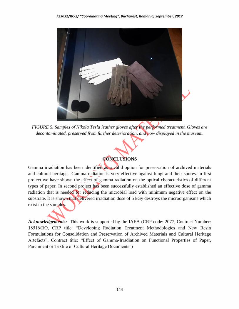
F23032/RC-2/ “Coordinating Meeting”, Bucharest, Romania, September, 2017
144
FIGURE 5. Samples of Nikola Tesla leather gloves after the performed treatment. Gloves are
decontaminated, preserved from further deterioration, and now displayed in the museum.
CONCLUSIONS
Gamma irradiation has been identified as a valid option for preservation of archived materials
and cultural heritage. Gamma radiation is very effective against fungi and their spores. In first
project we have shown the effect of gamma radiation on the optical characteristics of different
types of paper. In second project has been successfully established an effective dose of gamma
radiation that is needed for reducing the microbial load with minimum negative effect on the
substrate. It is shown that delivered irradiation dose of 5 kGy destroys the microorganisms which
exist in the samples.
Acknowledgements: This work is supported by the IAEA (CRP code: 2077, Contract Number:
18516/RO, CRP title: “Developing Radiation Treatment Methodologies and New Resin
Formulations for Consolidation and Preservation of Archived Materials and Cultural Heritage
Artefacts”, Contract title: “Effect of Gamma-Irradiation on Functional Properties of Paper,
Parchment or Textile of Cultural Heritage Documents”)

F23032/RC-2/ “Coordinating Meeting”, Bucharest, Romania, September, 2017
145
REFERENCES
[1] Medic, M, Vujcic, I, Masic, S, Milicevic, B, Dramicanin, M, Effect of Gamma-irradiation on
Functional Properties of Paper of Cultural Heritage Documents, InterRegioSci 2016, Novi Sad, decembar
2015, pp 73
[2] Vujčic, I, Mašic, S, Medic, M, Putic, S, Dramicanin, M - GAMMA IRRADIATION OF LEATHER
GLOVES IN TERMS OF CULTURAL HERITAGE PRESERVATION, Eco-Ist’17, Vrnjacka banja, jun
2017, pp. 531-535, ISBN 978-86-6305-062-4
[3] IAEA TECP-RER 8/015, Nuclear techniques for preservation of cultural heritage artefacts, 2009.
[4] IAEA Radiation Technology Series 2, Nuclear Techniques for Cultural Heritage Research, 2011.
[5] IAEA Nuclear Techniques for Cultural Heritage Research, IAEA RADIATION TECHNOLOGY
SERIES No. 2, 1-224, International Atomic Energy Agency Vienna International Centre, 2011
[6] Razem, B, Razem D, Braun, M, Irradiation treatment for the protection and conservation of cultural
heritage artefacts in Croatia, Radiation Physics and Chemistry, Volume 78, Issues 7–8, July–August
2009, Pages 729–731
[7] Bicchieri, M, Monti, M, Piantadina, G, Sodo, A, Effects of gamma irradiation on deteriorated paper,
Radiation Physics and Chemistry, Volume 125, August 2016, Pages 21–26
[8] Chio, J, Lim, S, Inactivation of fungal contaminants on Korean traditional cashbox by gamma
irradiation, Radiation Physics and Chemistry, Volume 118, January 2016, Pages 70–74
[9] M. Adamo, S. Baccaro, A. Cemmi Enea, Radiation Processing for Bio-Deteriorated Archived
Materials And Consolidation Of Porous Artefacts, Unità Tecnica Tecnologia dei Materiali Laboratorio
Tecnologie di Irraggiamento Centro Ricerche Casaccia, Roma, RT/2015/5/ENEA
[10] http://nikolateslamuseum.org/web/pages.php?p=4
11] Stanculescu, I, Moise, V, Ponta, C, Haiducu, M, Nisipeanu, S, Geba M, Miu, L, Iordache, O, Gamma
irradiation a chance for textile and leather heritage artefacts’ conservation, Proceedings of the Balkan
Symposium on Archaeometry Proceedings of the 29-30 October 2012 Bucharest, Romania
[12] ISO/ASTM 51538 - Practice for use of the ethanol-chlorobenzene dosimetry system, International
Organization for Standardization, 2009
F23032/RC-2/ “Coordinating Meeting”, Bucharest, Romania, September, 2017
146
Turkey
F23032-DEVELOPING RADIATION TREATMENT METHODOLOGIES AND NEW
RESIN FORMULATIONS FOR CONSOLIDATION AND PRESERVATION OF
ARCHIVED MATERIALS AND CULTURAL HERITAGE ARTEFACTS.
RADIATION INDUCED GRAFTING FOR THE PRESERVATION AND
CONSOLIDATION OF CELLULOSIC ARCHIVE MATERIALS.
(Research contract no: 18967)
DİLEK ŞOLPAN, MURAT TORUN, OLGUN GÜVEN
Hacettepe University, Department of Chemistry, 06800, Beytepe, Ankara/TURKEY
Email: [email protected]
Abstract
Museums, libraries and archives are preserving documents that are slowly degrading due to the inherent
ageing of the cellulose substrate or to the technological errors of the past (acid paper, iron gall ink).
Beside this, large quantities of paper are rapidly damaged by biological attacks, following natural
disasters and improper storage conditions. Cellulose is the major structural component of wood and plant
fibers and is the most abundant polymer synthesized by nature. Despite this great abundance, cellulosic
biomass has seen limited application outside of the paper industry. Its use as a feedstock for fuels and
chemicals has been limited because of its highly crystalline structure, inaccessible morphology and
limited solubility. Electron beams (EB), X-rays or gamma rays produce ions in a material which can then
initiate chemical reactions and cleavage of chemical bonds. Such ionizing radiation predominantly
scissions and degrades or depolymerizes cellulose. The gamma radiation will also be used for
decontamination and conservation purposes. Important advantages can be mentioned in its favour: no
toxic or radioactive residues remained in the treated item; large amount of objects can be treated quickly;
excellent reliability; attractive cost. There is also a potential side-effect. Interaction of gamma rays with
any substance may change its chemical and physical properties. The change is proportional with the
irradiation dose. The treatment of archives with gamma irradiation is an efficient and environmental
friendly alternative for biological decontamination of large volume of archives. For a successful
treatment, an optimal absorbed dose has to be established. An excessive dose may damage papers and an
insufficient one will not reduce bioburden to the desired level. The aim of this study is to investigate the
influence of gamma irradiation process on the mechanical and thermal properties and hydrophilicity of
whatman paper by grafting of N-butyl acrylate (NBA) and mixtures of methyl methacrylate (MMA) and
N-butyl acrylate of various compositions in presence of ethyl alcohol onto cellulose (Whatman No.1
paper) with γ-rays at 25°C in air.
1. INTRODUCTION
Cellulose, being the most abundant naturally occurring polymer on earth, is a highly interesting
material due to its renewability, low price, high availability, and good mechanical properties.
Cellulose is composed of repeating units of glucose joined together by hydrogen bonds to form

F23032/RC-2/ “Coordinating Meeting”, Bucharest, Romania, September, 2017
147
sheets. Paper is a good food supply for some microorganisms and insects. Hygroscopic
environment makes paper susceptible to biodegradation. The most important species involved in
the degradation of paper are insects, fungi and bacteria. They are changing the chemical and
physical structure of the paper by the alteration of cellulose links with products of metabolism.
The major causes of damage to works on paper are photo-oxidation, acidification or migration,
biological, mechanical injury, improper framing and storage [1]. The major concern for the using
radiation treatment in paper conservation is the large decrease of the degree of polymerisation
(DP) of cellulose. The main effect is the scission of β-glycoside bonds. These scissions are
reducing the average length of the polymeric chain in the amorphous region of cellulose and will
reduce the strength of cellulose fibers [2]. There are several strategies available for paper
conservation, targeting various aspects of deterioration, which are further specialized, depending
on the targets and limitations of the specific implementation [3].
In another study, Adamo et al. [4] suggested the use of doses of 2–3 kGy for the decontamination
of (strong) paper, since the negative effects of radiation were considered to be negligible. In a
later study though, Adamo et al. [5] concluded that even doses up to 10 Gy do not significantly
damage paper. Magaudda et al. [6] studied the effects of cradiation on library infesting insects
and found that the use of very low doses is effective since they induce sterility and molting to
them. In a later study, Valentin Moise et al. [7] suggest doses lower than 10 kGy for paper
decontamination, and consider that a small paper degradation is acceptable, taking into account
the overall preservation benefit. The good stability of the printing inks subjected to -radiation
was verified by Rocchetti et al. [8]. Magaudda et al. [9] and Adamo et al. [10] reported that
irradiated paper may be more prone to attack by cellulose eating insects and fungi growth. The
principles of using ionization technology for the disinfection and disinfestation of books and
documents, together with a review of several relevant experimental works are presented by
Adamo et al. [11]. The R&D work funded by the British Library was carried out at the
University of Surrey. The process selected for refinement and development is known as graft co-
polymerization and exploits, inter alia, the polymerization property of gamma radiation [12,13].
In this work, N-butyl acrylate (NBA) and methyl methacrylate (MMA) were chosen as the
monomers for grafting on cellulose surface. The grafted polymer, PBA, is hydrophobic and
expected to enhance the interface adhesion. Furthermore, PBA has a low glass-transition
temperature (Tg) and a soft chain [13]. The effect of gamma-irradiation process was investigated
for grafting of NBA and MMA/NBA mixtures on Whatman No.1 paper. NBA and MMA/NBA
mixtures were grafted on Whatman No.1 paper. The NBA and MMA/NBA grafted Whatman
No.1 papers were characterized by gravimetric measurements and also by chemical analysis,
such as Fourier Transform Infrared (FTIR) spectroscopy. In this report, thermal and mechanical
properties of Cellulose (Whatman No.1 paper)-g-poly(NBA) (Cell-g-poly(NBA) and Cellulose

F23032/RC-2/ “Coordinating Meeting”, Bucharest, Romania, September, 2017
148
(Whatman No.1 paper)-g-poly(MMA-co-NBA) (Cell-g-poly(MMA-co-NBA) were investigated
and we determined also the hydrophilicitiy properties of grafted and control samples.
2. EXPERIMENTAL
The irradiation of samples with 60
Co--rays was performed using a “Ob Servo Sangius, Gamma
irradiator” with a dose rate of 1.918 kGy/h. The cellulose sample used in this work was a
commercial sample, Whatman No.1 paper, Maidstone, UK, degree of polymerization, DP=2900,
R.S.D.=1.6%. We used N-butyl acrylate (NBA) and methyl methacrylate (MMA) for grafting
onto cellulose in this study. Samples were characterized by using spectroscopic techniques such
as FTIR, XPS drop contact angle, water uptake, thermal and mechanical resistance. Whatman
No.1 paper was used for our experiment: Whatman No.1 paper which is not a paper used for
documents, but is very suitable as a reference in paper conservation research, because it
comprises practically pure cellulose fibres [12]. The chemical structures of N-butyl acrylate,
methyl methacrylate utilized for surface modification of cellulose (Whatman No.1 paper) in this
work are presented in Figure 1.
a) b) c)
FIG.1. Structures of a) Cellulose, b) N-butyl acrylate, c) Methyl methacrylate.
Irradiation
Prior to -irradiation Whatman No.1 papers were cut into medium size as Dumpbell shaped with
a weight of ~0.06 g. Each Whatman No.1 papaer was washed in ethyl alcohol for 24 h and dried
under vacuum till constant weight. Measurements of dose levels to be used in the experimental
procedure were established using a Fricke dosimetry, according to standard regulations. Dose
levels used for irradiation were 0, 3, 9, 12, 18 kGy. The irradiation was performed using 60
Co
source with the dose rate 1.918 kGy/h in N2 atmosphere.
Grafting
In a typical irradiated grafting, to synthesize Cell-g-poly(NBA) (Whatman No.1 paper) and Cell-
g-poly(MMA-co-NBA), the conditions are N2 purged grafting solution containing the monomer
(NBA and MMA/NBA mixture, 1/1, 1/4, 1/8 v/v) solutions in 1/4, 1/10 v/v (monomer or
monomer mixture)/ethyl alcohol at room temperature under in air. All grafted samples were
extensively washed before analysis with a 2-propanol and finally with butyl alcohol to remove
F23032/RC-2/ “Coordinating Meeting”, Bucharest, Romania, September, 2017
149
traces of the unreacted monomer and the homopolymer that formed. The samples were dried in
vacuo at room temperature untill a constant weight and finally characterized.
Gravimetric measurements
The degree of grafting (DG, wt.%) was calculated by the weighing of the Whatman No.1 papers
before and after the grafting reactions were carried out. The degree of grafting was estimated as
follows:
DG=[(W2W1)/W1]x100 Eq.1
where W1 (g) is the weight of Whatman No.1 paper and W2 (g) is the dry weight of the Cell-g-
poly(NBA) and Cell-g-poly(MMA-co-NBA).
Tests on irradiated and grafted Cellulose (Whatman No.1 paper)
In order to evaluate the effect of irradiation on degradation of Whatman No.1 paper and grafting
of NBA and MMA/NBA mixtures on cellulose (Whatman No.1 paper) irradiation doses of
3,9,12,18 kGy were used. After irradiation, several types of tests were performed: spectroscopic
analysis (FTIR), thermogravimetric (TGA-DTG), mechanical, contact angle.
Spectroscopic analysis
The FTIR spectra for samples were recorded. FTIR spectroscopy (Thermo Scientific Nicolet
iS10 FT-IR Spectrometer) technique was utilized for characterization of the grafted, control and
irradiated samples. FTIR spectra were collected in the 400–4000 cm-1
region with resolution 4
cm-1
.
Thermal properties
Perkin Elmer Pyris1 Thermogravimetric Analyser was used for determination of thermal
properties of irradiated, control and grafted samples. Samples weighing 5-10 mg were heated in
dynamic nitrogen atmosphere from 20 °C to 800
°C at a heating rate of 20
ºC min
-1.
Mechanical properties
Mechanical properties i.e. tensile strength at break, elongation at break and young modulus of
films were determined by using a Zwick Z010 model Universal Testing Instrument and screw
grip module at room temperature. Strain rate for all samples is 50 mm/min. Dumbbell shaped
samples were prepared by using ISO 37 type 1 cutting die. Tear resistance was chosen as the
best method to measure the decrease of mechanical properties of paper [14]. In this test, the force
needed to the tear the sheet of paper in the cross direction (opposite to the machine direction)

F23032/RC-2/ “Coordinating Meeting”, Bucharest, Romania, September, 2017
150
was measured. Each reported value is a mean obtained from of 5 different measurements. Three
main types of mechanical tests were used. Tensile properties were measured for irradiated and
control samples.
Surface analysis
Contact-angle analysis
Static contact angles (CA) of samples were appraised using a Krüss DSA100 model contact
angle goniometer. With a syringe, a water drop was dropped carefully onto the grafted and
ungrafted cellulose samples. and the average CA value was obtained by measuring the same
sample at four different positions after a time period of 1 min. CAs of grafted and control
samples which were irradiated at two different doses were also measured after compressing the
samples (~0.5 cm x 0.5 cm) under 12 ton cm-2
pressure for 10 min using a manual hydraulic
press [15].
3. RESULTS and DISCUSSION
Paper is a good food supply for some microorganisms and insects. Hygroscopic environment
makes paper susceptible to biodegradation. The most important species involved in the
degradation of paper are insects, fungi and bacteria. They are changing the chemical and physical
structure of the paper by the alteration of cellulose links with products of metabolism. To
conserve and consolidate of cultural heritage we need two kinds of intervention: one to
strengthen it, and one to preserve it from further decay. The graft co-polymerization technology
seeks to achieve both goals in one process. The process selected for refinement and development
is known as graft co-polymerization [12] and exploits, inter alia, the polymerization property of
gamma radiation. In graft co-polymerization a monomer is introduced into the paper, and is
allowed to diffuse throughout.
In this work, the effect of gamma-irradiation process was investigated for grafting of NBA and
MMA/NBA mixtures on Whatman No.1 paper. The NBA and MMA/NBA grafted Whatman
No.1 papers were characterized by gravimetric measurements and also by chemical analysis,
such as Fourier Transform Infrared (FTIR) spectroscopy and XPS. Thermal and mechanical
properties of Cellulose (Whatman No.1 paper) and Cellulose (Whatman No.1 paper)-g-
poly(MMA-co-NBA) were investigated. The grafting reaction conditionas and parameters were
investigated and optimized. For this aim we changed the composition of monomer mixtures and
the composition of monomer mixtures and solvent, irradiation dose. The experimental conditions
and optimum conditions to use some characterization and analysis are shown in Tables 1 for
cellulose and grafted cellulose at different conditions.
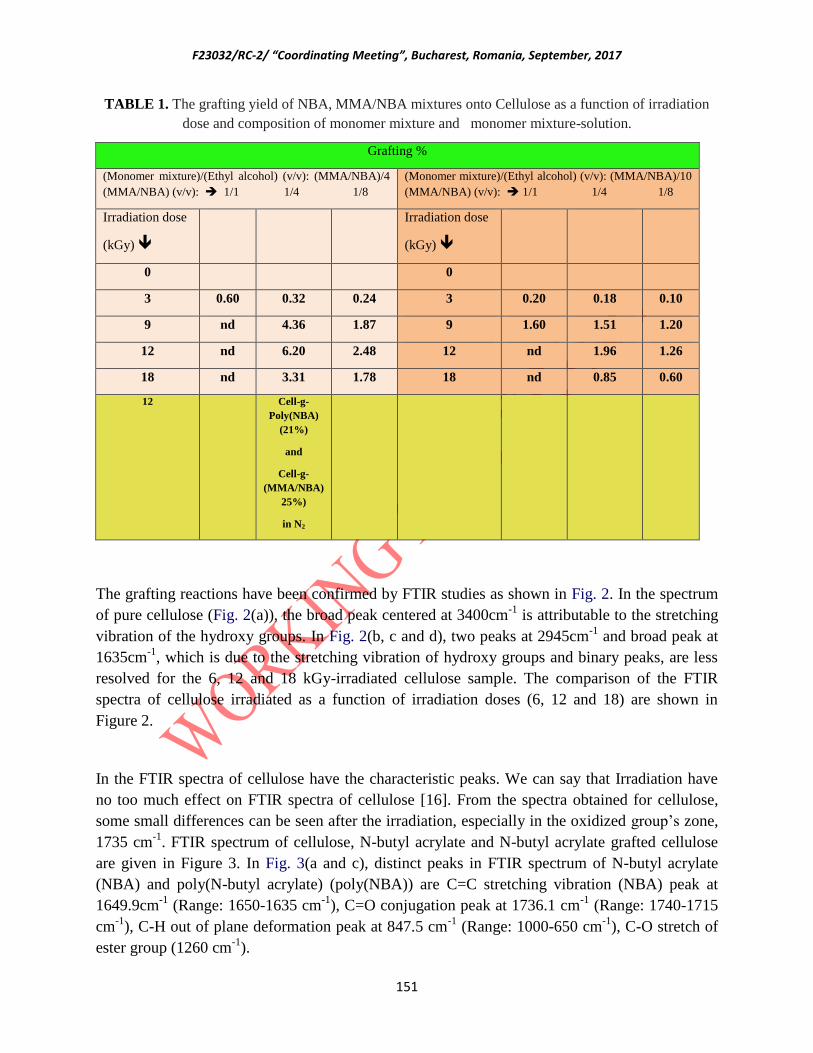
F23032/RC-2/ “Coordinating Meeting”, Bucharest, Romania, September, 2017
151
TABLE 1. The grafting yield of NBA, MMA/NBA mixtures onto Cellulose as a function of irradiation
dose and composition of monomer mixture and monomer mixture-solution.
Grafting %
(Monomer mixture)/(Ethyl alcohol) (v/v): (MMA/NBA)/4
(MMA/NBA) (v/v): 1/1 1/4 1/8
(Monomer mixture)/(Ethyl alcohol) (v/v): (MMA/NBA)/10
(MMA/NBA) (v/v): 1/1 1/4 1/8
Irradiation dose
(kGy)
Irradiation dose
(kGy)
0 0
3 0.60 0.32 0.24 3 0.20 0.18 0.10
9 nd 4.36 1.87 9 1.60 1.51 1.20
12 nd 6.20 2.48 12 nd 1.96 1.26
18 nd 3.31 1.78 18 nd 0.85 0.60
12 Cell-g-
Poly(NBA)
(21%)
and
Cell-g-
(MMA/NBA)
25%)
in N2
The grafting reactions have been confirmed by FTIR studies as shown in Fig. 2. In the spectrum
of pure cellulose (Fig. 2(a)), the broad peak centered at 3400cm-1
is attributable to the stretching
vibration of the hydroxy groups. In Fig. 2(b, c and d), two peaks at 2945cm-1
and broad peak at
1635cm-1
, which is due to the stretching vibration of hydroxy groups and binary peaks, are less
resolved for the 6, 12 and 18 kGy-irradiated cellulose sample. The comparison of the FTIR
spectra of cellulose irradiated as a function of irradiation doses (6, 12 and 18) are shown in
Figure 2.
In the FTIR spectra of cellulose have the characteristic peaks. We can say that Irradiation have
no too much effect on FTIR spectra of cellulose [16]. From the spectra obtained for cellulose,
some small differences can be seen after the irradiation, especially in the oxidized group’s zone,
1735 cm-1
. FTIR spectrum of cellulose, N-butyl acrylate and N-butyl acrylate grafted cellulose
are given in Figure 3. In Fig. 3(a and c), distinct peaks in FTIR spectrum of N-butyl acrylate
(NBA) and poly(N-butyl acrylate) (poly(NBA)) are C=C stretching vibration (NBA) peak at
1649.9cm-1
(Range: 1650-1635 cm-1
), C=O conjugation peak at 1736.1 cm-1
(Range: 1740-1715
cm-1
), C-H out of plane deformation peak at 847.5 cm-1
(Range: 1000-650 cm-1
), C-O stretch of
ester group (1260 cm-1
).

F23032/RC-2/ “Coordinating Meeting”, Bucharest, Romania, September, 2017
152
In Fig. 4 , the FTIR spectra of Cell-g-poly(MMA-co-NBA) contains the peaks for NBA, MMA
and cellulose and absorbance bands at 2856 (CH, CH2), 1740 (C=O), 1454 (CH2-), 1260 (CH3),
1200 and 1073cm-1
(COOCH3) and (C-O-C), respectively, appeared in comparison to cellulose
in agreement with the literature data. These results indicate the formation of NBA and
(MMA/NBA) mixtures grafted on cellulose. The band is also present in MMA (Fig. 4(a and c)),
but absent in the cellulose and NBA (Fig. 4(b)). We can say that FTIR spectra of Cell-g-
poly(MMA-co-NBA) contain peaks from both NBA and (MMA/NBA) and cellulose. This is
indicative of the participation of C=O and COOCH3 and C-O-C groups and grafting of MMA
and NBA on cellulose. From the Figs. 2-4 we identify the functional groups of the n-BA and also
(MMA/NBA) grafted cellulose by detecting the major absorbance peaks. IR observations show
that the studied whatman papers which grafted poly(MMA-co-NBA) showed substantial
structural changes and there was no important difference at our irradiation doses.
In Figures 5 and 6, the effect of monomer compositions and monomer composition/Ethyl alcohol
ratios for grafting of MMA/NBA monomer mixtures on cellulose at 12 kGy irradiaton dose are
shown. When the amount of NBA in monomer mixture is high, since the polymerization of NBA
is lower than MMA the grafting percent and related absorption peaks is smaller than the other
spectrum. The effect of irradiation dose to graft of MMA/NBA monomer mixtures on cellulose
is given in Figures 7 and 8. The success of grafting of poly(MMA-co-NBA) on cellulose was
confirmed via FTIR analysis, [16] in which an adsorption peak at 1729 cm-1
was observed after
grafting, corresponding to the carbonyl group in NBA and MMA in Figures 3-8. The spectra also
in Figures 5 and 6 depending on the monomer mixture compositions and solvent ratio and
Figures 7 and 8 showed an increase in the peak intensity for higher graft lengths at 12 kGy
irradiation dose. The highest grafting yield was at 12 kGy irradiation dose and MMA/NBA:1/4
and (MMA/NBA)/Ethyl alcohol ratio: (1/4)/4 conditions. FTIR spectra agree with these results.

F23032/RC-2/ “Coordinating Meeting”, Bucharest, Romania, September, 2017
153
60
65
70
75
80
85
90
95
100
%T
500 1000 1500 2000 2500 3000 3500 4000
Wav enumbers (cm-1)
FIG. 2. FTIR spectra of (a) unirradiated cellulose, (b) 6 kGy irradiated cellulose, (c) 12 kGy irradiated
cellulose, (d) 18 kGy irradiated cellulose.
82
84
86
88
90
92
94
96
98
100
102
104
%T
500 1000 1500 2000 2500 3000 3500 4000
Wav enumbers (cm-1)
FIG. 3. FTIR spectra of (a) Cell-g-poly(NBA), (b) unirradiated cellulose, (c) poly(NBA).
a
c
b
a
b
c
d

F23032/RC-2/ “Coordinating Meeting”, Bucharest, Romania, September, 2017
154
-120
-100
-80
-60
-40
-20
0
20
40
60
80
100
%T
500 1000 1500 2000 2500 3000 3500 4000
Wav enumbers (cm-1)
FIG. 4. FTIR spectra of (a) Cell-g-poly(MMA-co-NBA, (b) unirradiated cellulose, (c) poly(MMA-co-
NBA).
58
60
62
64
66
68
70
72
74
76
78
80
82
84
86
88
90
92
94
96
98
%T
500 1000 1500 2000 2500 3000 3500 4000
Wav enumbers (cm-1)
FIG.5. FTIR spectra of (a) Unirradiated cellulose, (b) 12 kGy irradiated cellulose, (c) Cell-g-poly(MMA-
co-NBA) 12 kGy irradiation (MMA/NBA)/EtOH: (1/4)/10, (d) Cell-g-poly(MMA-co-NBA) 12 kGy
irradiation (MMA/NBA)/EtOH: (1/8)/10.
a
b
c
(1/4)/10
(1/8)/10
a
b
c
d

F23032/RC-2/ “Coordinating Meeting”, Bucharest, Romania, September, 2017
155
62
64
66
68
70
72
74
76
78
80
82
84
86
88
90
92
94
96
98
%T
500 1000 1500 2000 2500 3000 3500 4000
Wav enumbers (cm-1)
FIG.6. FTIR spectra of (a) Unirradiated cellulose, (b) 12 kGy irradiated cellulose, (c) Cell-g-
poly(MMA-co-NBA) 12 kGy irradiation (MMA/NBA)/EtOH: (1/8)/4, (d) Cell-g-poly(MMA-co-NBA) 12
kGy irradiation (MMA/NBA)/EtOH: (1/4)/4.
FIG.7. FTIR spectra of (a) Unirradiated cellulose, (b) Cell-g-poly(MMA-co-NBA) 3 kGy irradiation, (c)
Cell-g-poly(MMA-co-NBA) 9 kGy irradiation (d) Cell-g-poly(MMA-co-NBA) 18 kGy irradiation, (e) Cell-
g-poly(MMA-co-NBA) 12 kGy irradiation. (MMA/NBA)/EtOH: (1/4)/10.
a
b
c
d
(1/4)/4
(1/8)/4
a
b
c
d
e
3 kGy
kGy
kGy
9 kGy
kGy
kGy 18 kGy
kGy
kGyk
Gy 12 kGy
kGyk
Gy
0 kGy
kGy
kGy
40
45
50
55
60
65
70
75
80
85
90
95
100
%T
500 1000 1500 2000 2500 3000 3500 4000
Wav enumbers (cm-1)

F23032/RC-2/ “Coordinating Meeting”, Bucharest, Romania, September, 2017
156
55
60
65
70
75
80
85
90
95
%T
500 1000 1500 2000 2500 3000 3500 4000
Wav enumbers (cm-1)
FIG.8. FTIR spectra of (a) Unirradiated cellulose, (b) Cell-g-poly(MMA-co-NBA) 3 kGy irradiation, (c)
Cell-g-poly(MMA-co-NBA) 9 kGy irradiation, (d) Cell-g-poly(MMA-co-NBA) 18 kGy irradiation, (e)
Cell-g-poly(MMA-co-NBA) 12 kGy irradiation. (MMA/NBA)/EtOH: (1/4)/4.
Thermal analysis
Thermal decomposition of cellulose, irradiated cellulose (6, 12 and 18 kGy), grafted cellulose,
cellulose-g-NBA and Cell-g-poly(MMA-co-NBA) were recorded and method was determined by
using TGA and was compared. Thermograms of unirradiated and irradiated cellulose are given in
Figure 9. Cellulose shows maximum decomposition at 397 ºC, 6, 12 and 18 kGy-irradiated
celluloses shows maximum decomposition at 397 ºC, and for unirradiated and irradiated
celluloses (6, 12 and 18 kGy) residual amounts are 0% at 750 and 650ºC, respectively. Cellulose
starts to degrade at 277 ºC and its maximum degradation temperature is 397 ºC. Two-stage
degradation was formed at 397 ºC (I) and 627 ºC (II) for the thermogram of unirradiated
cellulose under nitrogen atmosphere (Figures 9 and 10). Degradation temperature for irradiated
celluloses (6, 12 and 18 kGy) is observed at 387 ºC (I) and at a lower temperature, 627ºC.
Although the maximum decomposition temperatures are the same, there are small differences
between their residual amounts. The residual amounts can be seen in Figure 10.
In the thermograms of PolyNBA and Poly(MMA-co-NBA) (Figures 11 and 12) , Tmax=423ºC
and 419ºC and at 600°C the undecomposed residue is 4 and 0 %, respectively. For MMA, the
temperature for maximum weight loss is Tmax=378ºC and the temperature for half-life is T1/2 =
369ºC. Poly(methy methacrylate), PMMA, degrades by unzipping with separation of the
monomers from the end groups without any residual material and the product of degradation is
mostly MMA.
a
b
c
d
e
0 kGy
kGy
kGy 3 kGy
kGy
kGy 9 kGy
kGy
kGy 18 kGy
kGy
kGyk
Gy
12 kGy
kGyk
Gy

F23032/RC-2/ “Coordinating Meeting”, Bucharest, Romania, September, 2017
157
The thermograms of unirradiated and irradiated cellulose (12 kGy), Cell-g-poly(NBA) and Cell-
g-(MMA-co-NBA) are given in Figure 13. In the thermogram of the sample obtained for grafting
of NBA and MMA/NBA on cellulose, Tmax=423ºC and 419ºC, at 600ºC the undecomposed
residue is 4 and 0 %, respectively. The residual amounts can be seen in Figure 14. From these
results, it is seen that grafting of NBA and MMA/NBA) on cellulose improves thermal
properties of irradiated cellulose. Thermal degradation of cellulose was investigated by
thermogravimetry (TGA). Thermal degradation of cellulose under inert atmosphere produces
smaller molecules, mainly monomer components of macromolecular structure [17]. It can be
observed a higher thermal stability of grafted cellulose compared with unirradiated and irradiated
cellulose.
The effect of irradiation dose, the amount of poly(MMA-co-NBA) on cellulose and
(MMA(NBA)/Ethyl alcohol ratios on thermal stability of grafted cellulose are shown in Figures
15,16. Optimum parameters are very important to explain the thermal stability of grafted
cellulose samples is depending on the irradiation dose and the amount of grafted copolymer.
FIG.9. TGA of (a) unirradiated cellulose (red), (b) 6 kGy irradiated cellulose (pink), (c) 12 kGy
irradiated cellulose (black), (d) 18 kGy irradiated cellulose (green).
a Cellulose 0 kGy
b Cellulose 6 kGy
c Cellulose 12 kGy
d Cellulose 18 kGy
a
b c
d
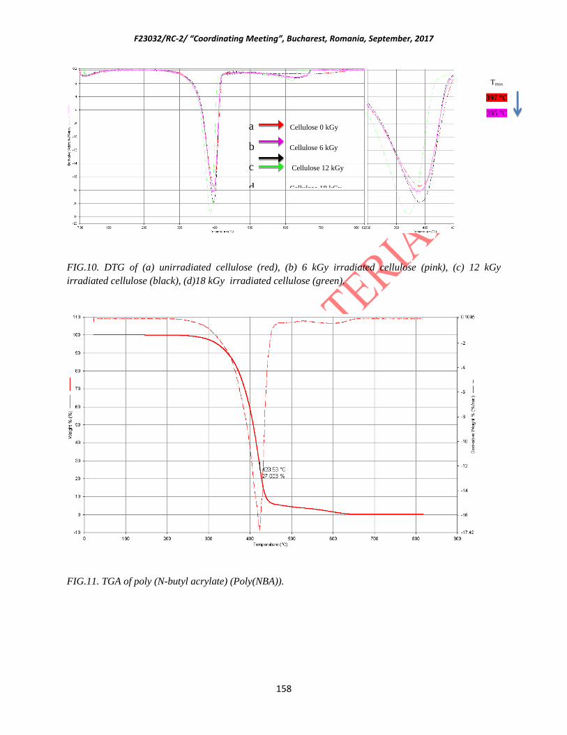
F23032/RC-2/ “Coordinating Meeting”, Bucharest, Romania, September, 2017
158
FIG.10. DTG of (a) unirradiated cellulose (red), (b) 6 kGy irradiated cellulose (pink), (c) 12 kGy
irradiated cellulose (black), (d)18 kGy irradiated cellulose (green).
FIG.11. TGA of poly (N-butyl acrylate) (Poly(NBA)).
Tmax
397 °C
395 °C
395 °C
387 °C, 627°C
a Cellulose 0 kGy
b Cellulose 6 kGy
c Cellulose 12 kGy
d Cellulose 18 kGy

F23032/RC-2/ “Coordinating Meeting”, Bucharest, Romania, September, 2017
159
FIG.12. TGA of poly (methylmethacrylate-co-N-butyl acrylate) (Poly(MMA-co-NBA)).
FIG.13. TGA of (a) unirradiated cellulose (red), (b) 12 kGy irradiated cellulose (green), (c) Cell-g-
poly(NBA) (blue), (21% grafting yield by direct grafting, 12 kGy), (d) Cell-g-poly(MMA-co-NBA) (pink)
(25% grafting yield, 12 kGy by direct grafting).
a b c
d
a Cellulose 0 kGy
b Cellulose 12 kGy
c Cell-g-poly(NBA) 12 kGy
d Cell-g-poly(MMA-co-NBA) 12 kGy

F23032/RC-2/ “Coordinating Meeting”, Bucharest, Romania, September, 2017
160
FIG.14. DTG of (a) unirradiated cellulose, (b) 12 kGy irradiated cellulose, (c) Cell-g-poly(NBA) (21%
grafting yield by direct grafting, 12 kGy), (d) Cell-g-poly(MMA-co-NBA) (25% grafting yield, 12 kGy by
direct grafting).
FIG.15. TGA of (a) unirradiated cellulose (red), (b) Cell-g-poly(MMA-co-NBA) 3 kGy irradiation (pink),
(c) Cell-g-poly(MMA-co-NBA) 9 kGy irradiation (green), (d) Cell-g-poly(MMA-co-NBA) 18 kGy
irradiation (blue), (e) Cell-g-poly(MMA-co-NBA) 12 kGy irradiation (orange). (MMA/NBA)/EtOH:
(1/4)/4.
a
b
c
d
a
b
c
d
e
Tmax
397 °C
395 °C
394°C,
414°C
392 °C
384°C
a Cellulose 0 kGy
b Cellulose 12 kGy
c Cell-g-poly(NBA) 12 kGy
d Cell-g-poly(MMA-co-NBA) 12 kGy
a Cellulose 0 kGy
b Cell-g-poly(MMA-co-NBA) (1/4)/4 3 kGy
c Cell-g-poly(MMA-co-NBA) (1/4)/4 9 kGy
d Cell-g-poly(MMA-co-NBA) (1/4)/4 18 kGy
e Cell-g-poly(MMA-co-NBA) (1/4)/4 12 kGy

F23032/RC-2/ “Coordinating Meeting”, Bucharest, Romania, September, 2017
161
FIG.16. DTG of (a) unirradiated cellulose (red), (b) Cell-g-poly(MMA-co-NBA) 3 kGy irradiation (pink),
(c) Cell-g-poly(MMA-co-NBA) 9 kGy irradiation (green), (d) Cell-g-poly(MMA-co-NBA) 18 kGy
irradiation (blue), (e) Cell-g-poly(MMA-co-NBA) 12 kGy irradiation (orange). (MMA/NBA)/EtOH:
(1/4)/4.
Mechanical properties
In order to investigate the physical and thermal behaviour of cellulose (Whatman No.1 paper),
mechanical (stress-strain) and thermal (TGA/DTG and DSC) tests should made. Stress-Strain %
curves for unirradiated and 6, 12, 18 kGy irradiated cellulose, and cellulose-g-poly(NBA) and
Cell-poly(MMA-co-NBA) are shown in Figures 23 and 24. For unirradiated cellulose and 6, 12,
18 kGy irradiated celluloses, Force at break (breaking force) are 17.0, 15.3, 12.5, 10.1 MPa but
for Cell-g-poly(NBA) and Cell-g-poly(MMA-co-NBA) are 8.28, 8.62 MPa, respectively. For
unirradiated cellulose and 6, 12, 18 kGy irradiated celluloses, Elongation at break (tensile strain)
are 3.1, 2.8, 2.6, 2.6 % and for Cell-g-poly(NBA) and Cell-g-poly(MMA-co-NBA) are 2.9 and
25.5 %. When we compared TGA results with mechanical experiment results they are agree with
eachother. The important factor is chain scission and crosslinking which is competitive depend
on irradiation dose. The ability of a paper to absorb energy when subjected to stress is expressed
by the tensile energy absorption (TEA) [18]. It is related to the paper toughness and its ability to
tolerate stressing or strain. Percent deformation or stretch indicates the ability of a paper to stand
tensile stress and to adapt to different conformations. Significant decreases caused by radiation
can be seen in three of the samples. It is well known that ionising radiation causes chain
breakage with lowering of the degree of polymerization of cellulose [19]. This effect would
diminish the mechanical properties of cellulose; the intensity of the effect will depend on the
applied dose, the conditions of irradiation and the initial quality of the cellulosic material. The
mechanical properties of paper determine its durability and resistance to environmental stress.
Tmax
397°C
395 °C
394°C,
414°C
392 °C
384°C
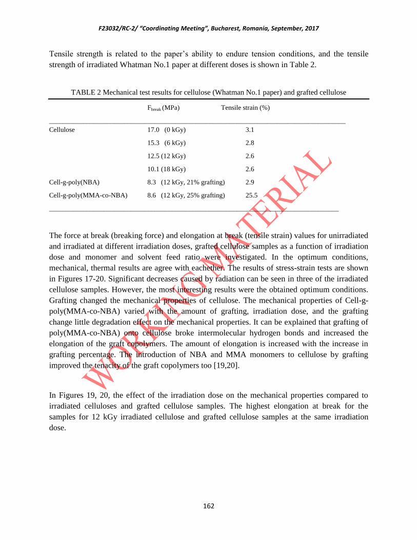
F23032/RC-2/ “Coordinating Meeting”, Bucharest, Romania, September, 2017
162
Tensile strength is related to the paper’s ability to endure tension conditions, and the tensile
strength of irradiated Whatman No.1 paper at different doses is shown in Table 2.
TABLE 2 Mechanical test results for cellulose (Whatman No.1 paper) and grafted cellulose
Fbreak (MPa) Tensile strain (%)
_______________________________________________________________________________________
Cellulose 17.0 (0 kGy) 3.1
15.3 (6 kGy) 2.8
12.5 (12 kGy) 2.6
10.1 (18 kGy) 2.6
Cell-g-poly(NBA) 8.3 (12 kGy, 21% grafting) 2.9
Cell-g-poly(MMA-co-NBA) 8.6 (12 kGy, 25% grafting) 25.5
_____________________________________________________________________________________
The force at break (breaking force) and elongation at break (tensile strain) values for unirradiated
and irradiated at different irradiation doses, grafted cellulose samples as a function of irradiation
dose and monomer and solvent feed ratio were investigated. In the optimum conditions,
mechanical, thermal results are agree with eachether. The results of stress-strain tests are shown
in Figures 17-20. Significant decreases caused by radiation can be seen in three of the irradiated
cellulose samples. However, the most interesting results were the obtained optimum conditions.
Grafting changed the mechanical properties of cellulose. The mechanical properties of Cell-g-
poly(MMA-co-NBA) varied with the amount of grafting, irradiation dose, and the grafting
change little degradation effect on the mechanical properties. It can be explained that grafting of
poly(MMA-co-NBA) onto cellulose broke intermolecular hydrogen bonds and increased the
elongation of the graft copolymers. The amount of elongation is increased with the increase in
grafting percentage. The introduction of NBA and MMA monomers to cellulose by grafting
improved the tenacity of the graft copolymers too [19,20].
In Figures 19, 20, the effect of the irradiation dose on the mechanical properties compared to
irradiated celluloses and grafted cellulose samples. The highest elongation at break for the
samples for 12 kGy irradiated cellulose and grafted cellulose samples at the same irradiation
dose.

F23032/RC-2/ “Coordinating Meeting”, Bucharest, Romania, September, 2017
163
0 1 2 3 4 5 6
0
4
8
12
16
20
Sta
nd
ard
Fo
rce
(M
Pa
)
Strain (%)
Cellulose 0 kGy
Cellulose 18 kGy
Cellulose 12 kGy
Cellulose 6 kGy
FIG. 17. Stress-strain curves of (a) unirradiated cellulose (black), (b) 6 kGy irradiated cellulose (green),
(c) 12 kGy irradiated cellulose (blue), (d) 18 kGy irradiated cellulose (red).
0 5 10 15 20 25
0
5
10
15
Strain in %
Forc
e in M
Pa
FIG.18. Stress-Strain curves of (a) unirradiated cellulose (red), (b) Cell-g-poly(NBA) (green) (21%
grafting yield by direct grafting, 12 kGy), (c) Cell-g-poly(MMA-co-NBA) (blue) (25% grafting yield by
direct grafting, 12 kGy).
a
b
a
a
b
c
c
d
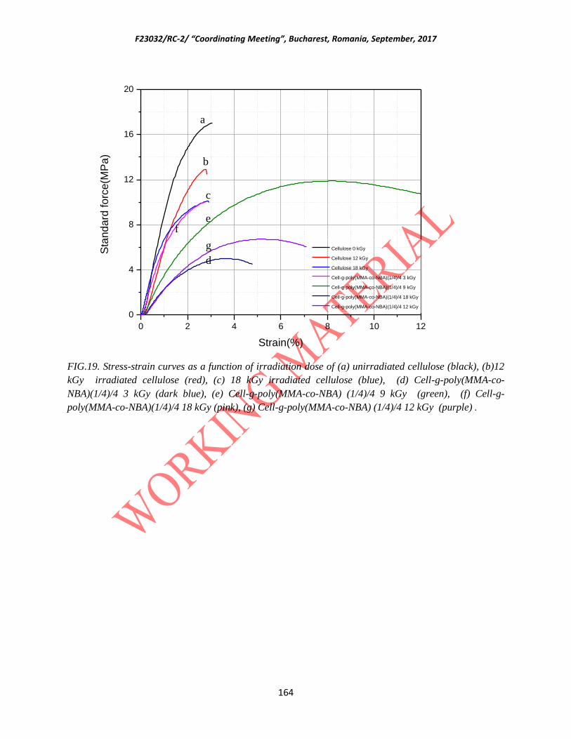
F23032/RC-2/ “Coordinating Meeting”, Bucharest, Romania, September, 2017
164
0 2 4 6 8 10 12
0
4
8
12
16
20
Sta
nd
ard
fo
rce
(MP
a)
Strain(%)
Cellulose 0 kGy
Cellulose 12 kGy
Cellulose 18 kGy
Cell-g-poly(MMA-co-NBA)(1/4)/4 3 kGy
Cell-g-poly(MMA-co-NBA)(1/4)/4 9 kGy
Cell-g-poly(MMA-co-NBA)(1/4)/4 18 kGy
Cell-g-poly(MMA-co-NBA)(1/4)/4 12 kGy
FIG.19. Stress-strain curves as a function of irradiation dose of (a) unirradiated cellulose (black), (b)12
kGy irradiated cellulose (red), (c) 18 kGy irradiated cellulose (blue), (d) Cell-g-poly(MMA-co-
NBA)(1/4)/4 3 kGy (dark blue), (e) Cell-g-poly(MMA-co-NBA) (1/4)/4 9 kGy (green), (f) Cell-g-
poly(MMA-co-NBA)(1/4)/4 18 kGy (pink), (g) Cell-g-poly(MMA-co-NBA) (1/4)/4 12 kGy (purple) .
e f
a
b
c
d
g

F23032/RC-2/ “Coordinating Meeting”, Bucharest, Romania, September, 2017
165
0 2 4 6 8 10 12
0
4
8
12
16
20
Sta
nd
ard
fo
rce
(MP
a)
Strain(%)
Cellulose 0 kGy
Cellulose 12 kGy
Cellulose 18 kGy
Cell-g-poly(MMA-co-NBA)(1/8)/4 3 kGy
Cell-g-poly(MMA-co-NBA)(1/8)/4 9 kGy
Cell-g-poly(MMA-co-NBA)(1/8)/4 18 kGy
Cell-g-poly(MMA-co-NBA)(1/8)/4 12 kGy
FIG.20. Stress-strain curves as a function of irradiation dose of (a) unirradiated cellulose (black), (b) 12
kGy irradiated cellulose (red), (c) 18 kGy irradiated cellulose (blue), (d) Cell-g-poly(MMA-co-
NBA)(1/8)/4 3 kGy (pink), (e) Cell-g-poly(MMA-co-NBA) (1/8)/4 9kGy (green), (f) Cell-g-poly(MMA-co-
NBA)(1/8)/4 18kGy (dark blue), (g)Cell-g-poly(MMA-co-NBA) (1/8)/4 12 kGy (purple).
Surface analysis
Contact angle analysis
When the more apolar Poly(NBA) grafted make cellulose more hydrophobic which is revealed
by significant increase in CA in flat surfaces [21]. As a result of the decrease in hydrophilicity
associated with grafting of Poly(NBA) was implied by a unique observation. It is possible to
accept that the hydrophilic surface of cellulose (Whatman No.1 paper) not provide an easier
sliding of the water molecules over surface. The water droplet can not stay on the Cell-g-
polyNBA surface for the Poly(NBA) grafted samples in contrast to Cellulose (Whatman No.1
paper). The contact angle is a measure of the wettability of a surface by a liquid. It broadly
defines the hydrophilic/hydrophobic character of the surface. The contact angle measurement
evaluates the hydrophobic or hydrophilic characteristics of a surface. As expected, results show
that the presence of NBA and MMA/NBA in the cellulose leads to higher contact angle,
signalizing that the tendency to absorb water is decreased and that the surfaces became more
hydrophobic. These results are in agreement with the findings of water uptake, which can be
directly related to contact angle. The results of contact angles (degree) measured on the surface
of cellulose, Cell-g-poly(NBA), Cell-g-poly(MMA-co-NBA) are given in Table 3.
a
b
c
e
d
f
g
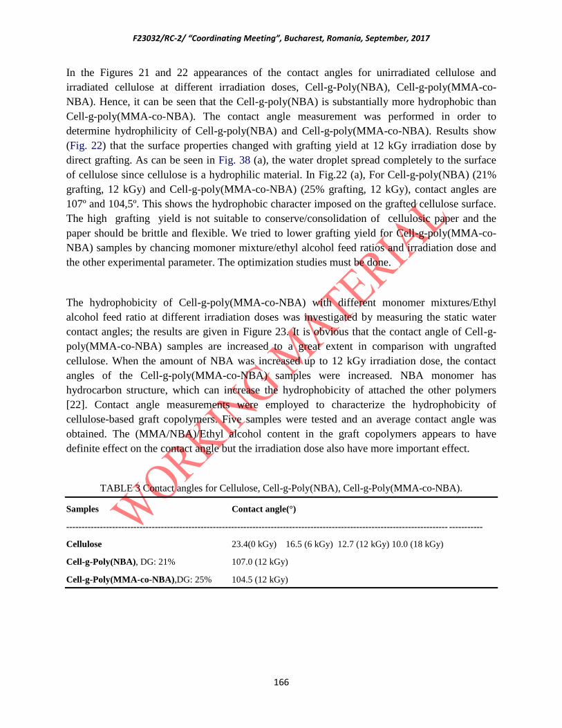
F23032/RC-2/ “Coordinating Meeting”, Bucharest, Romania, September, 2017
166
In the Figures 21 and 22 appearances of the contact angles for unirradiated cellulose and
irradiated cellulose at different irradiation doses, Cell-g-Poly(NBA), Cell-g-poly(MMA-co-
NBA). Hence, it can be seen that the Cell-g-poly(NBA) is substantially more hydrophobic than
Cell-g-poly(MMA-co-NBA). The contact angle measurement was performed in order to
determine hydrophilicity of Cell-g-poly(NBA) and Cell-g-poly(MMA-co-NBA). Results show
(Fig. 22) that the surface properties changed with grafting yield at 12 kGy irradiation dose by
direct grafting. As can be seen in Fig. 38 (a), the water droplet spread completely to the surface
of cellulose since cellulose is a hydrophilic material. In Fig.22 (a), For Cell-g-poly(NBA) (21%
grafting, 12 kGy) and Cell-g-poly(MMA-co-NBA) (25% grafting, 12 kGy), contact angles are
107º and 104,5º. This shows the hydrophobic character imposed on the grafted cellulose surface.
The high grafting yield is not suitable to conserve/consolidation of cellulosic paper and the
paper should be brittle and flexible. We tried to lower grafting yield for Cell-g-poly(MMA-co-
NBA) samples by chancing momoner mixture/ethyl alcohol feed ratios and irradiation dose and
the other experimental parameter. The optimization studies must be done.
The hydrophobicity of Cell-g-poly(MMA-co-NBA) with different monomer mixtures/Ethyl
alcohol feed ratio at different irradiation doses was investigated by measuring the static water
contact angles; the results are given in Figure 23. It is obvious that the contact angle of Cell-g-
poly(MMA-co-NBA) samples are increased to a great extent in comparison with ungrafted
cellulose. When the amount of NBA was increased up to 12 kGy irradiation dose, the contact
angles of the Cell-g-poly(MMA-co-NBA) samples were increased. NBA monomer has
hydrocarbon structure, which can increase the hydrophobicity of attached the other polymers
[22]. Contact angle measurements were employed to characterize the hydrophobicity of
cellulose-based graft copolymers. Five samples were tested and an average contact angle was
obtained. The (MMA/NBA)/Ethyl alcohol content in the graft copolymers appears to have
definite effect on the contact angle but the irradiation dose also have more important effect.
TABLE 3 Contact angles for Cellulose, Cell-g-Poly(NBA), Cell-g-Poly(MMA-co-NBA).
Samples Contact angle(°)
----------------------------------------------------------------------------------------------------------------------------- -----------
Cellulose 23.4(0 kGy) 16.5 (6 kGy) 12.7 (12 kGy) 10.0 (18 kGy)
Cell-g-Poly(NBA), DG: 21% 107.0 (12 kGy)
Cell-g-Poly(MMA-co-NBA),DG: 25% 104.5 (12 kGy)

F23032/RC-2/ “Coordinating Meeting”, Bucharest, Romania, September, 2017
167
FIG. 21. Water contact angle (CA) values for (a) unirradiated cellulose (Whatman No.1 paper) (b) 6 kGy
irradiated cellulose, (c) 12 kGy irradiated cellulose, (d)18 kGy irradiated cellulose.
FIG. 22. Water contact angle (CA) values of (a) Cell-g-poly(NBA) (21% grafting yield by direct grafting,
12 kGy), (b) Cell-g-poly(MMA-co-NBA) (25% grafting yield by direct grafting, 12 kGy).
(a) Irradiated Cellulose Cell-g-poly(MMA-co-NBA)(1/1)/4 Cell-g-poly(MMA-co-NBA)(1/4)/4 Cell-g-poly(MMA-co-NBA)(1/8)/4
12 kGy 12 kGy 12 kGy 12 kGy
a
107.0
b
104.5
a
23.4
b
16.5
c
12.7
d
10.0

F23032/RC-2/ “Coordinating Meeting”, Bucharest, Romania, September, 2017
168
(b) Irradiated Cellulose Cell-g-poly(MMA-co-NBA)(1/1)/10 Cell-g-poly(MMA-co-NBA)(1/4)/10 Cell-g-poly(MMA-co-NBA)(1/8)/10
12 kGy 12 kGy 12 kGy 12 kGy
(c) Cell-g-poly(MMA-co-NBA)(1/4)/10 Cell-g-poly(MMA-co-NBA)(1/4)/10 Cell-g-poly(MMA-co-NBA)(1/4)/10 Cell-g-poly(MMA-co-NBA)(1/4)/10
18 kGy 12 kGy 9 kGy 3 kGy
FIG. 23. Water contact angle (CA) values as a function of (MMA/NBA)/Ethyl alcohol feed ratio (a) and
(b) and irradiation dose (c) for Cellulose, irradiated cellulose and Cell-g-poly(MMA-co-NBA).
4. CONCLUSIONS
The grafting of NBA and MMA/NBA mixture on cellulose was performed by direct grafting
methods. For the highest grafting yield, required irradiation dose and monomer mixture ratios,
solvent amount were determined. The results of spectroscopic and thermal characterization,
mechanical show that NBA and MMA/NBA participated with cellulose and the stability of
cellulose is affected by grafting of NBA and MMA/NBA. The results of contact angle
measurements ans SEM results show that the surface properties changed with percent grafting at
12.0 kGy irradiation dose by grafting. Cellulosic papers used in the preparation of archives were
identified and the cellulosic materials were tested for the application of the methods
proposed.The work plan for the next year will be comprised of the following steps.
1. In addition to the used monomers, the effect of the different amounts of microcrystallline
cellulose will be investigated.
2. The grafting of cellulose papers with monomer solutions of above mentioned monomers in the
presence of microcrystalline cellulose and different concentrations will be tested.
3. Irradiation of impregnated papers with gamma rays and if possible by electron beams.
4. Chemical, mechanical, and surface property testing of (co)polymer loaded papers will be
performed.

F23032/RC-2/ “Coordinating Meeting”, Bucharest, Romania, September, 2017
169
5. Microbiological tests will be performed.
REFERENCES
[1] CARLMARK,A., LARSSON, E., MALMSTRÖM. E., “Grafting of cellulose by ring-opening
polymerization. A review.” European Polymer Journal, (2012), 646–1659.
[2] BRATU, E., MOISE, I.V., CUTRUBINIS, M., NEGUT, D.C., VIRGOLICI, M., “Archives
decontamination by gamma irradiation.” Nukleonika, 54(2), (2009), 77−84.
[3] ZERVOS, S., ALEXOPOULOU, I., “Paper conservation methods: a literature review.” Cellulose, 22,
(2015), 2859–2897.
[4] ADAMO, M., BRIZZ, M., MAGAUDDA, G., MARTINELLI, G., ZAPPALA, M.P., ROCCHETTI, F.,
SAVAGNONE. F., “Gamma radiation treatment of paper in different environmental conditions: chemical,
physical and microbiological analysis.” Restaurator, 22, (2001), 107–131.
[5] ADAMO, M., MAGAUDDA, G., OMARINI,S., “Biological measurement of damage occurring to the
inner structure of paper after gamma rays irradiation. Preliminary tests.” Restaurator, 28, (2007), 39–46.
[6] MAGAUDDA, G., ADAMO, M., PASQUALI, A., ROSSI. G., “The effect of ionizing gamma ray
radiation on the biology of the periplaneta Americana.” Restaurator, 21, (2000), 41–51.
[7] VALENTIN, V., MOISE, I., et al., “Establishing the irradiation dose for paper decontamination.” Radiat
Phys Chem 81, (2012), 1045–1050.
[8] ROCCHETTI, F., ADAMO, M., MAGAUDDA. G., “Fastness of printing inks subjected to gamma-ray
irradiation and accelerated ageing.” Restaurator, 23, (2002), 15–26.
[9] MAGAUDDA, G., ADAMO, M., ROCCHETTI, F., “Damage caused by destructive insects to cellulose
previously subjected to gamma-ray irradiation and artificial ageing.” Restaurator, 22, (2001), 242–250.
[10] ADAMO, M., MAGAUDDA, G., TRIONFETTI NISINI, P., TRONELLI. G., “Susceptibility of cellulose
to attack by cellulolytic microfungi after gamma irradiation and ageing.” Restaurator, 24, (2003), 145–151.
[11] ADAMO, M., MAGAUDDA, G., Tata, A., “Radiation technology for cultural heritage restoration.”
Restaurator, 25, (2004), 159–170.
[12] EGAN, A., Mardian, J., “The strengthening of embrittled books using gamma radiation.” Rad.Phys.Chem.,
46(4-6), (1995), 1303-1307.
[13] SHUZHAO, L., MIAOMIAO, X., ANNA, Z., HUINING, X., “Cellulose Microfibrils Grafted with PBA
via Surface-Initiated Atom Transfer Radical Polymerization for Biocomposite Reinforcement.”
Biomacromolecules, (2011), 3305–3312.
[14] GONZALEZ, M.E., CALVO, A.M., KAIRIYAMA, E., “Gamma radiation for preservation of biologically
damaged paper.” Radiation Physics and Chemistry, 63, (2002), 263–265.
[15] GONZALEZ, A., Alvarez Igarzabal, C., “Soy protein – Poly (lactic acid) bilayer films as biodegradable
material for active food packaging.” Food Hydrocolloids, 33, (2013), 289-296.
[16] HANNA, L., LINDA, F., MALMSTRÖM, E., HULT, A., “Surface grafting of microfibrillated cellulose
with poly(e-caprolactone)–Synthesis and characterization.” European Polymer Journal, 44, (2008), 2991–
2997.
[17] JANATA, M., MASAR, B., TOMAN, L., VLCEK, P., LATALOVA, P., Brus, J., HOLLER, P., “Synthesis
of novel types of graft copolymers by a n ‘‘grafting-from n” method using ring-opening polymerization of
F23032/RC-2/ “Coordinating Meeting”, Bucharest, Romania, September, 2017
170
lactones and lactides.” React Funct Polym., 57 (2-3), (2003), 137–46.
[18] YUPENG LIU, L., KEJIAN, Y., XIAOMING, C., JIFU, W., ZHONGKAI, W., HARRY, PLOEHN,J.,
“Sustainable thermoplastic elastomers derived from renewable cellulose, rosin and fatty acids.” Polym.
Chem., 5, (2014), 3170-3181.
[19] GÜCLÜ, G., GÜRDAĞ, G., ÖZGÜMÜŞ, S., “Competitive removal of heavy metal ions by cellulose graft
copolymers.” J Appl Polym Sci., 90, (2003), 2034–2039.
[20] WANG, J., YAO, K., KORICH, A.L., LI, S., MA, S., PLOEHN, H.J., IOVINE, P.M., WANG, CHU, C.,
F., TANG, C., “Combining renewable gum rosin and lignin: Towards hydrophobic polymer composites by
controlled polymerization.” J. Polym. Sci. Polym. Chem, A49, (2011), 3728–3738.
[21] NURKUVA, Z., AAL, A.A., KUPCHİSHİN, A., KHUTOYANSKİY, V., “Radiation grafting from binary
monomer mixtures. II. Vinyl ether of monoethanolamine and N-vinylpyrrolidone.” Radiat Phys Chem, 68,
(2003), 793-798.
[22] ANNA, C., EMMA, L., EVA, M., “Grafting of cellulose by ring-opening polymerisation–A review.”
European Polymer Journal, 48, (2012), 1646–1659.
[23] CARLMARK, A., MALMSTRÖM, E.E., “ATRP Grafting from Cellulose Fibers to Create Block-
Copolymer Grafts.” Biomacromolecules, 4, (2003), 1740–1745.
Ukraine
Determination of the Process Parameters of Archived Materials and Cultural
Heritage Artefacts Irradiation Treatment by Using Monte-Carlo Method
V.V. Morgunov
Ukrainian Engineering Pedagogics Academy
Kharkiv, Ukraine
Abstract. Radiation processing techniques are in wide use in for disinfection and consolidation of archived
materials and cultural heritage artefacts. The maximum dose (Dmax), which can be absorbed by product without
changing its properties, is known from research phase. So,minimal absorbed dose (Dmin) should be transferred to
product to achieve disinfection and this dose shouldn’t be more than maximum dose. The location and magnitude of
the dose minimum and maximum is critical to process control, optimized irradiation configurations and it affects
both disinfection and product properties. Reliable product dose-maps are necessary for identification of these critical
process parameters and may involve time consuming and laborious dosimetry. In some cases determination of the
dose-maps is difficult to produce by experiment. Such cases are very often occur during cultural heritage artefacts
radiation treatment. In such situations the numerical simulation can be used. After consideration of all possible
software toolkits for passage of ionization radiation through the matter GEANT4 was chosen. The CADMesh library
was implemented in developed code to input complicated geometry. The radiation sources (plaque and cylindrical)

F23032/RC-2/ “Coordinating Meeting”, Bucharest, Romania, September, 2017
171
were inputed into the code. Their activities, loading date into operation can be loaded from .csv file. The comparison
between measuraments and simulated results were made. The simulated results have shown a good agreement with
measured ones.
1. Collection data (density, composition, geometry shape and dimension)
Following data were collected:
Dimension and shape of irradiation facility of
Horia Hulubei National Institute for R&D in Physics and Nuclear Engineering (IFIN-
HH) (gamma rays, plaque) – Fig. 1;
Ruđer Boškovic Institute (gamma rays, cylindrical) – Fig. 2;
Slovak Medical University (electron beam) – Fig. 3.
Composition and density of irradiated objects materials and materials used in the
irradiation facilities:
wood;
paper;
polyethylene;
aluminum;
concrete;
stainless steel;
air.
Shapes of different objects:
pencils of 60
Co;
irradiation rack;
polyethylene bottles;
tote boxes and empty boxes filled with paper and dosimeters;
crumpled paper.

F23032/RC-2/ “Coordinating Meeting”, Bucharest, Romania, September, 2017
172
Fig. 1. Irraidation chamber and maze in IFIN-HH, Romania. 1 – radiation source; 2 – tote
boxes; 3 – cardboard boxes filled with paper and where dosimeters were placed, 4 – walls.
Fig 2. Irradiation room and maze in Ruđer Bošković Institute. 1 – radiation source; 2 – walls.

F23032/RC-2/ “Coordinating Meeting”, Bucharest, Romania, September, 2017
173
Pencils consist of two parts: 60
Co material and stainless steel cover.
Romanian radiation centre has radiation source rack which consist of 396 radioactive pencils.
These pencils constitute 12 modules. In it turns, module constitute 3 frames, 4 modules per
frame. Figure 3 represents scheme of radioactive rack in developed code.
Radiation source rack in Ruđer Boškovic Institute consist of 96 radioactive pencils – 24 pencils
in 4 frames. Scheme of this radiation source rack in the code is presented on Fig. 4.
IFIN-HH, Romania has conducted experiments to measure dose rate in cardboard filled with cru
paper. Tot boxes were filled by bottles made from polyethylene. To simulate these experiments
the following steps were made.
1. Tote boxes and boxes with paper and dosimeters were constructed as boolean
subdivision of two boxes. Differense of sizes of these boxes was equal to thickness of these tote
boxes and boxes with paper and dosimeters.
ig. 3. Plaque source rack (IFIN-HH, Romania) in the code. Blue is active pencils, grey –
inactive.

F23032/RC-2/ “Coordinating Meeting”, Bucharest, Romania, September, 2017
174
2. To construct polyethylene bottles the GEANT4 class G4Polycone was used. The
thickness of botlle was 1 mm, length – 230 mm, radius – 32,5 mm, volume – 0, 5 l. The scheme
of bottle is given on Fig. 5. Then, according to experiments made in IFIN-HH, Romania tote
boxes were filled by these bottles. The view of tote box filled by crumpled paper is given on Fig.
6 All 52 boxes in Romanian experiment were filled with polyethylene bottles.
3. Crumpled paper were built as hollow cylinders of paper. The view of box filled with
paper is given on Fig. 7.
2. Continue to verify developing software.
At these stage of 2nd
year fork on contract verification of code was conducted. The main
attention was paid to accurate placing of all object in simulation. As there are more than 1500
object in simulation so it is very easy to overlapp some objects. GEANT4 strictly recommends to
not overlapp objects in simulation. And a lot of time was spend to eliminate all overlappings.
Some bugs were eliminate during this year. These bugs were made due to human factor mostly.
These bugs were corrected during 2nd
year of contract.

F23032/RC-2/ “Coordinating Meeting”, Bucharest, Romania, September, 2017
175
Fig. 4. Cylindrical source rack in the code
Fig. 5. Polyethylene bottle.

F23032/RC-2/ “Coordinating Meeting”, Bucharest, Romania, September, 2017
176
3. Continue to carry out of numerical experiments. Simulation of radiation
treatment of complicated geometrical objects made from different
materials.
During this year the following simulation of real experiments were made.
1. Simulation of dose rate experiments in IFIN-HH, Romania. Experiments were made to
measure dose rate in cardboard filled with cru paper. Tot boxes were filled by bottles
made from polyethylene. The scheme of experiments is given on Fig. 1. The comparison
of measured and simulated is given in Table 1.
2. Simulation of Ruđer Boškovic Institute dose rate measurements. The geometry of this
simulation is given on Fig. 2. The results of measured and simulated dose rates are given
in Table 2.
4. Simulation X-Ray conversion of accelerated electrons and electron
beam treatment and X-Ray treatment of cultural heritage artefacts.
During 2nd
year of contract design of the electron beam facility of Slovak Medical University
(located in Trenčín) was inputed into the code (Fig. 3). The simulation of X-Rays conversion has
started. The debugging, testing is under the way. The purpose is to investigate possibility of
cultural heritage artefacts treatment by X-Rays.
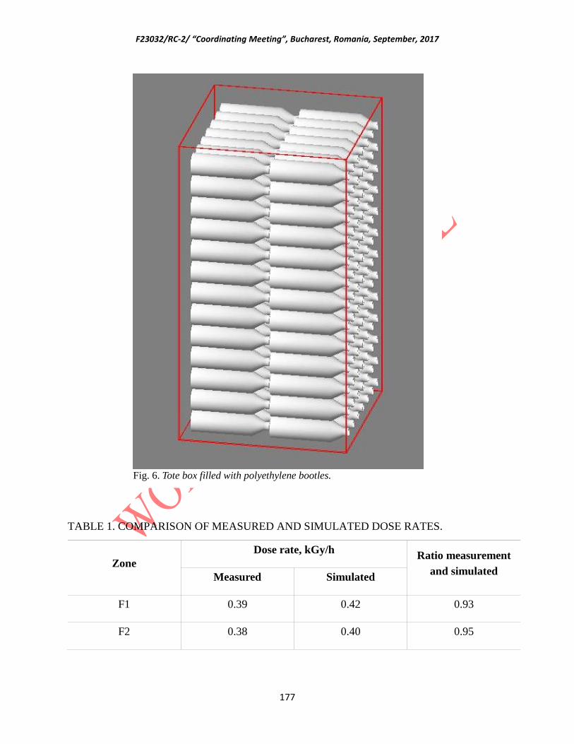
F23032/RC-2/ “Coordinating Meeting”, Bucharest, Romania, September, 2017
177
TABLE 1. COMPARISON OF MEASURED AND SIMULATED DOSE RATES.
Zone
Dose rate, kGy/h Ratio measurement
and simulated Measured Simulated
F1 0.39 0.42 0.93
F2 0.38 0.40 0.95
Fig. 6. Tote box filled with polyethylene bootles.
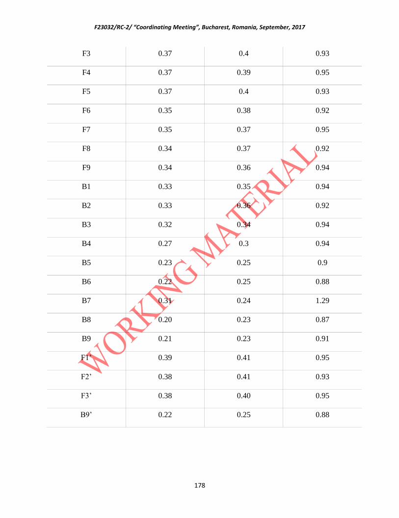
F23032/RC-2/ “Coordinating Meeting”, Bucharest, Romania, September, 2017
178
F3 0.37 0.4 0.93
F4 0.37 0.39 0.95
F5 0.37 0.4 0.93
F6 0.35 0.38 0.92
F7 0.35 0.37 0.95
F8 0.34 0.37 0.92
F9 0.34 0.36 0.94
B1 0.33 0.35 0.94
B2 0.33 0.36 0.92
B3 0.32 0.34 0.94
B4 0.27 0.3 0.94
B5 0.23 0.25 0.9
B6 0.22 0.25 0.88
B7 0.31 0.24 1.29
B8 0.20 0.23 0.87
B9 0.21 0.23 0.91
F1’ 0.39 0.41 0.95
F2’ 0.38 0.41 0.93
F3’ 0.38 0.40 0.95
B9’ 0.22 0.25 0.88

F23032/RC-2/ “Coordinating Meeting”, Bucharest, Romania, September, 2017
179
TABLE 2. THE RESULTS OF MEASURED AND SIMULATED DOSE RATES FOR RUĐER
BOŠKOVIĆ INSTITUTE’ EXPERIMENT
Distance, cm
Dose rate, Gy/s Ratio measurement and
simulated Measured Simulated
60 0.443 0.465 0.95
70 0.342 0.35 0.98
80 0.274 0.27 1.01
90 0.225 0.22 1.02
100 0.188 0.20 1.04
110 0.161 0.175 0.92
FIG. 7. Box filled with paper. Paper are in hollow cylinder view.
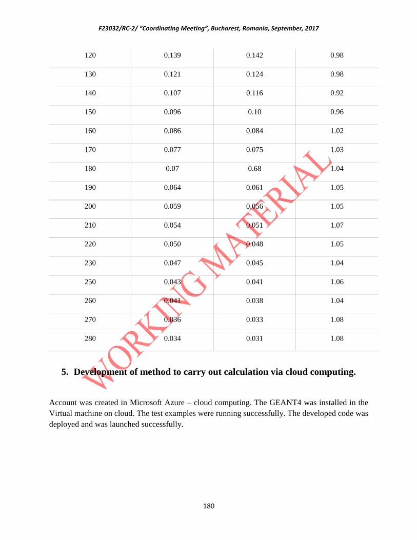
F23032/RC-2/ “Coordinating Meeting”, Bucharest, Romania, September, 2017
180
120 0.139 0.142 0.98
130 0.121 0.124 0.98
140 0.107 0.116 0.92
150 0.096 0.10 0.96
160 0.086 0.084 1.02
170 0.077 0.075 1.03
180 0.07 0.68 1.04
190 0.064 0.061 1.05
200 0.059 0.056 1.05
210 0.054 0.051 1.07
220 0.050 0.048 1.05
230 0.047 0.045 1.04
250 0.043 0.041 1.06
260 0.041 0.038 1.04
270 0.036 0.033 1.08
280 0.034 0.031 1.08
5. Development of method to carry out calculation via cloud computing.
Account was created in Microsoft Azure – cloud computing. The GEANT4 was installed in the
Virtual machine on cloud. The test examples were running successfully. The developed code was
deployed and was launched successfully.
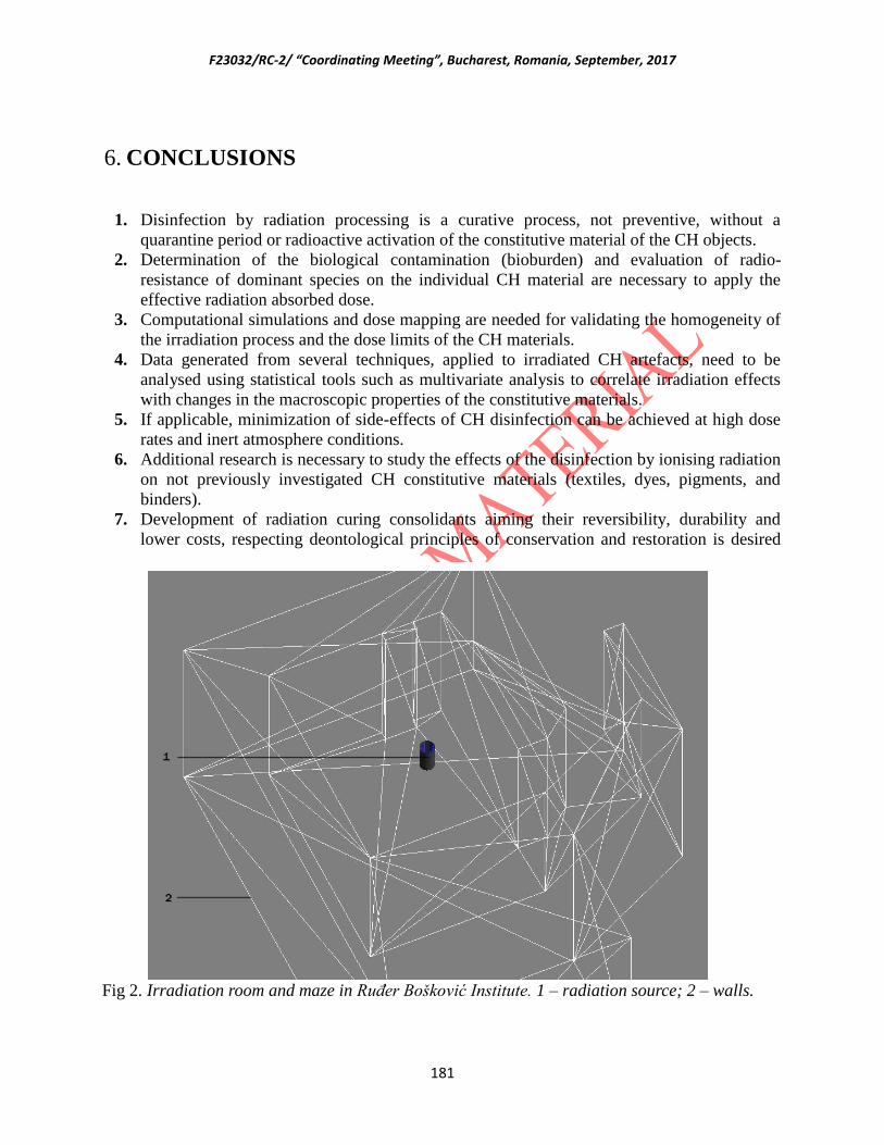
F23032/RC-2/ “Coordinating Meeting”, Bucharest, Romania, September, 2017
181
6. CONCLUSIONS
1. Disinfection by radiation processing is a curative process, not preventive, without a
quarantine period or radioactive activation of the constitutive material of the CH objects.
2. Determination of the biological contamination (bioburden) and evaluation of radio-
resistance of dominant species on the individual CH material are necessary to apply the
effective radiation absorbed dose.
3. Computational simulations and dose mapping are needed for validating the homogeneity of
the irradiation process and the dose limits of the CH materials.
4. Data generated from several techniques, applied to irradiated CH artefacts, need to be
analysed using statistical tools such as multivariate analysis to correlate irradiation effects
with changes in the macroscopic properties of the constitutive materials.
5. If applicable, minimization of side-effects of CH disinfection can be achieved at high dose
rates and inert atmosphere conditions.
6. Additional research is necessary to study the effects of the disinfection by ionising radiation
on not previously investigated CH constitutive materials (textiles, dyes, pigments, and
binders).
7. Development of radiation curing consolidants aiming their reversibility, durability and
lower costs, respecting deontological principles of conservation and restoration is desired
Fig 2. Irradiation room and maze in Ruđer Bošković Institute. 1 – radiation source; 2 – walls.
F23032/RC-2/ “Coordinating Meeting”, Bucharest, Romania, September, 2017
182
by CH community. Natural consolidants (in-situ application) and environmental friendly
materials should be investigated for this purpose.
7. RECOMMENDATIONS
The participants recommend the following:
1. Irradiation facilities involved in this CRP assure good and confident dosimetric procedures.
However, dosimetric inter-comparison is desirable. IAEA could help in this process by
supporting initial contacts between CRP partners and independent institutions (e.g. NIST)
and the inherent logistic expenses.
2. It is recommended to analyse the side and post-effects (long time behaviour) of CH
irradiated materials using different techniques related to change in colour, de-
polymerization, free radicals, mechanical properties, etc. and statistically analyse the
results.
3. It is recommended to perform statistical data tests to prove significance differences for all
obtained results.
4. It is recommended to survey CH objects that have been irradiated long time ago to assess
the stability of constitutive materials.
5. It is recommended to document the irradiation of CH artefacts (biological type and burden,
irradiation parameters and any other important information).
6. It is recommended that radiation processing of CH artefacts is to be performed in qualified
irradiation facilities.
7. Use of consolidants like graphene for protection of tangible CH materials should be
investigated for preventing corrosion and enhancing mechanical properties.
8. In order to meet the stated objectives of CRP, IAEA should improve the budget to facilitate
the acquisition of some essential scientific equipment and for practical training interchange
of the partner members.
9. Promotion of preservation of CH artefacts by ionizing radiation should be continued
through scientific publications, positive diffusion by media, strong interaction programs
with restorers and conservation community supported by national and international
organizations.
F23032/RC-2/ “Coordinating Meeting”, Bucharest, Romania, September, 2017
183
ANNEX 1: LIST OF PARTICIPANTS
2nd Coordination Meeting on CRP/F23032/
“DEVELOPING RADIATION TREATMENT METHODOLOGIES AND NEW RESIN
FORMULATIONS FOR CONSOLIDATION AND PRESERVATION OF ARCHIVED
MATERIALS AND CULTURAL HERITAGE ARTEFACTS”
25-30 September 2017
List of Participants (as of 25-09-2017)
List of non-local Participants
1 IAEA Mr Sunil SABHARWAL
International Atomic Energy Agency
Department of Nuclear Sciences and Applications
Division of Physical and Chemical Sciences
Physics Section
P.O. Box 100, Vienna International Centre
Wagramer Straße 5
1400 VIENNA
AUSTRIA
Tel.: 0043 1 2600 21744
Fax: 0043 1 26007
EMail: [email protected]
Internet: http://www.iaea.org
2 Brazil Mr Pablo Antonio VASQUEZ SALVADOR
Instituto de Pesquisas Energeticas e Nucleares (IPEN); Comissão
Nacional de Energia Nuclear (CNEN)
Av. Prof. Lineu Prestes, 2242; Cidade Universitaria
05508-000 SÃO PAULO
BRAZIL
EMail: [email protected]
3 Bulgaria Ms Petya Kovacheva
Radiochemical Laboratory; Faculty of Chemistry; Sofia University
St. Kliment Ohridski

F23032/RC-2/ “Coordinating Meeting”, Bucharest, Romania, September, 2017
184
1 James Bourchier Boulevard
1164 SOFIA
BULGARIA
EMail.: [email protected]
4 Croatia Ms Katarina MARUSIC
Bijenicka c. 54
10000 ZAGREB
CROATIA
EMail: [email protected]
5 Cuba Mr Iván Padrón DIAZ
Centro de Aplicaciones Tecnologicas y Desarrollo Nuclear (CEADEN)
Calle 30 #502 e/ 5ta y 7ma Avenida
P.O. Box 6122
Playa, LA HABANA
CUBA
EMail: [email protected]
6 Egypt Mr Hassan Ahmed Abd EL-REHIM
National Centre for Radiation Research and Technology (NCRRT)
Egyptian Atomic Energy Authority (EAEA)
P.O. Box 29, Nasr City, 3
Ahmed El-Zomor, Nasr City
CAIRO, El Zohoor
EGYPT
EMail: [email protected]
7 France Mr Quoc-Khoi TRAn
ARC-Nucleart; CEA/Grenoble
17, rue des martyrs
38054 GRENOBLE, CEDEX 09
FRANCE
Email: [email protected]
8 France Mr Laurent CORTELLA
Atelier Régional de Conservation ARC-Nucléart

F23032/RC-2/ “Coordinating Meeting”, Bucharest, Romania, September, 2017
185
CEA-Grenoble
17, rue des martyrs
38 054 GRENOBLE CEDEX 09
FRANCE
E-mail: [email protected]
9 Iran Ms Ramsina BET ESHO BABRUD
Nuclear Science and Technology Research Institute; Atomic Energy
Organization of Iran (AEOI)
P.O. Box 14155-1339
TEHRAN, North Kargar
IRAN-ISLAMIC REPUBLIC OF IRAN
EMail: [email protected]
10 Italy Ms Stefania BACCARO
Ente per le Nuove Tecnologie L'Energia e L'Ambiente (ENEA)
301, Via Anguillarese
00123 ROME
ITALY
EMail: [email protected]
11 Poland Ms Dagmara CHMIELEWSKA SMIETANKO
Institute of Nuclear Chemistry and Technology
ul. Dorodna 16
03-195 WARSAW
POLAND
EMail: [email protected]
12 Portugal Mr Luis Miguel MOTA FERREIRA
Centro de Ciências e Tecnologias Nucleares
Instituto Superior Técnico
Estrada Nacional 10, Km 139,7
2695-066 BOBADELA
PORTUGAL
EMail: [email protected]
F23032/RC-2/ “Coordinating Meeting”, Bucharest, Romania, September, 2017
186
13 Romania Mr Constantin Daniel NEGUT
HORIA HULUBEI National Institute for R&D in Physics and
Nuclear Engeneering (IFIN-HH)
IRASM Radiation Processing Department
30 Reactorului St., MAGURELE – Ilfov, RO 077125,
ROMANIA
EMail: [email protected]
14 Serbia Mr Slobodan MASIC
Vinča Institute of Nuclear Sciences
PO Box 522, 11001 BELGRADE
SERBIA
EMail: [email protected]
15 Turkey Ms Dilek SOLPAN
Department of Chemistry
Hacettepe University
Beytepe Campus
06800 Ankara
TURKEY
EMail: [email protected]
16 Ukraine Mr Volodymyr Victorovich MORGUNOV
Ukrainian Engineering Pedagogical Academy
Universitetskaya str. 16
61003 KHARKIV
UKRAINE
EMail: [email protected]
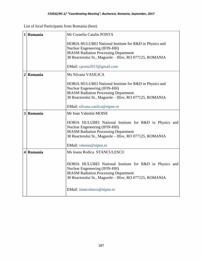
F23032/RC-2/ “Coordinating Meeting”, Bucharest, Romania, September, 2017
187
List of local Participants from Romania (host)
1 Romania Mr Corneliu Catalin PONTA
HORIA HULUBEI National Institute for R&D in Physics and
Nuclear Engeneering (IFIN-HH)
IRASM Radiation Processing Department
30 Reactorului St., Magurele – Ilfov, RO 077125, ROMANIA
EMail: [email protected]
2 Romania Ms Silvana VASILICA
HORIA HULUBEI National Institute for R&D in Physics and
Nuclear Engeneering (IFIN-HH)
IRASM Radiation Processing Department
30 Reactorului St., Magurele – Ilfov, RO 077125, ROMANIA
EMail: [email protected]
3 Romania Mr Ioan Valentin MOISE
HORIA HULUBEI National Institute for R&D in Physics and
Nuclear Engeneering (IFIN-HH)
IRASM Radiation Processing Department
30 Reactorului St., Magurele – Ilfov, RO 077125, ROMANIA
EMail: [email protected]
4 Romania Ms Ioana Rodica STANCULESCU
HORIA HULUBEI National Institute for R&D in Physics and
Nuclear Engeneering (IFIN-HH)
IRASM Radiation Processing Department
30 Reactorului St., Magurele – Ilfov, RO 077125, ROMANIA
EMail: [email protected]

F23032/RC-2/ “Coordinating Meeting”, Bucharest, Romania, September, 2017
188
ANNEX 2: AGENDA
Second Research Coordination Meeting of the
IAEA Coordinate Research Project F23032- on
“Developing Radiation Treatment Methodologies and New Resin Formulations for
Consolidation and Preservation of Archived Materials and Cultural Heritage Artefacts”
Bucharest, Romania, 25-30 September 2017
AGENDA
Monday, 25 September 2017
Session I: Introductory Session
9.00 – 9:30 Opening of the meeting
Election of the Chairperson and reporter
Adoption of the agenda
Session II: Participants’ Presentations

F23032/RC-2/ “Coordinating Meeting”, Bucharest, Romania, September, 2017
189
09:30-10:30
10.30-11.10
11.10 – 11.50
11.50-12.30
12.30-13.30
13.30-14.20
14.20-15.00
15:00-15:40
15.40-16.00
Mr. Pablo Antonio VASQUEZ SALVADOR – Brazil
Coffee Break
Ms. Petya KOVACHEVA- Bulgaria
Ms. Katarina MARUSIC– Croatia
Lunch break
Mr. Iván Padrón DIAZ – Cuba
Mr. Hassan Ahmed Abd EL-REHIM- Egypt
Mr. Quoc-Khoi TRAN – France
Coffee Break
16.00 – 16.40 Ms. Ramsina BET ESHO BABRUD – Iran
16.40 – 17.10
17:10 – 18:00
Ms. Stefania BACCARO - Italy
Ms. Dagmara CHMIELEWSKA SMIETANKO – Poland
19: 00 – 21:00 Welcome Reception
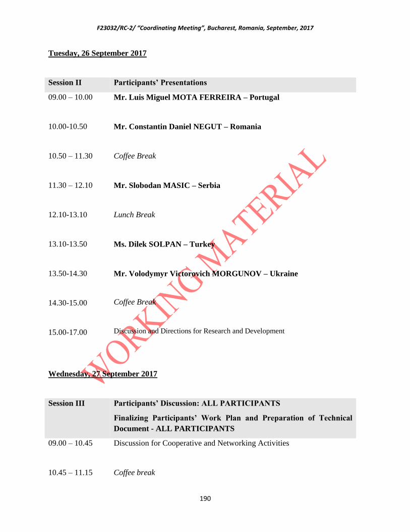
F23032/RC-2/ “Coordinating Meeting”, Bucharest, Romania, September, 2017
190
Tuesday, 26 September 2017
Session II Participants’ Presentations
09.00 – 10.00
10.00-10.50
10.50 – 11.30
11.30 – 12.10
12.10-13.10
13.10-13.50
13.50-14.30
14.30-15.00
15.00-17.00
Mr. Luis Miguel MOTA FERREIRA – Portugal
Mr. Constantin Daniel NEGUT – Romania
Coffee Break
Mr. Slobodan MASIC – Serbia
Lunch Break
Ms. Dilek SOLPAN – Turkey
Mr. Volodymyr Victorovich MORGUNOV – Ukraine
Coffee Break
Discussion and Directions for Research and Development
Wednesday, 27 September 2017
Session III Participants’ Discussion: ALL PARTICIPANTS
Finalizing Participants’ Work Plan and Preparation of Technical
Document - ALL PARTICIPANTS
09.00 – 10.45 Discussion for Cooperative and Networking Activities
10.45 – 11.15 Coffee break
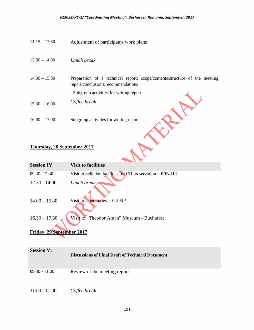
F23032/RC-2/ “Coordinating Meeting”, Bucharest, Romania, September, 2017
191
11.15 – 12.30 Adjustment of participants work plans
12.30 – 14.00 Lunch break
14.00 – 15.30
15.30 – 16.00
Preparation of a technical report: scope/contents/structure of the meeting
report/conclusions/recommendations
- Subgroup activities for writing report
Coffee break
16.00 – 17.00 Subgroup activities for writing report
Thursday, 28 September 2017
Session IV Visit to facilities
09.30- 12.30 Visit to radiation facilities for CH preservation – IFIN-HH
12.30 - 14.00 Lunch break
14.00 – 15.30 Visit to laboratories – ELI-NP
16.30 – 17.30 Visit to “Theodor Aman” Museum - Bucharest
Friday, 29 September 2017
Session V: Discussions of Final Draft of Technical Document
09.30 - 11.00 Review of the meeting report
11.00 - 11.30 Coffee break

F23032/RC-2/ “Coordinating Meeting”, Bucharest, Romania, September, 2017
192
11.30 – 12.30 Review and acceptance of the draft of the meeting report
12.30 – 13.30
13.30 -
Lunch break
Closing of the meeting

F23032/RC-2/ “Coordinating Meeting”, Bucharest, Romania, September, 2017
193
OUTPUT RELATED RESULTS
Publications
1. S. BACCARO, C. CASIERI, A. CEMMI, M. CHIARINI, V. D’AIUTO, M. TORTORA
Characterization of γ-radiation induced polymerization in ethyl methacrylate and methyl
acrylate monomers solutions,
Rad. Phys. Chem., 141 (2017) 131-137, https://doi.org/10.1016/j.radphyschem.2017.06.017
2. S. BACCARO, A. CEMMI
Radiation activities and application of ionizing radiation on Cultural heritage at ENEA
Calliope gamma facility (Casaccia R.C., Rome, Italy),
Nukleonika, accepted with minor revision (2017).
3. D. CHMIELEWSKA-ŚMIETANKO, U. GRYCZKA, W. MIGDAŁ, K. KOPEĆ
Electron beam for preservation of biodeteriorated cultural heritage paper-based objects
accepted for publication in Radiation Physics and Chemistry
4. N. SHEIKH, R. BETESHOBABRUD, F. KHATAMIFAR
Feasibility study of using gamma ray for fungal decontamination of historical oil painting
Journal of Nuclear Science and Technology 72 (2015) 39-45
5. KODAMA, Y.; RODRIGUES, O. JR.; GARCIA, R. H. L.; SANTOS, P. S.; VASQUEZ, P.
A. S.
Study of Free Radicals in Gamma Irradiated Cellulose of Cultural Heritage Materials Using
Electron Paramagnectic Resonance.
Radiation Physics and Chemistry, 124, 2016. 169-173
6. SANTOS, P. S.
Study and optimization of parameters of gamma ray processing in industrial scale considering
operational factors.
M.Sc. Thesis, University of São Paulo, 2017.
7. I. STANCULESCU, L. DRAGOMIR, M. MOCENCO, C. PINTILIE, B. LUNGU,
DANIEL NEGUT, M. CUTRUBINIS, M. VIRGOLICI, V. MOISE, L. CORTELLA, Q.
K.TRAN
Consolidation of wooden artefacts by resin impregnation and radiopolymerization » in
Restitutio, volume 8, 2014, pp. 271-275 (CONScience 2014, 7e édition, Bucarest
(Roumanie), 4-6 novembre 2014.
8. L. CORTELLA,
La momie soignée, in « Quatre momies et demie », sous la direction de Camille Perez, EAN :
9782757209868, Editeur(s) : Coédition Musée Joseph Déchelette, Roanne / Somogy éditions
d'Art, 2015, 81-90.
9. W. GLUSZEWSKI, Q.-K. TRAN, L. CORTELLA, D. ABBASOVA
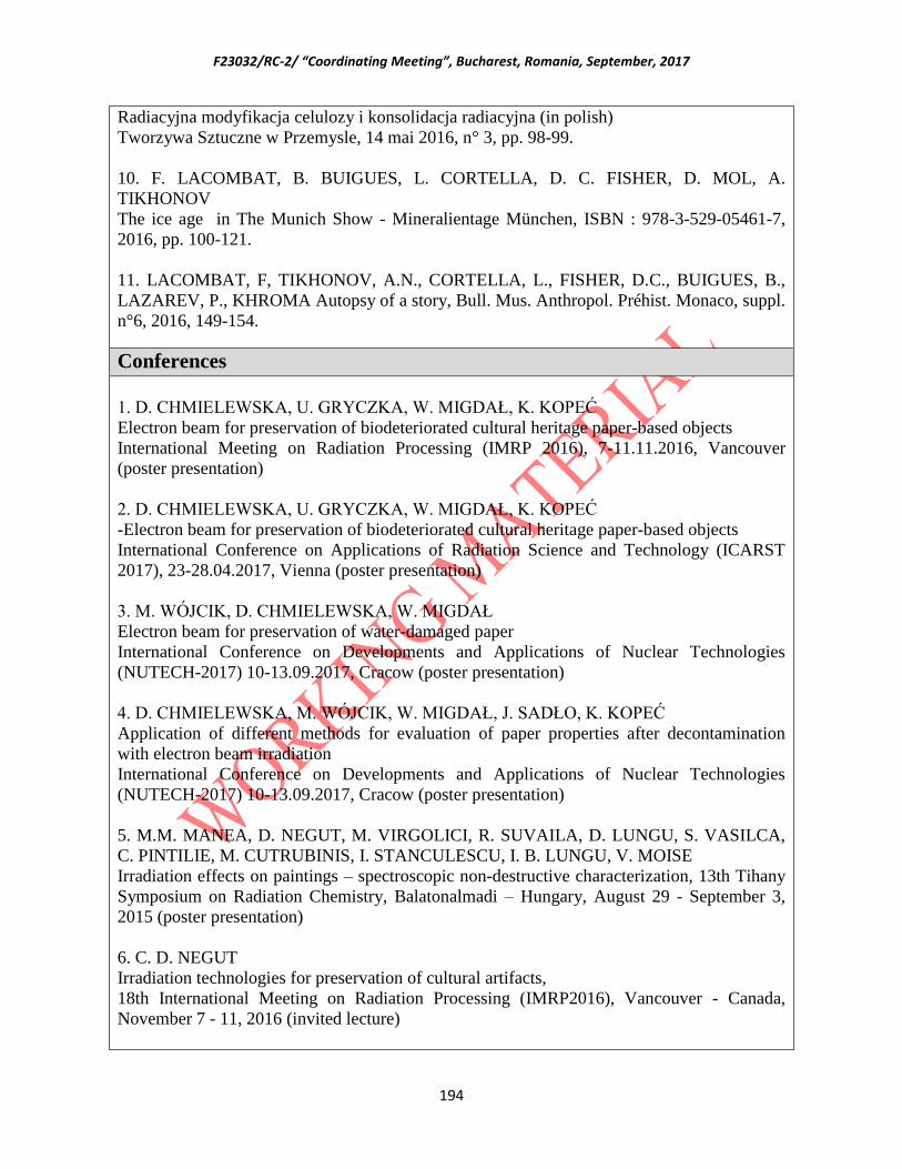
F23032/RC-2/ “Coordinating Meeting”, Bucharest, Romania, September, 2017
194
Radiacyjna modyfikacja celulozy i konsolidacja radiacyjna (in polish)
Tworzywa Sztuczne w Przemysle, 14 mai 2016, n° 3, pp. 98-99.
10. F. LACOMBAT, B. BUIGUES, L. CORTELLA, D. C. FISHER, D. MOL, A.
TIKHONOV
The ice age in The Munich Show - Mineralientage München, ISBN : 978-3-529-05461-7,
2016, pp. 100-121.
11. LACOMBAT, F, TIKHONOV, A.N., CORTELLA, L., FISHER, D.C., BUIGUES, B.,
LAZAREV, P., KHROMA Autopsy of a story, Bull. Mus. Anthropol. Préhist. Monaco, suppl.
n°6, 2016, 149-154.
Conferences
1. D. CHMIELEWSKA, U. GRYCZKA, W. MIGDAŁ, K. KOPEĆ
Electron beam for preservation of biodeteriorated cultural heritage paper-based objects
International Meeting on Radiation Processing (IMRP 2016), 7-11.11.2016, Vancouver
(poster presentation)
2. D. CHMIELEWSKA, U. GRYCZKA, W. MIGDAŁ, K. KOPEĆ
-Electron beam for preservation of biodeteriorated cultural heritage paper-based objects
International Conference on Applications of Radiation Science and Technology (ICARST
2017), 23-28.04.2017, Vienna (poster presentation)
3. M. WÓJCIK, D. CHMIELEWSKA, W. MIGDAŁ
Electron beam for preservation of water-damaged paper
International Conference on Developments and Applications of Nuclear Technologies
(NUTECH-2017) 10-13.09.2017, Cracow (poster presentation)
4. D. CHMIELEWSKA, M. WÓJCIK, W. MIGDAŁ, J. SADŁO, K. KOPEĆ
Application of different methods for evaluation of paper properties after decontamination
with electron beam irradiation
International Conference on Developments and Applications of Nuclear Technologies
(NUTECH-2017) 10-13.09.2017, Cracow (poster presentation)
5. M.M. MANEA, D. NEGUT, M. VIRGOLICI, R. SUVAILA, D. LUNGU, S. VASILCA,
C. PINTILIE, M. CUTRUBINIS, I. STANCULESCU, I. B. LUNGU, V. MOISE
Irradiation effects on paintings – spectroscopic non-destructive characterization, 13th Tihany
Symposium on Radiation Chemistry, Balatonalmadi – Hungary, August 29 - September 3,
2015 (poster presentation)
6. C. D. NEGUT
Irradiation technologies for preservation of cultural artifacts,
18th International Meeting on Radiation Processing (IMRP2016), Vancouver - Canada,
November 7 - 11, 2016 (invited lecture)

F23032/RC-2/ “Coordinating Meeting”, Bucharest, Romania, September, 2017
195
7. A. P. RODRIGUES, S. CABO VERDE, M.H. CASIMIRO AND L.M. FERREIRA
Hybrids: Preliminary results on biocide activity for conservation purposes
2nd
C2TN Workshop on Advanced Materials, Campus Tecnológico e Nuclear-IST, Bobadela
LRS, Portugal, November 2016 (invited lecture)
8. L.M. FERREIRA, A.P. RODRIGUES, S.C. VERDE, L.C. ALVES, M.H. CASIMIRO,
J.J.H. LANCASTRE, A.N. FALCÃO, F.M.A. MARGAÇA, M.F. ARAÚJO
Hybrid materials prepared by gamma irradiation for consolidation of ancient mosaics:
morphology and preliminary biocide activity studies
12nd
International Symposium on Ionizing Radiation and Polymers (IRaP 2016), Peninsula of
Giens, France, September 2016. (oral presentation)
9. L. M. FERREIRA, M. H. CASIMIRO, J.J.H. LANCASTRE, A.P. RODRIGUES, S.
CABO VERDE, L.C. ALVES, A.N. FALCÃO, S. R. GOMES, G. RODRIGUES, F.M.A.
MARGAÇA, J. P. LEAL, J. COROADO, V. HIPÓLITO CORREIA, M.F. ARAÚJO
Distinct polymeric based materials prepared/functionalized by gamma irradiation for
biomedical applications and Roman mosaics preservation
International Conference on Applications of Radiation Science and Technology (ICARST-
2017),Vienna – IAEA headquarters, Austria, April 2017 (poster presentation)
10. S. BACCARO, C. CASIERI, A. CEMMI, M. CHIARINI, V. D'AIUTO, M. TORTORA,
Gamma radiation induced in situ polymerization of consolidating products for the
conservation of cultural heritage manufacts
POLY 2015 4th Int. Symposium Frontiers in Polymer Science
(http://www.globaleventslist.elsevier.com/events/2015/05/frontiers-in-polymer-science/), 20–
22 May 2015, Riva del Garda, Italy (poster presentation)
11. S. BACCARO, A. CEMMI,
Application of ionizing radiation for cultural heritage
ICARST 2017 (https://www.iaea.org/events/icarst-2017) (invited lecture)
12. S. BACCARO, A. CEMMI,
Radiation activities at ENEA Calliope gamma facility (Casaccia R.C., Rome, Italy)
ICARST 2017 (https://www.iaea.org/events/icarst-2017) (poster presentation)
13. S. BACCARO, A. CEMMI, I. DI SARCINA,
Gamma radiation effects on cellulose-based materials in Cultural Heritage applications,
Miller Conference 2017, October 7-11 2017, Castellammare del Golfo, Sicily, Italy (invited
lecture).
14. R. BETESHO BABRUD, N. SHEIKH, F. KHATAMIFAR, M. E. MOGHADAM
Fungal decontamination of historical oil painting by using gamma ray
International Conference on Applications of Radiation Science and Technology (ICARST
2017), 23-28.04.2017, Vienna (poster presentation)
15. M. ŠEGVIĆ KLARIĆ, I. PUCIĆ, A. BOŽIĆEVIĆ, K. MARUŠIĆ, B. MIHALJEVIĆ,
F23032/RC-2/ “Coordinating Meeting”, Bucharest, Romania, September, 2017
196
Gamma-irradiation for cultural heritage – treatment of selected fungi on linen textile,
The First International Conference on Applications of Radiation Science and Technology
(ICARST 2017), Wienna, Austria, 24.-28.04.2017.
16. B. MIHALJEVIĆ, M. ŠEGVIĆ KLARIĆ, I. PUCIĆ, A. BOŽIČEVIĆ, K. MARUŠIĆ,
Gamma-irradiation of selected fungi on linen textile,
IAEA Technical Meeting on developing strategies for safe analysis of paint materials,
Netherlands Institute for Conservation, Art and Science, Amsterdam, Netherland 27.-
30.06.2017.
17. M. MEDIĆ, I. VUJČIĆ, S. MAŠIĆ, B. MILIĆEVIĆ, M.D. DRAMIĆANIN
Effect of Gamma-irradiation on Functional Properties of Paper of Cultural Heritage
Document;
InterRegioSci 2016, Novi Sad, December 2015, pp 73
18. I. VUJČIĆ, S. MAŠIĆ, M. MEDIĆ, S. PUTIĆ, M.D. DRAMIĆANIN
GAMMA IRRADIATION OF LEATHER GLOVES IN TERMS OF CULTURAL
HERITAGE PRESERVATION,
Eco-Ist’17, Vrnjacka banja, jun 2017
19. SANTOS, P. S.; VASQUEZ, S. P. A.
Two-Faces Stationary Irradiation Method and Dosimetric Considerations for Radiation
Processing at the Multipurpose Gamma Irradiation Facility/
IPEN-CNEN. International Nuclear Atlantic Conference - INAC 2015, São Paulo, 2015
20. SANTOS, P. S.; VASQUEZ, S. P. A.
Effects of the Interruption of the Irradiation Process on PMMA Harwell Industry Dosimetry
System.
International Nuclear Atlantic Conference - INAC 2015. São Paulo. 2015
21. KODAMA, Y.; RODRIGUES, O. JR.; GARCIA, R. H. L.; OTUBO, L.; SANTOS, P. S.;
VASQUEZ, P. A. S.
Kinetics of Free Radicals Decay Reactions in Cellulosic Based Heritage Materials
Disinfected by Gamma Radiation International,
Conference on Applications of Radiation Science and Technology (ICARST 2017) 24 to 28
April 2017, Vienna, Austria.
22. VASQUEZ, P.A.S.
Overview of Disinfection of Cultural Heritage Artefacts and Archive Materials by Ionizing
Radiation in Brazil: Culture meets Nuclear,
International Conference on Applications of Radiation Science and Technology (ICARST
2017) 24 to 28 April 2017, Vienna, Austria.
23. VASQUEZ, P.A.S. (Scientific Committee Member, Lecturer and Chairman),
Preservation of Cultural Heritage,
International Conference on Applications of Radiation Science and Technology (ICARST

F23032/RC-2/ “Coordinating Meeting”, Bucharest, Romania, September, 2017
197
2017), 24 to 28 April 2017, Vienna, Austria.
24. VASQUEZ, P.A.S., (Lecturer and Course Director),
Using nuclear techniques in support of conservation and preservation of cultural heritage
objects,
IAEA REGIONAL -LATIN AMERICA AND THE CARIBBEAN -TRAINING COURSE
C7-RLA/0/058-00112th to 16th September 2016. Nuclear and Energy Research Institute –
IPEN, São Paulo, Brazil.
25. VASQUEZ, P.A.S., (Organizer and Chairman),
Cultural Heritage,
2015 International Nuclear Atlantic Conference - INAC 2015, São Paulo, SP, Brazil, October
4-9, 2015, ASSOCIAÇÃO BRASILEIRA DE ENERGIA NUCLEAR – ABEN, ISBN: 978-
85-99141-06-9
26. VASQUEZ, P.A.S., (Lecturer),
IAEA- Training Course in the frame of an European Technical Co-operation Project
(Regional Training Course on Recent Developments in Irradiation Technology for CH
Preservation and Restoration for Junior Specialists - RER0039, 7th to 11th December 2015,
Bucharest, Romania
27. W. GŁUSZEWSKI, B. BORUC, Q.-K.TRAN, L. CORTELLA, D. ABBASOWA, P.
KOVACHEVA, N. BOSHNAKOVA
Preservation and protection of cultural heritage artefacts
NUTECH-2014, International Conference on Development and Applications of Nuclear
Technologies - Poster Session, Varsovie (Pologne), 21-24 septembre 2014.
28. I. STANCULESCU, V. MOISE, L. CORTELLA, Q.-K.TRAN
Decontamination of textile, leather and parchment artefacts by gamma irradiation
ETICH 2014, 3rd International Seminar and Workshop on Emerging Technology and
Innovation for Cultural Heritage : Advanced Technology for Diagnosis, Preservation and
Management of Historical and Archaeological Parchment, Leather and Textile Artefacts,
Sibiu (Roumanie), 15-18 octobre 2014.
29. L. CORTELLA,
Les traitements des objets du patrimoine par irradiation gamma et comportement des
matériaux aux doses insecticides et fongicides, Paris, université Paris-Sud, dans le cadre des
rencontres Paris-Saclay/Entreprises : Chimie sous rayonnement et applications industrielles, 5
juin 2015.
30. S. VASILCA, I. R. STANCULESCU, M. VIRGOLICI, C. PINTILIE, V. MOISE, B.
LUNGU, Q.-K. TRAN, L. CORTELLA,
Thermoanalytical and infrared studies of very degraded wooden artefacts consolidation with a
radiation-curing resin,
The 15th International Balkan Workshop on Applied Physics, 2-4 juillet 2015, Constanta
(Roumanie),.

F23032/RC-2/ “Coordinating Meeting”, Bucharest, Romania, September, 2017
198
31. L. CORTELLA, C. SALVAN, C. ALBINO, Q. K. TRAN,
Nouveaux développements concernant les techniques d’irradiation gamma pour le traitement
biocide des collections patrimoniales,
Paris, Musée du Louvre, 3e colloque international : Gestion intégrée des contaminants
biologiques (Integrated Pest Managements 2016 : IPM) dans les musées, archives,
bibliothèques et demeures historiques, 13-15 septembre 2016.
32. L. CORTELLA,
Nuclear techniques for conservation,
Sinaia, The 5th Balkan Symposium on Archaeometry, 27-30 septembre 2016.
33. Q.-K. TRAN,
Giens (Var),
IRaP 2016, The 12th meeting of the ionizing radiation & polymers symposium, 25-30
septembre 2016.
34. Q.-K. TRAN,
Development of New Radiation-Curing Monomers-Resins Systems for the Consolidation of
Wooden Cultural Heritage Artefacts,
Icarst 2017, Vienna, 24-27 april 2017
35. L. CORTELLA,
Uses and Prospects in Gamma Biocide Treatments for Cultural Heritage,
Icarst 2017, Vienna, 24-27 april 2017
36. L. CORTELLA,
Behavior of Polychromic Layers When Irradiated for Gamma Biocide Treatments,
IAEA Amsterdam Technical Meeting on Developing Strategies for Safe Analysis of Paintings
and Paint Materials, 27 – 30 June 2017, Rijksmuseum, Amsterdam, the Netherlands.
37. V. MORGUNOV
The numerical simulation of cultural heritage radiation treatment by Mnte-Carlo method.
International Conference on Applications of Radiation Science and Technology , 24-28 april
2017, Vienna, IAEA. p. 318 (poster presentation)
Country: Students and inter-institutional dissemination
Bulgaria
Cooperation:
-National Museum of History
-Bulgarian Academy of Sciences
Croatia
Collaborating institutions:

F23032/RC-2/ “Coordinating Meeting”, Bucharest, Romania, September, 2017
199
-Croatian Conservation Institute
-Department of Restoration, Academy of Fine Arts, University of Zagreb
-Central Laboratory for Conservation and Restoration of Archives, Croatian State Archives
-Conservation and Restoration Department, Museum of Contemporary Art (MSU)
-Etnographic Museum, Zagreb
-Technical Museum, Zagreb
-Museum of the Serbian Orthodox Church in Croatia, Zagreb
-National and University Library, Zagreb
-Museum of Arts and Crafts, Zagreb
-Musem of Contemporary Art, Zagreb
-Croatian History Museum, Zagreb
-Mimara Museum, Zagreb
-Museum of Cetinska Krajina, Sinj, ect.
-Many other museums, churches and private collectors
Students:
Up to now:
- 3 undergraduate students from the University of Zagreb, Faculty of Pharmacy and
Biochemistry
In the future:
- 2 undergraduate students from the University of Zagreb, Faculty of Pharmacy and
Biochemistry
- 1 undergraduate student from the University of Zagreb, Faculty of Chemical
Engineering and Technology
Poland
1 PhD student (doing his PhD in INCT in the field of application of EB irradiation to
paper decontamination)
Collaboration with Warsaw University and Technology (Faculty of Chemical and
Process Engineering)
Portugal
• 2 MSc students (1 Erasmus student from Lithuan, Univ. of Vilnius and another from
Univ. Nova de Lisboa)
• 1 PhD student (doing his PhD under my supervision);
• Invitations for collaboration in the area of application of radiation techniques in the
processing of new materials and treatment of CH artifacts:
- Dept. of Conservation and Restoration, Facudade de Ciências e Tecnologia, Univ. Nova
de Lisboa
- Dept. of Conservation and Restoration, Polytechnic Institute of Tomar

F23032/RC-2/ “Coordinating Meeting”, Bucharest, Romania, September, 2017
200
• Invitation to one workshop in the field of Cultural Heritage, (application of radiation
techniques in the processing of new materials and treatment of CH artifacts) from Department
of Conservation and Restoration, Facudade de Ciências e Tecnologia, Univ. Nova de Lisboa
Related Documents