Replica Exchange Molecular Dynamics Simulations of Coarse-grained Proteins in Implicit Solvent Yassmine Chebaro, † Xiao Dong, †,‡ Rozita Laghaei, ‡ Philippe Derreumaux,* ,† and Normand Mousseau* ,‡ Laboratoire de Biochimie The ´orique, UPR 9080 CNRS, Institut de Biologie Physico-Chimique et UniVersite ´ Paris 7 Denis Diderot, 13 rue Pierre et Marie Curie, 75005 Paris, France and De ´partement de Physique and Regroupement Que ´be ´cois sur les Mate ´riaux de Pointe, UniVersite ´ de Montre ´al, C.P. 6128, succursale centre-Ville, Montre ´al (Que ´bec), Canada ReceiVed: June 16, 2008; ReVised Manuscript ReceiVed: October 20, 2008 Current approaches aimed at determining the free energy surface of all-atom medium-size proteins in explicit solvent are slow and are not sufficient to converge to equilibrium properties. To ensure a proper sampling of the configurational space, it is preferable to use reduced representations such as implicit solvent and/or coarse- grained protein models, which are much lighter computationally. Each model must be verified, however, to ensure that it can recover experimental structures and thermodynamics. Here we test the coarse-grained implicit solvent OPEP model with replica exchange molecular dynamics (REMD) on six peptides ranging in length from 10 to 28 residues: two alanine-based peptides, the second -hairpin from protein G, the Trp-cage and zinc-finger motif, and a dimer of a coiled coil peptide. We show that REMD-OPEP recovers the proper thermodynamics of the systems studied, with accurate structural description of the -hairpin and Trp-cage peptides (within 1-2 Å from experiments). The light computational burden of REMD-OPEP, which enables us to generate many hundred nanoseconds at each temperature and fully assess convergence to equilibrium ensemble, opens the door to the determination of the free energy surface of larger proteins and assemblies. I. Introduction The computational determination of the free energy surface of proteins is an important aim in biology and chemistry because it can provide a more complete description of folding paths and intermediates than many serial molecular dynamics (MD) trajectories. Even with the advances in computing power, this calculation remains challenging because the energy landscape in explicit solvent is very complex and rugged. Fully converged all-atom free energy surfaces are therefore mainly reported for small-size systems with 20 amino acids or less. 1,2 Recently, several studies have underlined this challenge, pointing to the difficulty of ensuring enough sampling to determine reliable thermodynamical properties due to the very slow motion of biomolecules compared with the thermal vibrations, even at high temperature. 3-5 As can be expected, there has been numerous attempts at lifting these limitations. Although these approaches are varied, they can be sorted into two classes: development of accelerated sampling methods and use of reduced protein/solvent representations. In the context of accelerating the sampling of rare events, several thermodynamical techniques have recently emerged, including multicanonical algorithms, 6,7 replica exchange mo- lecular dynamics (REMD), 8 metadynamics, 9 and coupling between various techniques. 10 At this point, however, no method has really provided the efficiency gain necessary to fully characterize the free energy surface of even medium-size proteins in spite of ingenious attempts. For instance, Huang et al. developed REMD with solute temperature on simple peptides in explicit solvent but found that this technique is even less efficient than standard REMD. 11 The second direction for enhanced sampling is to reduce the number of degrees of freedom, and develop implicit or coarse- grained solvent and/or reduced protein representation. The challenge here is to simplify the description without changing the physics. Not surprisingly, this approach has been followed by many groups. 12-18 For instance, all-atom models in implicit solvent 17-19 have shown promising thermodynamic results for small peptides, but their applicability to large proteins remain to be determined. Ideally, we would like to use implicit solvent coarse-grained protein models, but the transferability of such force fields to predict the thermodynamics of R, , or mixed topologies is still problematic. 16 It is, however, the move that we make here as we present the application of a coarse-grained implicit solvent protein model, OPEP, to free energy calcula- tions, using REMD. OPEP, one of the best protein force fields to recognize native from decoys, 20 has already been coupled to Monte-Carlo 21,22 and MD simulations 23,24 as well as the activation-relaxation technique (ART) 25,26 to study protein folding 27-30 and the aggregation of amyloid-forming peptides. 31-34 MD-OPEP was found to describe protein dynamics at least qualitatively correctly since the absence of explicit solvent accelerates folding times by about 2 orders of magnitude compared with simulations in explicit water. 23,35 Here we report REMD-OPEP simulations on six test systems to validate OPEP predictions with respect to structural and thermodynamical properties and establish the scale of computational efforts to obtain the equilibrium ensemble. To this end, we first study two alanine-based peptides and a 16-residue -hairpin. We then turn to the 20-residue Trp-cage and a 28-residue R fold and finally examine a dimer of a 7-residue peptide with a coiled coil signature. * To whom correspondence should be addressed. E-mail: philippe. [email protected] (P.D.), [email protected] (N.M.). † Institut de Biologie Physico-Chimique et Universite ´ Paris. ‡ Universite ´ de Montre ´al. J. Phys. Chem. B 2009, 113, 267–274 267 10.1021/jp805309e CCC: $40.75 2009 American Chemical Society Published on Web 12/09/2008

Welcome message from author
This document is posted to help you gain knowledge. Please leave a comment to let me know what you think about it! Share it to your friends and learn new things together.
Transcript
Replica Exchange Molecular Dynamics Simulations of Coarse-grained Proteins in ImplicitSolvent
Yassmine Chebaro,† Xiao Dong,†,‡ Rozita Laghaei,‡ Philippe Derreumaux,*,† andNormand Mousseau*,‡
Laboratoire de Biochimie Theorique, UPR 9080 CNRS, Institut de Biologie Physico-Chimique et UniVersiteParis 7 Denis Diderot, 13 rue Pierre et Marie Curie, 75005 Paris, France and Departement de Physique andRegroupement Quebecois sur les Materiaux de Pointe, UniVersite de Montreal, C.P. 6128, succursalecentre-Ville, Montreal (Quebec), Canada
ReceiVed: June 16, 2008; ReVised Manuscript ReceiVed: October 20, 2008
Current approaches aimed at determining the free energy surface of all-atom medium-size proteins in explicitsolvent are slow and are not sufficient to converge to equilibrium properties. To ensure a proper sampling ofthe configurational space, it is preferable to use reduced representations such as implicit solvent and/or coarse-grained protein models, which are much lighter computationally. Each model must be verified, however, toensure that it can recover experimental structures and thermodynamics. Here we test the coarse-grained implicitsolvent OPEP model with replica exchange molecular dynamics (REMD) on six peptides ranging in lengthfrom 10 to 28 residues: two alanine-based peptides, the second !-hairpin from protein G, the Trp-cage andzinc-finger motif, and a dimer of a coiled coil peptide. We show that REMD-OPEP recovers the properthermodynamics of the systems studied, with accurate structural description of the !-hairpin and Trp-cagepeptides (within 1-2 Å from experiments). The light computational burden of REMD-OPEP, which enablesus to generate many hundred nanoseconds at each temperature and fully assess convergence to equilibriumensemble, opens the door to the determination of the free energy surface of larger proteins and assemblies.
I. Introduction
The computational determination of the free energy surfaceof proteins is an important aim in biology and chemistry becauseit can provide a more complete description of folding paths andintermediates than many serial molecular dynamics (MD)trajectories. Even with the advances in computing power, thiscalculation remains challenging because the energy landscapein explicit solvent is very complex and rugged. Fully convergedall-atom free energy surfaces are therefore mainly reported forsmall-size systems with 20 amino acids or less.1,2 Recently,several studies have underlined this challenge, pointing to thedifficulty of ensuring enough sampling to determine reliablethermodynamical properties due to the very slow motion ofbiomolecules compared with the thermal vibrations, even at hightemperature.3-5 As can be expected, there has been numerousattempts at lifting these limitations. Although these approachesare varied, they can be sorted into two classes: development ofaccelerated sampling methods and use of reduced protein/solventrepresentations.
In the context of accelerating the sampling of rare events,several thermodynamical techniques have recently emerged,including multicanonical algorithms,6,7 replica exchange mo-lecular dynamics (REMD),8 metadynamics,9 and couplingbetween various techniques.10 At this point, however, no methodhas really provided the efficiency gain necessary to fullycharacterize the free energy surface of even medium-sizeproteins in spite of ingenious attempts. For instance, Huang etal. developed REMD with solute temperature on simple peptides
in explicit solvent but found that this technique is even lessefficient than standard REMD.11
The second direction for enhanced sampling is to reduce thenumber of degrees of freedom, and develop implicit or coarse-grained solvent and/or reduced protein representation. Thechallenge here is to simplify the description without changingthe physics. Not surprisingly, this approach has been followedby many groups.12-18 For instance, all-atom models in implicitsolvent17-19 have shown promising thermodynamic results forsmall peptides, but their applicability to large proteins remainto be determined. Ideally, we would like to use implicit solventcoarse-grained protein models, but the transferability of suchforce fields to predict the thermodynamics of R, !, or mixedtopologies is still problematic.16 It is, however, the move thatwe make here as we present the application of a coarse-grainedimplicit solvent protein model, OPEP, to free energy calcula-tions, using REMD.
OPEP, one of the best protein force fields to recognize nativefrom decoys,20 has already been coupled to Monte-Carlo21,22
and MD simulations23,24 as well as the activation-relaxationtechnique (ART)25,26 to study protein folding27-30 and theaggregation of amyloid-forming peptides.31-34 MD-OPEP wasfound to describe protein dynamics at least qualitatively correctlysince the absence of explicit solvent accelerates folding timesby about 2 orders of magnitude compared with simulations inexplicit water.23,35 Here we report REMD-OPEP simulations onsix test systems to validate OPEP predictions with respect tostructural and thermodynamical properties and establish the scaleof computational efforts to obtain the equilibrium ensemble.To this end, we first study two alanine-based peptides and a16-residue !-hairpin. We then turn to the 20-residue Trp-cageand a 28-residue !!R fold and finally examine a dimer of a7-residue peptide with a coiled coil signature.
* To whom correspondence should be addressed. E-mail: [email protected] (P.D.), [email protected] (N.M.).
† Institut de Biologie Physico-Chimique et Universite Paris.‡ Universite de Montreal.
J. Phys. Chem. B 2009, 113, 267–274 267
10.1021/jp805309e CCC: $40.75 2009 American Chemical SocietyPublished on Web 12/09/2008
II. Simulation Details1. OPEP Force Field. Since the OPEP force field and its
parameters are described in details elsewhere,20 we limitourselves to a few comments. OPEP’s description includes allheavy backbone atoms and uses a single bead to represent theside chains of all amino acids, except the proline side chainwhere three beads are taken into account. Although a reducedrepresentation cannot offer the structural precision of all-atommolecular mechanics36 and spectroscopic37 force fields, theOPEP analytical form is sufficiently rich to predict lowest energystates of peptides consistent with experiment.38,22,39 The ap-plicability of OPEP in folding was recently revisited on the 60-residue B domain of protein A, and we found that ART-OPEPsimulations recovered the experimental three-helix bundlestarting from random states, but also explained the observedshift to another topology upon mutations.40
Solvent effects are incorporated directly into the interactionparameters, through a hydrogen-bonding potential consisting oftwo-body and four-body terms, and a pairwise contact potentialbetween side-chains represented by either a 12-6 potential ora 6-potential.20 In this work, we use the standard OPEP potentialfor all systems, except for the alanine-based peptides and thezinc-finger motif (BBA), where the simulations are repeatedwith the 12-6 potential replaced by the desolvation potential
where
with r the distance and rcm the van der Waals radius betweentwo particles, rssm ) rcm + 3 Å (where 3 Å is the diameter ofa water molecule).
The interaction U is dependent on the desolvation barrierheight (!db) and the depth of the solvent-separated minimum(!ssm). For the alanine-based and BBA systems, we use !db )0.1! and !ssm ) 0.2!, with ! equal to the OPEP !ij parameterdefined in eq 6 of ref 20, and following previous studies,41,42 k) 6, m ) 3, and n ) 2.
2. Replica Exchange Molecular Dynamics. The replicaexchange algorithm, implemented in our code, was firstproposed by Marinari and Parisi for spin glasses.43 Later, Sugitaand Okamoto coupled the scheme with MD to construct thefree energy landscape of proteins.8 The algorithm is simple andeasily parallelisable. N MD runs or replicas are launched atdifferent T. At regular intervals, configurations are exchangedbetween two adjacent Ti and Tj with a probability given byeq 3,
where Ei is the configurational energy in replica i.This procedure allows the system to escape from local basins
and explore, with the proper thermodynamical weight, theenergy landscape. In practice, the efficiency of REMD decreaseswith the number of atoms since the adjacent temperatures mustbe close to ensure overlaps in the configurational energydistributions. This limitation strongly favors the use of implicitsolvent and coarse-grained representation, which in additionmakes each time-step much less costly.
In the simulations reported here, MD is performed in an openbox with the temperature controlled by Berendsen’s thermostat44
with a coupling time of 500 fs. A time-step of 1.5 fs is used,and the RATTLE45 algorithm is applied with a tolerance of10-6 for the bond length constraints and 10-12 for the relativevelocities of the pairs of the bounded atoms. Because of theimportant mass difference between the H atom and the side-chain beads, a rapid kinetic energy transfer from the heavy tothe light atoms can cause instabilities. To circumvent thisproblem, we reassign the H atom velocity to that of the N amidewhenever the velocity on H corresponds to a displacement ofmore than 15% of the N-H bond length. Geometric corrections,that is, resetting the total momentum and total angular momen-tum to zero, are made every 500 time-steps.
For REMD, we use a logarithmic temperature distributionwith 10-16 replicas ranging from about 200 to 500 K.Exchanges are attempted every 10 000 time-steps, leading toan acceptation rate between 20 and 50%.
3. Analysis. In addition to ensuring a better sampling ofconformational space, the REMD data can serve to establishthe phase diagram through the use of reweighting methods. Here,we use the PTWHAM version (weighted-histogram method forparallel tempering) to take into account the correlations betweenthe REMD trajectories.46 The main advantage of reweightingmethods is that it is possible to fully determine the T-dependenceof various quantities, and by following the evolution of thesethermodynamical properties as a function of time, we can qualifythe convergence of the simulations with good precision, anessential step for establishing the internal consistency of ourresults. The details of the PTWHAM algorithm can be foundin the original paper.46
In what follows, secondary structure analysis is performedusing the STRIDE program.47 All generated conformations areclustered recursively using the C R root-mean square deviations(rmsd) as follows. After computing the list of neighbors for allconformations, we identify the structure with the largest numberof neighbors with a rmsd of 2.5 Å. The members of this largestcluster are removed, and the procedure is repeated until allconformations are clustered.
III. ResultsA. Alanine-based Peptides. The first peptide studied is
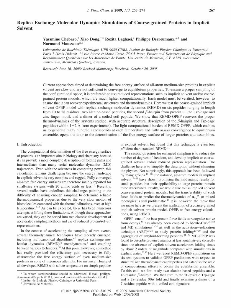
(AAQAA)3 blocked by Ace and NH2. Starting from the randomstate shown in Figure 1a, we explore its structural andthermodynamical properties by a single 100 ns REMD simula-tion using 12 replicas between 190 and 410 K. The plots of thermsd with respect to the starting structure (panel c) and the heatcapacity as a function of T (panel d) show convergence formultiple independent time intervals. Figure 1b shows thecalculated residue helicity at 269 K in comparison with thatderived from NMR chemical shift measurements at 274 K.48
Overall, the simulation yields a helix content of 28.3% versus
U(r,rcm, !, !db, !ssm) )
{!Z(r)[Z(r) - 2] for r < rcm
CY(r)n[Y(r)n/2 - (rdb - rcm)2n/2n] + !db
for rcm e r < rdb
-B[Y(r) - h1][Y(r)m + h2] for r g rdb
(1)
Z(r) ) (rcm/r)k
Y(r) ) (r - rdb)2
C ) 4n(! + !db)/(rdb - rcm)4n
B ) m!ssm(rssm - rdb)2(m-1)
h1 ) (1 - 1/m)(rssm - rdb)2/(!ssm/!db + 1)
h2 ) (m - 1)(rssm - rdb)2m/(1 + !db/!ssm),
(2)
p(i, j) ) min{1.0, exp[ 1kBTi
- 1kBTj](Ei - Ej)} (3)
268 J. Phys. Chem. B, Vol. 113, No. 1, 2009 Chebaro et al.
47% by NMR and 70%49 and 55%50 by MD and MC,respectively. Note that Chen et al. use this helicity as input datato tune their generalized Born (GB) implicit solvent param-eters,18 whereas our parameters are taken from ref 20.
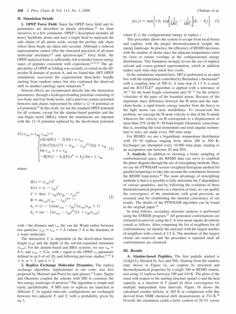
To further understand helix stability, we also examine thedecaalanine peptide blocked by Ace and NH2 using 16 replicasbetween 190 and 448 K. As reported in Figure 2a, there is atransition at 290 K between helical structures (T < Tm) andrandom coil structures (T > Tm). This value agrees very wellwith that obtained by all-atom multicanonical MC simulationsusing the Schiffer solvent-accessible surface parameters (Tm )285 K),51 but it is lower than that derived by coarse-grainedLangevin simulations (Tm ) 324 K).52
Figure 1d shows that the transition temperature of (AAQAA)3
is located at 310 K versus 278 K from experiments.48 Thisincrease in Tm can result from different sources: an overestima-tion of the backbone torsional parameters, but also, followingthe work of Chan et al. based on Go-based simulations,53 theabsence of an energy barrier to desolvation. Figure 2 reports,for both decaalanine (panel a) and (AAQAA)3 (panel b), theimpact of the desolvation potential given by eq 1 on the heatcapacity profiles. We see that a small desolvation barrier height,which has no effect during energy minimization, is sufficientto decrease Tm by 30-40 K, indicating that fitting theexperimental melting temperatures requires a delicate balanceof many components including the energy barriers to desolvation.
B. !-Hairpin. We now consider the second hairpin fromdomain B1 of protein G. This 16-residue peptide of sequenceGEWTYDDATKTFTVTE has been extensively studied, bothexperimentally54,55 and numerically,17,29,56-64 as a model forprotein folding. An early NMR study finds the hairpin to be
42% folded in water at 278 K,54 but on the basis of Trpfluorescence experiments, the hairpin population is 80% at 278K and Tm found at 297 K.55 Later, Fesinmeyer et al. revisitedthe hairpin population using CD and 2D NMR data and foundthe peptide 30% folded in water at 298 K.65 All thesemeasurements based on different probes point to a transitiontemperature around 280-300 K with an experimental structureresembling that observed in the full protein G. This structure,referred to as native, was however recently questioned. Compar-ing REMD simulations with experimental parameters includingHR, HN chemical shifts, JHR-HN scalar couplings and NOE,Weinstock et al. propose that the native ensemble of the peptideincludes a large population of conformations with non-nativeH-bonds.66
Numerically, results are also very diverse. Simulations basedon the all-atom implicit solvent model developed by Irbackidentify a melting temperature of 297 K,17 whereas simulationsbased on the all-atom OPLSAA force field show a transitiontemperature at 360 K.58 Generalized-ensemble simulations withGROMOS show 3 native H-bonds with a probability above 96%at 300 K with the hairpin stable at 320 K,64 but REMDsimulations using the same potential found instead that 56%and 25% of all conformations are native at 300 and 389 K,respectively.61 Finally, Lwin and Luo examine the effect of sixAMBER parameter sets on the phase diagram and find that thetwo best ff03 and ff99ci sets overestimate the transitiontemperature (365-380 K).62
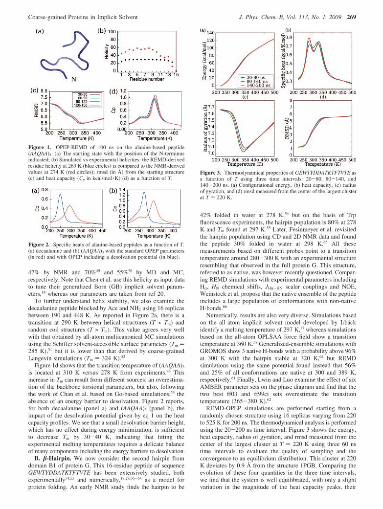
REMD-OPEP simulations are performed starting from arandomly chosen structure using 16 replicas varying from 220to 525 K for 200 ns. The thermodynamical analysis is performedusing the 20-200 ns time interval. Figure 3 shows the energy,heat capacity, radius of gyration, and rmsd measured from thecenter of the largest cluster at T ) 220 K using three 60 nstime intervals to evaluate the quality of sampling and theconvergence to an equilibrium distribution. This cluster at 220K deviates by 0.9 Å from the structure 1PGB. Comparing theevolution of these four quantities in the three time intervals,we find that the system is well equilibrated, with only a slightvariation in the magnitude of the heat capacity peaks, their
Figure 1. OPEP-REMD of 100 ns on the alanine-based peptide(AAQAA)3. (a) The starting state with the position of the N-terminusindicated; (b) Simulated vs experimental helicities: the REMD-derivedresidue helicity at 269 K (blue circles) is compared to the NMR-derivedvalues at 274 K (red circles); rmsd (in Å) from the starting structure(c) and heat capacity (Cp in kcal/mol ·K) (d) as a function of T.
Figure 2. Specific heats of alanine-based peptides as a function of T.(a) decaalanine and (b) (AAQAA)3 with the standard OPEP parameters(in red) and with OPEP including a desolvation potential (in blue).
Figure 3. Thermodynamical properties of GEWTYDDATKTFTVTE asa function of T using three time intervals: 20-80, 80-140, and140-200 ns. (a) Configurational energy, (b) heat capacity, (c) radiusof gyration, and (d) rmsd measured from the center of the largest clusterat T ) 220 K.
Coarse-grained Proteins in Implicit Solvent J. Phys. Chem. B, Vol. 113, No. 1, 2009 269
positions remaining unchanged (panel b), suggesting that thehairpin is equilibrated after 60 ns.
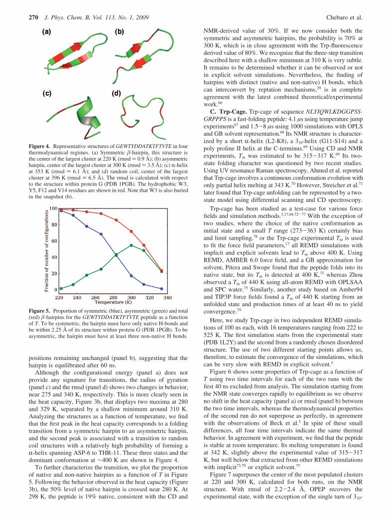
Although the configurational energy (panel a) does notprovide any signature for transitions, the radius of gyration(panel c) and the rmsd (panel d) shows two changes in behavior,near 275 and 340 K, respectively. This is more clearly seen inthe heat capacity, Figure 3b, that displays two maxima at 280and 329 K, separated by a shallow minimum around 310 K.Analyzing the structures as a function of temperature, we findthat the first peak in the heat capacity corresponds to a foldingtransition from a symmetric hairpin to an asymmetric hairpin,and the second peak is associated with a transition to randomcoil structures with a relatively high probability of forming aR-helix spanning ASP-6 to THR-11. These three states and thedominant conformation at !400 K are shown in Figure 4.
To further characterize the transition, we plot the proportionof native and non-native hairpins as a function of T in Figure5. Following the behavior observed in the heat capacity (Figure3b), the 50% level of native hairpin is crossed near 280 K. At298 K, the peptide is 19% native, consistent with the CD and
NMR-derived value of 30%. If we now consider both thesymmetric and asymmetric hairpins, the probability is 70% at300 K, which is in close agreement with the Trp-fluorescencederived value of 80%. We recognize that the three-step transitiondescribed here with a shallow minimum at 310 K is very subtle.It remains to be determined whether it can be observed or notin explicit solvent simulations. Nevertheless, the finding ofhairpins with distinct (native and non-native) H bonds, whichcan interconvert by reptation mechanisms,29 is in completeagreement with the latest combined theoretical/experimentalwork.66
C. Trp-Cage. Trp-cage of sequence NLYIQWLKDGGPSS-GRPPPS is a fast-folding peptide: 4.1 µs using temperature jumpexperiments67 and 1.5-8 µs using 1000 simulations with OPLSand GB solvent representation.68 Its NMR structure is character-ized by a short R-helix (L2-K8), a 310-helix (G11-S14) and apoly proline II helix at the C-terminus.69 Using CD and NMRexperiments, Tm was estimated to be 315-317 K.69 Its two-state folding character was questioned by two recent studies.Using UV resonance Raman spectroscopy, Ahmed et al. reportedthat Trp-cage involves a continuous conformation evolution withonly partial helix melting at 343 K.70 However, Streicher et al.71
later found that Trp-cage unfolding can be represented by a two-state model using differential scanning and CD spectroscopy.
Trp-cage has been studied as a test-case for various forcefields and simulation methods.3,17,68,72-77 With the exception oftwo studies, where the choice of the native conformation asinitial state and a small T range (273-363 K) certainly biasand limit sampling,78 or the Trp-cage experimental Tm is usedto fit the force field parameters,17 all REMD simulations withimplicit and explicit solvents lead to Tm above 400 K. UsingREMD, AMBER 6.0 force field, and a GB approximation forsolvent, Pitera and Swope found that the peptide folds into itsnative state, but its Tm is detected at 400 K,73 whereas Zhouobserved a Tm of 440 K using all-atom REMD with OPLSAAand SPC water.75 Similarly, another study based on Amber94and TIP3P force fields found a Tm of 440 K starting from anunfolded state and production times of at least 40 ns to yieldconvergence.79
Here, we study Trp-cage in two independent REMD simula-tions of 100 ns each, with 16 temperatures ranging from 222 to525 K. The first simulation starts from the experimental state(PDB 1L2Y) and the second from a randomly chosen disorderedstructure. The use of two different starting points allows us,therefore, to estimate the convergence of the simulations, whichcan be very slow with REMD in explicit solvent.5
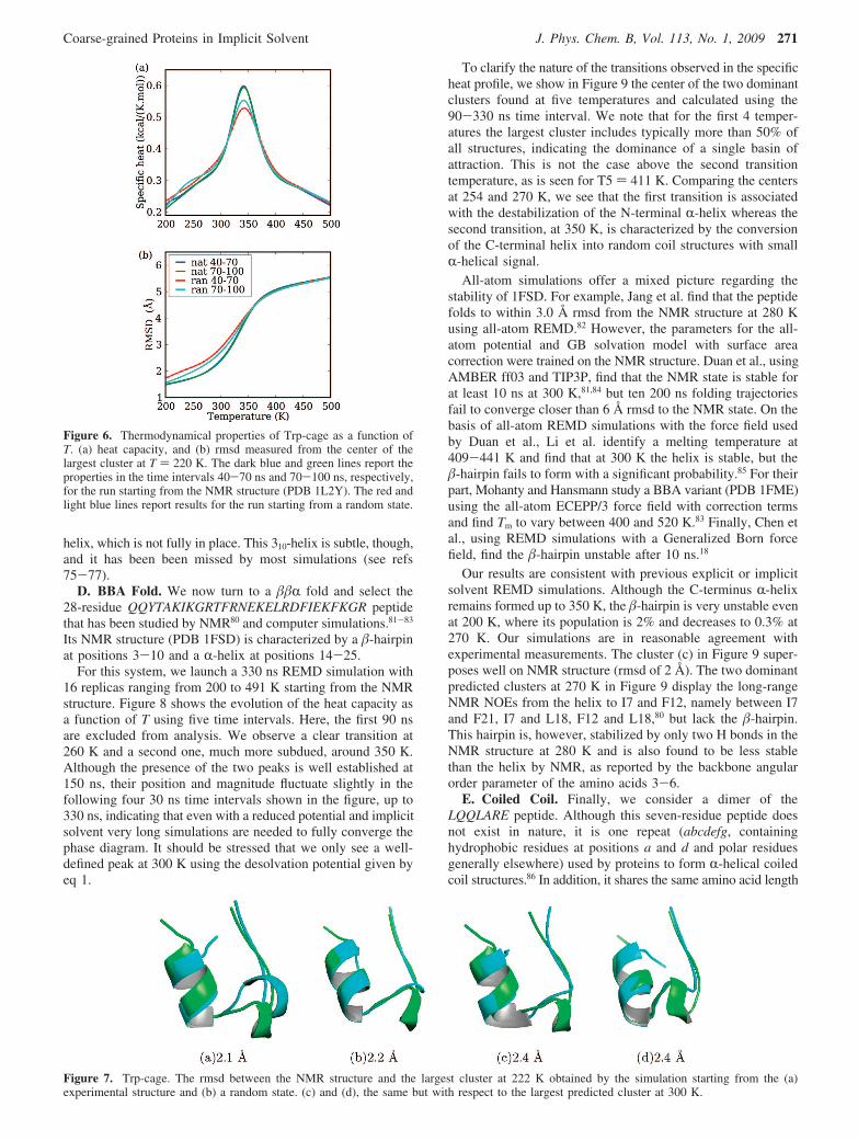
Figure 6 shows some properties of Trp-cage as a function ofT using two time intervals for each of the two runs with thefirst 40 ns excluded from analysis. The simulation starting fromthe NMR state converges rapidly to equilibrium as we observeno shift in the heat capacity (panel a) or rmsd (panel b) betweenthe two time intervals, whereas the thermodynamical propertiesof the second run do not superpose as perfectly, in agreementwith the observations of Beck et al.5 In spite of these smalldifferences, all four time intervals indicate the same thermalbehavior. In agreement with experiment, we find that the peptideis stable at room temperature. Its melting temperature is foundat 342 K, slightly above the experimental value of 315-317K, but well below that extracted from other REMD simulationswith implicit73,79 or explicit solvent.75
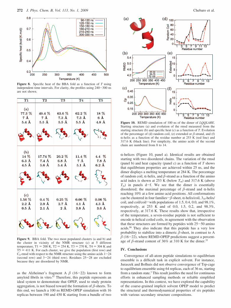
Figure 7 superposes the center of the most populated clustersat 220 and 300 K, calculated for both runs, on the NMRstructure. With rmsd of 2.2-2.4 Å, OPEP recovers theexperimental state, with the exception of the single turn of 310-
Figure 4. Representative structures of GEWTYDDATKTFTVTE in fourthermodynamical regimes. (a) Symmetric !-hairpin, this structure isthe center of the largest cluster at 220 K (rmsd ) 0.9 Å); (b) asymmetrichairpin, center of the largest cluster at 300 K (rmsd ) 3.5 Å); (c) R-helixat 353 K (rmsd ) 6.1 Å); and (d) random coil, center of the largestcluster at 396 K (rmsd ) 6.5 Å). The rmsd is calculated with respectto the structure within protein G (PDB 1PGB). The hydrophobic W3,Y5, F12 and V14 residues are shown in red. Note that W3 is also buriedin the snapshot (b).
Figure 5. Proportion of symmetric (blue), asymmetric (green) and total(red) !-hairpins for the GEWTYDDATKTFTVTE peptide as a functionof T. To be symmetric, the hairpin must have only native H-bonds andbe within 2.25 Å of its structure within protein G (PDB 1PGB). To beasymmetric, the hairpin must have at least three non-native H bonds.
270 J. Phys. Chem. B, Vol. 113, No. 1, 2009 Chebaro et al.
helix, which is not fully in place. This 310-helix is subtle, though,and it has been been missed by most simulations (see refs75-77).
D. BBA Fold. We now turn to a !!R fold and select the28-residue QQYTAKIKGRTFRNEKELRDFIEKFKGR peptidethat has been studied by NMR80 and computer simulations.81-83
Its NMR structure (PDB 1FSD) is characterized by a !-hairpinat positions 3-10 and a R-helix at positions 14-25.
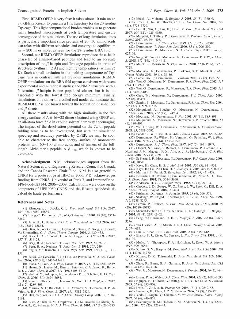
For this system, we launch a 330 ns REMD simulation with16 replicas ranging from 200 to 491 K starting from the NMRstructure. Figure 8 shows the evolution of the heat capacity asa function of T using five time intervals. Here, the first 90 nsare excluded from analysis. We observe a clear transition at260 K and a second one, much more subdued, around 350 K.Although the presence of the two peaks is well established at150 ns, their position and magnitude fluctuate slightly in thefollowing four 30 ns time intervals shown in the figure, up to330 ns, indicating that even with a reduced potential and implicitsolvent very long simulations are needed to fully converge thephase diagram. It should be stressed that we only see a well-defined peak at 300 K using the desolvation potential given byeq 1.
To clarify the nature of the transitions observed in the specificheat profile, we show in Figure 9 the center of the two dominantclusters found at five temperatures and calculated using the90-330 ns time interval. We note that for the first 4 temper-atures the largest cluster includes typically more than 50% ofall structures, indicating the dominance of a single basin ofattraction. This is not the case above the second transitiontemperature, as is seen for T5 ) 411 K. Comparing the centersat 254 and 270 K, we see that the first transition is associatedwith the destabilization of the N-terminal R-helix whereas thesecond transition, at 350 K, is characterized by the conversionof the C-terminal helix into random coil structures with smallR-helical signal.
All-atom simulations offer a mixed picture regarding thestability of 1FSD. For example, Jang et al. find that the peptidefolds to within 3.0 Å rmsd from the NMR structure at 280 Kusing all-atom REMD.82 However, the parameters for the all-atom potential and GB solvation model with surface areacorrection were trained on the NMR structure. Duan et al., usingAMBER ff03 and TIP3P, find that the NMR state is stable forat least 10 ns at 300 K,81,84 but ten 200 ns folding trajectoriesfail to converge closer than 6 Å rmsd to the NMR state. On thebasis of all-atom REMD simulations with the force field usedby Duan et al., Li et al. identify a melting temperature at409-441 K and find that at 300 K the helix is stable, but the!-hairpin fails to form with a significant probability.85 For theirpart, Mohanty and Hansmann study a BBA variant (PDB 1FME)using the all-atom ECEPP/3 force field with correction termsand find Tm to vary between 400 and 520 K.83 Finally, Chen etal., using REMD simulations with a Generalized Born forcefield, find the !-hairpin unstable after 10 ns.18
Our results are consistent with previous explicit or implicitsolvent REMD simulations. Although the C-terminus R-helixremains formed up to 350 K, the !-hairpin is very unstable evenat 200 K, where its population is 2% and decreases to 0.3% at270 K. Our simulations are in reasonable agreement withexperimental measurements. The cluster (c) in Figure 9 super-poses well on NMR structure (rmsd of 2 Å). The two dominantpredicted clusters at 270 K in Figure 9 display the long-rangeNMR NOEs from the helix to I7 and F12, namely between I7and F21, I7 and L18, F12 and L18,80 but lack the !-hairpin.This hairpin is, however, stabilized by only two H bonds in theNMR structure at 280 K and is also found to be less stablethan the helix by NMR, as reported by the backbone angularorder parameter of the amino acids 3-6.
E. Coiled Coil. Finally, we consider a dimer of theLQQLARE peptide. Although this seven-residue peptide doesnot exist in nature, it is one repeat (abcdefg, containinghydrophobic residues at positions a and d and polar residuesgenerally elsewhere) used by proteins to form R-helical coiledcoil structures.86 In addition, it shares the same amino acid length
Figure 7. Trp-cage. The rmsd between the NMR structure and the largest cluster at 222 K obtained by the simulation starting from the (a)experimental structure and (b) a random state. (c) and (d), the same but with respect to the largest predicted cluster at 300 K.
Figure 6. Thermodynamical properties of Trp-cage as a function ofT. (a) heat capacity, and (b) rmsd measured from the center of thelargest cluster at T ) 220 K. The dark blue and green lines report theproperties in the time intervals 40-70 ns and 70-100 ns, respectively,for the run starting from the NMR structure (PDB 1L2Y). The red andlight blue lines report results for the run starting from a random state.
Coarse-grained Proteins in Implicit Solvent J. Phys. Chem. B, Vol. 113, No. 1, 2009 271
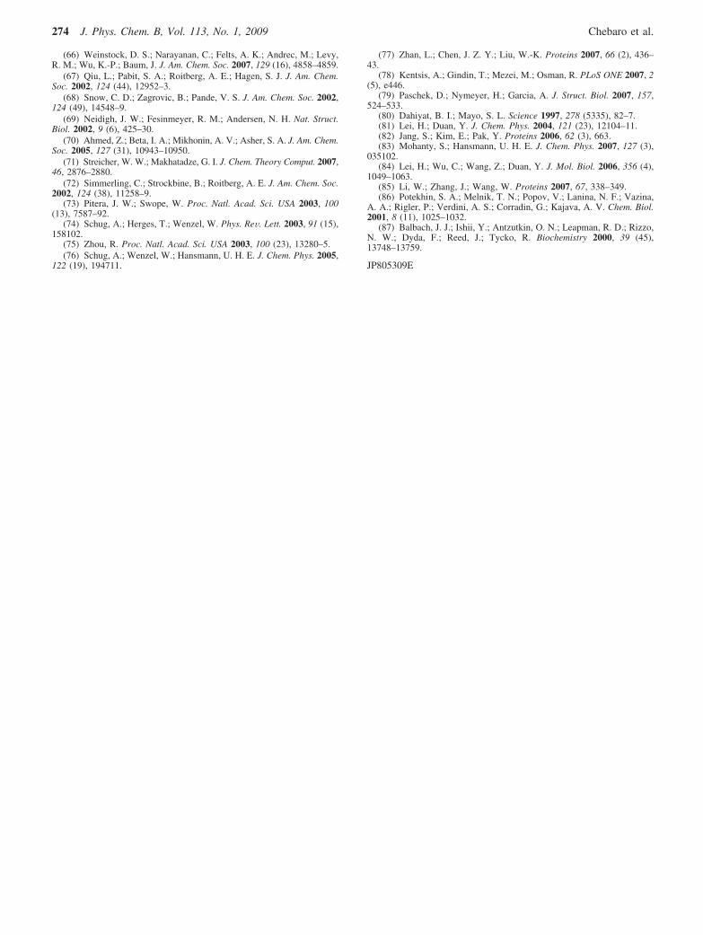
as the Alzheimer’s fragment A ! (16-22) known to formamyloid fibrils in vitro.87 Therefore, this peptide represents anideal system to demonstrate that OPEP, used to study proteinaggregation, is not biased toward the formation of !-sheets. Tothis end, we launch a 100 ns REMD-OPEP simulation with 16replicas between 190 and 450 K starting from a bundle of two
R-helices (Figure 10, panel a). Identical results are obtainedstarting with two disordered chains. The variation of the rmsd(panel b) and heat capacity (panel c) as a function of T showsthat equilibrium properties are achieved within 25 ns, and thedimer displays a melting temperature at 284 K. The percentageof random coil, R-helix, and !-strand as a function of the aminoacid index is shown at 253 K (below Tm) and 317.6 K (aboveTm) in panels d-f. We see that the dimer is essentiallydisordered; the maximal percentage of !-strand and R-helixreaching 10% at a few amino acid positions. All conformationscan be clustered in four familiess!-sheet, R-helix/coil, 310-helix/coil, and coil/coilswith populations of 1.5, 0.4, 0.0, and 98.1%,respectively, at 253 K and of 0.0, 1.5, 0.2, and 98.3%,respectively, at 317.6 K. These results show that, irrespectiveof the temperature, a seven-residue peptide is not sufficient toencode R-helical coiled coils, in agreement with the observationthat these structures are formed by peptides with 25-50 aminoacids.86 They also indicate that this peptide has a very lowprobability to stabilize into a dimeric !-sheet, in contrast to A! (16-22), where REMD-OPEP predictions suggest a percent-age of !-strand content of 36% at 310 K for the dimer.35
IV. Conclusions
Convergence of all-atom peptide simulations to equilibriumensemble is a difficult task in explicit solvent. For instance,Juraszek and Bolhuis did not observe convergence of Trp-cageto equilibrium ensemble using 64 replicas, each of 36 ns, startingfrom a random state.3 This result justifies the need for continuousefforts in enhanced sampling methods or reliable simplifiedrepresentations. In this context, we have explored the capabilityof the coarse-grained implicit solvent OPEP model to predictthe structural and thermodynamical properties of six peptideswith various secondary structure compositions.
Figure 8. Specific heat of the BBA fold as a function of T usingindependent time intervals. For clarity, the profiles using 240-300 nsare not shown.
Figure 9. BBA fold. The two most populated clusters (a and b) andthe cluster in vicinity of the NMR structure (c) at 5 differenttemperature, T1 ) 200 K, T2 ) 254 K, T3 ) 270 K, T4 ) 304 K andT5 ) 411 K. For each cluster, we give the population (first row) andCR-rmsd with respect to the NMR structure using the amino acids 1-24(second row) and 3-24 (third row). Residues 25-28 are excludedbecause they are disordered by NMR.
Figure 10. REMD simulation of 100 ns of the dimer of LQQLARE.Starting structure (a) and evolution of the rmsd measured from thestarting structure (b) and specific heat (c) as a function of T. Evolutionof the percentage of (d) random coil, (e) extended or !-strand, and (f)R-helix as a function of the residue number at 253 K (red line) and317.6 K (black line). For simplicity, the amino acids of the secondchain are numbered from 8 to 14.
272 J. Phys. Chem. B, Vol. 113, No. 1, 2009 Chebaro et al.
First, REMD-OPEP is very fast: it takes about 10 min on an3.0 GHz processor to generate a 1 ns trajectory for the 20-residueTrp-cage. This light computational burden enables us to generatemany hundred nanoseconds at each temperature and ensureconvergence of the simulations. The use of long simulation timesis particularly important since proteins of 20-30 amino acidscan relax with different schedules and converge to equilibriumin ! 200 ns or more, as seen for the 28-residue BBA fold.
Second, our REMD-OPEP simulations reproduce the R-helixcharacter of alanine-based peptides and lead to an accuratedescription of the !-hairpin and Trp-cage peptides in terms ofstructures (within 1-2 Å) and melting temperatures (within 25K). Such a small deviation in the melting temperature of Trp-cage runs in contrast with all previous simulations. REMD-OPEP simulations on the BBA fold appear consistent with recentexperimental and numerical studies; the NMR structure with aN-terminal !-hairpin is one populated cluster, but it is notassociated with the lowest free energy minimum. Finally,simulations on a dimer of a coiled coil model demonstrate thatREMD-OPEP is not biased toward the formation of R-helicesand !-sheets.
All these results along with the high similarity in the freeenergy surface of A ! 16-22 dimer obtained using OPEP andan all-atom force field in explicit solvent35 are very encouraging.The impact of the desolvation potential on the Tm of peptidefolding remains to be investigated, but with the simulationspeed-up and accuracy provided by OPEP, we may be soonable to characterize the free energy surface of monomericproteins with 60-100 amino acids and of trimers of the full-length Alzheimer’s peptide A !1-42, which is known to becytotoxic.
Acknowledgment. N.M. acknowledges support from theNatural Sciences and Engineering Research Council of Canadaand the Canada Research Chair Fund. N.M. is also grateful toCNRS for a poste rouge at IBPC in 2006. P.D. acknowledgesfunding from CNRS, Universite of Paris 7, and ImmunoPrion,FP6-Food-023144, 2006-2009. Calculations were done on thecomputers of UPR9080 CNRS and the Reseau quebecois decalcul de haute performance.
References and Notes
(1) Khandogin, J.; Brooks, C. L. Proc. Natl. Acad. Sci. USA 2007,104 (43), 16880–16885.
(2) Liang, C.; Derreumaux, P.; Wei, G. Biophys. J. 2007, 93 (10), 3353–3362.
(3) Juraszek, J.; Bolhuis, P. G. Proc. Natl. Acad. Sci. USA 2006, 103(43), 15859–15864.
(4) Okur, A.; Wickstrom, L.; Layten, M.; Geney, R.; Song, K.; Hornak,V.; Simmerling, C. J. Chem. Theory Comput. 2006, 2, 420–433.
(5) Beck, D. A. C.; White, G. W. N.; Daggett, V. J Struct Biol 2007,157 (3), 514–23.
(6) Berg, B. A.; Neuhaus, T. Phys. ReV. Lett. 1992, 68, 9–12.(7) Berg, B. A.; Neuhaus, T. Phys. Lett. B 1991, 267, 249.(8) Sugita, Y.; Okamoto, Y. Chem. Phys. Lett. 1999, 314 (1-2), 141–
151.(9) Bussi, G.; Gervasio, F. L.; Laio, A.; Parrinello, M. J. Am. Chem.
Soc. 2006, 128 (41), 13435–13441.(10) Piana, S.; Laio, A J. Phys. Chem. B. 2007, 111 (17), 4553–4559.(11) Huang, X.; Hagen, M.; Kim, B.; Friesner, R. A.; Zhou, R.; Berne,
B. J. J. Phys. Chem. B 2007, 111 (19), 5405–5410.(12) Shih, A. Y.; Arkhipov, A.; Freddolino, P. L.; Schulten, K. J J. Phys.
Chem. B. 2006, 110, 3674–3684.(13) Zhou, J.; Thorpe, I. F.; Izvekov, S.; Voth, G. A Biophys. J. 2007,
92 (12), 4289–303.(14) Marrink, S. J.; Risselada, H. J.; Yefimov, S.; Tieleman, D. P.; de
Vries, A. H J. Phys. Chem. B. 2007, 111, 7812–7824.(15) Han, W.; Wu, Y.-D. J. J. Chem. Theory Comp. 2007, 3, 2146–
2161.(16) Liwo, A.; Khalili, M.; Czaplewski, C.; Kalinowski, S.; Oldziej, S.;
Wachucik, K.; Scheraga, H. A. J. Phys. Chem. B. 2007, 111 (1), 260–285.
(17) Irback, A.; Mohanty, S. Biophys. J. 2005, 88 (3), 1560–9.(18) JChen, J.; Im, W.; Brooks, C. L. J. Am. Chem. Soc. 2006, 128
(11), 3728–3736.(19) Lei, H.; Wu, C.; Liu, H.; Duan, Y. Proc. Natl. Acad. Sci. USA
2007, 104 (12), 4925–4930.(20) Maupetit, J.; Tuffery, P.; Derreumaux, P. Proteins: Struct., Funct.,
Bioinf. 2007, 69, 394–408.(21) Derreumaux, P. J. Chem. Phys. 1999, 111 (5), 2301–2310.(22) Derreumaux, P. Phys. ReV. Lett. 2000, 85 (1), 206–209.(23) Derreumaux, P.; Mousseau, N. J. Chem. Phys. 2007, 126 (2),
025101.(24) Song, W.; Wei, G.; Mousseau, N.; Derreumaux, P. J. Phys. Chem.
B. 2008, 112 (14), 4410–4418.(25) Malek, R.; Mousseau, N. Phys. ReV. E 2000, 62 (6 Pt A), 7723–
7728.(26) Mousseau, N.; Derreumaux, P.; Barkema, G. T.; Malek, R. J. Mol.
Graph. Model 2001, 19 (1), 78–86.(27) Forcellino, F.; Derreumaux, P. Proteins 2001, 45 (2), 159–166.(28) Wei, G.; Mousseau, N.; Derreumaux, P. J. Chem. Phys. 2002, 117
(24), 11379–11387.(29) Wei, G.; Derreumaux, P.; Mousseau, N. J. Chem. Phys. 2003, 119
(13), 6403–6406.(30) Chen, W.; Mousseau, N.; Derreumaux, P J. Chem. Phys. 2006,
125 (8), 084911.(31) Santini, S.; Mousseau, N.; Derreumaux, P. J. Am. Chem. Soc. 2004,
126 (37), 11509–11516.(32) Melquiond, A.; Boucher, G.; Mousseau, N.; Derreumaux, P.
J. Chem. Phys. 2005, 122 (17), 174904.(33) Mousseau, N.; Derreumaux, P. NoV 2005, 38 (11), 885–891.(34) Melquiond, A.; Mousseau, N.; Derreumaux, P. Proteins 2006, 65
(1), 180–191.(35) Wei, G.; Song, W.; Derreumaux, P.; Mousseau., N. Frontiers Biosci.
2008, 13, 5681–5692.(36) Ponder, J. W.; Case, D. A. AdV. Protein Chem. 2003, 66, 27–85.(37) Derreumaux, P.; Wilson, K.; Vergoten, G.; Peticolas, W. J. Phys.
Chem. 1989, 93]?> (]?> (4), 1338–1350.(38) Derreumaux, P. J. Chem. Phys. 1997, 107 (6), 1941–1947.(39) Floquet, N.; Pasco, S.; Ramont, L.; Derreumaux, P.; Laronze, J. Y.;
Nuzillard, J. M.; Maquart, F. X.; Alix, A. J. P.; Monboisse, J. C. J. Biol.Chem. 2004, 279 (3), 2091–100.
(40) St-Pierre, J.-F.; Mousseau, N.; Derreumaux, P. J. Chem. Phys. 2008,128 (4), 045101.
(41) Kaya, H.; Chan, H. S. J. Mol. Biol. 2003, 326 (3), 911–931.(42) Kaya, H.; Liu, Z.; Chan, H. S. Biophys. J. 2005, 89 (1), 520–535.(43) Marinari, E.; Parisi, G. Europhys. Lett. 1992, 19, 451–458.(44) Berendsen, H.; Postma, J.; van Gunsteren, W.; Nola, A. D.; Haak,
J. J. Chem. Phys. 1984, 81, 3684–3690.(45) Andersen, H. C. J. Comput. Phys., 1983, 52 (1), 24-34.(46) Chodera, J. D.; Swope, W. C.; Pitera, J. W.; Seok, C.; Dill, K. A.
J. Chem. Theory Comput. 2007, 3, 26–41.(47) Frishman, D.; Argos, P. Proteins 1995, 23 (4), 566–579.(48) Shalongo, W.; Dugad, L.; Stellwagen, E. J. J. Am. Chem. Soc. 1994,
116, 8288–8293.(49) Ferrara, P.; Caflisch, A. Proc. Natl. Acad. Sci. U S A 2000, 97
(20), 10780–10785.(50) Shental-Bechor, D.; Kirca, S.; Ben-Tal, N.; Haliloglu, T. Biophys.
J. 2005, 88 (4), 2391–2402.(51) Peng, Y.; Hansmann, U. H. E. Biophys. J. 2002, 82 (6), 3269–
3276.(52) van Giessen, A. E.; Straub, J. E. J. Chem. Theory Comput. 2006,
2, 674–684.(53) Liu, Z.; Chan, H. S. Phys. Biol. 2005, 2 (4), S75-S85.(54) Blanco, F. J.; Rivas, G.; Serrano, L. Nat. Struct. Biol. 1994, 1 (9),
584–90.(55) Munoz, V.; Thompson, P. A.; Hofrichter, J.; Eaton, W. A. Nature
1997, 390, 6656.(56) Krivov, S. V.; Karplus, M. Proc. Natl. Acad. Sci. USA 2004, 101
(41), 14766–14770.(57) Klimov, D. K.; Thirumalai, D. Proc. Natl. Acad. Sci. USA 2000,
97 (6), 2544–9.(58) Zhou, R.; Berne, B. J.; Germain, R. Proc. Natl. Acad. Sci. USA
2001, 98 (26), 14931–6.(59) Wei, G.; Mousseau, N.; Derreumaux, P. Proteins 2004, 56 (3), 464–
74.(60) Evans, D. A.; Wales, D. J. Chem. Phys. 2004, 121 (2), 1080–1090.(61) Nguyen, P. H.; Stock, G.; Mittag, E.; Hu, C.-K.; Li, M. S. Proteins
2005, 61 (4), 795–808.(62) Lwin, T. Z.; Luo, R. Protein Sci. 2006, 15 (11), 2642–55.(63) Imamura, H.; Chen, J. Z. Y. Proteins 2006, 63 (3), 555–570.(64) Yoda, T.; Sugita, Y.; Okamoto, Y. Proteins: Struct., Funct., Bioinf.
2007, 66 (4), 846–859.(65) Fesinmeyer, R. M.; Hudson, F. M.; Andersen, N. H. J. Am. Chem.
Soc. 2004, 126 (23), 7238–43.
Coarse-grained Proteins in Implicit Solvent J. Phys. Chem. B, Vol. 113, No. 1, 2009 273
(66) Weinstock, D. S.; Narayanan, C.; Felts, A. K.; Andrec, M.; Levy,R. M.; Wu, K.-P.; Baum, J. J. Am. Chem. Soc. 2007, 129 (16), 4858–4859.
(67) Qiu, L.; Pabit, S. A.; Roitberg, A. E.; Hagen, S. J. J. Am. Chem.Soc. 2002, 124 (44), 12952–3.
(68) Snow, C. D.; Zagrovic, B.; Pande, V. S. J. Am. Chem. Soc. 2002,124 (49), 14548–9.
(69) Neidigh, J. W.; Fesinmeyer, R. M.; Andersen, N. H. Nat. Struct.Biol. 2002, 9 (6), 425–30.
(70) Ahmed, Z.; Beta, I. A.; Mikhonin, A. V.; Asher, S. A. J. Am. Chem.Soc. 2005, 127 (31), 10943–10950.
(71) Streicher, W. W.; Makhatadze, G. I. J. Chem. Theory Comput. 2007,46, 2876–2880.
(72) Simmerling, C.; Strockbine, B.; Roitberg, A. E. J. Am. Chem. Soc.2002, 124 (38), 11258–9.
(73) Pitera, J. W.; Swope, W. Proc. Natl. Acad. Sci. USA 2003, 100(13), 7587–92.
(74) Schug, A.; Herges, T.; Wenzel, W. Phys. ReV. Lett. 2003, 91 (15),158102.
(75) Zhou, R. Proc. Natl. Acad. Sci. USA 2003, 100 (23), 13280–5.(76) Schug, A.; Wenzel, W.; Hansmann, U. H. E. J. Chem. Phys. 2005,
122 (19), 194711.
(77) Zhan, L.; Chen, J. Z. Y.; Liu, W.-K. Proteins 2007, 66 (2), 436–43.
(78) Kentsis, A.; Gindin, T.; Mezei, M.; Osman, R. PLoS ONE 2007, 2(5), e446.
(79) Paschek, D.; Nymeyer, H.; Garcia, A. J. Struct. Biol. 2007, 157,524–533.
(80) Dahiyat, B. I.; Mayo, S. L. Science 1997, 278 (5335), 82–7.(81) Lei, H.; Duan, Y. J. Chem. Phys. 2004, 121 (23), 12104–11.(82) Jang, S.; Kim, E.; Pak, Y. Proteins 2006, 62 (3), 663.(83) Mohanty, S.; Hansmann, U. H. E. J. Chem. Phys. 2007, 127 (3),
035102.(84) Lei, H.; Wu, C.; Wang, Z.; Duan, Y. J. Mol. Biol. 2006, 356 (4),
1049–1063.(85) Li, W.; Zhang, J.; Wang, W. Proteins 2007, 67, 338–349.(86) Potekhin, S. A.; Melnik, T. N.; Popov, V.; Lanina, N. F.; Vazina,
A. A.; Rigler, P.; Verdini, A. S.; Corradin, G.; Kajava, A. V. Chem. Biol.2001, 8 (11), 1025–1032.
(87) Balbach, J. J.; Ishii, Y.; Antzutkin, O. N.; Leapman, R. D.; Rizzo,N. W.; Dyda, F.; Reed, J.; Tycko, R. Biochemistry 2000, 39 (45),13748–13759.
JP805309E
274 J. Phys. Chem. B, Vol. 113, No. 1, 2009 Chebaro et al.
Related Documents