Translational Neuroscience 1 Research Article • DOI: 10.2478/s13380-012-0022-0 • Translational Neuroscience • 3(2) • 2012 * E-mail: [email protected] REPETITIVE TRANSCRANIAL MAGNETIC STIMULATION (RTMS) MODULATES EVENT-RELATED POTENTIAL (ERP) INDICES OF ATTENTION IN AUTISM 1 Department of Psychiatry and Behavioral Sciences, University of Louisville School of Medicine, Louisville, KY 40202 2 Department of Anatomical Sciences and Neurobiology, University of Louisville School of Medicine, Louisville, KY 40202 3 Department of Bioengineering, University of Louisville J.B. Speed School of Engineering, Louisville, KY 40208 4 Department of Pediatrics, University of Louisville School of Medicine, Louisville, KY 40202 Manuel F. Casanova 1,2,3 , Joshua M. Baruth 1,2 , Ayman El-Baz 1,3 , Allan Tasman 1 , Lonnie Sears 4 , Estate Sokhadze 1,2,3, * Abstract Individuals with autism spectrum disorder (ASD) have previously been shown to have significantly augmented and prolonged event-related potentials (ERP) to irrelevant visual stimuli compared to controls at both early and later stages (e.g., N200, P300) of visual processing and evidence of an overall lack of stimulus discrimination. Abnormally large and indiscriminative cortical responses to sensory stimuli may reflect cortical inhibitory deficits and a disruption in the excitation/inhibition ratio. Low-frequency (≤1HZ) repetitive transcranial magnetic stimulation (rTMS) has been shown to increase inhibition of stimulated cortex by the activation of inhibitory circuits. It was our prediction that after 12 sessions of low-frequency rTMS applied bilaterally to the dorsolateral prefrontal cortices in individuals with ASD there would be a significant improvement in ERP indices of selective attention evoked at later (i.e., 200-600 ms) stages of attentional processing as well as an improvement in motor response error rate. We assessed 25 participants with ASD in a task of selective attention using illusory figures before and after 12 sessions of rTMS in a controlled design where a waiting-list group of 20 children with ASD performed the same task twice. We found a significant improvement in both N200 and P300 components as a result of rTMS as well as a significant reduction in response errors. We also found significant reductions in both repetitive behavior and irritability according to clinical behavioral questionnaires as a result of rTMS. We propose that rTMS has the potential to become an important therapeutic tool in ASD research and treatment. 1. Introduction Autism Spectrum Disorder (ASD) is characterized by severe disturbances in reciprocal social relations, varying degrees of language and communication difficulty, and restricted, repetitive and stereotyped behavioral patterns [1]. Additionally, it has been reported that individuals with ASD have abnormal reactions to the sensory environment [2] and visuo- perceptual abnormalities [3]. Aversive reactions to visual, auditory, and tactile stimuli have been commonly recorded in autistic individuals [4]. In fact, it has been suggested that sensory- perceptual abnormalities are present in approximately 90% of individuals with autism [5]. Previously our lab has investigated event- related potential (ERP) abnormalities of attention and novelty processing in ASD. ERPs provide a unique method of characterizing the magnitude and time course of brain activity associated with visual perception and attention and represent transient changes in the electrical activity of the brain in response to a given stimulus or event [6]. ERPs consist of component waveforms spanning from as early as 50 ms post- stimulus to up to 600-1000 ms post-stimulus. Generally components in the first 50-200 ms are considered early, exogenous field potentials reflecting sensory processes and the processing of physical attributes of a stimulus [7-10], while those after 200 ms represent endogenous field potentials reflecting polymodal associative processing and later-stage attentional processes (e.g. sustained attention, perceptual closure) [11-14]. In one of our investigations we assessed ERP indices of selective attention using a three-stimuli, novelty processing task in 11 high-functioning children and young adults with ASD and 11 age-matched, typically developing control subjects [15]. Patients with ASD showed significantly augmented and prolonged cortical responses to irrelevant, visual stimuli compared to controls and evidence of a lack of stimulus discrimination at both early (e.g., P100) and late stages (e.g., N200, P300) of visual processing. Additionally we confirmed these results in a following study where we also found abnormally large cortical responses to task irrelevant stimuli at early stages of visual processing and a significantly higher percentage of motor response errors in a similar visual ‘oddball’ task using illusory figures [16]. These preliminary results indicate that in ASD cortical responses may be augmented and indiscriminative during visual processing, and this may result in compromised selective attention. Abnormally large cortical responses to sensory stimuli (i.e. signal/sensory amplification) may be due to a disruption in the ratio between cortical excitation and inhibition [17,18]. Furthermore, a reduction in the ability to decrease these cortical responses may reflect inhibitory deficits which may play an important role in the manifestation of symptoms of ASD (e.g. sensory hypersensitivity, impaired social interaction). Received 25 April 2012 accepted 14 May 2012 Keywords • Autism • TMS • Event-related potentials • Attention • Perception © Versita Sp. z o.o.

Welcome message from author
This document is posted to help you gain knowledge. Please leave a comment to let me know what you think about it! Share it to your friends and learn new things together.
Transcript
Translational Neuroscience
1
Research Article • DOI: 10.2478/s13380-012-0022-0 • Translational Neuroscience • 3(2) • 2012
* E-mail: [email protected]
Repetitive tRanscRanial Magnetic stiMulation (RtMs)
Modulates event-Related potential (eRp) indices of
attention in autisM 1Department of Psychiatry and Behavioral Sciences, University of Louisville School of Medicine, Louisville, KY 402022Department of Anatomical Sciences and Neurobiology, University of Louisville School of Medicine, Louisville, KY 402023Department of Bioengineering, University of Louisville J.B. Speed School of Engineering, Louisville, KY 402084Department of Pediatrics, University of Louisville School of Medicine, Louisville, KY 40202
Manuel F. Casanova1,2,3, Joshua M. Baruth1,2, Ayman El-Baz1,3, Allan Tasman1, Lonnie Sears4, Estate Sokhadze1,2,3,*
abstractIndividuals with autism spectrum disorder (ASD) have previously been shown to have significantly augmented and prolonged event-related potentials (ERP) to irrelevant visual stimuli compared to controls at both early and later stages (e.g., N200, P300) of visual processing and evidence of an overall lack of stimulus discrimination. Abnormally large and indiscriminative cortical responses to sensory stimuli may reflect cortical inhibitory deficits and a disruption in the excitation/inhibition ratio. Low-frequency (≤1HZ) repetitive transcranial magnetic stimulation (rTMS) has been shown to increase inhibition of stimulated cortex by the activation of inhibitory circuits. It was our prediction that after 12 sessions of low-frequency rTMS applied bilaterally to the dorsolateral prefrontal cortices in individuals with ASD there would be a significant improvement in ERP indices of selective attention evoked at later (i.e., 200-600 ms) stages of attentional processing as well as an improvement in motor response error rate. We assessed 25 participants with ASD in a task of selective attention using illusory figures before and after 12 sessions of rTMS in a controlled design where a waiting-list group of 20 children with ASD performed the same task twice. We found a significant improvement in both N200 and P300 components as a result of rTMS as well as a significant reduction in response errors. We also found significant reductions in both repetitive behavior and irritability according to clinical behavioral questionnaires as a result of rTMS. We propose that rTMS has the potential to become an important therapeutic tool in ASD research and treatment.
1. introduction
Autism Spectrum Disorder (ASD) is characterized by severe disturbances in reciprocal social relations, varying degrees of language and communication difficulty, and restricted, repetitive and stereotyped behavioral patterns [1]. Additionally, it has been reported that individuals with ASD have abnormal reactions to the sensory environment [2] and visuo-perceptual abnormalities [3]. Aversive reactions to visual, auditory, and tactile stimuli have been commonly recorded in autistic individuals [4]. In fact, it has been suggested that sensory-perceptual abnormalities are present in approximately 90% of individuals with autism [5].
Previously our lab has investigated event-related potential (ERP) abnormalities of attention and novelty processing in ASD. ERPs provide a unique method of characterizing the magnitude and time course of brain activity associated with visual perception and attention
and represent transient changes in the electrical activity of the brain in response to a given stimulus or event [6]. ERPs consist of component waveforms spanning from as early as 50 ms post-stimulus to up to 600-1000 ms post-stimulus. Generally components in the first 50-200 ms are considered early, exogenous field potentials reflecting sensory processes and the processing of physical attributes of a stimulus [7-10], while those after 200 ms represent endogenous field potentials reflecting polymodal associative processing and later-stage attentional processes (e.g. sustained attention, perceptual closure) [11-14].
In one of our investigations we assessed ERP indices of selective attention using a three-stimuli, novelty processing task in 11 high-functioning children and young adults with ASD and 11 age-matched, typically developing control subjects [15]. Patients with ASD showed significantly augmented and prolonged cortical responses to irrelevant, visual stimuli compared to controls and
evidence of a lack of stimulus discrimination at both early (e.g., P100) and late stages (e.g., N200, P300) of visual processing. Additionally we confirmed these results in a following study where we also found abnormally large cortical responses to task irrelevant stimuli at early stages of visual processing and a significantly higher percentage of motor response errors in a similar visual ‘oddball’ task using illusory figures [16]. These preliminary results indicate that in ASD cortical responses may be augmented and indiscriminative during visual processing, and this may result in compromised selective attention. Abnormally large cortical responses to sensory stimuli (i.e. signal/sensory amplification) may be due to a disruption in the ratio between cortical excitation and inhibition [17,18]. Furthermore, a reduction in the ability to decrease these cortical responses may reflect inhibitory deficits which may play an important role in the manifestation of symptoms of ASD (e.g. sensory hypersensitivity, impaired social interaction).
Received 25 April 2012 accepted 14 May 2012
Keywords • Autism • TMS • Event-related potentials • Attention • Perception
© Versita Sp. z o.o.
2
One possible explanation for an increase in cortical excitation to inhibition ratio in ASD is the recent finding of abnormalities in cortical minicolumns [4]. Minicolumns are considered the basic anatomical and physiological unit of the cerebral cortex [19], and contain pyramidal cells that extend throughout laminae II-VI and are surrounded by a neuropil space consisting of, among other elements, several species of GABAergic, inhibitory interneurons (i.e. double-bouquet, basket, and chandelier cells) [20]. Double-bouquet cells in the peripheral neuropil space of minicolumns impose a strong vertically directed stream of inhibition [19] surrounding the minicolumnar core. In ASD our preliminary studies indicate that cortical minicolumns are reduced in size and increased in number, especially within the prefrontal cortex [4,17,21]. This reduction in size was mainly due to a significantly narrower minicolumnar width compared to controls, and most of this decrease was due to a significant reduction of the peripheral neuropil space [22].
There are considerable implications due to the significant reduction of minicolumnar neuropil in autism. Additionally the effect of loss of surround inhibition may result in an increase in the ratio of cortical excitation to inhibition which may explain signal/sensory amplification frequently recorded in autistic individuals. Furthermore, disturbances in the ratio of cortical excitation to inhibition may lead to an increase in cortical ‘noise’ which may influence functional cortical connectivity and may hinder the binding of associated cortical areas. In such over-wired networks signal is insufficiently differentiated from noise or task-irrelevant information, and as a result information capacity is drastically reduced [18,23]. Within the context of the prefrontal cortex a number of functions may be affected as the prefrontal cortex processes attentional components of working memory, oversees decisions, and regulates the ability to focus on task-relevant goals while excluding salient distracters [9,24,25].
Moreover, there has recently been considerable interest on the effects of repetitive transcranial magnetic stimulation (rTMS) on cortical excitability. TMS operates based on Faraday’s law of electromagnetic
induction which describes the process by which a changing magnetic field induces the flow of electric current in a nearby conductor preferentially standing at 90 degrees to the magnetic field. Studies have indicated that low-frequency or ‘slow’ rTMS (≤1Hz) increases inhibition of stimulated cortex [26], whereas high-frequency rTMS (>1Hz) increases excitability of stimulated cortex [27]. It has been proposed that the effect of ‘slow’ rTMS arises from increases in the activation of inhibitory circuits [28]. Hoffman and Cavus [29] in their review of ‘slow’ rTMS studies propose that long-term depression and long-term depotentiation may be models for understanding the mechanism of ‘slow’ rTMS. We theorize that contrary to other inhibitory cells (i.e., basket and chandelier), whose projections keep no constant relation to the surface of the cortex, the geometrically exact orientation of double-bouquet cells and their location at the periphery of the minicolumn (inhibitory surround) makes them an appropriate candidate for induction by a magnetic field applied parallel to cortex. Over a course of treatment ‘slow’ rTMS may selectively depotentiate enhanced synaptic weights associated with pathological conditions, and in the case of ASD, may lower the ratio of cortical excitation to cortical inhibition.
Specifically for this investigation we are interested in later-stage cognitive ERP components N200 and P300. The visual N200 is a negative endogenous ERP component found in a latency range of 180-350 ms post-stimulus over both frontal and centro-parietal scalp locations [30,31]. The visual N200 component is associated with categorization, perceptual closure and attention focusing ultimately signaling that a perceptual representation has been formed [32,33]; it is enhanced if the presented stimulus contains a perceptual feature or attribute defining the target in the task. The visual N200 can provide information about processes related to response conflict detection and processing, as well as inappropriate response inhibition [34-37]; it is thought to originate from the anterior cingulate cortex (ACC) and prefrontal sources [38,39].
The P300 directly follows the N200 and is one of the most studied ERP components; it is
elicited when a subject detects an unexpected (novel, rare) stimulus and consists of two components labeled P3a (frontal and fronto-central P300) and P3b (centro-parietal and parietal P300). The P3a (sometimes referred to as the novelty P300) is a fronto-central wave occurring within a time window of 300 to 600 ms; it reflects an aspect of the orienting response and has been related to evaluative attentional processes [12,40]. The P3b is a centro-parietal wave occurring between 320 and 560 ms that has been linked to task-relevance and the decision- related character of the eliciting stimulus; it reflects memory-updating processes and/or processing closure [11,41].
It is our prediction that after 12 sessions of low-frequency rTMS applied bilaterally to the dorsolateral prefrontal cortices (DLPFC) individuals with ASD will show significant normalization in ERP indices of selective attention evoked at later (i.e., 200-600 ms) stages of attentional processing as well as an improvement in motor response error rate. Mainly, there will be reduced amplitudes and latencies to task-irrelevant visual stimuli at later stages of visual processing and evidence of better stimulus discrimination, specifically between target and non-target illusory figures. We also anticipate improvements in social and behavioral functioning as evidenced by behavioral questionnaire outcomes. It may be proposed that that low-frequency rTMS may result in increased cortical inhibitory tone in the DLPFC and subsequently improved performance in the visual attention task.
2 Materials and Methods
2.1 ParticipantsParticipants with ASD (age range 9 to 19 years) were recruited through the University of Louisville Weisskopf Child Evaluation Center (WCEC). Diagnosis was made according to the Diagnostic and Statistical Manual of Mental Disorders (DSM-IV-TR) [1] and further ascertained with the Autism Diagnostic Interview – Revised (ADI-R) [42]. They also had a medical evaluation by a developmental pediatrician. All subjects had normal hearing based on past hearing screens. Participants
Translational Neuroscience
3
either had normal vision or wore corrective lenses. Participants with a history of seizure disorder, significant hearing or visual impairment, a brain abnormality conclusive from imaging studies or an identified genetic disorder were excluded. Forty one participants were high-functioning persons with autism diagnosis and 4 had Asperger Syndrome. All had full-scale IQ > 80 assessed using the Wechsler Intelligence Scale for Children, Fourth Edition (WISC-IV, [43]) or (for adolescents) the Wechsler Abbreviated Scale of Intelligence (WASI, [44]).
We enrolled 45 autistic patients, 39 males and 6 females, with a mean age of 13.0 ± 2.7 years. Twenty-five of them were randomly assigned to active 1.0 Hz TMS treatment (TMS group), while twenty were randomly assigned to the waiting-list group (WTL group). Mean age of subjects in the TMS group was 12.9 ± 3.1 years and 13.1 ± 2.2 years in the waiting-list group. There was not a significant difference in either age or full-scale IQ between the TMS and WTL groups.
The study complied with all relevant national regulations and institutional policies and has been approved by the local Institutional Review Board (IRB). Participating subjects and their parents (or legal guardians) were provided with full information about the study including the purpose, requirements, responsibilities, reimbursement, risks, benefits, alternatives, and role of the local IRB. The consent and assent forms approved by the IRB were reviewed and explained to all subjects who expressed interest to participate. All questions were answered before consent signature was requested. If the individual agreed to participate, both she/he and parent/guardian signed and dated the consent or assent form and received a copy countersigned by the investigator who obtained consent.
2.2 ERP Data Acquisition and Signal Processing
Electroencephalographic (EEG) data were acquired with a 128 channel Electrical Geodesics Inc. (EGI) system (v. 200) consisting of Geodesic Sensor Net electrodes, Net Amps and Net Station software (Electrical Geodesics Inc., Eugene, OR) running on a Macintosh G4
computer. EEG data were sampled at 500 Hz and filtered using a 0.1 - 200 Hz analog filter. Impedances were kept under 40 KW. According to the Technical Manual of EGI (2003) this Net Sensor electrode impedance level is sufficient for quality recording of EEG with this system. A study conducted by Ferree et al. [45] suggested that modern high input-impedance amplifiers and accurate digital filters for power noise provide excellent EEG signal collection with high scalp impedance (approximately 40 KW ).
The Geodesic Sensor Net is a lightweight elastic thread structure containing Ag/AgCl electrodes housed in a synthetic sponge on a pedestal. The sponges were soaked in a KCl solution to render them conductive. EEG data were recorded continuously. EEG channels with high impedance (> 50 KW) or visually detectable artifacts (e.g., channel drift, gross movement, etc.) were identified using Net Station event marker tools in ‘on-line’ mode and removed in the ‘off-line’ mode using Net Station Waveform Tools (NSWT). Stimulus-locked EEG data were segmented off-line into 1000 ms epochs spanning 200 ms pre-stimulus to 800 ms post-stimulus around the critical stimulus events: e.g., in an oddball task: (1) rare target (Kanizsa square) , (2) rare non-target distracter (Kanizsa triangle), (3) frequent non-target (non-Kanizsa standards). Data were digitally screened for artifacts (eye blinks, movements), and contaminated trials were removed using artifact rejection tools. The Net Station Waveform Tools’ Artifact Detection module in ‘off-line’ mode marks EEG channels ‘bad’ if fast average amplitude exceeds 200 mV, differential average amplitude exceeds 100 mV, or if the channel has zero variance. Segments were marked ‘bad’ if they contain more than 10 bad channels or if eye blinks or eye movements are detected (> 70 mV). After detection of bad channels, the NSWT’s ‘Bad channel replacement’ function was used for the replacement of data in bad channels with data interpolated from the remaining good channels (or segments) using spherical splines (more information on interpolation methods used in EGI Net Station systems can be found in [46-49].
The remaining data for correct trials was digitally filtered using 60 Hz Notch and 0.3-20 Hz bandpass filters and are then
segmented by condition and averaged to create ERPs. Averaged ERP data was baseline corrected and re-referenced into an average reference frame. All stimulus presentation and behavioral response collection was controlled by a PC computer running E-prime software (Psychology Software Tools Inc., PA). Visual stimuli were presented on a 15" display placed in 50 cm from participants face, and manual responses were collected with a 5-button keypad (Serial Box, Psychology Software Tools, Inc, PA).
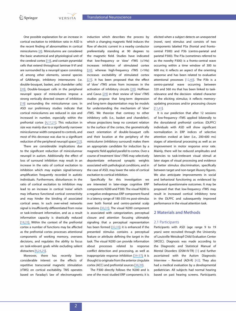
2.3 Kanizsa Illusory Figure TestIn this task subjects have to respond with a button-press to rare (25% probability) Kanizsa [50] squares (targets) among Kanizsa triangles (rare non-target distracters, 25% probability) and non-Kanizsa figures (standards, 50% probability). The stimuli are presented for 250 ms with inter-trial intervals (ITI) varying in the range of 1,100–1,300 ms. A fixation point (cross) was presented during ITI. White figures are displayed on a black background on a flat monitor. Subjects are instructed to press the first button on a 5-button keypad with their right index finger when a target appears, and ignore when the non-target Kanizsa or standard stimuli appear. Subjects were instructed to remain as still as possible with their eyes on the fixation mark and to refrain from blinking. Autistic patients had at least one session for EEG net conditioning and getting familiar with the experimental room.
The stimulus types used in the experiment are Kanizsa square (target), Kanizsa triangle (non-target), non-Kanizsa square, and non-Kanizsa triangle (standards). This task is a classic three-stimuli ‘oddball’ with rare Kanizsa target and rare Kanizsa distracter stimuli presented among frequent non-Kanizsa standards. The non-target Kanizsa triangle was introduced to differentiate processing of Kanizsa figures and targets. The stimuli consist of either three or four inducer disks which are considered the shape feature, and they either constitute an illusory figure (square, triangle) or not (collinearity feature) (Figure 1). One block of 240 trials was presented. Subjects with ASD in the TMS group were administered the Kanizsa, illusory figure test before (pre-TMS)
Translational Neuroscience
4
and after (post-TMS) treatment. There was also a randomly assigned waiting-list (WTL) group where individuals with ASD were administered the same Kanizsa illusory figure test twice (with an 8-10 week interval) to control for the TMS.
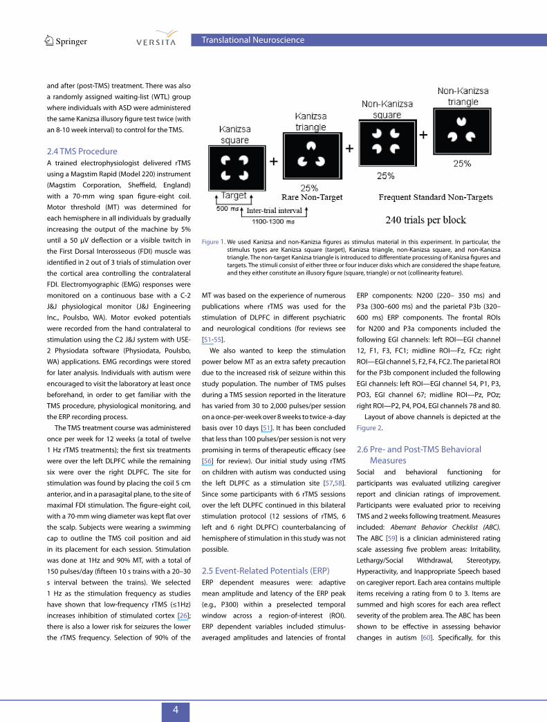
2.4 TMS ProcedureA trained electrophysiologist delivered rTMS using a Magstim Rapid (Model 220) instrument (Magstim Corporation, Sheffield, England) with a 70-mm wing span figure-eight coil. Motor threshold (MT) was determined for each hemisphere in all individuals by gradually increasing the output of the machine by 5% until a 50 μV deflection or a visible twitch in the First Dorsal Interosseous (FDI) muscle was identified in 2 out of 3 trials of stimulation over the cortical area controlling the contralateral FDI. Electromyographic (EMG) responses were monitored on a continuous base with a C-2 J&J physiological monitor (J&J Engineering Inc., Poulsbo, WA). Motor evoked potentials were recorded from the hand contralateral to stimulation using the C2 J&J system with USE-2 Physiodata software (Physiodata, Poulsbo, WA) applications. EMG recordings were stored for later analysis. Individuals with autism were encouraged to visit the laboratory at least once beforehand, in order to get familiar with the TMS procedure, physiological monitoring, and the ERP recording process.
The TMS treatment course was administered once per week for 12 weeks (a total of twelve 1 Hz rTMS treatments); the first six treatments were over the left DLPFC while the remaining six were over the right DLPFC. The site for stimulation was found by placing the coil 5 cm anterior, and in a parasagital plane, to the site of maximal FDI stimulation. The figure-eight coil, with a 70-mm wing diameter was kept flat over the scalp. Subjects were wearing a swimming cap to outline the TMS coil position and aid in its placement for each session. Stimulation was done at 1Hz and 90% MT, with a total of 150 pulses/day (fifteen 10 s trains with a 20–30 s interval between the trains). We selected 1 Hz as the stimulation frequency as studies have shown that low-frequency rTMS (≤1Hz) increases inhibition of stimulated cortex [26]; there is also a lower risk for seizures the lower the rTMS frequency. Selection of 90% of the
MT was based on the experience of numerous publications where rTMS was used for the stimulation of DLPFC in different psychiatric and neurological conditions (for reviews see [51-55].
We also wanted to keep the stimulation power below MT as an extra safety precaution due to the increased risk of seizure within this study population. The number of TMS pulses during a TMS session reported in the literature has varied from 30 to 2,000 pulses/per session on a once-per-week over 8 weeks to twice-a-day basis over 10 days [51]. It has been concluded that less than 100 pulses/per session is not very promising in terms of therapeutic efficacy (see [56] for review). Our initial study using rTMS on children with autism was conducted using the left DLPFC as a stimulation site [57,58]. Since some participants with 6 rTMS sessions over the left DLPFC continued in this bilateral stimulation protocol (12 sessions of rTMS, 6 left and 6 right DLPFC) counterbalancing of hemisphere of stimulation in this study was not possible.
2.5 Event-Related Potentials (ERP)ERP dependent measures were: adaptive mean amplitude and latency of the ERP peak (e.g., P300) within a preselected temporal window across a region-of-interest (ROI). ERP dependent variables included stimulus-averaged amplitudes and latencies of frontal
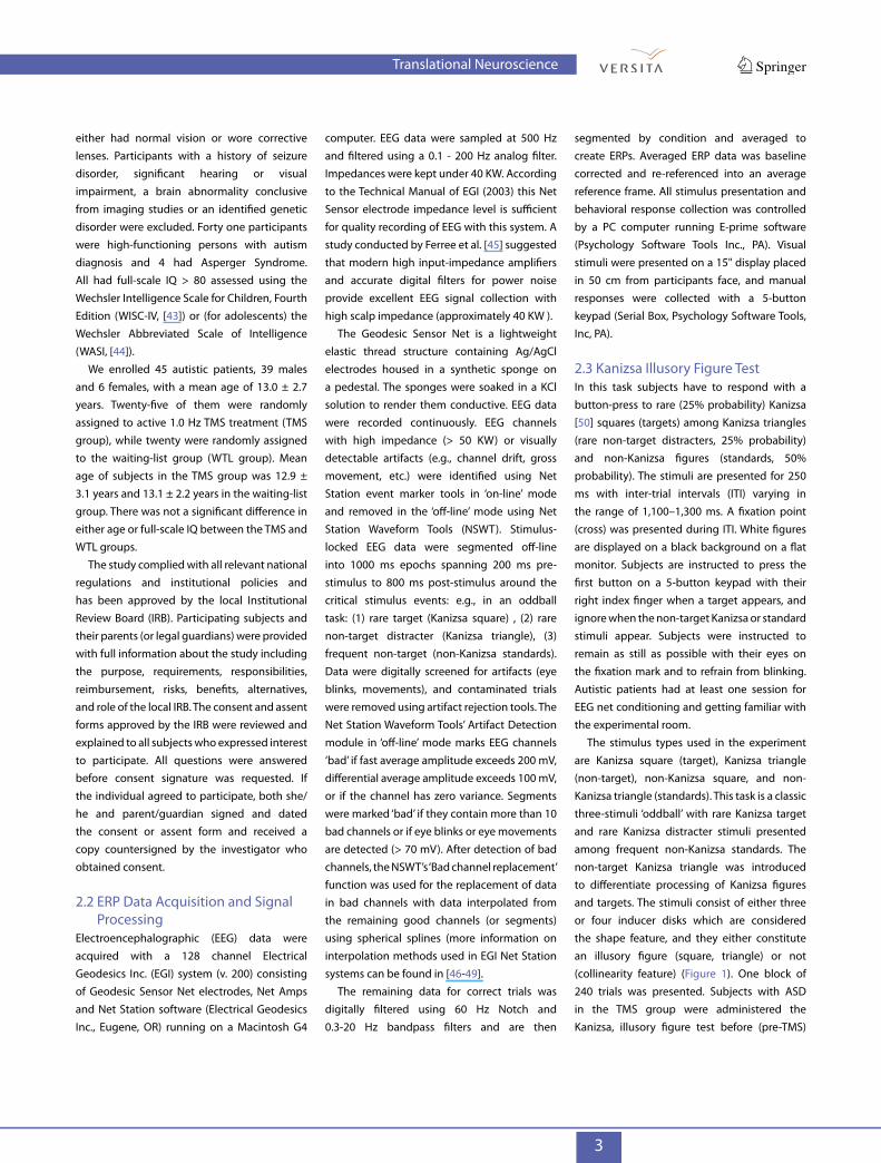
ERP components: N200 (220– 350 ms) and P3a (300–600 ms) and the parietal P3b (320–600 ms) ERP components. The frontal ROIs for N200 and P3a components included the following EGI channels: left ROI—EGI channel 12, F1, F3, FC1; midline ROI—Fz, FCz; right ROI—EGI channel 5, F2, F4, FC2. The parietal ROI for the P3b component included the following EGI channels: left ROI—EGI channel 54, P1, P3, PO3, EGI channel 67; midline ROI—Pz, POz; right ROI—P2, P4, PO4, EGI channels 78 and 80.
Layout of above channels is depicted at the Figure 2.
2.6 Pre- and Post-TMS Behavioral Measures
Social and behavioral functioning for participants was evaluated utilizing caregiver report and clinician ratings of improvement. Participants were evaluated prior to receiving TMS and 2 weeks following treatment. Measures included: Aberrant Behavior Checklist (ABC). The ABC [59] is a clinician administered rating scale assessing five problem areas: Irritability, Lethargy/Social Withdrawal, Stereotypy, Hyperactivity, and Inappropriate Speech based on caregiver report. Each area contains multiple items receiving a rating from 0 to 3. Items are summed and high scores for each area reflect severity of the problem area. The ABC has been shown to be effective in assessing behavior changes in autism [60]. Specifically, for this
Figure 1. We used Kanizsa and non-Kanizsa figures as stimulus material in this experiment. In particular, the stimulus types are Kanizsa square (target), Kanizsa triangle, non-Kanizsa square, and non-Kanizsa triangle. The non-target Kanizsa triangle is introduced to differentiate processing of Kanizsa figures and targets. The stimuli consist of either three or four inducer disks which are considered the shape feature, and they either constitute an illusory figure (square, triangle) or not (collinearity feature).
Translational Neuroscience
5
study we used the Irritability and Hyperactivity subscales of the ABC as outcome measures. Social Responsiveness Scale (SRS). The SRS [61] is a caregiver completed rating scale assessing social interest and interaction. The scale provides a dimensional measure of social interaction allowing the rating of social skills in Autism as well as non-autistic individuals. For this study we used the Social Awareness subscale of the SRS as an outcome measure. Repetitive Behavior Scale—Revised (RBS). The RBS [62] is a caregiver completed rating scale assessing repetitive and restricted behavior patterns. The RBS is a measure of different behaviors: stereotyped, self-injurious, compulsive, ritualistic, sameness, and restricted range [63]. Items from scales are summed to obtain a measure of severity of repetitive behavior.
2.7 Statistical Analysis Statistical analyses were performed on subject-averaged ERP and motor response data with
subject averages being observations. The primary analysis model was the repeated measures ANOVA, with dependent variables for electrocortical measures being specific ERP components’ amplitudes and latencies at selected ROIs. The data of each ERP dependent variable for each relevant ROI was analyzed using ANOVA with the following factors (all within participants): Stimulus (Target, Standard, non-target Kanizsa), Hemisphere (Left, Right), and Time (Pre-, Post). The between-subject factor included Group comparisons (TMS, Wait-List). Post-hoc analyses were conducted where appropriate. Behavioral responses (reaction time [RT], omission and commission error rate) were analyzed using Time and Group factor. For behavioral questionnaire data a Treatment (pre- vs. post-TMS/or waiting period) ANOVA was completed to determine changes associated with TMS and WTL conditions. In all ANOVAs Greenhouse-Geisser corrected P-values were employed where appropriate. IBM SPSS 19.0
(IBM, Somers, NY) and Sigma Stat 3.1 (Systat Software, Inc., Point Richmond, CA) packages were used for statistical analysis.
3. Results
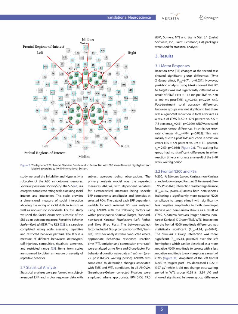
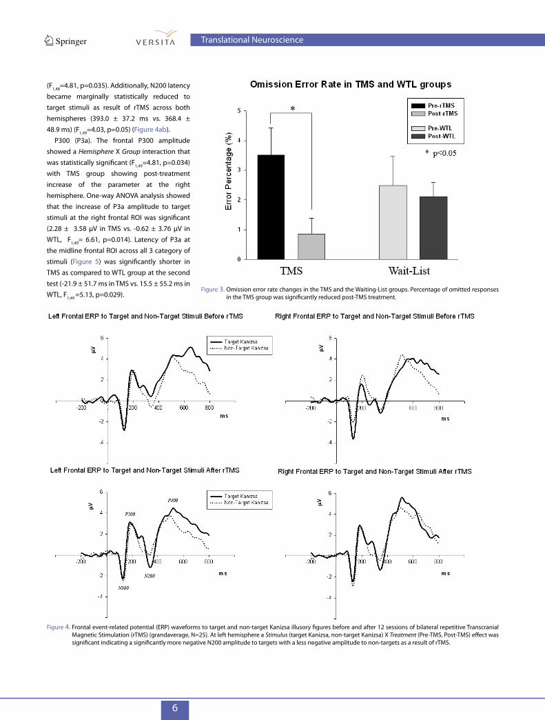
3.1 Motor ResponsesReaction time (RT) changes at the second test showed significant group differences (Time X Group effect, F1,49=6.71, p=0.031). However, post-hoc analysis using t-test showed that RT to targets was not significantly different as a result of rTMS (491 ± 118 ms pre-TMS vs. 470 ± 109 ms post-TMS, t24=0.983, p=0.299, n.s.). Post-treatment total accuracy differences between groups was not significant, but there was a significant reduction in total error rate as a result of rTMS (12.9 ± 17.9 percent vs. 5.5 ± 7.8 percent, t24=2.51, p=0.020). ANOVA revealed between group differences in omission error rate changes (F1,49=4.84, p=0.032). This was mainly due to a post-TMS reduction in omission errors (3.5 ± 5.9 percent vs. 0.9 ± 1.1 percent, t24= 2.59, p=0.016) (Figure 2a). The waiting-list group had no significant differences in either reaction time or error rate as a result of the 8-10 week waiting period.
3.2 Frontal N200 and P3aN200. A Stimulus (target Kanizsa, non-Kanizsa standard, non-target Kanizsa) X Treatment (Pre-TMS, Post-TMS) interaction reached significance (F2,96=3.42, p=0.037) across both hemispheres indicating a significantly more negative N200 amplitude to target stimuli with significantly less negative amplitudes to both non-target Kanizsa and non-Kanizsa stimuli as a result of rTMS. A Kanizsa Stimulus (target Kanizsa, non-target Kanizsa) X Group (TMS, WTL) interaction for the frontal N200 amplitude differences was statistically significant (F1,49=4.24, p=0.047). The Stimulus X Group interaction was more significant (F1,49=5.14, p=0.028) over the left hemisphere which can be described as a more negative N200 amplitude to targets with a less negative amplitude to non-targets as a result of rTMS (Figure 3a). Amplitude of the left frontal N200 to targets post-TMS decreased (-3.22 ± 5.97 µV) while it did not change post waiting period in WTL group (0.28 ± 3.39 µV) and showed significant between group difference
Figure 2. The layout of 128 channel Electrical Geodesics Inc. Sensor Net with EEG sites of interest highlighted and labeled according to 10-10 International System.
Translational Neuroscience
6
(F1,48=4.81, p=0.035). Additionally, N200 latency became marginally statistically reduced to target stimuli as result of rTMS across both hemispheres (393.0 ± 37.2 ms vs. 368.4 ± 48.9 ms) (F1,49=4.03, p=0.05) (Figure 4ab).
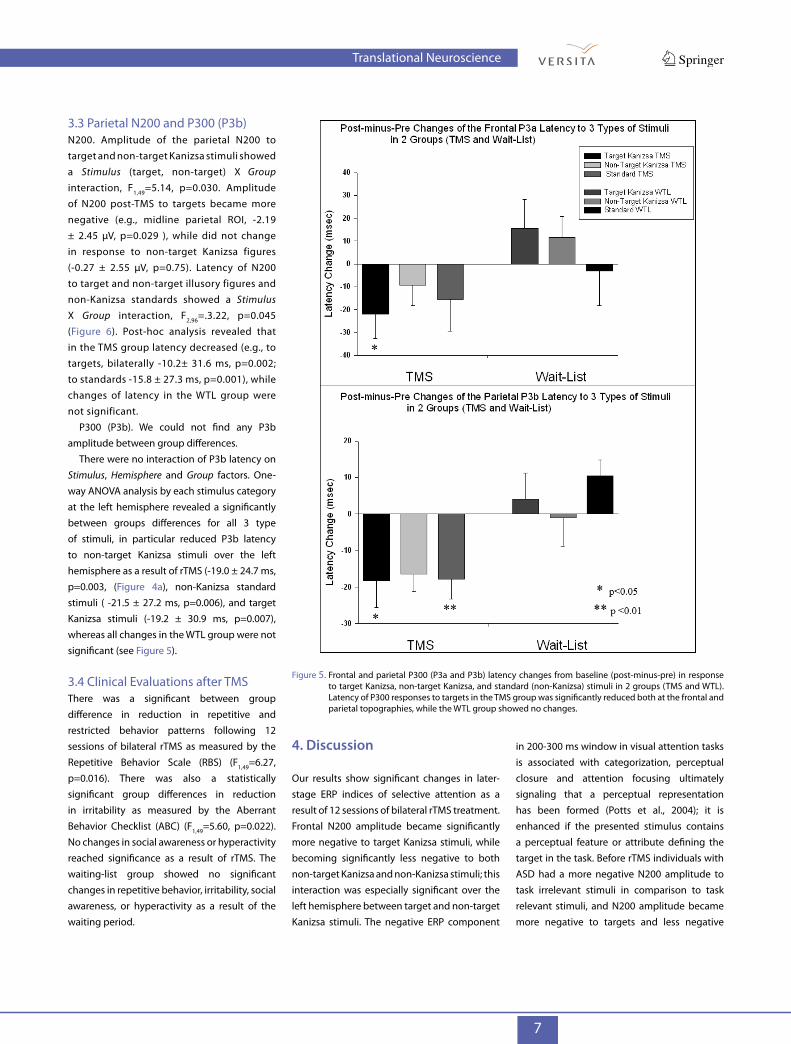
P300 (P3a). The frontal P300 amplitude showed a Hemisphere X Group interaction that was statistically significant (F1,49=4.81, p=0.034) with TMS group showing post-treatment increase of the parameter at the right hemisphere. One-way ANOVA analysis showed that the increase of P3a amplitude to target stimuli at the right frontal ROI was significant (2.28 ± 3.58 µV in TMS vs. -0.62 ± 3.76 µV in WTL, F1,49= 6.61, p=0.014). Latency of P3a at the midline frontal ROI across all 3 category of stimuli (Figure 5) was significantly shorter in TMS as compared to WTL group at the second test (-21.9 ± 51.7 ms in TMS vs. 15.5 ± 55.2 ms in WTL, F1,49 =5.13, p=0.029).
Figure 3. Omission error rate changes in the TMS and the Waiting-List groups. Percentage of omitted responses in the TMS group was significantly reduced post-TMS treatment.
Figure 4. Frontal event-related potential (ERP) waveforms to target and non-target Kanizsa illusory figures before and after 12 sessions of bilateral repetitive Transcranial Magnetic Stimulation (rTMS) (grandaverage, N=25). At left hemisphere a Stimulus (target Kanizsa, non-target Kanizsa) X Treatment (Pre-TMS, Post-TMS) effect was significant indicating a significantly more negative N200 amplitude to targets with a less negative amplitude to non-targets as a result of rTMS.
Translational Neuroscience
7
3.3 Parietal N200 and P300 (P3b)N200. Amplitude of the parietal N200 to target and non-target Kanizsa stimuli showed a Stimulus (target, non-target) X Group interaction, F1,49=5.14, p=0.030. Amplitude of N200 post-TMS to targets became more negative (e.g., midline parietal ROI, -2.19 ± 2.45 µV, p=0.029 ), while did not change in response to non-target Kanizsa figures (-0.27 ± 2.55 µV, p=0.75). Latency of N200 to target and non-target illusory figures and non-Kanizsa standards showed a Stimulus X Group interaction, F2,96=.3.22, p=0.045 (Figure 6). Post-hoc analysis revealed that in the TMS group latency decreased (e.g., to targets, bilaterally -10.2± 31.6 ms, p=0.002; to standards -15.8 ± 27.3 ms, p=0.001), while changes of latency in the WTL group were not significant.
P300 (P3b). We could not find any P3b amplitude between group differences.
There were no interaction of P3b latency on Stimulus, Hemisphere and Group factors. One-way ANOVA analysis by each stimulus category at the left hemisphere revealed a significantly between groups differences for all 3 type of stimuli, in particular reduced P3b latency to non-target Kanizsa stimuli over the left hemisphere as a result of rTMS (-19.0 ± 24.7 ms, p=0.003, (Figure 4a), non-Kanizsa standard stimuli ( -21.5 ± 27.2 ms, p=0.006), and target Kanizsa stimuli (-19.2 ± 30.9 ms, p=0.007), whereas all changes in the WTL group were not significant (see Figure 5).
3.4 Clinical Evaluations after TMSThere was a significant between group difference in reduction in repetitive and restricted behavior patterns following 12 sessions of bilateral rTMS as measured by the Repetitive Behavior Scale (RBS) (F1,49=6.27, p=0.016). There was also a statistically significant group differences in reduction in irritability as measured by the Aberrant Behavior Checklist (ABC) (F1,49=5.60, p=0.022). No changes in social awareness or hyperactivity reached significance as a result of rTMS. The waiting-list group showed no significant changes in repetitive behavior, irritability, social awareness, or hyperactivity as a result of the waiting period.
4. discussion
Our results show significant changes in later-stage ERP indices of selective attention as a result of 12 sessions of bilateral rTMS treatment. Frontal N200 amplitude became significantly more negative to target Kanizsa stimuli, while becoming significantly less negative to both non-target Kanizsa and non-Kanizsa stimuli; this interaction was especially significant over the left hemisphere between target and non-target Kanizsa stimuli. The negative ERP component
in 200-300 ms window in visual attention tasks is associated with categorization, perceptual closure and attention focusing ultimately signaling that a perceptual representation has been formed (Potts et al., 2004); it is enhanced if the presented stimulus contains a perceptual feature or attribute defining the target in the task. Before rTMS individuals with ASD had a more negative N200 amplitude to task irrelevant stimuli in comparison to task relevant stimuli, and N200 amplitude became more negative to targets and less negative
Figure 5. Frontal and parietal P300 (P3a and P3b) latency changes from baseline (post-minus-pre) in response to target Kanizsa, non-target Kanizsa, and standard (non-Kanizsa) stimuli in 2 groups (TMS and WTL). Latency of P300 responses to targets in the TMS group was significantly reduced both at the frontal and parietal topographies, while the WTL group showed no changes.
Translational Neuroscience
8
to irrelevant distracters as a result of rTMS treatment. Furthermore, N200 peak latency was significantly reduced to task relevant target stimuli across both hemispheres following rTMS pointing at more effective signal processing.
Our previous investigation of ERPs in 11 participants with ASD and 11 age-matched, typically developing control subjects [15] yielded significantly augmented and prolonged frontal N200 amplitude and latency baseline differences to irrelevant, novel visual stimuli in individuals with ASD relative to controls. The results of the current investigation indicate that rTMS treatment may have enhanced selective attention and stimulus discrimination by improving conflict processing between task relevant and task irrelevant visual stimuli.
P3b peak latency was significantly reduced to both non-target Kanzisa and non-Kanizsa
stimuli over the left hemisphere as a result of rTMS. The P3b component has been linked to task-relevance and the decision- related character of the eliciting stimulus; it reflects memory-updating processes and/or processing closure [11]. In our previous baseline investigation [15] we found that individuals with ASD had significantly prolonged P3b latency to irrelevant distracter stimuli in comparison to control subjects. The results of our current investigation may indicate enhanced visual discrimination processes and more efficient processing of irrelevant distracter stimuli as a result of rTMS. We also found a significant improvement in the percentage of total errors as a result of rTMS; this result further confirms improvement in selective attention, cognitive control, and irrelevant response inhibition after treatment with rTMS. In a recent relevant study
in ASD it was also shown that rTMS improves movement-related cortical potentials [64].
Previously we investigated the effects of six sessions of “slow” rTMS applied only to the left prefrontal cortex on performance in a visual task of selective attention in individuals with ASD [57,65]. Similar to the present investigation we found a significant reduction in the frontal N200 and parietal P3b latency to irrelevant distracter stimuli. However, we did not find any significant N200 amplitude changes after six sessions. Additionally, similar to the present investigation we also found a significant reduction in the percentage of motor response errors to target stimuli as a result of 6 sessions of rTMS applied to the left DLPFC [57,65]. It may plausibly be concluded that we found more robust changes in endogenous cognitive ERP components (i.e., N200 amplitude, P3b latency)
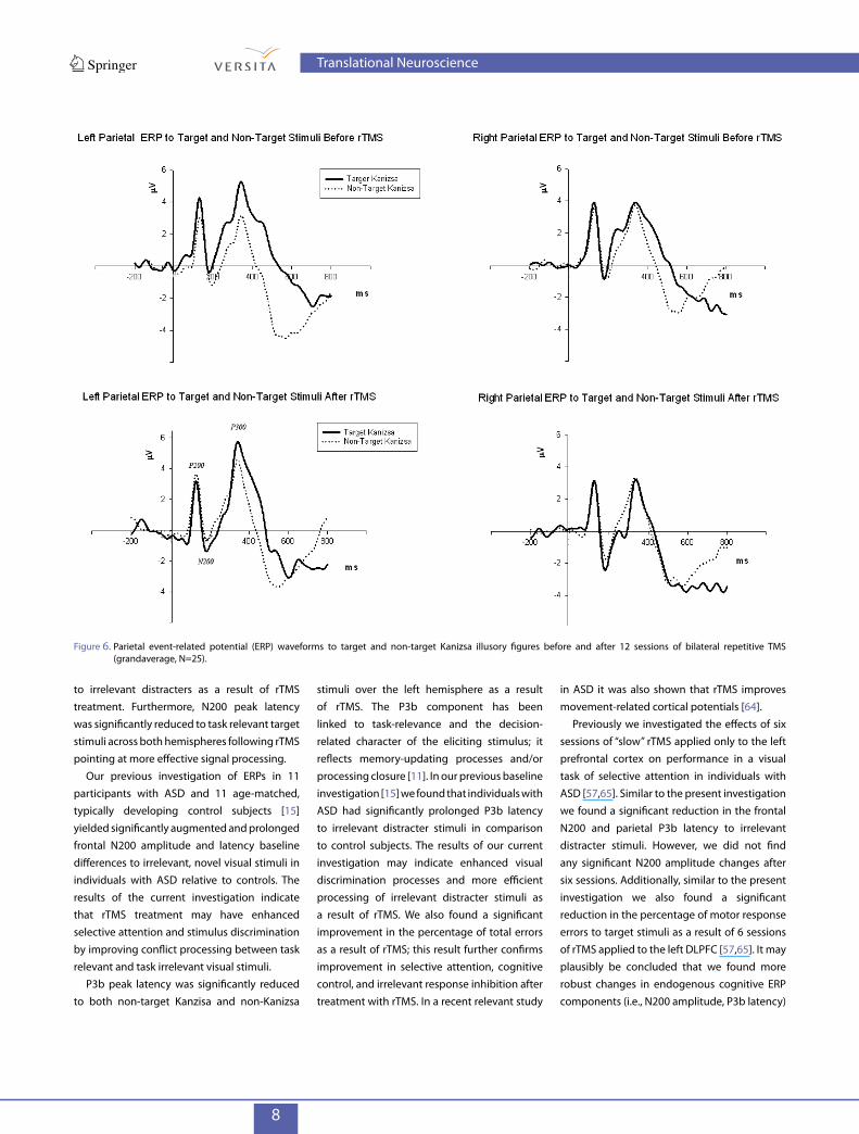
Figure 6. Parietal event-related potential (ERP) waveforms to target and non-target Kanizsa illusory figures before and after 12 sessions of bilateral repetitive TMS (grandaverage, N=25).
Translational Neuroscience
9
in this investigation due to the greater number of rTMS sessions and bilateral application.
In this investigation we also found a significant reduction in repetitive and restricted behavior patterns as well as a significant reduction in irritability according to clinical and behavioral questionnaires. These results confirm our previous findings of reductions in repetitive behavior [57,65,66] and irritability [66] as a result of low-frequency rTMS. It should be noted that we found significant reductions in irritability only as a result of 12 sessions of bilateral stimulation [66], whereas reductions in repetitive behavior have been significant after six sessions of stimulation to the left DLPFC [57,65].
The DLPFC processes attentional components of working memory, oversees decisions, and regulates the ability to focus on task-relevant goals while excluding salient distracters (i.e., selective attention) [24,25,35]. Evidence suggests there is a disruption in the ratio between cortical excitation and inhibition especially within the prefrontal cortex in individuals with autism [17,21]; this trend was confirmed in individuals with Asperger syndrome [22]. A lack of cortical inhibitory tone and an increased ratio of cortical excitation to inhibition could adversely affect patterns of cortical activation, possibly resulting in isolated
islands of coordinated excitatory activity (i.e., possible seizure foci). This may result in a high comorbidity rate of autism and seizure disorder [67]. Additionally, an increase in the ratio of cortical excitation to inhibition may explain the strong aversive reactions to auditory, tactile, and visual stimuli frequently reported in autistic individuals. Within the prefrontal cortex a lack of cortical inhibition could hinder the functional binding of associated cortical areas and drastically reduce selective attention and executive functioning. We propose in this study that over a course of treatment, “slow” rTMS may restore the balance between cortical excitation and cortical inhibition by selectively activating double-bouquet cells at the periphery of cortical minicolumns [20,21].
Previous studies have shown that minicolumnar abnormalities in autism are not equally distributed throughout the cerebral cortex. In autistic subjects topographical studies of different brain parcellations reveal the most significant minicolumnar abnormalities within the prefrontal cortex, more specifically, the DLPFC and the anterior cingulate (ACC) gyrus [21,22]. The involved areas exhibit different cytoarchitecture and functional subtypes (DLPFC- heteromodal association, ACC -paralimbic, see [68]) but
appear interlinked within an executive system that mediates metacognitive processing and top-down selection of strategic processes during complex attentional tasks [69]. It is postulated that the function of this system is to add flexibility to cognitive processes by making them less dependent on external cues [69].
In conclusion, this investigation showed that treatment with “slow” rTMS significantly improved both N200 and P300 ERP indices of selective attention and significantly reduced motor response errors to target stimuli. We also found significant reductions in both repetitive behavior and irritability according to clinical and behavioral questionnaires as a result of rTMS. It should also be noted that no lasting side effects were reported in this study as a result of “slow” rTMS treatment. We conclude that treatment with “slow” rTMS significantly improved selective attention and executive functioning in individuals with ASD. RTMS should be considered a potentially promising treatment modality targeting some of the core symptoms of ASD.
acknowledgments
Funding for this work was provided by NIH grant R01 MH86784 (MFC).
[1] American Psychiatric Association diagnostic and statistical manual of mental disorders (DSM-IV TR), 4th ed., American Psychiatric Association, Washington, DC, 2000
[2] Charman T., Autism spectrum disorders, Psychiatry, 2008, 7, 331–334[3] Happe F.G.E., Autism: cognitive deficit or cognitive style? Trends
Cogn. Sci., 1999, 3, 216-222[4] Casanova M.F., Buxhoeveden D., Gomez J., Disruption in the
inhibitory architecture of the cell minicolumn: implications for autism, Neuroscientist, 2003, 9, 496–507
[5] Gomes E., Pedroso F.S., Wagner M.B., Auditory hypersensitivity in the autistic spectrum disorder, Pro. Fono., 2008, 20, 279–284
[6] Jeste S.S., Nelson C.A. 3rd., Event related potentials in the understanding of autism spectrum disorders: an analytical review, J. Autism Dev. Disord., 2009, 39, 495-510
[7] Coles M.G.H., Rugg M.D., Event-related brain potentials: an introduction, In: Rugg M.D., Coles M.G.H. (Eds.), Electrophysiology of mind. Event-related brain potentials and cognition, Oxford University Press, Oxford, 1995
[8] Eichele T., Specht K., Moosmann M., Jongsma M.L., Quiroga R.Q., et al., Assessing the spatiotemporal evolution of neuronal activation with single-trial event-related potentials and functional MRI, Proc. Natl. Acad. Sci. U S A, 2005, 102, 17798-17803
[9] Folstein J.R., Van Petten C., Rose S.A., Novelty and conflict in the categorization of complex stimuli, Psychophysiology, 2008, 45, 467-479
[10] Herrmann C.S., Knight R.T., Mechanisms of human attention: event related potentials and oscillations, Neurosci. Biobehav. Rev., 2001, 25, 465-476
[11] Picton T.W., The P300 wave of the human event-related potential, J. Clin. Neurophysiol., 1992, 9, 456–479
[12] Polich J., Theoretical overview of P3a and P3b, In: Polich J. (Ed.), Detection of change. Event-related potential and fMRI findings, Kluwer Academic Press, Boston, 2003
[13] Polich J., Updating P300: an integrative theory of P3a and P3b, J. Clin. Neurophysiol., 2007, 118, 2128-2148
[14] Pritchard W.S., Psychophysiology of P300, Psychol. Bull., 1981, 89, 506-540
References
Translational Neuroscience
10
[15] Sokhadze E., Baruth J., Tasman A., Sears L., Mathai G., El-Baz A., Casanova M.F., Event-related potential study of novelty processing abnormalities in autism, Appl. Psychophysiol. Biofeedback, 2009, 34, 37-51
[16] Baruth J.M., Casanova M., Sears L., Sokhadze E., Early-stage visual processing abnormalities in autism spectrum disorder (ASD), Transl. Neurosci., 2010, 1, 177-187
[17] Casanova M.F., Buxhoeveden D.P., Brown C., Clinical and macroscopic correlates of minicolumnar pathology in autism, J. Child Neurol., 2002, 17, 692–695
[18] Rubenstein J.L.R., Merzenich M.M., Model of autism: increased ratio of excitation/inhibition in key neural systems, Gen. Brain Behav., 2003, 2, 255–267
[19] Mountcastle V.B., Introduction. Computation in cortical columns, Cereb. Cortex, 2003, 13, 2–4
[20] Casanova M.F., The neuropathology of autism, Brain Pathol., 2007, 17, 422-433
[21] Casanova M.F., van Kooten I., Switala A. E., van England H., Heinsen H., Steinbuch H.W.M., et al., Abnormalities of cortical minicolumnar organization in the prefrontal lobes of autistic patients, Clin. Neurosci. Res., 2006, 6, 127–133
[22] Casanova M.F., Buxhoeveden D.P., Switala A.E., Roy E., Asperger’s syndrome and cortical neuropathology, J. Child Neurol., 2002, 17, 142-145
[23] Belmonte M.K., Yurgelun-Todd D.A., Functional anatomy of impaired selective attention and compensatory processing in autism, Cognitive Brain Res., 2003, 17, 651-664
[24] Gray J.R., Chabris C.F., Braver T.S., Neural mechanisms of general fluid intelligence, Nat. Neurosci., 2003, 6, 316–322
[25] Matzel L.D., Kolata S., Selective attention, working memory, and animal intelligence, Neurosci. Biobehav. Rev., 2010, 34, 23–30
[26] Maeda F., Keenan J.P., Tormos J.M., Topka H., Pascual-Leone A., Modulation of corticospinal excitability by repetitive transcranial magnetic stimulation, Clin. Neurophysiol., 2000, 111, 800-805
[27] Pascual-Leone A., Valls-Sole J., Wasserman E.M., Hallett M., Responses to rapid-rate transcranial magnetic stimulation of the human cortex, Brain, 1994, 117, 847-858
[28] Pascual-Leone A., Walsh V., Rothwell J., Transcranial magnetic stimulation in cognitive neuroscience--virtual lesion, chronometry, and functional connectivity, Curr. Opin. Neurobiol., 2000, 10, 232-237
[29] Hoffman R.E., Cavus I., Slow transcranial magnetic stimulation, long-term depotentiation, and brain hyperexcitability disorders, Am. J. Psychiatr., 2002, 159, 1093–1102
[30] Näätänen R., Gaillard A.W.K., Mäntysalo S., Early selective attention effect on evoked potential reinterpreted, Acta Psychologica, 1978, 2, 313–329
[31] Näätänen R., Schröger E., Karakas S., Tervaniemi M., Paavilainen, P., Development of a memory trace for a complex sound in the human brain, Neuroreport, 1993, 4, 503–506
[32] Potts G.F., Patel S.H., Azzam P.N., Impact of instructed relevance on the visual ERP, Int. J. Psychophysiol., 2004, 52, 197–209
[33] Potts G.F., Wood S.M., Kothmann D., Martin L.E., Parallel perceptual enhancement and hierarchic relevance evaluation in an audio-visual conjunction task, Brain Res., 2008, 1236, 126-139
[34] Carter C.S., Braver T.S, Barch D.M., Botvinick M.M., Noll D., Cohen J.D., Anterior cingulate cortex, error detection, and the online monitoring of performance, Science, 1998, 280, 747-749
[35] Enriquez-Geppert S., Konrad C., Pantev C., Huster R.J., Conflict and inhibition differentially affect the N200/P300 complex in a combined go/nogo and stop-signal task, Neuroimage, 2010, 51, 877-887
[36] West R., Bowry R., McConville C., Sensitivity of medial frontal cortex to response and nonresponse conflict, Psychophysiol., 2004, 41, 739–748
[37] West R., Neural correlates of cognitive control and conflict detection in the Stroop and digit-location tasks, Neuropsychologia, 2003, 41, 1122–1135
[38] Donkers F.C.L., van Boxtel G.J.M., The N2 in go/no-go tasks reflects conflict monitoring not response inhibition, Brain Cogn., 2004, 56, 165–176
[39] Van Veen V., Carter C.S., The timing of action-monitoring process in the anterior cingulate cortex, J. Cogn. Neurosci., 2002, 14, 593-602
[40] Hruby T., Marsalek P., Event-related potentials--the P3 wave, Acta Neurobiol. Exp., 2003, 63, 55-63
[41] Polich J., Kok A., Cognitive and biological determinants of P300: an integrative review, Biol. Psychol., 1995, 41, 103-146
[42] Le Couteur A., Lord C., Rutter M., The autism diagnostic interview—revised (ADI-R), Western Psychological Services, Los Angeles, CA, 2003
[43] Wechsler D., Wechsler intelligence scale for children, 4th ed., Harcourt Assessment, Inc., San Antonio, TX, 2003
[44] Wechsler D., Wechsler abbreviated scale of intelligence, Harcourt Assessment, Inc., San Antonio, TX, 1999
[45] Ferree T.C., Luu P., Russell G.S., Tucker D.M., Scalp electrode impedance, infection risk, and EEG data quality, Clin. Neurophysiol., 2001, 112, 444–536
[46] Perrin E., Pernier J., Bertrand O., Giard M., Echallier J.F., Mapping of scalp potentials by surface spline interpolation, Electroencephalogr. Clin. Neurophysiol., 1987, 66, 75–81
[47] Fletcher E.M., Kussmaul C.L., Mangun G.R., Estimation of interpolation errors in scalp topographic mapping, Electroctoencephalogr. Clin. Neurophysiol., 1996, 98, 422–434
[48] Srinivasan R., Tucker D.M., Murias M., Estimating the spatial Nyquist of the human EEG, Behav. Res. Meth. Instrum. Comput., 1998, 30, 8–19
[49] Luu P., Tucker D.M., Englander R., Lockfeld A., Lutsep H., Oken B., Localizing acute stroke-related EEC changes: assessing the effects of spatial undersampling, J. Clin. Neurophysiol., 2001, 18, 302–317
[50] Kanizsa G., Subjective contours, Sci. American, 1976, 235, 48-52[51] Daskalakis Z.J., Christensen B.K., Fitzgerald P.B., Chen R., Transcranial
magnetic stimulation: a new investigational and treatment tool in psychiatry, J. Neuropsychiatry Clin. Neurosci., 2002, 14, 406–415
[52] Gershon A.A., Dannon P.N., Grunhaus L., Transcranial magnetic stimulation in the treatment of depression, Am. J. Psychiatr., 2003, 160, 835–845
Translational Neuroscience
11
[53] Greenberg B.D., Transcranial magnetic stimulation in anxiety disorders. In: George M.S., Belmaker R.H. (Eds.), Transcranial magnetic stimulation in clinical psychiatry, American Psychiatric Publishing, Inc., Washington, DC, 2007
[54] Loo C., Mitchell P., A review of the efficacy of transcranial magnetic stimulation (TMS) treatment for depression, and current and future strategies to optimize efficacy, J. Affect. Disord., 2005, 88, 255–267
[55] Wassermann E.M., Lisanby S.H., Therapeutic application of repetitive transcranial magnetic stimulation: a review, Clin. Neurophysiol., 2001, 112, 1367–1377
[56] Helmich R.C., Siebner H.R., Bakker M., Munchau A., Bloem B.R., Repetitive transcranial magnetic stimulation to improve mood and motor function in Parkinson’s disease, J. Neurol. Sci., 2006, 248, 84–96
[57] Sokhadze E., Baruth J., Tasman A., Mansoor M., Ramaswamy R., Sears L., Mathai G., El-Baz A., Casanova M.F., Low-frequency repetitive transcranial magnetic stimulation (rTMS) affects event-related potential measures of novelty processing in autism, Appl. Psychophysiol. Biofeedback, 2010, 35, 147-161
[58] Baruth J., Williams E., Sokhadze E., El-Baz A., Sears L., Casanova, M.F. (2011). Repetitive transcranial stimulation (rTMS) improves electroencephalographic and behavioral outcome measures in autism spectrum disorders (ASD). Autism Sci. Digest, 2011, 1(1), 52-57.
[59] Aman M.G., Singh N.N., Aberrant behavior checklist—community. Supplementary manual, Slosson Educational Publications, East Aurora, NY, 1994
[60] Aman M.G., Management of hyperactivity and other acting out problems in patients with autism spectrum disorder, Semin. Pediatr. Neurol., 2004, 11, 225–228
[61] Constantino J.N., Gruber C.P., The social responsiveness scale (SRS) manual, Western Psychological Services, Los Angeles, CA, 2005
[62] Bodfish J.W., Symons F.J., Lewis M.H., Repetitive behavior scale, Western Carolina Center Research Reports, Morganton, NC, 1999
[63] Bodfish J.W., Symons F.S., Parker D.E., Lewis M.H., Varieties of repetitive behavior in autism: comparisons to mental retardation, J. Autism Dev. Disord., 2000, 30, 237–243
[64] Enticott P.G., Rinehart N.J., Tonge B.J., Bradshaw J.L., Fitzgerald P.B., Repetitive transcranial magnetic stimulation (rTMS) improves movement-related cortical potentials in autism spectrum disorders, Brain Stimul., 2012, 5, 30-37
[65] Sokhadze E., El-Baz A., Baruth J., Mathai G., Sears L., Casanova M.F., Effects of low frequency repetitive transcranial magnetic stimulation (rTMS) on gamma frequency oscillations and event-related potentials during processing of illusory figures in autism, J. Autism Dev. Disord., 2009, 39, 619-634
[66] Baruth J.M., Casanova M., El-Baz A., Horrell T., Mathai G., Sears L., Sokhadze E., Low-frequency repetitive transcranial magnetic stimulation (rTMS) modulates evoked-gamma oscillations in autism spectrum disorder (ASD), J. Neurother., 2010, 14, 179-194
[67] Tuchman R.F., Rapin I., Regression in pervasive developmental disorders: seizures and epileptiform electroencephalogram correlates, Pediatrics, 1997, 99, 560 –566
[68] Mesulam M.M., Behavioral Neuroanatomy: Large-networks, association cortex, frontal syndromes, the limbic system, and hemispheric specializations. In Mesulam M.M (Ed) Principles of Behavioral and Cognitive Neurology, Oxford University Press:New York, 2nd edition, 2000, Ch. 1, pp. 1-120,
[69] Fernandez-Duque D., Baird J., Posner, M. , Executive attention and metacognitive regulation. Consc. Cogn. , 2000, 9, 288–307
Translational Neuroscience
Related Documents