Repetitive Orogastric Gavage Affects the Phenotype of Diet- Induced Obese Mice Vincent E. de Meijer, MD, MSc 1,2 , Hau D. Le, MD 1 , Jonathan A. Meisel, MD 1 , and Mark Puder, MD, PhD 1,* 1 Department of Surgery and the Vascular Biology Program Children's Hospital Boston and Harvard Medical School, Massachusetts, USA 2 Department of Surgery, Erasmus MC – University Medical Center Rotterdam, the Netherlands Abstract Interest in pharmacological intervention to combat metabolic syndrome and its complications is increasing as the prevalence of obesity is reaching epidemic proportions. The potential efficacy of drugs is often tested in animal models; however, the method of drug delivery is frequently overlooked and may act as a confounder due to stress. We hypothesized that long-term orogastric gavage would negatively influence the development of hepatic steatosis and the metabolic syndrome in a murine model of diet-induced obesity. C57BL/6J male mice were fed a high fat diet and were gavaged with a vehicle once or twice daily for 9 weeks. A group without orogastric gavaging served as control. A similar experiment was performed using leptin deficient ob/ob mice that were fed a standard diet for 4 weeks. Food intake was monitored, insulin resistance determined, and steatosis was assessed by histology and quantified via magnetic resonance spectroscopy. After 9 weeks, control C57BL/6J mice exhibited significantly more weight gain, insulin resistance and hepatic steatosis, compared to mice that were gavaged daily, or twice daily. This effect was likely due to decreased food consumption associated with gavage-induced stress. In contrast, the phenotype of leptin deficient ob/ob mice was not affected by orogastric gavage. Therefore, we concluded that orogastric gavage may lead to increased stress, thereby affecting food intake and the development of diet-induced obesity in a murine model. The effects of what may seem to be trivial laboratory routines, such as orogastric gavage, should be taken into account when designing animal studies for drug development. 1. Introduction Metabolic syndrome is a cluster of interrelated risk factors for diabetes and cardiovascular disease, including obesity, hyperglycemia, dyslipidemia and elevated blood pressure [1]. The prevalence of the metabolic syndrome is increasing worldwide, estimated to be 35–40% both in the US [1] and in the Middle East [2], depending on the criteria used. Risk factors for the metabolic syndrome are mainly related to a sedentary lifestyle and obesity. The prevalence of non-alcoholic fatty liver disease, which is considered to be the hepatic © 2010 Elsevier Inc. All rights reserved. * Corresponding Author: Mark Puder, MD, PhD, Postal address: 300 Longwood Avenue, Fegan 3, Boston, MA 02115, USA, Phone: 617-355-1838, Fax: 617-730-0298, [email protected]. Publisher's Disclaimer: This is a PDF file of an unedited manuscript that has been accepted for publication. As a service to our customers we are providing this early version of the manuscript. The manuscript will undergo copyediting, typesetting, and review of the resulting proof before it is published in its final citable form. Please note that during the production process errors may be discovered which could affect the content, and all legal disclaimers that apply to the journal pertain. NIH Public Access Author Manuscript Physiol Behav. Author manuscript; available in PMC 2012 June 19. Published in final edited form as: Physiol Behav. 2010 June 16; 100(4): 387–393. doi:10.1016/j.physbeh.2010.04.001. NIH-PA Author Manuscript NIH-PA Author Manuscript NIH-PA Author Manuscript

Welcome message from author
This document is posted to help you gain knowledge. Please leave a comment to let me know what you think about it! Share it to your friends and learn new things together.
Transcript
Repetitive Orogastric Gavage Affects the Phenotype of Diet-Induced Obese Mice
Vincent E. de Meijer, MD, MSc1,2, Hau D. Le, MD1, Jonathan A. Meisel, MD1, and MarkPuder, MD, PhD1,*
1Department of Surgery and the Vascular Biology Program Children's Hospital Boston andHarvard Medical School, Massachusetts, USA 2Department of Surgery, Erasmus MC – UniversityMedical Center Rotterdam, the Netherlands
AbstractInterest in pharmacological intervention to combat metabolic syndrome and its complications isincreasing as the prevalence of obesity is reaching epidemic proportions. The potential efficacy ofdrugs is often tested in animal models; however, the method of drug delivery is frequentlyoverlooked and may act as a confounder due to stress. We hypothesized that long-term orogastricgavage would negatively influence the development of hepatic steatosis and the metabolicsyndrome in a murine model of diet-induced obesity. C57BL/6J male mice were fed a high fat dietand were gavaged with a vehicle once or twice daily for 9 weeks. A group without orogastricgavaging served as control. A similar experiment was performed using leptin deficient ob/ob micethat were fed a standard diet for 4 weeks. Food intake was monitored, insulin resistancedetermined, and steatosis was assessed by histology and quantified via magnetic resonancespectroscopy. After 9 weeks, control C57BL/6J mice exhibited significantly more weight gain,insulin resistance and hepatic steatosis, compared to mice that were gavaged daily, or twice daily.This effect was likely due to decreased food consumption associated with gavage-induced stress.In contrast, the phenotype of leptin deficient ob/ob mice was not affected by orogastric gavage.Therefore, we concluded that orogastric gavage may lead to increased stress, thereby affectingfood intake and the development of diet-induced obesity in a murine model. The effects of whatmay seem to be trivial laboratory routines, such as orogastric gavage, should be taken into accountwhen designing animal studies for drug development.
1. IntroductionMetabolic syndrome is a cluster of interrelated risk factors for diabetes and cardiovasculardisease, including obesity, hyperglycemia, dyslipidemia and elevated blood pressure [1].The prevalence of the metabolic syndrome is increasing worldwide, estimated to be 35–40%both in the US [1] and in the Middle East [2], depending on the criteria used. Risk factorsfor the metabolic syndrome are mainly related to a sedentary lifestyle and obesity. Theprevalence of non-alcoholic fatty liver disease, which is considered to be the hepatic
© 2010 Elsevier Inc. All rights reserved.*Corresponding Author: Mark Puder, MD, PhD, Postal address: 300 Longwood Avenue, Fegan 3, Boston, MA 02115, USA, Phone:617-355-1838, Fax: 617-730-0298, [email protected].
Publisher's Disclaimer: This is a PDF file of an unedited manuscript that has been accepted for publication. As a service to ourcustomers we are providing this early version of the manuscript. The manuscript will undergo copyediting, typesetting, and review ofthe resulting proof before it is published in its final citable form. Please note that during the production process errors may bediscovered which could affect the content, and all legal disclaimers that apply to the journal pertain.
NIH Public AccessAuthor ManuscriptPhysiol Behav. Author manuscript; available in PMC 2012 June 19.
Published in final edited form as:Physiol Behav. 2010 June 16; 100(4): 387–393. doi:10.1016/j.physbeh.2010.04.001.
NIH
-PA Author Manuscript
NIH
-PA Author Manuscript
NIH
-PA Author Manuscript
manifestation of the metabolic syndrome [3], is increasing as well, affecting up to 30% ofindividuals with the syndrome [4]. Non-alcoholic fatty liver disease is a spectrum of diseaseranging from the simple build-up of fat in the liver (hepatic steatosis) to non-alcoholicsteatohepatitis, cirrhosis, and ultimately liver failure [5]. The main goal of treating metabolicsyndrome is to reduce obesity, especially abdominal girth, through a multimodality approachthat includes diet, excise, and pharmacologic intervention [6]. Pharmacologic agents can beused to target both energy intake and energy expenditure, reduce food consumption, and toimprove markers of insulin sensitivity, hypercholesterolemia and hepatic steatosis. Despitethe availability of several classes of drugs such as antihyperglycemics, antihypertensives,and lipid lowering agents, further drug development is warranted to overcome currentlimitations, potentially leading to improved clinical management of the metabolic syndromeand its associated complications.
In order to test the efficacy of drugs on treating the metabolic syndrome, animal models areessential to investigate the relative influence of various treatments on energy intake, energyexpenditure, and body weight. In animal models, drugs can be administered intravenously,intraperitoneally or via orogastric gavage, the last considered to be the least invasive route.However, even non-invasive laboratory routines such as personnel entering the animalhousing room, cage maintenance, and body weight measurements may elicit an acute stressresponse [7].
More stressful procedures such as restraining the animal, may induce catabolism byactivating the corticotrophin-releasing hormone system [8, 9]. Moreover, chronic activationof the stress response has been associated with altered energy homeostasis and reduction ofbody fat content [10]. Other reports, however, support a link between exposure to chronicstress and the development of obesity [11, 12]. To this day, it remains to be demonstratedwhether an acute stressor such as repetitive orogastric gavage may by itself influence themetabolic phenotype of the model studied.
The aim of this study was to investigate the degree of stress induced by different frequenciesof orogastric gavage on food intake, fat deposition, insulin resistance and hepatic steatosis inobesity-prone C57BL/6J and ob/ob mice. Using the classic model of diet-induced obesity aswell as the transgenic model of leptin deficiency, we hypothesized that long-term, repetitiveorogastric gavaging of mice would alter the development of the metabolic syndrome, and inparticular, hepatic steatosis.
2. Material and Methods2.1. Animals and general housing conditions
C57BL/6J mice (Jackson Laboratories, Bar Harbor, ME) fed a high fat diet ad libitumgradually develop visceral adiposity, hyperglycemia, insulin and leptin resistance, as well ashepatic steatosis, sharing many of the same obesity related phenotypes as humans [13].B6.V-Lepob/J mice (Jackson Laboratories, Bar Harbor, ME), commonly referred to as ob/obmice, are homozygous for the spontaneous mutation of the Lepob gene and exhibit obesity,hyperphagia, diabetes, and hepatic steatosis. They are both hypometabolic and hypothermic[14, 15]. Male 5-week-old mice were housed 4 (ob/ob) or 5 (C57BL/6J) animals per cage onpaper chip bedding in a barrier room with regulated temperature (21°C ± 1 .6°C), humidity(45% ± 10%), and an alternating 12-hour light and dark cycle. All animals had free access towater and standard rodent chow pellets (Prolab Isopro, RMH 3000 #25; containing 14% fat,26% protein, and 60% carbohydrate by calories; energy density 4.1 kcal/g; Prolabs Purina,Richmond, IN) for an acclimation period of 1 week prior to study initiation. During thestudy period, the animals were weighed twice a week and food intake was measured daily.Intake was measured per cage to avoid potential additional stress of individual housing.
de Meijer et al. Page 2
Physiol Behav. Author manuscript; available in PMC 2012 June 19.
NIH
-PA Author Manuscript
NIH
-PA Author Manuscript
NIH
-PA Author Manuscript
Animal protocols complied with the National Institutes of Health Animal Research AdvisoryCommittee guidelines, and were approved by the Children's Hospital Boston Animal Careand Use Committee (protocol no. A06-08-065R).
2.2. Study dietsStudy diets were stored at −80°C and provided fresh each day to avoid lipid peroxidation. Inthe high fat purified rodent diet (D12492; Research Diets, New Brunswick, NJ), the amountof calories derived from fat, protein, and carbohydrates were 60%, 20%, and 20%,respectively (energy density of 5.24 kcal/g). In the standard purified rodent diet (D12450B;Research Diets, New Brunswick, NJ), 10% of calories were derived from fat, 20% fromprotein, and 70% from carbohydrates (energy density of 3.85 kcal/g).
2.3. InterventionAnimals received 0.2 mL of a common vehicle, 0.45% (w/v) methylcellulose (Sigma-Aldrich, St. Louis, MO) in double distilled H2O, via orogastric gavage through a 30 mm 20Gauge feeding needle (Fine Sciences Tools Inc., Foster City, CA). Mice were gavaged dailybetween 07:00 and 09:00 h, or if twice, again between 19:00 and 21:00 h. Control mice weredisturbed and manually handled daily without gavaging. Orogastric gavage was routinelyperformed by one experienced investigator (VM) throughout the study, with 0% mortality.
2.3. Study designAfter the one week acclimation period, fifteen C57BL/6J mice were placed on the high fatdiet and randomized into three groups. In a separate study, twelve leptin deficient ob/obmice were acclimatized for one week, were fed a standard purified rodent diet andrandomized into three groups. In both experiments, animals in the control group (0x/day)were not gavaged and served as controls, whereas animals in the second (1x/day) and thirdgroup (2x/day) were gavaged daily, or twice daily, respectively, throughout the study period.
2.4. Specimen collectionAt the end of the feeding experiments, mice were fasted for 6 hours. Glucose concentrationwas determined from tail vein blood using the OneTouch UltraSmart Blood GlucoseMonitoring System (LifeScan, Milpitas, CA). Mice were then anesthetized with 2.5%Avertin (2,2,2-Tribromoethanol, Sigma-Aldrich, St. Louis, MO) by intraperitoneal injection.Blood was then collected via retro-orbital sinus puncture and centrifuged at 14000 rpm at4°C for 10 min to obtain serum. Serum was delivered to the Clinical Laboratory atChildren’s Hospital Boston for analysis of alanine aminotransferase (ALT), total cholesteroland triglyceride levels.
We then performed a midline laparotomy to observe, excise, and weigh the liver. The frontallobe of the liver was fixed in 10% formalin at 4°C overn ight, washed with phosphate-buffered saline, and then embedded in paraffin. The left lateral lobe was excised andcollected for magnetic resonance (MR) spectroscopy analysis. The remaining liver wasimmediately snap-frozen in liquid nitrogen and stored at −80°C.
White adipose tissue was dissected according to previously defined anatomic landmarks[16]. It was then weighed, snap-frozen in liquid nitrogen, and stored at −80°C. Inguinal (allsubcutaneous fat between the lower part of the rib cage and mid thigh), mesenteric (all fatalong the mesentery from the lesser curvature of the stomach to the sigmoid colon),retroperitoneal and epidydimal fat pads were weighed and expressed relative to evisceratedbody weight. A white adipose tissue fat-index was calculated using the sum of the individualfat pads as a percentage of the eviscerated body weight [16].
de Meijer et al. Page 3
Physiol Behav. Author manuscript; available in PMC 2012 June 19.
NIH
-PA Author Manuscript
NIH
-PA Author Manuscript
NIH
-PA Author Manuscript
2.5. Surrogate indexes of insulin sensitivity and resistanceInsulin levels were measured using a rat/mouse insulin ELISA kit (Linco Research, St.Charles, MO). Surrogate indexes for insulin sensitivity were calculated, includingquantitative insulin-sensitivity check index (QUICKI), homeostasis model assessment(HOMA) and log(HOMA) [17]. The calculations were performed as follows: QUICKI=1/[log(I0)+log(G0)], where I0 is the fasting insulin (µU/mL) and G0 is the fasting glucose (mg/dL); HOMA=(G0xI0)/22.5 (with glucose expressed as mmol/L and insulin expressed as µU/mL); and log(HOMA).
2.6. Hepatic histologyParaffin-embedded sections of the liver were stained with hematoxylin and eosin andperiodic acid Schiff’s/diastase to examine cellular architecture, glycogen deposition andlipid accumulation. Frozen tissue sections embedded in tissue medium (Optimal CuttingTemperature OCT, Sakura Fenetek, Torrance, CA) were stained with Oil Red-O to detect fatdroplets. A pathologist blinded to the treatment groups conducted a histological analysis ofthe liver sections.
2.7. Hepatic MR spectroscopyMR spectroscopy was performed by the MR Laboratory at Beth Israel Deaconess MedicalCenter [18]. Samples were thawed at room temperature for 1 hour prior to analysis, blottedfree of excess water and connective tissue, and placed in 5 mm diameter glass tubes for MRspectroscopy. An 8.5 T vertical bore magnet (DRX system, Bruker Instruments, Billerica,MA) was used for spectroscopic measurements of fat and water resonances. Specifically, apoint resolved echo spectroscopic acquisition was applied to homogenous regions of liver,as identified from fast low angle shot images of the liver specimen. Voxel volumesinterrogated spectroscopically with the point resolved echo spectroscopic sequence were 2mm3. The repetition and echo times were 8 s and 12 ms, respectively, and 16 signal averageswere acquired per spectrum. The water resonance and the methylene/methyl resonanceswere numerically integrated using the manufacturer supplied Paravision 4.0 software(Bruker Instruments, Billerica, MA). The methylene/methyl area was divided by the sum ofthe methylene/methyl area plus the water area to obtain the MR spectroscopy parameterrepresenting hepatic fat fraction used for group comparisons.
2.8. Statistical analysesContinuous data are expressed as means ± standard error of the mean (SEM). TheKolmogorov-Smirnov one sample test was used to check Gaussianity of the continuous data.Data sets involving more than two groups were assessed by analysis of variance (ANOVA),or if nonparametric, using the Kruskal-Wallis test. Repeated measures ANOVA was used toanalyze body weight gain. If the ANOVA showed significant effects, group means werefurther compared using the unpaired two-tailed Student’s t test, or if nonparametric, by usingthe Mann-Whitney U test. P≤0.05 was considered statistically significant. All data werecollected in a computerized Microsoft Excel database (Microsoft Inc., Redmond, WA). Theanalysis was performed with SPSS version 16.0 (SPSS Inc., Chicago, IL) statisticalsoftware, and figures were created using GraphPad Prism version 5.0 (GraphPad SoftwareInc., La Jolla, CA) software.
3. Results3.1. C57BL/6J mice fed a 60% high fat diet for 9 weeks
3.1.1. Repetitive orogastric gavage of C57BL/6J mice on a high fat dietdecreased body weight gain, energy intake, liver weights and adiposity,
de Meijer et al. Page 4
Physiol Behav. Author manuscript; available in PMC 2012 June 19.
NIH
-PA Author Manuscript
NIH
-PA Author Manuscript
NIH
-PA Author Manuscript
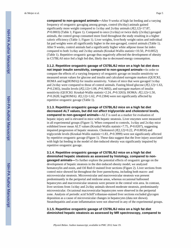
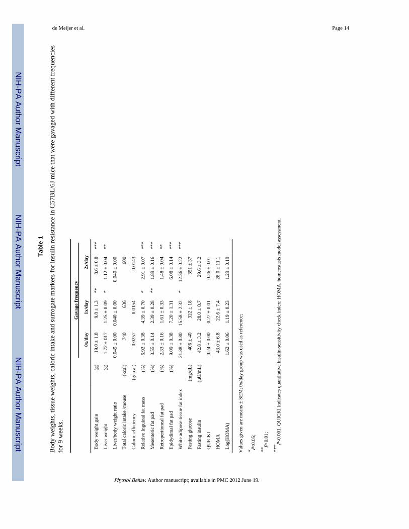
compared to non-gavaged animals—After 9 weeks of high fat feeding and a varyingfrequency of orogastric gavaging among groups, control (0x/day) animals gainedsignificantly more weight compared to 1x/day and 2x/day animals (F(2,12)=17.87,P=0.0003) (Table 1; Figure 1). Compared to once (1x/day) or twice daily (2x/day) gavagedanimals, the control group consumed more food throughout the study resulting in a highercaloric efficiency (Table 1; Figure 1). Liver weights, liver/body weight ratios and individualfat pad weights were all significantly higher in the non-gavaged, control animals (Table 1).After 9 weeks, control animals had a significantly higher white adipose tissue fat indexcompared to both 1x/day and 2x/day animals (Kruskal-Wallis statistic=10.50, P=0.0052)(Table 1). Repetitive orogastric gavage thus negatively affected the development of obesityin C57BL/6J mice fed a high fed diet, likely due to decreased energy consumption.
3.1.2. Repetitive orogastric gavage of C57BL/6J mice on a high fat diet doesnot impair insulin sensitivity, compared to non-gavaged animals—In order tocompare the effects of a varying frequency of orogastric gavage on insulin sensitivity wemeasured serum values for glucose and insulin and calculated surrogate markers (QUICKI,HOMA and log(HOMA)) for insulin sensitivity. Values of mice that were gavaged 1x/dayand 2x/day were compared to those of control animals. Fasting blood glucose (F(2,12)=1.63,P=0.2365), insulin levels (F(2,12)=1.08, P=0.3692), and surrogate markers of insulinsensitivity (QUICKI: Kruskal-Wallis statistic=2.24, P=0.3263); HOMA: F(2,12)=1.50,P=0.2628; log(HOMA): F(2,12)=1.62, P=0.2384) were not significantly affected byrepetitive orogastric gavage (Table 1).
3.1.3. Repetitive orogastric gavage of C57BL/6J mice on a high fat dietdecreased ALT values, but did not affect triglyceride and cholesterol levels,compared to non-gavaged animals—ALT is used as a marker for evaluation ofhepatic injury and is elevated in mice with hepatic steatosis. Liver enzymes were measuredin all experimental groups (Figure 1). When compared to controls, 1x/day and 2x/day miceexhibited lower mean ALT values (Kruskal-Wallis statistic=7.75, P=0.0207), suggestingimpaired progression of hepatic steatosis. Cholesterol (F(2,12)=0.22, P=0.8050) andtriglyceride levels (Kruskal-Wallis statistic=1.83, P=0.3999) were not significantly affectedby repetitive orogastric gavage (Figure 1). These data suggest that the liver injury associatedwith high fat feeding in the model of diet-induced obesity was significantly impaired byrepetitive orogastric gavage.
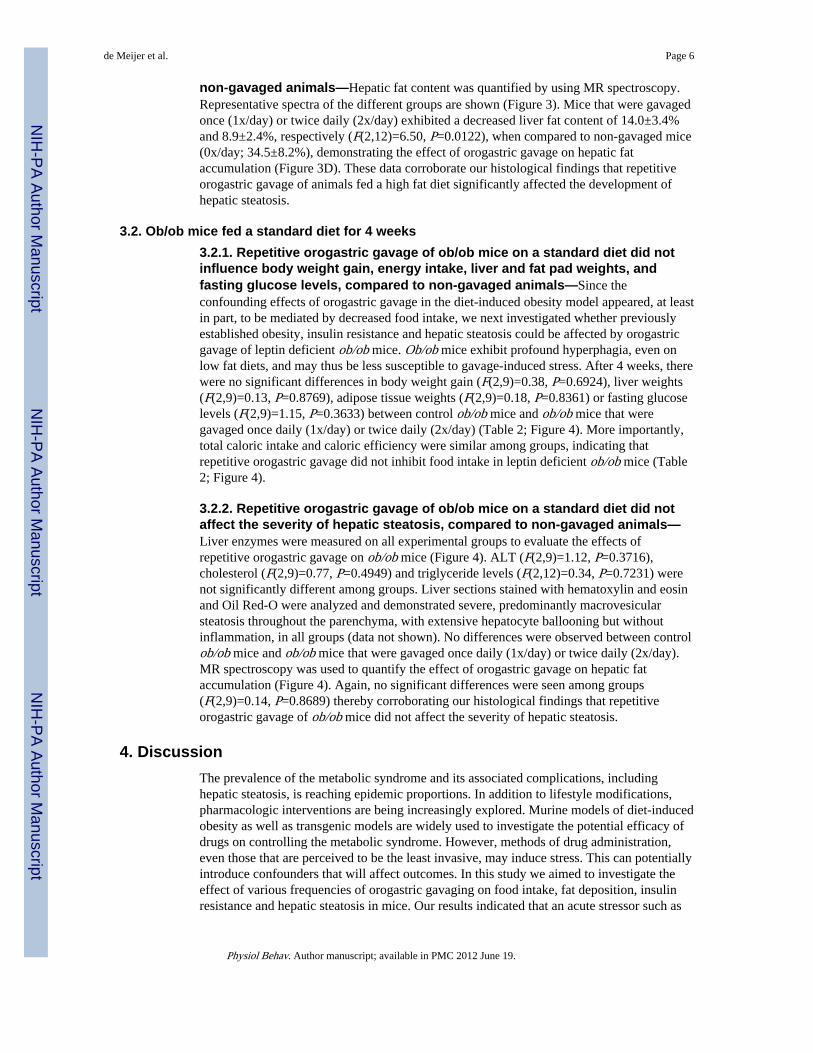
3.1.4. Repetitive orogastric gavage of C57BL/6J mice on a high fat dietdiminished hepatic steatosis as assessed by histology, compared to non-gavaged animals—To further explore the potential effects of orogastric gavage on thedevelopment of hepatic steatosis in the diet-induced obesity model, we analyzedhematoxylin and eosin, and Oil Red-O stained liver sections (Figure 2). Liver sections fromcontrol mice showed fat throughout the liver parenchyma, including both macro- andmicrovesicular steatosis. Microvesicular and macrovesicular steatosis was presentpredominantly in the periportal and midzone areas, whereas occasional balloonedhepatocytes and macrovesicular steatosis were present in the central vein area. In contrast,liver sections from 1x/day and 2x/day animals showed moderate steatosis, predominantlymicrovesicular. Occasional macrovesicular hepatocytes were observed in the periportalzone. Analysis of periodic acid Schiff’s/diastase-stained liver sections excluded glycogendeposition as a cause of microvesicular changes in hepatocytes (data not shown).Steatohepatitis and acute inflammation were not observed in any of the experimental groups.
3.1.5. Repetitive orogastric gavage of C57BL/6J mice on a high fat dietdiminished hepatic steatosis as assessed by MR spectroscopy, compared to
de Meijer et al. Page 5
Physiol Behav. Author manuscript; available in PMC 2012 June 19.
NIH
-PA Author Manuscript
NIH
-PA Author Manuscript
NIH
-PA Author Manuscript
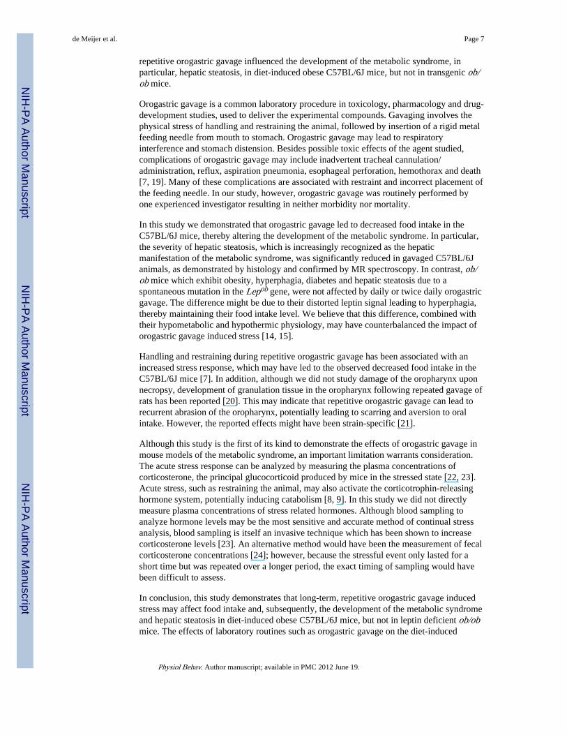
non-gavaged animals—Hepatic fat content was quantified by using MR spectroscopy.Representative spectra of the different groups are shown (Figure 3). Mice that were gavagedonce (1x/day) or twice daily (2x/day) exhibited a decreased liver fat content of 14.0±3.4%and 8.9±2.4%, respectively (F(2,12)=6.50, P=0.0122), when compared to non-gavaged mice(0x/day; 34.5±8.2%), demonstrating the effect of orogastric gavage on hepatic fataccumulation (Figure 3D). These data corroborate our histological findings that repetitiveorogastric gavage of animals fed a high fat diet significantly affected the development ofhepatic steatosis.
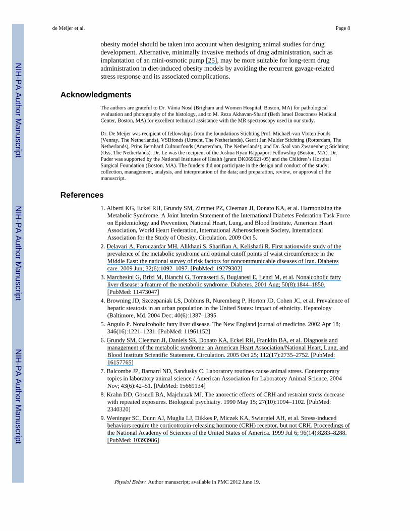
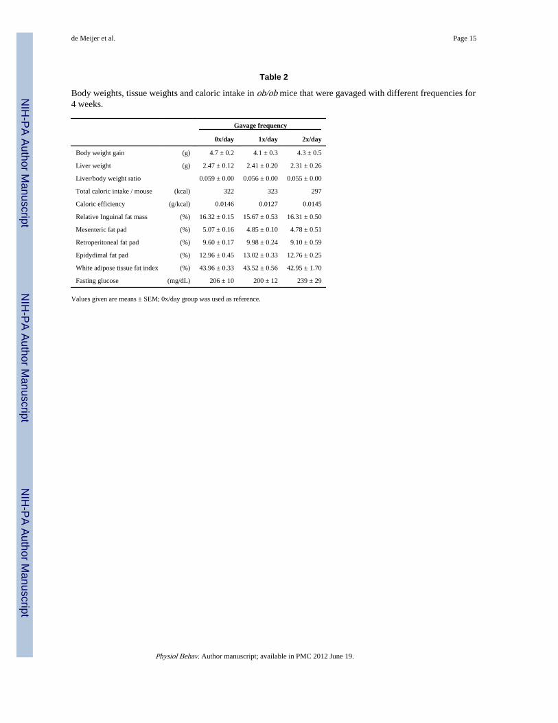
3.2. Ob/ob mice fed a standard diet for 4 weeks3.2.1. Repetitive orogastric gavage of ob/ob mice on a standard diet did notinfluence body weight gain, energy intake, liver and fat pad weights, andfasting glucose levels, compared to non-gavaged animals—Since theconfounding effects of orogastric gavage in the diet-induced obesity model appeared, at leastin part, to be mediated by decreased food intake, we next investigated whether previouslyestablished obesity, insulin resistance and hepatic steatosis could be affected by orogastricgavage of leptin deficient ob/ob mice. Ob/ob mice exhibit profound hyperphagia, even onlow fat diets, and may thus be less susceptible to gavage-induced stress. After 4 weeks, therewere no significant differences in body weight gain (F(2,9)=0.38, P=0.6924), liver weights(F(2,9)=0.13, P=0.8769), adipose tissue weights (F(2,9)=0.18, P=0.8361) or fasting glucoselevels (F(2,9)=1.15, P=0.3633) between control ob/ob mice and ob/ob mice that weregavaged once daily (1x/day) or twice daily (2x/day) (Table 2; Figure 4). More importantly,total caloric intake and caloric efficiency were similar among groups, indicating thatrepetitive orogastric gavage did not inhibit food intake in leptin deficient ob/ob mice (Table2; Figure 4).
3.2.2. Repetitive orogastric gavage of ob/ob mice on a standard diet did notaffect the severity of hepatic steatosis, compared to non-gavaged animals—Liver enzymes were measured on all experimental groups to evaluate the effects ofrepetitive orogastric gavage on ob/ob mice (Figure 4). ALT (F(2,9)=1.12, P=0.3716),cholesterol (F(2,9)=0.77, P=0.4949) and triglyceride levels (F(2,12)=0.34, P=0.7231) werenot significantly different among groups. Liver sections stained with hematoxylin and eosinand Oil Red-O were analyzed and demonstrated severe, predominantly macrovesicularsteatosis throughout the parenchyma, with extensive hepatocyte ballooning but withoutinflammation, in all groups (data not shown). No differences were observed between controlob/ob mice and ob/ob mice that were gavaged once daily (1x/day) or twice daily (2x/day).MR spectroscopy was used to quantify the effect of orogastric gavage on hepatic fataccumulation (Figure 4). Again, no significant differences were seen among groups(F(2,9)=0.14, P=0.8689) thereby corroborating our histological findings that repetitiveorogastric gavage of ob/ob mice did not affect the severity of hepatic steatosis.
4. DiscussionThe prevalence of the metabolic syndrome and its associated complications, includinghepatic steatosis, is reaching epidemic proportions. In addition to lifestyle modifications,pharmacologic interventions are being increasingly explored. Murine models of diet-inducedobesity as well as transgenic models are widely used to investigate the potential efficacy ofdrugs on controlling the metabolic syndrome. However, methods of drug administration,even those that are perceived to be the least invasive, may induce stress. This can potentiallyintroduce confounders that will affect outcomes. In this study we aimed to investigate theeffect of various frequencies of orogastric gavaging on food intake, fat deposition, insulinresistance and hepatic steatosis in mice. Our results indicated that an acute stressor such as
de Meijer et al. Page 6
Physiol Behav. Author manuscript; available in PMC 2012 June 19.
NIH
-PA Author Manuscript
NIH
-PA Author Manuscript
NIH
-PA Author Manuscript
repetitive orogastric gavage influenced the development of the metabolic syndrome, inparticular, hepatic steatosis, in diet-induced obese C57BL/6J mice, but not in transgenic ob/ob mice.
Orogastric gavage is a common laboratory procedure in toxicology, pharmacology and drug-development studies, used to deliver the experimental compounds. Gavaging involves thephysical stress of handling and restraining the animal, followed by insertion of a rigid metalfeeding needle from mouth to stomach. Orogastric gavage may lead to respiratoryinterference and stomach distension. Besides possible toxic effects of the agent studied,complications of orogastric gavage may include inadvertent tracheal cannulation/administration, reflux, aspiration pneumonia, esophageal perforation, hemothorax and death[7, 19]. Many of these complications are associated with restraint and incorrect placement ofthe feeding needle. In our study, however, orogastric gavage was routinely performed byone experienced investigator resulting in neither morbidity nor mortality.
In this study we demonstrated that orogastric gavage led to decreased food intake in theC57BL/6J mice, thereby altering the development of the metabolic syndrome. In particular,the severity of hepatic steatosis, which is increasingly recognized as the hepaticmanifestation of the metabolic syndrome, was significantly reduced in gavaged C57BL/6Janimals, as demonstrated by histology and confirmed by MR spectroscopy. In contrast, ob/ob mice which exhibit obesity, hyperphagia, diabetes and hepatic steatosis due to aspontaneous mutation in the Lepob gene, were not affected by daily or twice daily orogastricgavage. The difference might be due to their distorted leptin signal leading to hyperphagia,thereby maintaining their food intake level. We believe that this difference, combined withtheir hypometabolic and hypothermic physiology, may have counterbalanced the impact oforogastric gavage induced stress [14, 15].
Handling and restraining during repetitive orogastric gavage has been associated with anincreased stress response, which may have led to the observed decreased food intake in theC57BL/6J mice [7]. In addition, although we did not study damage of the oropharynx uponnecropsy, development of granulation tissue in the oropharynx following repeated gavage ofrats has been reported [20]. This may indicate that repetitive orogastric gavage can lead torecurrent abrasion of the oropharynx, potentially leading to scarring and aversion to oralintake. However, the reported effects might have been strain-specific [21].
Although this study is the first of its kind to demonstrate the effects of orogastric gavage inmouse models of the metabolic syndrome, an important limitation warrants consideration.The acute stress response can be analyzed by measuring the plasma concentrations ofcorticosterone, the principal glucocorticoid produced by mice in the stressed state [22, 23].Acute stress, such as restraining the animal, may also activate the corticotrophin-releasinghormone system, potentially inducing catabolism [8, 9]. In this study we did not directlymeasure plasma concentrations of stress related hormones. Although blood sampling toanalyze hormone levels may be the most sensitive and accurate method of continual stressanalysis, blood sampling is itself an invasive technique which has been shown to increasecorticosterone levels [23]. An alternative method would have been the measurement of fecalcorticosterone concentrations [24]; however, because the stressful event only lasted for ashort time but was repeated over a longer period, the exact timing of sampling would havebeen difficult to assess.
In conclusion, this study demonstrates that long-term, repetitive orogastric gavage inducedstress may affect food intake and, subsequently, the development of the metabolic syndromeand hepatic steatosis in diet-induced obese C57BL/6J mice, but not in leptin deficient ob/obmice. The effects of laboratory routines such as orogastric gavage on the diet-induced
de Meijer et al. Page 7
Physiol Behav. Author manuscript; available in PMC 2012 June 19.
NIH
-PA Author Manuscript
NIH
-PA Author Manuscript
NIH
-PA Author Manuscript
obesity model should be taken into account when designing animal studies for drugdevelopment. Alternative, minimally invasive methods of drug administration, such asimplantation of an mini-osmotic pump [25], may be more suitable for long-term drugadministration in diet-induced obesity models by avoiding the recurrent gavage-relatedstress response and its associated complications.
AcknowledgmentsThe authors are grateful to Dr. Vânia Nosé (Brigham and Women Hospital, Boston, MA) for pathologicalevaluation and photography of the histology, and to M. Reza Akhavan-Sharif (Beth Israel Deaconess MedicalCenter, Boston, MA) for excellent technical assistance with the MR spectroscopy used in our study.
Dr. De Meijer was recipient of fellowships from the foundations Stichting Prof. Michaël-van Vloten Fonds(Venray, The Netherlands), VSBfonds (Utrecht, The Netherlands), Gerrit Jan Mulder Stichting (Rotterdam, TheNetherlands), Prins Bernhard Cultuurfonds (Amsterdam, The Netherlands), and Dr. Saal van Zwanenberg Stichting(Oss, The Netherlands). Dr. Le was the recipient of the Joshua Ryan Rappaport Fellowship (Boston, MA). Dr.Puder was supported by the National Institutes of Health (grant DK069621-05) and the Children’s HospitalSurgical Foundation (Boston, MA). The funders did not participate in the design and conduct of the study;collection, management, analysis, and interpretation of the data; and preparation, review, or approval of themanuscript.
References1. Alberti KG, Eckel RH, Grundy SM, Zimmet PZ, Cleeman JI, Donato KA, et al. Harmonizing the
Metabolic Syndrome. A Joint Interim Statement of the International Diabetes Federation Task Forceon Epidemiology and Prevention, National Heart, Lung, and Blood Institute, American HeartAssociation, World Heart Federation, International Atherosclerosis Society, InternationalAssociation for the Study of Obesity. Circulation. 2009 Oct 5.
2. Delavari A, Forouzanfar MH, Alikhani S, Sharifian A, Kelishadi R. First nationwide study of theprevalence of the metabolic syndrome and optimal cutoff points of waist circumference in theMiddle East: the national survey of risk factors for noncommunicable diseases of Iran. Diabetescare. 2009 Jun; 32(6):1092–1097. [PubMed: 19279302]
3. Marchesini G, Brizi M, Bianchi G, Tomassetti S, Bugianesi E, Lenzi M, et al. Nonalcoholic fattyliver disease: a feature of the metabolic syndrome. Diabetes. 2001 Aug; 50(8):1844–1850.[PubMed: 11473047]
4. Browning JD, Szczepaniak LS, Dobbins R, Nuremberg P, Horton JD, Cohen JC, et al. Prevalence ofhepatic steatosis in an urban population in the United States: impact of ethnicity. Hepatology(Baltimore, Md. 2004 Dec; 40(6):1387–1395.
5. Angulo P. Nonalcoholic fatty liver disease. The New England journal of medicine. 2002 Apr 18;346(16):1221–1231. [PubMed: 11961152]
6. Grundy SM, Cleeman JI, Daniels SR, Donato KA, Eckel RH, Franklin BA, et al. Diagnosis andmanagement of the metabolic syndrome: an American Heart Association/National Heart, Lung, andBlood Institute Scientific Statement. Circulation. 2005 Oct 25; 112(17):2735–2752. [PubMed:16157765]
7. Balcombe JP, Barnard ND, Sandusky C. Laboratory routines cause animal stress. Contemporarytopics in laboratory animal science / American Association for Laboratory Animal Science. 2004Nov; 43(6):42–51. [PubMed: 15669134]
8. Krahn DD, Gosnell BA, Majchrzak MJ. The anorectic effects of CRH and restraint stress decreasewith repeated exposures. Biological psychiatry. 1990 May 15; 27(10):1094–1102. [PubMed:2340320]
9. Weninger SC, Dunn AJ, Muglia LJ, Dikkes P, Miczek KA, Swiergiel AH, et al. Stress-inducedbehaviors require the corticotropin-releasing hormone (CRH) receptor, but not CRH. Proceedings ofthe National Academy of Sciences of the United States of America. 1999 Jul 6; 96(14):8283–8288.[PubMed: 10393986]
de Meijer et al. Page 8
Physiol Behav. Author manuscript; available in PMC 2012 June 19.
NIH
-PA Author Manuscript
NIH
-PA Author Manuscript
NIH
-PA Author Manuscript
10. Michel C, Duclos M, Cabanac M, Richard D. Chronic stress reduces body fat content in bothobesity-prone and obesity-resistant strains of mice. Hormones and behavior. 2005 Aug; 48(2):172–179. [PubMed: 15894318]
11. Tamashiro KL, Hegeman MA, Nguyen MM, Melhorn SJ, Ma LY, Woods SC, et al. Dynamic bodyweight and body composition changes in response to subordination stress. Physiology & behavior.2007 Jul 24; 91(4):440–448. [PubMed: 17512562]
12. Bartolomucci A, Cabassi A, Govoni P, Ceresini G, Cero C, Berra D, et al. Metabolic consequencesand vulnerability to diet-induced obesity in male mice under chronic social stress. PloS one. 2009;4(1):e4331. [PubMed: 19180229]
13. Collins S, Martin TL, Surwit RS, Robidoux J. Genetic vulnerability to diet-induced obesity in theC57BL/6J mouse: physiological and molecular characteristics. Physiology & behavior. 2004 Apr;81(2):243–248. [PubMed: 15159170]
14. Pelleymounter MA, Cullen MJ, Baker MB, Hecht R, Winters D, Boone T, et al. Effects of theobese gene product on body weight regulation in ob/ob mice. Science (New York, NY. 1995 Jul28; 269(5223):540–543.
15. Halaas JL, Gajiwala KS, Maffei M, Cohen SL, Chait BT, Rabinowitz D, et al. Weight-reducingeffects of the plasma protein encoded by the obese gene. Science (New York, NY. 1995 Jul 28;269(5223):543–546.
16. Parekh PI, Petro AE, Tiller JM, Feinglos MN, Surwit RS. Reversal of diet-induced obesity anddiabetes in C57BL/6J mice. Metabolism: clinical and experimental. 1998 Sep; 47(9):1089–1096.[PubMed: 9751238]
17. Muniyappa R, Lee S, Chen H, Quon MJ. Current approaches for assessing insulin sensitivity andresistance in vivo: advantages, limitations, and appropriate usage. American journal of physiology.2008 Jan; 294(1):E15–E26. [PubMed: 17957034]
18. Alwayn IP, Andersson C, Lee S, Arsenault DA, Bistrian BR, Gura KM, et al. Inhibition of matrixmetalloproteinases increases PPAR-alpha and IL-6 and prevents dietary-induced hepatic steatosisand injury in a murine model. Am J Physiol Gastrointest Liver Physiol. 2006 Dec; 291(6):G1011–G1019. [PubMed: 16844679]
19. Brown AP, Dinger N, Levine BS. Stress produced by gavage administration in the rat.Contemporary topics in laboratory animal science / American Association for Laboratory AnimalScience. 2000 Jan; 39(1):17–21. [PubMed: 11178310]
20. Germann PG, Ockert D. Granulomatous inflammation of the oropharyngeal cavity as a possiblecause for unexpected high mortality in a Fischer 344 rat carcinogenicity study. Laboratory animalscience. 1994 Aug; 44(4):338–343. [PubMed: 7983845]
21. Germann PG, Ockert D, Heinrichs M. Pathology of the oropharyngeal cavity in six strains of rats:predisposition of Fischer 344 rats for inflammatory and degenerative changes. Toxicologicpathology. 1998 Mar-Apr;26(2):283–289. [PubMed: 9547869]
22. Hunt C, Hambly C. Faecal corticosterone concentrations indicate that separately housed male miceare not more stressed than group housed males. Physiology & behavior. 2006 Mar 30; 87(3):519–526. [PubMed: 16442135]
23. Mostl E, Palme R. Hormones as indicators of stress. Domestic animal endocrinology. 2002 Jul;23(1–2):67–74. [PubMed: 12142227]
24. Good T, Khan MZ, Lynch JW. Biochemical and physiological validation of a corticosteroidradioimmunoassay for plasma and fecal samples in oldfield mice (Peromyscus polionotus).Physiology & behavior. 2003 Nov; 80(2–3):405–411. [PubMed: 14637242]
25. Rowland RR, Reyes E, Chukwuocha R, Tokuda S. Corticosteroid and immune responses of micefollowing mini-osmotic pump implantation. Immunopharmacology. 1990 Nov-Dec;20(3):187–190. [PubMed: 2289873]
de Meijer et al. Page 9
Physiol Behav. Author manuscript; available in PMC 2012 June 19.
NIH
-PA Author Manuscript
NIH
-PA Author Manuscript
NIH
-PA Author Manuscript
Figure 1.Absolute mean caloric intake per C57BL/6J mouse per week (A) was calculated per groupon a daily base. Body weight gain (B) was calculated relative to the weight of eachindividual animal before initiation of the experiment. Plasma alanine aminotransferase(ALT; C), total cholesterol (D) and triglyceride (E) levels in the different groups. Valuesrepresent the mean ± SEM. Statistical significance is calculated between the control animals(0x/day) and the difference between 1x/day animals and 2x/day animals (*, P<0.05).
de Meijer et al. Page 10
Physiol Behav. Author manuscript; available in PMC 2012 June 19.
NIH
-PA Author Manuscript
NIH
-PA Author Manuscript
NIH
-PA Author Manuscript
Figure 2.Representative liver sections stained with hematoxylin and eosin (H&E; top panels; originalmagnification 400x), and Oil Red O (lower panels; original magnification 200x). Liversfrom C57BL/6J animals that had not been gavaged revealed extensive, microvesicular andmacrovesicular steatosis after 9 weeks (left panels). Livers from animals that had beengavaged once (middle panels), or twice daily (right panels), exhibited moderate,predominantly microvesicular steatosis. P indicates portal tract; CV, central vein.
de Meijer et al. Page 11
Physiol Behav. Author manuscript; available in PMC 2012 June 19.
NIH
-PA Author Manuscript
NIH
-PA Author Manuscript
NIH
-PA Author Manuscript
Figure 3.Magnetic resonance spectra for 0x/day (A), 1x/day (B) and 2x/day (C) livers from C57BL/6J mice. Percent fat content was determined relative to water (100%) by numericalintegration of the areas under the lipid and water peaks. Mean hepatic fat fraction asmeasured by magnetic resonance spectroscopy (D). Statistical significance is calculatedbetween the 0x/day animals and the difference between 1x/day animals and 2x/day animals,respectively (*, P<0.05). Variance statistic is SEM.
de Meijer et al. Page 12
Physiol Behav. Author manuscript; available in PMC 2012 June 19.
NIH
-PA Author Manuscript
NIH
-PA Author Manuscript
NIH
-PA Author Manuscript
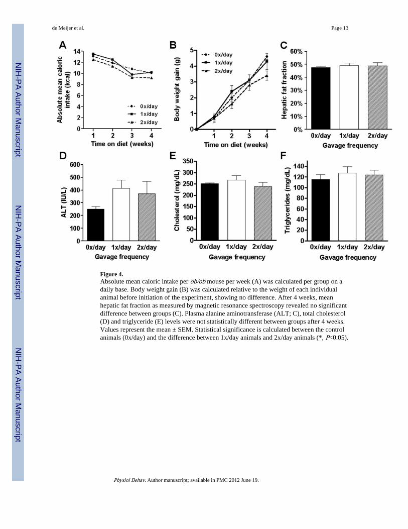
Figure 4.Absolute mean caloric intake per ob/ob mouse per week (A) was calculated per group on adaily base. Body weight gain (B) was calculated relative to the weight of each individualanimal before initiation of the experiment, showing no difference. After 4 weeks, meanhepatic fat fraction as measured by magnetic resonance spectroscopy revealed no significantdifference between groups (C). Plasma alanine aminotransferase (ALT; C), total cholesterol(D) and triglyceride (E) levels were not statistically different between groups after 4 weeks.Values represent the mean ± SEM. Statistical significance is calculated between the controlanimals (0x/day) and the difference between 1x/day animals and 2x/day animals (*, P<0.05).
de Meijer et al. Page 13
Physiol Behav. Author manuscript; available in PMC 2012 June 19.
NIH
-PA Author Manuscript
NIH
-PA Author Manuscript
NIH
-PA Author Manuscript
NIH
-PA Author Manuscript
NIH
-PA Author Manuscript
NIH
-PA Author Manuscript
de Meijer et al. Page 14
Tabl
e 1
Bod
y w
eigh
ts, t
issu
e w
eigh
ts, c
alor
ic in
take
and
sur
roga
te m
arke
rs f
or in
sulin
res
ista
nce
in C
57B
L/6
J m
ice
that
wer
e ga
vage
d w
ith d
iffe
rent
fre
quen
cies
for
9 w
eeks
.
Gav
age
freq
uenc
y
0x/d
ay1x
/day
2x/d
ay
Bod
y w
eigh
t gai
n(g
)19
.0 ±
1.8
9.8
± 1
.3**
8.6
± 0
.8**
*
Liv
er w
eigh
t(g
)1.
72 ±
017
1.25
± 0
.09
*1.
12 ±
0.0
4**
Liv
er/b
ody
wei
ght r
atio
0.04
5 ±
0.0
00.
040
± 0
.00
0.04
0 ±
0.0
0
Tot
al c
alor
ic in
take
/mou
se(k
cal)
740
636
600
Cal
oric
eff
icie
ncy
(g/k
cal)
0.02
570.
0154
0.01
43
Rel
ativ
e In
guin
al f
at m
ass
(%)
6.92
± 0
.38
4.39
± 0
.70
*2.
91 ±
0.0
7**
*
Mes
ente
ric
fat p
ad(%
)3.
55 ±
0.1
42.
39 ±
0.2
8**
1.89
± 0
.16
***
Ret
rope
rito
neal
fat
pad
(%)
2.33
± 0
.16
1.61
± 0
.33
1.48
± 0
.04
**
Epi
dydi
mal
fat
pad
(%)
9.09
± 0
.38
7.20
± 1
.31
6.08
± 0
.14
***
Whi
te a
dipo
se ti
ssue
fat
inde
x21
.88
± 0
.80
15.5
8 ±
2.3
2*
12.3
6 ±
0.2
2**
*
Fast
ing
gluc
ose
(mg/
dL)
406
± 4
032
2 ±
18
351
± 3
7
Fast
ing
insu
lin(µ
U/m
L)
42.8
± 3
.228
.0 ±
8.7
29.6
± 3
.2
QU
ICK
I0.
24 ±
0.0
00.
27 ±
0.0
10.
26 ±
0.0
1
HO
MA
43.0
± 6
.822
.6 ±
7.4
28.0
± 1
1.1
Log
(HO
MA
)1.
62 ±
0.0
61.
19 ±
0.2
31.
29 ±
0.1
9
Val
ues
give
n ar
e m
eans
± S
EM
; 0x/
day
grou
p w
as u
sed
as r
efer
ence
;
* P<0.
05;
**P<
0.01
;
*** P<
0.00
1. Q
UIC
KI
indi
cate
s qu
antit
ativ
e in
sulin
-sen
sitiv
ity c
heck
inde
x; H
OM
A, h
omeo
stas
is m
odel
ass
essm
ent.
Physiol Behav. Author manuscript; available in PMC 2012 June 19.
NIH
-PA Author Manuscript
NIH
-PA Author Manuscript
NIH
-PA Author Manuscript
de Meijer et al. Page 15
Table 2
Body weights, tissue weights and caloric intake in ob/ob mice that were gavaged with different frequencies for4 weeks.
Gavage frequency
0x/day 1x/day 2x/day
Body weight gain (g) 4.7 ± 0.2 4.1 ± 0.3 4.3 ± 0.5
Liver weight (g) 2.47 ± 0.12 2.41 ± 0.20 2.31 ± 0.26
Liver/body weight ratio 0.059 ± 0.00 0.056 ± 0.00 0.055 ± 0.00
Total caloric intake / mouse (kcal) 322 323 297
Caloric efficiency (g/kcal) 0.0146 0.0127 0.0145
Relative Inguinal fat mass (%) 16.32 ± 0.15 15.67 ± 0.53 16.31 ± 0.50
Mesenteric fat pad (%) 5.07 ± 0.16 4.85 ± 0.10 4.78 ± 0.51
Retroperitoneal fat pad (%) 9.60 ± 0.17 9.98 ± 0.24 9.10 ± 0.59
Epidydimal fat pad (%) 12.96 ± 0.45 13.02 ± 0.33 12.76 ± 0.25
White adipose tissue fat index (%) 43.96 ± 0.33 43.52 ± 0.56 42.95 ± 1.70
Fasting glucose (mg/dL) 206 ± 10 200 ± 12 239 ± 29
Values given are means ± SEM; 0x/day group was used as reference.
Physiol Behav. Author manuscript; available in PMC 2012 June 19.
Related Documents


