BASIC RESEARCH www.jasn.org Renal CD133 + /CD73 + Progenitors Produce Erythropoietin under Hypoxia and Prolyl Hydroxylase Inhibition Benedetta Bussolati,* Carola Lauritano,* Aldo Moggio,* Federica Collino, † Massimiliano Mazzone, ‡§ and Giovanni Camussi † *Department of Molecular Biotechnology and Health Sciences, Molecular Biotechnology Center, † Department of Medical Sciences, University of Torino, Torino, Italy; ‡ Laboratory of Molecular Oncology and Angiogenesis, Vesalius Research Center, VIB, Leuven, Belgium; and § Laboratory of Molecular Oncology and Angiogenesis, Vesalius Research Center, Department of Oncology, KU Leuven, Leuven, Belgium ABSTRACT The identity of the peritubular population of cells with mesenchymal phenotype thought responsible for producing erythropoietin in humans remains unclear. Here, renal CD133 + /CD73 + progenitor cells, isolated from the human renal inner medulla and described as a population of mesenchymal progenitors, released erythropoietin under hypoxic conditions. CD133 2 cells did not synthesize erythropoietin, and CD133 + progenitor cells stopped producing erythropoietin when they differentiated and acquired an epithelial phenotype. Inhibition of prolyl hydroxylases, using either dimethyloxalylglycine or a small hairpin RNA against prolyl hydroxylase-2, increased both hypoxia-inducible factor-2a (HIF-2a) expression and eryth- ropoietin transcription. Moreover, under hypoxic conditions, inhibition of prolyl hydroxylase significantly increased erythropoietin release by CD133 + progenitors. Finally, blockade of HIF-2a impaired erythro- poietin synthesis by CD133 + progenitors. Taken together, these results suggest that it is the renal CD133 + progenitor cells that synthesize and release erythropoietin under hypoxia, via the prolyl hydroxylase-HIF- 2a axis, in the human kidney. In addition, this study provides rationale for the therapeutic use of prolyl hydroxylase inhibitors in the setting of acute or chronic renal injury. J Am Soc Nephrol 24: 1234–1241, 2013. doi: 10.1681/ASN.2012080772 The glycoprotein hormone erythropoietin (EPO) regulates blood red cell production, linking de- creased tissue oxygenation to an adequate erythro- poietic response. In adults, the kidney is responsible for .90% of EPO production. Many efforts have been made to identify renal EPO-producing cells. In the rodent anemic kidney, EPO production is restricted to interstitial peritubular fibroblast-like cells localized in the deep cortex and outer medulla and coexpressing EPO mRNA and the mesenchy- mal marker CD73. 1–6 In the human kidney, the precise localization of EPO-producing cells is un- known. It is conceivable that, similarly to rodents, a peritubular population of mesenchymal cells/fibro- blasts is responsible for EPO production. Data obtained from a human EPO-producing cell line isolated from human kidney showed that these cells possess mesenchymal characteristics and the ability to synthesize EPO in response to hypoxia-depen- dent hypoxia-inducible factor-2a (HIF-2a) stabili- zation and activation. 7 However, in human renal tissue, in situ hybridization studies showed EPO production in cells of renal tubules. In particular, EPO mRNA was expressed by epithelial distal tu- bular cells, collecting tubules and additionally by glomerular cells. 8 High EPO levels are also released Received August 3, 2012. Accepted March 13, 2013. Published online ahead of print. Publication date available at www.jasn.org. Correspondence: Dr. Benedetta Bussolati, Department of Mo- lecular Biotechnology and Health Sciences, Molecular Bio- technology Center, University of Torino, via Nizza 52, 10126 Torino, Italy. Email: [email protected]. Copyright © 2013 by the American Society of Nephrology 1234 ISSN : 1046-6673/2408-1234 J Am Soc Nephrol 24: 1234–1241, 2013

Welcome message from author
This document is posted to help you gain knowledge. Please leave a comment to let me know what you think about it! Share it to your friends and learn new things together.
Transcript
BASIC RESEARCH www.jasn.org
Renal CD133+/CD73+ Progenitors ProduceErythropoietin under Hypoxia and Prolyl HydroxylaseInhibition
Benedetta Bussolati,* Carola Lauritano,* Aldo Moggio,* Federica Collino,†
Massimiliano Mazzone,‡§ and Giovanni Camussi†
*Department of Molecular Biotechnology and Health Sciences, Molecular Biotechnology Center, †Department ofMedical Sciences, University of Torino, Torino, Italy; ‡Laboratory of Molecular Oncology and Angiogenesis, VesaliusResearch Center, VIB, Leuven, Belgium; and §Laboratory of Molecular Oncology and Angiogenesis, Vesalius ResearchCenter, Department of Oncology, KU Leuven, Leuven, Belgium
ABSTRACTThe identity of the peritubular population of cells with mesenchymal phenotype thought responsible forproducing erythropoietin in humans remains unclear. Here, renal CD133+/CD73+progenitor cells, isolatedfrom the human renal inner medulla and described as a population of mesenchymal progenitors, releasederythropoietin under hypoxic conditions. CD1332 cells did not synthesize erythropoietin, and CD133+
progenitor cells stopped producing erythropoietin when they differentiated and acquired an epithelialphenotype. Inhibition of prolyl hydroxylases, using either dimethyloxalylglycine or a small hairpin RNAagainst prolyl hydroxylase-2, increased both hypoxia-inducible factor-2a (HIF-2a) expression and eryth-ropoietin transcription. Moreover, under hypoxic conditions, inhibition of prolyl hydroxylase significantlyincreased erythropoietin release by CD133+ progenitors. Finally, blockade of HIF-2a impaired erythro-poietin synthesis by CD133+ progenitors. Taken together, these results suggest that it is the renal CD133+
progenitor cells that synthesize and release erythropoietin under hypoxia, via the prolyl hydroxylase-HIF-2a axis, in the human kidney. In addition, this study provides rationale for the therapeutic use of prolylhydroxylase inhibitors in the setting of acute or chronic renal injury.
J Am Soc Nephrol 24: 1234–1241, 2013. doi: 10.1681/ASN.2012080772
The glycoprotein hormone erythropoietin (EPO)regulates blood red cell production, linking de-creased tissue oxygenation to an adequate erythro-poietic response. In adults, the kidney is responsiblefor .90% of EPO production. Many efforts havebeen made to identify renal EPO-producing cells.In the rodent anemic kidney, EPO production isrestricted to interstitial peritubular fibroblast-likecells localized in the deep cortex and outer medullaand coexpressing EPO mRNA and the mesenchy-mal marker CD73.1–6 In the human kidney, theprecise localization of EPO-producing cells is un-known. It is conceivable that, similarly to rodents, aperitubular population of mesenchymal cells/fibro-blasts is responsible for EPO production. Dataobtained from a human EPO-producing cell lineisolated from human kidney showed that these cellspossess mesenchymal characteristics and the ability
to synthesize EPO in response to hypoxia-depen-dent hypoxia-inducible factor-2a (HIF-2a) stabili-zation and activation.7 However, in human renaltissue, in situ hybridization studies showed EPOproduction in cells of renal tubules. In particular,EPO mRNA was expressed by epithelial distal tu-bular cells, collecting tubules and additionally byglomerular cells.8 High EPO levels are also released
Received August 3, 2012. Accepted March 13, 2013.
Published online ahead of print. Publication date available atwww.jasn.org.
Correspondence: Dr. Benedetta Bussolati, Department of Mo-lecular Biotechnology and Health Sciences, Molecular Bio-technology Center, University of Torino, via Nizza 52, 10126Torino, Italy. Email: [email protected].
Copyright © 2013 by the American Society of Nephrology
1234 ISSN : 1046-6673/2408-1234 J Am Soc Nephrol 24: 1234–1241, 2013
by tumor cells of renal carcinomas,9 considered to derive fromtransformed tubular or progenitor/stem cells. In addition,murine embryonic renal stem cells organized in an organoidand implanted in vivo in rats produced murine EPO, suggest-ing that EPO-producing cells derive from renal stem cells.10
Using CD133 as a marker, a population of renal residentprogenitors has been localized in different segments of thenephron.11–14 In particular, CD133+ progenitors are enrichedin theHenle’s loop and thin segments of the papillary region ofmedulla, which is characterized by a very low oxygen ten-sion.15 In vitro, hypoxia was shown to be a key factor in themaintenance of the progenitor phenotype and stem propertiesof these cells.13
In this study, we investigated whether CD133+ renal pro-genitors could be a source of EPO within the kidney.
RESULTS
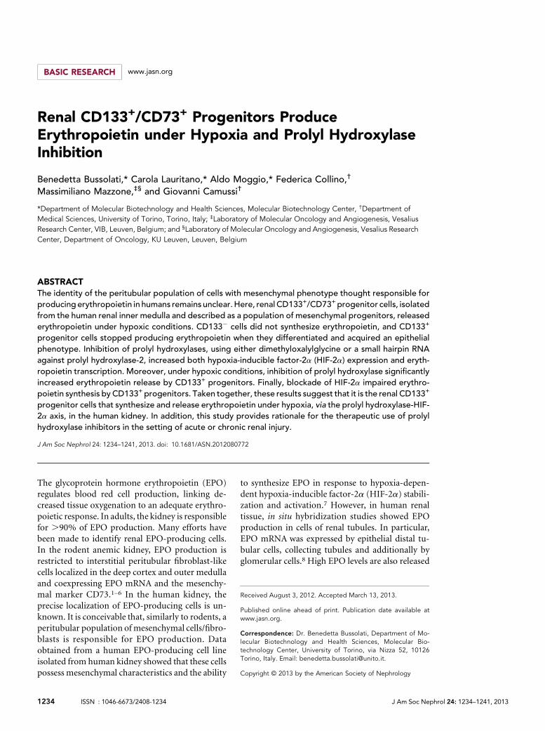
EPO Production by Renal CD133+ Cells in Normoxiaand HypoxiaCD133+ cells located within the Henle’s loop in the humaninner medulla coexpressed the hypoxic marker carbonic
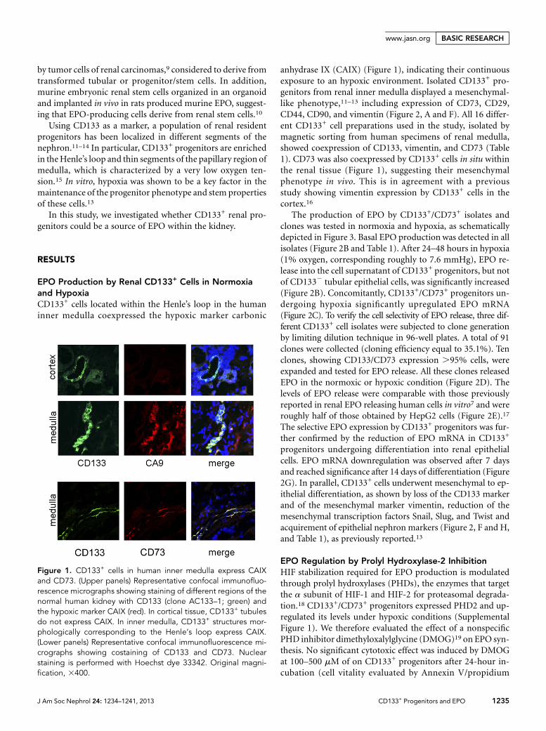
anhydrase IX (CAIX) (Figure 1), indicating their continuousexposure to an hypoxic environment. Isolated CD133+ pro-genitors from renal inner medulla displayed a mesenchymal-like phenotype,11–13 including expression of CD73, CD29,CD44, CD90, and vimentin (Figure 2, A and F). All 16 differ-ent CD133+ cell preparations used in the study, isolated bymagnetic sorting from human specimens of renal medulla,showed coexpression of CD133, vimentin, and CD73 (Table1). CD73 was also coexpressed by CD133+ cells in situ withinthe renal tissue (Figure 1), suggesting their mesenchymalphenotype in vivo. This is in agreement with a previousstudy showing vimentin expression by CD133+ cells in thecortex.16
The production of EPO by CD133+/CD73+ isolates andclones was tested in normoxia and hypoxia, as schematicallydepicted in Figure 3. Basal EPO production was detected in allisolates (Figure 2B and Table 1). After 24–48 hours in hypoxia(1% oxygen, corresponding roughly to 7.6 mmHg), EPO re-lease into the cell supernatant of CD133+ progenitors, but notof CD1332 tubular epithelial cells, was significantly increased(Figure 2B). Concomitantly, CD133+/CD73+ progenitors un-dergoing hypoxia significantly upregulated EPO mRNA(Figure 2C). To verify the cell selectivity of EPO release, three dif-ferent CD133+ cell isolates were subjected to clone generationby limiting dilution technique in 96-well plates. A total of 91clones were collected (cloning efficiency equal to 35.1%). Tenclones, showing CD133/CD73 expression .95% cells, wereexpanded and tested for EPO release. All these clones releasedEPO in the normoxic or hypoxic condition (Figure 2D). Thelevels of EPO release were comparable with those previouslyreported in renal EPO releasing human cells in vitro7 and wereroughly half of those obtained by HepG2 cells (Figure 2E).17
The selective EPO expression by CD133+ progenitors was fur-ther confirmed by the reduction of EPO mRNA in CD133+
progenitors undergoing differentiation into renal epithelialcells. EPO mRNA downregulation was observed after 7 daysand reached significance after 14 days of differentiation (Figure2G). In parallel, CD133+ cells underwent mesenchymal to ep-ithelial differentiation, as shown by loss of the CD133 markerand of the mesenchymal marker vimentin, reduction of themesenchymal transcription factors Snail, Slug, and Twist andacquirement of epithelial nephron markers (Figure 2, F and H,and Table 1), as previously reported.13
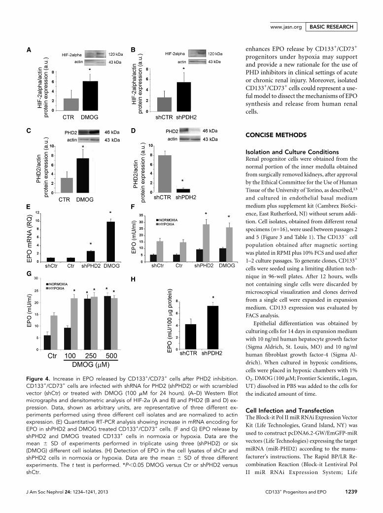
EPO Regulation by Prolyl Hydroxylase-2 InhibitionHIF stabilization required for EPO production is modulatedthrough prolyl hydroxylases (PHDs), the enzymes that targetthe a subunit of HIF-1 and HIF-2 for proteasomal degrada-tion.18 CD133+/CD73+ progenitors expressed PHD2 and up-regulated its levels under hypoxic conditions (SupplementalFigure 1). We therefore evaluated the effect of a nonspecificPHD inhibitor dimethyloxalylglycine (DMOG)19 on EPO syn-thesis. No significant cytotoxic effect was induced by DMOGat 100–500 mM of on CD133+ progenitors after 24-hour in-cubation (cell vitality evaluated by Annexin V/propidium
Figure 1. CD133+ cells in human inner medulla express CAIXand CD73. (Upper panels) Representative confocal immunofluo-rescence micrographs showing staining of different regions of thenormal human kidney with CD133 (clone AC133–1; green) andthe hypoxic marker CAIX (red). In cortical tissue, CD133+ tubulesdo not express CAIX. In inner medulla, CD133+ structures mor-phologically corresponding to the Henle’s loop express CAIX.(Lower panels) Representative confocal immunofluorescence mi-crographs showing costaining of CD133 and CD73. Nuclearstaining is performed with Hoechst dye 33342. Original magni-fication, 3400.
J Am Soc Nephrol 24: 1234–1241, 2013 CD133+ Progenitors and EPO 1235
www.jasn.org BASIC RESEARCH
Figure 2. EPO synthesis by renal CD133+/CD73+ cells under hypoxia and during differentiation. (A) FACS analysis showing expressionof CD133 and of mesenchymal markers CD73, CD44, and CD29, but not CD105 by isolated cells. Data are representative of all celllines in the study. (B) Increase in EPO release by CD133+ but not CD1332 cells after 24 or 48 hours of hypoxia. Data are mean 6 SD ofdifferent experiments performed in triplicate using six different cell isolates. (C) Quantitative RT-PCR analysis showing increase inmRNA encoding for EPO after 24 or 48 hours of hypoxia. Data are normalized to TBP mRNA and to 1 for time 0 and are the mean 6 SDof three different experiments performed using three different cell isolates. ANOVA with Dunnett’s comparison test was performed forB and C. *P,0.05 versus time 0. (D and E) EPO release by 10 different CD133+/CD73+ cell clones or by HepG2 cells in normoxia andafter 24 hours of hypoxia. EPO release is tested in triplicate by cells plated in 24-well plates (250,000 cells per well). A typical cyto-fluorimetric analysis of CD133/CD73 expression is also shown. (F) Representative cytofluorimetric histogram of CD133 expression andrepresentative micrographs of the immunofluorescence staining of cells at day 0 and 14 after differentiation. Nuclei are stained in bluewith Hoechst dye 33342. Data are representative of all cell lines in the study. (G and H) Quantitative RT-PCR analysis showing decreasein mRNAs encoding for EPO (E) and mesenchymal transcription factors (F) after 7 or 14 days of epithelial differentiation. Data arenormalized to TBP mRNA and to 1 for time 0 and are the mean 6 SD of three different experiments performed using three different cellisolates. ANOVA with Dunnett’s comparison test (G) or t test (H). *P,0.05. TBP, TATA binding protein. Original magnification, 3400 in F.
1236 Journal of the American Society of Nephrology J Am Soc Nephrol 24: 1234–1241, 2013
BASIC RESEARCH www.jasn.org
iodide staining using cytofluorimetric analysis, was.95%). Inaddition, we specifically inhibited PHD2, the PHD isoformknown to be involved in EPO synthesis,20 using two differentsmall hairpin RNA (shRNA). PHD inhibition with 100 mMDMOG induced HIF-2a protein increase in CD133+/CD73+
cells (Figure 4A). An increase inHIF-1a protein levels was alsoobserved (Supplemental Figure 2A). In parallel, PHD2 wasupregulated in CD133+/CD73+ cells treated with DMOG (Fig-ure 4C), as expected in virtue of the rapid induction of itssynthesis by HIFs.18 Indeed, DMOG does not affect thePHD protein levels because it acts as a competitive inhibi-tor.19 The specific PHD2 inhibition by lentiviral shRNA forPHD2 (shPHD2 cells) enhanced HIF-2a protein levels inCD133+ cells and downregulated PHD2 expression (Figure4, B and D). HIF-1a protein levels were also increased (Sup-plemental Figure 2B). In CD133+/CD73+ cells, PHD2 inhibi-tion, using either shPHD2 delivery with two differentlentiviral vectors or DMOG administration, induced anupregulation of EPO mRNA (Figure 4E and SupplementalFigure 3A). In particular, DMOG treatment upregulated theexpression of EPO mRNA .9-fold. The evaluation of EPOrelease into the cell supernatant, however, did not show a sig-nificant increase in normoxia (Figure 4F and SupplementalFigure 3B). At variance, PHD2 inhibition significantly en-hanced the EPO release at 24 hours when cells were submittedto hypoxia (1% oxygen) (Figure 4F and Supplemental Figure3B). In normoxia, increasing concentrations of the nonspecificPHD inhibitor DMOG were required to induce EPO release(Figure 4G), whereas its release in hypoxia was already at pla-teau at the lower DMOG concentration. Indeed, in shPHD2cells, EPO increased in normoxia at an intracellular level
(Figure 4H), indicating a possible hypoxia-based PHD2 inde-pendent control of EPO release.
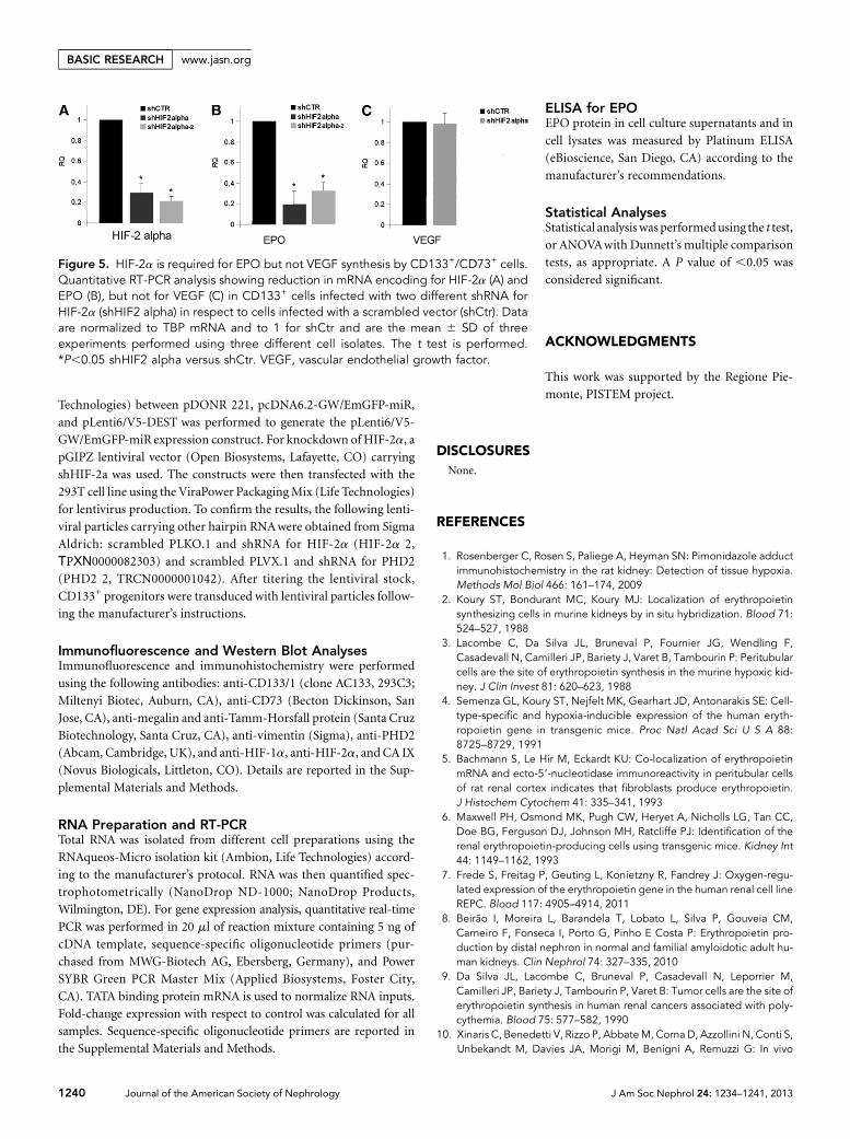
Role of HIF-2a in EPO ProductionEPOproduction is known tobemodulated byhypoxia throughHIF-2a activation.7,21,22 The involvement of HIF-2a in EPOproduction by CD133+/CD73+ progenitors was investigatedby generation of negative progenitors for HIF-2a (shHIF-2a)by infection with two different specific shRNA lentiviruses(Figure 5A). shHIF-2a cells almost completely lost the abilityto synthesize EPO (Figure 5B and Supplemental Figure 3, Aand B) but not vascular endothelial growth factor, used ascontrol for maintenance of cellular activity (Figure 5C).
DISCUSSION
Taken together, the results of this study identify CD133+/CD73+
renal progenitors as a possible source of EPO and show thatEPO release in hypoxic conditions can be enhanced by PHD2inhibition. CD133+/CD73+ progenitors therefore appear as anew source of EPO production within the human kidney.Similarly to EPO-producing renal cells described in murinestudies or isolated from human kidney, CD133+/CD73+ cellspresent a mesenchymal phenotype. The ability to synthesizeEPO was restricted to CD133+ progenitors because it was lostafter epithelial differentiation and it was absent in CD1332
cells. Indeed, in epithelial cells, the mechanisms underlyingthe repression of EPO gene expression were recently shown todepend on the GATA promoter motif,23 underlying a celltype– dependent inhibitory mechanism that could be relevant
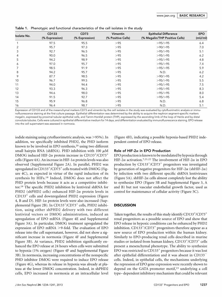
Table 1. Phenotypic and functional characteristics of the cell isolates in the study
Isolate No.CD133
(% Expression)CD73
(% Expression)Vimentin
(% Positive Cells)Epithelial Difference
(% Megalin/THP Positive Cells)EPO
(mU/ml)
1 97.5 95.5 .95 .95/.95 6.42 95.7 97.3 .95 .90/.95 7.03 92.7 96.5 .95 .95/.95 5.14 90.0 96.5 .95 .95/.95 6.25 94.2 98.9 .95 .95/.95 4.86 97.0 95.7 .95 .95/.95 7.47 98.5 91.9 .95 .95/.95 5.98 94.2 95.7 .95 N.D. 6.29 87.7 98.5 .95 .90/.95 4.210 96.7 99.5 .95 .95/.95 5.511 94.0 94.4 .95 .95/.95 7.512 93.3 96.3 .95 .95/.95 8.313 94.6 98.0 .95 .95/.95 8.014 91.5 92.7 .95 .95/.95 5.415 95.9 96.8 .95 N.D. 6.816 96.4 98.7 .95 N.D. 5.1
Expression of CD133 and of the mesenchymal markers CD73 and vimentin by the cell isolates in the study was evaluated by cytofluorimetric analysis or immu-nofluorescence staining at the first culture passage. Epithelial differentiation was determined by the ability to acquire the nephron segment-specific markersmegalin, expressed by proximal tubular epithelial cells, and Tamm-Horsfall protein (THP), expressed by the ascending limb of the loop of Henle and by distalconvolute tubules. Cells were cultured in epithelial differentiativemedium for 14 days, and differentiation evaluated by immunofluorescence staining. EPO releaseinto the cell supernatant was assessed in normoxia.
J Am Soc Nephrol 24: 1234–1241, 2013 CD133+ Progenitors and EPO 1237
www.jasn.org BASIC RESEARCH
in the epithelial differentiation of renal cells. In contrast withthe described localization of EPO-producing cells in the in-terstitium of rat and mouse kidney,1–6,21 CD133+ progenitorshave been identified along the renal nephron.11–14 This ob-servation overlaps the described localization of EPO-produc-ing cells in different segments of the human nephron by in situhybridization studies.8,9 These dissimilar results may dependon differences in species. Indeed, a population of cells corre-sponding to the human CD133+ cells has not been identifiedin mice and rats because the AC133 antibodies recognize aglycosylation-dependent stem cell–specific isoform of CD133only in human cells.24 Alternatively, it can be speculated thatCD133+/CD73+ cells within the nephron in the innermedullarepresent an additional source of EPO within the kidney, to-gether with a fibroblast-like interstitial population. Indeed,
nonclassic sites of EPO production have alsobeen described in different organs such asbrain, lung, heart, and bone marrow.25 Inthis context, EPO may play tissue-specificphysiologic roles possibly unrelated to eryth-ropoiesis such asmodulationof angiogenesisand cell survival.26,27
The molecular pathways involved in thecontrol of oxygen sensing and leading toEPO synthesis have been fully elucidated inrecent years.27 In vivo studies in rats as wellas in vitro studies on EPO-producing celllines clearly showed that EPO productiondepends on HIF-2a activation,7,21,22
which, in turn, is modulated by the hydrox-ylation of its proline residues by PHD2.27
This was supported both in genetic murinestudies as well as in human clinical settings.In fact, the loss of PHD2 function, either byinherited mutation or by genetic deletionsof Egln1 (the gene codifying for PHD2), orthe increase in HIF-2a function in patientswithHif2a inherited mutation is associatedwith excessive EPO and polycythemia.28–30
We here confirmed the involvement ofPHD2–HIF-2a axis in hypoxia-inducedEPO synthesis by renal CD133+/CD73+
progenitor cells. However, as PHD2 inhibi-tion in normoxia increased intracellularEPO production, but not its release, at var-iance with the nonspecific PHD inhibitorDMOG, the presence of additional PHD2-independent hypoxia-related mechanismscontrolling EPO release can be envisaged.Indeed, hypoxia is known to control exo-cytosis trafficking31 and to modulate spe-cific receptor turnover independently byPHD2.32 Moreover, a previous work in arenal carcinoma cell line showed the pres-ence of an intracellular pool of preformed
EPO that could be rapidly released in response to an increaseof cAMP.33 Although these mechanisms still require eluci-dation, the hypoxic environment within the renal medullaappears instrumental for EPO release by CD133+/CD73+
progenitors.Pharmacologic modulation of the local renal EPO pro-
duction is a major goal in nephrology and several phar-maceutical companies are believed to have an interest inPHD-based drug discovery.34 Indeed, PHD inhibitors havebeen shown to induce EPO production in mouse and rhesusmacaque models.35,36 Besides EPO production, PHD inhibi-tors were also shown to display renoprotective activity inAKI,37 possibly due to the effect of EPO on survival of endo-thelial and renal tubular cells. In this context, the finding thatpharmacologic inhibition of PHD, and particularly of PHD2,
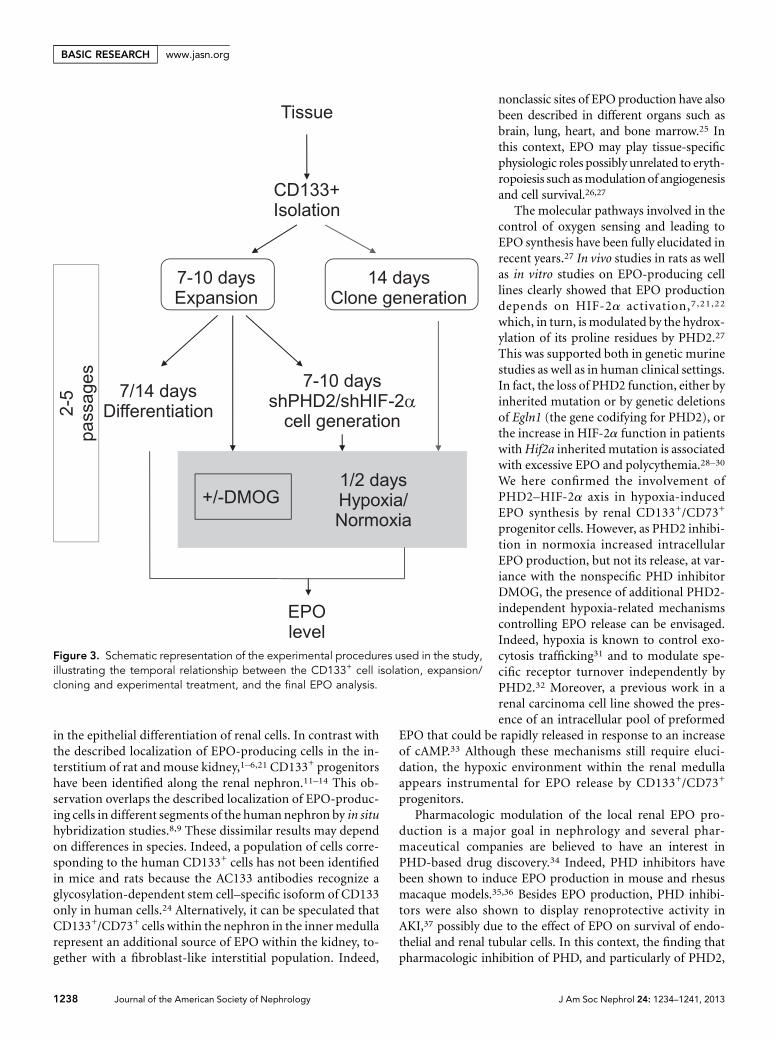
Figure 3. Schematic representation of the experimental procedures used in the study,illustrating the temporal relationship between the CD133+ cell isolation, expansion/cloning and experimental treatment, and the final EPO analysis.
1238 Journal of the American Society of Nephrology J Am Soc Nephrol 24: 1234–1241, 2013
BASIC RESEARCH www.jasn.org
enhances EPO release by CD133+/CD73+
progenitors under hypoxia may supportand provide a new rationale for the use ofPHD inhibitors in clinical settings of acuteor chronic renal injury. Moreover, isolatedCD133+/CD73+ cells could represent a use-fulmodel to dissect themechanisms of EPOsynthesis and release from human renalcells.
CONCISE METHODS
Isolation and Culture ConditionsRenal progenitor cells were obtained from the
normal portion of the inner medulla obtained
from surgically removed kidneys, after approval
by the Ethical Committee for the Use of Human
Tissue of the University of Torino, as described,13
and cultured in endothelial basal medium
medium plus supplement kit (Cambrex BioSci-
ence, East Rutherford, NJ) without serum addi-
tion. Cell isolates, obtained from different renal
specimens (n=16), were used between passages 2
and 5 (Figure 3 and Table 1). The CD1332 cell
population obtained after magnetic sorting
was plated in RPMI plus 10% FCS and used after
1–2 culture passages. To generate clones, CD133+
cells were seeded using a limiting dilution tech-
nique in 96-well plates. After 12 hours, wells
not containing single cells were discarded by
microscopical visualization and clones derived
from a single cell were expanded in expansion
medium. CD133 expression was evaluated by
FACS analysis.
Epithelial differentiation was obtained by
culturing cells for 14 days in expansion medium
with 10 ng/ml human hepatocyte growth factor
(Sigma Aldrich, St. Louis, MO) and 10 ng/ml
human fibroblast growth factor-4 (Sigma Al-
drich). When cultured in hypoxic conditions,
cells were placed in hypoxic chambers with 1%
O2. DMOG (100mM; Frontier Scientific, Logan,
UT) dissolved in PBS was added to the cells for
the indicated amount of time.
Cell Infection and TransfectionThe Block-it Pol II miR RNAi Expression Vector
Kit (Life Technologies, Grand Island, NY) was
used to construct pcDNA6.2-GW/EmGFP-miR
vectors (Life Technologies) expressing the target
miRNA (miR-PHD2) according to the manu-
facturer’s instructions. The Rapid BP/LR Re-
combination Reaction (Block-it Lentiviral Pol
II miR RNAi Expression System; Life
Figure 4. Increase in EPO released by CD133+/CD73+ cells after PHD2 inhibition.CD133+/CD73+ cells are infected with shRNA for PHD2 (shPHD2) or with scrambledvector (shCtr) or treated with DMOG (100 mM for 24 hours). (A–D) Western Blotmicrographs and densitometric analysis of HIF-2a (A and B) and PHD2 (B and D) ex-pression. Data, shown as arbitrary units, are representative of three different ex-periments performed using three different cell isolates and are normalized to actinexpression. (E) Quantitative RT-PCR analysis showing increase in mRNA encoding forEPO in shPHD2 and DMOG treated CD133+/CD73+ cells. (F and G) EPO release byshPHD2 and DMOG treated CD133+ cells in normoxia or hypoxia. Data are themean 6 SD of experiments performed in triplicate using three (shPHD2) or six(DMOG) different cell isolates. (H) Detection of EPO in the cell lysates of shCtr andshPHD2 cells in normoxia or hypoxia. Data are the mean 6 SD of three differentexperiments. The t test is performed. *P,0.05 DMOG versus Ctr or shPHD2 versusshCtr.
J Am Soc Nephrol 24: 1234–1241, 2013 CD133+ Progenitors and EPO 1239
www.jasn.org BASIC RESEARCH
Technologies) between pDONR 221, pcDNA6.2-GW/EmGFP-miR,
and pLenti6/V5-DEST was performed to generate the pLenti6/V5-
GW/EmGFP-miR expression construct. For knockdown of HIF-2a, a
pGIPZ lentiviral vector (Open Biosystems, Lafayette, CO) carrying
shHIF-2a was used. The constructs were then transfected with the
293T cell line using the ViraPower PackagingMix (Life Technologies)
for lentivirus production. To confirm the results, the following lenti-
viral particles carrying other hairpin RNAwere obtained from Sigma
Aldrich: scrambled PLKO.1 and shRNA for HIF-2a (HIF-2a 2,
ΤPΧΝ0000082303) and scrambled PLVX.1 and shRNA for PHD2
(PHD2 2, TRCN0000001042). After titering the lentiviral stock,
CD133+ progenitors were transduced with lentiviral particles follow-
ing the manufacturer’s instructions.
Immunofluorescence and Western Blot AnalysesImmunofluorescence and immunohistochemistry were performed
using the following antibodies: anti-CD133/1 (clone AC133, 293C3;
Miltenyi Biotec, Auburn, CA), anti-CD73 (Becton Dickinson, San
Jose, CA), anti-megalin and anti-Tamm-Horsfall protein (Santa Cruz
Biotechnology, Santa Cruz, CA), anti-vimentin (Sigma), anti-PHD2
(Abcam, Cambridge, UK), and anti-HIF-1a, anti-HIF-2a, and CA IX
(Novus Biologicals, Littleton, CO). Details are reported in the Sup-
plemental Materials and Methods.
RNA Preparation and RT-PCRTotal RNA was isolated from different cell preparations using the
RNAqueos-Micro isolation kit (Ambion, Life Technologies) accord-
ing to the manufacturer’s protocol. RNA was then quantified spec-
trophotometrically (NanoDrop ND-1000; NanoDrop Products,
Wilmington, DE). For gene expression analysis, quantitative real-time
PCR was performed in 20 ml of reaction mixture containing 5 ng of
cDNA template, sequence-specific oligonucleotide primers (pur-
chased from MWG-Biotech AG, Ebersberg, Germany), and Power
SYBR Green PCR Master Mix (Applied Biosystems, Foster City,
CA). TATA binding protein mRNA is used to normalize RNA inputs.
Fold-change expression with respect to control was calculated for all
samples. Sequence-specific oligonucleotide primers are reported in
the Supplemental Materials and Methods.
ELISA for EPOEPO protein in cell culture supernatants and in
cell lysates was measured by Platinum ELISA
(eBioscience, San Diego, CA) according to the
manufacturer’s recommendations.
Statistical AnalysesStatistical analysiswasperformedusing the t test,
or ANOVAwith Dunnett’s multiple comparison
tests, as appropriate. A P value of ,0.05 was
considered significant.
ACKNOWLEDGMENTS
This work was supported by the Regione Pie-
monte, PISTEM project.
DISCLOSURESNone.
REFERENCES
1. Rosenberger C, Rosen S, Paliege A, Heyman SN: Pimonidazole adductimmunohistochemistry in the rat kidney: Detection of tissue hypoxia.Methods Mol Biol 466: 161–174, 2009
2. Koury ST, Bondurant MC, Koury MJ: Localization of erythropoietinsynthesizing cells in murine kidneys by in situ hybridization. Blood 71:524–527, 1988
3. Lacombe C, Da Silva JL, Bruneval P, Fournier JG, Wendling F,Casadevall N, Camilleri JP, Bariety J, Varet B, Tambourin P: Peritubularcells are the site of erythropoietin synthesis in the murine hypoxic kid-ney. J Clin Invest 81: 620–623, 1988
4. Semenza GL, Koury ST, Nejfelt MK, Gearhart JD, Antonarakis SE: Cell-type-specific and hypoxia-inducible expression of the human eryth-ropoietin gene in transgenic mice. Proc Natl Acad Sci U S A 88:8725–8729, 1991
5. Bachmann S, Le Hir M, Eckardt KU: Co-localization of erythropoietinmRNA and ecto-59-nucleotidase immunoreactivity in peritubular cellsof rat renal cortex indicates that fibroblasts produce erythropoietin.J Histochem Cytochem 41: 335–341, 1993
6. Maxwell PH, Osmond MK, Pugh CW, Heryet A, Nicholls LG, Tan CC,Doe BG, Ferguson DJ, Johnson MH, Ratcliffe PJ: Identification of therenal erythropoietin-producing cells using transgenic mice. Kidney Int44: 1149–1162, 1993
7. Frede S, Freitag P, Geuting L, Konietzny R, Fandrey J: Oxygen-regu-lated expression of the erythropoietin gene in the human renal cell lineREPC. Blood 117: 4905–4914, 2011
8. Beirão I, Moreira L, Barandela T, Lobato L, Silva P, Gouveia CM,Carneiro F, Fonseca I, Porto G, Pinho E Costa P: Erythropoietin pro-duction by distal nephron in normal and familial amyloidotic adult hu-man kidneys. Clin Nephrol 74: 327–335, 2010
9. Da Silva JL, Lacombe C, Bruneval P, Casadevall N, Leporrier M,Camilleri JP, Bariety J, Tambourin P, Varet B: Tumor cells are the site oferythropoietin synthesis in human renal cancers associated with poly-cythemia. Blood 75: 577–582, 1990
10. Xinaris C, Benedetti V, Rizzo P, AbbateM, Corna D, Azzollini N, Conti S,Unbekandt M, Davies JA, Morigi M, Benigni A, Remuzzi G: In vivo
Figure 5. HIF-2a is required for EPO but not VEGF synthesis by CD133+/CD73+ cells.Quantitative RT-PCR analysis showing reduction in mRNA encoding for HIF-2a (A) andEPO (B), but not for VEGF (C) in CD133+ cells infected with two different shRNA forHIF-2a (shHIF2 alpha) in respect to cells infected with a scrambled vector (shCtr). Dataare normalized to TBP mRNA and to 1 for shCtr and are the mean 6 SD of threeexperiments performed using three different cell isolates. The t test is performed.*P,0.05 shHIF2 alpha versus shCtr. VEGF, vascular endothelial growth factor.
1240 Journal of the American Society of Nephrology J Am Soc Nephrol 24: 1234–1241, 2013
BASIC RESEARCH www.jasn.org
maturation of functional renal organoids formed from embryonic cellsuspensions. J Am Soc Nephrol 23: 1857–1868, 2012
11. Bussolati B, Bruno S, GrangeC, Buttiglieri S, DeregibusMC, CantinoD,Camussi G: Isolation of renal progenitor cells from adult human kidney.Am J Pathol 166: 545–555, 2005
12. Sallustio F, De Benedictis L, Castellano G, Zaza G, Loverre A,Costantino V, Grandaliano G, Schena FP: TLR2 plays a role in the ac-tivation of human resident renal stem/progenitor cells. FASEB J 24:514–525, 2010
13. Bussolati B, Moggio A, Collino F, Aghemo G, D’Armento G, Grange C,Camussi G: Hypoxia modulates the undifferentiated phenotype ofhuman renal inner medullary CD133+ progenitors through Oct4/miR-145 balance. Am J Physiol Renal Physiol 302: F116–F128, 2012
14. Ward HH, Romero E,Welford A, Pickett G, Bacallao R, Gattone VH 2nd,Ness SA, Wandinger-Ness A, Roitbak T: Adult human CD133/1(+)kidney cells isolated from papilla integrate into developing kidney tu-bules. Biochim Biophys Acta 1812: 1344–1357, 2011
15. Heyman SN, Khamaisi M, Rosen S, Rosenberger C: Renal parenchymalhypoxia, hypoxia response and the progression of chronic kidney dis-ease. Am J Nephrol 28: 998–1006, 2008
16. Smeets B, Boor P, Dijkman H, Sharma SV, Jirak P, Mooren F, Berger K,Bornemann J, Gelman IH, Floege J, van der Vlag J, Wetzels JF, MoellerMJ: Proximal tubular cells contain a phenotypically distinct, scatteredcell population involved in tubular regeneration. J Pathol 229: 645–659, 2013
17. Goldberg MA, Glass GA, Cunningham JM, Bunn HF: The regulatedexpression of erythropoietin by two human hepatoma cell lines. ProcNatl Acad Sci U S A 84: 7972–7976, 1987
18. Epstein AC, Gleadle JM, McNeill LA, Hewitson KS, O’Rourke J, MoleDR, Mukherji M, Metzen E, Wilson MI, Dhanda A, Tian YM, Masson N,Hamilton DL, Jaakkola P, Barstead R, Hodgkin J, Maxwell PH, PughCW, Schofield CJ, Ratcliffe PJ: C. elegans EGL-9 and mammalian ho-mologs define a family of dioxygenases that regulate HIF by prolylhydroxylation. Cell 107: 43–54, 2001
19. Cunliffe CJ, Franklin TJ, Hales NJ, Hill GB: Novel inhibitors of prolyl 4-hydroxylase. 3. Inhibition by the substrate analogue N-oxaloglycineand its derivatives. J Med Chem 35: 2652–2658, 1992
20. Berra E, Benizri E, Ginouvès A, Volmat V, Roux D, Pouysségur J: HIFprolyl-hydroxylase 2 is the key oxygen sensor setting low steady-statelevels of HIF-1alpha in normoxia. EMBO J 22: 4082–4090, 2003
21. Pan X, Suzuki N, Hirano I, Yamazaki S, Minegishi N, Yamamoto M:Isolation and characterization of renal erythropoietin-producing cellsfrom genetically produced anemia mice. PLoS ONE 6: e25839, 2011
22. Rankin EB, Biju MP, Liu Q, Unger TL, Rha J, Johnson RS, Simon MC,Keith B, Haase VH: Hypoxia-inducible factor-2 (HIF-2) regulates hepaticerythropoietin in vivo. J Clin Invest 117: 1068–1077, 2007
23. Obara N, Suzuki N, Kim K, Nagasawa T, Imagawa S, Yamamoto M:Repression via the GATA box is essential for tissue-specific erythro-poietin gene expression. Blood 111: 5223–5232, 2008
24. Yin AH,Miraglia S, Zanjani ED, Almeida-PoradaG,OgawaM, Leary AG,Olweus J, Kearney J, Buck DW: AC133, a novel marker for human he-matopoietic stem and progenitor cells. Blood 90: 5002–5012, 1997
25. Haase VH: Hypoxic regulation of erythropoiesis and iron metabolism.Am J Physiol Renal Physiol 299: F1–F13, 2010
26. Bahlmann FH, Fliser D: Erythropoietin and renoprotection. Curr OpinNephrol Hypertens 18: 15–20, 2009
27. Wenger RH, Hoogewijs D: Regulated oxygen sensing by protein hy-droxylation in renal erythropoietin-producing cells. Am J Physiol RenalPhysiol 298: F1287–F1296, 2010
28. Minamishima YA, Kaelin WG Jr: Reactivation of hepatic EPO synthesisin mice after PHD loss. Science 329: 407, 2010
29. Percy MJ, Zhao Q, Flores A, Harrison C, Lappin TR, Maxwell PH,McMullin MF, Lee FS: A family with erythrocytosis establishes a role forprolyl hydroxylase domain protein 2 in oxygen homeostasis. Proc NatlAcad Sci U S A 103: 654–659, 2006
30. Perrotta S, Della Ragione F: The HIF2A gene in familial erythrocytosis.N Engl J Med 358: 1966, author reply 1966–1967, 2008
31. Wang Y, Roche O, Xu C, Moriyama EH, Heir P, Chung J, Roos FC, ChenY, Finak G, Milosevic M, Wilson BC, Teh BT, Park M, Irwin MS, Ohh M:Hypoxia promotes ligand-independent EGF receptor signaling viahypoxia-inducible factor-mediated upregulation of caveolin-1. ProcNatl Acad Sci U S A 109: 4892–4897, 2012
32. Mazzone M, Dettori D, Leite de Oliveira R, Loges S, Schmidt T, JonckxB, Tian YM, Lanahan AA, Pollard P, Ruiz de Almodovar C, De Smet F,Vinckier S, Aragonés J, Debackere K, Luttun A, Wyns S, Jordan B,Pisacane A, Gallez B, Lampugnani MG, Dejana E, SimonsM, Ratcliffe P,Maxwell P, Carmeliet P: Heterozygous deficiency of PHD2 restorestumor oxygenation and inhibits metastasis via endothelial normaliza-tion. Cell 136: 839–851, 2009
33. Sherwood JB, Burns ER, Shouval D: Stimulation by cAMP of erythro-poietin secretion by an established human renal carcinoma cell line.Blood 69: 1053–1057, 1987
34. Muchnik E, Kaplan J: HIF prolyl hydroxylase inhibitors for anemia. Ex-pert Opin Investig Drugs 20: 645–656, 2011
35. Hsieh MM, Linde NS, Wynter A, Metzger M, Wong C, Langsetmo I, LinA, Smith R, Rodgers GP, Donahue RE, Klaus SJ, Tisdale JF: HIF prolylhydroxylase inhibition results in endogenous erythropoietin induction,erythrocytosis, and modest fetal hemoglobin expression in rhesusmacaques. Blood 110: 2140–2147, 2007
36. Safran M, Kim WY, O’Connell F, Flippin L, Günzler V, Horner JW,Depinho RA, Kaelin WG Jr: Mouse model for noninvasive imaging ofHIF prolyl hydroxylase activity: Assessment of an oral agent that stim-ulates erythropoietin production. Proc Natl Acad Sci U S A 103: 105–110, 2006
37. Bernhardt WM, Câmpean V, Kany S, Jürgensen JS, Weidemann A,Warnecke C, Arend M, Klaus S, Günzler V, Amann K, Willam C,Wiesener MS, Eckardt KU: Preconditional activation of hypoxia-in-ducible factors ameliorates ischemic acute renal failure. J Am SocNephrol 17: 1970–1978, 2006
This article contains supplemental material online at http://jasn.asnjournals.org/lookup/suppl/doi:10.1681/ASN.2012080772/-/DCSupplemental.
J Am Soc Nephrol 24: 1234–1241, 2013 CD133+ Progenitors and EPO 1241
www.jasn.org BASIC RESEARCH
Related Documents


