Renal Association Clinical Practice Guidelines on Acute Kidney Injury Dr Andrew Lewington a and Dr Suren Kanagasundaram b a Consultant Nephrologist, Leeds Teaching Hospitals, Leeds b Consultant Nephrologist, Freeman Hospital, Newcastle Key Words acute kidney injury . definition . epidemiology . prevention . treatment facilities . choice of renal replacement modality . vascular access . anticoagulation . therapy prescription . timing of initiation of therapy Introduction Acute kidney injury (AKI) has now replaced the term acute renal failure and an universal definition and staging system has been proposed to allow earlier detection and management of AKI. The new terminology enables healthcare professionals to consider the disease as a spec- trum of injury. This spectrum extends from less severe forms of injury to more advanced injury when acute kidney failure may require renal replacement therapy (RRT). Clinically AKI is characterised by a rapid reduc- tion in kidney function resulting in a failure to maintain fluid, electrolyte and acid-base homoeostasis. There have previously been many different definitions of AKI used in the literature which has made it difficult to determine the epidemiology and outcomes of AKI. Over recent years there has been increasing recognition that relatively small rises in serum creatinine in a variety of clinical settings are associated with worse outcomes [1]. To address the lack of an universal definition for AKI a collaborative network of international experts represent- ing nephrology and intensive care societies established the Acute Dialysis Quality Initiative (ADQI) and devised the RIFLE definition and staging system for AKI [2]. Shortly after this many of the original members of the ADQI group collaborated to form the Acute Kidney Injury Network (AKIN) [3, 4]. The AKIN group modi- fied the RIFLE staging system to reflect the clinical significance of relatively small rises in serum creatinine. Most recently the international guideline group, Kidney Disease: Improving Global Outcomes (KDIGO) has brought together international experts from many different specialties to produce a definition and staging system that harmonises the previous definitions and staging systems proposed by both ADQI and AKIN [5]. It is anticipated that this definition and staging system Fax +41 61 306 12 34 E-Mail [email protected] www.karger.com # 2011 S. Karger AG, Basel 1018–8665/09/0000–0349$00.00/0 Accessible online at: www.karger.com/nec Dr Andrew Lewington and Dr Suren Kanagasundaram Email: [email protected] or [email protected] Nephron Clin Pract 2011;118(suppl 1):c349–c390 DOI: 10.1159/000328075 Received: September 18, 2010 Accepted: March 14, 2011 Published online: May 6, 2011

Welcome message from author
This document is posted to help you gain knowledge. Please leave a comment to let me know what you think about it! Share it to your friends and learn new things together.
Transcript
Renal Association Clinical PracticeGuidelines on Acute Kidney Injury
Dr Andrew Lewingtona and Dr Suren Kanagasundaramb
aConsultant Nephrologist, Leeds Teaching Hospitals, LeedsbConsultant Nephrologist, Freeman Hospital, Newcastle
Key Wordsacute kidney injury . definition . epidemiology . prevention .
treatment facilities . choice of renal replacement modality .
vascular access . anticoagulation . therapy prescription .
timing of initiation of therapy
Introduction
Acute kidney injury (AKI) has now replaced the termacute renal failure and an universal definition and stagingsystem has been proposed to allow earlier detection andmanagement of AKI. The new terminology enableshealthcare professionals to consider the disease as a spec-trum of injury. This spectrum extends from less severeforms of injury to more advanced injury when acutekidney failure may require renal replacement therapy(RRT). Clinically AKI is characterised by a rapid reduc-tion in kidney function resulting in a failure to maintainfluid, electrolyte and acid-base homoeostasis. There havepreviously been many different definitions of AKI used in
the literature which has made it difficult to determine theepidemiology and outcomes of AKI. Over recent yearsthere has been increasing recognition that relativelysmall rises in serum creatinine in a variety of clinicalsettings are associated with worse outcomes [1].
To address the lack of an universal definition for AKI acollaborative network of international experts represent-ing nephrology and intensive care societies establishedthe Acute Dialysis Quality Initiative (ADQI) and devisedthe RIFLE definition and staging system for AKI [2].Shortly after this many of the original members of theADQI group collaborated to form the Acute KidneyInjury Network (AKIN) [3, 4]. The AKIN group modi-fied the RIFLE staging system to reflect the clinicalsignificance of relatively small rises in serum creatinine.
Most recently the international guideline group,Kidney Disease: Improving Global Outcomes (KDIGO)has brought together international experts from manydifferent specialties to produce a definition and stagingsystem that harmonises the previous definitions andstaging systems proposed by both ADQI and AKIN [5].It is anticipated that this definition and staging system
Fax +41 61 306 12 34E-Mail [email protected]
# 2011 S. Karger AG, Basel1018–8665/09/0000–0349$00.00/0
Accessible online at:www.karger.com/nec
Dr Andrew Lewington and Dr Suren KanagasundaramEmail: [email protected] or [email protected]
Nephron Clin Pract 2011;118(suppl 1):c349–c390
DOI: 10.1159/000328075
Received: September 18, 2010Accepted: March 14, 2011Published online: May 6, 2011
will be adopted globally. This will enable future com-parisons of the incidence, outcomes and efficacy oftherapeutic interventions for AKI.
To date there is a paucity of data on the incidence ofAKI whether community or hospital-acquired. Thereported prevalence of AKI from US data ranges from1% (community-acquired) up to 7.1% (hospital-acquired) of all hospital admissions [6, 7]. The popu-lation incidence of AKI from UK data ranges from 172per million population (pmp) per year from early data[8] up to 486–630 pmp/year from more recent series[9–11], again depending on definition. The incidenceof AKI requiring renal replacement therapy (RRT)ranges from 22 pmp/year [7] to 203 pmp/year [10]. Anestimated 5–20% of critically ill patients experience anepisode of AKI during the course of their illness andAKI receiving RRT has been reported in 49% of alladmissions to intensive-care units (ICU) [12]. Datafrom the Intensive Care National Audit ResearchCentre (ICNARC) suggests that AKI accounts fornearly 10% of all ICU bed days [13].
Acute kidney injury is common in hospitalisedpatients and also has a poor prognosis with the mortalityranging from 10%–80% dependent upon the patientpopulation studied. Patients who present with uncom-plicated AKI, have a mortality rate of up to 10% [14,15]. In contrast, patients presenting with AKI and multi-organ failure have been reported to have mortality ratesof over 50%. If renal replacement therapy is required themortality rate rises further to as high as 80% [16, 17].
Acute kidney injury is no longer considered to be aninnocent bystander merely reflecting co-existent pathol-ogies. It has been demonstrated to be an independentrisk factor for mortality [18–20]. The cause of this isunclear but is possibly associated with an increased riskof ‘non-renal’ complications such as bleeding andsepsis [17]. An alternative explanation may lie in experi-mental work that has demonstrated the ‘distant effects’ ofischaemic AKI on the other organs. In these experimentalmodels isolated ischaemic AKI upregulates inflammatorymediators in other organs including the brain, lungs andheart [21].
The UK National Confidential Enquiry into PatientOutcome and Death (NCEPOD) adding insult to injuryacute kidney injury report was published last year [22].This report examined the care of patients who died witha diagnosis of AKI. It identified many deficiencies in the
care of patients who developed AKI and reported thatonly 50% of patients received good care. There was poorattention to detail, inadequate assessment of risk factorsfor AKI and an unacceptable delay in recognising postadmission AKI. The report made a number of recom-mendations which included the following
. all emergency admissions should have a risk assess-ment for AKI
. all emergency admissions should have electrolyteschecked on admission and appropriately thereafter
. predictable avoidable AKI should not occur
. all acute admission should receive adequate seniorreviews (consultant review within 12 hours)
. there should be sufficient critical care and renalbeds to allow rapid step up care
. undergraduate medical training should include therecognition of the acutely ill patient and the preven-tion, diagnosis and management of AKI
. postgraduate training in all specialties shouldinclude training in the detection, prevention andmanagement of AKI.
The NCEPOD report was used to support a successfulproposal made to the National Institute for Health andClinical Excellence (NICE) for an AKI guideline. It ishoped that the guideline will be available in the nearfuture.
Once a patient has developed AKI the therapeuticoptions are limited with the mainstay of treatmentbeing renal replacement therapy (RRT). However thereare many important aspects surrounding the care of apatient with AKI that must be considered which includetimely referral and transfer to renal services if appro-priate. There is a paucity of evidence to guide the optimaltime to initiate RRT and the decision remains the choiceof the individual physician. If a patient commences RRTthen there are number of practical issues to be consideredincluding the modality, the choice of filter membrane,the optimal site of vascular access, anticoagulation andthe intensity of the treatment. The purpose of theseclinical practice guidelines is to review the availableevidence and provide a pragmatic approach to the patientwith AKI. There is a pressing need for renal physicians toengage in undergraduate and postgraduate educationalprogrammes to improve the current management ofAKI.
c350 Nephron Clin Pract 2011;118(suppl 1):c349–c390 Lewington/Kanagasundaram
References
1 Praught ML, Shlipak MG. Are small changes in serum creatinine animportant risk factor? Curr Opin Nephrol Hypertens 2005;14:265–270
2 Bellomo R, Ronco C, Kellum JA, Mehta RL, Palevsky P, and the ADQIworkgroup. Acute renal failure – definition, outcome measures, animalmodels, fluid therapy and information technology needs. The secondinternational consensus conference of Acute Dialysis Quality Initiative(ADQI) Group. Crit Care 2004;8:R204–R212
3 Mehta RL, Kellum JA, Shah SV, et al. Acute Kidney Injury Network(AKIN): report of an initiative to improve outcomes in acute kidneyinjury. Crit. Care 2007;11:R31
4 Molitoris BA, Levin A, Warnock D, et al. Improving outcomes of acutekidney injury: report of an initiative. Nat Clin Pract Nephrol 2007;3[8]:439–442
5 Kidney Disease: Improving Global Outcomes. Clinical practice guide-line on acute kidney injury. 2011. www.kdigo.org
6 Nash K, Hafeez A, Hou S. Hospital-acquired renal insufficiency. Am JKidney Dis 2002;39:930–936
7 Kaufman J, Dhakal M, Patel B, et al. Community-acquired acute renalfailure. Am J Kidney Dis 1991;17:191–198
8 Feest TG, Round A, Hamad S. Incidence of severe acute renal failure inadults: results of a living community based study. BMJ 1993;306:481–483
9 Stevens PE, Tamimi NA, Al Hasani MK, et al. Non-specialist manage-ment of acute renal failure. QJM 2001;94:533–540
10 Metcalfe W, Simpson KM, Khan IH, et al. Acute renal failure requiringrenal replacement therapy: incidence and outcome. QJM 2002;95:579–583
11 Hegarty J, Middleton R, Krebs M et al. Severe acute renal failure. Place ofcare, incidence and outcomes. QJM 2005;98:661–666
12 Metnitz PGH, Krenn CG, Steltzer H, et al. Effect of acute renal failurerequiring renal replacement therapy on outcome in critically ill patients.Crit Care Med 2002;30:2051–2058
13 Intensive-Care National Audit Research Centre 2005 www.icnarc.org14 Hou SH, Bushinsky DA, Wish JB, Cohen JJ, Harrington JT. Hospital-
acquired renal insufficiency: a prospective study. Am J Med 1983;74:243–248
15 Shusterman N, Strom BL, Murray TG, Morrison G, West SL, Maislin G.Risk factors and outcome of hospital-acquired acute renal failure. Clin-ical epidemiologic study. Am J Med 1987;83:65–71
16 Liano F, Junco E, Pascual J, Madero R, Verde E. The spectrum of acuterenal failure in the intensive care unit compared with that seen in othersettings. The Madrid Acute Renal Failure Study Group. Kidney Int1998;53:S16–S24
17 Cosentino F, Chaff C, Piedmonte M. Risk factors influencing survival inICU acute renal failure. Nephrol Dial Transplant 1994;9:179–182
18 Chertow GM, Levy EM, Hammermeister KE et al. Independent associa-tion between acute renal failure and mortality following cardiac surgery.Am J Med 1998;104:343–348
19 Levy EM, Viscoli CM, Horwitz RI. The effect of acute renal failure onmortality: a cohort analysis. JAMA 1996;275:1489–1494
20 Uchino S, Bellomo R, Goldsmith D et al. An assessment of the RIFLEcriteria for acute renal failure in hospitalized patients. Crit Care Med2006;34:1913–1917
21 Li X, Hassoun HT, Santora R, Rabb H. Organ crosstalk: the role of thekidney. Curr Opin Crit Care. 2009 Dec;15[6]:481–487
22 National Confidential Enquiry into Patient Outcome and Death,Adding Insult to Injury 2009. www.ncepod.org
Renal Association Clinical PracticeGuidelines on Acute Kidney Injury
Nephron Clin Pract 2011;118(suppl 1):c349–c390 c351
Summary of Clinical Practice Guideline on AcuteKidney Injury
1. Acute Kidney Injury (AKI) (Guidelines AKI1.1–1.3)
Guideline 1.1 – AKI: Definition, Epidemiology andOutcomesWe recommend that the international Kidney Disease:
Improving Global Outcomes (KDIGO) definition ofacute kidney injury (AKI) should be adopted. (NotGraded)
Acute kidney injury is defined when one of thefollowing criteria is met
. Serum creatinine rises by 526mmol/L within 48hours or
. Serum creatinine rises51.5 fold from the referencevalue, which is known or presumed to haveoccurred within one week or
. urine output is <0.5ml/kg/hr for >6 consecutivehours
The reference serum creatinine should be the lowestcreatinine value recorded within 3 months of the event.
If a reference serum creatinine value is not availablewithin 3 months and AKI is suspected
. repeat serum creatinine within 24 hours
. a reference serum creatinine value can be estimatedfrom the nadir serum creatinine value if patientrecovers from AKI
Guideline 1.2 – AKI: Definition, Epidemiology andOutcomesWe recommend that the international Kidney Disease:
Improving Global Outcomes (KDIGO) staging classifi-cation* of acute kidney injury (AKI) should be adopted.(Not Graded)
Guideline 1.3 – AKI: Definition, Epidemiology andOutcomesWe recommend that serum creatinine and urine
output remain the best biomarkers for AKI. Serumcreatinine should be measured using the enzymatictechnique. (1B)
2. Acute Kidney Injury (AKI) (Guidelines AKI2.1–2.2)
Guideline 2.1 – AKI: Clinical Assessment; History,ExaminationWe recommend that all patients presenting with AKI
should have a comprehensive history and examinationperformed to help determine the aetiology of the AKI.(1A)
Guideline 2.2 – AKI: Clinical Assessment;InvestigationsWe recommend that all patients presenting with AKI
should have appropriate baseline investigations per-formed which should include a urinalysis and a renaltract ultrasound within 24 hours (if renal tract obstruc-tion is suspected). (1A)
3. Acute Kidney Injury (AKI) (Guidelines AKI3.1–3.4)
Guideline 3.1 – AKI: Prevention; Risk AssessmentWe recommend that patients at risk of AKI should be
identified and appropriate preventative measures shouldbe instituted as early as possible. (1B)
Guideline 3.2 – AKI: Prevention; Fluid TherapyWe recommend that prescription of appropriate intra-
venous fluid should be carefully considered followingassessment of the patient’s volume status. Thereafterthe patient’s clinical response should be monitoredclosely. (1B)
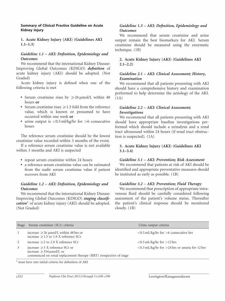
Stage Serum creatinine (SCr) criteria Urine output criteria
1 increase 526 mmol/L within 48 hrs orincrease 51.5 to 1.9 X reference SCr
<0.5mL/kg/hr for >6 consecutive hrs
2 increase 52 to 2.9 X reference SCr <0.5mL/kg/hr for >12 hrs
3 increase 53 X reference SCr orincrease 5354 mmol/L orcommenced on renal replacement therapy (RRT) irrespective of stage
<0.3mL/kg/hr for >24 hrs or anuria for 12 hrs
*must have met initial criteria for definition of AKI
c352 Nephron Clin Pract 2011;118(suppl 1):c349–c390 Lewington/Kanagasundaram
Guideline 3.3 – AKI: Prevention; Contrast-InducedAKI (CI-AKI)We recommend that patients identified as being at risk
of contrast induced-AKI (CI-AKI) should have a carefulassessment of volume status and receive pre-procedurevolume expansion with 0.9% sodium chloride or iso-tonic sodium bicarbonate if clinically indicated. (1A)
Guideline 3.4 – AKI: Prevention; AKI secondary toRhabdomyolysisWe recommend that patients identified as being at risk
of developing AKI secondary to rhabdomyolysis shouldreceive intravenous volume expansion with 0.9%sodium chloride and sodium bicarbonate. (1B)
4. Acute Kidney Injury (AKI) (Guidelines AKI4.1–4.5)
Guideline 4.1 – AKI: Management; GeneralManagementWe recommend that general supportive measures
include optimisation of haemodynamic status byappropriate fluid therapy, administration of vasopressorsand/or inotropes and treatment of any underlying sepsis.Nephrotoxic medications should be stopped. (1A)
Guideline 4.2 – AKI: Management; PharmacologicalTherapyWe recommend that therapeutic drug dosing must be
adapted to altered kinetics in AKI. (1B)
Guideline 4.3 – AKI: Management; PharmacologicalTherapyWe recommend that there is no specific pharmacolo-
gical therapy proven to effectively treat AKI secondary tohypoperfusion injury and/or sepsis. (1B)
Guideline 4.4 – AKI: Management; NutritionalSupportWe recommend that patients with AKI receiving renal
replacement therapy (RRT) should be referred to adietician for individual assessment. (1D)
Guideline 4.5 – AKI: Management; NutritionalSupportWe recommend that patients with AKI should receive
25–35 kcal/kg/day and up to a maximum of 1.7 g aminoacids/kg/day if hypercatabolic and receiving continuousrenal replacement therapy. Trace elements and water solu-ble vitamins should be supplemented as required. (1C)
5. Acute Kidney Injury (AKI) (Guidelines AKI5.1–5.7)
Guideline 5.1 – AKI: Treatment facilities and referralto renal servicesWe recommend that renal services shouldwork together
with other specialties to develop guidelines for the man-agement of AKI. These should include clear guidelineswith respect to when to request a renal referral. (1A)
Guideline 5.2 – AKI: Treatment facilities and referralto renal servicesWe recommend that specialist renal advice should be
given with consultant renal physician input. (1B)
Guideline 5.3 – AKI: Treatment facilities and referralto renal servicesWe recommend that transfer protocols should be
developed based on local physiological early warningscores to ensure appropriate triage of in-patients withAKI arriving from other hospitals. (1C)
Guideline 5.4 – AKI: Treatment facilities and referralto renal servicesWe recommend that physiological surveillance should
be performed for all patients with AKI to identify earlysigns of physiological deterioration which may requireescalation in the level of care. (1A)
Guideline 5.5 – AKI: Treatment facilities and referralto renal servicesWe suggest that renal physicians and intensivists
should work together to provide care for patients withAKI on the intensive care unit (ICU). Nephrologytrainees should be trained to care for acutely ill patientswith AKI. (2C)
Guideline 5.6 – AKI: Treatment facilities and referralto renal servicesWe suggest that intensive care units should contact
renal services to discuss patients likely to require ongoingsingle organ renal support prior to step-down. Advancewarning of such patients will facilitate forward planningand continued follow-up. (2C)
Guideline 5.7 – AKI: Treatment facilities and referralto renal servicesWe recommend that AKI survivors with residual renal
impairment should be managed according to localchronic kidney disease (CKD) guidelines. Discharge
Renal Association Clinical PracticeGuidelines on Acute Kidney Injury
Nephron Clin Pract 2011;118(suppl 1):c349–c390 c353
planning should include plans for CKD management,where relevant. (1A)
6. Acute Kidney Injury (AKI) (Guideline AKI 6.1)
Guideline 6.1 – AKI: Choice of renal replacementtherapy modalityWe recommend that the choice of renal replacement
therapy modality should be guided by the individualpatient’s clinical status, medical and nursing expertise,and availability of modality. (1B)
7. Acute Kidney Injury (AKI) (Guidelines AKI7.1–7.3)
Guideline 7.1 – AKI: Choice of dialyser/haemofiltermembraneWe recommend that synthetic or modified cellulosic
membranes should be used in preference to unmodifiedcellulose membranes. (1B)
Guideline 7.2 – AKI: Choice of dialysate/replacementfluidWe recommend that bicarbonate should be the pre-
ferred buffer for dialysate and replacement fluid in con-tinuous renal replacement therapy (CRRT) techniquesunless regional citrate anticoagulation is employed. (1C)
Guideline 7.3 – AKI: Microbial standards for fluidsWe recommend that microbial standards for fluids
used for chronic haemodialysis (HD)/haemodiafiltration(HDF) should be also applied to extracorporeal therapyfor AKI. (1A)
8. Acute Kidney Injury (AKI) (Guidelines AKI8.1–8.9)
Guideline 8.1 – AKI: Vascular access for RRTWe recommend that acute access for renal replace-
ment therapy should be veno-venous rather thanarterio-venous. (1A)
Guideline 8.2 – AKI: Vascular access for RRTWe recommend that dialysis catheters should be of an
adequate length to minimise the risks of access recircula-tion. (1C)
Guideline 8.3 – AKI: Vascular access for RRTWe suggest that the access site and catheter type
should be chosen with regard to the phase of the patient’sillness. (2C)
Guideline 8.4 – AKI: Vascular access for RRTWe recommend that access should be placed by
experienced or appropriately supervised staff. Real-timeultrasound guidance should be used to aid placementof upper body access. (1A)
Guideline 8.5 – AKI: Vascular access for RRTWe recommend that it is advisable that real-time
ultrasound guidance be used for the insertion of femoralaccess. (1D)
Guideline 8.6 – AKI: Vascular access for RRTWe recommend that subclavian access should be
avoided in patients at risk of progressing to CKD stage4 or 5 due to the risks of compromising future, perma-nent vascular access. (1D)
Guideline 8.7 – AKI: Vascular access for RRTWe suggest that non-dominant arm upper limb
vasculature should be preserved as a contingency forfuture permanent access. (2C)
Guideline 8.8 – AKI: Vascular access for RRTWe recommend that temporary access should be
changed at appropriate intervals (as per local protocol)to minimise the risk of infection. (1C)
Guideline 8.9 – AKI: Vascular access for RRTWe suggest that local policies on prevention of
catheter-related infection should be optimised byreserving the catheter for extracorporeal treatmentonly. (2D)
9. Acute Kidney Injury (AKI) (Guidelines AKI9.1–9.4)
Guideline 9.1 – AKI: Anticoagulation forextracorporeal therapiesWe recommend that anticoagulation for RRT should
be tailored according to patient characteristics and themodality of RRT chosen. (1C)
Guideline 9.2 – AKI: Anticoagulation forextracorporeal therapiesWe recommend that regional anticoagulation with
citrate reduces risk of haemorrhage compared to sys-temic heparinisation. The complexity of the techniquemeans that this should be in routine use on any uniton which it is employed in order to allow sufficientlevels of expertise to be maintained. (1C)
c354 Nephron Clin Pract 2011;118(suppl 1):c349–c390 Lewington/Kanagasundaram
Guideline 9.3 – AKI: Anticoagulation forextracorporeal therapiesWe suggest that prostacyclin is a suitable alternative to
unfractionated heparin in those at increased risk ofbleeding but may cause haemodynamic instability. (2C)
Guideline 9.4 – AKI: Anticoagulation forextracorporeal therapiesWe suggest that a no-anticoagulation, saline flush
strategy can be used in patients receiving continuousand intermittent RRT who are at high risk of bleeding.However, ultrafiltration requirements are increased,effective intermittent HD time is reduced and the tech-nique runs the risk of membrane fibre rupture. (2C)
10. Acute Kidney Injury (AKI) (Guidelines AKI10.1–10.5)
Guideline 10.1 – AKI: Renal Replacement TherapyprescriptionWe recommend that the delivered dose of RRT should
be assessed to ensure the adequacy of the prescription.(1A)
Guideline 10.2 – AKI: Renal Replacement TherapyprescriptionWe recommend that the prescribed dose should be
assessed at each session (for intermittent haemodialysis)and daily (for continuous RRT) to account for any meas-ured shortfalls in delivered dose. (1A)
Guideline 10.3 – AKI: Renal Replacement TherapyprescriptionWe recommend that patients with AKI and multi-
organ failure treated by continuous renal replacementtherapy (CRRT) should receive treatment doses equiva-lent to post dilution ultrafiltration rates 525ml/kg/hr.A proportionate upward adjustment to the prescribedultrafiltration rate should be made in pre-dilutionalcontinuous haemofiltration. (1A)
Guideline 10.4 – AKI: Renal Replacement TherapyprescriptionWe recommend that patients with AKI and multi-
organ failure treated by intermittent haemodialysisshould receive either alternate day haemodialysis withat least the minimum dose considered appropriate forend-stage renal disease (ESRD), urea reduction ratio(URR) >65% or eKt/V >1.2 or daily haemodialysis.(1B)
Guideline 10.5 – AKI: Renal Replacement TherapyprescriptionWe suggest that renal replacement therapy dosing
methods that require an assessment of patient weightshould use a measured weight rather than an extrapo-lated weight from pre-morbid readings. (2B)
11. Acute Kidney Injury (AKI) (Guidelines AKI11.1–11.5)
Guideline 11.1 – AKI: Timing of initiation of renalreplacement treatmentWe recommend that the decision to start RRT in
patients with AKI should remain a clinical decisionbased on fluid, electrolyte and metabolic status of eachindividual patient. (1C)
Guideline 11.2 – AKI: Timing of initiation of renalreplacement treatmentWe recommend that RRTshould be initiated once AKI
is established and unavoidable but before overt compli-cations have developed. (1B)
Guideline 11.3 – AKI: Timing of initiation of renalreplacement treatmentWe recommend that the threshold for initiating RRT
should be lowered when AKI occurs as part of multi-organ failure. (1C)
Guideline 11.4 – AKI: Timing of initiation of renalreplacement treatmentWe recommend that the initiation of RRT may be
deferred if the underlying clinical condition is improvingand there are early signs of renal recovery. (1D)
Guideline 11.5 – AKI: Timing of discontinuation ofrenal replacement treatmentWe recommend that an improvement in the patient’s
clinical condition and urine output would justify tem-porary discontinuation of ongoing renal support to seeif AKI is recovering. (1D)
12. Acute Kidney Injury (AKI) (Guideline AKI12.1)
Guideline 12.1 – AKI: EducationWe recommend that undergraduate and postgraduate
medical trainees should be taught the principles ofprevention, recognition and management of AKI. (1C)
Renal Association Clinical PracticeGuidelines on Acute Kidney Injury
Nephron Clin Pract 2011;118(suppl 1):c349–c390 c355
Summary of Audit Measures
It is recommended that the following audit measuresare recorded for all patients diagnosed with acutekidney injury. However it is recognised that it mayonly be possible for renal units to record these auditmeasures for patients that have been referred for arenal specialist opinion.
The Renal Association encourages other specialties torecord these audit measures for all patients diagnosedwith AKI irrespective of whether or not they are referredto renal services. From a pragmatic point of view interms of available resources it is proposed that otherspecialties initially collect data on patients with AKIstage 3. Once a robust data collection system has beenestablished an incremental collection of data extendingto AKI stage 2 and then AKI stage 1 could follow.
1. Incidence and outcomes of patients diagnosedwith. community-acquired AKI. hospital acquired AKI
2. Incidence and outcomes of patients with differentcauses of AKI
3. Incidence of acute admissions/patients under-going major surgery who had. the risk of AKI assessed on admission/pre-surgery
. electrolytes checked on admission/pre-surgeryand rechecked within 24 hours
4. Proportion of patients who had a urinalysisperformed within 24 hours of the diagnosis ofAKI unless anuric
5. Proportion of patients where there has been adelay of >48 hours in recognising the diagnosisof AKI
6. Proportion of patients developing AKI secondaryto obstruction who had a renal ultrasoundexamination <24 hrs after a diagnosis of AKIestablished
7. Proportion of patients with or at risk of AKI whoare prescribed intravenous fluids without anassessment of volume status
8. Proportion of patients with AKI who did not havethe appropriate adjustment of medication doses
9. Proportion of patients with or at risk of AKI whoreceive nephrotoxic medications
10. Proportion of patients at high risk of contrastinduced AKI (CI-AKI) who developed AKI anddid not
. receive pre-procedure volume assessment
. receive appropriate volume expansion
. have appropriate adjustments to medications11. Proportion of patients with severe AKI where
there is documented evidence of patient involve-ment in decision making with respect to com-mencing renal replacement therapy (RRT)
12. Incidence of delays of transfer of patients withAKI >24 hours following referral to renal servicesdue to a lack of resources on renal unit
13. Incidence of patients with single organ AKIadmitted to ICU for RRT due to a lack ofresources on the renal unit
14. Number of AKI inpatient transfers requiring escala-tion of care within 24 hours of arrival on renal unit
15. Incidence of dialysis catheter-related bacteraemiaand sepsis in patients with AKI
16. Incidence of heparin induced thrombocytopenia17. Proportion of critically ill patients with AKI
treated with alternate day haemodialysis whoreceive eKt/V 51.2 per session
18. Proportion of critically ill patients with AKItreated with continuous renal replacement ther-apy who receive >25mls/kg/hr post dilutionultrafiltration
19. Proportion of patients with AKI receiving renalreplacement therapy reviewed by dieticianwithin 24 hours
20. Proportion of patients with AKI receiving renalreplacement therapy prescribed the recom-mended nutritional support
21. Proportion of patients with AKI who recoverkidney function within 90 days of episode asdefined by. return of serum creatinine to within 20% ofbaseline value (most recent value within 3months but accepting value up to one year)
. dialysis independence (if previously requiringdialysis)
22. Proportion of AKI survivors with residual chronickidney disease with post-discharge CKD planning
23. Proportion of AKI survivors who are given infor-mation on the cause of AKI and how this might beavoided in the future
24. Outcome measures for patients developing AKIshould include. length of hospital stay. hospital mortality. 90 day mortality. one year mortality
c356 Nephron Clin Pract 2011;118(suppl 1):c349–c390 Lewington/Kanagasundaram
Rationale for Clinical Practice Guideline on AcuteKidney Injury
1. Acute Kidney Injury (AKI) (Guidelines AKI1.1–1.3)
Guideline 1.1 – AKI: Definition, Epidemiology andOutcomesWe recommend that the international Kidney Disease:
Improving Global Outcomes (KDIGO) definition ofacute kidney injury (AKI) should be adopted. (NotGraded)
Audit measures1. Incidence and outcomes of patients diagnosed with
. community-acquired AKI
. hospital acquired AKI2. Proportion of patients where there has been a delay
of >48 hours in recognising the diagnosis of AKI3. Proportion of patients with AKI who recover kidney
function within 90 days of episode as defined by. return of serum creatinine to within 20% ofbaseline value (most recent value within 3months but accepting up to one year)
. dialysis independence (if previously requiringdialysis)
4. Outcome measures for patients developing AKIshould include. length of hospital stay. hospital mortality. 90 day mortality. one year mortality
RationaleOver recent years it has been recognised that even
small increases in serum creatinine (SCr) are associatedwith worse patient outcomes [1]. To reflect the impor-tance of these changes in SCr the term acute kidneyinjury (AKI) has now replaced acute renal failure(ARF). This allows AKI to be considered as a spectrumof severity that if not detected or recognised in its earlystages may ultimately result in acute kidney failure andthe need for renal replacement therapy (RRT).
The most recent definitions proposed by the AcuteDialysis Quality Initiative (ADQI), RIFLE, and theAcute Kidney Injury Network (AKIN) have been basedon rises in serum creatinine or reductions in urineoutput. These definitions aimed to promote the earlierdetection and recognition of AKI triggering appropriatetreatment prior to progressive injury and kidney failure.
The application of these definitions in more than500,000 patients has validated the increased risk ofmortality associated with developing AKI [2–4]. Thesestudies have also indicated that the incidence of AKI inhospitalised patients may be as high as 18%. Thesedefinitions have recently been harmonised by theKidney Diseases: Improving Global Outcomes Inter-national (KDIGO) guideline group [5].
It is important to note that the diagnosis of AKIshould be made initially based on the definition below.Once the diagnosis of AKI has been established its sever-ity can be determined using the staging system (shownin Table 1 in the rationale for guideline recommendation1.2).
Acute kidney injury is defined when one of the follow-ing criteria is met
. Serum creatinine rises by 526mmol/L within 48hours or
. Serum creatinine rises51.5 fold from the referencevalue, which is known or presumed to haveoccurred within one week or
. urine output is <0.5ml/kg/hr for >6 consecutivehours
The reference serum creatinine should be the lowestcreatinine value recorded within 3 months of theevent.
If a reference serum creatinine value is not availablewithin 3 months and AKI is suspected
. repeat serum creatinine within 24 hours
. a reference serum creatinine value can be estimatedfrom the nadir serum creatinine value if patientrecovers from AKI
It is recognised that outside of ICU the accuracy ofurine output measurements will be less reliable. Theuse of urine output criteria for both the diagnosis andstaging of AKI has been less well studied. Clinicaljudgement is necessary in patient assessment and therecognition that patients may develop oliguric as wellas nonoliguric AKI.
Guideline 1.2 – AKI: Definition, Epidemiology andOutcomesWe recommend that the international Kidney Disease:
Improving Global Outcomes (KDIGO) staging classifi-cation of acute kidney injury (AKI) should be adopted.(Not Graded)
Renal Association Clinical PracticeGuidelines on Acute Kidney Injury
Nephron Clin Pract 2011;118(suppl 1):c349–c390 c357
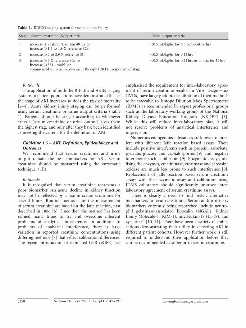
RationaleThe application of both the RIFLE and AKIN staging
systems to patient populations have demonstrated that asthe stage of AKI increases so does the risk of mortality[2–4]. Acute kidney injury staging can be performedusing serum creatinine or urine output criteria (Table1). Patients should be staged according to whichevercriteria (serum creatinine or urine output) gives themthe highest stage and only after they have been identifiedas meeting the criteria for the definition of AKI.
Guideline 1.3 – AKI: Definition, Epidemiology andOutcomesWe recommend that serum creatinine and urine
output remain the best biomarkers for AKI. Serumcreatinine should be measured using the enzymatictechnique. (1B)
RationaleIt is recognised that serum creatinine represents a
poor biomarker. An acute decline in kidney functionmay not be reflected by a rise in serum creatinine forseveral hours. Routine methods for the measurementof serum creatinine are based on the Jaffe reaction, firstdescribed in 1886 [6]. Since then the method has beenrefined many times to try and overcome inherentproblems of analytical interference. In addition, toproblems of analytical interference, there is largevariation in reported creatinine concentrations usingdiffering methods [7] that reflect calibration differences.The recent introduction of estimated GFR (eGFR) has
emphasised the requirement for inter-laboratory agree-ment of serum creatinine results. In Vitro Diagnostics(IVDs) have largely adopted calibration of their methodsto be traceable to Isotope Dilution Mass Spectrometry(IDMS) as recommended by expert professional groupssuch as the laboratory working group of the NationalKidney Disease Education Program (NKDEP) [8].Whilst this will reduce inter-laboratory bias, it willnot resolve problems of analytical interference andimprecision.
Numerous endogenous substances are known to inter-fere with different Jaffe reaction based assays. Theseinclude positive interferents such as protein, ascorbate,pyuvate, glucose and cephalosporins [9] and negativeinterferents such as bilirubin [9]. Enzymatic assays, uti-lising the enzymes, creatininase, creatinase and sarcosineoxidase are much less prone to such interference [9].Replacement of Jaffe reaction based serum creatinineassays with the enzymatic assay and calibration usingIDMS calibrators should significantly improve inter-laboratory agreement of serum creatinine assays.
There is clearly a need to find better, alternativebio-markers to serum creatinine. Serum and/or urinarybiomarkers currently being researched include neutro-phil gelatinase-associated lipocalin (NGAL), KidneyInjury Molecule-1 (KIM-1), interleukin-18 (IL-18), andcystatin C [10–14]. There have been a variety of publi-cations demonstrating their utility in detecting AKI indifferent patient cohorts. However further work is stillrequired to understand their application before theycan be recommended as superior to serum creatinine.
Table 1. KDIGO staging system for acute kidney injury
Stage Serum creatinine (SCr) criteria Urine output criteria
1 increase 526 mmol/L within 48 hrs orincrease 51.5 to 1.9 X reference SCr
<0.5 mL/kg/hr for >6 consecutive hrs
2 increase 52 to 2.9 X reference SCr <0.5 mL/kg/hr for >12 hrs
3 increase 53 X reference SCr orincrease 5354 mmol/L orcommenced on renal replacement therapy (RRT) irrespective of stage
<0.3 mL/kg/hr for >24 hrs or anuria for 12 hrs
c358 Nephron Clin Pract 2011;118(suppl 1):c349–c390 Lewington/Kanagasundaram
References
1 Praught ML, Shlipak MG. Are small changes in serum creatinine andimportant risk factor? Current Opinions in Nephrology and Hyper-tension 2005;14:265–270
2 Hoste EA, Clermont G, Kersten A, Venkataraman R, Angus DC, DeBacquer D, Kellum JA. RIFLE criteria for acute kidney injury areassociated with hospital mortality in critically ill patients: a cohortanalysis. Crit Care. 2006;10[3]:R73. Epub 2006 May 12.
3 Uchino S, Bellomo R, Goldsmith D, Bates S, Ronco C. An assessment ofthe RIFLE criteria for acute renal failure in hospitalized patients. CritCare Med. 2006 Jul;34[7]:1913–1917.
4 Thakar CV, Christianson A, Freyberg R, Almenoff P, Render ML.Incidence and outcomes of acute kidney injury in intensive care units:a Veterans Administration study. Crit Care Med. 2009 Sep;37[9]:2552–2558.
5 Kidney Disease: Improving Global Outcomes. Clinical practice guide-line on acute kidney injury. 2011. www.kdigo.org
6 Jaffe M. Uber den niederschlag, welchen pikrinsaure in normalen hrnerzgeugt und uber eine neue reaction des kreatinins. Z Physiol Chem1886;10:391–400
7 Lawson N, Lang T, Broughton A, Prinsloo P, Turner C, Marenah C.Creatinine assays: time for action? Ann Clin Biochem 2002;39:599–602
8 Recommendations for improving serum creatinine measurement: Areport from the laboratory working group of the National KidneyDisease Education Preogram. Clin Chem 2006;52:5–18
9 Peake M, Whiting M. Measurement of serum creatinine – current statusand future goals. Clin Biochem Rev 2006;27:173–184
10 Hewitt SM, Dear J, Star RA. Discovery of protein biomarkers for renaldiseases. J Am Soc Nephrol. 2004;15[7]:1677–1689
11 Vaidya VS, Ramirez V, Ichimura T, Bobadilla NA, Bonventre JV. Urinarykidney injury molecule-1: a sensitive quantitative biomarker for earlydetection of kidney tubular injury. Am J Physiol Renal Physiol 2006;290[2]:F517–F529
12 Parikh CR, Mishra J, Thiessen-Philbrook H, Dursun B, Ma Q, Kelly C,Dent C, Devarajan P, Edelstein CL. Urinary IL-18 is an early predictivebiomarker of acute kidney injury after cardiac surgery. Kidney Int2006;70:199–203
13 Mishra J, Ma Q, Prada A, Mitsnefes M, Zahedi K, Yang J, Barasch J,Devarajan P. Identification of Neutrophil Gelatinase-AssociatedLipocalin as a Novel Early Urinary Biomarker for Ischemic RenalInjury. J Am Soc Nephrol 2003;14:2534–2543
14 Herget-Rosenthal S, Marggraf G, Husing J, Goring F, Pietruck F, JanssenO, Philipp T, Kribben A. Early detection of acute renal failure by serumcystatin C. Kidney Int 2004;66:1115–1122
Renal Association Clinical PracticeGuidelines on Acute Kidney Injury
Nephron Clin Pract 2011;118(suppl 1):c349–c390 c359
2. Acute Kidney Injury (AKI) (Guidelines AKI2.1–2.2)
Guideline 2.1 – AKI: Clinical Assessment; History,ExaminationWe recommend that all patients presenting with AKI
should have a comprehensive history and examinationperformed to help determine the cause of the AKI. (1A)
Audit measure1. Incidence and outcomes of patients with different
causes of AKI
RationaleAcute kidney injury is most frequently caused by
ischaemia, sepsis or nephrotoxic insults to the kidney.In patients with hospital-acquired AKI the cause is fre-quently multi-factorial in patients with multiple riskfactors. However it is essential to consider the underlyingcause of AKI as a smaller percentage of cases may becaused by acute interstitial nephritis or acute glomerulo-nephritis which will require specific therapy [1]. It ishoped that earlier detection and recognition of AKImay provide an earlier opportunity to provide specifictherapy to these forms of esoteric AKI [2].
Clinical assessment of the patient with AKI starts witha comprehensive history including a review of:
. patient notes
. AKI risk factorsk age >75 yrsk chronic kidney disease (CKD, eGFR <60mls/min/1.73m2)
k Cardiac failurek Atherosclerotic peripheral vascular diseasek Liver diseasek Diabetes mellitusk Nephrotoxic medications
. potential causes for AKI includingk reduced fluid intakek increased fluid lossesk urinary tract symptomsk recent drug ingestionk sepsis
. systemic clinical featuresk feverk rashk joint pains
Clinical examination must include
. generalk rashk uveitisk joint swelling
. assessment of volume statusk core temperaturek peripheral perfusionk heart ratek blood pressurek jugular venous pressure
. signs of renovascular diseasek audible bruitsk impalpable peripheral pulses
. abdominal examinationk palpable bladder
Guideline 2.2 – AKI: Clinical Assessment;InvestigationsWe recommend that all patients presenting with AKI
should have appropriate baseline investigations per-formed which should include a urinalysis and a renaltract ultrasound within 24 hours (if renal tract obstruc-tion is suspected). (1A)
Audit measures1. Proportion of patients who had a urinalysis per-
formed within 24 hours of the diagnosis of AKIunless anuric
2. Proportion of patients developing AKI secondary toobstructionwho had a renal ultrasound examination<24hrs after a diagnosis of AKI established
RationaleClinical assessment to establish a working diagnosis
requires a number of investigations to be performed.A baseline set of laboratory investigations should be
sent including:
. biochemistryk Urea and electrolytes
. haematologyk FBC
. urinalysis (�microscopy)
. microbiologyk urine culture (if infection is suspected)k blood culture (if infection is suspected)
More specific renal investigations are dependent uponthe clinical presentation and may include:
. renal immunology
c360 Nephron Clin Pract 2011;118(suppl 1):c349–c390 Lewington/Kanagasundaram
. urinary biochemistryk electrolytesk osmolality
. ECG
. chest X-ray
. abdominal X-ray
. renal tract ultrasound (within 24 hrs if obstructionsuspected or esoteric cause suspected requiring akidney biopsy)
. kidney biopsy
Urinalysis can provide important clinical informationto patients with AKI. Positive protein values of 3þ and4þ on reagent strip testing of the urine suggest intrinsicglomerular disease. A reagent strip positive for bloodsuggests the presence of red blood cells (>5/highpower field). Although red cell morphology may not beparticularly useful [3] the observation of large numbersof red cells in the presence of proteinuria suggests aglomerular aetiology for AKI. The suspicion is strength-ened by the finding of red cell casts on a freshly collectedsample of urine (this is rarely performed in the UK).
Haematuria may also be found in cases of lowerurinary tract obstruction often in association withtumours and less commonly associated with calculi,infection or severe renal ischaemia due to arterial orvenous thrombosis. Characteristically myoglobinuriawill cause a positive reagent strip reaction for haematuriawithout evidence of red cells on urine microscopy.
Increased numbers of white cells (>5 per high powerfield) are non-specific but are found more commonlywith acute interstitial nephritis, infection and glomerulo-nephritis. Eosinophiluria is not a very specific test forinterstitial nephritis and has a very poor positive pre-dictive value. However, the value of eosinophiluria ininterstitial nephritis is in ruling out the disease, thenegative predictive value for patients with AKI is greaterthan 90% [4].
Urine microscopy can be informative in particularclinical scenarios such as suspected poisoning. The pre-sence of crystalluria may provide vital information andin the case of ethylene glycol poisoning oxalate crystalsmay be visible [5]. Patients who suffer from tumourlysis syndrome can produce urate crystal deposition. Anumber of drugs may lead to AKI and crystalluriaincluding sulphonamides, acyclovir, triamterene, indina-vir and cathartics high in phosphates.
Various measures have been claimed to aid in thediagnosis of AKI including urine osmolality, urine/plasma creatinine and urea ratios, urinary sodium, frac-tional excretion of sodium (FENa), fractional excretion ofurea (FEUrea), freewater clearance and creatinine clear-ance. All of these have limitations and their specificityand sensitivity in clinical practice often means that asingle measurement may be inconclusive except inextreme circumstances [6–8].
In pre-renal AKI there is increased urinary sodiumreabsorption and increased urinary urea reabsorption.This should therefore be reflected by low urine sodiumconcentrations, low FENa and low FEUrea, and increasedblood urea:creatinine ratios.
Urinary electrolytes should be interpreted with cau-tion, particularly in the elderly (who may already havean impaired concentrating ability), and in patients ondiuretics or with a salt-losing state. In such patients theFEUrea may possibly be a more useful index [8]. Thenormal FEUrea is greater than 45%. Levels of less than35% are associated with pre-renal AKI. Patients withpre-renal AKI not on diuretics have both low FENa(<1%) and low FEUrea. However patients with pre-renal AKI on diuretics have levels of FENa greater than2% but still have low levels of FEUrea. In comparison,patients with ATN have both high FENa and high FEUrea.
One clinical situation where measurement of urinaryelectrolytes may have clinical utility is in the diagnosisof hepatorenal syndrome as the cause of AKI in patientswith liver disease. The diagnostic criteria for hepatorenalfailure include a urine sodium of less than 10mmol/L(although not a major diagnostic criterion) [9].
Ultrasound is the gold standard test for diagnosis ofupper tract obstruction through the finding of hydro-nephrosis and/or hydroureter. However upper urinarytract obstruction may not be initially detected by ultra-sound in a patient who is volume depleted. It is thereforerecommended to repeat the renal tract ultrasound ifupper urinary tract obstruction is suspected once thepatient is adequately fluid resuscitated.
There are other circumstances when ultrasound maynot be diagnostic, such as in retroperitoneal fibrosis orearly in the course of obstructive disease, in which caseadditional imaging studies may be considered such asdynamic nuclear medicine studies or CT. Dynamicnuclear medicine studies will be of little diagnostic useif the patient has oligo-anuric AKI.
Renal Association Clinical PracticeGuidelines on Acute Kidney Injury
Nephron Clin Pract 2011;118(suppl 1):c349–c390 c361
References
1 Liano F, Pascual J and the Madrid Acute Renal Failure Study Group. Epi-demiology of acute renal failure: a prospective, multicenter, community-based study. Kidney International 1996;50:811–818
2 Lines S, Lewington A. Acute kidney injury. Clin Med. 2009 Jun;9[3]:273–277
3 Favaro S, Bonfant L, D’Angelo A, Giacomini A, Normanno M, Calo L. Isthe red cell morphology really useful to detect the source of haematuria?Am J Nephrol 1997;17:172–175
4 Rossert J. Drug-induced acute interstitial nephritis. Kidney Int 2001;60:804–817
5 Fogazzi GB. Crystalluria: a neglected aspect of urinary sediment analysis.Nephrol Dialysis Transplant 1996;11:379–387
6 Kellen M, Aronson S, Roizen MF, Barnard J, Thisted RA. Predictive anddiagnostic tests of renal failure: A review. Anesth Analg 1994;78:134–142
7 Espinel CH, Gregory AW. Differential diagnosis of acute renal failure.Clin Nephrol 1980;13:73–77
8 Carvounis CP, Nisar S, Guro-Razuman S. Significance of the fractionalexcretion of urea in the differential diagnosis of acute renal failure.Kidney Int 2002;62:2223–2229
9 Arroyo V, Gines P, Gerbes AL et al. Definition and diagnostic criteria ofrefractory ascites and hepatorenal syndrome in cirrhosis. InternationalAscites Club Hepatology 1996;23:164–176
c362 Nephron Clin Pract 2011;118(suppl 1):c349–c390 Lewington/Kanagasundaram
3. Acute Kidney Injury (AKI) (Guidelines AKI3.1–3.4)
Guideline 3.1 – AKI: Prevention; Risk AssessmentWe recommend that patients at risk of AKI should be
identified and appropriate preventative measures shouldbe instituted as early as possible. (1B)
Audit measures1. Incidence of acute admissions/patients undergoing
major surgery who had. risk of AKI assessed on admission/pre-surgery. electrolytes checked on admission/pre-surgeryand rechecked within 24 hours
2. Proportion of patients at risk of AKI who receivenephrotoxic medications
RationalePublished series of AKI suggest that up to 30% of cases
may be preventable, with a further significant percentagepotentially remediable through simple interventionssuch as volume repletion, discontinuing and/or avoidingcertain potentially nephrotoxic agents and earlierrecognition of conditions causing rapid progression ofAKI [1–3].
Risk factors for developing AKI include:
. age >75 yrs
. chronic kidney disease (CKD, eGFR <60mls/min/1.73m2)
. cardiac failure
. atherosclerotic peripheral vascular disease
. liver disease
. diabetes mellitus
. nephrotoxic medication
. hypovolaemia
. sepsis
Guideline 3.2 – AKI: Prevention; Fluid TherapyWe recommend that prescription of appropriate intra-
venous fluid should be carefully considered followingassessment of the patient’s volume status. Thereafterthe patient’s clinical response should be monitoredclosely. (1B)
Audit measure1. Proportion of patients at risk of AKI who are
prescribed intravenous fluids without an assess-ment of volume status
RationaleIn hospital AKI following surgery is an important
contributor to postoperative morbidity and mortality.The causes are multifactorial and therefore involve theidentification of the high risk patients and institutionof preventative measures. Avoidance of pre- and peri-operative hypovolaemia is an essential component ofpatient management.
Prescription of intravenous fluid should follow acareful assessment of patient volume status i.e. hypo-volaemic, euvolaemic, hypervolaemic. Considerationshould then be made regarding the nature of the fluidlost and therefore the nature of the fluid that needs tobe replaced. There is no evidence base to favour the pre-scription of crystalloid or colloids to protect kidneyfunction in the peri-operative period, although therehave only been a handful of studies looking at this [4].Following the selection of the appropriate fluid the rateof fluid replacement must be guided by clinical assess-ment with consideration for safety limits. The patient’svolume status must be continually monitored and a deci-sion made regarding when to stop intravenous fluids.
It is important to recognise that the daily sodium intakein health is between 70 and 100mmol/day. Following sur-gery the body’s physiological response is to retain sodiumandwater. The selection of the type of fluid to be prescribedis important as excessive peri-operative fluid therapy with0.9% sodium chloride (Na 154mmol/l, Cl 154mmol/l) canpotentially lead to hyperchloraemic acidosis, sodium,chloride and water overload which contributes to post-operative morbidity and mortality [5] whereas excessiveperi-operative fluid replacement with 5% dextrose willincrease the risk of developing hyponatraemia.
Fluid replacement prescriptions should be tailored tothe needs of the patient. Potassium containing solutions(Hartmann’s and Ringer’s Lactate) should be usedcautiously in patients who develop progressive AKI,due to the potential risk of exacerbating hyperkalaemia.
Guideline 3.3 – AKI: Prevention; Contrast-InducedAKI (CI-AKI)We recommend that patients identified at being at risk
of contrast induced-AKI (CI-AKI) should have a carefulassessment of volume status and receive pre-procedurevolume expansion with 0.9% sodium chloride or iso-tonic sodium bicarbonate if clinically indicated. (1A)
Audit measure1. Proportion of patients at high risk of contrast induced
AKI (CI-AKI) who developed AKI and did not
Renal Association Clinical PracticeGuidelines on Acute Kidney Injury
Nephron Clin Pract 2011;118(suppl 1):c349–c390 c363
. receive pre-procedure volume assessment
. receive appropriate volume expansion
. have appropriate adjustments to medications
RationaleContrast-induced acute kidney injury (CI-AKI) sec-
ondary to radiological contrast media is uncommon inthe general population. It classically occurs within 72hours of receiving the contrast media and usuallyrecovers over the following five days. Its incidenceincreases significantly in patients with risk factors andis associated with an increased short and long-termmortality [6]. Acute kidney injury results from a combi-nation of afferent arteriolar vasoconstriction and directtoxicity of the contrast media to the tubule epithelialcells.
Prevention is important as there is no specific treat-ment and involves the evaluation of potential risk factors(see guideline 3.1) and clinical assessment of the patient’svolume status [7]. It should also be considered whetheralternative imaging could be utilised such as magneticresonance angiography or whether carbon dioxide canbe used to reduce the amount of contrast agent required[8]. Patients identified as at high risk of CI-AKI shouldbe discussed with a renal physician to assess the indivi-dual risk/benefit to the patient. It is recognised that insome patients the risk of CI-AKI is outweighed by thepotential benefit from the contrast study.
Potentially nephrotoxic medications such as non-steroidal anti-inflammatory drugs and aminoglycosidesshould be withheld or avoided. Currently there is insuf-ficient evidence to support the routine discontinuationof angiotensin-converting enzyme inhibitors (ACE-I)or angiotensin receptor blockers (ARBs) in stable out-patients [9].
Metformin is not nephrotoxic but is exclusivelyexcreted via the kidneys. Patients on metformin whodevelop AKI are at risk of developing lactic acidosis.The current advice from the Royal College of Radiolo-gists is that there is no need to stop metformin afterreceiving contrast if the serum creatinine is within thenormal range and/or eGFR >60ml/min/1.73m2. Ifserum creatinine is above the normal reference rangeor eGFR is <60 ml/min/1.73m2, any decision to stop itfor 48 hours should be made in consultation with thereferring clinician [10].
Patients at risk of CI-AKI must receive appropriatevolume expansion prior to the procedure. Intravenous0.9% sodium chloride at a rate of 1mL/kg/hour for 12hours pre- and post-procedure has been shown to be
more effective than 0.45% sodium chloride in reducingCI-AKI [11]. More recently it has been demonstratedthat intravenous isotonic sodium bicarbonate signifi-cantly reduces the risk of CI-AKI [12, 13]. Subsequentlythere have been a number of studies that have comparedintravenous isotonic sodium bicarbonate to intravenous0.9% sodium chloride [14, 15]. Systematic reviews andmeta-analyses have provided conflicting conclusionsand have recognised a significant degree of heterogeneityand publication bias. It is currently recommended thateither intravenous 0.9% sodium chloride or isotonicsodium bicarbonate should be used for volume expan-sion in patients at risk of CI-AKI [16, 17].
It is generally accepted that high osmolar contrastmedia should be avoided in patients at risk of CI-AKI[18]. More controversial is the debate regarding whetheriso-osmolar contrast media is safer than low-osmolarcontrast media in patients at risk of CI-AKI. Therehave been a number of studies that have comparedthese two media and currently there is no clear benefitin preventing CI-AKI in at risk patients by using iso-osmolar contrast media in preference to low-osmolarcontrast media [19].
The volume of contrast media should be minimisedand further exposure to contrast media should bedelayed until full recovery of renal function unlessabsolutely necessary [20]. Renal function should bechecked up to 48–72 hours following the procedure ina high risk group to ensure stable renal function.
Following the seminal paper demonstrating the benefi-cial effects of N-acetylcysteine in preventing CI-AKI therehas been a multitude of publications which have beensubject to a number of meta-analyses [21]. These meta-analyses have commented on the heterogeneity of thestudies making a definitive conclusion difficult [22, 23].Currently there is no compelling evidence for the routineuse of N-acetylcysteine to prevent CI-AKI.
Guideline 3.4 – AKI: Prevention; AKI secondary toRhabdomyolysisWe recommend that patients identified as being at risk
of developing AKI secondary to rhabdomyolysis shouldreceive intravenous volume expansion with 0.9%sodium chloride and sodium bicarbonate. (1B)
RationaleRhabdomyolysis induced AKI results from skeletal
muscle injury and cell lysis with the release of myoglobinand other muscle breakdown products. Myoglobin isfreely filtered by the kidneys and is directly toxic to the
c364 Nephron Clin Pract 2011;118(suppl 1):c349–c390 Lewington/Kanagasundaram
tubule epithelial cells particularly in the setting of hypo-volaemia and acidosis. There are a number of causesincluding trauma, burns, compartment syndrome anddrugs (cocaine, ecstasy, statins). Management includesvolume assessment and close monitoring with aggressivefluid resuscitation and alkalinisation of the urine.
Fluid resuscitation with 0.9% sodium chloride is pre-ferred at a rate of 10–15ml/kg/hr to achieve high urinaryflow rates (>100ml/hr), with the cautious addition ofsodium bicarbonate 1.26% to maintain urinary pH >6.5
[24]. Throughout this process the patient’s volume statusmust be carefully evaluated and once the patient hasbeen adequately fluid resuscitated care must be taken notto precipitate pulmonary oedema.
Mannitol is still used because of its properties as anosmotic diuretic and free radical scavenger by manycentres although there is little clinical data to supportits use [25]. Inappropriate use of mannitol can pre-cipitate pulmonary oedema particularly if used withhypertonic sodium bicarbonate.
References
1 Stevens PE, Tamimi NA, Al Hasani MK et al. Non-specialist manage-ment of acute renal failure. QJM 2001;94:533–540
2 Vijayan A, Miller SB. Acute renal failure: prevention and nondialytictherapy. Semin Nephrol 1998;18:523–532
3 Davidman M, Olson P, Kohen J, Leither T, Kjellstrand C. Iatrogenicrenal disease. Arch Intern Med 1991;151:1809–1812
4 Zacharias M, Gilmore ICS, Herbison GP et al. Interventions for protect-ing renal function in the perioperative period (Review). CochraneDatabase of Systematic Reviews 2005;Issue 3:CD003590
5 Lobo DN, Macafee DAL, Allison SP. How perioperative fluid balanceinfluences post-operative outcomes. Best Practice and Research ClinicalAnaesthesiology 2006;20[3];439–455
6 Levy EM, Viscoli CM, Horwitz RI. The effect of acute renal failure onmortality: a cohort analysis. JAMA 1996;275:1489–1494
7 Stacul F, Adam A, Becker CR, et al. Strategies to reduce the risk ofcontrast-induced nephropathy. Am J Cardiol 2006;98, 59K–77K
8 Shaw DR, Kessel DO. The current status of the use of carbon dioxide indiagnostic and interventional angiographic procedures. CardiovascIntervent Radiol 2006;29:323–331
9 Rosenstock JL, Bruno R, Kim JK, Lubarsky L, Schaller R, PanagopoulosG, DeVita MV, Michelis MF. The effect of withdrawal of ACE inhibitorsor angiotensin receptor blockers prior to coronary angiography on theincidence of contrast-induced nephropathy. Int Urol Nephrol. 2008;40[3]:749–755
10 Metformin: updated guidance for use in diabetics with renal impair-ment. London: The Royal College of Radiologists www.RCR.ac.uk 2009
11 Mueller C, Buerkle G, Buettner HJ, et al. Prevention of contrast mediaassociated nephropathy: randomised comparison of 2 hydrationregimens in 1620 patients undergoing coronary angioplasty. ArchIntern Med 2002;162:329–336
12 Merten GJ, Burgess WP, Gray LV, et al. Prevention of contrast inducednephropathy with sodium bicarbonate: a randomized controlled trial.JAMA 2004;291:2328–2334
13 Zoungas S, Ninomiya T, Huxley R, Cass A, Jardine M, Gallagher M,Patel A, Vasheghani-Farahani A, Sadigh G, Perkovic V. Systematicreview: sodium bicarbonate treatment regimens for the prevention ofcontrast-induced nephropathy. Ann Intern Med. 2009 Nov 3;151[9]:631–638
14 Adolph E, Holdt-Lehmann B, Chatterjee T, Paschka S, Prott A,Schneider H, Koerber T, Ince H, Steiner M, Schuff-Werner P, NienaberCA. Renal Insufficiency Following Radiocontrast Exposure Trial(REINFORCE): a randomized comparison of sodium bicarbonate
versus sodium chloride hydration for the prevention of contrast-induced nephropathy. Coron Artery Dis. 2008 Sep;19[6]:413–419
15 Ozcan EE, Guneri S, Akdeniz B, Akyildiz IZ, Senaslan O, Baris N, AslanO, Badak O. Sodium bicarbonate, N-acetylcysteine, and saline forprevention of radiocontrast-induced nephropathy. A comparison of 3regimens for protecting contrast-induced nephropathy in patientsundergoing coronary procedures. A single-center prospective controlledtrial. Am Heart J. 2007 Sep;154[3]:539–544
16 Hoste EA, De Waele JJ, Gevaert SA, Uchino S, Kellum JA. Sodiumbicarbonate for prevention of contrast-induced acute kidney injury: asystematic review and meta-analysis. Nephrol Dial Transplant. 2010Mar;25[3]:747–758
17 Brar SS, Hiremath S, Dangas G, Mehran R, Brar SK, Leon MB. Sodiumbicarbonate for the prevention of contrast induced-acute kidney injury:a systematic review and meta-analysis. Clin J Am Soc Nephrol. 2009Oct;4[10]:1584–1592
18 Barrett BJ, Carlisle EJ. Meta-analysis of the relative nephrotoxicity ofhigh-and low-osmolality iodinated contrast media. Radiology 1993;188:171–178
19 Heinrich MC, Haberle L, Muller V, Bautz W, Uder M. Nephrotoxicity ofiso-osmolar iodixanol compared with nonionic low-osmolar contrastmedia: meta-analysis of randomized controlled trials. Radiology. 2009Jan;250[1]:68–86
20 Cigarroa RG, Lange RA, Williams RH, Hillis LD. Dosing of contrastmaterial to prevent contrast nephropathy in patients with renal disease.Am J Med. 1989 Jun;86(6 Pt 1):649–652
21 Tepel M, van der Giet M, Schwarzfeld C, Laufer U, Liermann D, ZidekW. Prevention of radiographic-contrast-agent-induced reductions inrenal function by acetylcysteine. N Engl J Med. 2000 Jul 20;343[3]:180–184
22 Kshirsagar AV, Poole C, Mottl A et al. N-acetylsysteine for theprevention of radio contrast induced nephropathy: a meta-analysis ofprospective controlled trials. J Am Soc Nephrol 2004;15:761–769
23 Nallamothu BK, Shojania KG, Saint et al. Is N-acetylsysteine effective inpreventing contrast-related nephropathy? A meta-analysis. Am J Med2004;117:938–947
24 Sever MS, Vanholder R, Lameire N. Management of crush relatedinjuries after disasters. N Eng J Med 2006;354:1052–1063
25 Brown CVR, Rhee P, Chan L et al. Preventing renal failure in patientswith rhabdomyolysis: do bicarbonate and mannitol make a difference?J Trauma 2004;56:1191–1196
Renal Association Clinical PracticeGuidelines on Acute Kidney Injury
Nephron Clin Pract 2011;118(suppl 1):c349–c390 c365
4. Acute Kidney Injury (AKI) (Guidelines AKI4.1–4.5)
Guideline 4.1 – AKI: Management; GeneralManagementWe recommend that general supportive measures
include optimisation of haemodynamic status byappropriate fluid therapy, administration of vasopressorsand/or inotropes and treatment of any underlying sepsis.Nephrotoxic medications should be stopped. (1A)
Audit measures1. Proportion of patients with AKI who are prescribed
intravenous fluids without an assessment of volumestatus
2. Proportion of patients with AKI who receivenephrotoxic medications
3. Proportion of patients with severe AKI where thereis documented evidence of patient involvement indecision making with respect to commencingrenal replacement therapy (RRT)
4. Proportion of AKI survivors who are given infor-mation on the cause of AKI and how this mightbe avoided in the future
RationaleIn the majority of cases AKI can be effectively treated
and resolved by adequate volume replacement, treatmentof the underlying medical condition (e.g. sepsis, haemor-rhage) and avoidance of nephrotoxic medications.However it is important to remember that the moreesoteric forms of AKI will require specific therapywhich is outside of the remit of this guideline.
In the hypovolaemic patient fluid replacement is bestachieved through the rapid infusion of repeated smallvolumes (250ml of crystalloid or colloid) and closemonitoring using a CVP line and urinary tract catheter(if clinically indicated, as its use is associated with anincreased risk of infection)). Lactate and base excessmeasurementsmay also be helpful in conjunctionwith clin-ical judgment in assessing response to volume loading [1].
With respect to the use of colloids it should beacknowledged that there have been earlier reportsregarding the use of high molecular weight hydroxyethyl starch and an increased risk of AKI [2, 3]. Themulti-centre German trial, Efficacy of Volume Substi-tution and Insulin Therapy in Severe Sepsis Trial(VISEP), reported a significantly higher incidence ofAKI in patients receiving 10% hydroxyethyl starchcompared to Ringer’s lactate [4, 5]. It is therefore
probably prudent to recommend that high molecularweight hydroxyethyl starches be used cautiously inpatients with severe sepsis at risk of developing AKI.The French equivalent of the UK Blood Transfusionservice recommends an upper limit on the volume ofstarch solutions used in resuscitation of patients [6]. Alarge well controlled prospective study is needed to con-clusively prove the safety of administering hydroxyethylstarch on a daily basis in this patient group.
A decreasing urine output is a sensitive indicator of AKIand oliguric AKI is associated with a poorer prognosis.Documentation of urine volume is part of fluid balancemanagement in any acutely ill patient. However thereare a number of caveats to consider. Urine volume maynot be diagnostic, particularly when diuretics have alreadybeen administered. It must also be recognised that part ofthe usual stress response to surgery is an increasedsecretion of antidiuretic hormone (ADH) and an upregu-lation of the renin-angiotensin-aldosterone system result-ing in avid salt and water retention [7]. As a consequencethere is decreased urine output and free water clearance inthe first 12–24 hours following surgery [8]. A carefulevalution of volume status is required and not necessarilythe prescription of more fluid. If the patient is nothypovolaemic there is evidence that demonstrates thereis no association between urine output per se and thedevelopment of AKI [9].
In patients with severe AKI there may be no otheroption than to commence renal replacement therapy(RRT). Such decisions should be discussed with thepatient if they have mental capacity. The NCEPODadding insult to injury report detected a concerning lackof such discussions with patients or relatives documentedin the notes. The commonly accepted indications forcommencing RRT are listed in Table 3, section 11.
It is important to monitor the patient’s volume statusthroughout the episode of AKI. This remains an essentialpart of patient management in the recovery phase.Patients may develop a polyuric phase during whichthey are at increased risk of developing a negative fluidbalance and electrolyte disturbance including hyper-natraemia and hypokalaemia. There will need to be care-ful consideration of when to reintroduce medicationssuch as antihypertensives and diuretics. Unfortunatelythis can be overlooked placing the patient at risk offuture readmission.
Following an episode of AKI the patient should receiveinformation regarding the cause and how this may bepotentially avoided in the future. This may involveeducating and empowering the patient with respect to
c366 Nephron Clin Pract 2011;118(suppl 1):c349–c390 Lewington/Kanagasundaram
their risk factors for developing AKI and advice as towhen to consider contacting their general practitionerin the future if they develop intercurrent illness in thecommunity.
Guideline 4.2 – AKI: Management; PharmacologicalTherapyWe recommend that therapeutic drug dosing must be
adapted to altered kinetics in AKI. (1B)
Audit measure1. Proportion of patients with AKI who did not have
the appropriate adjustment of medication doses
RationaleInappropriate drug dosing of patients with AKI is an
important cause of adverse drug events [10]. Pharmaco-kinetics including the volume of distribution, clearanceand protein binding are altered by organ failure in thecritically ill patient. Drug doses need to be adjustedappropriately with the correct assessment of kidneyfunction to reduce toxicity. There is an important rolefor the clinical pharmacist on the ICU. A number ofpublications have demonstrated the clinical and eco-nomic benefits of the critical care pharmacist [11].
Guideline 4.3 – AKI: Management; PharmacologicalTherapyWe recommend that there is no specific pharmaco-
logical therapy proven to effectively treat AKI secondaryto hypoperfusion injury and/or sepsis. (1B)
RationaleThere is currently no evidence to support the use of a
specific pharmacological therapy in the treatment of AKIsecondary to hypoperfusion injury and/or sepsis. Therationale behind the use of loop diuretics was based ontheir putative ability to reduce the energy requirementsof the cells of the ascending limb of Henle and thereforeameliorate the resultant ischaemic damage [12]. Loopdiuretics have also been used to convert patients witholiguric AKI to non-oliguric AKI (recognised to have abetter prognosis), to facilitate the management of fluidand electrolyte disturbances and reduce the requirementfor renal replacement therapy (RRT). Of concern hasbeen the demonstration that the use of loop diuretics isassociated with an increased risk of failure to recoverrenal function and mortality, perhaps related to theresultant delay in commencing RRT appropriately [13].A recent meta-analysis of nine randomised controlled
trials concluded that furosemide is not associated withany significant clinical benefits in the prevention andtreatment of AKI in adults [14]. High doses can be asso-ciated with an increased risk of ototoxicity which is animportant consideration particularly in those patientsventilated on the ICU.
Dopamine is a non-selective dopamine receptor ago-nist which at low-dose (0.5–3.0mg/kg/min) induces adose-dependent increase in renal blood flow, natriuresisand diuresis in healthy humans [15]. It has been pro-posed that dopamine may potentially reduce ischaemiccell injury in patients with AKI by improving renalblood flow and reducing oxygen consumption throughinhibition of sodium transport. There have been a multi-tude of studies investigating the use of dopamine in theprevention and treatment of AKI which were mostrecently reviewed in a meta-analysis that concludedthat there is no good evidence to support any importantclinical benefits to patients with or at risk of AKI [16]. Apossible explanation as to why dopamine is not beneficialhas been provided by a study demonstrating that low-dose dopamine can worsen renal perfusion in patientswith AKI [17]. Additionally the use of dopamine isassociated with side-effects which include cardiac arrhyth-mias and myocardial and intestinal ischaemia [18].
Fenoldopam, in contrast to dopamine is a selectivedopamine A-1 receptor agonist which decreases systemicvascular resistance whilst increasing renal blood flow toboth the cortex and medullary regions in the kidney[19]. It has been used in patients with hypertensiveemergencies [20] and has been noted to improve renalfunction in patients with severe hypertension [21]. Themajority of small clinical studies that have beenperformed to date have investigated fenoldopam’s abilityto prevent the development of AKI without providingconclusive evidence. A beneficial effect of fenoldopamin critically ill patients with or at risk of AKI has beensuggested by a meta-analysis of 16 randomised studies[22]. The meta-analysis concluded that fenoldopamreduces the need for renal replacement therapy andmortality in patients with AKI. Such results highlightthe need for large multicentre randomised controlledtrials to be performed before the use of fenoldopamcan be recommended.
Guideline 4.4 – AKI: Management; NutritionalSupportWe recommend that patients with AKI receiving renal
replacement therapy should be referred to a dietician forindividual assessment. (1D)
Renal Association Clinical PracticeGuidelines on Acute Kidney Injury
Nephron Clin Pract 2011;118(suppl 1):c349–c390 c367
Guideline 4.5 – AKI: Management; NutritionalsupportWe recommend that patients with AKI should receive
25–35 kcal/kg/day and up to a maximum of 1.7 g aminoacids/kg/day if hypercatabolic and receiving continuousrenal replacement therapy. Trace elements and watersoluble vitamins should be supplemented as required.(1C)
Audit measures1. Proportion of patients with AKI receiving renal
replacement therapy reviewed by dietician within24 hours
2. Proportion of patients with AKI receiving renalreplacement therapy prescribed the recommendednutritional support
Rationale for 4.4 and 4.5Malnutrition has been identified as a predictor of
in-hospital mortality for patients with AKI independentof complications and co-morbidities [23]. AKI isassociated with significant metabolic and immunologicdisturbances along with the induction of a pro-inflam-matory state which is exacerbated by malnutrition[24]. Appropriate nutritional support could potentiallymitigate these disturbances and improve outcomes.However very few systematic studies have been per-formed assessing the impact of nutrition on recognisedclinical endpoints. Recommendations are thereforebased on expert opinion.
AKI results in perturbations of fluid, electrolyte andacid base metabolism in association with specific altera-tions in protein and amino acid, carbohydrate and lipidmetabolism. Negative nitrogen balance results fromprotein catabolism and the release of amino acids fromskeletal muscle [25]. Hyperglycaemia may occur due toinsulin resistance [26], decreased glucose uptake byskeletal muscle and accelerated hepatic gluconeogenesis[27]. Impaired lipolysis is the major contributor to lipidabnormalities including hypertriglyceridaemia [28].
Another consequence of AKI is disruption of vitaminand trace element balance. Levels of water-solublevitamins are usually low with the exception of vitaminC. It is therefore important to avoid inappropriate sup-plementation of vitamin C due to the risk of developingsecondary oxalosis. The levels of fat soluble vitamins Aand E are reduced, whilst vitamin K levels are normalor even elevated. The trace element selenium has been
shown to be profoundly decreased in patients with AKI[29].
Nutritional support for patients with AKI must takeinto account not only the specific metabolic disturbancesassociated with the kidney injury but also the underlyingdisease process. It is recognised that patients with AKIrepresent a heterogeneous group rarely presenting withan isolated disease process but often in association withsepsis and multi-organ failure.
Renal replacement therapy results in loss of bothmacronutrients and micronutrients which must there-fore be supplemented. The impact made by RRT dependson the method utilised and its intensity. Continuousrenal replacement therapy (CRRT) results in significantloss of water-soluble, small molecular weight substancesincluding nutrients. A total daily loss of 10–15 g aminoacids and 5–10 g protein has been reported along withsignificant losses of water-soluble vitamins [30].
Enteral nutrition is the recommended form of nutri-tional support for patients with AKI. The provision ofnutrients via the gut lumen helps maintain gut integrity,decreases gut atrophy and decreases bacterial and endo-toxin translocation. If oral feeding is not possible thenenteral feeding (tube feeding) should be initiatedwithin 24 hours, which has been shown to be safe andeffective [31]. A nasogastric tube is recognised as thestandard access for administration of enteral nutrition.However a jejunal tube may be indicated in the presenceof impaired gastrointestinal motility.
Total parenteral nutrition should be considered tosupplement the enteral route or in those patients withouta functioning gut. Referral to a dietician for individualassessment is recommended as nutrient requirementsfor patients will vary considerably dependent upon thecourse of the AKI, underlying disease and need forRRT [32].
Guidelines on enteral nutrition in patients with AKIhave been developed by an interdisciplinary expertgroup and published by the European Society for ClinicalNutrition and Metabolism Patients [33]. Nutritionalrequirements are dependent upon the severity of theunderlying disease and the type and intensity of RRT.As a general rule patients with AKI should receive20–35 kcal/kg/day and up to a maximum of 1.7 gamino acids/kg/day if hypercatabolic and receivingCRRT. Electrolytes must be monitored closely to avoidhypokalaemia and/or hypophosphataemia following theinitiation of enteral nutrition.
c368 Nephron Clin Pract 2011;118(suppl 1):c349–c390 Lewington/Kanagasundaram
References
1 Pinsky MR et al. Fluid and volume monitoring. Int J Artif Organs2008;31:111–126
2 Schortgen F et al. Effects of hydroxyethyl starch and gelatin on renalfunction in severe sepsis: a multicentre randomised study. Lancet2001;357:911–916
3 Cittanova ML et al. Effects of hydroxy ethyl starch in brain-dead kidneydonors on renal function in a kidney transplant recipients. Lancet1996;348:1620–1622
4 Bagshaw S, et al. Fluid resuscitation and the septic kidney. CurrentOpinions in Critical Care 2006;12:527–530
5 Brunkhorst et al. German Competence Network Sepsis (SepNet). Inten-sive insulin therapy and pentastarch resuscitation in severe sepsis. NEngl J Med. 2008;358:125–139
6 Schortgen F et al. Effects of hydroxyethylstarch and gelatin on renalfunction in severe sepsis: a multicentre randomised study. Lancet2001;357:911–916
7 Lobo DN, Macafee DAL, Allison SP. How perioperative fluid balanceinfluences post-operative outcomes. Best Practice and Research ClinicalAnaesthesiology 2006;20[3]:439–455
8 Gann DS, Kenney PR. Special problems of fluid and electrolytemanagement in surgery. Chan JCM and Gill JR eds, Kidney ElectrolyteDisorders. Churchill Livingstone Inc USA 1990;343–362
9 Alpert RA, Roizen MF, Hamilton WK et al. Intraoperative UrinaryOutput Does Not Predict Postoperative Renal Function in PatientsUndergoing Abdominal Aortic Revascularisation. Surgery 1984;95:707–711
10 Schiff GD, Klass D, Peterson J et al. Linking laboratory and pharmacy:opportunities for reducing errors and improving care. Arch Intern Med2003;163:893–900
11 Kane SL, Weber RJ, Dasta JF. The impact of critical care pharmacists onenhancing patient outcomes. Intensive Care Med 2003;29:691–698
12 Heyman SN, Rosen S, Epstein FH et al. Loop diuretics reduce hypoxicdamage to proximal tubules of the isolated perfused rat kidney. KidneyInt 1994;45:981–985
13 Mehta RL, Pascual MT, Soroko S et al. Diuretics, mortality, and non-recovery of renal function in acute renal failure. JAMA 2002;288:2547–2553
14 Ho KM, Sheridan DJ. Meta-analysis of frusemide to prevent or treatacute renal failure. BMJ 2006;333(7565):420–425
15 Denton MD, Chertow GM, Brady HR. Renal-dose dopamine for thetreatment of acute renal failure: scientific rationale, experimental studiesand clinical trials. Kidney Int 1996;49:4–14
16 Friedrich JO, Adhikari N, Herridge MS. Meta-analysis: low-dose dopa-mine increases urine output but does not prevent renal dysfunction ordeath. Ann Intern Med 2005;142:510–524
17 Lauschke A, Teichgraber UKM, Frei U et al. Low-dose dopamineworsens renal perfusion in patients with acute renal failure. KidneyInt 2006;69:1669–1674
18 Schenarts PA, Sagraves SG, Bard M et al. Low-dose dopamine: a physio-logically based review. Current Surgery 2006;63[3]:219–225
19 Mathur VS, Swan SK, Lambrecht LJ et al. The effects of fenoldapam, aselective dopamine receptor agonist, on systemic and renal haemo-dynamics in normotensive subjects. Crit Care Med 1999;29:1832–1837
20 Murphy MB, Murray C, Shorten GD. Fenoldapam: a selective peripheraldopamine receptor agonist for the treatment of severe hypertension. NEngl J Med 2001;345:1548–1557
21 Schusterman NH, Elliott WJ, White WB. Fenoldapam, but not nitro-prusside, improves renal function in severely hypertensive patientswith impaired renal function. Am J Med 1993;95:161–168
22 Landoni G, Biondi-Zoccai GGL, Tumlin JA et al. Beneficial impact offenoldapam in critically ill patients with or at risk for acute renal failure:a meta-analysis of randomised clinical trials. Am J Kidney Dis 2007;49[1]:56–68
23 Fiaccadori E, Lombardi M, Leonardi S, Rotelli CF, Tortorella G,Borghetti A. Prevalence and clinical outcome associated with pre-existing malnutrition in acute renal failure: a prospective cohortstudy. J Am Soc Nephrol 1999;10:581–593
24 Druml W. Nutritional management of acute renal failure. J Renal Nutri-tion 2005;15:63–70
25 Druml W. Protein metabolism in acute renal failure. Miner ElectrolyteMetab 1998;24:47–54
26 May RC, Clark AS, Goheer MA et al. Specific defects in insulin mediatedmuscle metabolism in acute uraemia. Kidney Int 1985;28:490–497
27 Cianciaruso B, Bellizzi V, Napoli R et al. Hepatic uptake and release ofglucose, lactate, and amino acids in acutely uraemic dogs. Metabolism1991;40:261–269
28 Druml W, Zechner R, Magometschnigg D et al. Post heparin lipolyticactivity in acute renal failure. Clin Nephrol 1985;23:289–293
29 Metnitz GH, Fischer M, Bartens C et al. Impact of acute renal failure onantioxidant status in multiple organ failure. Acta Anaesthesiol Scand2000;44:236–240
30 Bellomo R, Martin H, Parkin G et al. Continuous arteriovenous haemo-diafiltration in the critically ill: influence on major nutrient balances.Intensive Care Med 1991;17:399–402
31 Fiaccadori E, Maggiore U, Giacosa R et al. Enteral nutrition in patientswith acute renal failure. Kidney Int 2004;65:999–1008
32 Kreymann KG, Berger MM, Deutz NEP et al. ESPEN guidelines onenteral nutrition: intensive care. Clin Nutrition 2006;25:210–223
33 Cano N, Fiaccadori E, Tesinsky P et al. ESPEN guidelines on enteralnutrition: adult renal failure. Clin Nutrition 2006;25:295–310
Renal Association Clinical PracticeGuidelines on Acute Kidney Injury
Nephron Clin Pract 2011;118(suppl 1):c349–c390 c369
5. Acute Kidney Injury (AKI) (Guidelines AKI5.1–5.7)
Guideline 5.1 – AKI: Treatment facilities and referralto renal servicesWe recommend that renal services should work
together with other specialties to develop guidelines forthe management of AKI. These should include clearguidelines with respect to when to request a renalreferral. (1A)
Guideline 5.2 – AKI: Treatment facilities and referralto renal servicesWe recommend that specialist renal advice should be
given with consultant renal physician input. (1B)
Guideline 5.3 – AKI: Treatment facilities and referralto renal servicesWe recommend that transfer protocols should be
developed based on local physiological early warningscores to ensure appropriate triage of in-patients withAKI arriving from other hospitals. (1C)
Guideline 5.4 – AKI: Treatment facilities and referralto renal servicesWe recommend that physiological surveillance should
be performed for all patients with AKI to identify earlysigns of physiological deterioration which may requireescalation in the level of care. (1A)
Guideline 5.5 – AKI: Treatment facilities and referralto renal servicesWe suggest that renal physicians and intensivists
should work together to provide care for patients withAKI on the intensive care unit (ICU). Nephrologytrainees should be trained to care for acutely ill patientswith AKI. (2C)
Guideline 5.6 – AKI: Treatment facilities and referralto renal servicesWe suggest that intensive care units should contact
renal services to discuss patients likely to require ongoingsingle organ renal support prior to step-down. Advancewarning of such patients will facilitate forward planningand continued follow-up. (2C)
Guideline 5.7 – AKI: Treatment facilities and referralto renal servicesWe recommend that AKI survivors with residual renal
impairment should be managed according to local
chronic kidney disease (CKD) guidelines. Dischargeplanning should include plans for CKD management,where relevant. (1A)
Audit measures1. Incidence of delays of transfer of patients with AKI
>24 hours following referral to renal services dueto a lack of resources on renal unit
2. Incidence of patients with single organ AKIadmitted to ICU for RRT due to a lack of resourceson the renal unit
3. Number of AKI in-patient transfers requiringescalation of care within 24 hours of arrival onrenal unit
4. Proportion of AKI survivors with residual chronickidney disease with post-discharge CKD planning
Rationale for 5.1–5.7Almost all AKI develops outside of the renal unit and
it should be possible to manage the majority of patientseither in the non-specialist ward or in critical care areas.The most appropriate facility for care will depend on thepresence or absence of non-renal organ failure, the needfor renal support and the need for renal specialist input.The latter will be determined, in part, by the likelihoodthat AKI will be transient and self-limiting, and by theaetiology of AKI – particularly if an esoteric diagnosisis possible [1].
There are three, key interfaces which may well be geo-graphically remote but whose smooth function will helpdetermine the most appropriate venue for management.These exist between the non-specialist ward and criticalcare (critical care outreach), between renal services andcritical care (the critical care/nephrology interface) andbetween renal services and the non-specialist ward(acute renal outreach). Organisation of these interfaceswill be dictated by local geography, practice and resource.The need for clarity in these interactions has been high-lighted by a range of studies that have suggested bothclinical and organisational deficiencies in management.
Shortfalls in the basics of initial assessment andmanagement on non-specialist wards have been welldemonstrated in both regional [2] and national studies[3]. In those who might need it, referral for a renalspecialist opinion may be delayed [3, 4] or not evenundertaken [3, 5]. These deficiencies, coupled withfailures in the timely recognition of the acutely ill patientand the need to escalate care [3], may place unnecessarypressure on critical care and renal services frompathology that might, otherwise, have been mitigated.
c370 Nephron Clin Pract 2011;118(suppl 1):c349–c390 Lewington/Kanagasundaram
Care of the AKI patient on non-specialist wards maybe facilitated in two ways. The first is through the useof physiological severity scores to aid the recognition,management and placement of the acutely ill patient.These should now be established in routine practice[6]. The second is by enhancing the initial assessmentand treatment of evolving AKI [1] to both optimise themanagement of those who could remain in that non-spe-cialist area, and also, for those who need it, ensure timelytransfer to renal services. How this goal might beachieved is unclear but a suggested solution may includethe development and dissemination of clear, writtenguidelines. A supplementary educational package maybe of benefit. Both non-specialists and renal servicesshould have an understanding of the indications forseeking specialist renal advice and of transfer and treat-ment protocols. Renal advice should be provided withconsultant input given the evidence that this can bepoor when offered at a more junior level [3]. Mechan-isms to monitor and assure success have yet to be estab-lished but could include longitudinal audit of theincidence of severe AKI, augmented by root cause analy-sis.
Most AKI managed in critical care areas is parochial inorigin [7] and usually associated with other organ dys-function. Nevertheless, vigilance needs to be maintainedfor those causes that may be esoteric and requirespecialist renal input. This may be especially relevantfor those ICUs who cannot call upon bedside nephrolo-gical assessment (around one third in a recent survey[8]). The main interaction between renal services andcritical care will, however, be the flow of sick ESRDpatients in one direction [9] and the reciprocal step-down of AKI patients still requiring single organ, renalsupport. The latter may represent a specific bottleneckin patient flow due to renal capacity constraints andwidening provision of RRT on ICU, with 43% havingno on-site step-down facility of any nature on whichrenal support can continue [8].
Although evidence for delayed step-down fromcritical care was found in a short, observational surveyof severe, single-organ AKI in Greater Manchester [4],a 12 month survey of patient flow across the NorthEast and Cumbria Critical Care Network showed thatsuch delays were relatively short (median 2 days) andamounted to a relatively modest number of criticalcare bed days consumed (113 in that year) [9]. Thestudy found that the period of single organ renal supportwas significantly longer on those ICUs without a renalunit on site but the results, overall, did not support the
anecdotal impression of frequently delayed step-downof these patients. It is recommended, nevertheless, thatearly contact is made with renal services to allow forwardplanning for those patients likely to step-down stillrequiring renal support.
The UK Department of Health (DH) has recom-mended that patients with single organ failure requiringobservation or intervention should receive level 2 (highdependency unit, HDU) care [10]. A failure to do somay place undue pressure on level 3 facilities and,furthermore, may increase mortality [11]. DH recom-mendations would clearly include AKI but a pragmaticinterpretation would limit the scope to the more severe(AKIN stage 3) cases. In addition, although many renalunits do contain level 2 facilities, diversion of patientsaway from those that do not is not only impractical but,we feel, is also disadvantageous when renal, not critical,care is required. Such units must, however, maintainphysiological surveillance of AKI patients under theircare and clear pathways should be established to allowrapid escalation of care for those that are deteriorating.
Although timely renal transfer may be a key goal, thearrival of patients on the renal unit with unheraldedcritical illness is a potential disaster in terms of bothsafety and the unexpected burden that this might placeon local critical care services. A prospective, singlecentre observational study examined the utility of theSOFA (Sequential Organ Failure Assessment) score as apredictor of later escalation of care in AKI patientstransferring from outside hospitals [12]. Those requiringescalation of care within the first 24 hours after transferhad high scores. The tool could not determine the mostappropriate venue for transfer but might augment sub-jective assessment of illness severity by the referringteam, trigger pre-emptive responses by the receivingteam, such as early liaison with critical care, and warnof the need for more frequent physiological observationafter arrival on the renal unit. The local MEWS (Modi-fied Early Warning System) score seems to have similarutility to the SOFA score but has the added advantageof harmonising with physiological assessment withinthe receiving institution, as a whole (unpublished data,N.S. Kanagasundaram).
A further consideration for AKI survivors is thelong term management of persisting renal dysfunctionafter hospital discharge. A recent observational study ofsurvivors of AKI requiring renal support found incom-plete recovery of renal function to be an independentdeterminant of long term survival [13] in keeping withthe role of CKD as an independent risk factor for
Renal Association Clinical PracticeGuidelines on Acute Kidney Injury
Nephron Clin Pract 2011;118(suppl 1):c349–c390 c371
death. A subsequent observational study of survivors ofRRT-requiring ICU AKI confirmed these findings [14];patients with de novo CKD following AKI had poorerlong term survival than those with full recovery ofrenal function although the highest post-discharge mor-tality was in survivors with CKD that had pre-dated AKI.Ten per cent of these survivors to hospital dischargeeventually reached ESRD with those with pre-existingCKD seeming to be at highest risk.
The burden of CKD in survivors of AKI and the num-bers progressing to ESRD may be under-appreciated.Planning for long term management of persisting CKDmay be particularly poor for those patients residing onnon-renal wards who have been discharged from follow-up by the acute renal outreach service (unpublisheddata, N.S. Kanagasundaram). There is a need for AKI sur-vivors to have a clear post-discharge plan for follow-upand management of residual renal dysfunction, if present.
References
1 Hussein HK, Lewington AJP, Kanagasundaram NS. General manage-ment of acute kidney injury. British Journal of Hospital Medicine2009;70:M104–M107
2 Stevens PE, Tamimi NA, Al-Hasani MK, Mikhail AI, Kearney E, Lap-worth R, Prosser DI, Carmichael P. Non-specialist management ofacute renal failure. QJM 2001;94:533–540
3 Stewart J, Findlay G, Smith N, Kelly K, Mason M. Adding insult toinjury: A review of the care of patients who died in hospital with aprimary diagnosis of acute kidney injury (acute renal failure).London, National Confidential Enquiry into Patient Outcome andDeath, 2009
4 Hegarty J, Middleton RJ, Krebs M, Hussain H, Cheung C, Ledson T,Hutchison AJ, Kalra PA, Rayner HC, Stevens PE, O’Donoghue DJ.Severe acute renal failure in adults: Place of care, incidence and out-comes. QJM 2005;98:661–666. Epub 2005 Jul 2029
5 Feest TG, Round A, Hamad S. Incidence of severe acute renal failurein adults: Results of a community based study. Br Med J 1993;306:481–483
6 Armitage M, Eddleston J, Stokes T on behalf of the Guideline Develop-ment Group. Recognising and responding to acute illness in adults inhospital: Summary of NICE guidance. BMJ 2007;335:258–259
7 Liano F, Junco E, Pascual J, Madero R, Verde E. The spectrum ofacute renal failure in the intensive care unit compared with that seen
in other settings. The Madrid acute renal failure study group. KidneyInternational 1998;53:S16–S24
8 Wright SE, Bodenham A, Short AI, Turney JH. The provision andpractice of renal replacement therapy on adult intensive care units inthe United Kingdom. Anaesthesia 2003;58:1063–1069
9 Wright SE, Baudouin SV, Kaudeer N, Shrestha S, Malone J, Burn L,Kanagasundaram NS. Patient flow from critical care to renal services:A year-long survey in a critical care network. QJM 2008;101:643–648
10 Anonymous: Comprehensive critical care: A review of adult services inUK (ed. London, Department of Health UK, 2000)
11 Lyons RA,Wareham K, Hutchings HA, Major E, Ferguson B. Populationrequirement for adult critical-care beds: A prospective quantitative andqualitative study. Lancet 2000;355:595–598
12 Kanagasundaram NS, Jones KE. Transfer of patients with acute kidneyinjury to specialist renal services – physiological early-warning systems,applied prior to transfer from outside hospitals, can identify those atrisk of deterioration. QJM 2008;101:249–250
13 Schiffl H, Fischer R. Five-year outcomes of severe acute kidney injuryrequiring renal replacement therapy. Nephrology Dialysis Transplanta-tion 2008;23:2235–2241
14 Triverio P-A, Martin P-Y, Romand J, Pugin J, Perneger T, Saudan P.Long-term prognosis after acute kidney injury requiring renal replace-ment therapy. Nephrol Dial Transplant 2009;24:2186–2189
c372 Nephron Clin Pract 2011;118(suppl 1):c349–c390 Lewington/Kanagasundaram
6. Acute Kidney Injury (AKI) (Guideline AKI 6.1)
Guideline 6.1 – AKI: Choice of renal replacementtherapy modalityWe recommend that the choice of renal replacement
therapy modality should be guided by the individualpatient’s clinical status, medical and nursing expertise,and availability of modality. (1B)
RationaleAnalysis of the current literature does not allow evi-
dence-based guidelines for the selection of RRTmodalityfor the treatment of AKI. In the early 1980s the optionsfor RRT therapy were generally limited to intermittenthaemodialysis (IHD) and peritoneal dialysis (PD). Thecurrently available therapies in industrialised societiesnow include various forms of continuous renal replace-ment therapy (CRRT) and newer ‘hybrid’ therapiessuch as extended duration dialysis (EDD), sustainedlow-efficiency dialysis (SLED) and the Genius1 system.Despite the increasing technological sophistication ofRRT, key clinical management issues such as the optimaldosing of therapy and whether the selection of treatmentmodality impact on patient and renal survival remain tobe determined.
Although it is widely perceived that CRRT is superiorto IHD in haemodynamically unstable critically illpatients, prospective randomised clinical trials havefailed to confirm this supposition. In many of the earliertrials there was a bias for the more critically ill patients toreceive CRRT rather than IHD. For example, Swartz andcolleagues [1] retrospectively compared patients treatedwith CVVH or IHD and reported a two-fold greatermortality in patients treated with CVVH. However afteradjusting for severity of illness, there was no difference.Similarly, in a prospective study, mortality was 79% inpatients treated with CRRT compared to 59% in theIHD treated group, but after adjustment for co-morbid-ities, the modality of RRT was no longer a risk factor foroutcome [2].
Six randomised prospective controlled trials compar-ing CRRT and IHD from Europe and the USA havebeen published recently [3–8]. The smallest of thesetrials was designed to compare the effects of CVVHand IHD on systemic haemodynamics and splanchnicperfusion in patients with septic shock, with an overallmortality of 70% in both the CVVH and IHD groups[3]. In a US multi-centre trial of 166 patients withAKI, Mehta and colleagues reported intensive careunit and hospital mortality rates of 59.5% and 65.5%,
respectively, in patients randomised to CRRT as com-pared to 41.5% and 47.6%, respectively, in patients ran-domised to IHD (p< 0.02). Again, after covariateadjustment, there was no difference in mortality attribu-table to modality of RRT [4]. In addition in this studythere was a high rate of crossover between the treatmentmodalities. A US single-centre trial, randomised 80patients to either CVVHD or IHD, and although greaterhaemodynamic stability and fluid removal rates werereported with the former, there was no difference insurvival [5]. Similarly a Swiss study randomising 125patients to either CVVHDF or IHD, reported an ICUmortality of 34% and 38% respectively for the twomodes of RRT, with no difference in final hospitalmortality [6]. Once again, the Hemodiafe study, a multi-center randomised controlled trial of 359 patients, alsoreported no difference in mortality according to modeof RRTused (IHD vs. CVVHDF) [7]. This study is note-worthy as IHD was successfully delivered to patientsdespite marked haemodynamic instability with verylittle crossover between treatment groups. The authorsdeliberately chose cooled dialysate in combination witha very high dialysate sodium concentration to minimisecardiovascular instability during IHD, and compared toother studies delivered the highest Kt/V dose in theIHD group. Finally, the SHARF multi-centre collabora-tion randomised 316 AKI patients to receive eitherIHD or CVVH and found no impact on outcome [7]although no analysis of the effect or equivalence ofdelivered dose was provided in this paper.
A number of meta-analyses have been performed. Earlywork was hampered by inclusion of non-randomised trialdata [9] and by limited numbers of randomised trialsavailable at that time [10]. The authors of a more recentmeta-analysis were unable to draw any conclusions onmodality choice, noting methodological problems in eventhe most rigorous studies [11]. Another recent meta-analysis reached broadly similar conclusions on modalityand although they noted CRRT-treated patients to havehigher mean arterial pressures, no significant differenceswere found in other haemodynamic indices [12].
Noting the absence of outcome difference betweenmodalities, some have turned their attention to cost.Both a retrospective, cohort study [13] and a prospectiveassessment of cost [14] in a randomised trial [4], havesuggested that IHD is cheaper than CRRT. Given thenuances of local resource availability, purchasing practiceand clinical practice, this limited data cannot be used as abasis for modality choice at this stage. Studies comparingother forms of RRT have been limited.
Renal Association Clinical PracticeGuidelines on Acute Kidney Injury
Nephron Clin Pract 2011;118(suppl 1):c349–c390 c373
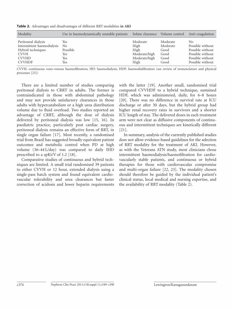
There are a limited number of studies comparingperitoneal dialysis to CRRT in adults. The former iscontraindicated in those with abdominal pathologyand may not provide satisfactory clearances in thoseadults with hypercatabolism or a high urea distributionvolume due to fluid overload. Two studies reported anadvantage of CRRT, although the dose of dialysisdelivered by peritoneal dialysis was low [15, 16]. Inpaediatric practice, particularly post cardiac surgery,peritoneal dialysis remains an effective form of RRT, insingle organ failure [17]. Most recently, a randomisedtrial from Brazil has suggested broadly equivalent patientoutcomes and metabolic control when PD at highvolume (36–44 L/day) was compared to daily IHDprescribed to a spKt/V of 1.2 [18].
Comparative studies of continuous and hybrid tech-niques are limited. A small trial randomised 39 patientsto either CVVH or 12 hour, extended dialysis using asingle-pass batch system and found equivalent cardio-vascular tolerability and urea clearances but fastercorrection of acidosis and lower heparin requirements
with the latter [19]. Another small, randomised trialcompared CVVHDF to a hybrid technique, sustainedHDF, which was administered, daily, for 6–8 hours[20]. There was no difference in survival rate at ICUdischarge or after 30 days, but the hybrid group hadhigher renal recovery rates in survivors and a shorterICU length of stay. The delivered doses in each treatmentarm were not clear as diffusive components of continu-ous and intermittent techniques are kinetically different[21].
In summary, analysis of the currently published studiesdoes not allow evidence-based guidelines for the selectionof RRT modality for the treatment of AKI. However,as with the Veterans ATN study, most clinicians choseintermittent haemodialysis/haemofiltration for cardio-vascularly stable patients, and continuous or hybridtherapies for those with cardiovascular compromiseand multi-organ failure [22, 23]. The modality chosenshould therefore be guided by the individual patient’sclinical status, local medical and nursing expertise, andthe availability of RRTmodality (Table 2).
Table 2. Advantages and disadvantages of different RRTmodalities in AKI
Modality Use in haemodynamically unstable patients Solute clearance Volume control Anti-coagulation
Peritoneal dialysis Yes Moderate Moderate NoIntermittent haemodialysis No High Moderate Possible withoutHybrid techniques Possible High Good Possible withoutCVVH Yes Moderate/high Good Possible withoutCVVHD Yes Moderate/high Good Possible withoutCVVHDF Yes High Good Possible without
CVVH: continuous veno-venous haemofiltration; HD: haemodialysis; HDF: haemodiafiltration (see review of nomenclature and physicalprocesses [23])
c374 Nephron Clin Pract 2011;118(suppl 1):c349–c390 Lewington/Kanagasundaram
References
1 Swartz RD, Messana JM, Orzol S, Port FK. Comparing continuoushemofiltration with hemodialysis in patients with severe acute renalfailure. Am J Kidney Dis 1999;34:424–432
2 Guerin C, Girard R, Selli JM, Ayzac L. Intermittent versus continuousrenal replacement therapy for acute renal failure in intensive careunits: Results from a multicenter prospective epidemiological survey.Intensive Care Medicine 2002;28:1411–1418
3 John S, Griesbach D, Baumgartel M, Weihprecht H, Schmieder RE,Geiger H. Effects of continuous haemofiltration vs. intermittent haemo-dialysis on systemic haemodynamics and splanchnic regional perfusionin septic shock patients: A prospective, randomized clinical trial.Nephrol Dial Transplant 2001;16:320–327
4 Mehta RL, McDonald B, Gabbai FB, Pahl M, Pascual MT, Farkas A,Kaplan RM, Collaborative Group for Treatment of ARF in ICU. Arandomized clinical trial of continuous versus intermittent dialysis foracute renal failure. Kidney Int 2001;60:1154–1163
5 Augustine JJ, Sandy D, Seifert TH, Paganini EP. A randomized con-trolled trial comparing intermittent with continuous dialysis in patientswith ARF. Am J Kidney Dis 2004;44:1000–1007
6 Uehlinger DE, Jakob SM, Ferrari P, Eichelberger M, Huynh-Do U, MartiHP, Mohaupt MG, Vogt B, Rothen HU, Regli B, Takala J, Frey FJ.Comparison of continuous and intermittent renal replacement therapyfor acute renal failure. Nephrol Dial Transplant 2005;20:1630–1637.Epub 2005 May 1610
7 Vinsonneau C, Camus C, Combes A, Costa de Beauregard MA, KloucheK, Boulain T, Pallot JL, Chiche JD, Taupin P, Landais P, Dhainaut JF.Continuous venovenous haemodiafiltration versus intermittenthaemodialysis for acute renal failure in patients with multiple-organdysfunction syndrome: A multicentre randomised trial. Lancet 2006;368:379–385
8 Lins RL, Elseviers MM, Van der Niepen P, Hoste E, Malbrain ML,Damas P, Devriendt J. Intermittent versus continuous renal replacementtherapy for acute kidney injury patients admitted to the intensive careunit: Results of a randomized clinical trial. Nephrol Dial Transplant2009;24:512–518
9 Kellum JA, Angus DC, Johnson JP, Leblanc M, Griffin M, RamakrishnanN, Linde-Zwirble WT. Continuous versus intermittent renal replace-ment therapy: A meta-analysis. Intensive Care Medicine 2002;28:29–37
10 Tonelli M, Manns B, Feller-Kopman D. Acute renal failure in the inten-sive care unit: A systematic review of the impact of dialytic modality onmortality and renal recovery.[comment]. Am J Kidney Dis 2002;40:875–885
11 Bagshaw SM, Berthiaume LR, Delaney A, Bellomo R. Continuous versusintermittent renal replacement therapy for critically ill patients withacute kidney injury: A meta-analysis. Critical Care Medicine 2008;36:610–617
12 Rabindranath K, Adams J, Macleod AM, Muirhead N. Intermittentversus continuous renal replacement therapy for acute renal failure inadults. Cochrane Database of Systematic Reviews 2007:CD003773
13 Manns B, Doig CJ, Lee H, Dean S, Tonelli M, Johnson D, Donaldson C.Cost of acute renal failure requiring dialysis in the intensive careunit: Clinical and resource implications of renal recovery. CriticalCare Medicine 2003;31:449–455
14 Farese S, Jakob SM, Kalicki R, Frey FJ, Uehlinger DE. Treatment of acuterenal failure in the intensive care unit: Lower costs by intermittentdialysis than continuous venovenous hemodiafiltration. ArtificialOrgans 2009;33:634–640
15 Gangji AS, Rabbat CG, Margetts PJ, Gangji AS, Rabbat CG, Margetts PJ.Benefit of continuous renal replacement therapy in subgroups of acutelyill patients: A retrospective analysis. Clin Nephrol 2005;63:267–275
16 Swartz RD, Bustami RT, Daley JM, Gillespie BW, Port FK, Swartz RD,Bustami RT, Daley JM, Gillespie BW, Port FK. Estimating the impactof renal replacement therapy choice on outcome in severe acute renalfailure. Clin Nephrol 2005;63:335–345
17 Strazdins V, Watson AR, Harvey B, European Pediatric PeritonealDialysis Working G, Strazdins V, Watson AR, Harvey B, EuropeanPediatric Peritoneal Dialysis Working Group. Renal replacementtherapy for acute renal failure in children: European guidelines.Pediatric Nephrology 2004;19:199–207
18 Gabriel DP, Caramori JT, Martim LC, Barretti P, Balbi AL. High volumeperitoneal dialysis vs. daily hemodialysis: A randomized, controlled trialin patients with acute kidney injury. Kidney Int 2008;(Suppl):S87–S93
19 Kielstein JT, Kretschmer U, Ernst T, Hafer C, BahrMJ, Haller H, Fliser D.Efficacy and cardiovascular tolerability of extended dialysis in criticallyill patients: A randomized controlled study. Am J Kidney Dis 2004;43:342–349
20 Abe M, Okada K, Suzuki M, Nagura C, Ishihara Y, Fujii Y, Ikeda K, KaizuK, Matsumoto K. Comparison of sustained hemodiafiltration with con-tinuous venovenous hemodiafiltration for the treatment of critically illpatients with acute kidney injury. Artificial Organs 2010;34:331–338
21 Kanagasundaram NS, Paganini EP: Critical care dialysis – a gordianknot (but is untying the right approach?). Nephrol Dial Transplant1999;14:2590–2594
22 The VA NIH Acute Renal Failure Trial Network, Palevsky PM, ZhangJH, O’Connor TZ, Chertow GM, Crowley ST, Choudhury D, FinkelK, Kellum JA, Paganini E, Schein RM, Smith MW, Swanson KM,Thompson BT, Vijayan A, Watnick S, Star RA, Peduzzi P. Intensity ofrenal support in critically ill patients with acute kidney injury. N EnglJ Med 2008;359:7–20
23 Kanagasundaram NS. Renal replacement therapy in acute kidney injury:An overview. British Journal of Hospital Medicine 2007;68:292–297
Renal Association Clinical PracticeGuidelines on Acute Kidney Injury
Nephron Clin Pract 2011;118(suppl 1):c349–c390 c375
7. Acute Kidney Injury (AKI) (Guidelines AKI7.1–7.3)
Guideline 7.1 – AKI: Choice of dialyser/haemofiltermembraneWe recommend that synthetic or modified cellulosic
membranes should be used in preference to unmodifiedcellulose membranes. (1B)
Guideline 7.2 – AKI: Choice of dialysate/replacementfluidWe recommend that bicarbonate should be the pre-
ferred buffer for dialysate and replacement fluid in con-tinuous renal replacement therapy (CRRT) techniquesunless regional citrate anticoagulation is employed. (1C)
Guideline 7.3 – AKI: Microbial standards for fluidsWe recommend that microbial standards for fluids
used for chronic haemodialysis (HD)/haemodiafiltration(HDF) should be also applied to extracorporeal therapyfor AKI. (1A)
Rationale for 7.1–7.3Laboratory experiments have shown that synthetic
membranes tend to cause less activation of complementand mononuclear cells. Results from clinical trials have,however, been conflicting with some suggesting a survivaladvantage for more biocompatible membranes [1–4] andothers showing no benefit [5–8]. Comparison of individualstudies has been compromised by variability in method-ology, definitions of AKI, definitions of ‘biocompatibility’,and in other aspects of dialysis provision, such as timing ofinitiation and adequacy. No study was blinded.
Meta-analyses have yielded varying results. Subrama-nian et al. found worse survival with the use of non-biocompatible membranes although this effect mayhave been confined to unsubstituted rather than modi-fied cellulose membranes [9]. This meta-analysis did,however, include a large, observational study that mayhave skewed results in favour of biocompatibility. Themost recent Cochrane meta-analysis continues to showno outcome advantage with biocompatible membranes[10]. The definition of non-biocompatibility includedboth unsubstituted and modified cellulose membranes.Neither meta-analysis could demonstrate a differencein rates of renal recovery. No recommendation can there-fore be made about the use of synthetic over modifiedcellulosic membranes for treating patients with AKI.Adverse survival found with the use of unsubstitutedcellulose membranes favours the use of either synthetic
or modified cellulose membrane materials. The maindeterminants of membrane choice will remain technical,with prescription of the dialyser/haemofilter dictated byits intended use.
Lactate and acetate have been largely replaced bybicarbonate as the primary buffer for dialysate used inIHD for established renal failure and this practice haspropagated, by default, to IHD for AKI. In a similarfashion, bicarbonate has become the primary buffer forboth replacement and dialysate fluids in CRRT. Drivenby concerns about exacerbating existing lactic acidosis,particularly in those with liver failure, the developmentof commercially available bicarbonate-based fluids thatcircumvent the inherent instability of such solutionshas led to their increasing utilisation. Evidence of benefitover lactate-based solutions is inconsistent with somestudies showing no substantive differences in metabolicparameters, pH, or haemodynamic status [11] whilstothers have shown improved haemodynamic stability[12, 13] and more rapid control of systemic acidosis[14]. Despite these conflicting data, the likelihood ofbenefit, especially in the sickest patients, and the readyavailability of commercially-prepared bicarbonate fluid,seems to justify its use in CRRT.
Little comparative data exists on the metabolic effectsof citrate- and bicarbonate-based solutions. A single-centre, prospective sequential cohort study suggestedequivalent acid-base and electrolyte control in patientsreceiving CVVH [15] but numbers were small andsubstitution fluid infusion rates differed between thetwo treatment cohorts. Given the absence of high qualityevidence no recommendation can be made on theoptimal choice for metabolic control between citrate-and bicarbonate-based solutions.
A final consideration in the use of dialysate/replace-ment fluids is their microbial integrity. The potentialfor clinically significant transfer of pyrogen-inducingmaterial in dialysate and substitution fluids is wellrecognised in the setting of chronic haemodialysis andhaemodiafiltration and has led to the establishment ofstrict standards for microbiological purity. In the absenceof specific evidence for renal support for AKI, it isrecommended that these same standards should apply.Intermittent HD will tend to be provided under theauspices of a renal unit so the same water qualitystandards should already be in place across both acuteand chronic services. The need to assure microbialintegrity of fluids for CRRTs has not, however, beenwell recognised although evidence now exists thatbreaches may well be frequent [16, 17].
c376 Nephron Clin Pract 2011;118(suppl 1):c349–c390 Lewington/Kanagasundaram
References
1 Himmelfarb J, Tolkoff Rubin N, Chandran P, Parker RA, Wingard RL,Hakim R. A multicenter comparison of dialysis membranes in thetreatment of acute renal failure requiring dialysis. J Am Soc Nephrol1998;9:257–266
2 Hakim RM, Wingard RL, Parker RA. Effect of the dialysis membrane inthe treatment of patients with acute renal failure. N Engl J Med1994;331:1338–1342
3 Schiffl H, Lang SM, Konig A, Strasser T, Haider MC, Held E. Biocom-patible membranes in acute renal failure: Prospective case-controlledstudy. Lancet 1994;344:570–572
4 Schiffl H, Sitter T, Lang S, Konig A, Haider M, Held E. Bioincompatiblemembranes place patients with acute renal failure at increased risk ofinfection. ASAIO Journal 1995;41:M709–M712
5 Albright RC, Jr., Smelser JM, McCarthy JT, Homburger HA, BergstralhEJ, Larson TS. Patient survival and renal recovery in acute renal failure:Randomized comparison of cellulose acetate and polysulfone membranedialyzers. Mayo Clinic Proceedings 2000;75:1141–1147
6 Gastaldello K, Melot C, Kahn RJ, Vanherweghem JL, Vincent JL,Tielemans C. Comparison of cellulose diacetate and polysulfonemembranes in the outcome of acute renal failure. A prospectiverandomized study. Nephrol Dial Transplant 2000;15:224–230
7 Jorres A, Gahl GM, Dobis C, Polenakovic MH, Cakalaroski K,Rutkowski B, Kisielnicka E, Krieter DH, Rumpf KW, Guenther C,Gaus W, Hoegel J. Haemodialysis-membrane biocompatibility andmortality of patients with dialysis-dependent acute renal failure: Aprospective randomised multicentre trial. International multicentrestudy group. Lancet 1999;354:1337–1341
8 Kurtal H, von Herrath D, Schaefer K. Is the choice of membraneimportant for patients with acute renal failure requiring hemodialysis?Artificial Organs 1995;19:391–394
9 Subramanian S, Venkataraman R, Kellum JA. Influence of dialysismembranes on outcomes in acute renal failure: A meta-analysis.Kidney Int 2002;62:1819–1823
10 Alonso A, Lau J, Jaber BL. Biocompatible hemodialysis membranes foracute renal failure. Cochrane Database of Systematic Reviews2008:CD005283
11 Kierdorf H, Leue C, Heintz B, Riehl J, Melzer H, Sieberth HG. Contin-uous venovenous hemofiltration in acute renal failure: Is a bicarbonate-or lactate-buffered substitution better? Contributions to Nephrology1995;116:38–47
12 Thomas AN, Guy JM, Kishen R, Geraghty IF, Bowles BJ, Vadgama P.Comparison of lactate and bicarbonate buffered haemofiltration fluids:Use in critically ill patients. Nephrol Dial Transplant 1997;12:1212–1217
13 Barenbrock M, Hausberg M, Matzkies F, de la Motte S, Schaefer RM.Effects of bicarbonate- and lactate-buffered replacement fluids on cardio-vascular outcome in CVVH patients. Kidney Int 2000;58:1751–1757
14 McLean AG, Davenport A, Cox D, Sweny P. Effects of lactate-bufferedand lactate-free dialysate in CAVHD patients with and without liverdysfunction. Kidney Int 2000;58:1765–1772
15 Aman J, Nurmohamed SA, Vervloet MG, Groeneveld AB: Metaboliceffects of citrate- vs. bicarbonate-based substitution fluid in continuousvenovenous hemofiltration: A prospective sequential cohort study.Journal of Critical Care 2010;25:120–127
16 Kanagasundaram NS, Larive AB, Paganini EP. A preliminary survey ofbacterial contamination of the dialysate circuit in continuous veno-venous hemodialysis. Clin Nephrol 2003;59:47–55
17 Moore I, Bhat R, Hoenich NA, Kilner AJ, Prabhu M, Orr KE,Kanagasundaram NS. A microbiological survey of bicarbonate-basedreplacement circuits in continuous veno-venous hemofiltration. CriticalCare Medicine 2009;37:496–500
Renal Association Clinical PracticeGuidelines on Acute Kidney Injury
Nephron Clin Pract 2011;118(suppl 1):c349–c390 c377
8. Acute Kidney Injury (AKI) (Guidelines AKI8.1–8.6)
Guideline 8.1 – AKI: Vascular access for RRTWe recommend that acute access for renal replace-
ment therapy should be veno-venous rather thanarterio-venous. (1A)
Guideline 8.2 – AKI: Vascular access for RRTWe recommend that dialysis catheters should be of an
adequate length to minimise the risks of access recircula-tion. (1C)
Guideline 8.3 – AKI: Vascular access for RRTWe suggest that the access site and catheter type
should be chosen with regard to the phase of the patient’sillness. (2C)
Guideline 8.4 – AKI: Vascular access for RRTWe recommend that access should be placed by
experienced or appropriately supervised staff. Real-timeultrasound guidance should be used to aid placementof upper body access. (1A)
Guideline 8.5 – AKI: Vascular access for RRTWe recommend that it is advisable that real-time
ultrasound guidance be used for the insertion of femoralaccess. (1D)
Guideline 8.6 – AKI: Vascular access for RRTWe recommend that subclavian access should be
avoided in patients at risk of progressing to CKD stage4 or 5 due to the risks of compromising future,permanent vascular access. (1D)
Guideline 8.7 – AKI: Vascular access for RRTWe suggest that non-dominant arm upper limb
vasculature should be preserved as a contingency forfuture permanent access. (2C)
Guideline 8.8 – AKI: Vascular access for RRTWe recommend that temporary access should be
changed at appropriate intervals (as per local protocol)to minimise the risk of infection. (1C)
Guideline 8.9 – AKI: Vascular access for RRTWe suggest that local policies on prevention of
catheter-related infection should be optimised byreserving the catheter for extracorporeal treatment only.(1D)
Audit measure1. Incidence of dialysis catheter-related bacteraemia
and sepsis in patients with AKI
Rationale for 8.1–8.9In industrialised societies, the vast majority of
continuous therapy is now provided using pumped,veno-venous methods [1]. Not only does this techniquesupport the requirement for adequate blood flow rates toachieve the higher ultrafiltration/dialysate flow rates usedin modern CRRT, but it also avoids the potential hazardsof the acute arterio-venous access used historically [2].The adequacy of intermittent techniques is much moredependent on delivered, extracorporeal blood flow.Catheter failure is a frequent cause of under-delivery ofthe prescribed IHD dose [3] and should be borne inmind as a cause of any prescription-delivery shortfall.Temporary vascular access used in acute dialysis maylead to levels of access recirculation of nearly 40%depending on the site and length of access, blood flowand reversal of the lines [4].
Several venous catheters are available, with the dual-lumen design being the most popular because of easeof insertion and good flow characteristics [5]. Suchcatheters usually have a double-D cross-sectional profileand are amenable to guide wire changes [6]. Cathetersmade of semi-rigid polyurethane or softer silicone areregarded as the best in terms of thrombogenicity [1].The former are a reasonable short-term option (<3weeks) while the latter might be best utilised for longerterm dialysis because of the lower propensity to causeendovascular trauma [1]. Such catheters, used with sub-cutaneous tunnelling, are highly desirable if RRT is likelyto be prolonged (>3 weeks) [7]. A small, single-centre,randomised control trial compared the performance oftunnelled to non-tunnelled femoral catheters insertedin AKI patients prior to initiation of renal support [8].Tunnelled access was found to give better flow character-istics, fewer post-insertion complications, greater long-evity and less likelihood of a prescription-deliveryshortfall. There were, however, more insertion failuresin this group and successful catheter placements tooksignificantly longer. Of note, all catheters insertionswere performed by a single operator. The applicabilityof these findings to routine practice is unclear.
Use of real-time ultrasound guidance for catheterplacement at upper body sites has been demonstratedto be associated with greater success and fewer complica-tions [9]. It is advisable that similar guidance be used forfemoral catheter insertion.
c378 Nephron Clin Pract 2011;118(suppl 1):c349–c390 Lewington/Kanagasundaram
A number of factors should be taken into considera-tion in choosing a site for insertion and appropriatecatheter length. Femoral catheters shorter than 20 cmfrom hub to tip are associated with higher degrees ofaccess recirculation [4, 10]. Femoral catheters of atleast 24 cm in length may produce improved flow rates[6]. Because of the risks of infection and femoral veinthrombosis, it is recommended that femoral cathetersbe removed and replaced on at least a weekly basis[6, 11]. It is advisable that femoral catheters be replacedby upper body access once the patient starts to mobilise.
The subclavian approach carries with it the long-termrisk of venous stenosis that may compromise future,ipsilateral, permanent upper limb arteriovenous access.Subclavian access is thus best avoided in those with alikelihood of progressing to CKD stage 4 or 5. Theinternal jugular approach may be associated with alower incidence of both accidental pneumothorax [6]and long-term venous stenosis [12] in comparison withsubclavian access, and is the preferred upper bodyaccess. Infection may be somewhat more common thanat the subclavian site, however, especially in patientswith tracheostomies [12].
For the average adult, internal jugular vein cathetersshould be around 20 cm in length on the right and24 cm on the left [12], to ensure safe positioning ofthe catheter tip in the lower superior vena cava. Withappropriate infection control and catheter care, upperbody access may only need replacement every 2–3 weeks[12, 13]. Local guidelines may suggest a more frequentschedule of replacement and should be adhered to.
Catheter-related bacteraemia and exit site infectionare significant risks of temporary access for acute RRT[14]. Fastidious insertion technique by experienced orappropriately supervised staff and rigorous cathetercare can reduce this risk [15]. It is advisable that dialysiscatheters be reserved solely for the purpose of RRT asrepeated manipulations for non-RRT related reasonsmay increase the risk of contamination. Guidewire-exchange of catheters for non-infection related reasonsmay not increase bacteraemia rates [11] but cannot berecommended in the presence of catheter-related bacter-aemia or exit-site infection. Between periods of RRTcatheters may be locked with heparin 1000 units/ml tolumen volumes unless there is a clear contraindication.It is recommended that higher concentrations be avoideddue to the risks associated with over-dosing and leakageof the lock into the systemic circulation. Alternatives toheparin to reduce infection risks include heparin andantibiotic combinations, citralock and taurolock althoughno high quality data currently exists that would supporttheir routine use. Recently, antimicrobial catheters havebeen introduced for vascular access, either impregnatedwith silver or antibiotic coated. Preliminary trials havesuggested a reduction in the incidence of catheterassociated bacteraemia but larger trials will be requiredbefore the use of these catheters can be recommended asstandard practice.
In patients who are likely to progress to stage 4 or 5CKD, upper limb vasculature should be preserved as acontingency for future permanent vascular access [7].
Renal Association Clinical PracticeGuidelines on Acute Kidney Injury
Nephron Clin Pract 2011;118(suppl 1):c349–c390 c379
References
1 Canaud B, Leray-Moragues H, Leblanc M, Klouche K, Vela C, Beraud JJ.Temporary vascular access for extracorporeal renal replacement thera-pies in acute renal failure patients. Kidney Int 1998;53:S142–S150
2 Storck M, Hartl WH, Zimmerer E, Inthorn D. Comparison of pump-driven and spontaneous continuous haemofiltration in postoperativeacute renal failure. Lancet 1991;337:452–455
3 Kanagasundaram NS, Greene T, Larive AB, Daugirdas JT, Depner TA,Garcia M, Paganini EP. Prescribing an equilibrated intermittenthemodialysis dose in intensive care unit acute renal failure. KidneyInt 2003;64:2298–2310
4 Kelber J, Delmez JA, Windus DW. Factors affecting delivery of high-efficiency dialysis using temporary vascular access. Am J Kidney Dis1993;22:24–29
5 Tapson JS, Hoenich NA, Wilkinson R, Ward MK. Dual lumen sub-clavian catheters for haemodialysis. International Journal of ArtificialOrgans 1985;8:195–200
6 Uldall R. Vascular access for continuous renal replacement therapy.Seminars in Dialysis 1996;9:93–97
7 Canaud B, Desmeules S, Klouche K, Leray-Moragues H, Beraud JJ.Vascular access for dialysis in the intensive care unit. Best Practice &Research Clinical Anaesthesiology 2004;18:159–174
8 Klouche K, Amigues L, Deleuze S, Beraud JJ, Canaud B. Complications,effects on dialysis dose, and survival of tunneled femoral dialysiscatheters in acute renal failure. Am J Kidney Dis 2007;49:99–108
9 Anonymous: NICE technology appraisal guidance, no. 49 – guidance onthe use of ultrasound locating devices for placing central venouscatheters, National Institute for Clinical Excellence, 2002
10 Leblanc M, Fedak S, Mokris G, Paganini EP. Blood recirculation intemporary central catheters for acute hemodialysis. Clin Nephrol1996;45:315–319
11 Oliver MJ, Callery SM, Thorpe KE, Schwab SJ, Churchill DN. Risk ofbacteremia from temporary hemodialysis catheters by site of insertionand duration of use: A prospective study. Kidney Int 2000;58:2543–2545
12 Cimochowski GE, Worley E, Rutherford WE, Sartain J, Blondin J,Harter H. Superiority of the internal jugular over the subclavianaccess for temporary dialysis. Nephron 1990;54:154–161
13 Weijmer MC, Vervloet MG, ter Wee PM. Compared to tunnelled cuffedhaemodialysis catheters, temporary untunnelled catheters are associatedwith more complications already within 2 weeks of use. Nephrol DialTransplant 2004;19:670–677
14 Kairaitis LK, Gottlieb T. Outcome and complications of temporaryhaemodialysis catheters. Nephrol Dial Transplant 1999;14:1710–1714
15 Deshpande KS, Hatem C, Ulrich HL, Currie BP, Aldrich TK, Bryan-Brown CW, Kvetan V. The incidence of infectious complications ofcentral venous catheters at the subclavian, internal jugular, and femoralsites in an intensive care unit population. Crit Care Med 2005;33:13–20;discussion 234–235
c380 Nephron Clin Pract 2011;118(suppl 1):c349–c390 Lewington/Kanagasundaram
9. Acute Kidney Injury (AKI) (Guidelines AKI9.1–9.4)
Guideline 9.1 – AKI: Anticoagulation forextracorporeal therapiesWe recommend that anticoagulation for RRT should
be tailored according to patient characteristics and themodality of RRT chosen. (1C)
Guideline 9.2 – AKI: Anticoagulation forextracorporeal therapiesWe recommend that regional anticoagulation with
citrate reduces risk of haemorrhage compared to sys-temic heparinisation. The complexity of the techniquemeans that this should be in routine use on any uniton which it is employed in order to allow sufficientlevels of expertise to be maintained. (1C)
Guideline 9.3 – AKI: Anticoagulation forextracorporeal therapiesWe suggest that prostacyclin is a suitable alternative to
unfractionated heparin in those at increased risk ofbleeding, but may cause haemodynamic instability. (2C)
Guideline 9.4 – AKI: Anticoagulation forextracorporeal therapiesWe suggest that a no-anticoagulation, saline flush
strategy can be used in patients receiving continuousand intermittent RRT who are at high risk of bleeding.However, ultrafiltration requirements are increased,effective intermittent HD time is reduced and the tech-nique runs the risk of membrane fibre rupture. (2C)
Audit measure1. Incidence of heparin induced thrombocytopenia
Rationale for 9.1–9.4Clotting of the extracorporeal circuit is a significant
source of under-delivery of the prescribed dose of RRTand is the most frequent cause of therapy interruptionin CRRT. The hypercoaguable state of the critically-illpatient with AKI [1] compounds various technicalfactors such as non-laminar flow within both the vascu-lar access and circuit, blood-membrane interactions, theair-blood interface in the venous bubble trap and thehaemoconcentration induced by high ultrafiltrationvolumes used in CVVH/CVVHDF.
The most widely used anticoagulant for RRT inpatients with AKI is unfractionated heparin (UFH) [2,3]. Although an effective anticoagulant for IHD in
patients with CKD, UFH may be less effective in AKI,as many critically ill patients have reduced levels ofantithrombin, especially when used for patients treatedwith CRRT. In addition, systemic heparinisation isassociated with a risk of bleeding and also with thedevelopment of heparin-induced thrombocytopenia(HIT) [4, 5]. Low molecular weight heparins havegenerally not been shown to be superior over UFH andhave an extended half life in AKI and require monitoringwith anti-Xa activity [3].
Regional heparin protocols, with reversal of heparinby infusion of protamine into the return line, havebeen developed to prevent systemic anticoagulationand minimise bleeding risk. Unfortunately, these proto-cols are cumbersome, may be associated with paradoxicalincreased risk of bleeding if excess protamine is infused,and do not alter the risk of HIT. Other anticoagulantsthat can be used as alternatives for anticoagulation ofthe extracorporeal circuit in patients with a history ofHIT include prostacyclin (prostaglandin I2 – which isused in non-HIT patients who are at high risk of bleed-ing) [6–9], hirudin, nafamostat, and argatroban [3]. Thesynthetic heparinoids, danaparoid and fondiparinuxmay also be used, although cross reactivity with theHIT antibodies has occasionally been reported. If theseagents are used and the peripheral platelet count doesnot increase within 72 hours cross reactivity should beexcluded Argatroban is currently not licensed in theUK, and has to be given by continuous infusion. Dana-paroid, fondiparinux and hirudin are all renally excretedand therefore have extended half lives in AKI.
The synthetic heparinoids require monitoring ofanti-Xa activity and hirudin by either its plasmaconcentration or the ecarin clotting time. Hirudin ispartially cleared by high flux membranes, but themajority of patients given hirudin for CRRT developantibodies to hirudin, which reduce its clearance andextend its half life so increasing the risk of haemorrhage.In cases of excess anticoagulation associated withbleeding, there are no specific antidotes for theseagents, unlike protamine for unfractionated heparin.Although activated factor VII has been shown to beeffective and hirudin can be cleared by high fluxdialysis/CRRT plasma exchange is required in cases ofhirudin antibodies [3].
Over the last decade citrate has emerged as a veryeffective regional anticoagulant for use in CRRT [10–14]. Citrate is infused into the pre-filter line and worksby chelating calcium. Calcium is then re-infusedseparately, or into the return line, to maintain normal
Renal Association Clinical PracticeGuidelines on Acute Kidney Injury
Nephron Clin Pract 2011;118(suppl 1):c349–c390 c381
systemic ionised calcium concentrations. Commerciallyavailable citrate systems have not been available untilrecently, so individual units developed their own proto-cols for citrate anticoagulation. Citrate comes as asodium salt, and each molecule is indirectly convertedto three bicarbonates, so there can potentially be changesin sodium balance and acid-base status depending uponthe citrate load and the ability of the patient toadequately metabolise citrate.
There have been few prospective comparative studiesof UFH and citrate anticoagulation. In two CRRTstudiesthe median circuit survival time was significantly pro-longed with citrate (70 hr v. 40 hr and 124 v. 38 hr) andthere was reduced blood transfusion requirements and/or haemorrhage in the citrate groups [10, 11]. Howeverpatients that cannot adequately metabolise citrate tobicarbonate, such as those with acute liver failure, maydevelop a ‘calcium gap’ due to the accumulation ofcalcium citrate complex. The ‘calcium gap’ is the calciumcomplexed with citrate, and is the difference between thetotal calcium measured and that due to ionised calciumand plasma protein bound calcium. As these patientscannot adequately metabolise citrate, they will developa metabolic acidosis with hypercitrataemia. On theother hand, over administration of citrate to patientswho can metabolise the citrate load will result in a sys-temic alkalosis. In Japan, nafamostat is used as a regional
anticoagulant, and appears to have similar efficacy andsafety profile to citrate.
Although UFH remains the most commonlyemployed extracorporeal anticoagulant for RRT inpatients with AKI, there is emerging data to supportthe safety and potential superiority of regional citrateanticoagulation for CRRT. Now that citrate based anti-coagulation systems have been developed for CRRT bythe major commercial companies, the proportion ofpatients with AKI treated by citrate systems may increase.
The short duration of intermittent techniques mayallow a ‘minimal’ heparin (e.g. 500 IU/hour) or evenno heparin strategy. Regular saline flushes, used to sus-tain the latter, may, however, reduce the effective dialysistime. No heparin use in CRRT is possible and can achieveadequate solute clearances but its disadvantages includethe need for increased ultrafiltration, the potential riskof dialyser fibre rupture and additional nursing workload[15, 16].
Finally, pre-dilutional fluid replacement during con-tinuous haemofiltration can help minimise the haemo-concentration induced by large ultrafiltration volumesbut comes at the price of the inefficiency of ultrafilteringa mixture of just-infused replacement fluid and plasma –the proportions of which are important considerationsin the CRRT prescription.
c382 Nephron Clin Pract 2011;118(suppl 1):c349–c390 Lewington/Kanagasundaram
References
1 Davenport A. The coagulation system in the critically ill patient withacute renal failure and the effect of an extracorporeal circuit. Am JKidney Dis 1997;30:S20–S27
2 Davenport A, Mehta S. The Acute Dialysis Quality Initiative – part VI:access and anticoagulation in CRRT. Adv Ren Replace Ther. 2002;9:273–281
3 Oudemans-van-Straaten HM, Wester JPJ, Pont ACJM de, SchetzMRC. Anticoagulation strategies in continuous renal replacementtherapy: can the choice be evidence based? Intensive Care Med 2006;32:188–202
4 Mehta RL. Anticoagulation strategies for continuous renal replacementtherapies: what works? Am J Kidney Dis 28:S8–S14, 1996
5 Davenport A. Management of heparin-induced thrombocytopeniaduring continuous renal replacement therapy. Am J Kidney Dis 1998;32:E3
6 Ponikvar R, Kandus A, Buturovic J, Kveder R. Use of prostacyclin asthe only anticoagulant during continuous venovenous hemofiltration.Contributions to Nephrology 1991;93:218–220
7 Journois D, Chanu D, Pouard P, Mauriat P, Safran D. Assessment ofstandardized ultrafiltrate production rate using prostacyclin in contin-uous venovenous hemofiltration. Contributions to Nephrology 1991;93:202–204
8 Davenport A, Will EJ, Davison AM. Comparison of the use of standardheparin and prostacyclin anticoagulation in spontaneous and pump-driven extracorporeal circuits in patients with combined acute renaland hepatic failure. Nephron 1994;66:431–437
9 Langenecker SA, Felfernig M, Werba A, Mueller CM, Chiari A, ZimpferM. Anticoagulation with prostacyclin and heparin during continuousvenovenous hemofiltration. Crit Care Med 1994;22:1774–1781
10 Monchi M, Berghmans D, Ledoux D et al. Citrate vs. heparin foranticoagulation in continuous venovenous hemofiltration: a prospec-tive randomized study. Intensive Care Med. 2004;30:260–265
11 Kutsogiannis DJ, Gibney RT, Stollery D, Gao J. Regional citrate versussystemic heparin anticoagulation for continuous renal replacement incritically ill patients. Kidney Int. 2005;67:2361–2367
12 Palsson R, Niles JL. Regional citrate anticoagulation in continuousvenovenous hemofiltration in critically ill patients with a high risk ofbleeding. Kidney Int 1999;55:1991–1997
13 Mehta RL, McDonald BR, Aguilar MM, Ward DM. Regional citrateanticoagulation for continuous arteriovenous hemodialysis in criticallyill patients. Kidney Int 1990;38:976–981
14 Kirschbaum B, Galishoff M, Reines HD: Lactic acidosis treated withcontinuous hemodiafiltration and regional citrate anticoagulation.Crit Care Med 1992;20:349–353
15 Smith D, Paganini EP, Suhoza K, Eisele G, Swann S, Nakamoto S. Non-heparin continuous renal replacement therapy is possible, in Progress inArtificial Organs – 1985, edited by Nose Y, Kjellstrand C, Ivanovich P,Cleveland, ISAO Press, 1986, pp 226–230
16 Ramesh Prasad GV, Palevsky PM, Burr R, Lesko JM, Gupta B, GreenbergA. Factors affecting system clotting in continuous renal replacementtherapy: results of a randomized, controlled trial. Clin Nephrol53:55–60;2000
Renal Association Clinical PracticeGuidelines on Acute Kidney Injury
Nephron Clin Pract 2011;118(suppl 1):c349–c390 c383
10. Acute Kidney Injury (AKI) (Guidelines AKI10.1–10.5)
Guideline 10.1 – AKI: Renal Replacement TherapyprescriptionWe recommend that the delivered dose of RRT should
be assessed to ensure the adequacy of the prescription.(1A)
Guideline 10.2 – AKI: Renal Replacement TherapyprescriptionWe recommend that the prescribed dose should be
assessed at each session (for intermittent haemodialysis)and daily (for continuous RRT) to account for anymeasured shortfalls in delivered dose. (1A)
Guideline 10.3 – AKI: Renal Replacement TherapyprescriptionWe recommend that patients with AKI and multi-
organ failure treated by continuous renal replacementtherapy (CRRT) should receive treatment doses equiva-lent to post dilution ultrafiltration rates 525ml/kg/hr.A proportionate upward adjustment to the prescribedultrafiltration rate should be made in pre-dilutionalcontinuous haemofiltration. (1A)
Guideline 10.4 – AKI: Renal Replacement TherapyprescriptionWe recommend that patients with AKI and multi-
organ failure treated by intermittent haemodialysisshould receive either alternate day haemodialysis withat least the minimum dose considered appropriate forend-stage renal disease (ESRD), urea reduction ratio(URR) >65% or eKt/V >1.2 or daily haemodialysis.(1B)
Guideline 10.5 – AKI: Renal Replacement TherapyprescriptionWe suggest that renal replacement therapy dosing
methods that require an assessment of patient weightshould use a measured weight rather than an extrapo-lated weight from pre-morbid readings. (2B)
Audit measures1. Proportion of critically ill patients treated by
alternate day haemodialysis who receive eKt/V51.2 per session
2. Proportion of critically ill patients with AKI treatedwith continuous renal replacement therapy whoreceive >25mls/kg/hr post-dilution ultrafiltration
Rationale for 10.1–10.5Traditionally in studies in patients with AKI, the dose
(or intensity) of treatment has been assessed by ureaclearance in dialysis based modalities, and by ultra-filtration volume (a surrogate of urea clearance), in theconvective therapies. Urea is relatively non-toxic andregarded as a surrogate for other low molecular weighturaemic toxins. It is recognised that using urea as amarker of intensity or dose of RRT has a number oflimitations particularly in critically ill patients. Ureageneration rates will differ between patients, due topatient specific factors (age, sex and race etc), due todisease specific factors (the catabolic rate, the presenceof muscle injury and/or breakdown, sepsis and liverdisease) and due to medical therapy such as nutritionalsupport and steroid treatment. However given thecurrent absence of any other more suitable marker ureaclearance is accepted as the best way to compare intensityor dose of RRT. The urea clearance achieved duringCRRT is approximately equal to the effluent flow rate(dialysis and ultrafiltration flow rate combined).
The dose of RRT delivered to patients not onlyincludes small solute clearances but also larger ‘middle’molecules. The amount of these other moleculesremoved will depend on the modality used and is greaterfor convective (haemofiltration) than diffusion (dialysis)based techniques. Middle molecule clearance by inter-mittent therapies is also affected by both frequency andduration of therapy. In addition to solute clearances,the prescription and delivery of renal support to patientswith AKI also includes other key aspects of medicalmanagement, including sodium and water balance(patients are often grossly salt-and volume-loaded bythe time they reach the need for RRT; drug carriagesolutions and colloids will compound this, even whenthe period of ‘active’ renal re-perfusion has ceased),and correction of acid-base imbalance. There are funda-mental differences in provision of RRT to patients withestablished renal failure compared to those with AKIincluding the wide, intra- and inter-individual variabilityin key clinical and dialytic factors, such as total bodywater and the catabolic rate [1]. Thus, the prescriptionof a dose of RRT and assessment of its delivery willneed to be undertaken daily (for CRRT) and at eachsession (for IRRT).
There is a paucity of data regarding ‘adequate’ treat-ment doses of intermittent haemodialysis (IHD) to bedelivered in AKI. Analysis of a prospectively collecteddatabase has shown that higher doses of intermittenthaemodialysis, defined as a urea reduction ratio (URR)
c384 Nephron Clin Pract 2011;118(suppl 1):c349–c390 Lewington/Kanagasundaram
>58%, improved survival [2]. It should be noted thatthis cut-off dose, equivalent to a Kt/V of around 1, islower than that recommended for IHD for establishedrenal failure. In this study dialysis dose had no impacton patient survival in patients at the extremes of illnessseverity. Whereas, for those patients with intermediateseverity of illness, the delivery of dialysis dose in excessof the 50th percentile (Kt/V �1) was associated withlower mortality risk than lower doses [3].
Due to the lack of prospective studies addressing theminimum dose of RRT required in AKI a consensuspanel convened by the multinational Acute DialysisQuality Initiative (ADQI) recommended that patientswith AKI receive at least the minimum dose that isconsidered appropriate for patients with establishedrenal failure [4]. Recently the Veterans trial reportedthat there was no significant improvement in patientoutcomes provided a Kt/V of 1.2–1.4 per session wasdelivered [5]. Due to the difficulty in assessing thevolume of distribution of urea in patients with AKI,several studies have shown that the delivered dose ofIHD can be markedly lower than that prescribed [2, 6],and is not routinely measured in clinical practice. How-ever it must be stressed that weight-based RRT dosingis important and should be performed. To achieve aURR above 65% or eKt/V above 1.2 consistently in thevast majority of the haemodialysis population cliniciansshould aim for a minimum target URR of 70% orminimum eKt/V of 1.4 in individual patients.
Only one study has evaluated the effect of daily andalternate day IHD on the outcome among patientswith AKI [7]. This reported both lower mortality (28%v. 46%, p¼ 0.01) and shorter duration of AKI (9� 2 v.16� 6 days, p¼ 0.001) in the daily IHD group. Howeverthe dose of haemodialysis delivered to the alternate daygroup was low (mean delivered Kt/V of 0.94� 0.11).This probably accounted for the markedly increasedtime-averaged urea concentration, and the high incidenceof complications including gastrointestinal bleeding,mental status alteration, and infection reported in thisgroup.
Several studies have looked at dose in CRRT [5, 8–11].In a large single centre randomised controlled trial the435 enrolled patients were randomised to one of threecontinuous venovenous haemofiltration (CVVH) doses(post dilution). The doses were defined by achieveddaily ultrafiltration rates of 20ml/kg/hr, 35ml/kg/hr,and 45ml/kg/hr [8]. Mortality was markedly lower inthe intermediate and high dose arms (43% and 42%,respectively) compared to the low dose arm (59%,
p< 0.001). Subsequently there have been three furtherstudies comparing the dose of CRRT. Two smaller studiesfailed to show any survival benefit [10, 11]. Howeversurvival benefit was demonstrated in a study thatadded an additional dose of dialysis to haemofiltrationproviding an equivalent dose of 35ml/kg/hr [11].
An important technical consideration is that the slowdialysate flow rates employed in continuous venovenoushaemodialysis (CVVHD) and haemodiafiltration (HDF)ensures that the effluent fluid will be fully equilibratedwith plasma with respect to small solutes by the time itleaves the dialyser [12]. Ultrafiltration rates in convectivetreatments (haemofiltration) can thus be used, inter-changeably, with dialysate flow rates for the CRRTswhen considering urea clearances [13].
Two large multicentre randomised controlled studieshave been published recently to provide much neededguidance on the optimal dose of CRRT in critically illpatients. The Veteran Affairs/National Institute ofHealth Acute Renal Failure Trial Network (ATN) studywas performed in ICUs across the USA [5] whilst theRandomised Evaluation of Normal Versus AugmentedLevel Renal Replacement Therapy (RENAL) was con-ducted in ICUs in Australia and New Zealand [14].The ATN study showed no additional beneficial patientoutcome with a delivered CVVHDF dose (pre-dilution)of 35ml/kg/h compared to 20ml/kg/h, although therewas a non significant trend for better outcome in themore critically ill patients with the higher dose of RRT[5]. The RENAL trial failed to demonstrate any survivalbenefit from receiving post dilution CVVHDF at a doseof 40ml/kg/hr versus 25ml/kg/hr. These studies havenow provided evidence that there is no survival benefitin critically ill patients receiving ultrafiltration doses>25ml/kg/hr. This suggests that a minimum delivereddose of 25ml/kg/h is required, and to allow for circuitclotting, a higher dose should be prescribed, particularlyfor the critically ill patient.
There is little data on dose comparisons in critically illpatients receiving peritoneal dialysis (PD). A more recentstudy CRRTwas reported to be superior to PD in treatingpatients with malaria induced AKI [15], and this mayhave well have been due to the dose of PD delivered, asthe rate of creatinine clearance and correction of acidosiswere much inferior during PD therapy. Peritonealdialysis has been shown to be an effective therapy inchildren post cardiac surgery [16], when a PD dose inexcess of a weekly Kt/V urea of 2.1 was delivered, witha median creatinine clearance of 74.3 L/wk/1.73m2.Automated peritoneal dialysis machines are the preferred
Renal Association Clinical PracticeGuidelines on Acute Kidney Injury
Nephron Clin Pract 2011;118(suppl 1):c349–c390 c385
method for delivering individualised peritoneal dialysisdose and accurately measuring ultrafiltration.
Just as there are no studies looking at the dose ofperitoneal dialysis required for patients with singleorgan and multiple organ failure, there is a similarpaucity of data on patient outcomes with the recentlyintroduced ‘hybrid’ treatments (such as Genius1,extended daily dialysis (EDD), and sustained low-efficiency dialysis (SLED)). Only a relatively smallnumber of patients were treated by hybrid therapiesin the Veterans study, but there was no obviousimprovement in patient outcomes with more intensivetherapy above alternate day sessions delivering a Kt/V
of 51.3 [5]. However it must be remembered that ifintermittent haemodialysis, EDD and/or SLED tech-niques are used in the intensive care unit, unless thereis a dedicated water treatment plant available, thesimple treatment of domestic water with a single reverseosmosis unit and ultrafilters may not provide the qualityof water required for haemodiafiltration, unless batchdialysate systems (Genius1) are used. The ATN studydid not show any major benefit for either intensivehaemodialysis, hybrid therapies or CVVHDF [5], butthe amount of intermittent haemodialysis and/orhybrid therapy delivered per session was greater than inearlier studies.
References
1 Kanagasundaram NS, Paganini P. Acute renal failure on the intensivecare unit. Clin Med 2005;5:435–440
2 Kanagasundaram NS, Greene T, Larive AB, Daugirdas JT, Depner TA,Garcia M, Paganini EP. Prescribing an equilibrated intermittent hemo-dialysis dose in intensive care unit acute renal failure. Kidney Int2003;64:2298–2310
3 Paganini EP, Tapolyai M, Goormastic M, Halstenberg W, Kozlowski L,Leblanc M, Lee JC, Moreno L, Sakai K. Establishing a dialysistherapy/patient outcome link in intensive care unit acute dialysis forpatients with acute renal failure. Am J Kidney Dis 1996;28:S81–S89
4 Kellum JA, Mehta RL, Angus DC, Palevsky P, Ronco C. The firstinternational consensus conference on continuous renal replacementtherapy. Kidney Int. 2002;62:1855–1863
5 The VA/NIH acute renal failure trial network. Intensity of renal supportin critically ill patients with acute kidney injury. N Engl J Med 2008;359
6 Evanson JA, Himmelfarb J, Wingard R et al. Prescribed versus delivereddialysis in acute renal failure patients. Am J Kidney Dis. 1998;32:731–738
7 Schiffl H, Lang SM, Fischer R. Daily hemodialysis and the outcome ofacute renal failure. N Engl J Med. 2002;346:305–310
8 Ronco C, Bellomo R, Homel P et al. Effects of different doses in con-tinuous veno-venous haemofiltration on outcomes of acute renalfailure: a prospective randomised trial. Lancet. 2000;356:26–30
9 Bouman CS, Oudemans-Van Straaten HM, Tijssen JG et al. Effects ofearly high-volume continuous venovenous hemofiltration on survival
and recovery of renal function in intensive care patients with acuterenal failure: a prospective, randomized trial. Crit Care Med.2002;30:2205–2211
10 Tolwani AJ, Campbell RC, Stofan BS et al. Standard versus high doseCVVHDF for intensive care unit related acute renal failure. J Am SocNephrol 2008 Epub PMID 18337480
11 Saudan P, Niederberger M, De Seigneux S, Romand J, Pugin J, PernegerT, Martin PY. Adding a dialysis dose to continuous haemofiltrationincreases survival in patients with acute renal failure. Kidney Int2006;70:1312–1317
12 Bellomo R. Do we know the optimal dose for renal replacement therapyin the intensive care unit? Kidney Int 2006;70:1202–1204
13 Sigler MH. Transport characteristics of the slow therapies: implicationsfor achieving adequacy of dialysis in acute renal failure. Advances inRenal Replacement Therapy 1997;4:68–80
14 RENAL Replacement Therapy Study Investigators. Intensity of con-tinuous renal replacement therapy in critically ill patients. N Engl JMed. 2009;361:1627–1638
15 Phu NH, Hien TT, Mai NTet al. Hemofiltration and peritoneal dialysisin infection-associated acute renal failure in Vietnam. N Engl J Med.2002;347:895–902
16 McNeice KL, Ellis EE, Drummond-Webb JJ, Fontenot EE, O’Grady CM,Blaszak RT. Adequacy of peritoneal dialysis in patients followingcardiopulmonary bypass surgery. Pediatr Nephrol 2005;20:972–976
c386 Nephron Clin Pract 2011;118(suppl 1):c349–c390 Lewington/Kanagasundaram
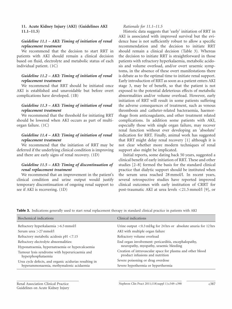
11. Acute Kidney Injury (AKI) (Guidelines AKI11.1–11.5)
Guideline 11.1 – AKI: Timing of initiation of renalreplacement treatmentWe recommend that the decision to start RRT in
patients with AKI should remain a clinical decisionbased on fluid, electrolyte and metabolic status of eachindividual patient. (1C)
Guideline 11.2 – AKI: Timing of initiation of renalreplacement treatmentWe recommend that RRT should be initiated once
AKI is established and unavoidable but before overtcomplications have developed. (1B)
Guideline 11.3 – AKI: Timing of initiation of renalreplacement treatmentWe recommend that the threshold for initiating RRT
should be lowered when AKI occurs as part of multi-organ failure. (1C)
Guideline 11.4 – AKI: Timing of initiation of renalreplacement treatmentWe recommend that the initiation of RRT may be
deferred if the underlying clinical condition is improvingand there are early signs of renal recovery. (1D)
Guideline 11.5 – AKI: Timing of discontinuation ofrenal replacement treatmentWe recommend that an improvement in the patient’s
clinical condition and urine output would justifytemporary discontinuation of ongoing renal support tosee if AKI is recovering. (1D)
Rationale for 11.1–11.5Historic data suggests that ‘early’ initiation of RRT in
AKI is associated with improved survival but the evi-dence base is not sufficiently robust to allow a specificrecommendation and the decision to initiate RRTshould remain a clinical decision (Table 3). Whereasthe decision to initiate RRT is straightforward in thosepatients with refractory hyperkalaemia, metabolic acido-sis and volume overload, and/or overt uraemic symp-toms, in the absence of these overt manifestations thereis debate as to the optimal time to initiate renal support.Early introduction of RRTas soon as a patient enters AKIstage 3, may be of benefit, so that the patient is notexposed to the potential deleterious effects of metabolicabnormalities and/or volume overload. However, earlyinitiation of RRT will result in some patients sufferingthe adverse consequences of treatment, such as venousthrombosis and catheter-related bacteraemia, haemor-rhage from anticoagulants, and other treatment relatedcomplications. In addition some patients with AKI,especially those with single organ failure, may recoverrenal function without ever developing an ‘absolute’indication for RRT. Finally, animal work has suggestedthat RRT might delay renal recovery [1] although it isnot clear whether more modern techniques of renalsupport also might be implicated.
Initial reports, some dating back 50 years, suggested aclinical benefit of early initiation of RRT. These and otherstudies [2–8] formed the basis for the standard clinicalpractice that dialytic support should be instituted whenthe serum urea reached 28mmol/l. In recent years,several retrospective studies have reported improvedclinical outcomes with early institution of CRRT forpost-traumatic AKI at urea levels <21.5mmol/l [9], or
Table 3. Indications generally used to start renal replacement therapy in standard clinical practice in patients with AKI
Biochemical indications Clinical indications
Refractory hyperkalaemia >6.5mmol/l
Serum urea >27mmol/l
Refractory metabolic acidosis pH <7.15
Refractory electrolyte abnormalities:
Hyponatraemia, hypernatraemia or hypercalcaemia
Tumour lysis syndrome with hyperuricaemia andhyperphosphataemia
Urea cycle defects, and organic acidurias resulting inhyperammonaemia, methymalonic acidaemia
Urine output <0.3ml/kg for 24 hrs or absolute anuria for 12 hrs
AKI with multiple organ failure
Refractory volume overload
End organ involvement: pericarditis, encephalopathy,neuropathy, myopathy, uraemic bleeding
Creation of intravascular space for plasma and other bloodproduct infusions and nutrition
Severe poisoning or drug overdose
Severe hypothermia or hyperthermia
Renal Association Clinical PracticeGuidelines on Acute Kidney Injury
Nephron Clin Pract 2011;118(suppl 1):c349–c390 c387
initiation of CRRT in post cardiac surgery patients with aurine output of<100mL/8 hr [10, 11]. A recent observa-tional study from the US multi-centre PICARD group,reported that starting RRT at urea values >27mmol/Lwas associated with a two-fold increased risk of mortality[12]. However, a prospective randomised study of bothdose and timing of initiation of CVVH did not showany survival advantage of early therapy [13]. An earlystart was defined as initiation within 12 hours of meetingthe following criteria: a urine output <30ml/hr for >6hours despite attempts at optimisation, and a measuredurinary creatinine clearance <20ml/min. Therapy wascommenced in the late start group when a conventionalindication was met (severe pulmonary oedema, urea>40mmol/l, Kþ >6.5mmol/l). The study was, however,underpowered.
A recent systematic review of the literature identified23 studies including only 4 RCTs and 1 quasi-RCT[14]. Most of the other studies were comparativecohort studies and mainly retrospective. The primarymeta-analysis of the RCTs, only, suggested a 36% mor-tality risk reduction, although this was non-significant.Meta-analysis of cohort studies indicated a statisticallysignificant mortality risk reduction. Both analyses werehampered by varying definitions of early and late starts
and by the fact that studies encompassed at least 4decades of experience and widely different populations.Further prospective observational studies, publishedsubsequent to this meta-analysis have yielded conflictingresults [15, 16].
Finally, the evidence base for discontinuation of renalsupport with recovering renal function is even less clearthan that for its initiation. A post hoc analysis from aninternational, multi-centre study found that urineoutput at the time of first stopping CRRT was the mostimportant predictor of sustained discontinuation,especially if not enhanced by diuretics. Those whoreturned to RRT within 7 days had a higher mortalitythan those who did not although this could have beendue to an intercurrent deterioration in the patient’soverall condition rather than early cessation of renalsupport [17].
Thus, the current consensus from retrospective andobservational studies suggests that ‘early’ initiation ofRRT in AKI is associated with improved patient survival,although this remains to be confirmed by adequatelypowered, prospective, randomised trials. In every dayclinical practice, clinicians typically start RRT earlier inpatients with multiple organ failure than in those withAKI alone.
c388 Nephron Clin Pract 2011;118(suppl 1):c349–c390 Lewington/Kanagasundaram
References
1 Conger JD. Does hemodialysis delay recovery from acute renal failure?Seminars in Dialysis 1990;3:146–148
2 Teschan PE, Baxter CR, O’Brien TF, Freyhof JN, Hall WH. Prophylactichemodialysis in the treatment of acute renal failure. Annals of internalmedicine, 53:992–1016, 1960 [classical article]. J Am Soc Nephrol1998;9:2384–2397
3 Easterling RE, Forland M. A five year experience with prophylacticdialysis for acute renal failure. Transactions – American Society forArtificial Internal Organs 1964;10:200–208
4 Fischer RP, Griffen WO, Jr., Reiser M, Clark DS. Early dialysis in thetreatment of acute renal failure. Surgery, Gynecology & Obstetrics1966;123:1019–1023
5 Parsons FM, Hobson SM, Blagg CR, McCracken BH. Optimum time fordialysis in acute reversible renal failure. Description and value of animproved dialyser with large surface area. Lancet 1961;1:129–134
6 Kleinknecht D, Jungers P, Chanard J, Barbanel C, Ganeval D. Uremicand non-uremic complications in acute renal failure: Evaluation ofearly and frequent dialysis on prognosis. Kidney Int 1972;1:190–196
7 Conger JD. A controlled evaluation of prophylactic dialysis in post-traumatic acute renal failure. Journal of Trauma-Injury Infection &Critical Care 1975;15:1056–1063
8 Gillum DM, Dixon BS, Yanover MJ, Kelleher SP, Shapiro MD, BenedettiRG, Dillingham MA, Paller MS, Goldberg JP, Tomford RC. The roleof intensive dialysis in acute renal failure. Clin Nephrol 1986;25:249–255
9 Gettings LG, Reynolds HN, Scalea T. Outcome in post-traumatic acuterenal failure when continuous renal replacement therapy is applied earlyvs. Late. Intensive Care Medicine 1999;25:805–813
10 Demirkilic U, Kuralay E, Yenicesu M, Caglar K, Oz BS, Cingoz F, GunayC, Yildirim V, Ceylan S, Arslan M, Vural A, Tatar H. Timing ofreplacement therapy for acute renal failure after cardiac surgery. JCard Surg 2004;19:17–20
11 Elahi MM, Lim MY, Joseph RN, Dhannapuneni RR, Spyt TJ. Earlyhemofiltration improves survival in post-cardiotomy patients withacute renal failure. Eur J Cardiothorac Surg 2004;26:1027–1031
12 Liu KD, Himmelfarb J, Paganini E, Ikizler TA, Soroko SH, Mehta RL,Chertow GM. Timing of initiation of dialysis in critically ill patientswith acute kidney injury Clin J Am Soc Nephrol 2006;1:915–919
13 Bouman CS, Oudemans-Van Straaten HM, Tijssen JG, Zandstra DF,Kesecioglu J. Effects of early high-volume continuous venovenoushemofiltration on survival and recovery of renal function in intensivecare patients with acute renal failure: A prospective, randomized trial.Crit Care Med 2002;30:2205–2211
14 Seabra VF, Balk EM, Liangos O, Sosa MA, Cendoroglo M, Jaber BL.Timing of renal replacement therapy initiation in acute renal failure:A meta-analysis. Am J Kidney Dis 2008;52:272–284
15 Bagshaw SM, Uchino S, Bellomo R, Morimatsu H, Morgera S, Schetz M,Tan I, Bouman C, Macedo E, Gibney N, Tolwani A, Oudemans-vanStraaten HM, Ronco C, Kellum JA. Beginning and Ending SupportiveTherapy for the Kidney I: Timing of renal replacement therapy andclinical outcomes in critically ill patients with severe acute kidneyinjury. Journal of Critical Care 2009;24:129–140
16 Shiao CC, Wu VC, Li WY, Lin YF, Hu FC, Young GH, Kuo CC, Kao TW,Huang DM, Chen YM, Tsai PR, Lin SL, Chou NK, Lin TH, Yeh YC,Wang CH, Chou A, KoWJ, Wu KD, National Taiwan University SurgicalIntensive Care Unit-Associated Renal Failure Study G. Late initiation ofrenal replacement therapy is associated with worse outcomes in acutekidney injury after major abdominal surgery. Critical Care (London,England) 2009;13:R171
17 Uchino S, Bellomo R, Morimatsu H, Morgera S, Schetz M, Tan I,Bouman C, Macedo E, Gibney N, Tolwani A, Straaten HO, Ronco C,Kellum JA. Discontinuation of continuous renal replacement therapy:A post hoc analysis of a prospective multicenter observational study.Crit Care Med 2009;37:2576–2582
Renal Association Clinical PracticeGuidelines on Acute Kidney Injury
Nephron Clin Pract 2011;118(suppl 1):c349–c390 c389
12. Acute Kidney Injury (AKI) (Guideline AKI 12.1)
Guideline 12.1 – AKI: EducationWe recommend that undergraduate and postgraduate
medical trainees should be taught the principles ofprevention and treatment of AKI. (1C)
RationaleAcute kidney injury may be encountered in all
branches of medicine and the opportunity to teachtrainees should be embraced by nephrologists. TheNCEPOD adding insult to injury AKI study recom-mended that both undergraduate and postgraduate
medical training for all specialties should include therecognition of the acutely ill patients and the prevention,diagnosis and management of AKI [1]. There is othersupportive evidence that currently medical trainees donot receive adequate training in the management ofAKI [2].
The importance of the association between small risesin serum creatinine and adverse patient outcomes shouldbe highlighted. Medical students and trainees should betaught the principles of volume assessment and fluidprescribing. There should be a consolidation of inter-specialty training and an emphasis on the developmentof AKI in the acutely ill patient.
References
1 National Confidential Enquiry into Patient Outcome and Death,Adding Insult to Injury 2009. www.ncepod.org
2 Ali M, Lewington AJP. Do medical trainees receive adequate training in
the management of acute kidney injury? Abstract. American Society ofNephrology 2010
Acknowledgements
The authors wish to thank the following individualsfor providing helpful feedback on this guideline:
Dr Mike Jones and Dr Sian Finlay on behalf of Societyfor Acute Medicine
Dr Tim Gould on behalf of the Intensive Care SocietyMr Nicholas Palmer on behalf of the National Kidney
Federation
This guideline on AKI has been endorsed by theSociety of Acute Medicine and the Intensive CareSociety.
Declarations of interestThe authors do not have any perceived conflicts of
interest to declare.
c390 Nephron Clin Pract 2011;118(suppl 1):c349–c390 Lewington/Kanagasundaram
Related Documents