Removal of sialic acid involving Klotho causes cell-surface retention of TRPV5 channel via binding to galectin-1 Seung-Kuy Cha* † , Bernardo Ortega* † , Hiroshi Kurosu ‡ , Kevin P. Rosenblatt ‡ , Makoto Kuro-o ‡ , and Chou-Long Huang* § Departments of *Medicine and ‡ Pathology, University of Texas Southwestern Medical Center, Dallas, TX 75390 Communicated by Melanie H. Cobb, University of Texas Southwestern Medical Center, Dallas, TX, April 16, 2008 (received for review December 26, 2007) Klotho is a mammalian senescence-suppression protein that has homology with glycosidases. The extracellular domain of Klotho is secreted into urine and blood and may function as a humoral factor. Klotho-deficient mice have accelerated aging and imbalance of ion homeostasis. Klotho treatment increases cell-surface abun- dance of the renal epithelial Ca 2 channel TRPV5 by modifying its N-linked glycans. However, the precise sugar substrate and mech- anism for regulation by Klotho is not known. Here, we report that the extracellular domain of Klotho activates plasma-membrane resident TRPV5 through removing terminal sialic acids from their glycan chains. Removal of sialic acids exposes underlying disac- charide galactose-N-acetylglucosamine, a ligand for a ubiquitous galactoside-binding lectin galectin-1. Binding to galectin-1 lattice at the extracellular surface leads to accumulation of functional TRPV5 on the plasma membrane. Knockdown of -galactoside 2,6-sialyltransferase (ST6Gal-1) by RNA interference, but not other sialyltransferases, in a human cell line prevents the regula- tion by Klotho. Moreover, the regulation by Klotho is absent in a hamster cell line that lacks endogenous ST6Gal-1, but is restored by forced expression of recombinant ST6Gal-1. Thus, Klotho partici- pates in specific removal of 2,6-linked sialic acids and regulates cell surface retention of TRPV5 through this activity. This action of Klotho represents a novel mechanism for regulation of the activity of cell-surface glycoproteins and likely contributes to maintenance of calcium balance by Klotho. T he Klotho gene was identified from a mouse strain in which mutation of the gene causes multiple phenotypes closely re- sembling human aging, including shortened life span, infertility, muscle and skin atrophy, pulmonary emphysema, osteopenia, hyperphosphatemia, and vascular and soft tissue calcification (1). The encoded protein, Klotho, is a single-pass transmembrane protein with a large extracellular domain (952 aa in human), a membrane-spanning segment, and a short (11 aa) intracellular carboxyl terminus (1). Overexpression of Klotho extends life span in mice, supporting that Klotho is an aging-suppression molecule (2). The role of Klotho in human aging is suggested by reports that polymorphisms of KLOTHO are correlated with life span, osteo- porosis, and coronary artery disease in humans (3–5). The biological function of Klotho and how deficiency of Klotho contributes to aging-associated phenotypes remains elusive. Klotho is predominantly expressed in distal renal tubules, parathyroid glands, and epithelial cells of the choroids plexus (1, 2, 6). The extracellular domain of Klotho is shed extracellularly (in urine, blood, and cerebrospinal fluid) and elicits biological effects on target cells (2, 7, 8), suggesting that it functions as a circulating hormone. With respect to the mechanism of hyperphosphatemia, recent studies report that both full-length Klotho and Klotho ectodomain bind to multiple FGF receptors and increase their affinity for FGF23 (9, 10). FGF23 is a circulating hormone that decreases serum phosphate levels by suppressing renal phosphate reabsorption. These findings underscore that Klotho / mice and Fgf23 / mice share many phenotypes, including hyperphos- phatemia (11). To support an important role of Klotho in the regulation of mineral metabolism in humans, Ichikawa et al. (12) recently reported that a mutation in KLOTHO causes severe tumoral calcinosis in a young girl. The extracellular domain of Klotho is composed of two internal repeats, KL1 and KL2, each sharing amino acid sequence homology to family 1 glycosidases (1, 13). Recently, Chang et al. (14) reported that Klotho treatment increases cell-surface abundance of the renal epithelial Ca 2 channel TRPV5 by modifying its N-linked glycans. However, the precise sugar substrate for Klotho and how modifi- cation of glycans by Klotho increases surface expression of TRPV5 proteins are not known. Here, we report that Klotho ectodomain increases surface abundance of TRPV5 through mediating and removing terminal sialic acids from their glycan chains. Removal of sialic acids exposes underlying disaccharide N-acetyllactosamine (LacNAc), a ligand for galectin-1. Binding to galectin-1 at the extracellular surface leads to accumulation of functional TRPV5 on the plasma membrane. This action of Klotho represents a novel mechanism for regulation of the activity of cell-surface glycopro- teins and likely contributes to the maintenance of calcium ho- meostasis by Klotho. Results The Extracellular Domain of Klotho Participates in Removal of Sialic Acids from N-Glycans of TRPV5, Causing Its Retention at the Cell Surface. Sialic acid (N-acetylneuraminic acid) residues often cap the terminal galactose residues of glycoproteins (15). We tested the hypothesis that Klotho increases surface abundance of TRPV5 by hydrolyzing terminal sialic acid residues of the glycan chains. The abundance of functional TRPV5 channels at the cell surface was examined by measuring whole-cell current density in transfected HEK cells (16) (Fig. 1A). The extracellular domain of Klotho (KLe) likely functions as a humoral factor (2, 7, 8). Incubation of TRPV5- transfected cells with purified recombinant murine KLe for 16–24 h increased TRPV5 current density dose-dependently (Fig. 1 A). The median effective concentration (EC 50 ) for KLe is 50 pM. Mutation of a single N-linked glycosylation site, Asn-358, to glu- tamine (N358Q) prevented the regulation of TRPV5 by Klotho (Fig. 1 A), supporting the notion that Klotho modifies an N-glycan of TRPV5 (14). A sialidase from Clostridium perfringens had similar effects on TRPV5 (Fig. 1B). The effect of bacterial sialidase was not additive to that of Klotho. A specific inhibitor of sialidase, 2-deoxy- 2,3-dehyro-N-acetylneuraminic acid (DANA) (17), prevented the effect of Klotho (Fig. 1C) and bacterial sialidase (data not shown). The effects of Klotho and bacterial sialidase on surface abun- Author contributions: S.-K.C., B.O., H.K., M.K.-o., and C.-L.H. designed research; S.-K.C., B.O., and H.K. performed research; K.P.R. contributed new reagents/analytic tools; S.-K.C., B.O., H.K., M.K.-o., and C.-L.H. analyzed data; and S.-K.C., M.K.-o., and C.-L.H. wrote the paper. The authors declare no conflict of interest. † S.-K.C. and B.O. contributed equally to this work. § To whom correspondence should be addressed. E-mail: chou-long.huang@ utsouthwestern.edu. This article contains supporting information online at www.pnas.org/cgi/content/full/ 0803223105/DCSupplemental. © 2008 by The National Academy of Sciences of the USA www.pnas.orgcgidoi10.1073pnas.0803223105 PNAS July 15, 2008 vol. 105 no. 28 9805–9810 PHYSIOLOGY

Welcome message from author
This document is posted to help you gain knowledge. Please leave a comment to let me know what you think about it! Share it to your friends and learn new things together.
Transcript
Removal of sialic acid involving Klotho causescell-surface retention of TRPV5 channelvia binding to galectin-1Seung-Kuy Cha*†, Bernardo Ortega*†, Hiroshi Kurosu‡, Kevin P. Rosenblatt‡, Makoto Kuro-o‡, and Chou-Long Huang*§
Departments of *Medicine and ‡Pathology, University of Texas Southwestern Medical Center, Dallas, TX 75390
Communicated by Melanie H. Cobb, University of Texas Southwestern Medical Center, Dallas, TX, April 16, 2008 (received for review December 26, 2007)
Klotho is a mammalian senescence-suppression protein that hashomology with glycosidases. The extracellular domain of Klotho issecreted into urine and blood and may function as a humoralfactor. Klotho-deficient mice have accelerated aging and imbalanceof ion homeostasis. Klotho treatment increases cell-surface abun-dance of the renal epithelial Ca2� channel TRPV5 by modifying itsN-linked glycans. However, the precise sugar substrate and mech-anism for regulation by Klotho is not known. Here, we report thatthe extracellular domain of Klotho activates plasma-membraneresident TRPV5 through removing terminal sialic acids from theirglycan chains. Removal of sialic acids exposes underlying disac-charide galactose-N-acetylglucosamine, a ligand for a ubiquitousgalactoside-binding lectin galectin-1. Binding to galectin-1 latticeat the extracellular surface leads to accumulation of functionalTRPV5 on the plasma membrane. Knockdown of �-galactoside�2,6-sialyltransferase (ST6Gal-1) by RNA interference, but notother sialyltransferases, in a human cell line prevents the regula-tion by Klotho. Moreover, the regulation by Klotho is absent in ahamster cell line that lacks endogenous ST6Gal-1, but is restored byforced expression of recombinant ST6Gal-1. Thus, Klotho partici-pates in specific removal of �2,6-linked sialic acids and regulatescell surface retention of TRPV5 through this activity. This action ofKlotho represents a novel mechanism for regulation of the activityof cell-surface glycoproteins and likely contributes to maintenanceof calcium balance by Klotho.
The Klotho gene was identified from a mouse strain in whichmutation of the gene causes multiple phenotypes closely re-
sembling human aging, including shortened life span, infertility,muscle and skin atrophy, pulmonary emphysema, osteopenia,hyperphosphatemia, and vascular and soft tissue calcification (1).The encoded protein, Klotho, is a single-pass transmembraneprotein with a large extracellular domain (952 aa in human), amembrane-spanning segment, and a short (11 aa) intracellularcarboxyl terminus (1). Overexpression of Klotho extends life spanin mice, supporting that Klotho is an aging-suppression molecule(2). The role of Klotho in human aging is suggested by reports thatpolymorphisms of KLOTHO are correlated with life span, osteo-porosis, and coronary artery disease in humans (3–5).
The biological function of Klotho and how deficiency of Klothocontributes to aging-associated phenotypes remains elusive. Klothois predominantly expressed in distal renal tubules, parathyroidglands, and epithelial cells of the choroids plexus (1, 2, 6). Theextracellular domain of Klotho is shed extracellularly (in urine,blood, and cerebrospinal fluid) and elicits biological effects ontarget cells (2, 7, 8), suggesting that it functions as a circulatinghormone. With respect to the mechanism of hyperphosphatemia,recent studies report that both full-length Klotho and Klothoectodomain bind to multiple FGF receptors and increase theiraffinity for FGF23 (9, 10). FGF23 is a circulating hormone thatdecreases serum phosphate levels by suppressing renal phosphatereabsorption. These findings underscore that Klotho�/� mice andFgf23�/� mice share many phenotypes, including hyperphos-phatemia (11). To support an important role of Klotho in theregulation of mineral metabolism in humans, Ichikawa et al. (12)
recently reported that a mutation in KLOTHO causes severetumoral calcinosis in a young girl.
The extracellular domain of Klotho is composed of two internalrepeats, KL1 and KL2, each sharing amino acid sequence homologyto family 1 glycosidases (1, 13). Recently, Chang et al. (14) reportedthat Klotho treatment increases cell-surface abundance of the renalepithelial Ca2� channel TRPV5 by modifying its N-linked glycans.However, the precise sugar substrate for Klotho and how modifi-cation of glycans by Klotho increases surface expression of TRPV5proteins are not known. Here, we report that Klotho ectodomainincreases surface abundance of TRPV5 through mediating andremoving terminal sialic acids from their glycan chains. Removal ofsialic acids exposes underlying disaccharide N-acetyllactosamine(LacNAc), a ligand for galectin-1. Binding to galectin-1 at theextracellular surface leads to accumulation of functional TRPV5 onthe plasma membrane. This action of Klotho represents a novelmechanism for regulation of the activity of cell-surface glycopro-teins and likely contributes to the maintenance of calcium ho-meostasis by Klotho.
ResultsThe Extracellular Domain of Klotho Participates in Removal of SialicAcids from N-Glycans of TRPV5, Causing Its Retention at the CellSurface. Sialic acid (N-acetylneuraminic acid) residues often cap theterminal galactose residues of glycoproteins (15). We tested thehypothesis that Klotho increases surface abundance of TRPV5 byhydrolyzing terminal sialic acid residues of the glycan chains. Theabundance of functional TRPV5 channels at the cell surface wasexamined by measuring whole-cell current density in transfectedHEK cells (16) (Fig. 1A). The extracellular domain of Klotho (KLe)likely functions as a humoral factor (2, 7, 8). Incubation of TRPV5-transfected cells with purified recombinant murine KLe for �16–24h increased TRPV5 current density dose-dependently (Fig. 1A).The median effective concentration (EC50) for KLe is �50 pM.Mutation of a single N-linked glycosylation site, Asn-358, to glu-tamine (N358Q) prevented the regulation of TRPV5 by Klotho(Fig. 1A), supporting the notion that Klotho modifies an N-glycanof TRPV5 (14). A sialidase from Clostridium perfringens had similareffects on TRPV5 (Fig. 1B). The effect of bacterial sialidase was notadditive to that of Klotho. A specific inhibitor of sialidase, 2-deoxy-2,3-dehyro-N-acetylneuraminic acid (DANA) (17), prevented theeffect of Klotho (Fig. 1C) and bacterial sialidase (data not shown).
The effects of Klotho and bacterial sialidase on surface abun-
Author contributions: S.-K.C., B.O., H.K., M.K.-o., and C.-L.H. designed research; S.-K.C.,B.O., and H.K. performed research; K.P.R. contributed new reagents/analytic tools; S.-K.C.,B.O., H.K., M.K.-o., and C.-L.H. analyzed data; and S.-K.C., M.K.-o., and C.-L.H. wrote thepaper.
The authors declare no conflict of interest.
†S.-K.C. and B.O. contributed equally to this work.
§To whom correspondence should be addressed. E-mail: [email protected].
This article contains supporting information online at www.pnas.org/cgi/content/full/0803223105/DCSupplemental.
© 2008 by The National Academy of Sciences of the USA
www.pnas.org�cgi�doi�10.1073�pnas.0803223105 PNAS � July 15, 2008 � vol. 105 � no. 28 � 9805–9810
PHYS
IOLO
GY
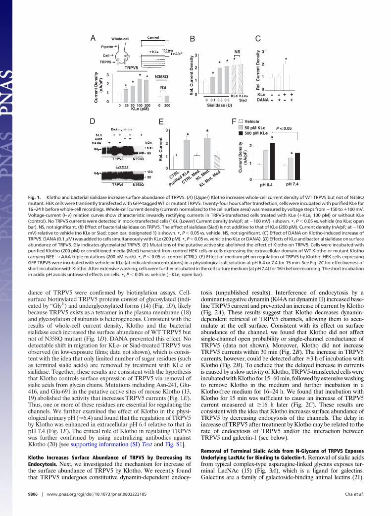
dance of TRPV5 were confirmed by biotinylation assays. Cell-surface biotinylated TRPV5 proteins consist of glycosylated (indi-cated by ‘‘Gly’’) and underglycosylated forms (14) (Fig. 1D), likelybecause TRPV5 exists as a tetramer in the plasma membrane (18)and glycosylation of subunits is heterogeneous. Consistent with theresults of whole-cell current density, Klotho and the bacterialsialidase each increased the surface abundance of WT TRPV5 butnot of N358Q mutant (Fig. 1D). DANA prevented this effect. Nodetectable shift in migration for KLe- or Siad-treated TRPV5 wasobserved (in low-exposure films; data not shown), which is consis-tent with the idea that only limited number of sugar residues (suchas terminal sialic acids) are removed by treatment with KLe orsialidase. Together, these results are consistent with the hypothesisthat Klotho controls surface expression of TRPV5 via removal ofsialic acids from glycan chains. Mutations including Asn-241, Glu-416, and Glu-691 in the putative active sites of mouse Klotho (13,19) abolished the activity that increases TRPV5 currents (Fig. 1E).Thus, one or more of these residues are essential for regulating thechannels. We further examined the effect of Klotho in the physi-ological urinary pH (�6.4) and found that the regulation of TRPV5by Klotho was enhanced in extracellular pH 6.4 relative to that inpH 7.4 (Fig. 1F). The critical role of Klotho in regulating TRPV5was further confirmed by using neutralizing antibodies againstKlotho (20) [see supporting information (SI) Text and Fig. S1].
Klotho Increases Surface Abundance of TRPV5 by Decreasing ItsEndocytosis. Next, we investigated the mechanism for increase ofthe surface abundance of TRPV5 by Klotho. We recently foundthat TRPV5 undergoes constitutive dynamin-dependent endocy-
tosis (unpublished results). Interference of endocytosis by adominant-negative dynamin (K44A rat dynamin II) increased base-line TRPV5 current and prevented an increase of current by Klotho(Fig. 2A). These results suggest that Klotho decreases dynamin-dependent retrieval of TRPV5 channels, allowing them to accu-mulate at the cell surface. Consistent with its effect on surfaceabundance of the channel, we found that Klotho did not affectsingle-channel open probability or single-channel conductance ofTRPV5 (data not shown). Moreover, Klotho did not increaseTRPV5 currents within 30 min (Fig. 2B). The increase in TRPV5currents, however, could be detected after �3 h of incubation withKlotho (Fig. 2B). To exclude that the delayed increase in currentsis caused by a slow activity of Klotho, TRPV5-transfected cells wereincubated with Klotho for 15–60 min, followed by extensive washingto remove Klotho in the medium and further incubation in aKlotho-free medium for 16–24 h. We found that incubation withKlotho for 15 min was sufficient to cause an increase of TRPV5current measured at �16 h later (Fig. 2C). These results areconsistent with the idea that Klotho increases surface abundance ofTRPV5 by decreasing endocytosis of the channels. The delay inincrease of TRPV5 after treatment by Klotho may be related to therate of endocytosis of TRPV5 and/or the interaction betweenTRPV5 and galectin-1 (see below).
Removal of Terminal Sialic Acids from N-Glycans of TRPV5 ExposesUnderlying LacNAc for Binding to Galectin-1. Removal of sialic acidsfrom typical complex-type asparagine-linked glycans exposes ter-minal LacNAc (15) (Fig. 3A), which is a ligand for galectins.Galectins are a family of galactoside-binding animal lectins (21).
B C
E
KLe+Siad0 0.1 0.3 0.5
Sialidase (U)
KLeRel
. Cu
rren
t D
ensi
ty
0
1
2
3
*
* * *
NS
KLeDANA
+ +++
- -- -
Rel
. Cu
rren
t D
ensi
ty
0
1
2
3
*
D
KLe (pM)
A Whole-cell
0 25 50 100 200 0 200
Cu
rren
t D
ensi
ty
(nA
/pF
)
2
3
0
1
TRPV5
N358Q
Pipette
Cell
TRPV5
NS
***
*
F
Rel
. Cu
rren
t
0
1
2
CTRL
Purified KLe
CTRL Med.
KL Med.
KL Mut. Med.
**3
*
*
P < 0.05
pH 6.4 pH 7.4
Cu
rren
t D
ensi
ty
(nA
/pF
)
0
1
2
Vehicle
50 pM KLe500 pM KLe
Fig. 1. Klotho and bacterial sialidase increase surface abundance of TRPV5. (A) (Upper) Klotho increases whole-cell current density of WT TRPV5 but not of N358Qmutant. HEK cells were transiently transfected with GFP-tagged WT or mutant TRPV5. Twenty-four hours after transfection, cells were incubated with purified KLe for16–24 h before whole-cell recordings. Whole-cell current density (currents normalized to the cell surface area) was measured by voltage steps from �150 to �100 mV.Voltage-current (I–V) relation curves show characteristic inwardly rectifying currents in TRPV5-transfected cells treated with KLe (�KLe; 100 pM) or without KLe(control). No TRPV5 currents were detected in mock-transfected cells (16). (Lower) Current density (nA/pF; at �100 mV) is shown. *, P � 0.05 vs. vehicle (no KLe; openbar). NS, not significant. (B) Effect of bacterial sialidase on TRPV5. The effect of sialidase (Siad) is not additive to that of KLe (200 pM). Current density (nA/pF; at �100mV) relative to vehicle (no KLe or Siad; open bar, designated 1) is shown. *, P � 0.05 vs. vehicle. NS, not significant. (C ) Effect of DANA on Klotho-induced increase ofTRPV5. DANA (0.1 �M) was added to cells simultaneously with KLe (200 pM). *, P � 0.05 vs. vehicle (no KLe or DANA). (D) Effects of KLe and bacterial sialidase on surfaceabundance of TRPV5. Gly indicates glycosylated TRPV5. (E ) Mutations of the putative active site abolished the effect of Klotho on TRPV5. Cells were incubated withpurified Klotho (200 pM) or conditioned media (Med) harvested from control HEK cells or cells expressing the extracellular domain of WT Klotho or mutant Klothocarrying NEE3 AAA triple mutations (200 pM each). *, P � 0.05 vs. control (CTRL). (F ) Effect of medium pH on regulation of TRPV5 by Klotho. HEK cells expressingGFP-TRPV5 were incubated with vehicle or KLe (at indicated concentrations) in a physiological salt solution at pH 6.4 or 7.4 for 15 min. See Fig. 2C for effectiveness ofshort incubationwithKlotho.Afterextensivewashing, cellswerefurther incubated inthecell culturemedium(atpH7.4) for16hbeforerecording.Theshort incubationin acidic pH avoids untoward effects on cells. *, P � 0.05 vs. vehicle (�KLe; open bar).
9806 � www.pnas.org�cgi�doi�10.1073�pnas.0803223105 Cha et al.
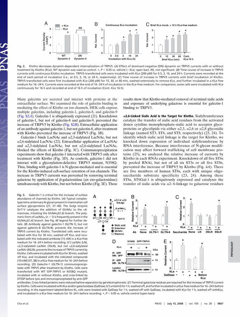
Many galectins are secreted and interact with proteins at theextracellular surface. We examined the role of galectin binding inmediating the effect of Klotho on ion channels. HEK cells expressmultiple galectins, including galectin-1, galectin-8, and galectin-9(Fig. S2A). Galectin-1 is ubiquitously expressed (21). Knockdownof galectin-1, but not of galectin-8 and galectin-9, prevented theincrease of TRPV5 by Klotho (Fig. S2B). Extracellular applicationof an antibody against galectin-1, but not galectin-8, after treatmentwith Klotho prevented the increase of TRPV5 (Fig. 3B).
Galectin-1 binds LacNAc and �2,3-sialylated LacNAc, but not�2,6-sialylated LacNAc (22). Extracellular application of LacNAcand �2,3-sialylated LacNAc, but not �2,6-sialylated LacNAc,blocked the effects of Klotho (Fig. 3C). Coimmunoprecipitationexperiments show that galectin-1 interacted with TRPV5 only aftertreatment with Klotho (Fig. 3D). As controls, galectin-1 did notinteract with a glycosylation-defective TRPV5 mutant, N358Q.Thus, binding with galectin-1 is N-glycan-mediated and is essentialfor the Klotho-induced cell-surface retention of ion channels. Theincrease in TRPV5 currents was prevented by removing terminalgalactose by application of �-galactosidase (an exo-galactosidase)simultaneously with Klotho, but not before Klotho (Fig. 3E). These
results show that Klotho-mediated removal of terminal sialic acidsand exposure of underlying galactose is essential for galectin-1binding to TRPV5.
�2,6-Linked Sialic Acid Is the Target for Klotho. Sialyltransferasescatalyze the transfer of sialic acid residues from the activateddonor cytidine monophosphate-sialic acid to acceptor glyco-proteins or glycolipids via either �2,3, �2,6 or �2,8 glycosidiclinkage (named ST3, ST6, and ST8, respectively) (23, 24). Toidentify which sialic acid linkage is the target for Klotho, weknocked down expression of individual sialyltransferase byRNA interference. Because interference of N-glycan modifi-cation may affect forward trafficking of cell membrane pro-teins (25), we analyzed the relative increase of currents byKlotho in each RNAi experiment. Knockdown of all five ST6sby pooled RNAi, but not of all six ST3s or all five ST8s,prevented the increase of TRPV5 by Klotho (Fig. 4A). Thereare five members of human ST6s, each with unique oligo-saccharide substrate specificity (23, 24). Among theseST6s, ST6Gal-1 is ubiquitously expressed and catalyzes thetransfer of sialic acids via �2–6-linkage to galactose residues
A
0
2
4
6
*
WTDynamin II
DNDynamin II
Cu
rren
t D
ensi
ty
(nA
/pF
)
+ KLe- KLe NS
B
Rel
. Cu
rren
t
0
1
2
3
*
* *
0 0.5 3 16 24Incubation with KLe (h)
Continuous KLe incubation
Rel
. Cu
rren
t
0
1
2
3
* ** *
0 15 30 60Brief KLe (min)
Brief KLe incub. + 16 h KLe-free medium
Cont.KLe 16 h
C
Fig. 2. Klotho decreases dynamin-dependent internalization of TRPV5. (A) Effect of dominant-negative (DN) dynamin on TRPV5 currents with or withouttreatment by Klotho (KLe). WT dynamin was used as control. *, P � 0.05 vs. vehicle (�KLe; open bar). NS, not significant. (B) Time course of increase in TRPV5currents with continuous Klotho incubation. TRPV5-transfected cells were incubated with KLe (200 pM) for 0.5, 3, 16, and 24 h. Currents were recorded at theend of each period of incubation (i.e., at 0.5, 3, 16, or 24 h, respectively). (C) Time course of increase in TRPV5 currents with brief incubation of Klotho.TRPV5-transfected cells were first incubated with KLe (200 pM) for 15, 30, or 60 min, washed extensively to remove KLe, and further incubated in a KLe-freemedium for 16–24 h. Currents were recorded at the end of 16–24 h of incubation in the KLe-free medium. For comparison, some cells were incubated with KLecontinuously for 16 h and recorded at end of 16 h of incubation (Cont. KLe 16 h).
A B
Rel
ativ
e C
urr
ent
1
2
3
0KLe + + +
None GLTN-1 GLTN-8Ab
**
C
Rel
ativ
e C
urr
ent
1
2
3
0KLe + + +
None LN 3SLNSaccharide
* *
+6SLN
E
Rel
ativ
e C
urr
ent
1
2
3
0KLe + + +
**
GalDase + + BeforeKLe
D
Fig. 3. Galectin-1 is critical for the increase of surfaceabundance of channels by Klotho. (A) Typical complex-typetetra-antennaryN-glycanpresent inmammaliancellsurface glycoproteins (27, 37–40). The Golgi enzymeGnT-V catalyzes the addition of GlcNAc to the �6-mannose, initiating the GlcNAc�(1,6) branch. The poly-meric form of LacNAcn (n � 1) is frequently present in theGlcNAc�(1,6) branch. See Fig. 4E legend for further de-tails. (B) Antibody against galectin-1 (GLTN-1), but notagainst galectin-8 (GLTN-8), prevents the increase ofTRPV5 current by Klotho. Transfected cells were incu-bated with KLe for 30 min, washed off KLe, and incu-bated with the indicated antibody (15 nM) in a KLe-freemedium for 16–24 h before recording. (C ) LacNAc (LN),�2,3-sialylated LacNAc (3SLN), but not �2,6-sialylatedLacNAc (6SLN), prevents the increase of TRPV5 current byKlotho.Cellswere incubatedwithKLefor30min,washedoff KLe, and incubated with the indicated compounds(10 mM) (37, 38) in a KLe-free medium for 16–24 h beforerecording. (D) Galectin-1 (GLTN-1) coimmunoprecipi-tates with TRPV5 after treatment by Klotho. Cells weretransfected with WT GFP-TRPV5 or N358Q mutant,incubated with or without Klotho, and cross-linked byDTSSP before lysis and immunoprecipitated by anti-GFPantibodies. Cross-linked proteins were reduced before separation by gel electrophoresis. (E ) Terminal galactose residues are required for the increase of TRPV5 currentbyKlotho.Cellswere incubatedwithKLeand/orgalactosidase (GalDase;0.5units/ml) for1h,washedoff,andfurther incubated inaKLe-freemediumfor16–24hbeforerecording. In the experiment labeled Before KL, cells were treated with GalDase for 1 h, washed off with GalDase, incubated with KLe for 1 h, washed off withKLe,and incubated in a KLe-free medium for 16–24 h before recording. *, P � 0.05 vs. vehicle control (open bars).
Cha et al. PNAS � July 15, 2008 � vol. 105 � no. 28 � 9807
PHYS
IOLO
GY
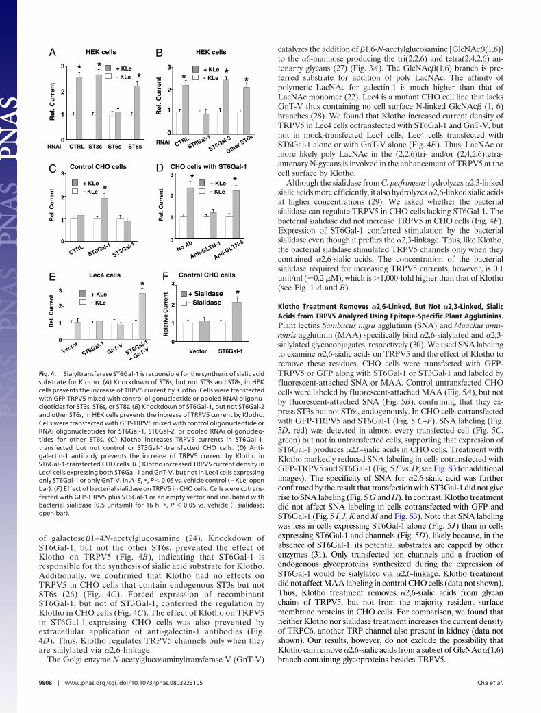
of galactose�1–4N-acetylglucosamine (24). Knockdown ofST6Gal-1, but not the other ST6s, prevented the effect ofKlotho on TRPV5 (Fig. 4B), indicating that ST6Gal-1 isresponsible for the synthesis of sialic acid substrate for Klotho.Additionally, we confirmed that Klotho had no effects onTRPV5 in CHO cells that contain endogenous ST3s but notST6s (26) (Fig. 4C). Forced expression of recombinantST6Gal-1, but not of ST3Gal-1, conferred the regulation byKlotho in CHO cells (Fig. 4C). The effect of Klotho on TRPV5in ST6Gal-1-expressing CHO cells was also prevented byextracellular application of anti-galectin-1 antibodies (Fig.4D). Thus, Klotho regulates TRPV5 channels only when theyare sialylated via �2,6-linkage.
The Golgi enzyme N-acetylglucosaminyltransferase V (GnT-V)
catalyzes the addition of �1,6-N-acetylglucosamine [GlcNAc�(1,6)]to the �6-mannose producing the tri(2,2,6) and tetra(2,4,2,6) an-tenarry glycans (27) (Fig. 3A). The GlcNAc�(1,6) branch is pre-ferred substrate for addition of poly LacNAc. The affinity ofpolymeric LacNAc for galectin-1 is much higher than that ofLacNAc monomer (22). Lec4 is a mutant CHO cell line that lacksGnT-V thus containing no cell surface N-linked GlcNAc� (1, 6)branches (28). We found that Klotho increased current density ofTRPV5 in Lec4 cells cotransfected with ST6Gal-1 and GnT-V, butnot in mock-transfected Lec4 cells, Lec4 cells transfected withST6Gal-1 alone or with GnT-V alone (Fig. 4E). Thus, LacNAc ormore likely poly LacNAc in the (2,2,6)tri- and/or (2,4,2,6)tetra-antenary N-gycans is involved in the enhancement of TRPV5 at thecell surface by Klotho.
Although the sialidase from C. perfringens hydrolyzes �2,3-linkedsialic acids more efficiently, it also hydrolyzes �2,6-linked sialic acidsat higher concentrations (29). We asked whether the bacterialsialidase can regulate TRPV5 in CHO cells lacking ST6Gal-1. Thebacterial sialidase did not increase TRPV5 in CHO cells (Fig. 4F).Expression of ST6Gal-1 conferred stimulation by the bacterialsialidase even though it prefers the �2,3-linkage. Thus, like Klotho,the bacterial sialidase stimulated TRPV5 channels only when theycontained �2,6-sialic acids. The concentration of the bacterialsialidase required for increasing TRPV5 currents, however, is 0.1unit/ml (�0.2 �M), which is �1,000-fold higher than that of Klotho(see Fig. 1 A and B).
Klotho Treatment Removes �2,6-Linked, But Not �2,3-Linked, SialicAcids from TRPV5 Analyzed Using Epitope-Specific Plant Agglutinins.Plant lectins Sambucus nigra agglutinin (SNA) and Maackia amu-rensis agglutinin (MAA) specifically bind �2,6-sialylated and �2,3-sialylated glycoconjugates, respectively (30). We used SNA labelingto examine �2,6-sialic acids on TRPV5 and the effect of Klotho toremove these residues. CHO cells were transfected with GFP-TRPV5 or GFP along with ST6Gal-1 or ST3Gal-1 and labeled byfluorescent-attached SNA or MAA. Control untransfected CHOcells were labeled by fluorescent-attached MAA (Fig. 5A), but notby fluorescent-attached SNA (Fig. 5B), confirming that they ex-press ST3s but not ST6s, endogenously. In CHO cells cotransfectedwith GFP-TRPV5 and ST6Gal-1 (Fig. 5 C–F), SNA labeling (Fig.5D, red) was detected in almost every transfected cell (Fig. 5C,green) but not in untransfected cells, supporting that expression ofST6Gal-1 produces �2,6-sialic acids in CHO cells. Treatment withKlotho markedly reduced SNA labeling in cells cotransfected withGFP-TRPV5 and ST6Gal-1 (Fig. 5 F vs. D; see Fig. S3 for additionalimages). The specificity of SNA for �2,6-sialic acid was furtherconfirmed by the result that transfection with ST3Gal-1 did not giverise to SNA labeling (Fig. 5 G and H). In contrast, Klotho treatmentdid not affect SNA labeling in cells cotransfected with GFP andST6Gal-1 (Fig. 5 I, J, K and M and Fig. S3). Note that SNA labelingwas less in cells expressing ST6Gal-1 alone (Fig. 5J) than in cellsexpressing ST6Gal-1 and channels (Fig. 5D), likely because, in theabsence of ST6Gal-1, its potential substrates are capped by otherenzymes (31). Only transfected ion channels and a fraction ofendogenous glycoproteins synthesized during the expression ofST6Gal-1 would be sialylated via �2,6-linkage. Klotho treatmentdid not affect MAA labeling in control CHO cells (data not shown).Thus, Klotho treatment removes �2,6-sialic acids from glycanchains of TRPV5, but not from the majority resident surfacemembrane proteins in CHO cells. For comparison, we found thatneither Klotho nor sialidase treatment increases the current densityof TRPC6, another TRP channel also present in kidney (data notshown). Our results, however, do not exclude the possibility thatKlotho can remove �2,6-sialic acids from a subset of GlcNAc �(1,6)branch-containing glycoproteins besides TRPV5.
Rel
. Cu
rren
t
0
1
2
3
CTRL ST3s ST6s ST8sRNAi
* **
+ KLe
- KLe
A
Rel
. Cu
rren
t
0
1
2
3
CTRLST6Gal-1
ST6Gal-2
Other ST6sRNAi
* **
B
C
Rel
. Cu
rren
t
0
1
2
3
CTRLST6Gal-1
ST3Gal-1
*
Control CHO cells R
el. C
urr
ent
0
1
2
3
No Ab
Anti-GLTN-1
Anti-GLTN-8
* *CHO cells with ST6Gal-1D
+ KLe
- KLe
+ KLe
- KLe
+ KLe
- KLe
E Lec4 cells
Rel
. Cu
rren
t
0
1
2
3
Vector
ST6Gal-1
GnT-VST6Gal-1
+ GnT-V
*+ KLe
- KLe
HEK cells HEK cells
F
Vector ST6Gal-1
Rel
ativ
e C
urr
ent
1
2
3
0
+ Sialidase- Sialidase
*
Control CHO cells
Fig. 4. Sialyltransferase ST6Gal-1 is responsible for the synthesis of sialic acidsubstrate for Klotho. (A) Knockdown of ST6s, but not ST3s and ST8s, in HEKcells prevents the increase of TRPV5 current by Klotho. Cells were transfectedwith GFP-TRPV5 mixed with control oligonucleotide or pooled RNAi oligonu-cleotides for ST3s, ST6s, or ST8s. (B) Knockdown of ST6Gal-1, but not ST6Gal-2and other ST6s, in HEK cells prevents the increase of TRPV5 current by Klotho.Cells were transfected with GFP-TRPV5 mixed with control oligonucleotide orRNAi oligonucleotides for ST6Gal-1, ST6Gal-2, or pooled RNAi oligonucleo-tides for other ST6s. (C ) Klotho increases TRPV5 currents in ST6Gal-1-transfected but not control or ST3Gal-1-transfected CHO cells. (D) Anti-galectin-1 antibody prevents the increase of TRPV5 current by Klotho inST6Gal-1-transfected CHO cells. (E ) Klotho increased TRPV5 current density inLec4 cells expressing both ST6Gal-1 and GnT-V, but not in Lec4 cells expressingonly ST6Gal-1 or only GnT-V. In A–E, *, P � 0.05 vs. vehicle control (�KLe; openbar). (F ) Effect of bacterial sialidase on TRPV5 in CHO cells. Cells were cotrans-fected with GFP-TRPV5 plus ST6Gal-1 or an empty vector and incubated withbacterial sialidase (0.5 units/ml) for 16 h. *, P � 0.05 vs. vehicle (�sialidase;open bar).
9808 � www.pnas.org�cgi�doi�10.1073�pnas.0803223105 Cha et al.
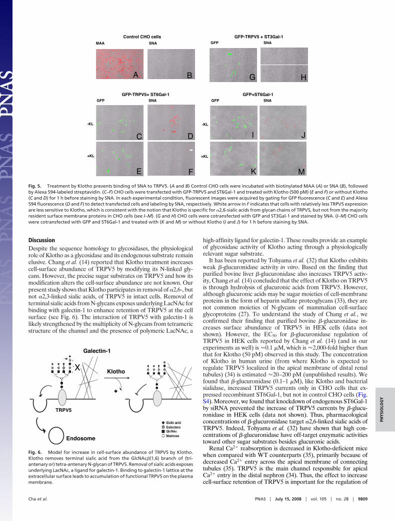
DiscussionDespite the sequence homology to glycosidases, the physiologicalrole of Klotho as a glycosidase and its endogenous substrate remainelusive. Chang et al. (14) reported that Klotho treatment increasescell-surface abundance of TRPV5 by modifying its N-linked gly-cans. However, the precise sugar substrates on TRPV5 and how itsmodification alters the cell-surface abundance are not known. Ourpresent study shows that Klotho participates in removal of �2,6-, butnot �2,3-linked sialic acids, of TRPV5 in intact cells. Removal ofterminal sialic acids from N-glycans exposes underlying LacNAc forbinding with galectin-1 to enhance retention of TRPV5 at the cellsurface (see Fig. 6). The interaction of TRPV5 with galectin-1 islikely strengthened by the multiplicity of N-glycans from tetramericstructure of the channel and the presence of polymeric LacNAc, a
high-affinity ligand for galectin-1. These results provide an exampleof glycosidase activity of Klotho acting through a physiologicallyrelevant sugar substrate.
It has been reported by Tohyama et al. (32) that Klotho exhibitsweak �-glucuronidase activity in vitro. Based on the finding thatpurified bovine liver �-glucuronidase also increases TRPV5 activ-ity, Chang et al. (14) concluded that the effect of Klotho on TRPV5is through hydrolysis of glucuronic acids from TRPV5. However,although glucuronic acids may be sugar moieties of cell-membraneproteins in the form of heparin sulfate proteoglycans (33), they arenot common moieties of N-glycans of mammalian cell-surfaceglycoproteins (27). To understand the study of Chang et al., weconfirmed their finding that purified bovine �-glucuronidase in-creases surface abundance of TRPV5 in HEK cells (data notshown). However, the EC50 for �-glucuronidase regulation ofTRPV5 in HEK cells reported by Chang et al. (14) (and in ourexperiments as well) is �0.1 �M, which is �2,000-fold higher thanthat for Klotho (50 pM) observed in this study. The concentrationof Klotho in human urine (from where Klotho is expected toregulate TRPV5 localized in the apical membrane of distal renaltubules) (34) is estimated �20–200 pM (unpublished results). Wefound that �-glucuronidase (0.1–1 �M), like Klotho and bacterialsialidase, increased TRPV5 currents only in CHO cells that ex-pressed recombinant ST6Gal-1, but not in control CHO cells (Fig.S4). Moreover, we found that knockdown of endogenous ST6Gal-1by siRNA prevented the increase of TRPV5 currents by �-glucu-ronidase in HEK cells (data not shown). Thus, pharmacologicalconcentrations of �-glucuronidase target �2,6-linked sialic acids ofTRPV5. Indeed, Tohyama et al. (32) have shown that high con-centrations of �-glucuronidase have off-target enzymatic activitiestoward other sugar substrates besides glucuronic acids.
Renal Ca2� reabsorption is decreased in Klotho-deficient micewhen compared with WT counterparts (35), primarily because ofdecreased Ca2� entry across the apical membrane of connectingtubules (35). TRPV5 is the main channel responsible for apicalCa2� entry in the distal nephron (34). Thus, the effect to increasecell-surface retention of TRPV5 is important for the regulation of
MAA SN A
C D
GFP-TRPV5+ ST6Gal-1
E F
SNA GFP
-K L
+KL
Control CHO cells
G A B H
GFP-TRPV5 + ST3Gal-1 GFP SN A
GFP+ST6Gal-1 GFP SN A
-K L
+KL
J
M K
I
Fig. 5. Treatment by Klotho prevents binding of SNA to TRPV5. (A and B) Control CHO cells were incubated with biotinylated MAA (A) or SNA (B), followedby Alexa 594-labeled streptavidin. (C–F) CHO cells were transfected with GFP-TRPV5 and ST6Gal-1 and treated with Klotho (500 pM) (E and F) or without Klotho(C and D) for 1 h before staining by SNA. In each experimental condition, fluorescent images were acquired by gating for GFP fluorescence (C and E) and Alexa594 fluorescence (D and F) to detect transfected cells and labeling by SNA, respectively. White arrow in F indicates that cells with relatively less TRPV5 expressionare less sensitive to Klotho, which is consistent with the notion that Klotho is specific for �2,6-sialic acids from glycan chains of TRPV5, but not from the majorityresident surface membrane proteins in CHO cells (see I–M). (G and H) CHO cells were cotransfected with GFP and ST3Gal-1 and stained by SNA. (I–M) CHO cellswere cotransfected with GFP and ST6Gal-1 and treated with (K and M) or without Klotho (I and J) for 1 h before staining by SNA.
KlothoX
TRPV5
Endosome
Galectin-1
Fig. 6. Model for increase in cell-surface abundance of TRPV5 by Klotho.Klotho removes terminal sialic acid from the GlcNAc�(1,6) branch of (tri-antenary or) tetra-antenary N-glycan of TRPV5. Removal of sialic acids exposesunderlying LacNAc, a ligand for galectin-1. Binding to galectin-1 lattice at theextracellular surface leads to accumulation of functional TRPV5 on the plasmamembrane.
Cha et al. PNAS � July 15, 2008 � vol. 105 � no. 28 � 9809
PHYS
IOLO
GY
renal Ca2� transport by Klotho. Klotho also regulates calciumhomeostasis through other mechanisms. Klotho inhibits the ex-pression of 25-hydroxyvitamin D 1�-hydroxylase (36), the keyenzyme for synthesis of 1,25-dihydroxyvitamin D. Accordingly,Klotho-deficient mice have higher circulating levels of 1,25-dihydroxyvitamin D and hypercalcemia from increased gastroin-testinal absorption of calcium (1, 36). Furthermore, Klotho pro-motes the release of parathyroid hormone from parathyroid glandsin response to decreases in the extracellular Ca2� levels (6). Overall,the effects of Klotho to increase cell-surface retention of TRPV5and release of parathyroid hormone will enhance renal Ca2�
reabsorption and counteract the effect of decrease in the synthesisof 1,25-dihydroxyvitamin D on calcium metabolism.
The regulation of TRPV5 by Klotho represents a novel mecha-nism for regulation of the activity of cell-surface glycoproteins.Increasing evidence indicates that N-glycan-mediated binding withgalectins is important for regulating the residence time of cellsurface glycoproteins (37–40). Up- and down-regulation of N-acetylglucosaminyltransferases (GnT-IV or GnT-V; also known asMgat4 and Mgat5, respectively) in the Golgi increases and de-creases LacNAc content of cytokine receptors and Glut-2 trans-porters, respectively (37, 38). These effects underscore the en-hanced responses of tumor cells to growth-promoting factors andthe reduced surface abundance of Glut-2 in pancreatic �-cellscaused by high-fat diets, respectively (37, 38). Additionally, Lau etal. (39) recently reports that an increase in intracellular concen-tration of N-acetylglucosamine stimulates N-glycan processing inthe Golgi and enhances association of receptors with galectins. Incontrast to the regulation of synthesis and branching of N-glycansoccurring at the Golgi, Klotho functions from the outside of cell asa humoral factor and by modifying N-glycans of mature cell surface
glycoproteins at the plasma membrane. Whether Klotho regulatesother potential targets related to aging phenotypes via the samemechanism remains to be determined.
Materials and MethodsGFP-tagged TRPV5 have been described (16). cDNAs for human ST6Gal-1 andST3Gal-1 were cloned into pEF1 expression vector. Sequences for RNAi oligonu-cleotides are in Table S1. Preparation of purified KLe has been described (2). Cellswere transiently transfected with cDNAs encoding GFP-TRPV5 with or withoutadditional constructs. Whole-cell currents were recorded by using an Axopatch200B amplifier as described (16). For immunoprecipitation of GFP-TRPV5 withgalectins-1, cell surface proteins were cross-linked by using a membrane-impermeable dithio-bis-suffosuccinimydyl propionate (DTSSP) and lysed, andsupernatants were immunoprecipitated by a monoclonal anti-GFP antibody.Immunoprecipitates were reduced by DTT, separated by SDS/PAGE, and analyzedfor galectin-1 by using a polyclonal anti-galectin-1 antibody. For staining by SNAand MAA, fixed CHO cells were incubated with biotinylated SNA or MAA fol-lowed by Alexa 594-conjugated streptavidin. Fluorescent images were obtainedwith a Nikon Eclipse TE2000-U fluorescent microscope and overlaid with differ-ential interference contrast images as described (41). In each experiment, gain forfluorescence detection was adjusted equally by using untransfected cells as acontrol.
ACKNOWLEDGMENTS. WethankDrs.PamelaStanley (AlbertEinsteinCollegeofMedicine, New York, NY) and Mike Pierce (University of Georgia, Athens, GA) forLec4 cells and cDNA for GnT-V, Drs. James Paulson and Ola Blixt (The ScrippsResearch Institute, La Jolla, CA), and the Consortium for Functional Glycomics(National Institutes of Health Grant GM62116) for glycan compounds, and Drs.Mark Lehrman, Carolyn Bertozzi, Michel Baum, and Orson Moe for discussionsand comments. This work was supported by National Institutes of Health GrantsDK20543 and DK59530 (to C.-L.H.) and AG19712 and AG25326 (to M.K.-o. andK.P.R.), American Heart Association Grant 0440019N (to C.-L.H.), the Eisai Re-search Fund (M.K.-o., the Ellison Medical Foundation (M.K.-o.), and the Ted NashLong Life Foundation (M.K.-o.).
1. Kuro-o M, et al. (1997) Mutation of the mouse klotho gene leads to a syndromeresembling aging. Nature 390:45–51.
2. Kurosu H, et al. (2005) Suppression of aging in mice by the hormone Klotho. Science309:1829–1833.
3. Arking DE, et al. (2002) Association of human aging with a functional variant of Klotho.Proc Natl Acad Sci USA 99:856–861.
4. Arking DE, Atzmon G, Arking A, Barzilai N, Dietz HC (2005) Association between afunctional variant of the KLOTHO gene and high-density lipoprotein cholesterol,blood pressure, stroke, and longevity. Circ Res 96:412–418.
5. Ogata N, et al. (2002) Association of klotho gene polymorphism with bone density andspondylosis of the lumbar spine in postmenopausal women. Bone 31:37–41.
6. Imura A, et al. (2007) Alpha-Klotho as a regulator of calcium homeostasis. Science316:1615–1618.
7. Imura A, et al. (2004) Secreted Klotho protein in sera and CSF: Implication for post-translational cleavage in release of Klotho protein from cell membrane. FEBS Lett565:143–147.
8. Chen CD, Podvin S, Gillespie E, Leeman SE, Abraham CR (2007) Insulin stimulates thecleavage and release of the extracellular domain of Klotho by ADAM10 and ADAM17.Proc Natl Acad Sci USA 104:19796–19801.
9. Kurosu H, et al. (2006) Regulation of fibroblast growth factor-23 signaling by Klotho.J Biol Chem 281:6120–6123.
10. Urakawa I, et al. (2006) Klotho converts canonical FGF receptor into a specific receptorfor FGF23. Nature 444:770–774.
11. Razzaque MS, Sitara D, Taguchi T, St-Arnaud R, Lanske B (2006) Premature aging-likephenotype in fibroblast growth factor 23 null mice is a vitamin D-mediated process.FASEB J 20:720–722.
12. Ichikawa S, et al. (2007) A homozygous missense mutation in human KLOTHO causessevere tumoral calcinosis. J Clin Invest 117:2684–2691.
13. Ito S, Fujimori T, Hayashizaki Y, Nabeshima Y (2002) Identification of a novel mousemembrane-bound family 1 glycosidase-like protein, which carries an atypical active sitestructure. Biochim Biophys Acta 1576:341–345.
14. Chang Q, et al. (2005) The �-glucuronidase klotho hydrolyzes and activates the TRPV5channel. Science 310:490–493.
15. Schauer R (1991) Biosynthesis and function of N- and O-substituted sialic acids. Glyco-biology 1:449–452.
16. Yeh BI, Kim YK, Jabbar W, Huang CL (2005) Conformational changes of pore helixcoupled to gating of TRPV5 by protons. EMBO J 24:3224–3234.
17. Usuki S, Hoops P, Sweeley CC (1988) Growth control of human foreskin fibroblasts andinhibition of extracellular sialidase activity by 2-deoxy-2,3-dehydro-N-acetylneura-minic acid. J Biol Chem 263:10595–10599.
18. Hoenderop JG, et al. (2003) Homo- and heterotetrameric architecture of the epithelialCa2� channels TRPV5 and TRPV6. EMBO J 22:1–10.
19. Rye CS, Withers SG (2000) Glycosidase mechanisms. Curr Opin Chem Biol 4:573–580.20. Kato Y, et al. (2000) Establishment of the anti-Klotho monoclonal antibodies and
detection of Klotho protein in kidneys. Biochem Biophys Res Commun 267:597–602.
21. Barondes SH, Cooper DN, Gitt MA, Leffler H (1994) Galectins: Structure and function ofa large family of animal lectins. J Biol Chem 269:20807–20810.
22. Leppanen A, Stowell S, Blixt O, Cummings RD (2005) Dimeric galectin-1 binds with highaffinity to �2,3-sialylated and nonsialylated terminal N-acetyllactosamine units onsurface-bound extended glycans. J Biol Chem 280:5549–5562.
23. Patel RY, Balaji PV (2006) Identification of linkage-specific sequence motifs in sialyl-transferases. Glycobiology 16:108–116.
24. Yasukawa Z, Sato C, Kitajima K (2005) Inflammation-dependent changes in �2,3-,�2,6-, and �2,8-sialic acid glycotopes on serum glycoproteins in mice. Glycobiology15:827–837.
25. Vagin O, Turdikulova S, Sachs G (2004) The H,K-ATPase � subunit as a model to studythe role of N-glycosylation in membrane trafficking and apical sorting. J Biol Chem279:39026–39034.
26. Fukuta K, Yokomatsu T, Abe R, Asanagi M, Makino T (2000) Genetic engineering ofCHO cells producing human interferon-� by transfection of sialyltransferases. Glyco-conj J 17:895–904.
27. Demetriou M, Granovsky M, Quaggin S, Dennis JW (2001) Negative regulation of T cellactivation and autoimmunity by Mgat5 N-glycosylation. Nature 409:733–739.
28. Chaney W, Sundaram S, Friedman N, Stanley P (1989) The Lec4A CHO glycosylationmutant arises from miscompartmentalization of a Golgi glycosyltransferase. J Cell Biol109:2089–2096.
29. Corfield AP, Higa H, Paulson JC, Schauer R (1983) The specificity of viral and bacterialsialidases for �2–3- and �2–6-linked sialic acids in glycoproteins. Biochim Biophys Acta744:121–126.
30. Brinkman-Van der Linden EC, Sonnenburg JL, Varki A (2002) Effects of sialic acidsubstitutions on recognition by Sambucus nigra agglutinin and Maackia amurensishemagglutinin. Anal Biochem 303:98–104.
31. Martin LT, Marth JD, Varki A, Varki NM (2002) Genetically altered mice with differentsialyltransferase deficiencies show tissue-specific alterations in sialylation and sialicacid 9-O-acetylation. J Biol Chem 277:32930–32938.
32. Tohyama O, et al. (2004) Klotho is a novel �-glucuronidase capable of hydrolyzingsteroid �-glucuronides. J Biol Chem 279:9777–9784.
33. Bishop JR, Schuksz M, Esko JD (2007) Heparan sulphate proteoglycans fine-tunemammalian physiology. Nature 446:1030–1047.
34. Hoenderop JG, Nilius B, Bindels RJ (2002) Molecular mechanism of active Ca2� reab-sorption in the distal nephron. Annu Rev Physiol 64:529–549.
35. Tsuruoka S, et al. (2006) Nephrol Dial Transplant 21:2762–2767.36. Yoshida T, Fujimori T, Nabeshima Y (2002) Mediation of unusually high concentrations
of 1,25-dihydroxyvitamin D in homozygous klotho mutant mice by increased expres-sion of renal 1�-hydroxylase gene. Endocrinology 143:683–689.
37. Partridge EA, et al. (2004) Regulation of cytokine receptors by Golgi N-glycans pro-cessing and endocytosis. Science 306:120–124.
38. Ohtsubo K, et al. (2005) Dietary and genetic control of glucose transporter 2 glycosyl-ation promotes insulin secretion in suppressing diabetes. Cell 123:1307–1321.
39. Lau KS, et al. (2007) Complex N-glycan number and degree of branching cooperate toregulate cell proliferation and differentiation. Cell 129:123–134.
40. Stanley P (2007) A method to the madness of N-glycan complexity? Cell 129:27–29.41. Zeng WZ, et al. (2002) Evidence for endocytosis of ROMK potassium channels via
clathrin-coated vesicles. Am J Physiol 283:F630–F639.
9810 � www.pnas.org�cgi�doi�10.1073�pnas.0803223105 Cha et al.
Related Documents