Removal of Artifacts T-61.182, Biomedical Image Analysis Seminar presentation 19.2.2005 Hannu Laaksonen Vibhor Kumar

Removal of Artifacts T-61.182, Biomedical Image Analysis Seminar presentation 19.2.2005 Hannu Laaksonen Vibhor Kumar.
Dec 29, 2015
Welcome message from author
This document is posted to help you gain knowledge. Please leave a comment to let me know what you think about it! Share it to your friends and learn new things together.
Transcript
Removal of Artifacts
T-61.182, Biomedical Image AnalysisSeminar presentation 19.2.2005
Hannu LaaksonenVibhor Kumar
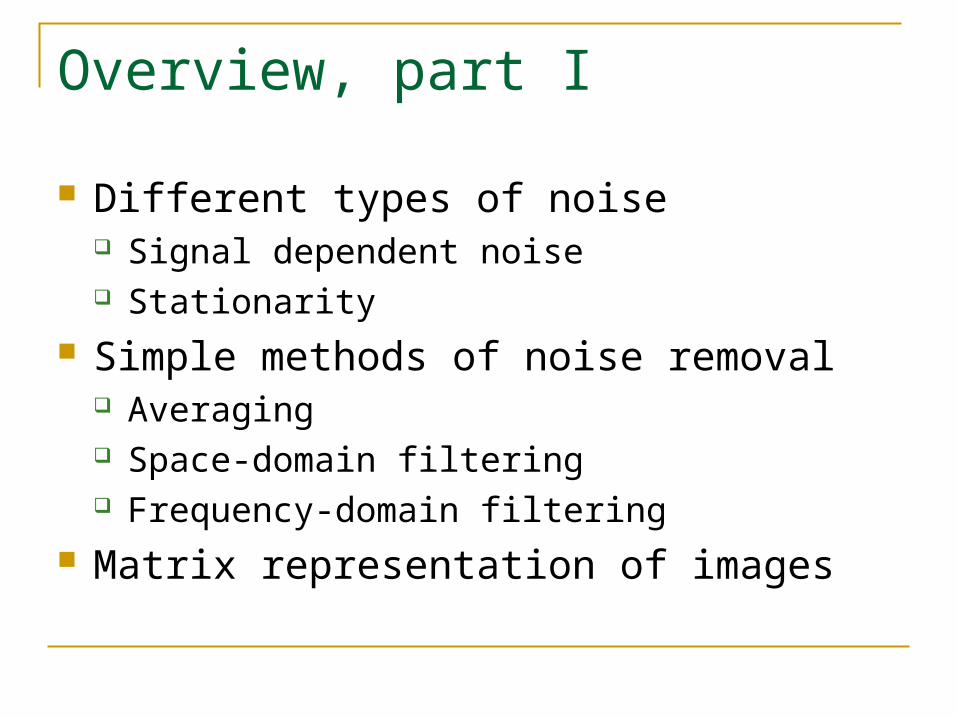
Overview, part I
Different types of noise Signal dependent noise Stationarity
Simple methods of noise removal Averaging Space-domain filtering Frequency-domain filtering
Matrix representation of images

Introduction
Noise: any part of the image that is of no interest
Removal of noise (artifacts) crucial for image analysis
Artifact removal should not cause distortions in the image
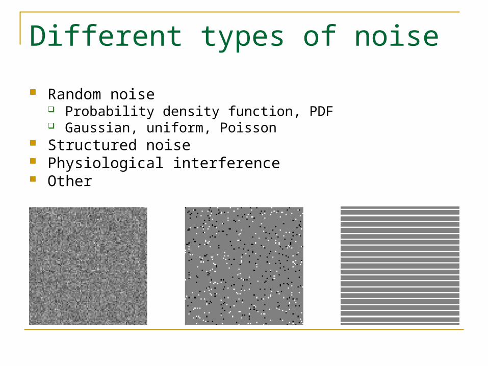
Different types of noise
Random noise Probability density function, PDF Gaussian, uniform, Poisson
Structured noise Physiological interference Other
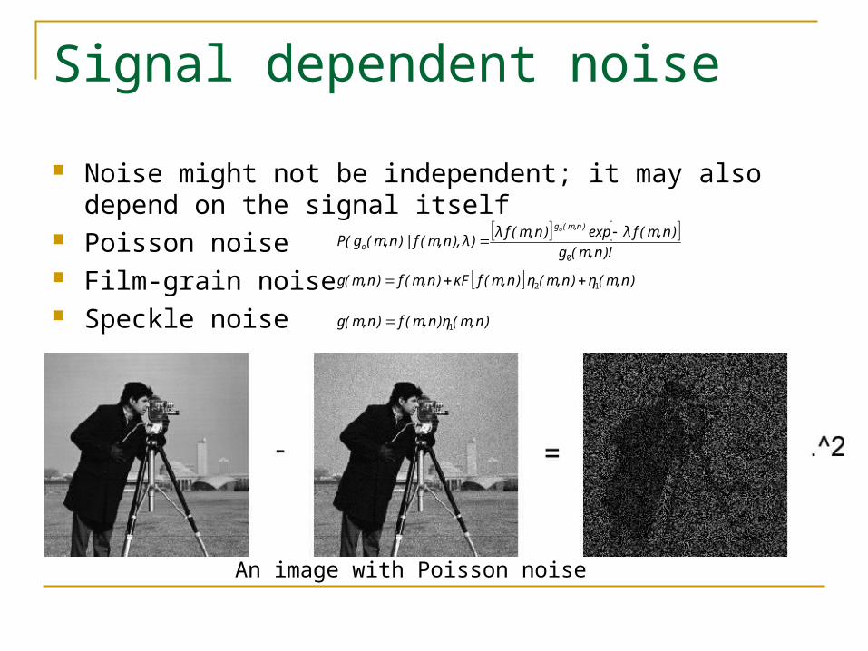
Signal dependent noise
Noise might not be independent; it may also depend on the signal itself
Poisson noise Film-grain noise Speckle noise
An image with Poisson noise
)n,m(η)n,m(η)n,m(fFκ)n,m(f)n,m(g 12
)n,m(η)n,m(f)n,m(g 1
)!n,m(g
)n,m(fλexp)n,m(fλ)λ),n,m(f|)n,m(g(P
)n,m(g
o
o
0

Stationarity
Strongly stationary Stationary in the wide sense Nonstationary Quasistationary (block-wise stationary)
Short-time analysis Cyclo-stationary

Synchronized or multiframe averaging If several time instances of the image are available, the noise
can be reduced by averaging Synchronized averaging: frames are acquired in the same
phase Changes (motion, displacement) between frames will cause
distortion

Space-domain filters
Images often nonstationary as a whole, but ma be stationary in small segments
Moving-window filter Sizes, shapes and weights vary Parameters are estimated in the window and
applied to the pixel in center
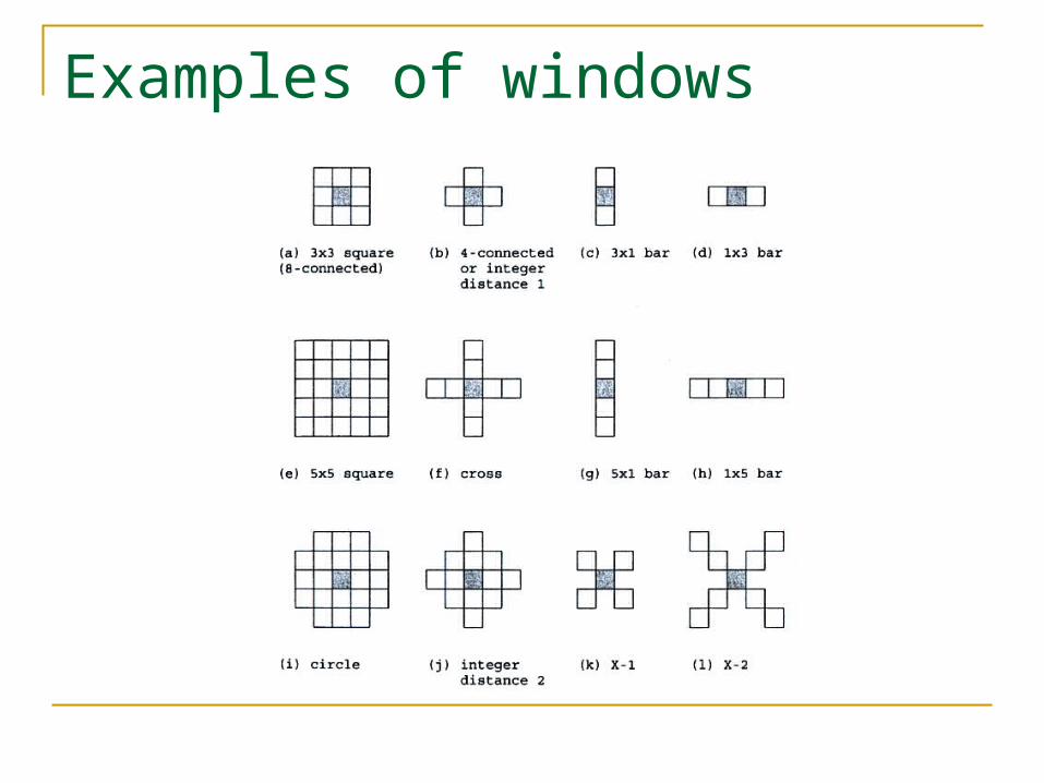
Examples of windows

Examples of space-domain filters Mean filter
Mean of the values in window Median filter
Median of the values in window Nonlinear
Order-statistic filter A large class of nonlinear filters

Filters in use

Frequency-domain filters
In natural images, usually the most important information is located at low frequencies
Frequency-domain filtering: 2D Fourier transform is calculated of the image The transformed image passed through a transfer
function (filter) The image is then transformed back
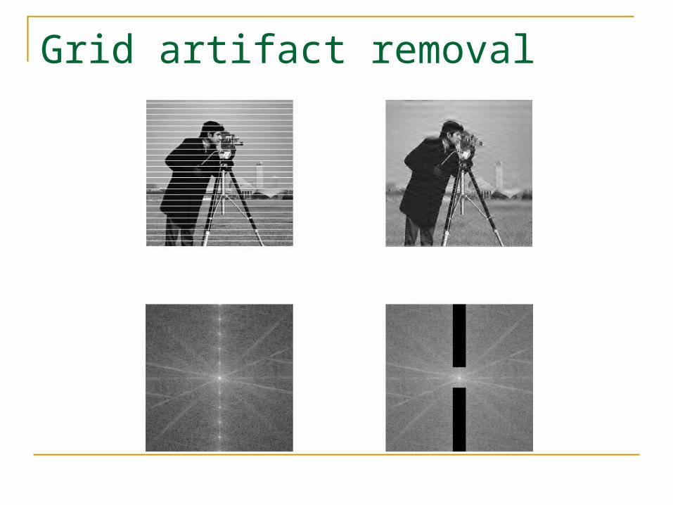
Grid artifact removal

Matrix representation of image processing Image may be presented as a matrix:f = {f(m,n) : m = 0,1,2,…M-1; n = 0,1,2,…,N-1}
Can be converted into vector by row ordering:f = [f1, f2, …, fM]T
Image properties can be calculated using matrix notation Mean m = E[f] Covariance σ = E[(f - m)(f - m)T] Autocorrelation Φ = E[f fT]
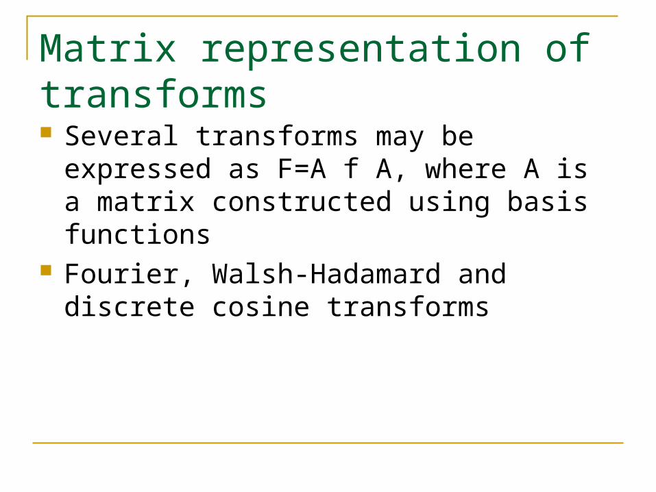
Matrix representation of transforms Several transforms may be expressed as
F=A f A, where A is a matrix constructed using basis functions
Fourier, Walsh-Hadamard and discrete cosine transforms
Related Documents