S101 VETERINARSKI ARHIV 76 (Suppl.), S101-S109, 2006 Remarks on cranial lesions in the European polecat (Mustela putorius) caused by helminth parasites Uwe Kierdorf 1 *, Horst Kierdorf 1 , Dean Konjević 2 , and Peter Lazar 3 1 Department of Biology, University of Hildesheim, Hildesheim, Germany 2 Department for Game Biology, Pathology and Breeding, Faculty of Veterinary Medicine, University of Zagreb, Zagreb, Croatia 3 Department of Parasitology and Diseases of Fish, Bees and Game, University of Veterinary Medicine, Kosice, Slovak Republic KIERDORF, U., H. KIERDORF, D. KONJEVIĆ, P. LAZAR: Remarks on cranial lesions in the European polecat (Mustela putorius) caused by helminth parasites. Vet. arhiv 76, S101-S109, 2006. ABSTRACT The paper briefly reviews the literature on cranial lesions in Mustela putorius caused by the trematode Troglotrema acutum and the nematode Skrjabingylus nasicola. Additional macroscopic, radiographic and scanning electron microscopy findings from the study of dried skulls and formalin-fixed heads of European polecats are presented. Previous observations on the possibility of concurrent infestation of Mustela putorius with both parasite species are confirmed. Key words: Troglotrema acutum, Skrjabingylus nasicola, cranial lesions, paranasal sinuses, Mustela putorius Introduction Adult individuals of different parasitic helminth species can be found in the paranasal sinuses of mustelids. These parasites are the trematode Troglotrema acutum (Fig. 1) and species of the nematode genus Skrjabingylus (HANSSON, 1968, 1970; COLYN and VAN ROMPAEY, 1989; KOUBEK et al., 2004a). The chief definitive host of Troglotrema acutum is the European polecat (Mustela * Contact address: Dr. Uwe Kierdorf, Department of Biology, University of Hildesheim, Marienburger Platz 22, 31141 Hildesheim, Germany, Phone: +49-5121-883 913; E-mail: [email protected] ISSN 0372-5480 Printed in Croatia

Welcome message from author
This document is posted to help you gain knowledge. Please leave a comment to let me know what you think about it! Share it to your friends and learn new things together.
Transcript

S101
VETERINARSKI ARHIV 76 (Suppl.), S101-S109, 2006
Remarks on cranial lesions in the European polecat (Mustela putorius) caused by helminth parasites
Uwe Kierdorf1*, Horst Kierdorf1, Dean Konjević2, and Peter Lazar3
1Department of Biology, University of Hildesheim, Hildesheim, Germany2Department for Game Biology, Pathology and Breeding, Faculty of Veterinary Medicine, University of
Zagreb, Zagreb, Croatia3Department of Parasitology and Diseases of Fish, Bees and Game, University of Veterinary Medicine,
Kosice, Slovak Republic
KIERDORF, U., H. KIERDORF, D. KONJEVIĆ, P. LAZAR: Remarks on cranial lesions in the European polecat (Mustela putorius) caused by helminth parasites. Vet. arhiv 76, S101-S109, 2006.
ABSTRACTThe paper briefly reviews the literature on cranial lesions in Mustela putorius caused by the trematode
Troglotrema acutum and the nematode Skrjabingylus nasicola. Additional macroscopic, radiographic and scanning electron microscopy findings from the study of dried skulls and formalin-fixed heads of European polecats are presented. Previous observations on the possibility of concurrent infestation of Mustela putorius with both parasite species are confirmed.
Key words: Troglotrema acutum, Skrjabingylus nasicola, cranial lesions, paranasal sinuses, Mustela putorius
IntroductionAdult individuals of different parasitic helminth species can be found in the paranasal
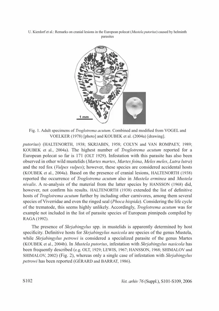
sinuses of mustelids. These parasites are the trematode Troglotrema acutum (Fig. 1) and species of the nematode genus Skrjabingylus (HANSSON, 1968, 1970; COLYN and VAN ROMPAEY, 1989; KOUBEK et al., 2004a).
The chief definitive host of Troglotrema acutum is the European polecat (Mustela
* Contact address: Dr. Uwe Kierdorf, Department of Biology, University of Hildesheim, Marienburger Platz 22, 31141 Hildesheim, Germany, Phone: +49-5121-883 913; E-mail: [email protected]
ISSN 0372-5480Printed in Croatia
S102
Fig. 1. Adult specimens of Troglotrema acutum. Combined and modified from VOGEL and VOELKER (1978) [photo] and KOUBEK et al. (2004a) [drawing].
putorius) (HALTENORTH, 1938; SKRJABIN, 1958; COLYN and VAN ROMPAEY, 1989; KOUBEK et al., 2004a). The highest number of Troglotrema acutum reported for a European polecat so far is 171 (OLT 1929). Infestation with this parasite has also been observed in other wild mustelids (Martes martes, Martes foina, Meles meles, Lutra lutra) and the red fox (Vulpes vulpes); however, these species are considered accidental hosts (KOUBEK et al., 2004a). Based on the presence of cranial lesions, HALTENORTH (1938) reported the occurrence of Troglotrema acutum also in Mustela erminea and Mustela nivalis. A re-analysis of the material from the latter species by HANSSON (1968) did, however, not confirm his results. HALTENORTH (1938) extended the list of definitive hosts of Troglotrema acutum further by including other carnivores, among them several species of Viverridae and even the ringed seal (Phoca hispida). Considering the life cycle of the trematode, this seems highly unlikely. Accordingly, Troglotrema acutum was for example not included in the list of parasite species of European pinnipeds compiled by RAGA (1992).
The presence of Skrjabingylus spp. in mustelids is apparently determined by host specificity. Definitive hosts for Skrjabingylus nasicola are species of the genus Mustela, while Skrjabingylus petrowi is considered a specialized parasite of the genus Martes (KOUBEK et al., 2004b). In Mustela putorius, infestation with Skrjabingylus nasicola has been frequently described (e.g. OLT, 1929; LEWIS, 1967; HANSSON, 1968; SHIMALOV and SHIMALOV, 2002) (Fig. 2), whereas only a single case of infestation with Skrjabingylus petrowi has been reported (GÉRARD and BARRAT, 1986).
Vet. arhiv 76 (Suppl.), S101-S109, 2006
U. Kierdorf et al.: Remarks on cranial lesions in the European polecat (Mustela putorius) caused by helminth parasites

S103
Fig. 3. Cranial lesions in a European polecat (Mustela putorius) from Croatia, characteristic of infestation with Troglotrema acutum. Note occurrence of lesions in the area of the skull roof
(A) and also more laterally (B) in the region of the postorbital process.
First intermediate hosts of Troglotrema acutum are prosobranch snails of the genus Bythinella, while anurans act as second intermediate hosts (VOGEL and VOELKER, 1978; KOUBEK et al., 2004a). Invasion of a polecat occurs when it eats a frog or toad containing metacercariae.
Intermediate hosts of Skrjabingylus nasicola are terrestrial mollusks (slugs and snails). However, these are only rarely found in the diet of the European polecat (WOLSAN, 1993) and other mustelids, which mainly feed on small vertebrates. Therefore, most authors consider small mammals (shrews and rodents) to act as paratenic hosts of the parasite (HANSSON, 1967; VAN SOEST et al., 1972; KING, 1977; WEBER and MERMOD, 1985).
Depending on the host, the adult individuals of both Troglotrema acutum and Skrjabingylus nasicola are attached to the mucosal surface or lie in cysts beneath the sinus mucosa that are formed by a suppurative granulation process (JUBB and KENNEDY, 1963). The bone reaction caused by infestation with the parasites was described as a local rarefying osteomyelitis, which may eventually perforate the bone and discharge into the cranial cavity, to the exterior, or into the nasal cavity (JUBB and KENNEDY, 1963). The bone destruction results in the cranial lesions that can be observed in mustelids infested with the helminths.
Some authors (e.g. BAER, 1931; HALTENORTH, 1938) claim that it is possible to morphologically distinguish cranial lesions caused by the trematode from those provoked by the nematode. According to these investigators, Skrjabingylus nasicola causes
Vet. arhiv 76 (Suppl.), S101-S109, 2006
U. Kierdorf et al.: Remarks on cranial lesions in the European polecat (Mustela putorius) caused by helminth parasites
S104
relatively small holes which are mostly present in the supraorbital region of the skull. In contrast, bone perforations caused by Troglotrema acutum are typically larger and affect more extended areas of the frontals and sometimes also neighboring bones. HANSON (1968, 1970), however, later cautioned that in practice it may not always be possible to distinguish cranial lesions caused by the trematode from those caused by the nematode.
With regard to Mustela putorius, OLT (1929) holds that only Troglotrema acutum causes perforations of the cranium, while infestation with Skrjabingylus nasicola does not result in such damage. More recently, VAN BREE and KOMPANJE (1997, p. 103) stated ”that in polecats and other larger mustelids the visible damage of the frontal sinuses by these parasitic worms [Skrjabingylus nasicola, present authors] is very rare indeed, and restricted to a small hole in the area of the frontal sinuses.” However, LEWIS (1967, p. 562) observed ”three large perforations around the supra-orbital region of the skull of a male polecat” from which 16 adult Skrjabingylus nasicola (13 females, 3 males) were recovered. KOUBEK et al. (2004a) undertook a study of the geographical distribution of Troglotrema acutum in the Czech Republic based on the analysis of dried skulls of Mustela putorius and other carnivores. These authors state that a typical attribute of the lesions caused by the trematode is a changed bone structure over a large area of the cranium.
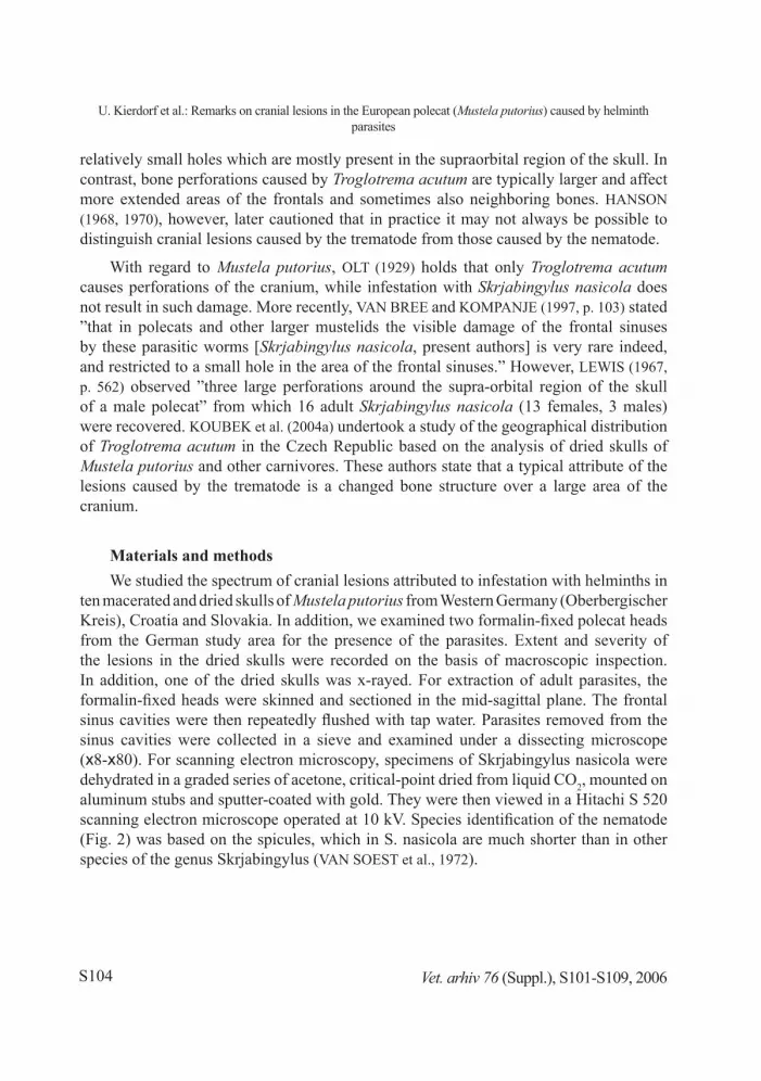
Materials and methodsWe studied the spectrum of cranial lesions attributed to infestation with helminths in
ten macerated and dried skulls of Mustela putorius from Western Germany (Oberbergischer Kreis), Croatia and Slovakia. In addition, we examined two formalin-fixed polecat heads from the German study area for the presence of the parasites. Extent and severity of the lesions in the dried skulls were recorded on the basis of macroscopic inspection. In addition, one of the dried skulls was x-rayed. For extraction of adult parasites, the formalin-fixed heads were skinned and sectioned in the mid-sagittal plane. The frontal sinus cavities were then repeatedly flushed with tap water. Parasites removed from the sinus cavities were collected in a sieve and examined under a dissecting microscope (x8-x80). For scanning electron microscopy, specimens of Skrjabingylus nasicola were dehydrated in a graded series of acetone, critical-point dried from liquid CO2, mounted on aluminum stubs and sputter-coated with gold. They were then viewed in a Hitachi S 520 scanning electron microscope operated at 10 kV. Species identification of the nematode (Fig. 2) was based on the spicules, which in S. nasicola are much shorter than in other species of the genus Skrjabingylus (VAN SOEST et al., 1972).
Vet. arhiv 76 (Suppl.), S101-S109, 2006
U. Kierdorf et al.: Remarks on cranial lesions in the European polecat (Mustela putorius) caused by helminth parasites

S105
Results and discussionThe damage seen in the dried skulls showed great variation in extent and severity
(Figs. 3, 4). The lesions visible on external inspection were mostly present in the frontal bones, but also neighboring bones could be affected (Fig. 4d). The most conspicuous feature was the occurrence of multiple bone perforations of originally more or less circular outline (Fig. 4b) that had frequently become confluent, thus forming more extended defect areas with a more irregular outline (Fig. 4c,d). The edges of the holes were mostly rather smooth (Fig. 4b,c). Formation of the holes had led to an exposure of the frontal sinus and nasal cavity. In some of the skulls, the surface of the bone surrounding the perforations was smooth (Fig. 4b). In other specimens, however, the bone surface was in places rough and bumpy (Fig. 4d), indicating that bone apposition had occurred along with bone destruction. Furthermore, an expansion of the frontal sinus with an ”in-pushing” of the inner table of the frontal bone into the cranial cavity was observed on radiographic examination (Fig. 5).
Our observations on the skulls correspond well with earlier descriptions of the cranial damage in Mustela putorius caused by Troglotrema acutum (OLT, 1929; LEHMENSICK, 1942; SKRJABIN, 1958; VOGEL and VOELKER, 1978).
Fig. 3. Cranial lesions in a European polecat (Mustela putorius) from Croatia, characteristic of
infestation with Troglotrema acutum. Note occurrence of lesions in the area of the skull roof (A) and also more laterally (B) in the region of the postorbital process.
Vet. arhiv 76 (Suppl.), S101-S109, 2006
U. Kierdorf et al.: Remarks on cranial lesions in the European polecat (Mustela putorius) caused by helminth parasites
S106
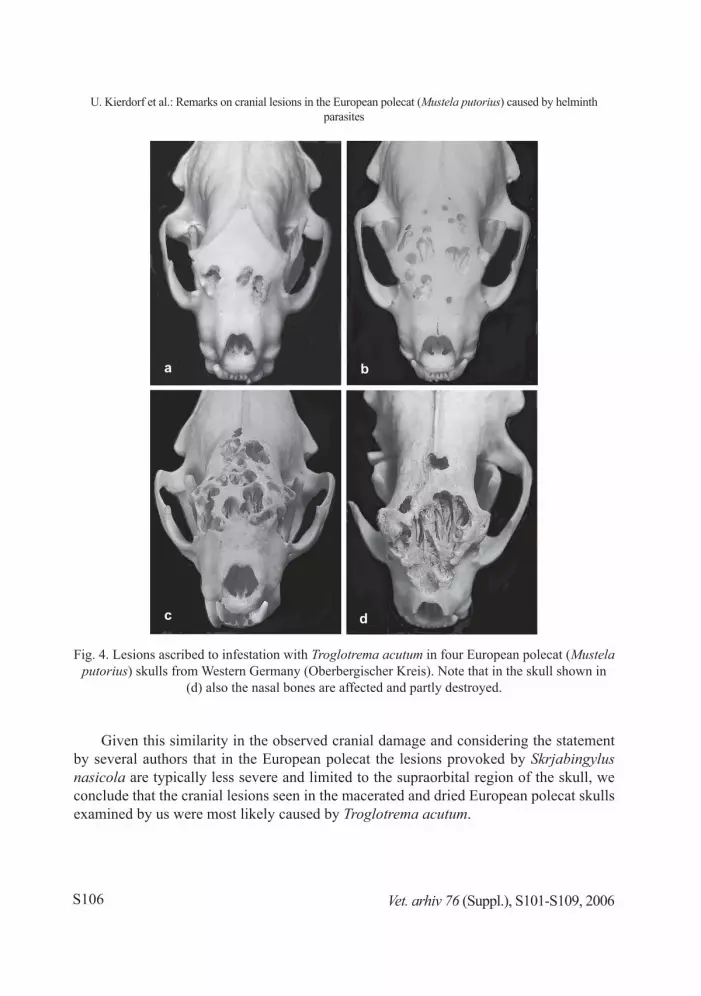
Fig. 4. Lesions ascribed to infestation with Troglotrema acutum in four European polecat (Mustela putorius) skulls from Western Germany (Oberbergischer Kreis). Note that in the skull shown in
(d) also the nasal bones are affected and partly destroyed.
Given this similarity in the observed cranial damage and considering the statement by several authors that in the European polecat the lesions provoked by Skrjabingylus nasicola are typically less severe and limited to the supraorbital region of the skull, we conclude that the cranial lesions seen in the macerated and dried European polecat skulls examined by us were most likely caused by Troglotrema acutum.
Vet. arhiv 76 (Suppl.), S101-S109, 2006
U. Kierdorf et al.: Remarks on cranial lesions in the European polecat (Mustela putorius) caused by helminth parasites
S107
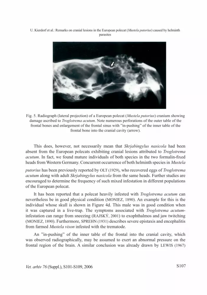
Fig. 5. Radiograph (lateral projection) of a European polecat (Mustela putorius) cranium showing damage ascribed to Troglotrema acutum. Note numerous perforations of the outer table of the frontal bones and enlargement of the frontal sinus with ”in-pushing” of the inner table of the
frontal bone into the cranial cavity (arrow).
This does, however, not necessarily mean that Skrjabingylus nasicola had been absent from the European polecats exhibiting cranial lesions attributed to Troglotrema acutum. In fact, we found mature individuals of both species in the two formalin-fixed heads from Western Germany. Concurrent occurrence of both helminth species in Mustela
putorius has been previously reported by OLT (1929), who recovered eggs of Troglotrema acutum along with adult Skrjabingylus nasicola from the same heads. Further studies are encouraged to determine the frequency of such mixed infestation in different populations of the European polecat.
It has been reported that a polecat heavily infested with Troglotrema acutum can nevertheless be in good physical condition (MONIEZ, 1890). An example for this is the individual whose skull is shown in Figure 4d. This male was in good condition when it was captured in a live-trap. The symptoms associated with Troglotrema acutum-infestation can range from sneezing (RAJSKÝ, 2001) to exophthalmos and jaw twitching (MONIEZ, 1890). Furthermore, SPREHN (1931) describes severe epistaxis and encephalitis from farmed Mustela vison infested with the trematode.
An ”in-pushing” of the inner table of the frontal into the cranial cavity, which was observed radiographically, may be assumed to exert an abnormal pressure on the frontal region of the brain. A similar conclusion was already drawn by LEWIS (1967)
Vet. arhiv 76 (Suppl.), S101-S109, 2006
U. Kierdorf et al.: Remarks on cranial lesions in the European polecat (Mustela putorius) caused by helminth parasites

S108
who observed corresponding changes in Mustela nivalis infested with Skrjabingylus nasicola. Behavioral alterations may occur as a consequence of this abnormal pressure. Further investigations, including histological studies on the brains of infested animals and behavioral studies on living animals, are needed to evaluate the effects of the cranial deformations caused by the parasites on brain structure and behavior in mustelids.
ReferencesBAER, J. G. (1931): Quelques helminthes rares ou peu connus du putois. Rev. Suisse Zool. 38,
313-334.COLYN, M., H. VAN ROMPAEY (1989): Review of the frontal sinus parasites of Mustelidae and
Viverridae, with new data from tropical Africa. Revue Zool. Afr. 103, 5-20.GÉRARD, Y., J. BARRAT (1986): Parasitisme de mustélidés par Skrjabingylus petrowi: Premier
rapport en Europe occidentale. Ann. Parasitol. Hum. Comp. 61, 575-579.HALTENORTH, T. (1938): Neue Wirte und Verbreitungsgebiete von Troglotrema acutum Leuck.
und Skrjabingylus nasicola Leuck. Sitzungsber. Gesellsch. Naturf. Freunde Berlin 1937, 74-80.
HANSSON, I. (1967): Transmission of the parasitic nematode Skrjabingylus nasicola (Leuckart, 1842) to species of Mustela (Mammalia). Oikos 18, 247-252.
HANSSON, I. (1968): Cranial helminth parasites in species of Mustelidae. I. Frequency and damage in fresh mustelids from Sweden. Oikos 19, 217-233.
HANSSON, I. (1970): Cranial helminth parasites in species of Mustelidae. II. Regional frequencies of damage in preserved crania from Denmark, Finland, Sweden, Greenland and the northeast of Canada compared with helminth invasion in fresh mustelid skulls from Sweden. Ark. Zool. 22, 571-594.
JUBB, K. V. F., P. C. KENNEDY (1963): Pathology of domestic animals Vol I. Academic Press Inc., New York, USA, pp. 123-124.
KING, C. (1977): The effects of the nematode parasite Skrjabingylus nasicola on British weasels (Mustela nivalis). J. Zool. (Lond.) 182, 225-249.
KOUBEK, P., V. BARUŠ, B. KOUBKOVÁ (2004a): Troglotrema acutum (Digenea) from carnivores in the Czech Republic. Helminthologia 41, 25-31.
KOUBEK, P., V. BARUŠ, B. KOUBKOVÁ (2004b): Presence of Skrjabingylus petrowi (Nematoda) in central Europe. Parasitol. Res. 94, 301-303.
LEHMENSICK, R. (1942): Über Veränderungen am Iltisschädel durch den Befall mit Troglotrema acutum. Z. Parasitenkd. 12, 659-664.
LEWIS, J. W. (1967): Observations on the skull of Mustelidae infected with the nematode, Skrjabingylus nasicola. J. Zool. (Lond.) 153, 561-564.
MONIEZ, R. (1890): Sur un parasite (Distoma acutum, F.S. Lkt) qui vit dans l’os ethmoïde et dans les sinus frontaux du putois. Rev. Biol. N. France 2, 242.
OLT, A. (1929): Untersuchungen von Iltisköpfen auf Parasiten in der Nasen- und Stirnhöhle. Deutsche Jägerz. 93, 83-84, 155.
Vet. arhiv 76 (Suppl.), S101-S109, 2006
U. Kierdorf et al.: Remarks on cranial lesions in the European polecat (Mustela putorius) caused by helminth parasites
S109
RAGA, J. A. (1992): Parasitismus bei den Pinnipedia. In: Handbuch der Säugetiere Europas, Band 6, Teil 2. (Niethammer, J., F. Krapp, Eds.). Aula, Wiesbaden, Germany, pp 41-75.
RAJSKÝ, D. (2001): Parazitárne choroby mäsožravcov. In: Starostlivost o zver a choroby zveri. (Ciberej J., M. Trávniček, G. Kováč, D. Rajský, P. Lazar, P. Zubrický, Eds.). Parpress, Bratislava, Slovakia, p 156.
SHIMALOV, V. V., V. T. SHIMALOV (2002): Helminth fauna of the European polecat (Mustela putorius Linnaeus, 1758) in Belorussian Polesie. Parasitol. Res. 88, 259-260.
SKRJABIN, K. I. (1958): Trematodes of animals and humans - Vol XIV (in Russian). USSR Academy of Sciences, Moscow, Russia, pp. 11-17.
SPREHN, C. (1931): Befunde am Untersuchungsmaterial aus Pelztierfarmen. Deut. Pelztierzüchter 12, 323-325.
VAN BREE, P. J. H., E. J. O. KOMPANJE (1997): Three aberrant polecats Mustela putorius (Mammalia: Carnivora, Mustelidae). Deinsea 4, 103-105.
VAN SOEST, R. W. M., J. VAN DER LAND, P. J. H. VAN BREE (1972): Skrjabingylus nasicola (Nematoda) in skulls of Mustela erminea and Mustela nivalis (Mammalia) from the Netherlands. Beaufortia 20, 85-97.
VOGEL, H., J. VOELKER (1978): Über den Lebenszyklus von Troglotrema acutum. Tropenmed. Parasit. 29, 385-405.
WEBER, J. M., C. MERMOD (1985): Quantitative aspects of the life cycle of Skrjabingylus nasicola, a parasitic nematode of the frontal sinuses of mustelids. Z. Parasitenkd. 71, 631-638.
WOLSAN, M. (1993): Mustela putorius Linnaeus, 1758 – Waldiltis, Europäischer Iltis, Iltis. In: Handbuch der Säugetiere Europas, Band 5, Teil 2. (Niethammer, J., F. Krapp, Eds.). Aula, Wiesbaden, Germany, pp 699-769.
Received: 15 August 2005Accepted: 4 April 2006
KIERDORF, U., H. KIERDORF, D. KONJEVIĆ, P. LAZAR: Osvrt na oštećenja lubanja europskih običnih tvorova (Mustela putorius) uzrokovana helmintima: Vet. arhiv 76, S101-S109, 2006.
SAŽETAKU radu je ukratko prikazan pregled literature o oštećenjima lubanja europskih običnih tvorova (Mustela
putorius) uzrokovanih metiljima vrste Troglotrema acutum i oblićima vrste Skrjabingylus nasicola. Također su prikazana i makroskopska, rengenološka, mikroskopska i elektronsko-mikroskopska opažanja na očišćenim, suhim lubanjama tvorova, kao i na čitavim glavama pohranjenima u formalinu. Prethodna opažanja o mogućnostima istodobne invazije objema vrstama potvrđena su u ovom radu.
Ključne riječi: Troglotrema acutum, Skrjabingylus nasicola, oštećenja lubanje, Mustela putorius
Vet. arhiv 76 (Suppl.), S101-S109, 2006
U. Kierdorf et al.: Remarks on cranial lesions in the European polecat (Mustela putorius) caused by helminth parasites
Related Documents