APPENDIX A Relativistic Bethe Theory Even for 100-keV incident electrons, it is necessary to use relativistic kine- matics to calculate inelastic cross sections (see Section 3.6.2). Above about 200 keY, an additional relativistic effect becomes significant, in the form of a "retarded" interaction. At high incident energies, Eq. (3.26) should be replaced by (Mpller, 1932; Perez et aI., 1977) d 2 (T _ 2 2 2 (kl) [ 1 2y - 1 1 dD dE - 4y af,ft ko Q2 - y2Q(Eo - Q) + (Eo _ Q)2 (AI) + (Eo +lmOC2)2] 17](q, E)12 where y = 1/(1 - V2/C 2 )1I2, v is the incident velocity, ao = 52.92 X 10- 12 m is the Bohr radius, R = 13.6 eV is the Rydberg energy, and moc 2 = 511 keY is the rest energy of an electron. For most collisions, the last three terms within the brackets of Eq. (AI) can be neglected and the ratio (kl/ko) of the wavevectors of the fast electron (after and before scattering) taken as unity. The quantity Q has dimensions of energy and is defined by /i2 q 2 E2 E2 Q = 2mo - 2moc2 = R(q a o)2 - 2moc2 (A2) where q is the scattering vector and E represents energy loss. The E2/2mod term in Eq. (A2) is significant at small scattering angles. In Eq. (AI), 17](q, E)12 is an energy-differential relativistic form factor, equal to the nonrelativistic form factor le(q,E)12 for high-angle collisions but given by (Inokuti, 1971) (A3) 403

Welcome message from author
This document is posted to help you gain knowledge. Please leave a comment to let me know what you think about it! Share it to your friends and learn new things together.
Transcript
APPENDIX A Relativistic Bethe Theory
Even for 100-ke V incident electrons, it is necessary to use relativistic kinematics to calculate inelastic cross sections (see Section 3.6.2). Above about 200 keY, an additional relativistic effect becomes significant, in the form of a "retarded" interaction. At high incident energies, Eq. (3.26) should be replaced by (Mpller, 1932; Perez et aI., 1977)
d 2(T _ 2 2 2 (kl) [ 1 2y - 1 1
dD dE - 4y af,ft ko Q2 - y2Q(Eo - Q) + (Eo _ Q)2 (AI)
+ (Eo +lmOC2)2] 17](q, E)12
where y = 1/(1 - V2/C2)1I2, v is the incident velocity, ao = 52.92 X 10⁻¹² m is the Bohr radius, R = 13.6 eV is the Rydberg energy, and moc2 = 511 keY is the rest energy of an electron. For most collisions, the last three terms within the brackets of Eq. (AI) can be neglected and the ratio (kl/ko) of the wavevectors of the fast electron (after and before scattering) taken as unity. The quantity Q has dimensions of energy and is defined by
/i2q2 E2 E2 Q = 2mo - 2moc2 = R(qao)2 - 2moc2 (A2)
where q is the scattering vector and E represents energy loss. The E2/2mod term in Eq. (A2) is significant at small scattering angles.
In Eq. (AI), 17](q, E)12 is an energy-differential relativistic form factor, equal to the nonrelativistic form factor le(q,E)12 for high-angle collisions but given by (Inokuti, 1971)
(A3)
403
404 Appendix A
for qao « 1, dfidE being the energy-differential generalized oscillator strength as employed in Sections 3.2.2 and 3.6.I.
Fano (1956) has shown that the differential cross section can be written as a sum of two independent terms. Within the dipole region, ()« (E1Eo)1I2, his result can be written as
d2u 4aB (df ) [ 1 (Vlc)2()2()1] dO dE = (EIR)(TIR) dE ()2+ ()1 + «()2+ ()1)«()2+ ()lIy2)2 (A4)
where T = mov2/2 and ()E = EI(2yT) as previously. The first term is identical to Eq. (3.29) and provides the Lorentzian angular distribution observed at lower incident energies; it arises from Coulomb (electrostatic) interaction between the incident and atomic electrons and involves forces parallel to the scattering vector q.
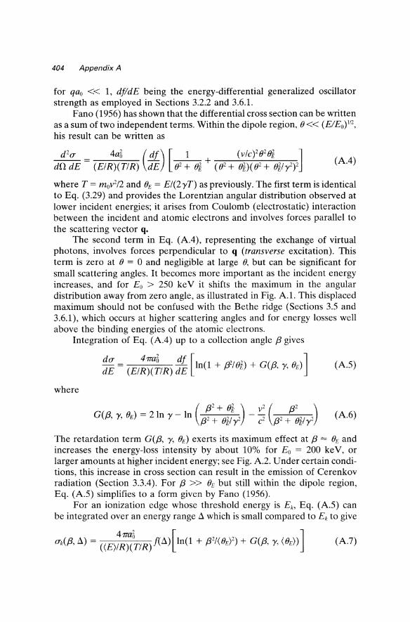
The second term in Eq. (A4), representing the exchange of virtual photons, involves forces perpendicular to q (transverse excitation). This term is zero at () = 0 and negligible at large (), but can be significant for small scattering angles. It becomes more important as the incident energy increases, and for Eo > 250 ke V it shifts the maximum in the angular distribution away from zero angle, as illustrated in Fig. A!. This displaced maximum should not be confused with the Bethe ridge (Sections 3.5 and 3.6.1), which occurs at higher scattering angles and for energy losses well above the binding energies of the atomic electrons.
Integration of Eq. (A4) up to a collection angle {3 gives
du 47mB df [ 2 ] dE = (EIR)(TIR) dE In(1 + ;J21()E) + G({3, y, ()E) (AS)
where
(A6)
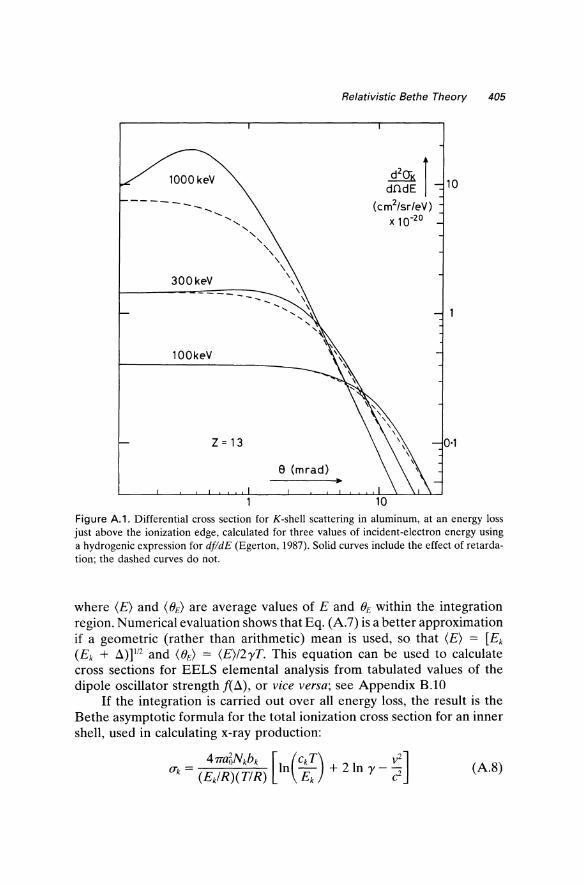
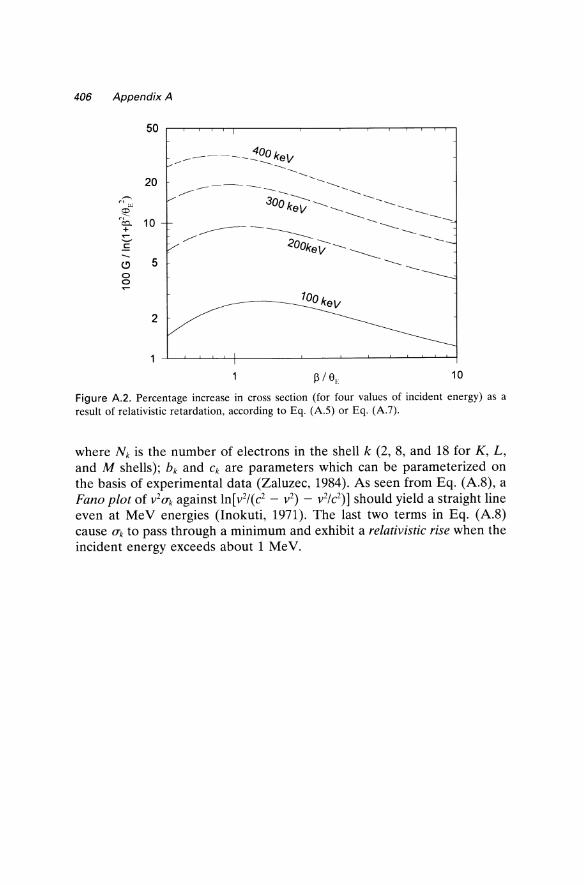
The retardation term G({3, y, ()E) exerts its maximum effect at {3 = ()E and increases the energy-loss intensity by about 10% for Eo = 200 ke V, or larger amounts at higher incident energy; see Fig. A2. Under certain conditions, this increase in cross section can result in the emission of Cerenkov radiation (Section 3.3.4). For {3 » ()E but still within the dipole region, Eq. (AS) simplifies to a form given by Fano (1956).
For an ionization edge whose threshold energy is Eb Eq. (AS) can be integrated over an energy range a which is small compared to Ek to give
-----
300keV -----
100keV
Z = 13
e (mrad)
Relativistic Bethe Theory 405
d2CTK I dndE
(cm2/sr/eV) x 10-20
10
0·1
Figure A.1. Differential cross section for K-shell scattering in aluminum, at an energy loss just above the ionization edge, calculated for three values of incident-electron energy using a hydrogenic expression for dJldE (Egerton, 1987). Solid curves include the effect of retardation; the dashed curves do not.
where (E) and (BE) are average values of E and BE within the integration region. Numerical evaluation shows that Eq. (A.7) is a better approximation if a geometric (rather than arithmetic) mean is used, so that (E) = [Ek (Ek + .:l)P/2 and (BE) = (E)/2yT. This equation can be used to calculate cross sections for EELS elemental analysis from tabulated values of the dipole oscillator strength f(.:l), or vice versa; see Appendix B.IO
If the integration is carried out over all energy loss, the result is the Bethe asymptotic formula for the total ionization cross section for an inner shell, used in calculating x-ray production:
(A.8)
406 Appendix A
50
20 ~ c,
"" (:I:l CC:J.. + 10 ~
E
(!) 5 0 0
2
10
Figure A.2. Percentage increase in cross section (for four values of incident energy) as a result of relativistic retardation, according to Eq. (A.S) or Eq. (A.7).
where Nk is the number of electrons in the shell k (2, 8, and 18 for K, L, and M shells); bk and Ck are parameters which can be parameterized on the basis of experimental data (Zaluzec, 1984). As seen from Eq. (A.8), a Fano plot of V 20"k against In[v2f(c2 - v2) - V 2/C2)] should yield a straight line even at MeV energies (Inokuti, 1971). The last two terms in Eq. (A.8) cause O"k to pass through a minimum and exhibit a relativistic rise when the incident energy exceeds about 1 MeV.
APPENDIX B Computer Programs
The computer codes discussed in this appendix are available by anonymous FTP to ftp.phys.ualberta.ca (directory /pub/eels); contact the author by email (egerton@phys. ualberta.ca) for further information. The programs are also listed in the Microscopy Society of America (MSA) public-domain library, accessible by anonymous FTP to www.amc.anl.govorftp.msa.microscopy.com (ANL Software Library, EMMPDLlEels subdirectory) or via WWW sites at Argonne National Laboratory (URL = http://www.amc. anl.gov or http://ftp.msa.microscopy.com).
B.1. Matrix Deconvolution
The following program removes plural scattering from a low-loss spectrum using the matrix method (see page 255) described by Schattschneider (1983) and by Su and Schattschneider (1992a), and is based on the FORTRAN program MATRIX written by these authors. The spectrum is read from a two-column (energy loss, intensity) file; the channel number NZ and integral AD of the zero-loss peak are found as in the FLOG program (p. 410). The data are normalized by dividing by AD, prior to matrix evaluation up to order n (typically 5 to 10). For correct scaling, the SSD is multiplied by AD before writing to an output file.
The output contains the single-scattering distribution over an energy range of approximately n times the mean single-scattering loss, followed by spurious data (which may involve large numbers, positive or negative). Larger n therefore increases the range of useful data, although at the expense of increased computing time. The method is mathematically exact for scattering up to the nth order.
Unlike the Fourier-log program listed in Section B.2, MATMOD ignores instrumental broadening of the energy-loss peaks. Artifacts may
407
408 Appendix 8 Sec. 8.1
therefore occur at multiples of the energy of the single-scattering peak, just as for a Fourier-log program which uses the delta function approximation: Eq. (4.13). These artifacts are more noticeable for thicker specimens and when the measured width of an main inelastic peak is comparable to that of the zero-loss peak.
Advantages over the Fourier-log method are that the data need not fall to zero at each end of their range; a limited number of data points (not necessarily a power of 2) can be processed, resulting in a short computing time. The specimen thickness can, in principle, be arbitrarily large, although errors involved in the measurement of the area of the zero-loss peak and from neglect of the instrumental resolution are likely to be significant for thicker specimens.
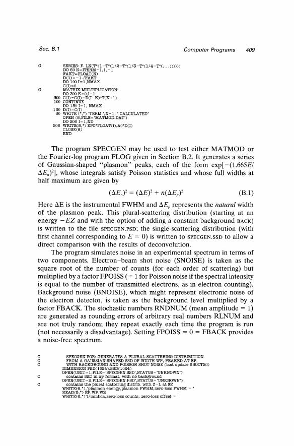
C MATMOD. FOR Last update: 95NOV03 C MATRIX DECONVOLUTION (P. SCHATTSCHNEIDER & D.S. SU, 199()"'1992) C This program is intended for deconvolution of multiple C inelastic scattering in image·mode (angle, integrated) low· loss C spectra. It utilizes a method originally described in C P. Schattschneider, Phil. Mag. B 47 (1983), 555-560. C SSD is written to the file MATMOD.DAT C CHARACTER'20 LABEL
DIMENSION T( 1 024),D( I 024),C(1 024),DATA( 1024),E( I 024) CHARACTER'12 INFILE,OUTFILE,TEXT WRlTE(', ') , NAME OF INPUT FILE ? ' READ(6,15) INFILE
15 FORMAT(A12) OPEN(13, FILE~INFILE) WRlTE(6, ') , NUMBER OF DATA POINTS TO BE READ ~ , READ(5,*) NSPEC ND~O
DO 107 I~l,NSPEC READ(13, ',END~50) E(l),DATA(l)
107 ND~I 50 CLOSE(13)
C FIND ZERO·LOSS CHANNEL: DO III I~I,ND NZ~I
III IF(E(l)+E(I+ l).GT.O.) GO TO 112 112 CONTINUE
C FIND SEPARATION POINT AS MINIMUM IN J(E)/E: DO 201 I~NZ,ND NSEP~I TANDIF ~ DATA(l + 1)/FLOAT(I - NZ+ 1) - DATA(I)/FLOAT(I - NZ) IF(TANDIF.GT.O.) GO TO 202
201 CONTINUE 202 EPC~(E(5)-E(1»/4.
BACK~(DATA(1)+DATA(2)+DATA(3)+DATA(4)+DATA(5»/5. SUM ~ O. DO 205 I~l,NSEP
205 SUM ~ SUM + DATA(I) AO ~ SUM - BACK'NSEP WRlTE(6, ')'ND,NZ,NSEP,BACK,AO,EPC ~ , ,ND,NZ,NSEP,BACK,AO,EPC WRlTE(6,*) 'eV/channel ~ ',EPC,', zero-loss intensity ~ ',AO DO 3 I~l,ND
3 T(l)~O.
C TRANSFER SHIFTED DATA TO ARRAY T(l): DO 302 I~NZ,ND
302 T(I-NZ+ 1)~DATA(l)-BACK C REMOVE ZERO-LOSS PEAK:
DO 4 I~l, NSEP-NZ+l 4 T(l)~O.
NMAX~ND-NZ+ I C NORMALIZE THE ARRAY T(I):
DO 30 I~ l,NMAX 30 T(l)~T(I)/AO
WRlTE(',') 'NUMBER OF TERMS IN LOG EXPANSION ~ , READ (6,') rrERM
C INITIALIZING THE ARRAY D(J): DO lOJ~l,NMAX
10 D(J)~T(J)/FLOAT(rrERM) WRlTE(',') 'SERIES EXPANSION OF LOG.
Sec. B.1
C SERIES: F. LNCT·(l-T·(l!2-T*(l!3-T·(l!4-T"(. .. )))))) DO 60 N=ITERM-l,l,-l FAKT=FLOAT(N) D(l)=-l./FAKT DO 100 1= 1 ,NMAX C(1)=O.
C MATRIX MULTIPLICATION: DO 300 K=O,I-l .
300 C(1) =C(1)-DCI - K)'TCK + 1) 100 CONTINUE
DO 1501=1, NMAX 150 D(1)=CCI)
50 WRITE C",') 'TERM ',N+l, 'CALCULATED' OPEN (6,FILE='MATMOD.DAT') DO 206 I=l,ND
206 WRITEC8,') EPC'FLOAT(1),AO"D(1) CLOSE(8) END
Computer Programs 409
The program SPECGEN may be used to test either MATMOD or the Fourier-log program FLOG given in Section B.2. It generates a series of Gaussian-shaped "plasmon" peaks, each of the form exp[ -(1.665EI aEn)2], whose integrals satisfy Poisson statistics and whose full widths at half maximum are given by
(B.1)
Here aE is the instrumental FWHM and aEp represents the natural width of the plasmon peak. This plural-scattering distribution (starting at an energy - EZ and with the option of adding a constant background BACK)
is written to the file SPECGEN.PSD; the single-scattering distribution (with first channel corresponding to E = 0) is written to SPECGEN.SSD to allow a direct comparison with the results of deconvolution.
The program simulates noise in an experimental spectrum in terms of two components. Electron-beam shot noise (SNOISE) is taken as the square root of the number of counts (for each order of scattering) but multiplied by a factor FPOISS (= 1 for Poisson noise if the spectral intensity is equal to the number of transmitted electrons, as in electron counting). Background noise (BNOISE), which might represent electronic noise of the electron detector, is taken as the background level multiplied by a factor FBACK. The stochastic numbers RNDNUM (mean amplitude = 1) are generated as rounding errors of arbitrary real numbers RLNUM and are not truly random; they repeat exactly each time the program is run (not necessarily a disadvantage). Setting FPOISS = 0 = FBACK provides a noise-free spectrum.
C SPECGEN.FOR: GENERATES A PLURAL-SCATTERING DISTRIBUTION C FROM A GAUSSIAN-SHAPED SSD OF WIDTH WP, PEAKED AT EP, C WITH BACKGROUND AND POISSON SHOT NOISE Clast update 950CT20)
DIMENSION PSD(l024),SSD(l024) OPENCUNIT= l,FILE= 'SPECGEN.SSD' ,STATUS = 'UNKNOWN')
C contains SSD in XJI format, with no background OPENCUNIT= 2,FILE = 'SPECGEN.PSD',STATUS= 'UNKNOWN')
C contains the plural scattering d1Strib. with Z-L at EZ WRITEC6, "), 'plasmon energy,plasmon FWHM,zero-loss FWHM = ' READC5,') EP,WP,WZ WRITE(6,*)'t!lambda,zero-loss counts, zero-loss offset = '
410 Appendix B
READ(5, *) TOL,AO,EZ WRlTE(6,*)'background level,eV/channel, number of channels = ' READ(5, *) BACK,EPC,ND WRlTE(6, *)'background noise fraction, Poisson noise fraction = ' READ(5, *) FBACK,FPOISS SZ = WZ/I.665 SP = WP/I.665 HZ = EPC* AO/Sz/ 1. 772 RLNUM= 1.23456
I = 1 C calculate intensity at each energy loss E :
10 E = FLOAT(l)*EPC - EZ FAC = 1. ORDER = o. PSD(I) = o.
C sum contribution from each order of scattering: 20 SN=SQRTCSZ*SZ+ORDER*SP*SP)
XPNT=(E-ORDER'EP)**2/SN/SN IF(XPNT.GT.20.0) EXPO=O.O IFCXPNT.LE.20.0) EXPO=EXP( - XPNT) DNE=HZ*SZ/SN'EXPO/FAC*TOL"ORDER RNDNUM=2. 'CFLOAT(IFIX(RLNUM))-RLNUM) SNOISE=FPOISS*CSQRTCDNE)'RNDNUM) RLNUM=9.8765'RNDNUM IFCORDER.NE.I.) GO TO 25 BNOISE=FBACK'BACK'RNDNUM SSD(I) = DNE+SQRT(SNOISE'SNOISE+ BNOISE'BNOISE) WRITE(l,') E,SSD(l)+BACK
25 CONTINUE PSDCl) = PSD(1) + DNE FAC=FAC*(ORDER+ I.) ORDER=ORDER + I. IF(ORDER.LT.15.) GO TO 20 SNOISE=FPOISS'CSQRTCPSD(1))'RNDNUM)
WRITEC2, *) E,PSDCl) +SQRTCSNOISE'SNOISE'+ BNOISE*BNOISE)+ BACK 1=1+1 IFCI.LE.ND) GO TO 10 CLOSE(l) CLOSE(2) END
B.2. Fourier-Log Deconvolution
Sec. B.1
The program FLOG calculates a single scattering distribution based on Eq. (4.11) and Eq. (4.10). It differs from the Fourier-log program used in the first edition of this book (and in Gatan ELIP software) by avoiding the delta-function approximation inherent in Eq. (4.13), and should therefore be preferable for processing low-loss or core-loss spectra containing sharp peaks.
The first ND data points are read from a named two-column file, which is presumed to consist of energy-intensity pairs of floating point numbers such as the ASCII x-y option provided by Gatan's ELiP software. The background level BACK is estimated from the first five intensity (y) values; if these points are not representative, they should be removed by editing the file, or else BACK set to zero in the program. The e V/channel value EPC is obtained from the first and fifth energy (x) values; the zero-loss channel number NZ is found by detecting positive x-values, on the assumption that the spectrum has been previously calibrated (for the zero-loss peak, at least). The separation point NSEP between the elastic and inelastic components is taken as the subsequent minimum in J(E)/E; the liE
Sec. B.2 Computer Programs 411
weighting discriminates against glitches on the zero-loss profile. The zeroloss intensity AO is taken as the sum of channel counts (above background) up to I=NSEP. Any discontinuities in the data (e.g., gain change during serial recording) are assumed to have been removed by prior editing.
The data are transferred to odd-numbered elements of the array D(J), subtracting any background and shifting the spectrum to the left so that J = 1 corresponds to the zero-loss channel. D(J) is extrapolated to the end of the array (J = MM-l) by fitting the last 10 data channels to an inverse power law, using Eq. (4.51). A cosine-bell function is subtracted to make the data approach zero at the end of the array without causing a discontinuity in intensity or slope at the last recorded data point (J = MFIN). The zeroloss peak Z(J) is copied from D(J) and the discontinuity at the separation point (MSEP) removed by subtracting a cosine-bell function, a procedure which preserves the zero-loss integral as AO. Even-numbered elements of D(J) and Z(J) are set to zero, indicating real data.
An effective width FWHMI of the zero-loss peak is estimated from the peak height and area, taking the peak shape to be Gaussian, and the operator enters a choice of reconvolution function: either the zero-loss peak (enter a negative number) or a Gaussian peak of specified width FWHM2. If this width is the same as FWHMl, there is no peak sharpening (and no noise amplification) but peak-shape distortion due to an asymmetric Z(E) is corrected. With the direction bit positive (ISIGN = 1), a fastFourier subroutine (Higgins, 1976) calculates cosine and sine coefficients of the Fourier transform, replacing the original data in D(J) and Z(J). The Fourier coefficients are manipulated according to Eqs. (4.14)-(4.17) and the phase term () in Eq. (4.17) extended to ± 7T to enable scattering parameters up to tl A = 3 to be accommodated. The higher-J coefficients are attenuated to avoid noise amplification, using a Gaussian filter function or by multiplying by the Fourier transform of Z(E). With ISIGN = -1, the FFT subroutine performs an inverse transform, placing the single-scattering distribution (without zero-loss peak) into odd elements of D(J) and into an output file; prior division by the number NN of real data points ensures that the output is correctly scaled. For spectra which extend to high energy loss and cover a very large dynamic range, the program may need to be modified (for some computers) to make use of double-precision arithmetic.
The fast-Fourier subroutine FFT (Higgins, 1976) uses a process known as the Danielson-Lanczos lemma. The N-point transform is divided into two NI2 transforms (by taking alternate data points), each of which is split into two and so on, ending in the requirement for N I-point transforms provided that N is of the form 2k where k is an integer. Ordering of the data involves bit-reversal sorting; overall, the number of mathematical operations is reduced from N2 to approximately N(10g2 N), a great saving
412 Appendix 8 Sec. 8.2
in computing time when N is large. An even shorter subroutine, which does not involve sorting, has been published by Uhrich (1969).
C FLOG.FOR LAST EDIT 95NOV03 C FOURIER-LOG DECONVOLUTION USING EXACT METHODS (A) OR (B) C Details in Egerton: EELS in the EM, 2nd edn.(Plenum Press, 1996) C Single·scattering distribution Is wrItten to the file FLOG.DAT
DIMENSION DATA(1024),E(1024),D(4096),Z(4096),SSD(4096) CHARACTER*12 INFILE WRITE(*,*) 'FLOG. FOR: name of mput file ~ , READ(5,15) INFILE
15 FORMAT(A12) OPEN(13,FILE~INFILE) OPEN(UNlT~ 14,FILE~ 'FLOG.DAT' ,STATUS~ 'UNKNOWN') NN ~ 2046 WRITE (6, *) 'Number of data pOints to be read ~ , READ(5, *) NSPEC DO 100 I~1, NSPEC READ(l3,*,END~50) E(I),DATACD ND~I
100 CONTINUE 50 EPC~(E(5)-E(l))/4.
BACK~(DATA( 1)+ DATA(2)+ DATA(3)+ DATA( 4)+ DATA(5))/5. C Find zero· loss channel:
DO 101 I~ 1,ND NZ~I
101 IF (E(l)+E(I+l).GE.O.) GO TO 102 102 CONTINUE
C Find minimum in J (E) /E to separate zero-loss peak: DO 201 I~NZ,ND IF(DATA(I+ 1)/FLOAT(I-NZ+ l).GT.DATACI)/FLOAT(l-NZ)) GO TO 202
201 NSEP ~ I 202 SUM ~ O.
DO 205 I~1,NSEP 205 SUM ~ SUM + DATA(l)
AO ~ SUM - BACK*FLOAT(NSEP) MSEP~2*(NSEP-NZ)+ 1 MFIN~2*(ND-NZ)+1
MM~2*NN C TRANSFER SHIFTED DATA TO ARRAY D(J):
DO 302J~1,MFIN, 2 302 D(J)~DATA((J -1)/2+ NZ)-BACK
C EXTRAPOLATE THE SPECTRUM TO ZERO AT J~MM -1: Al ~ D(MFIN-10)+D(MFIN-12)+D(MFIN-14)+D(MFIN-16)+D(MFIN-18) A2 ~ D(MFIN)+D(MFIN-2)+D(MFIN-4)+D(MFIN-6)+D(MFIN-8) R ~ 2.*ALOG((A1+.2)/(A2+.1))/ALOG(FLOAT(ND-NZ)/FLOAT(ND-NZ-1O)) IF(R.GT.O.) GO TO 303 R~O.
303 DEXT ~ D(MF!N)*(FLOAT(MFIN-l)/FLOAT(MM-4-2*NZ))**R 00 304 J~MFIN,MM,2 COSB ~ 0.5 - 0.5*COS(3.1416*FLOAT(J-MFIN)/FLOAT(MM-MFIN-2*NZ-3))
304 D(J) ~ D(MF!N)*(FLOAT(MFIN-1)/FLOAT(J-1))**R - COSB*DEXT C SET IMAGINARY COEFFICIENTS OF SPECTRAL DATA TO ZERO:
DO 305 J~1,MM,2 D(J+l)~ O. Z(J)~ O.
305 Z(J+1)~0. C COPY ZERO-LOSS PEAK AND SMOOTH RIGHT·HAND END:
DO 307 J~1, MSEP,2 Z(J)~D(J)-D(MSEP)/2. *(l.-COS(l.571*FLOAT(J -1)/FLOAT(MSEP-1)))
307 Z(2*MSEP-J)~D(MSEP)/2. *(l. -COS(l.571*FLOAT(J -l)/FLOAT(MSEP-1))) C ADD LEFT HALF OF Z(E) TO END CHANNELS OF ARRAYS D AND Z:
DO 306 I~1,NZ-l D(2*I-2*NZ+MM+1) ~ DATA(I)-BACK
306 Z(2*I-2*NZ+MM+1) ~ DATA (I)-BACK WRITE(6,*) 'Nread,NZ,NSEP,BACK ~ ',ND,NZ,NSEP,BACK WRITE(6,*) 'eV /channel ~ ',EPC,', zero-loss intenSity ~ ',AO FWHM1 ~0.9394*AO/D(l) WRITE(6,650) FWHM1
650 FORMAT(, FWHM ~',F4.1,' channels; enter new FWHM or -1. ') READ(5, *) FWHM2
551 CALL FFT(NN, + 1,Z) CALL FFT(NN, + I,D)
C Process the Fourier coefficients: DO 403 J~l,MM,2 DR~D(J)+ 1E-10 DI~D(J+1)
ZR~Z(J)+ 1E-10 ZI~Z(J+1) TOP ~ DI*ZR - DR*ZI
Sec. B.2
C
C
C
C
BOT = DR"ZR + m"ZI RL=0.5* ALOG(BOT**2+TOP""2)-ALOG(ZR"ZR +ZI"ZI) TH=ATAN(TOP/BOT) Extend range of arctan to + / - pi radians: IF(BOT.GE.O.O) GO TO 350 TH = TH + 3.14159265 IF(TOP.GE.O.O) GO TO 350 TH = TH - 3.14159265*2. Apply ZLP fUter and scaJ.i.ng factor for inverse transform:
350 D(J) = (ZR"RL-ZI"TH)/FLOAT(NN) D(J + 1)=(ZI"RL+ZR"TH)/FLOAT(NN) IFCFWHM2.LT.0.) GO TO 400 Next 5 lines replace ZLP fUter with a Gaussian: X= 1.687"FWHM2"FLOAT(J -1)/FLOAT (MM) IF(J.LT.NN) GO TO 380 X= 1.887"FWHM2"FLOAT(MM -J + 1 )/FLOAT(MM)
380 GAUSS = O. IFCX.GT.9.0) GO TO 390 GAUSS = EXP( - X"X)
390 D(J) = RL"AO"GAUSS/FLOAT(NN) D(J + 1 )=TH* AO"GAUSS/FLOAT(NN)
400 CONTINUE 403 CONTINUE
CALL FFr(NN, -1 ,D) DO 902 J=1,MM,2
902 WRITE(l4,") EPC"FLOATceJ -1)/2),D(J) CLOSE(l3) CLOSE(l4) END
SUBROUTINE FFr(NN,ISIGN,D) DIMENSION D(4096) N=2"NN J=1 D05I=1,N,2 IF(I-J)1,2,2 TR=D(J) TI=D(J+1) D(J)=D(I) D(J+1)=DG+1) D(I)=TR D(I+1)=TI
2 M=N/2 3 IF(J-M)5,5,4 4 J=J-M
M=M/2 IF(M-2)5,3,3
5 J=J+M MM=2
6 ML=MM-N IF(ML)7,10,10
7 IS=2*MM TH=6.283185/FLOAT(ISIGN"MM) HT=TH/2. ST=SIN(HT) W1=-2."ST"ST W2=SIN(TH) WR=l. WI=O. DO 9 M=1,MM,2 DO 8 I=M,N,IS J=I+MM A=WR*D(J) B=WI*D(J + 1) TR=A-B TI=WR*D(J+ 1)+WI*D(J) D(J)=DG)-TR
11 D(J+ 1)=DG+ 1)-TI D(I) = D(I) +TR
6 D(I+ 1)=DG+ 1)+TI TR=WR WR=WR"W1-WI*W2+WR
9 WI=WI"W1+TR*W2+WI MM=IS GO TO 6
10 RETURN END
Computer Programs 413
414 Appendix B
B.3. Kramers-Kronig Analysis and Thickness Determination
Sec. B.3
The program KRAKRO calculates the real part el(E) and imaginary part e2(E) of the dielectric function, the specimen thickness t, and the mean free path A(f3) for inelastic scattering. It employs the Fourier procedure for Kramers-Kronig analysis described by Johnson (1972), but using fastFourier transforms. As input, it requires a single-scattering distribution starting at the first channel, together with appropriate values of the incidentelectron energy, energy increment per channel, collection semiangle f3 and optical refractive index n. In the case of a metallic specimen, a large value (> 100) should be entered for n.
The SSD is read into an array SSD(I) and copied to odd elements of the array D(J). Assuming a Lorentzian angular distribution, an aperture correction is applied to the intensity SeE) to make it proportional to 1m ( -lie); the proportionality constant RK is evaluated by utilizing the Kramers-Kronig sum rule, Eq. (4.27). Since RK = lotl(7TUomov2) according to Eq. (4.26), this leads to an initial estimate of specimen thickness and mean free path, evaluated as A = tl(tIA) = tIof/l where II is the integral of the SSD intensity. Im( -lie) is copied to the array DI before being converted to its Fourier transform. Even-number elements of D(J) then contain the sine transform of Im( -lie). These coefficients are transferred to oddnumber elements so that inverse (cosine) transformation yields Re(1/e)-1, accompanied by its reflection about the midrange (J = NN) axis, due to aliasing. Taking the high-energy tail to be proportional to E-2, this energy dependence is subtracted from the low-energy (J < NN) data and used to extrapolate the high-energy values (a procedure which becomes less critical as the number of real data points NN is increased).
The real part EPS1 and imaginary part EPS2 of e are computed, followed by the surface energy-loss function SRFELF and the surfacescattering intensity SRFINT, and written to the output file EPSILON.DAT.
Calculation of the surface-mode scattering is based on Eq. (4.31), assuming clean (unoxidized) and smooth surfaces which are perpendicular to the incident beam, and neglecting coupling between the surfaces (liRe = 1 +e). The volume-loss intensity, obtained by subtracting SRFINT from SSD(J), is then renormalized by applying the K-K sum rule, leading to revised estimates for the specimen thickness and inelastic mean free path. KramersKronig analysis is then repeated to yield revised values of the dielectric data.
By setting NLOOPS > 2, further iterations are possible. Whether convergence is obtained depends largely on the behaviour of the data at low energy loss (E < 5 eV). To aid stability, E has been replaced by E + 1 in the expression for the surface-scattering angular dependence ANGDEP,
Sec. B.3 Computer Programs 415
thereby avoiding a non-zero value of SRFINT at E does not significantly bias the thickness estimates.
O. This modification
C KRAKRO.FOR Last update 95NOV05 C Kramers-Kronig analysis using J ohnsan method and FFI' subroutine C as described in EElB in the Electron Microscope C2nd edition). C Program generates output into the 5·column file KRAKRO.DAT
DIMENSION EN(4096), D(8192), DI(8192), DS(4096), SSD(4096) CHARACTER*12 INFILE WRITEC',*) 'NAME OF INPUT FILE ~ , READ(6,6) INFILE
6 FORMAT(A12) NN~4096 MM~2*NN DO lOI~l,NN
10 SSD(I)~O. OPEN(3,FlLE~INFILE) DO 12 I ~ 2, NN, 1 READC3, * ,END~50) EN(I),SSDCI)
12 D(2*I-1) ~ SSD(I) 50 D(I)~O.
SSD(I) ~ O. EPC~(ENC5)-EN(l))/4. WRITEC6, *) 'zero·loss sum, EO(keV),BETACmrad),ref.index ~ , READ(5,*) AO,EO,BETA,RI WRITEC6, *) 'no. of iterations, no. of lines into KRAKRO.DAT ~ , READ(5,*) NLOOPS,NLINES T ~ EO*Cl.+EOIl022.12)/(I.+EO/51 1.06)**2 RKO ~ 2590. * (l.+EO/511.06)*SQRT(2.*T/511.06) TGT ~ EO*(1022.12 + EO)/C51 1.06 + EO) OPEN(4,FlLE~'KRAKRO.DAT',STATUS~'UNKNOWN') LOOP ~ 1
C Calculate Im( -I /EPS), Re(l/EPS), EPS1, EPS2 and SRFINT data: DO 85 NUM~l,NLOOPS SUM ~ O. AREA ~ O.
C Apply aperture correction APC at each energy loss: Do 15J~3,MM,2 AREA ~ AREA+D(J) E~EPC*FLOATCJ -1)/2. APC~ALOG(I.+CBETA*TGT/E)**2) D(J)~DCJ)/APC
15 SUM~SUM+D(J)/E RK~SUM/ 1.571 /(1. -l./RI/RI)*EPC TNM ~ 332.5*RK/CAO*EPC)*EO*(I.+E0I1022.12)/(l.+EO/511.06)**2 TOL ~ AREA/ AO RLAM ~ TNM/TOL WRITEC6,61) NUM,TNM,TOL,RLAM
61 FORMATe LOOP',I2,': t(nm) ~',F6.1,', t/1ambda ~',F5.2, /!e', 1ambdaCnm) ~',F6.1)
C Apply normalization factor RK; store ImC -1 /EPS) in array DI: DO 20 J~1,MM,2 D(J)~D(J)/RK DI((J + 1 )/2)~D(J)
20 D(J + 1)~0 CALL FFT(NN, + I,D)
C Transfer sine coefficients to cosine locations: DO 30J~1,MM,2 D(J) ~2. *D(J + l)*FLOAT(CJ - NN)/IABS(J - NN))/FLOATCNN)
30 DCJ + 1) ~O. CALL FFT(NN,-l,D) DO 40 J~1,NN,2
C Correct the even function for reflected tail: D(J)~DCJ)+ 1.-D(NN-1)/2. *CFLOAT(NN-1)/FLOAT(MM-J))**2
40 DCNN+J) ~ 1.+D(NN-1)*CFLOAT(NN-1)/FLOAT(NN+J))**2/2. T ~ EO*Cl. + E0I1022.12)/(1. + EO/51 1.06)**2 RKO ~ 2590. * (1.+EO/51 1.06)*SQRTC2.*T/51 1.06) DO 80 J~3,MM,2 E~FLOATC(J -l)/2)*EPC RE ~ DCJ) DEN~DCJ)*DCJ)+ DIC(J + l)/2)*DICCJ + 1)/2) EPS1 ~DCJ)/DEN EPS2~DICCJ+ 1)/2)/DEN
C Calculate surface energy· loss function and surface intensity: SRFELF ~ 4.*EPS2/C(I.+EPS1)**2+EPS2**2) - DIC(J+1)/2) ADEP~TGT/CE+ 1.)* ATANCBETA *TGT/E)-BETA/ 1000./CBETA **2+ E*'2/TGT**2) SRFINT ~ 2000.*RK/RKO/TNM*ADEP*SRFELF DCJ) ~ SSDCCJ+ 1)/2)-SRFINT IF CNUM.NE.NLOOPS) GO TO 69 IF CJ/2.GT.NLINES) GO TO 69
416 Appendix B
WRITE( 4,66) E,EPSl.EPS2,RE,DI((J + 1 )/2) 66 FORMAT(6F12.4) 69 CONTINUE 60 CONTINUE
D(l) ~ O. 85 CONTINUE
CLOSE(4) END
Sec. B.3
For testing KRAKRO (or other purposes), the FORTRAN program DRUDE calculates the single-scattering plasmon-loss spectrum for a specimen of a given thickness TNM (in nm), recorded with electrons of a specified incident energy EO by a spectrometer which accepts scattering up to a specified collection semi angle BETA. It is based on the free-electron model (Section 3.3,1), with the volume energy-loss function ELF given by Eq. (3.42) and the surface-scattering energy-loss function SRFELF as in Eq. (4.31). The surface term can be made negligible by entering a large specimen thickness. The spectral intensity is written to the file DRUDE.SSD, while the real and imaginary parts of the dielectric function are written to DRUDE.DAT
for comparison with the results of Kramers-Kronig analysis.
C DRUDE.FOR Last update 950CT02 C Given the plasmon energy (EP) and plasmon FWHM (EW). this program C generates EPS 1, EPS2 from Eq. (3.40), ELF~Im( ~ l/EPS) from Eq. (3.42), C single soattermg intensltles VOLINT from Eq. (426) and SRFINT C from Eq. (4.31) of EEL'3 in the EM (Plenum Press, 2nd edition). C The output is E,SSD into the flie DRUDE.SSD and C E,EPS 1 ,EPS2,Re(l /eps),Im( ~ l/eps) mto DRUDE.DAT C
WRITE(6,') 'plasmon energy,plasmon width,eV/channel ~ , READ(5,') EP,EW,EPC WRITE(6,') 'Beta(mr),EO(keV),t(nm) ~ , READ(5, ') BETA,EO,TNM WRITE(6,') 'zero-loss integral, number of data points ~ , READ(5,') AO,NN B ~ BETA/lOoo. T ~ 1000. 'EO'(l.+EO/1022.12)/(l. +EO/511.06)"2 TGT ~ l(X)O. 'EO'(1022.12 + E)/(511.06 + EO) rum ~ 2590.' (1.-rEO/511.06)'SQRT(2.'T/511060) OPEN(l4,FILE~'DRUDE.DAT',STATUS~'UNKNOWN') OPEN(l3,FILE~ 'DRUDE.SSD' ,STATUS~ 'UNKNOWN') DO 12 IW ~ 2, NN, 1 E ~ EPC'FLOAT(IW - 1) EPSI ~ 1. ~ EP"2/(E'*2+EW"2) EPS2 ~ EW*EP"2/E/(E**2+EW"2) ELF = EP**2*E*EW/((E**2-EP**2)**2+(E*EW)**2) REREPS ~ EPSl/(EPS1*EPS1+EPS2*EPS2) THE ~ E/TGT SRFELF ~ 4.'EPS2/((l.+EPS1)"2+EPS2**2) ~ ELF ANGDEP ~ ATAN(B/THE)/THE - B/(B'B+THE'THE) SRFINT ~ EPC' AO* ANGDEP*SRFELF / (3.1416*O.0529*RKO'T) ANGLOG ~ ALOG(l.+ B'B/THE/THE) VOLINT ~ EPC*AO/3.1416*TNM/O.0529/T/2. *ELF'ANGLOG SSD ~ VOLINT + SRFINT WRITE(l3,') E,SSD
12 WRITE(l4,') E,EPS1,EPS2,REREPS,ELF CLOSE(l4) CLOSE(l3) STOP END
Sec. 8.4 Computer Programs 417
8.4. Fourier-Ratio Deconvolution
The program FRAT removes plural scattering from an ionization edge (whose background has previously been subtracted) using the Fourier-ratio method described in Section 4.3.2. It requires a low-loss spectrum, recorded from the same region of specimen at the same e V/channel, but this spectrum need not be contiguous with the core-loss region or match it in terms of absolute intensity. The method is therefore a more practical alternative to the Fourier-log program in the case of ionization edges recorded using a parallel-recording spectrometer. Other advantages are that the zero-loss peak does not need to be extracted from the low-loss spectrum (which involves some approximation) and that the specimen thickness is in principle unlimited, since there are no phase ambiguities in the Fourier components.
The low-loss spectrum is read as two-column (x-y) data (up to 1024 channels) from a named file and the zero-loss channel found from the energy (x) data. The first minimum is found in order to estimate the zeroloss integral AO and the energy resolution (obtained from AO and the zeroloss peak height, assuming a Gaussian shape). The spectrum is transferred to odd elements of the working array D(J), shifted so that the first channel represents zero loss and with any background (average of the first five channels) subtracted. D(J) is extrapolated to zero at the last odd-numbered channel, using a power-law extrapolation and a cosine-bell termination, and the left half of the zero-loss peak added to the end channels. The energy resolution (FWHM of the zero-loss peak) is printed to serve as a guide in specifying the width of the reconvolution function; smaller widths lead to peak sharpening but with a noise penalty (page 266). Even without such sharpening, the effect of any tails on the zero-loss peak (due for example to the point-spread function of a parallel-recording detector) is removed from the core-loss data.
The core-loss spectrum is read into odd elements of an array C(J) and extrapolated to zero in the same way as D(J). After taking Fourier transforms, using the FFT subroutine listed in Section B.2, the Fourier coefficients are processed according to Eq. (4.38) and Eq. (4.43), with a Gaussian reconvolution function GAUSS. If the coefficient of this function is the zero-loss integral (AO), plural scattering is subtracted from the ionization-edge intensity; if the coefficient is changed to the total integral (AT) of the low-loss spectrum, the core-loss SSD will have the same integral as the original edge, as required for absolute analysis of thick specimens (Wong and Egerton, 1995).
418 Appendix 8 Sec. 8.4
C FRAT.FOR Last update: 950CT31 C FOURIER· RATIO DECONVOLUTION USING EXACT METHOD (A) C (R.F.Egerton: EELS in the Electron Microscope, 2nd edition) C WITH LEFT SHIFI' BEFORE FORWARD TRANSFORM. C RECONVOLUTION FUNCTION R(F) IS EXP( - X*X) . C DATA IS READ IN FROM NAMED INPUT FILES (umts 14 and 15) C OUTPUT DATA APPEARS in named OUTFILE (umt 16) as NC x·y PAIRS C
DIMENSION DATA(I024),E(1024),D(4096),C(4096) CHARACTER'I2 LFILE,CFILE,OUTFILE NN~2048 WRITE(*, ') 'NAME OF LOWLOSS FILE ~ , READ(5,15) LFILE
15 FORMAT(AI2) OPEN(l5,FILE~LFILE) WRITE (6,*) 'NUMBER OF LOWLOSS CHANNELS TO BE READ ~ , READ(5, ') NREAD DO 501 I~I,NREAD READ(l5,*,END~50) E(I),DATA(I)
501 ND~I 50 CLOSE(l5)
EPC~(E(5)-E(l»/4. BACK~DATA(l)+ DATA(2)+ DATA(3)+ DATA(4)+ DATA(5)
C Set BACK~O. if zero· loss tail dominates first 5 channels C Find zero-loss channel:
DO 101 I~I,ND NZ~I
101 IF (E(I)+ECI+l).GE.O.) GO TO 102 102 CONTINUE
C Find minimum in J(E)/E to estimate zero-loss sum AO: DO 201 I~NZ,ND IFCDATACI + I )/FLOAT(!- NZ+ 1).GT.DATACD/FLOAT(I - NZ» GO TO 202
201 NSEP~I 202 SUM~O.
DO 205 13~I,NSEP 205 SUM ~ SUM + DATACI3)
AO ~ SUM - BACK*FLOAT(NSEP) MSEP~2*(NSEP-NZ)+ I MFIN~2*(ND-NZ)+ I MM~2*NN
C TRANSFER SHIFTED DATA TO ARRAY D(J): DO 302 J~I,MFIN,2
302 D(J)~DATA((J-l)/2+NZ)-BACK C EXTRAPOLATE THE SPECTRUM TO ZERO AT J ~ MM -I:
Al ~ D(MFIN-IO)+D(MFIN-12)+D(MFIN-14)+D(MFIN-16)+D(MFIN-18) A2 ~ D(MFIN)+D(MFIN-2)+D(MFIN-4)+D(MFIN-6)+D(MFIN-8) R ~ 2. *ALOG((AI +.2)/(A2+.l)/ALOG(FLOAT(ND-NZ)/FLOAT(ND-NZ-IO» DEND ~ D(MFIN)*(FLOAT(MFIN-l)/FLOAT(MM -4-2*NZ»**R DO 304 J~MFIN,MM,2 COSB ~ 0.5 - 0.5*COS(3.1416*FLOAT(J-MFIN)/FLOAT(MM-MFIN-2*NZ-3»
304 D(J) ~ D(MFIN)*(FLOAT(MFIN-l)/FLOAT(J-I»**R - COSB*DEND C Compute total area and set imaginary coefficients to zero:
AT~O.
DO 305 J~I,MM,2 AT~AT+D(J) D(J+I)~ O. C(J)~ O.
305 C(J + l)~ O. C Add left half of Z(E) to end channels in the array D(J):
C
DO 306 I~I,NZ-I 306 D(2*I-2*NZ+MM+l) ~ DATACI)-BACK
WRITE(6,*) 'ND,NREAD,NZ,NSEP,DATA(NZ),BACK,AO,THRESH,DEND,EPC:' WRITE(6,640) ND,NREAD,NZ,NSEP,DATA(NZ),BACK,AO,THRESH,DEND,EPC
640 FORMAT(4I6,6EI4.3) FWHMI ~O.9394*AO/D(l)*EPC WRITE(6,650) FWHMI
650 FORMAT('FWHM ~ ',F4.1, 'eV; FWHM of coreloss SSD ~ ') READ(5,*) FWHM2 FWHM2~FWHM2/EPC
WRrrE(*, *) 'Name of careless file = 1
READ(5,17) CFILE 17 FORMAT(AI2)
OPEN(l4,FILE~CFILE) WRITE (6,*) 'NUMBER OF CORELOSS CHANNELS TO BE READ ~ , READ(5, *) NC DO 551 I~I,NC READ(l4,*,END~55) E(I),C(2*I-I) NREAD~I
551 CONTINUE 55 EPC~(E(5)-E(l»/4.
NC~NREAD
CLOSE(l4)
Sec. 8.5 Computer Programs
C EXTRAPOLATE THE SPECTRUM TO ZERO AT J=MM -1: MFIN=2*NC-l Al = C(MFIN-I0)+C(MFIN-12)+C(MFIN-14)+C(MFIN-16)+C(MFIN-16) A2 = C(MFIN+C(MFIN-2)+C(MFIN-4)+C(MFIN-6)+C(MFIN-8) R = 2. * ALOG((AI + .2)/(A2+ .1))/ ALOG(ECNC)/E(NC-9)) CEND = A2/5.*(E(NC-2)/(E(l)+EPC*FLOAT(NN-l)))**R DO 314 J=MFIN,MM,2 COSB = 0.5 - 0.5*COS(3.1416*FLOAT(J-MFlN)/FLOAT(MM-I-MFlN))
314 C(J) = A2/ 5. *(E(NC-2)/ (E( 1)+ EPC*FLOAT((J - 5)/2)))**R -COSB*CEND CALL FFT(NN, + 1,C) CALL FFT(NN,+ I,D)
C PROCESS THE FOURIER COEFFICIENTS: DO 403 J=1,MM,2
C GAUSSIAN RECONVOLUTION FUNCTION: X= 1.887*FWHM2*FLOAT(J -l)/FLOAT(MM) IF(J.LT.NN) GO TO 381 X= 1.887*FWHM2*FLOAT(MM -J + 1 )/FLOAT(MM)
381 GAUSS = AO/2.718**(X*X)/FLOAT(NN) C Replace AO by AT for equal areas in SSD and PSD.
DR = D(J)+ lE-10 DI=D(J+l) CR=C(J)+ lE-10 CI=C(J+l) D(J) = GAUSS*(CR*DR+CI*DI)/(DR*DR+DI*DI) D(J + 1 )=GAUSS*(CI*DR-CR*DI)/(DR*DR + DI*DD
403 CONTINUE CALL FFT(NN, -I,D) WRITEC*, *) 'NAME OF OUTPUT FILE = ' READ(5,15) OUTFILE OPEN(l6,FILE=OUTFILE) DO 902 1= 1 ,NC WRITE(l6,*) E(l),D(2*I-l)
902 CONTINUE CLOSE(l6) END
B.5. Incident-Convergence Correction
419
The following program (CONCOR2) evaluates the factor FJ by which inelastic intensity (at energy loss E and recorded using a collection semiangle (3) is reduced as a result of the convergence of the electron beam (semiangle a). The program also evaluates a factor F2 (for use in absolute quantification) and an effective collection angle defined in Section 4.5.3. For inner-shell scattering, the energy loss E can be taken as the edge energy Ek or (more exactly) as an average energy loss (Ek+1112) within the integration window.
Whereas the program (CON COR) given in the first edition was based on Eq. (4.71), this version uses an analytical formula (Scheinfein and Isaacson, 1984) based on a Lorentzian angular distribution of inelastic scattering and assuming that the incident-beam intensity per steradian is constant up to the angle a. Double-precision arithmetic is used because the formula involves subtraction of terms which are nearly equal. Minor differences in output, compared to the earlier program, arise from correction of an error in the expression previously used to evaluate the characteristic angle THE.
When analyzing for two elements, a and b, incident-beam convergence is taken into account by multiplying the areal-density ratio Nj Nb, derived from Eq. (4.66), by Flbl Fla' If the absolute areal density Na of an element
420 Appendix 8 Sec. 8.5
a is being calculated from Eq. (4.65), the result should be divided by F2a•
For ll' < /3, F2 = FI; for ll' > /3, F2 is larger than FI (and may exceed unity; see Fig. 4.16) since the collection angle cuts off part of the low-loss angular cone. As a simpler alternative to applying the correction factors FI or F2, incident-beam convergence can be incorporated by computing each ionization cross section for the effective collection angle BST AR which is a function of energy loss and therefore different for each element.
C CONCOR2: EVALUATION OF CONVERGENCE CORRECTION F USING THE C FORMULAE OF SCHEINFEIN AND ISAACSON (SEM/1994, PP. 1685-6). C FOR ABSOLtrrE QUANTITATION, DIVIDE THE AREAL DENSITY BY F2. C FOR ELEMENTAL RATIOS, DIVIDE EACH CONCENTRATION BY F2 OR Fl. C C ALPHA AND BETA SHOULD BE IN MRAD, E IN EV, EO IN KEV . C
DOUBLE PRECISION ALPHA,BETA,THE,A2,B2,T2,Fl,F2 WRITE(6,601)
601 FORMAT(,O', 'CONCOR2: Enter ALPHA(mr),BETA(mr),E(eV),EO(keV)' I) READ(5, *) ALPHA,BETA,E,EO
501 FORMAT(4F1O.0) TGT=EO'O. + EOI 1022.)/(1. + EO/51 1.) THETAE=E/TGT
C A2,B2,T2 ARE SQUARES OF ANGLES IN RADlANS"2 A2=ALPHA'ALPHA'lE-6 B2=BETA*BETA'lE-6 T2=THETAE'THETAE'lE-6 ETA 1 =DSQRT((A2+ B2+T2)"2-4. 'A2'B2)-A2-B2-T2 ETA2=2.*B2'DWG(0.5/T2*(DSQRT((A2+T2-B2)"2+4.'B2'T2)+A2+T2-B2)) ETA3=2.'A2'DLOG(0.5/T2'(DSQRT((B2+T2-A2)"2+4.'A2'T2)+B2+T2-A2)) ETA=CETA1 + ETA2+ ETA3) I A2/DWG(4./T2) Fl = (ETA 1 + ETA2+ ETA3)/21 A2/DWG(1. + B2/T2) F2=Fl IF (ALHPA/BETA.LE.l.)GO TO 107 F2=Fl'A2/B2
107 CONTINUE BSTAR=THETAE'DSQRT(DEXP(F2'DWGO. + B2/T2))-1.) WRITE(6,602)
602 FORMATC/' ',5X,'Fl',8X,'F2',6X,'effective BETA'/) WRITE(6,6oo)Fl,F2,BSTAR
600 FORMAT(2FI0.3,F14.2) END
sample data: CONCOR2: Enter ALPHA(mr),BETA(mr),E(eV),EO(keV) 18,12,500,100
Fl 0.571
F2 1.285
effective BETA 16.05
8.6. Hydrogenic K-Shell Cross Sections
The FORTRAN program SIGMAK3 uses the hydrogenic approximation for generalized oscillator strength, Eqs. (3.125)-(3.127), in order to calculate differential (DSBYDE) and integrated cross sections and dipole oscillator strengths (SIGMA andfO) for K-shell ionization. Unlike the corresponding program (SIGMAK2) given in the first edition of this book, the reduction in effective nuclear charge (due to screening by the second 1s electron) is taken as 0.50 rather than the value of 0.3125 calculated by Zener (1930) for first-row elements, in order to provide a closer match to EELS, photoabsorption, and Hartree-Slater data (Egerton, 1993). The changes becomes
Sec. B.6 Computer Programs 421
more significant at low atomic number: 1(100 e V) is reduced from 0.46 to 0.42 for oxygen and from 2.02 to 1.58 for lithium. Two lines have been added before label 101 to prevent occasional error messages (from some compilers) when exp(-kH) becomes too small.
Relativistic kinematics are employed, based on Eqs. (3.139), (3.140), (3.144), and (3.146), but retardation effects (Appendix A) are not included. The energy-differential cross section DSBYDE is obtained from Eq. (3.151) with limits of integration given by Eqs. (3.152) and (3.153). The outer DO loop integrates DSBYDE to obtain the partial cross section SIGMA, making use of the energy dependence described by Eq. (3.154). IMAX sets the number of increments within the inner DO loop (integration over scattering vector); IMAX = 5 is sufficient for the evaluation of energy-loss partial cross sections (small f3 and d), but a larger number should be used when calculating total cross sections in order to accurately include the Bethe ridge. Because Eq. (3.154) is utilized in the integration over energy loss, relatively few energy increments are needed; EINC can be chosen as a convenient submultiple (e.g., 1/5 or 1/10) of the required energy window d. Typical input and output data are shown after the program listing.
Total K-shell cross sections (as required for EDX spectroscopy) are obtained by entering f3 = 3142 mrad, EINC = EK/lO, and taking the asymptotic value of SIGMA corresponding to large d; for Eo ::; 300ke V, the resulting values are within 3% of the Hartree-Slater values given by Scofield (1978) for Ar and Ni. Since the energy-differential cross section dafdE is independent of K-edge threshold energy EK, its value DSBYDE at any scattering angle BETA and energy loss E can be printed out by setting EINC=O and EK=E in the input data. Likewise, a cross section SIGMA integrated between any two values of energy loss can be obtained by entering the lower energy loss as EK and some submultiple of the energy difference as EINC.
C SIGMAK3 : CALCULATION OF K-SHELL IONIZATION CROSS SECTIONS C USING RELATIVISTIC KINEMATICS AND A HYDROGENIC MODEL WITH C INNER-SHELL SCREENING CONSTANT OF 0.6 (last update: 31Aug96) C
C INPUT DATA IS ENTERED AS REAL OR lNTEGER NUMBERS, C ON THE SAME LINE AND EACH FOLLOWED BY A COMMA, AS FOLLOWS:
C
Z - atomIC number of the element of Interest EK - K-shelJ Ionization energy, In eV
EINC - energy Increment of output data, In eV EO - inCident-electron energy, In ke V
BETA - maximum scattering angle (In milliradlans) contributing to the cross section
REAL KH2, LNQA02,LQA021,LQA02M,LQ2INC WRITE(6,601)
601 FORMAT(,O','Z,EK,EINC,EO,BETA ~ 'f) READ ',Z,EK,EINC,EO,BETA WRITE(6,602)
602 FORMAT(' ',6X,'E(EV)',7X,'DSBYDE',8X,'DELTA',7X,'SIGMA',7X,'fO'f) IMAX~IOO R~13.606 ZS~l.

422 Appendix B
RNK> 1. IF CZ.EQ.1.) GO TO 105 ZS ~ Z - 0.50 RNK>Z.
105 E ~ EK B ~ BETAIlOOO. T ~ 511060.*(l.-1./(l.+EO/(511.06))"Z)/Z. GG~ 1. + EO/51 1.06 POZ~T/R/(l. -Z. *T/511060.) F ~ O. S ~ O. SIGMA ~ O.
C CALCULATE FOR EACH ENERGY LOSS: DO III J~1,30 QAOZI ~ E**Z/(4.'R'T)+E"3/(8.'R*T**2*GG*'3) PP2 ~ P02 - E/R'(GG-EIl0221Z0.) LQAOZI ~ ALOG(QAOZl) QAOZM ~ QA021 + 4. *SQRT(P02*PP2)*(SIN(B/Z))"2 LQAOZM ~ ALOGCQA02M) LQ2INC ~ (LQA02M - LQA02l)/FLOAT(IMAX-l) LNQA02 ~ LQAOZI DFDIP ~ O. DSBYDE ~ O. GOSP ~ O.
C INTEGRATE OVER SCATTERING ANGLE: DO 109 I~l, IMAX
100 QA02 ~ EXP(LNQA02) Q ~ QA02/ZS"Z KHZ ~ E/R/ZS**2 - 1. AKH ~ SQRT(ABS(KH2)) IF(AKH.GT.O.l) GO TO 101 AKH~O.l
101 IF (KH2.LT.0.) GO TO 103 D ~ 1. - EXPC-2.*3.14159/AKH) BP ~ ATANC2*AKH/CQ'-KH2+ 1.)) IF (BP.GE.O.) GO TO 104 BP ~ BP + 3.14159
104 C ~ EXP((-2./AKH)*BP) IF (KH2.GE.O.) GO TO 102
C SUM OVER EQUIVALENT BOUND STATES: 103 D ~ 1.
Y ~ (-1./AKH'ALOG(CQ+1.-KH2+2.*AKH)/CQ+l -KH2-2.'AKH))) C ~ EXP(y)
102 A ~ ((Q-KH2+1.)*'2 + 4.'KH2)*'3 GOS~ 128. 'RNK*E/R/ZS"4'C/D'CQ+ KH2/3. + 1./3.)/ A/R
C GOS (~DF/DE) IS PER EV AND PER ATOM DSBYDE~DSBYDE + 3. 5166E- 16*(R/T)*CR/E)'(GOS +GOSP)/2. *LQ2INC
C DSBYDE IS THE ENERGY·DIFFERENTIAL X-SECN (CM*'2/EV/ATOM) IF (LGT.l) GO TO 115 DFDIPL ~ GOS DSBYDE ~ O.
115 LNQA02 ~ LNQA02 + LQZINC 109 GOSP ~ GOS
DELTA ~ E - EK IF (J.EQ.l) GO TO 11 7 S ~ ALOG(DSBDEP/DSBYDE)/ALOG(E/CE-EINC)) SGINC ~ (E*DSBYDE-(E-EINC)*DSBDEP)/(l.-S) SIGMA ~ SIGMA + SGINC
C SIGMA IS THE PARTIAL CROSS SECTION IN CM"2 PER ATOM F~F+(DFDIPL+ DFPREV)/2. *EINC
117 WRITE(8,605) E,DSBYDE,DELTA,SIGMA,F 605 FORMAT(, ',FIO.l,2X,E12.3,2X,FlO.1,2X,E12.3,2X,F7.3)
IF (EINC.EQ.O.) GO TO 112 IF CDELTA.LT.IOO.) GO TO 107 IF (SGINC.LT.O.OOI *SIGMA) GO TO 112 EINC ~ EINC*2
107 E ~ E + EINC IF CE.GT.T) GO TO 112 DFPREV ~ DFDIPL
III DSBDEP ~ DSBYDE 112 STOP
END
Sec. B.6
Sec. 8.7 Computer Programs 423
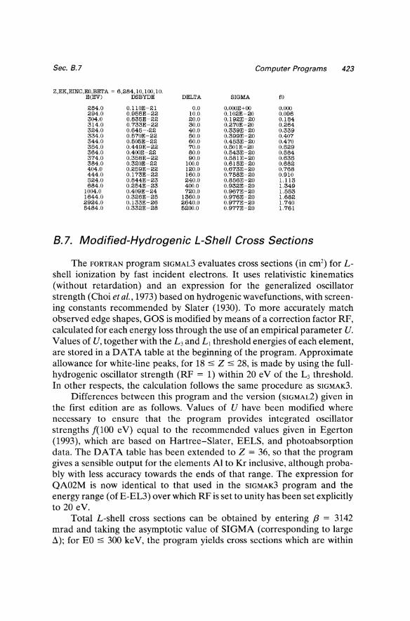
Z,EK,EINC,EO,BETA ~ 6,284,10,100,10. E(EV) DSBYDE DELTA SIGMA to
284.0 0.110E-21 0.0 O.oooE+OO 0.000 294.0 0.965E-22 10.0 0.102E-20 0.096 304.0 0.835E-22 20.0 0.192E-20 0.184 314.0 0.733E-22 30.0 0.270E-20 0.264 324.0 0.645-·22 40.0 0.339E-20 0.339 334.0 0.570E-22 50.0 0.399E-20 0.407 344.0 0.505E-22 60.0 0.453E-20 0.470 354.0 0.449E-22 70.0 0.501E-20 0.529 364.0 0.400E-22 80.0 0.543E-20 0.584 374.0 0.358E-22 90.0 0.581E-20 0.635 384.0 0.320E-22 100.0 0.615E-20 0.682 404.0 0.259E-22 120.0 0.673E-20 0.768 444.0 0.173E-22 160.0 0.758E-20 0.910 524.0 0.844E-23 240.0 0.856E-20 1.113 684.0 0.254E-23 400.0 0.932E-20 1.349
1004.0 0.409E-24 720.0 0.967E-20 1.553 1644.0 0.326E-25 1360.0 0.976E-20 1.682 2924.0 0.133E-26 2640.0 0.977E-20 1.740 5484.0 0.332E-28 5200.0 0.977E-20 1.761
B.7. Modified-Hydrogenic L-Shell Cross Sections
The FORTRAN program SIGMAL3 evaluates cross sections (in cm2) for Lshell ionization by fast incident electrons. It uses relativistic kinematics (without retardation) and an expression for the generalized oscillator strength (Choi et al., 1973) based on hydrogenic wavefunctions, with screening constants recommended by Slater (1930). To more accurately match observed edge shapes, GOS is modified by means of a correction factor RF, calculated for each energy loss through the use of an empirical parameter U. Values of U, together with the L3 and LJ threshold energies of each element, are stored in aDA T A table at the beginning of the program. Approximate allowance for white-line peaks, for 18 ::::; Z ::::; 28, is made by using the fullhydrogenic oscillator strength (RF = 1) within 20 eV of the L3 threshold. In other respects, the calculation follows the same procedure as SIGMAK3.
Differences between this program and the version (SIGMAL2) given in the first edition are as follows. Values of U have been modified where necessary to ensure that the program provides integrated oscillator strengths [(100 e V) equal to the recommended values given in Egerton (1993), which are based on Hartree-Slater, EELS, and photo absorption data. The DATA table has been extended to Z = 36, so that the program gives a sensible output for the elements Al to Kr inclusive, although probably with less accuracy towards the ends of that range. The expression for QA02M is now identical to that used in the SIGMAK3 program and the energy range (of E-EL3) over which RF is set to unity has been set explicitly to 20 eV.
Total L-shell cross sections can be obtained by entering f3 = 3142 mrad and taking the asymptotic value of SIGMA (corresponding to large .£l); for EO::::; 300 keY, the program yields cross sections which are within
424 Appendix 8 Sec. 8.7
8% of those calculated by Scofield (1978) for Ar and Ni. However, the algorithm is not designed to provide realistic values of differential cross section DSBYDE within 50 eV of the ionization edge or to accurately simulate the Bethe ridge at high scattering angle.
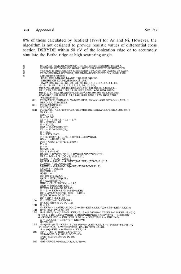
C SIGMAL3 : CALCULATION OF L-SHELL CROSS·SECTIONS USING A C MODIFIED HYDROGENIC MODEL WITH RELATIVISTIC KINEMATICS. C THE GOS IS REDUCED BY A SCREENING FACTOR RF, BASED ON DATA C FROM SEVERAL SOURCES; SEE ULTRAMICROSCOPY 60 (1993) P.22. C Last update: 96SepOl
REAL KH2,LNQA02,LQA021,LQA02M,LQ2INC DIMENSION XU(24),IE3(24),IEl(24) DATA XU/.62,.42,.30,.29,.22,.30,.22,.16,.12,.13,.13,.14,.16,
@.18,.19,.22,.14,.11,.12,.12,.12,.1O,.1O,.1O/, &!IE3/73,99, 136, 164,200,246,294,347,402,466,613,576,641, @710,779,866,931,1021,1116,1217,1323,1436,1660,1675/, &!IEI / 118, 149, 189,229,270,320,377,438,500,864,628,696,769, @846,926,loo8,1096,1194,1142,1248,1369,1476,1696,1727/
WRrrE(6,60l) 601 FORMAT(,0','SIGMAL3: VALUES OF Z, EO(keV) AND BETA(mr) ARE: ')
READ(6, ') Z,EO,BETA 601 FORMAT(3FI0.0)
WRITE(6,602) 602 FORMATC/' ',6X,'E(eV)',7X,'DSBYDE',8X,'DELTA',7X,'SIGMA',8X,'fO'/)
IMAX~10
EINC ~ 10. R ~ 13.606 ZS ~ Z - 0.36'(8. -1.) - 1. 7 IZ ~ IFIX(Z) - 12 U ~ XU(IZ) EL3 ~ FLOAT(IE3(IZ)) ELI ~ FLOAT(IEl(IZ)) E ~ EL3 B ~ BETA/lOoo. T ~ 611060.'(1.-1./(1.+EO/(611.06))"2)/2. GG ~ 1.+EO/611.06 P02 ~ T/R/(1.-2.'T/611060.) F ~ O. S ~ O. SIGMA ~ O. DO III J~1,40 QA021 ~ E"2/(4.'T'R) + E"3/(8.'R'T"2'GG"3) PP2 ~ P02-E/R'(GG-E/1022120.) LQA021 ~ ALOG(QA021) QA02M ~ QA021 + 4.'SQRT(P02'PP2)'(SIN(B/2.))"2 LQA02M ~ ALOG(QA02M) LQ2INC ~ (LQA02M-LQA021)/FLOAT(IMAX-l) LNQA02 ~ LQA021 DSBYDE ~ O. GOSP ~ O. DO 109 I~l,IMAX QA02 ~ EXP(LNQA02) Q ~ QA02/(ZS"2) KH2 ~ (E/(R'ZS"2)) - 0.26 AKH ~ SQRT(ABS(KH2)) IF(KH2.LT.0.0) GO TO 103 D ~ 1. - EXP(-2.'3.14169/AKH) BP ~ ATAN(AKH/(Q-KH2 + 0.26)) IF(BP.GE.O.O) GO TO 104 BP ~ BP + 3.14169
104 C ~ EXP((-2./AKH)'BP) IF(KH2.GE.0.0) GO TO 102
103 D ~ 1.0 C~EXP(( -1./AKH)'ALOG((Q+0.26-KH2+AKH)/(Q+0.26-KH2-AKH)))
102 IF(E-ELl)170,170,176 170 G~2.26'Q"4-(0. 76+3. 'KH2)'Q"3+(0.69376-0. 76'KH2-0.6'KH2"2)'Q'Q
@+(0.11146+0.86417'KH2+1.8833'KH2'KH2+KH2"3)'Q + 0.0036807 @+KH2/21.333 + KH2'KH2/4.6714 + KH2"3/2.4 + KH2"4/4.
A ~ ((Q-KH2 + 0.26)"2 + KH2)"6 GO TO 180
176 G~Q"3-(6./3. 'KH2+ 11./12.)'Q"2+(KH2'KH2/3. + 1.6'KH2+66./48.)'Q @+KH2"3/3.+0.76'KH2'KH2+23./48.'KH2+6./64.
A ~ ((Q-KH2 + 0.26)"2 + KH2)"4 180 RF ~ ((E+.I-EL3)/1.8/Z/Z)"U
IF(IABS(IZ-11).GT.5) GO TO 200 IF(E-EL3.GT.20) GO TO 200 RF~l.
200 GOS~RF'32. 'G'C/A/D'E/R/R/ZS"4
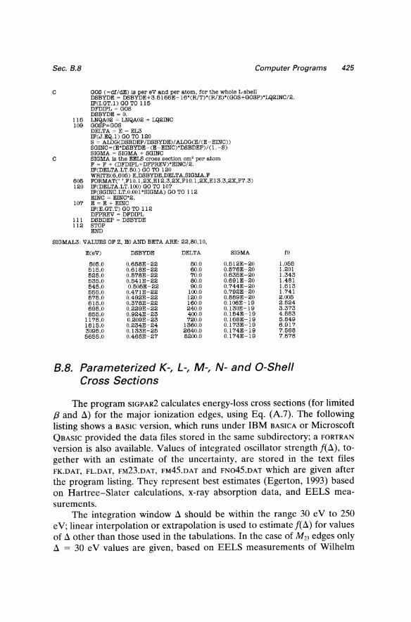
Sec. 8.8 Computer Programs
C GOS (~df/dE) is per eV and per atom, for the whole L·shell DSBYDE ~ DSBYDE+3.5166E~ 16*(R/T)*(R/E)*(GOS+GOSP)*LQ2INC/2. IF(I.GT.l) GO TO 115 DFDIPL ~ GOS DSBYDE ~ O.
115 LNQA02 ~ LNQA02 + LQ2INC 109 GOSP~GOS
DELTA ~ E ~ EL3 IF(J.EQ.l) GO TO 120 S ~ ALOG(DSBDEP/DSBYDE)/ALOG(E/(E~EINC)) SGINC~(E*DSBYDE~(E~ EINC)*DSBDEP)/(l. ~S) SIGMA ~ SIGMA + SGINC
C SIGMA is the EELS cross section em' per atom F ~ F + (DFDIPL+DFPREV)'EINC/2. IF(DELTA.LT.50.) GO TO 120 WRITE(6,605) E,DSBYDE,DELTA,SIGMA,F
605 FORMATe' ',FI0.1,2X,E12.3,2X,FI0.1,2X,E13.3,2X,F7.3) 120 IF(DELTA.LT.I00) GO TO 107
IF(SGINC.LT.O.OOl'SIGMA) GO TO 112 EINC ~ EINC'2.
107 E ~ E + EINC IF(E.GT.T) GO TO 112 DFPREV ~ DFDIPL
III DSBDEP ~ DSBYDE 112 STOP
END
SIGMAL3: VALUES OF Z, EO AND BETA ARE: 22,80,10,
E(eV) DSBYDE DELTA
505.0 0.658E~22 50.0 515.0 0.618E~22 60.0 525.0 0.578E~22 70.0 535.0 0.541E~22 80.0 545.0 0.505E~22 90.0 555.0 0.471E~22 100.0 575.0 0.492E~22 120.0 615.0 0.378E~22 160.0 695.0 0.229E~22 240.0 855.0 0.924E~23 400.0
1175.0 0.209E~23 720.0 1815.0 0.234E~24 1360.0 3095.0 0.133E~25 2640.0 5655.0 0.465E~27 5200.0
SIGMA
0.512E~20 0.576E~20 0.635E~20 0.691E~20 0.744E~20 0.792E~20 0.889E~20 0.106E~19 0.130E~ 19 0.154E~ 19 0.168E~ 19 0.173E~ 19 0.174E~19 0.174E~19
fO
1.055 1.201 1.343 1.481 1.613 1.741 2.005 2.524 3.373 4.553 5.849 6.917 7.568 7.878
8.8. Parameterized K-, L-, M-, N- and O-Shell Cross Sections
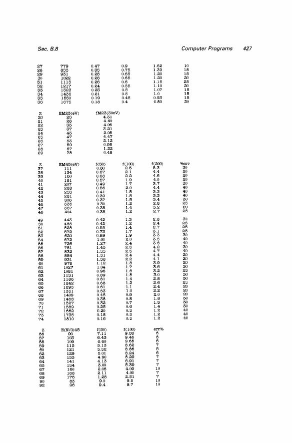
425
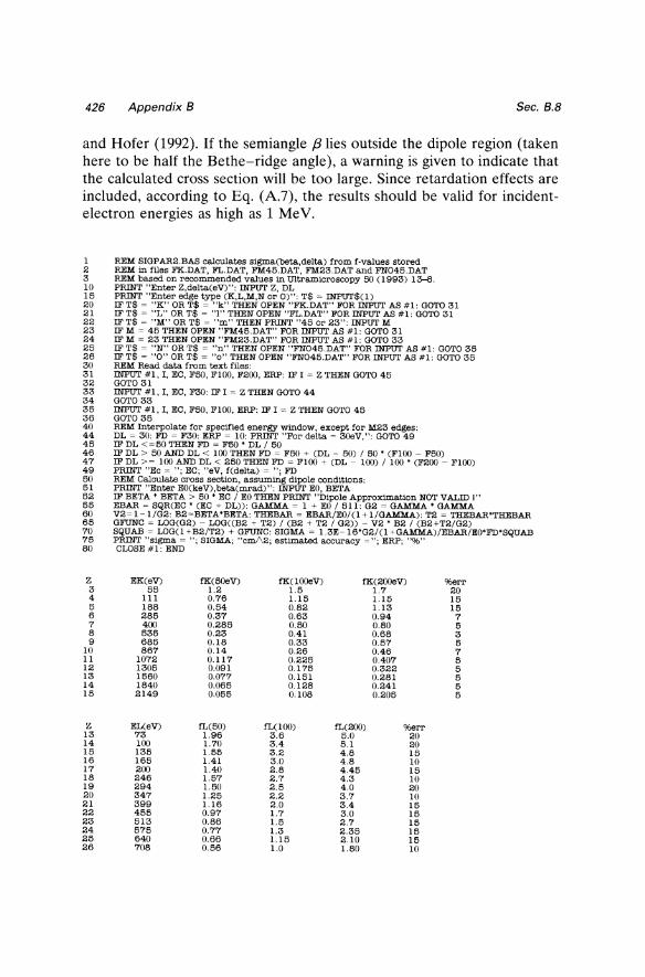
The program SIGPAR2 calculates energy-loss cross sections (for limited f3 and Ll) for the major ionization edges, using Eq. (A.7). The following listing shows a BASIC version, which runs under IBM BASICA or Microscoft QBASIC provided the data files stored in the same subdirectory; a FORTRAN
version is also available. Values of integrated oscillator strength feLl), together with an estimate of the uncertainty, are stored in the text files FK.DAT, FL.DAT, FM23.DAT, FM45.DAT and FN045.DAT which are given after the program listing. They represent best estimates (Egerton, 1993) based on Hartree-Slater calculations, x-ray absorption data, and EELS measurements.
The integration window Ll should be within the range 30 e V to 250
e V; linear interpolation or extrapolation is used to estimate feLl) for values of Ll other than those used in the tabulations. In the case of M23 edges only Ll = 30 e V values are given, based on EELS measurements of Wilhelm
426 Appendix B Sec. B.B
and Hofer (1992). If the semiangle f3lies outside the dipole region (taken here to be half the Bethe-ridge angle), a warning is given to indicate that the calculated cross section will be too large. Since retardation effects are included, according to Eq. (A.7), the results should be valid for incidentelectron energies as high as 1 MeV.
1 2 3 10 15 20 21 22 23 24 25 26 30 31 32 33 34 35 36 40 44 45 46 47 49 50 51 52 55 60 65 70 75 80
Z 3 4 5 6 7 8 9
10 11 12 13 14 15
Z 13 14 15 16 17 18 19 20 21 22 23 24 25 26
REM SIGPAR2.BAS calculates sigIna(beta,delta) from f·values stored REM in files FK.DAT, FL.DAT, FM45.DAT, FM23.DAT and FN045.DAT REM based on reco=ended values in Ultramicroscopy 50 (1993) 13-8. PRINT "Enter Z,delta(eV)": INPUT Z, DL PRINT "Enter edge type (K,L,M,N or 0)": T$ ~ INPUT$(1) IF T$ ~ "K" OR T$ ~ "k" TEEN OPEN "FK.DAT" FOR INPUT AS #1: GOTO 31 IF T$ ~ "L" OR T$ ~ "I" THEN OPEN "FL.DAT" FOR INPUT AS #1: GOTO 31 IF T$ ~ "M" OR T$ ~ "m" THEN PRINT "45 or 23": INPUT M IF M ~ 45 THEN OPEN "FM45.DAT" FOR INPUT AS #1: GOTO 31 IF M ~ 23 THEN OPEN "FM23.DAT" FOR INPUT AS #1: GOTO 33 IF T$ ~ "N" OR T$ ~ "n" THEN OPEN "FN045.DAT" FOR INPUT AS #1: GOTO 35 IF T$ ~ "0" OR T$ ~ "0" THEN OPEN "FN045.DAT" FOR INPUT AS #1: GOTO 35 REM Read data from text files: INPUT #1, I, EC, F50, FlOO, F200, ERP: IF I ~ Z THEN GOTO 45 GOTO 31 INPUT #1, I, EC, F30: IF I ~ Z THEN GOTO 44 GOTO 33 INPUT #1, I, EC, F50, Fl00, ERP: IF I ~ Z THEN GOTO 45 GOTO 35 REM Interpolate for specified energy window, except for M23 edges: DL ~ 30: FD ~ F30: ERP ~ 10: PRINT "For delta ~ 30eV,": GOTO 49 IF DL <~50 THEN FD ~ F50' DL 150 IF DL > 50 AND DL < 100 THEN FD ~ F50 + (DL - 50) I 50 ' (Fl00 - F50) IF DL >~ !(Xl AND DL < 250 THEN FD ~ Fl(Xl + (DL - 100) I 100' (F200 - Fl00) PRINT "Ec ~ "; EC; "eV, f(delta) ~ "; FD REM Calculate cross section, assuming dipole conditions: PRINT "Enter EO(keV),beta(mrad)": INPUT EO, BETA IF BETA' BETA> 50' EC I EO THEN PRINT "Dipole Approximation NOT VALID I" EBAR ~ SQR(EC' (EC + DL»: GAMMA ~ 1 + EO I 511: G2 ~ GAMMA' GAMMA V2~1-I/G2: B2~BETA'BETA: THEBAR ~ EBAR/EO/(l + I/GAMMA): T2 ~ THEBAR'THEBAR GFUNC ~ LOG(G2) - LOG«B2 + T2) I (B2 + T2 I G2» - V2 ' B2 I (B2+T2/G2) SQUAB ~ LOG(l+B2/T2) + GFUNC: SIGMA ~ 1.3E-16'G2/(l+GAMMA)/EBAR/EO'FD'SQUAB PRINT "sigma ~ "; SIGMA; "cml\2; estimated accuracy ~"; ERP; "%" CLOSE #1: END
EK(eV) fK(50eV) fK(lOOeV) fK(200eV) %err 55 1.2 1.5 1.7 20
III 0.76 1.15 1.15 15 188 0.54 0.82 1.13 15 285 0.37 0.63 0.94 7 400 0.285 0.50 0.80 5 535 0.23 0.41 0.68 3 685 0.18 0.33 0.57 5 867 0.14 0.26 0.46 7
1072 0.117 0.225 0.407 5 1305 0.091 0.175 0.322 5 1560 0.077 0.151 0.281 5 1840 0.065 0.128 0.241 5 2149 0.055 0.108 0.205 5
EL(eV) fL(50) fL(lOO) fL(200) %err 73 1.96 3.6 5.0 20 100 1.70 3.4 5.1 20 135 1.55 3.2 4.8 15 165 1.41 3.0 4.8 10 200 1.40 2.8 4.45 15 246 1.57 2.7 4.3 10 294 1.50 2.5 4.0 20 347 1.25 2.2 3.7 10 399 1.16 2.0 3.4 15 455 0.97 1.7 3.0 15 513 0.86 1.5 2.7 15 575 0.77 1.3 2.35 15 640 0.66 1.15 2.10 15 708 0.56 1.0 1.80 10
Sec. B.8 Computer Programs 427
27 779 0.47 0.9 1.62 10 28 855 0.35 0.75 1.39 15 29 931 0.28 0.65 1.20 15 30 1022 0.28 0.65 1.20 20 31 1115 0.26 0.6 1.15 25 32 1217 0.24 0.55 1.10 20 33 1323 0.23 0.5 1.07 15 34 1436 0.21 0.5 1.0 15 35 1550 0.19 0.45 0.93 15 36 1675 0.18 0.4 0.80 20
Z EM23(eV) fM23(30eV) 20 25 4.31 21 28 4.40 22 33 4.06 23 37 3.21 24 43 2.05 25 47 4.47 26 53 2.12 27 59 0.95 28 67 1.22 29 78 0.48
Z EM45(eV) f(50) f(lOO) f(200) %err 37 III 0.80 2.5 5.3 30 38 134 0.67 2.1 4.4 20 39 160 0.68 2.2 4.6 20 40 181 0.57 1.9 4.0 20 41 207 0.49 1.7 3.7 30 42 228 0.56 2.0 4.4 40 43 253 0.41 1.5 3.3 40 44 281 0.39 1.5 3.3 40 45 308 0.37 1.5 3.4 30 46 335 0.30 1.2 2.8 25 47 367 0.38 1.4 3.2 20 48 404 0.35 1.2 2.7 25
49 443 0.42 1.3 2.8 30 50 485 0.42 1.2 2.4 25 51 528 0.55 1.4 2.7 25 52 572 0.73 1.7 3.1 25 53 620 0.89 1.9 3.3 30 54 672 1.00 2.0 3.5 40 55 726 1.27 2.4 3.8 40 56 781 1.45 2.5 4.2 30 57 832 1.55 2.5 4.7 40 58 884 1.51 2.4 4.4 20 59 931 1.38 2.2 4.1 20 60 978 1.12 1.8 3.9 20 61 1027 1.04 1.7 3.6 30 62 1081 0.96 1.6 3.2 25 63 1131 0.89 1.5 3.0 30 64 1186 0.81 1.4 2.8 30 65 1242 0.68 1.2 2.6 25 66 1295 0.61 1.1 2.4 20 67 1351 0.53 1.0 2.2 20 68 1409 0.45 0.9 2.0 20 69 1468 0.38 0.8 1.8 30 70 1527 0.32 0.7 1.3 30 71 1589 0.25 0.6 1.4 30 72 1662 0.20 0.5 1.2 40 73 1735 0.18 0.5 1.2 40 74 1810 0.16 0.5 1.2 40
Z E(NIO)45 f(50) f(lOO) err% 56 90 7.11 9.03 6 57 103 6.43 9.46 8 58 109 6.60 9.65 6 59 115 5.13 8.62 7 60 121 5.52 8.86 5 62 129 5.01 8.24 6 63 133 4.90 8.29 7 64 141 4.13 6.91 7 65 154 3.00 5.39 7 67 160 2.06 4.09 10 68 168 2.11 4.00 7 69 176 1.28 2.51 7 90 83 9.0 9.5 10 92 96 9.4 9.7 10
428 Appendix B Sec. 8.8
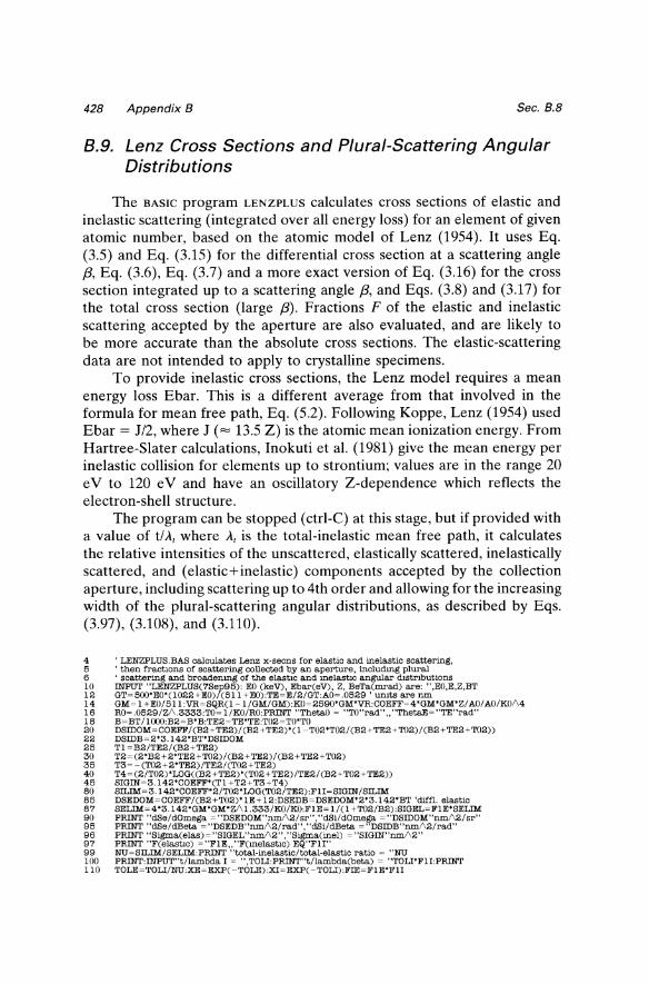
B.9. Lenz Cross Sections and Plural-Scattering Angular Distributions
The BASIC program LENZPLUS calculates cross sections of elastic and inelastic scattering (integrated over all energy loss) for an element of given atomic number, based on the atomic model of Lenz (1954). It uses Eq. (3.5) and Eq. (3.15) for the differential cross section at a scattering angle {3, Eq. (3.6), Eq. (3.7) and a more exact version of Eq. (3.16) for the cross section integrated up to a scattering angle {3, and Eqs. (3.8) and (3.17) for the total cross section (large {3). Fractions F of the elastic and inelastic scattering accepted by the aperture are also evaluated, and are likely to be more accurate than the absolute cross sections. The elastic-scattering data are not intended to apply to crystalline specimens.
To provide inelastic cross sections, the Lenz model requires a mean energy loss Ebar. This is a different average from that involved in the formula for mean free path, Eq. (5.2). Following Koppe, Lenz (1954) used Ebar = J/2, where J (= 13.5 Z) is the atomic mean ionization energy. From Hartree-Slater calculations, Inokuti et al. (1981) give the mean energy per inelastic collision for elements up to strontium; values are in the range 20 e V to 120 e V and have an oscillatory Z-dependence which reflects the electron-shell structure.
The program can be stopped (ctrl-C) at this stage, but if provided with a value of t/ A, where A, is the total-inelastic mean free path, it calculates the relative intensities of the unscattered, elastically scattered, inelastically scattered, and (elastic+inelastic) components accepted by the collection aperture, including scattering up to 4th order and allowing for the increasing width of the plural-scattering angular distributions, as described by Eqs. (3.97), (3.108), and (3.110).
4 5 6 10 12 14 16 18 20 22 25 30 35 40 45 80 85 87 90 95 96 97 99 100 110
, LENZPLUS.BAB calcrulates Lanz x·seens for elastic and Inelastic scattering, , then fractJ.ons of scattering collected by an aperture, includmg plural • scattering and broadenmg of the elastIc and melastlc angular d.stMbutlons INPUT "LENZPLUS(7Sep95): EO (keV), Ebar(eV), Z, BeTa(mrad) are: ",EO,E,Z,BT GT~500'EO'(l022+EO)/(511 +EO):TE~E/2/GT:AO~.0529 ' uruts are run GM~ 1 +EO/511 :VR~SQR(I-1 /GM/GM):KO~2590'GM'VR:COEFF~4'GM'GM'Z/ AO/ AO/KO/\4 RO~.0529/Z/\.3333:TO~ I/KO/RO:PRINT "ThetaO ~ .. TO .. rad ...... ThetaE~ .. TE .. rad .. B~BT/1000:B2~B·B:TE2~TE·TE:T02~TO·TO DSIDOM~COEFF/(B2+TE2)/(B2+TE2)·(I-T02·T02/(B2+TE2+T02)/(B2+TE2+T02)) DSIDB~2·3.142·BT·DSIDOM Tl ~B2/TE2/(B2+TE2) T2~(2'B2+2'TE2+T02)/(B2+TE2)/(B2+TE2+T02) T3~-CT02+2'TE2)/TE2/CT02+TE2) T4~(2/T02)'LOG((B2+TE2)'(T02+TE2)/TE2/(B2+T02+TE2)) SIGIN~3.142·COEFF·(TI +T2+T3+T4) SILIM~3.142·COEFF·2/T02·LOG(T02/TE2):FII~SIGIN/SILIM DSEDOM~COEFF/(B2+T02)'IE+ 12:DSEDB~DSEDOM·2·3.142·BT 'diffl. elastic SELIM~4·3.142·GM·GM·Z/\ 1.333/KO/KO:FIE~ 1/( 1 +T02/B2):SIGEL~FIE'SELIM PRINT "dSe/dOmega ~"DSEDOM"nm/\2/sr","dSi/dOmega. ~"DSIDOM"run/\2/sr" PRINT "dSe/dBeta ~"DSEDB"nm/\2/rad","dSi/dBeta ~"DSIDB"nm/\2/rad" PRINT "S!gma(elas)~"SIGEL"nm/\2","Sigma(inel) ~"SIGIN"run/\2" PRINT "F(elastIc) ~ .. FIE .... F(melastIc) EQ"FlI" NU~SILIM/SELIM:PRINT .. total-inelastic/total-elastic ratio ~ "NU PRINT:INPUT .. t/Iambda I ~ .. ,TOLI:PRINT .. t/lambda(beta) ~ "TOLI'FlI:PRINT TOLE~TOLI/NU:XE~EXP( -TOLE):XI~EXP( -TOLI):FIE~FIE'FI1
Sec. B.10 Computer Programs
115 120 121 122 124 125 130 135 136 139 140 142 143 145 150 170
RUN
PUN~XE'XI:PEL~(I-XE)'XI'FIE:PZ~PUN+PEL pm~XE'(l-XI)'FlI:PIE~(l-XI)'(l-XE)'FIE:PI~pm + PIE PRINT "P(unscat) ~ "PUN, "P(el) ~ "PEL"neglectmg elastic broadening" PRINT "PCinel) ~ "pm, "P(in +el) ~ "PIE "neglecting inelastic broadening" PRINT "1011 ~ "PZ, "11/1 ~ "PI"neglecting angular broadening" PI' ~ PZ + PI: LR ~ LOG(PI' IPZ): PRINT ''In(ItIIO) ~ "LR"without broadening": PRINT F2E~ 1/0 + 1.7/\2'T02/B2):F3E~ 1/0 +2.2/\2"r02/B2):F4E~ 1/0 +2. 71\2'T02/B2) PE~XE'(TOLE*FIE+TOLE/\2*F2E/2+TOLE/\'F3E/6+TOLE/\4'F4E/24) PEID~PE*XI:PU~XI*XE:RZ~PU+PEID 'unscattered and el/no-inel compts. REM PEL~XI*XE'(EXP(TOLE'FIE)-I):PZ~PUN+PEL 'not used PI~XI'(EXP(TOLI'FID-l) :PmE~ XE'PI:PIE~PI'PE:RJ~ PmE + PIE PRINT "P(unscat) ~ "PU,"P(el only) ~ "PEID"with elastIC broadening" REM ang dlstrIb of inel +el taken same as broadened elastlC PRINT "P(inel only) ~ "PmE,"P(in+el) ~ "PIE"Wltb inelastic broadening" PRINT "1011 ~ "RZ, "Ii/l ~ "RJ"with angular broadening" RT~RZ+RJ:LR~LOG(RT/RZ):PRINT ''In(ItIIO) ~ "LR"with angular broadening"
LENZPLUS (7Sep95): EO(keV), Ebar(eV), Z, BeTa(mrad) are: 100,40,6,10. ThetaO ~ 2.023144E-02 rad ThetaE ~ 2. 178253E-04 rad dSe/dOmega ~ 2897719 nm/\2/sr dSi/dOmega ~ 5.223308E-02 nmi\2/sr dSe/dBeta~ 1.820926E+08 nm/\2/rad dSi/dBeta~ 3.282326 nm/\2/rad SigIDa(elas) ~ 1.33367E-05 nmi\2 SigIDa(inel) ~ 1.662413E- 04 nm/\2 F(elastic) ~ .1963436 F(melastic) ~ .809588 total-inelastic/total-elastic ratio ~3.023035
t/lambdaI ~ 1.5 t/lambda(beta) ~ 1.214382
P(unscat) ~ .1358519 peel) ~ 1.713653E-02 neglecting elastic broadening PCinel) ~.3829302 P(in+el) ~4.830332E-02 neglecting melastic broadening 1011 ~.1529884 Ii/l ~.4312336 neglecting angular broadening In(ltIIO) ~ 1.339919 without broadenlng
P(unscat) ~.1358519 P(elonly) ~ 1.468281E-2 witb elastic broadening P(inel only) ~.321726 P(in+el) ~3.477201E-02 with melastic broadening 1011 ~.1505347 Ii/l ~ .356498 with angular broadening In(ltIIO) ~ 1.214382 with angular broadening Ok
8.10. Conversion between Oscillator Strength and Cross Section
429
The program FTOS.BAS converts a dipole oscillator strength (integrated over an energy range DL above an ionization edge) to a corresponding core-loss cross section, using Eq. (A.7) and the same procedure as in the SIGPAR2 program. Program STOF.BAS does the reverse and enables measured inner-shell cross sections to be parameterized in terms of a dipole oscillator strength which is independent of collection angle and incident energy, as described in Section 4.5.2.
10 REM: FTOS calculates a core· loss partial cross section 12 REM: from an f-value integrated over energy range DeLta 25 lli'PUT "FTOS (7Sep95): f-value ~ ",FC 35 SO~3.516E-16 :REM 4*PI*AO**2 40 lli'PUT "Edge energy(eV),DeLta(eV),EO(keV) ~ ",EC,DL,EO 45 ER~SQR(EC*(EC+DL))/l3.6 50 TK~EO*O +EO/I022)/0 +EO/511)/\2 55 TR~TK·I000/l3.6 60 GT~EO*(1022+EO)/(511 +EO)/2 65 TE~ER'13.6/2/GT: PRINT "ThetaE(mrad) ~ ",TE 70 lli'PUT "effective collection semlangle BETA (mrad) ~ ",BS 75 B2~BS*BS:T2~TE*TE:V2~2'TK/511 :G2~ 10-V2) 80 RET~LOG(G2)-LOG«B2+T2)/(B2+T2/G2))-V2*B2/(B2+T2/G2) 85 LG~LOGO + B2/T2):SC~FC'SO/ER/TR*(LG+ RET)
430 Appendix 8
90 FR=I00'RET/LO: PRINT "% increase due to retardation"FR 95 PRINT "SIGMA(DeLta,BeTa) = "SC"cmI\2"
FTOS(7Sep95): f-value = 0.41 Edge energy(eV), DeLta(eV),EO(keV) = 535,100,100 ThetaE(mrad) = 3.174038 effective collection semlangle BETA (mrad) = 10. % increase due to retardation 2.040891 SIGMA (DeLta,BeTa) = 1.453538E-21 eml\2 Ok
10 REM: STOF calculates OSCILLATOR STRENGTH FOR CORE-LOSS 12 REM: FROM A GIVEN PARTIAL CROSS SECTION SC 25 INPUT "STOF(7Sep95): CROSS SECTION (in eml\2) = ",SC 35 SO=3.516E-16 :REM 4'PI'AO"2 40 INPUT "Edge energy(eV), DeLta(eV), EO(keV) = ",EC,DL,EO 45 ER=SQRCEC·CEC+DL))/13.6 50 TK=EO'O +EO/1022)/0 +EO/511)1\2 55 TR=TK·I000/13.8 60 GT=EO'0022+ EO)/(511 +EO)/2 65 TE=ER·13.6/2/GT: PRINT "THETAE(mrad) = "TE 70 INPUT "effectlve BETA (mrad) = ",BS 75 LG=LOG(1 + BS'BS/TE/TE) 78 V2=2'TK/511: G2=1/0-V2): B2=BS'BS: T2=TE'TE 80 RET=LOG(G2)-LOG(B2+T2)/(B2+T2/G2))-V2'B2/(B2+T2/G2) 85 FC=SC/SO'ER'TR/(LG+RET) 90 FR= l00'RET ILO: PRINT "%inerease due to retardation= "FR 95 PRINT "f(DeLta) = "FC
RUN STOF(7Sep95); CROSS SECTION (in eml\2) = 1.453538e-21 Edge energy(eV), DeLta(eV), EO(keV) = 535,100,100 THETA(mrad) = 3.174038 effective BETA (mrad) = 10. %increase due to retardation = 2.040891 f(DeLta) =.4100002 Ok
Sec. 8.10
B.11. Conversion between Mean Energy and Inelastic Mean Free Path
The short routine EM2MFP.BAS uses Eq. (5.2) to convert a mean energy loss Em, as listed in Table 5.2, to the corresponding inelastic mean free path for energy-loss data recorded with an angle-limiting collection aperture of semiangle f3. It is not appropriate for f3 > 20 mrad, as explained in Section 5.1. The algorithm MFP2EM.BAS applies Eq. (5.2) in reverse, obtaining a value of Em from a measured value of ;'(f3) within a few iterations.
10 15 20 30 40 50 RUN
REM EM2MFP : CALCULATION OF TOTAL-INELASTIC MFP REM based on Malis et aI. (JEMT 8, 1988, 193-2(0) INPUT "ENTER Em(eV),EO(KeV),BETA(mrad):" ,EM,EO,BETA F=(1 + EO) 1 1022) 10 + E0/511)1\2 LAMBDA= 106'F'EO/EM/LOG(2'BETA'EO/EM) PRINT "MFP(nm) = ",LAMBDA
ENTER Em(eV),EO(KeV),BETA(mrad):20,200,5. MRP(nm) = 142.1619
10 15 20 30 32 35 RUN
REM: MFP2EM calculates Em parameter for MFP formula: REM:J. Electron Microse. Technique 8 (988) 193-100. INPUT "MFP(nm),EO(keV),beta(mrad) = ",LAM,EO,B:EB=15 EM= 106'0 + EO)/1022)/(1 +EO/511)1\2'EO/LAM/LOG(2'B'EO/EB) IF ABS CEB-EM)<EM/l000 THEN PRINT "Em(eV) = ",EM:END EB=EM:GOTO 30 ' Last edit 95Sep07
MFP(nm),EO(keV),beta(mrad) = 142.2,200,5. EM(eV) = 19.99049
APPENDIX c Plasmon Energies of Some Elements and Compounds
The following table lists the measured energy Ep and full width at halfmaximum !l.Ep of the principal low-loss peak observable in the energy-loss spectrum of some common materials. The data is taken mainly from Daniels et al. (1970), Colliex et al. (1976a), Raether (1980), and Colliex (1984a). In some instances, a free-electron plasmon energy is shown in parentheses, calculated from Eq. (3.41) with m = mo and n equal to the density of outer-shell electrons (including 3d electrons in the case of transition metals).
Layer crystals such as graphite and boron nitride have much weaker bonding in a direction perpendicular to the basal (cleavage) plane than within the basal plane, giving rise to two groups of valence electrons and two distinct plasmon energies. Furthermore, in anisotropic materials the dielectric function e( q, E) is actually a tensor eij and the peak structure in the energy-loss spectrum depends on the direction of the scattering vector q. For a uniaxial crystal such as graphite, axes can be chosen such that offdiagonal components are zero, in which case e( q, E) = eL sin20 + ell cos2
0, where eL = ell = e22 and en = e33 are components of e(E) perpendicular and parallel to the c-axis; 0 is the angle between q and the c-axis, which depends on the scattering angle and the specimen orientation. If the c-axis is parallel to the incident beam, the greatest contribution (for f3 » (JE)
comes from perpendicular excitations and two plasmon peaks are observed (see table). Under special conditions (e.g., small collection angle f3), the qllc excitations may predominate and the higher-energy peak is displaced downwards in energy. Further detail on EELS of anisotropic materials is given in Daniels et al. (1970) and Browning et al. (1991b).
431
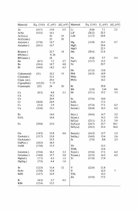
Material Eq. (3.41) Ep (eV) !:J.Ep (eV) Material Eq. (3.41) Ep (eV) !:J.Ep (eV)
Al (15.7) 15.0 0.5 Li (8.0) 7.1 2.2 AlAs (15.5) 16.1 LiF (26.2) 25.3 AI,03(a) 26 10 LiH (12.7) 20.9 AI,03(am.) 23 20 As(cryst.) (17.8) 18.7 Mg (10.9) 10.3 0.7 As(amor.) (16.1 ) 16.7 MgF, 24.6
MgO 22.3 B(amor.) 22.7 18 Mn (28.4) 21.6 BN(hex.) 9; 26 BN(amor.) 24 Na (6.0) 5.7 0.4 Ba (6.7) 7.2 2.7 NaCI (15.7) 15.5 Be (18.4) 18.7 4.8 Ni 20.7 Bi (14.0) 14.2 6.5
Pb (13.5) 13.0 C(diamond) (31) 33.2 13 PbS (16.3) 14.9 C(lonsdal.) 32.4 PbSe 15.0 C(am. tet.). 29.5 PbTe 14.2 C(graphite) (13;22) 7; 27 C(amorph) (22) 24 20 Pt 35.0
Rb (3.9) 3.41 0.6 Ca (8.2) 8.8 2.1 Sb (15.1) 15.2 3.3 Cd 19.2 Co 20.9 Sc (13.6) 14.0 Cr (26.8) 24.9 ScH, 17.2 Cs (3.4) 2.9 Se(cr.) (17.4) 17.1 6.2 Cu (16.0) 19.3 Se(am.) (16.4) 16.3 6.2
Er 14.0 SiC cr.) (16.6) 16.7 3.2 ErH, 16.8 Si(am.) 16.3 3.9
SiC(a) (23.1) 21.5 3.9 Fe (29.8) 23.0 Si3N,(a) (24.7) 23.7 10.1
SiO,(a) (24.2) 22.4 16.6
Ga (14.5) 13.8 0.6 Sn(sol.) (14.3) 13.7 1.3 GaAs(cr.) (15.7) 15.8 Sn(liq.) (14.0) 13.4 1.6 GaAs(am.) 15.5 Sr (7.0) 8.0 2.3 GaP(cr.) (16.5) 16.5 GaSb (13.8) 13.3 Tb 13.3
TbH, 15.6 Ge(cryst.) (15.6) 16.2 3.3 Te(cr.) (15.6) 17.1 6.2 Ge(amor.) (14.8) 15.7 3.8 Te(am.) (15.3) 16.3 6.2 Hg(sol.) (7.7) 6.3 1.5 Ti (17.8) 17.9 Hg(liq.) (7.5) 6.4 1.0
In (12.5) 11.4 12 V (22.8) 21.8 InAs (13.8) 13.8 Y 12.5 7 InSb (12.7) 12.9 YH, 15.3
Zn (13.9) 17.2 K (4.3) 3.7 0.3 KBr (12.4) 13.2
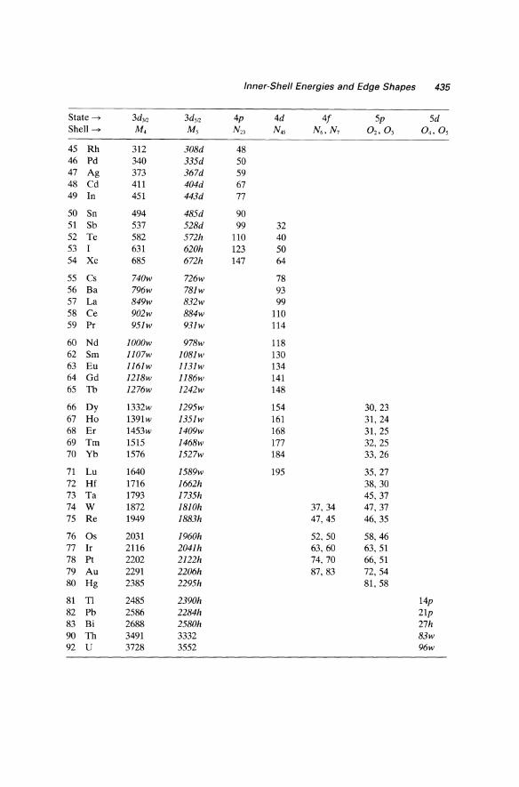
APPENDIX D Inner-Shell Energies and Edge Shapes
The following table gives threshold energies Ek (in e V) of the ionization edges observable by EELS, based on data of Bearden and Burr (1967), Siegbahn et al. (1967), Zaluzec (1981), Ahn and Krivanek (1983), and Colliex (1985). The most prominent edges (those most suitable for elemental analysis) are shown in italics. Where possible, an accompanying symbol is used to indicate the observed edge shape:
h denotes a hydrogenic edge with sawtooth profile (rapid rise at the threshold followed by more gradual decay), as in Fig. 3.43.
d denotes a delayed maximum due to centrifugal-barrier effects (Section 3.7.1), giving a rounded edge with a maximum at least 10 eV above the threshold energy as in Figs. 3.47.
w denotes sharp white-line peaks at the edge threshold, due to excitation to empty d-states (in the transition metals) or [-states (in the rare earths), as in Fig. 3.48a.
p denotes a low-energy edge which may appear more like a plasmon peak than a typical edge. However, the energy given is that of the edge onset, not the intensity maximum.
Because of near-edge structure, which depends on the chemical and crystallographic structure of a specimen, this classification can serve only as a rough guide. Elements such as copper can exist in different valence states, giving rise to dissimilar edge shapes (Fig. 3.46). The edge energies themselves may vary by several e V depending on the chemical environment of the excited energy; see Section 3.7.4.
433
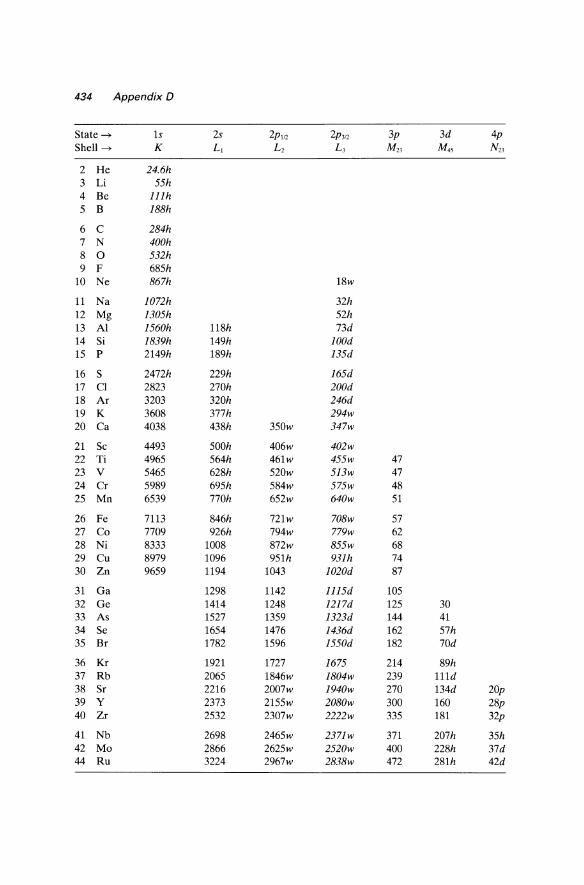
434 Appendix 0
State --+ Is 2s 2p1f2 2P31Z 3p 3d 4p Shell --+ K LJ Lz L3 MZ3 M45 NZ3
2 He 24.6h 3 Li 55h 4 Be l11h 5 B 188h
6 C 284h 7 N 400h 8 0 532h 9 F 685h
10 Ne 867h 18w
11 Na J072h 32h 12 Mg 1305h 52h 13 Al 1560h 118h 73d 14 Si 1839h 149h lOOd 15 P 2149h 189h 135d
16 S 2472h 229h 165d 17 CI 2823 270h 200d 18 Ar 3203 320h 246d 19 K 3608 377h 294w 20 Ca 4038 438h 350w 347w
21 Sc 4493 500h 406w 402w 22 Ti 4965 564h 461w 455w 47 23 V 5465 628h 520w 513w 47 24 Cr 5989 695h 584w 575w 48 25 Mn 6539 770h 652w 640w 51
26 Fe 7113 846h 721w 708w 57 27 Co 7709 926h 794w 779w 62 28 Ni 8333 1008 872w 855w 68 29 Cu 8979 1096 951h 931h 74 30 Zn 9659 1194 1043 1020d 87
31 Ga 1298 1142 1115d 105 32 Ge 1414 1248 1217d 125 30 33 As 1527 1359 1323d 144 41 34 Se 1654 1476 1436d 162 57h 35 Br 1782 1596 1550d 182 70d
36 Kr 1921 1727 1675 214 89h 37 Rb 2065 1846w 1804w 239 111d 38 Sr 2216 2007w 1940w 270 134d 20p 39 Y 2373 2155w 2080w 300 160 28p 40 Zr 2532 2307w 2222w 335 181 32p
41 Nb 2698 2465w 2371w 371 207h 35h 42 Mo 2866 2625w 2520w 400 228h 37d 44 Ru 3224 2967w 2838w 472 281h 42d
Inner-Shell Energies and Edge Shapes 435
State --> 3d3Q 3d'12 4p 4d 4/ 5p 5d Shell --> M, Ms N23 N4, N6 ,N, a" oJ 0 4 , a, 45 Rh 312 308d 48 46 Pd 340 335d 50 47 Ag 373 367d 59 48 Cd 411 404d 67 49 In 451 443d 77
50 Sn 494 485d 90 51 Sb 537 528d 99 32 52 Te 582 572h 110 40 53 I 631 620h 123 50 54 Xe 685 672h 147 64
55 Cs 740w 726w 78 56 Ba 796w 781w 93 57 La 849w 832w 99 58 Ce 902w 884w 110 59 Pr 951w 931w 114
60 Nd lOOOw 978w 118 62 Sm l107w lO81w 130 63 Eu 1161w 1131w 134 64 Gd 1218w 1186w 141 65 Tb 1276w 1242w 148
66 Dy 1332w 1295w 154 30, 23 67 Ho 1391w 1351w 161 31, 24 68 Er 1453w 1409w 168 31, 25 69 Tm 1515 1468w 177 32, 25 70 Yb 1576 1527w 184 33, 26
71 Lu 1640 1589w 195 35,27 72 Hf 1716 1662h 38, 30 73 Ta 1793 1735h 45, 37 74 W 1872 18lOh 37, 34 47, 37 75 Re 1949 1883h 47, 45 46, 35
76 Os 2031 1960h 52, 50 58,46 77 Ir 2116 2041h 63,60 63, 51 78 Pt 2202 2122h 74, 70 66,51 79 Au 2291 2206h 87,83 72,54 80 Hg 2385 2295h 81,58
81 Tl 2485 2390h 14p
82 Pb 2586 2284h 21p
83 Bi 2688 2580h 27h
90 Th 3491 3332 83w
92 U 3728 3552 96w
436 Appendix 0
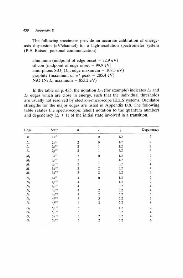
The following specimens provide an accurate calibration of energyaxis dispersion (eV/channel) for a high-resolution spectrometer system (P.E. Batson, personal communication):
aluminum (midpoint of edge onset = 72.9 eV) silicon (midpoint of edge onset = 99.9 e V) amorphous Si02 (L 23 edge maximum = 108.3 e V) graphite (maximum of 7T* peak = 285.4 eV) NiO (Ni L3 maximum = 853.2 e V)
In the table On p. 435, the notation L23 (for example) indicates L2 and L3 edges which are close in energy, such that the individual thresholds are usually not resolved by electron-microscope EELS systems. Oscillator strengths for the major edges are listed in Appendix B.8. The following table relates the spectroscopic (shell) notation to the quantum numbers and degeneracy (2j + 1) of the initial state involved in a transition.
Edge State n j Degeneracy
K 1s!!2 0 1/2 2
L! 2S!!2 2 0 112 2 L2 2p!12 2 112 2 L3 2p 3l2 2 1 3/2 4
M! 3S!!2 3 0 1/2 2
M2 3p"2 3 1/2 2 M3 3p3!' 3 1 3/2 4 M, 3d3!' 3 2 3/2 4 Ms 3dS!2 3 2 5/2 6
N! 4sln 4 0 1/2 2 N, 4p"2 4 112 2 N3 4p 3!2 4 1 3/2 4 N, 4d3!2 4 2 3/2 4 Ns 4ds12 4 2 5/2 6 N6 4['1' 4 3 5/2 6 N7 4f12 4 3 7/2 8
O 2 5p '!2 5 1/2 2 0 3
5p 3!2 5 3/2 4 0 4 5d 312 5 2 3/2 4 Os 5d5!2 5 2 5/2 6
APPENDIX E Electron Wavelengths and Relativistic Factors; Fundamental Constants
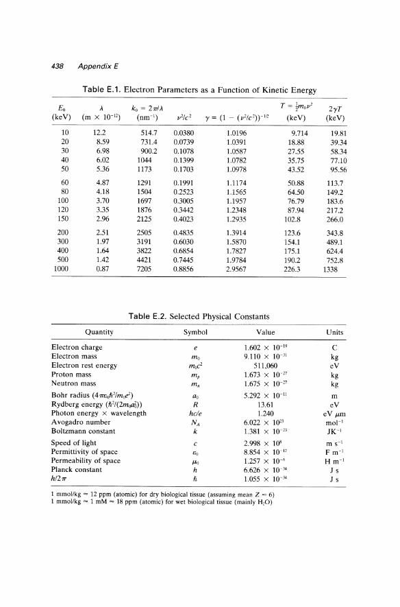
Table E.1lists (as a function of the kinetic energy Eo of an electron) values of the wavelength A, wave number ko, velocity v, relativistic factor y = (1 - V2/C2)-1I2, effective kinetic energy T = mov2/2, and parameter 2yTwhich is used to calculate the characteristic scattering angle BE = E/(2yT). For values of Eo not tabulated, these parameters can be calculated from the following equations:
ko = ymov/h = 2590( yv/c) nm- I
y = 1 + EoI(moc2)
T = E 1 + EoI(2moc2) = E 1 + y o [1 + Eo/(moc2)]2 0 2y2
2 T - E (2moc2 + Eo) - 2 Y - 0 2 - ymoV moc + Eo
(j _~_ E _ E (Eo+moc2) E - ymov2 - Eo(1 + y-I) - Eo Eo + 2moc2
Values of the fundamental constants for use in these (and other) equations are given in Table E.2, extracted from page F-162 of the 65th edition of Handbook of Chemistry and Physics (CRC Press, 1984).
437
438 Appendix E
Table E.1. Electron Parameters as a Function of Kinetic Energy
Eo A ko = 2mA T = ~mo1'2
(keV) (m x 10-12) (nm-I) 1'2/C 2 y = (1 - (1'2/C')>-1I2 (keV)
10 12.2 514.7 0.0380 1.0196 9.714 20 8.59 731.4 0.0739 1.0391 18.88 30 6.98 900.2 0.1078 1.0587 27.55 40 6.02 1044 0.1399 1.0782 35.75 50 5.36 1173 0.1703 1.0978 43.52
60 4.87 1291 0.1991 1.1174 50.88 80 4.18 1504 0.2523 1.1565 64.50
100 3.70 1697 0.3005 1.1957 76.79 120 3.35 1876 0.3442 1.2348 87.94 150 2.96 2125 0.4023 1.2935 102.8
200 2.51 2505 0.4835 1.3914 123.6 300 1.97 3191 0.6030 1.5870 154.1 400 1.64 3822 0.6854 1.7827 175.1 500 1.42 4421 0.7445 1.9784 190.2
1000 0.87 7205 0.8856 2.9567 226.3
Table E.2. Selected Physical Constants
Quantity Symbol Value
Electron charge e 1.602 X 10⁻¹⁹
Electron mass ma 9.110 x 10-)1
Electron rest energy mue? 511,060 Proton mass mp 1.673 X 10⁻²⁷
Neutron mass mn 1.675 X 10⁻²⁷
Bohr radius (471Eoh'/moe) ao 5.292 X 10⁻¹¹
Rydberg energy (h2/(2moa3» R 13.61 Photon energy x wavelength hc/e 1.240 Avogadro number NA 6.022 X 10²³
Boltzmann constant k 1.381 X 10⁻²³
Speed of light C 2.998 X 10⁸
Permittivity of space eo 8.854 X 10⁻¹²
Permeability of space fLo 1.257 X 10⁻⁶
Planck constant h 6.626 X 10⁻³⁴
h/21T h 1.055 X 10⁻¹⁴
1 mmollkg = 12 ppm (atomic) for dry biological tissue (assuming mean Z = 6) 1 mmollkg = 1 mM = 18 ppm (atomic) for wet biological tissue (mainly H20)
2yT (keV)
19.81 39.34 58.34 77.10 95.56
113.7 149.2 183.6 217.2 266.0
343.8 489.1 624.4 752.8
1338
Units
C kg eV kg kg
m eV
eV fLm mol-I JK-I
m S-I F m-I H m-I
J S J s
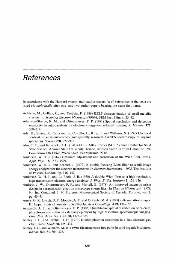
References
In accordance with the Harvard system, multiauthor papers (et al. references in the text) are listed chronologically after one- and two-author papers bearing the same first name.
Acheche, M., Colliex, C., and Trebbia, P. (1986) EELS characterization of small metallic clusters. In Scanning Electron Microscopy/1986/1, SEM Inc., Illinois, 25-32.
Adamson-Sharpe, K. M., and Ottensmeyer, F. P. (1981) Spatial resolution and detection sensitivity in microanalysis by electron energy-loss selected imaging. J. Microsc. 122, 309-314.
Ade, H., Zhang, X., Cameron, S., Costello, c., Kirz, J., and Williams, S. (1992) Chemical contrast in x-ray microscopy and spatially resolved XANES spectroscopy of organic specimens. Science 258, 972-975.
Ahn, C. C., and Krivanek, O. L. (1983) EELS Atlas. Copies ($US15) from Center for Solid State Science, Arizona State University, Tempe, Arizona 85287, or from Gatan Inc., 780 Commonwealth Drive, Warrendale, Pennsylvania 15086.
Andersen, W. H. J. (1967) Optimum adjustment and correction of the Wien filter. Brit J. Appl. Phys. 18,1573-1579.
Andersen, W. H. J., and Kramer, J. (1972) A double-focusing Wien filter as a full-image energy analyzer for the electron microscope. In Electron Microscopy-1972, The Institute of Physics, London, pp. 146-147.
Andersen, W. H. J., and Le Poole, J. B. (1970) A double Wien filter as a high resolution, high-transmission electron energy analyser. J. Phys. E (Sci. Instrum) 3, 121-126.
Andrew, J. W., Ottensmeyer, F. P., and Martell, E. (1978) An improved magnetic prism design for a transmission electron microscope energy filter. In Electron Microscopy-1978, 9th Int. Cong., ed. J. M. Sturgess; Microscopical Society of Canada, Toronto, vol. 1, pp.40-4l.
Anstis, G. R., Lynch, D. F., Moodie, A. F., and O'Keefe, M. A. (1973) n-Beam lattice images: III Upper limits of ionicity in W.Nb'6077. Acta Crystallogr. A29,138-152.
Arsenault, A. L., and Ottensmeyer, F. P. (1983) Quantitative spatial distribution of calcium, phosphorus and sulfur in calcifying epiphysis by high resolution spectroscopic imaging. Proc. Natl. Acad. Sci. USA 80, 1322-1326.
Ashley, J. c., and Ritchie, R. H. (1970) Double-plasmon excitation in a free-electron gas. Phys. Status Solidi 38, 425-434.
Ashley, J. C., and Williams, M. W. (1980) Electron mean free paths in solid organic insulators. Radiat. Res. 81, 364-378.
439
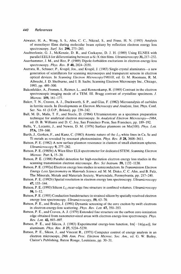
440 References
Atwater, H. A., Wong, S. S., Ahn, C. c., Nikzad, S., and Frase, H. N. (1993) Analysis of monolayer films during molecular beam epitaxy by reflection electron energy loss spectroscopy. Surf Sci. 298, 273-283.
Auchterlonie, G. J., McKenzie, D. R., and Cockayne, D. J. H. (1989) Using ELNES with parallel EELS for differentiating between a-Si :X thin films. Ultramicroscopy 31, 217 -232.
Auerhammer, J. M., and Rez, P. (1989) Dipole-forbidden excitations in electron-energy-loss spectroscopy. Phys. Rev. B 40, 2024-2030.
Autrata, R, Schauer, P., Kvapil, Jos., and Kvapil, J. (1983) Single-crystal aluminates-a new generation of scintillators for scanning microscopes and transparent screens in electron optical devices. In Scanning Electron Microscopy/1983/1I, ed. G. M. Roomans, R M. Albrecht, J. D. Shelburne, and I. B. Sachs; Scanning Electron Microscopy Inc., Chicago, 1993, pp. 489-500.
Bakenfelder, A., Fromm, I., Reimer, L., and Rennenkamp, R. (1989) Contrast in the electron spectroscopic imaging mode of a TEM. III. Bragg contrast of crystalline specimens. J. Microsc. 159,161-177.
Baker, T. N., Craven, A. J., Duckworth, S. P., and Glas, F. (1982) Microanalysis of carbides in ferritic steels. In Developments in Electron Microscopy and Analysis, Inst. Phys. Conf. SeT. No. 61 (I.O.P., Bristol), pp. 239-242.
Ball, M. D., Malis, T. F., and Steele, D. (1984) Ultramicrotomy as a specimen preparation technique for analytical electron microscopy. In Analytical Electron Microscopy-1984, ed. D. B. Williams and D. C. Joy, San Francisco Press, San Francisco, pp. 189-192.
Ballu, Y., Lecante, J., and Newns, D. M. (1976) Surface plasmons on Mo(100). Phys. Lett. 57A,159-160.
Barth, J., Gerken, F., and Kunz, C. (1983) Atomic nature of the L23 white lines in Ca, Sc and Ti metals as revealed by resonant photoemission. Phys. Rev. B 28, 3608-3611.
Batson, P. E. (1982) A new surface plasmon resonance in clusters of small aluminum spheres. Ultramicroscopy 9, 277-282.
Batson, P. E. (1985b) A Wien filter ELS spectrometer for dedicated STEM. Scanning Electron Microsc. Part 1, 15-20.
Batson, P. E. (1988) Parallel detection for high-resolution electron energy loss studies in the scanning transmission electron microscope. Rev. Sci Instrum. 59, 1132-1138.
Batson, P. E. (1992a) Electron energy loss studies in semiconductors. In Transmission Electron Energy Loss Spectrometry in Materials Science, ed. M. M. Disko, C. C. Ahn, and B. Fulz, The Minerals, Metals and Materials Society, Warrendale, Pennsylvania, pp. 217-240.
Batson, P. E. (1992b) Spatial resolution in electron energy loss spectroscopy. Ultramicroscopy 47,133-144.
Batson, P. E. (1993) Silicon L2•3 near-edge fine structure in confined volumes. Ultramicroscopy 50,1-12.
Batson, P. E. (1995) Conduction bandstructure in strained silicon by spatially resolved electron energy loss spectroscopy. Ultramicroscopy, 59, 63-70.
Batson, P. E., and Bruley, J. (1991) Dynamic screening of the core exciton by swift electrons in electron-energy-loss scattering. Phys. Rev. Lett. 67, 350-353.
Batson, P. E., and Craven, A. J. (1979) Extended fine structure on the carbon core-ionization edge obtained from nanometer-sized areas with electron energy-loss spectroscopy. Phys. Rev. Lett. 42, 893-897.
Batson, P. E., and Silicox, J. (1983) Experimental energy-loss function, Im[ -1/E(q,W»), for aluminum. Phys. Rev. B 27, 5224-5239.
Batson, P. E., Silcox, J., and Vincent R, (1971) Computer control of energy analysis in an electron microscope, 29th Ann. Proc. Electron Microsc. Soc. Am., ed. G. W. Bailey, Claitor's Publishing, Baton Rouge, Louisiana, pp. 30-31.
References 441
Batson, P. E., Pennycook, S. J., and Jones, L. G. P. (1981) A new technique for the scanning and absolute calibration of electron energy-loss spectra. Ultramicroscopy 6, 287-289.
Batson, P. E., Kavanagh, K. L., Woodall, J. M., and Mayer, J. W. (1986) Electron-energyloss scattering near a single misfit dislocation at the GaAs/GaInAs interface. Phys. Rev. Lett. 57, 1729-1732.
Batson, P. E., Chisholm, M. F., Clarke, D. R., Dimos, D., and Shaw, T. (1989) Energy-loss studies of carbon content in yttrium barium cuprate. Proc. 47th Ann. Meet. Electr. Microsc. Soc. Amer., ed. G. W. Bailey, San Francisco Press, San Francisco, pp. 196-197.
Batson, P. E., Johnson, D. W., and Spence, J. C. H. (1992) Resolution enhancement by deconvolution using a field emission source in electron energy loss spectroscopy. Ultramicroscopy 41, 137-145.
Bauer, R., Hezel, U., and Kurz, D. (1987) High resolution imaging of thick biological specimens with an imaging electron energy loss spectrometer. Optik 77,171-174.
Baumann, W., Niemietz, A., Reimer,L., and Volbert, B. (1981) Preparation ofP-47 scintillators for STEM. J. Microsc. 122,181-186.
Baumeister, W., and Hahn, M. (1976) An improved method for preparing single-crystal specimen supports: H20, exfoliation of vermiculite. Micron 7, 247-251.
Bazett-Jones, D. P., and Ottensmeyer, F. P. (1981) Phosphorus distribution in the nucleosome. Science 211, 169-170.
Beamson, G., Porter, H. Q., and Turner, D. W. (1981) Photoelectron spectromicroscopy. Nature 290, 556-561.
Bearden,J. A., and Burr, A. F. (1967) X-ray atomic energy levels. Rev. Mod. Phys. 39,125-142. Bell, A. E., and Swanson, L. W. (1979) Total energy distributions of field-emitted electrons
at high current density. Phys. Rev. B 19, 3353-3364. Bell, M. G., and Liang, W. Y. (1976) Electron energy loss studies in solids; the transition
metal dichalcogenides. Adv. Phys. 25, 53-86. Bendayan, M., Barth, R. F., Gingras, D., Londono, l., Robinson, P. T., Alam, F., Adams,
D. M., and Mattiazzi, L. (1989) Electron spectroscopic imaging for high-resolution immunocytochemistry: use of boronated protein A. J. Histochem. Cytochem. 37, 573-580.
Bennett, J. c., and Egerton, R. F. (1995) NiO test specimens for analytical electron microscopy: round-robin results. J. Microsc. Soc. Amer. 1,143-149.
Bentley, J. (1992) Applications of EELS to ceramics and catalysts. In Transmission Electron Energy Loss Spectrometry in Materials Science, ed. M. M. Disko, C. C. Ahn, and B. Fulz, The Minerals, Metals and Materials Society, Warrendale, Pennsylvania, 155-181.
Bentley, J., Angelini, P., and Sklad, P. S. (1984) Secondary fluorescence effects on x-ray microanalysis. In Analytical Electron Microscopy-1984, ed. D. B. Williams and D. C. Joy, San Francisco Press, San Francisco, 315-317.
Berger, A., and Kohl, H. (1993) Optimum imaging parameters for elemental mapping in an energy filtering transmission electron microscope. Optik 92, 175-193.
Berger, M. J., and Seltzer, S. M. (1982) Stopping powers and ranges of electrons and positrons. National Bureau of Standards report: NBSIR 82-2550, u.s. Dept. of Commerce, Washington, D.C. 20234, 162 pages.
Berger, S. D., and McMullan, D. (1989) Parallel recording for an electron spectrometer on a scanning transmission electron microscope. Ultramicroscopy 28,122-125.
Berger, S. D., Salisbury, I. G., Milne, R. H., Imeson, D., and Humphreys, C. J. (1987) Electron energy-loss spectroscopy studies of nanometre-scale structures in alumina produced by intense electron-beam irradiation. Phil. Mag. B 55, 341-358.
Berger, S. D., McKenzie, D. R., and Martin, P. J. (1988) EELS analysis of vacuum arcdeposited diamond-like films. Phil. Mag. Lett. 57, 285-290.
442 References
Bevington, P. R. (1969) Data Reduction and Error Analysis for the Physical Sciences (McGrawHill, New York).
Bethe, H. (1930) Zur Theorie des Durchgangs schneller Korpuskularstrahlen durch Materie. Ann. Phys. (Leipzig) 5,325-400.
Bianconi, A. (1983) XANES spectroscopy for local structures in complex systems. In EXAFS and Near Edge Structure, ed. A Bianconi, L. Incoccia, and S. Stipcich, Springer-Verlag, New York, pp. 118-129.
Bianconi, A, Dell'Ariccia, M., Durham, P. J., and Pendry, P. J. (1982) Multiple scattering resonances and structural effects in the x-ray absorption near edge spectra of Fe II and Fe. III:Hexacyanide complexes. Phys. Rev. B 26, 6502-6508.
Bianconi, A, Dell'Ariccia, M., Gargano, A, and Natoli, C. R. (1983a) Bond length determination using XANES. In EXAFS and Near Edge Structure, ed. A Bianconi, L. Incoccia, and S. Stipcich, Springer-Verlag, New York, pp. 57-6l.
Bianconi, A, Giovannelli, A, Ascone, I., Alema, S., Durham, P., and Fasella, P. (1983b) XANES of calmodulin: Differences and homologies between calcium-modulated proteins. In EXAFS and Near Edge Structure, ed. A Bianconi, L. Incoccia, and S. Stipcich, SpringerVerlag, New York, pp. 355-357.
Bihr, J., Benner, G., Krahl, D., Rilk, A, and Weimer, E. (1991) Design of an analytical TEM with integrated imaging il-spectrometer. Proc. 49th Ann. Meet. Electron Microsc. Soc. Amer., ed. G. W. Bailey San Francisco Press, San Francisco, pp. 354-355.
Blackstock, A W., Birkhoff, R. D., and Slater, M. (1955) Electron accelerator and high resolution analyser. Rev. Sci. Instrum. 26, 274-275.
Blaha, P., and Schwarz, K. (1983) Electron densities in TiC, TiN, and TiO derived from energy band calculations. Int. J. Quantum Chern. 23, 1535-1552.
Blake, D., Freund, F., Krishnan, K. F. M., Echer, C. J., Shipp, R., Bunch, T. E., Tielens, A G., Lipari, R. J., Hetherington, C. J. D., and Chang, S. (1988) The nature and origin of interstellar diamond. Nature 332, 611-613.
Blanche, G., Hug, G., Jaouen, M., and Flank, A. M. (1993) Comparison of the TiK extended fine structure obtained from electron energy loss spectroscopy and x-ray absorption spectroscopy. Ultramicroscopy 50, 141-145.
Blasse, G., and Bri!, A (1967) A new phosphor for flying-spot cathode-ray tubes for color television: Yellow-emitting Y3Als012-Ce3+. Appl. Phys. Lett. 11.53-54.
Boersch, H. (1954) Experimentelle Bestimmung der Energieverteilung in thermisch ausgelosten Elektronenstrahlen. Z. Phys. 139,115-146.
Boersch, H., Geiger, J., and Hellwig, H. (1962) Steigerung der Auflosung bei der ElektronenEnergieanalyse. Phys. Lett. 3, 64-66.
Boersch, H., Geiger, J., and W. Stickel (1964) Das Auflosungsvermogen des elektrostatischmagnetischen Energieanalysators fUr schnelle Elektronen. Z. Phys. 180,415-424.
Bohm, D., and Pines, D. (1951) A collective description of electron interactions: II. Collective vs. individual particle aspects of the interactions. Phys. Rev. 85, 338-353.
Bohmer, J., and Rahmann, H. (1990) Ultrastructural localization of aluminum in amphibian larvae. Ultramicroscopy 32, 18-25.
Bonham, R. A, and Fink, M. (1974) High Energy Electron Scattering, Van Nostrand Reinhold, New York.
Bonney, L. A. (1990) Measurement of the inelastic mean free path by EELS analyses of submicron spheres. Proc. Xllth Int. Congo for Electron Microscopy, San Francisco Press, San Francisco, pp. 74-75.
Booker, G. R., and Stickler, R. (1962) Method of preparing Si and Ge specimens for examination by transmission electron microscopy. Brit. J. Appl. Phys. 13, 446-449.
Boothroyd, C. 8., Sato, K., and Yamada, K. (1990) The detection of 0.5 at% boron in Ni3Al
References 443
using parallel energy loss spectroscopy. Proc. XIIth Int. Congo Electron Microscopy, San Francisco Press, San Francisco, vol. 2, pp. 80-81.
Botton, G., and L'Esperance, G. (1994) Development, quantitative performance and applications of a parallel electron energy-loss spectrum imaging system. J. Microsc. 173,9-25.
Bourdillon, A. J. (1984) The measurement of impact parameters by crystallographic orientation effects in electron scattering. Phil. Mag. 50, 839-848.
Bourdillon, A. J., and Stobbs, W. M. (1986) EELS by a dual parallel and serial detection spectrometer. Proc. XIth Int. Congo on Electron Microscopy, Japanese Society for Electron Microscopy, Tokyo, vol. I, pp. 523-524.
Bourdillon, A. J., Jepps, N. W., Stobbs, W. M., and Krivanek, O. L. (1981a) An application of EELS in the examination of inclusions and grain boundaries of a SiC ceramic. J. Microsc. 124, 49-56.
Bourdillon, A., Self, P. G., and Stobbs, W. M. (1981b) Crystallographic orientation in energy dispersive x-ray analysis. Phil. Mag. A 44, 1335-1350.
Bourdillon, A., Hall, D. J., Morrison, C. J., and Stobbs, W. M. (1983) The relative importance of mUltiple inelastic scattering in the quantification of EELS. J. Microsc. 130, 177-186.
Bourdillon, A. J., El Mashri, S. M., and Forty, A. J. (1984) Application of extended electron energy loss fine structure to the study of aluminum oxide films. Phil. Mag. 49,341-352.
Bourret, A., and Colliex, C. (1982) Combined HREM and STEM microanalysis on decorated dislocation cores. Ultramicroscopy 9, 183-190.
Bracewell, R. N. (1978) The Fourier Transform and its Applications. McGraw-Hill, New York. Bravman, J. c., and Sinclair, R. (1984) The preparation of cross-section specimens for transmis
sion electron microscopy. J. Electron Microscope Technique 1, 53-61. Brigham, E. O. (1974) The Fast Fourier Transform. Prentice-Hall, Englewood Cliffs, New
Jersey. Bringans, R. D., and Liang, W. Y. (1981) Energy bands of the cadmium halides from electron
energy loss spectroscopy. J. Phys. C. (Solid State) 14, 1065-1092. Brown, K. L. (1967) A first- and second-order matrix theory for the design of beam transport
systems and charged particle spectrometers. Adv. Part. Phys. 1,71-135. Brown, K. L., Belbeoch, R., and Bounin, P. (1964) First- and second-order magnetic optics
matrix equations for the midplane of uniform-field wedge magnets. Rev. Sci. Instr. 35, 481-485.
Brown, K. L., Rothacker, F., Carey, D. C., and Iselin, Ch. (1977) TRANSPORT: A computer program for designing charged particle beam transport systems. SLAC-91 report, National Technical Information Service, Springfield, Virginia 22161.
Brown, L. M., Colliex, C., and Gasgnier, M. (1984) Fine structure in EELS from rare-earth sesquioxide thin films. J. Phys. (Paris) 45 (ColI. C2), 433-436.
Browne, M. T. (1979) Electron energy analysis in a Vacuum Generators HB5 STEM. In Scanning Electron Microscopy, SEM Inc., A. M. F. O'Hare, Illinois, part 2, pp. 827-834.
Browne, M. T., and Ward, J. F. L. (1982) Detectors for STEM and the measurement of their detective quantum efficiency. Ultramicroscopy 7, 249-262.
Browning, N. D., and Pennycook, S. J. (1993) Atomic resolution spectroscopy for the microanalysis of materials. Microbeam Anal. 2,81-89.
Browning, N. D., Yuan, Y., and Brown, L. M. (1991a) Investigation of fluctuations on oxygen stoichiometry near grain boundaries in ion-beam thinned YBa2CU307-8 by scanning transmission electron microscopy. Inst. Phys. Conf. Ser. No. 119 (EMAG 91). Institute of Physics, Bristol, pp. 283-286.
Browning, N. D., Yuan, J., and Brown, L. M. (1991b) Real-space determination ot anisotropic electronic structure by electron energy loss spectroscopy. Ultramicroscopy 38, 291-298.
444 References
Browning, N. D., Chisholm, M. F., and Pennycook, S. 1. (1993a) Cell-by-cell mapping of carrier concentrations in high temperature superconductors. Interface Sci 1, 309-319.
Browning, N. D., Chisholm, M. F., and Pennycook, S. 1. (1993b) Atomic-resolution chemical analysis using a scanning transmission electron microscope. Nature 366,143-146.
Bruley, 1. (1992) Detection of nitrogen at (100) platelets in a type IaA/B diamond. Phil. Mag. Lett. 66,47-56.
Bruley, 1. (1993) Spatially resolved electron energy-loss near-edge structure analysis of a near ~ = 11 tilt boundary in sapphire. Microsc. Microanal. Microstruct. 4, 23-39.
Bruley, 1., and Batson, P. E. (1989) Electron-energy-Ioss studies of dislocations in diamond. Phys. Rev. B 40, 9888-9894.
Bruley, 1., and Brown, L. M. (1989) Quantitative electron energy-loss spectroscopymicroanalysis of platelet and voidite defects in diamond. Phil. Mag. A 59, 247-261.
Bruley, 1., Brydson, R., Miillejans. H., Mayer, 1., Gutekunst, G., Mader, W., Knauss, D., and RUhle, M. (1994) Investigations of the chemistry and bonding at niobium-sapphire interfaces. 1. Mater. Res. 9,2574-2583.
Bruley, 1., Williams, D. B., Cuomo, 1. 1., and Pappas, D. P. (1995) Quantitative near-edge structure analysis of diamond-like carbon in the electron microscope using a two-window method. 1. Microsc., submitted.
Brydson. R (1991) Interpretation of near-edge structure in the electron energy-loss spectrum. EMSA Bull. 21, pp. 57-67.
Brydson, R, Sauer, H., Engel, W., Thomas, 1. M., and Zeitler, E. (1989) Co-ordination fingerprints in electron loss near-edge structures: determination of the local site symmetry of aluminum and beryllium in ultrafine minerals. 1. Chem. Soc., Chem. Commun., Issue 15, pp. 1010-1012.
Brydson, R, Hansen, P. L., and McComb, D. W. (1992a) p --? p-like transitions in tetrahedrally coordinated cation L23 ELNES. In Electron Microscopy 1992, Proc. EUREM, Granada, vol. 1, pp. 251-252.
Brydson, R, Sauer, H., and Engel. W. (1992b) Electron energy-loss near edge structure as an analytical tool-the study of minerals. In Transmission Electron Energy Loss Spectrometry in Materials Science, ed. M. M. Disko, C. C. Ahn, and B. Fulz, The Minerals, Metals and Materials Society, Warrendale, Pennsylvania, pp. 131-154.
Brydson, R, Richardson, I. G., McComb, D. W., and Groves, G. W. (1993) Parallel electron energy loss spectroscopy study of AI-substituted calcium silicate hydrate (C-S-H) phases present in hardened cement pastes. Solid State Commun. 88, pp. 183-187.
Brydson, R, Mullejans, H., Bruley, 1., Trusty, P. A., Sun, X., Yeomans, 1. A., and Ruhle, M. (1995) Spatially resolved electron energy-loss studies of metal ceramic interfaces in transition metal/alumina cermets. 1. Microsc. 177,369-386.
Buffat, B., and Tuilier, M. H. (1987) X-ray absorption edges of iron and cobalt with six-fold coordination in oxides: influence of site distortion and oxidation state. Solid State Commun. 64,401-406.
Buggy, T. W .. and Craven, A. 1. (1981) Optimization of post-specimen lenses for use in STEM. In Developments in Electron Microscopy and Analysis, lnst. Phys. Conf. Ser. No. 61, pp.197-200.
Burch, S. F., Gull, S. F., and Skilling, 1. (1983) Image restoration by a powerful maximumentropy method. Comput. Vision, Graphics Image Process. 23, 113-128.
Burgess, W. G., Preston, A. R., Botton, G. A., Zaluzec, N. 1., and Humphreys, C. 1. (1994) Benefits of energy filtering for advanced convergent beam electron diffraction patterns. Ultramicroscopy 55, 276-283.
Bursill, L. A., Egerton, R F., Thomas, 1. M" and Pennycook, S. (1981) High-resolution imaging and electron energy-loss studies of platelet defects in diamond. 1. Chem. Soc., Faraday Trans. 77,1367-1373.
References 445
Buseck, P. R., Cowley, J. M., and Eyring, L. (1988) High-Resolution Transmission Electron Microscopy and Associated Techniques. Oxford University Press, New York and Oxford.
Butler, J. H., Watari, F., and Higgs, A. (1982) Simultaneous collection and processing of energy-filtered STEM images using a fast digital data acquisition system. Ultramicroscopy 8,327-334.
Carey, D. C (1978) TURTLE: A computer program for simulating charged particle beam transport systems. Report NAL-64, National Accelerator Laboratory, Batavia, lllinois 60510.
Carlemalm, E., and Kellenberger, E. (1982) The reproducible observation of unstained embedded cellular material in this section: Visualization of an integral membrane by a new mode of imaging for STEM. EMBO J. 1,63-67.
Carlemalm, E., Acetarin, J. D., Villinger, W., Collie x, C, and Kellenberger, E. (1982) Heavy metal containing surroundings provide much more "negative" contrast by Z-imaging in STEM than with conventional modes. J. Ultrastruc. Res. 80, 339-343.
Castaing, R. (1975) Energy filtering in electron microscopy and electron diffraction. In Physical Aspects of Electron Microscopy and Microbeam Analysis, ed. B. Siegel, Wiley, New York, pp. 287-301.
Castaing, R., and Henry, L. (1962) Filtrage magnetique des vitesses en microscopie electronique. c.R. Acad. Sci. Paris 8255,76-78.
Castaing, R., Hennequin, J. F., Henry, L., and Slodzian, G. (1967) The magnetic prism as an optical system: In Focussing of Charged Particles. ed. A. Septier, Academic Press, New York, pp. 265-293.
Castro-Fernandez, F. R., Sellars, C. M., and Whiteman, J. A. (1985) Measurement of foil thickness and extinction distance by convergent beam transmission electron microscopy. Phil. Mag. A 52,289-303.
Catalano, M., Kim, M. J., Carpenter, R. W., Chowdhury, K. D., and Wong, J. (1993) The composition and structure of SIPOS: a high spatial resolution electron microscopy study. J. Mater. Res. 8,2893-2901.
Cazaux, J. (1983) Another expression for the minimum detectable mass in EELS, EPMA and AES. Ultramicroscopy 12, 83-86.
Cazaux, J. (1984) Detection limits in Auger electron spectroscopy. Surf Sci. 140,85-100. Chadderton, L. T. (1965) Radiation Damage in Crystals, Methuen, London. Chan, H. M., and Williams, D. B. (1985) Quantitative analysis of lithium in AI-Li alloys by
ionization energy loss spectroscopy. Phil. Mag. B 52, 1019-1032. Chen, C H., Silcox, J., and Vincent, R. (1975) Electron energy losses in silicon: Bulk and
surface plasmons and Cerenkov radiation. Phys. Rev. B 12, 64-71. Chen, C H., Joy, D. C., Chen, H. S., and Hauser, J. J. (1986) Observation of anomalous
plasmon linewidth in the icosahedral AI-Mn quasicrystals. Phys. Rev. Lett. 57, 743-746.
Chen, M. Y., Li, D., Dravid, V. P., Chung, Y-W., Wong, M-S., and Sproul, W. D. (1993) Analytical electron microscopy and Raman spectroscopy studies of carbon nitride thin films. J. Vac. Sci. Technol. A 11,521-524.
Chen, Z., Cochrane, R., and Loretto, M. H. (1984) Microanalysis of extracted precipitates from extracted steels. In Analytical Electron Microscopy-1984, ed. D. B. Williams and D. C. Joy, San Francisco Press, San Francisco, pp. 243-246.
Cheng, S. C, and Egerton, R. F. (1985) Signal/background ratio of ionization edges in EELS. Ultramicroscopy, 16, 279-282.
Cheng, S. C, and Egerton, R. F. (1993) Elemental analysis of thick amorphous specimens by EELS. Micron 24, 251-256.
Cherns, D., Howie, A., and Jacobs, M. H. (1973) Characteristic x-ray production in thin crystals. Z. Naturforsch. 28a, 565-571.
446 References
Choi, B.-H. (1973) Cross section for M-shell ionization in heavy atoms by collision of simple heavy charged particles. Phys. Rev. A 7, 2056-2062.
Choi, B.-H., Merzbacher, E., and Khandelwal, G. S. (1973) Tables for Born approximation calculations of L-subshell ionization by simple heavy charged particles. At. Data 5, 291-304.
Ciliax, B. J., Kirk, K. L., and Leapman, R. D. (1993) Radiation damage of fluorinated organic compounds measured by parallel electron energy loss spectroscopy. Ultramicroscopy 48,13-25.
Citrin, P. H .. Eisenberger, P., and Kincaid, B. M. (1976) Transferabilty of phase shifts in extended x-ray absorption fine structure. Phys. Rev. Lett. 36, 1346-1349.
Cliff, G., and Kenway, P. B. (1982) The effects of spherical aberration in probe-forming lenses on probe size, image resolution and x-ray spatial resolution in scanning transmission electron microscopy. In Microbeam Analysis-1982, ed. K. F. J. Heinrich, San Francisco Press, San Francisco, 1982, pp. 107-110.
Cochran, W. T. (1967) What is the fast Fourier transform? IEEE Trans. Audio Electroacoust. AVIS, 45-55.
Cockayne, D. J. H., McKenzie. D., and Muller, D. (1991) Electron diffraction of amorphous thin films using PEELS. Microsc. Microanal. Microstruct. 2, 359-366.
Collett, S. A., Brown, L. M., and Jacobs, M. H. (1981) Microanalytical electron microscopy on type 304 steel: Correlation between EDX and EELS results. In Quantitative Microanalysis with High Spatial Resolution, The Metals Society, London, 1981, pp. 159-164.
Collett, S. A., Brown. L. M., and Jacobs, M. H. (1984) Demonstration of superior resolution of EELS over EDX in microanalysis. In Developments in Electron Microscopy and Analysis 1983, ed. P. Doig, Inst. Phys. Conf. Ser. No. 68, I.O.P., Bristol, pp. 103-106.
Colliex, C. (1982) Electron energy-loss analysis in materials science. In Electron Microscopy-1982, 10th Int. Cong., Deutsche Gesellschaft fUr Elektronenmikroskopie, Vol. 1, pp. 159-166.
Colliex, C. (1984a) Electron energy-loss spectroscopy in the electron microscope. In Advances in Optical and Electron Microscopy, ed. V. E. Cosslett and R. Barer, Academic Press, London, Vol. 9. pp. 65-177.
Colliex, C. (1985) An illustrated review on various factors governing the high spatial resolution capabilities in EELS microanalysis. Ultramicroscopy 18, 131-150.
Colliex, c., and Jouffrey, B. (1972) Diffusion inelastique des electrons dans une solide par excitation de niveaus atomiques profonds. Phil. Mag. 25,491-514.
Colliex, c., and Mory, C. (1984) Quantitative aspects of scanning transmission electron microscopy. In Quantitative Electron Microscopy, ed. J. N. Chapman and A. J. Craven, SUSSP Publications, Edinburgh, pp. 149-216.
Colliex, c., Cosslett, V. E., Leapman, R. D., and Trebbia, P. (1976a) Contribution of electron energy-loss spectroscopy to the development of analytical electron microscopy. Ultramicroscopy 1, 301-315.
Colliex, c., Gasgnier, M., and Trebbia, P. (1976b) Analysis of the electron excitation spectra in heavy rare earth metals, hydrides and oxides. J. Phys. (Paris) 37,397-406.
Colliex, c., Jeanguillaume, c., and Trebbia P. (1981a) Quantitative local microanalysis with EELS. In Microprobe Analysis of Biological Systems, ed. T. E. Hutchinson and A. P. Somlyo, Academic Press, New York, pp. 251-271.
Colliex, c., Manoubi, T., Gasgnier, M., and Brown, L. M. (1985) Near-edge fine structures on EELS core-loss edges. In Scanning Electron Microscopy-J985, SEM Inc., A. M. F. O'Hare, Illinois. Part 2, pp. 489-512.
Colliex, c., Mory, c., Olins, A. L., Olins, D. E., and Tence, M. (1989) Energy-filtered STEM of thick biological sections. J. Microsc. 153, 1-21.
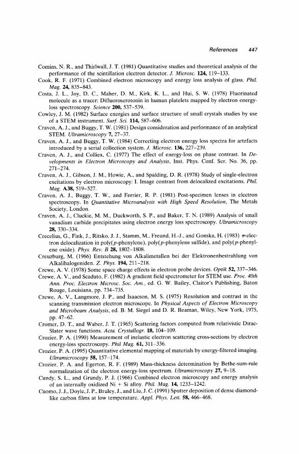
References 447
Comins, N. R., and Thirlwall, J. T. (1981) Quantitative studies and theoretical analysis of the performance of the scintillation electron detector. 1. Microsc. 124, 119-133.
Cook, R. F. (1971) Combined electron microscopy and energy loss analysis of glass. Phil. Mag. 24, 835-843.
Costa, J. L., Joy, D. c., Maher, D. M., Kirk, K. L., and Hui, S. W. (1978) Fluorinated molecule as a tracer: Difluoroserotonin in human platelets mapped by electron energyloss spectroscopy. Science 200, 537-539.
Cowley, J. M. (1982) Surface energies and surface structure of small crystals studies by use of a STEM instrument. Surf Sci. 114,587-606.
Craven, A. J., and Buggy, T. W. (1981) Design consideration and performance of an analytical STEM. Ultramicroscopy 7, 27-37.
Craven, A. J., and Buggy, T. W. (1984) Correcting electron energy loss spectra for artefacts introduced by a serial collection system. 1. Microsc. 136,227-239.
Craven, A. J., and Colliex, C. (1977) The effect of energy-loss on phase contrast. In Developments in Electron Microscopy and Analysis, Inst. Phys. Conf. Ser. No. 36, pp. 271-274.
Craven, A. J., Gibson, J. M., Howie, A, and Spalding, D. R. (1978) Study of single-electron excitations by electron microscopy: I. Image contrast from de localized excitations. Phil. Mag. A38,519-527.
Craven, A. J., Buggy, T. W., and Ferrier, R. P. (1981) Post-specimen lenses in electron spectroscopy. In Quantitative Microanalysis with High Speed Resolution, The Metals Society, London.
Craven, A. J., Cluck ie, M. M., Duckworth, S. P., and Baker, T. N. (1989) Analysis of small vanadium carbide precipitates using electron energy loss spectroscopy. Ultramicroscopy 28, 330-334.
Crecelius, G., Fink, J., Ritsko, J. J., Stamm, M., Freund, H.-J., and Gonska, H. (1983) 1T-elec
tron delocalization in poly(p-phenylene), poly(p-phenylene sulfide), and poly(p-phenylene oxide). Phys. Rev. B 28, 1802-1808.
Creuzburg, M. (1966) Entstehung von Alkalimetallen bei der Elektronenbestrahlung von Alkalihalogeniden. Z. Phys. 194,211-218.
Crewe, A V. (1978) Some space charge effects in electron probe devices. Optik 52, 337-346. Crewe, A. V., and Scaduto, F. (1982) A gradient field spectrometer for STEM use. Proc. 40th
Ann. Proc. Electron Microsc. Soc. Am., ed. G. W. Bailey, Claitor's Publishing, Baton Rouge, Louisiana, pp. 734-735.
Crewe, A. V., Langmore, J. P., and Isaacson, M. S. (1975) Resolution and contrast in the scanning transmission electron microscope. In Physical Aspects of Electron Microscopy and Microbeam Analysis, ed. B. M. Siegel and D. R. Beaman, Wiley, New York, 1975, pp.47-62.
Cromer, D. T., and Waber, J. T. (1965) Scattering factors computed from relativistic DiracSlater wave functions. Acta. Crystallogr. 18, 104-109.
Crozier, P. A. (1990) Measurement of inelastic electron scattering cross-sections by electron energy-loss spectroscopy. Phil Mag. 61,311-336.
Crozier, P. A (1995) Quantitative elemental mapping of materials by energy-filtered imaging. Ultramicroscopy 58, 157-174.
Crozier, P. A. and Egerton, R. F. (1989) Mass-thickness determination by Bethe-sum-rule normalization of the electron energy-loss spectrum. Ultramicroscopy 27, 9-18.
Cundy, S. L., and Grundy, P. J. (1966) Combined electron microscopy and energy analysis of an internally oxidized Ni + Si alloy. Phil. Mag. 14,1233-1242.
Cuomo, J. J., Doyle,J. P., Bruley, J., and Liu, J. C. (1991) Sputter deposition of dense diamondlike carbon films at low temperature. Appl. Phys. Lett. 58, 466-468.
448 References
Curtis, G. H .• and Silcox, J. (1971) A Wien filter for use as an energy analyzer with an electron microscope. Rev. Sci. Instrum. 42,630-637.
Daberkow, I., Herrmann, K.-H., Liu. L.. and Rau, W. D. (1991) Performance of electron image converters with Y AG single-crystal screen and CCD sensor. Ultramicroscopy 38, 215-223.
Daniels, J., and Kruger, P. (1971) Electron energy losses and optical constants of Ne and CH,. Phys. Status Solidi (b) 43,659-664.
Daniels, J., Festenberg, C. V., Raether, H., and Zeppenfeld, K. (1970) Optical constants of solids by electron spectroscopy. Springer Tracts in Modern Physics, Springer-Verlag, New York, Vol. 54, pp. 78-135.
Darlington, E. H., and Sparrow, T. G. (1975) A magnetic prism spectrometer for a high voltage electron microscope. f. Phys. E (Sci. lnstrum.) 8, 596-600.
Davis, C. A, McKenzie, D. R, Yin, Y., Kravtchinskaia, E., Amaratunga, G. A J., and Veerasamy, V. S. (1994) Substitutional nitrogen doping of tetrahedral amorphous carbon. Phil. Mag. B 69, 1133-1140.
Davis, L. C., and Feldkamp. L. A. (1976) Interpretation of 3p-core-excitation spectra in Cr, Mn, Co, and Ni. Solid State Commun. 19, 413-416.
Dehmer, J. L., and Dill, D. (1977) Molecular effects on inner-shell photoabsorption K-shell spectrum of Nz. f. Chern. Phys. 65,5327-5337.
Devenish, R W., Eaglesham, D. J., Maher, D. M., and Humphreys, C. J. (1989) Nanolithography using field emission and conventional thermionic electron sources. Ultramicroscopy 28, 324-329.
Dexpert, H., Lynch, J. P., and Freund. E. (1982) Sensitivity limits in x-ray emission spectroscopy of catalysts. In Developments in Electron Microscopy and Analysis, Inst. Phys. Conf. Ser. No. 61 (LO.P.), pp. 171-174.
Diociaiuti, M., and Paoletti, L. (1991) Structural characterization of air-oxidized chromium particles by extended energy-loss fine-structure spectroscopy. f. Microsc. 162,279-289.
Diociaiuti, M., Bascelli, A, and Paoletti, L. (1992a) Extended electron energy loss fine structure and selected area electron diffraction combined study of copper cluster oxidation. Vacuum 43, 575-581.
Diociaiuti, M., Picozzi, P., Santucci, S., Lozzi, L., and de Crescenzi, M. (1992b) Extended electron energy-loss fine structure and selected-area diffraction studies of small palladium clusters. f. Microsc. 166,231-245.
Diociaiuti, M., Falchi, M., and Paoletti, L. (1995) Electron energy loss spectroscopy study of iron deposition in human alveolar macrophages: ferritin or hemosiderin? Microsc. Microanal. Microstruct. 6, 33-40.
Disko, M. M. (1981) An EXELFS analysis system and the preliminary orientation dependence of EXELFS in graphite. In Analytical Electron Microscopy-198J, ed. R H. Geiss, San Francisco Press, San Francisco, pp. 214-220.
Disko, M. M., Meitzner, G., Ahn, C. c., and Krivanek, O. L. (1989) Temperature-dependent transmission extended electron energy-loss fine-structure of aluminum. f. Appl. Phys. 65,3295-3297.
Disko, M. M., Luton, M. J., and Shuman, H. (1991) Energy-loss near-edge fine structure and composition profiles of cryomilled oxide-dispersion-strengthened aluminum. Ultramicroscopy 37, 202-209.
Disko, M. M., Ahn, C. c., and Fultz, B., eds. (1992) Transmission Electron Energy Loss Spectrometry in Materials Science. The Minerals, Metals and Materials Society, Warrendale, Pennsylvania, 272 pages.
Ditchfield, R W., and Cullis, A G. (1976) Plasmon energy-loss analysis of epitaxial layers in siliwn and germanium. Micron 7, 133-140.
References 449
Ditchfield, R. W., and Whelan, M. J. (1977) Energy broadening of the electron beam in the electron microscope. Optik 48,163-172.
Ditchfield, R. W., Grubb, D. T., and Whelan, M. J. (1973) Electron energy-loss studies of polymers during radiation damage. Phil. Mag. 27, 1267-1280.
Donald, A. M., and Craven, A. J. (1979) A study of grain boundary segregation in Cu-Bi alloys using STEM. Phil. Mag. A 39, 1-11.
Downing, K. K., Ho, M.-H., and Glaeser, R. M. (1980) A charge coupled device readout system for electron microscopy, 38th Ann. Proc. Electron Microsc. Soc. Am., ed. G. W. Bailey, Claitor's Publishing, Baton Rouge, Louisiana, pp. 234-235.
Dravid, V. P., and Zhang, H. (1992) Hole formation and charge transfer in Yj.xCaxSr2Cu2Ga07, a new oxide superconductor. Physica C 200, 349-358.
Dravid, V. P., Zhang, H., and Wang, Y. Y. (1993) Inhomogeneity of charge carrier concentration along the grain boundary plane in oxide superconductors. Physica C 213, 353-358.
Du Chesne, A., Lieser, G., and Wegner, G. (1992) ESI for investigation of polymer microdomain morphology. Electron Microscopy 1992, Proc. EUREM, Granada, vol. 1, pp. 255-256.
Duckworth, S. P., Craven, A. J., and Baker, T. N. (1984) Comparison of carbon and noncarbon replicas for ELS. In Analytical Electron Microscopy-1984, ed. D. B. Williams and D. C. Joy, San Francisco Press, San Francisco, pp. 235-238.
Durham, P. J. (1983) Multiple scattering calculations of XANES. In EXAFS and Near Edge Structure, ed. A. Bianconi, L. Incoccia, and S. Stipcich, Springer-Verlag, New York, pp.37-42.
Durham, P. J., Pendry, J. B., and Hodges, C. H. (1981) XANES: determination of bond angles and multi-atom correlations in ordered and disordered systems. Solid State Commun. 38, 159-162.
Durham, P. J., Pendry, J. B., and Hodges, C. H. (1982) Calculation of x-ray absorption nearedge structure, XANES, Comput. Phys. Commun. 25, 193-205.
Eaglesham, D. J., and Berger, S. D. (1994) Energy filtering the "thermal diffuse" background in electron diffraction. Ultramicroscopy 53, 319-324.
Echenique, P. M., Ritchie, R. H., and Brandt, W. (1979) Spatial excitation patterns by swift ions in condensed matter. Phys. Rev. B 20, 2567-2580.
Echenique, P. M., Bausells, J., and Rivacoba, A. (1987) Energy-loss probability in electron microscopy. Phys. Rev. B 35, 1521-1524.
Egerton, R. F. (1975) Inelastic scattering of 80-keV electrons in amorphous carbon. Phil. Mag. 31, 199-215.
Egerton, R. F. (1976a) Measurement of inelastic/elastic scattering ratio for fast electrons and its use in the study of radiation damage. Phys. Status Solidi (a) 37, 663-668.
Egerton, R. F. (1976b) Coupling between plasmon and K-shell excitation in electron energyloss spectra of amorphous carbon, graphite and beryllium. Solid State Commun. 19, 737-740.
Egerton, R. F. (1976c) Inelastic scattering and energy filtering in the transmission electron microscope. Phil. Mag. 34, 49-66.
Egerton, R. F. (1978a) Formulae for light-element microanalysis by electron energy-loss spectrometry. Ultramicroscopy 3, 243-251.
Egerton, R. F. (1978b) A simple electron spectrometer for energy analysis in the transmission microscope. Ultramicroscopy 3, 39-47.
Egerton, R. F. (1979) K-shell ionization cross-sections for use in microanalysis. Ultramicroscopy 4, 169-179.
Egerton, R. F. (1980a) The use of electron lenses between a TEM specimen and an electron spectrometer. Optik 56, 363-376.
450 References
Egerton, R. F. (1980b) Design of an aberration-corrected electron spectrometer for the TEM. Optik 57, 229-242.
Egerton, R. F. (1980c) Chemical measurements of radiation damage at and below room temperature. Ultramicroscopy 5, 521-523.
Egerton, R. F. (1980d) Instrumentation and software for energy-loss microanalysis. In Scanning Electron Microscopy, SEM Inc., A. M. F. O'Hare, Illinois, Vol. 1, pp. 41-52.
Egerton, R. F. (1980e) An automated system for energy-loss microanalysis. 38th Ann. Proc. Electron Microsc. Soc. Am., ed. G. W. Bailey, Claitor's Publishing, Baton Rouge, Louisiana, pp. 130-131.
Egerton, R. F. (1980f) Measurement of radiation damage by electron energy-loss spectroscopy, J. Microsc. 118,389-399.
Egerton, R. F. (1981a) SIGMAL: a program for calculating L-shell ionization cross sections. 39th Ann. Proc. Electron Microsc. Soc. Am., ed. G. W. Bailey, Claitor's Publishing, Baton Rouge, Louisiana, pp. 198-199.
Egerton, R. F. (1981b). Alignment and characterization of an electron spectrometer. Ultramicroscopy 6, 93-96.
Egerton, R. F. (1981c) The range of validity of EELS microanalysis formulae. Ultramicroscopy 6,297-300.
Egerton, R. F. (1981d) Applications of energy-loss microanalysis. In Analytical Electron Microscopy-J98J, ed. R. H. Geiss, San Francisco Press, San Francisco, pp. 154-160.
Egerton, R. F. (1982a) A revised expression for signal/noise ratio in EELS. Ultramicroscopy 9,387-390.
Egerton, R. F. (1982b) Organic mass loss at lOOK and 300K. J. Microsc. 126,95-100. Egerton, R. F. (1982c) Electron energy-loss spectroscopy for chemical analysis. Phil. Trans.
R. Soc. London A305, 521-533. Egerton, R. F. (1982d) Thickness dependence of the STEM ratio image. Ultramicroscopy
9,297-299. Egerton, R. F. (1984a) Parallel-recording systems for electron energy-loss spectroscopy
(EELS). J. Electron Microsc. Tech. 1,37-52. Egerton, R. F. (1987) EELS at intermediate voltage. In Intermediate Voltage Microscopy and
Its Application to Materials Science, ed. K. Rajan, Philips Electron Optics, Mahwah, pp.67-71.
Egerton, R. F. (1992a) A data base for energy-loss cross sections and mean free paths. 50th Ann. Proc. Electron Microsc. Soc. Amer., San Francisco Press, San Francisco, pp. 1264-1265.
Egerton, R. F. (1992b) Electron energy-loss spectroscopy-EELS. In Quantitative Microbeam Analysis, ed. A. G. Fitzgerald, B. E. Storey, and D. Fabian, SUSSP, Edinburgh, and lOP, Bristol, pp. 145-168.
Egerton, R. F. (1993) Oscillator-strength parameterization of inner-shell cross sections. Ultramicroscopy 50, 13-28.
Egerton, R. F., and Cheng, S. C. (1982) The use of photodiode arrays to record 100 keV electrons. J. Microsc. 127, RP3-RP4.
Egerton, R. F., and Cheng, S. C. (1985) Thickness measurement by EELS. 43rd Ann. Proc. Electron Microsc. Soc. Amer., ed. G. W. Bailey, San Francisco Press, San Francisco, pp. 389-399.
Egerton, R. F., and Cheng, S. C. (1987) Measurements of local thickness by electron energyloss spectroscopy. Ultramicroscopy 21, 231-244.
Egerton, R. F., and Crozier, P. A. (1987) A compact parallel-recording detector for EELS. J. Microsc. 148, 305-312.
Egerton, R. F., and Crozier, P. A. (1988) The use of Fourier techniques in electron energy-loss
References 451
spectroscopy. Scanning Microscopy, Supplement, 2, Scanning Microscopy International, Chicago, 245-254.
Egerton, R. F., and Kenway, D. (1979) An acquisition, storage, display and processing system for electron energy-loss spectra. Ultramicroscopy 4, 221-225.
Egerton, R. F., and Rossouw, C. J. (1976) Direct measurement of contamination and etching rates in an electron beam. J. Phys. D 9, 659-663.
Egerton, R. F., and Sevely, J. (1983) Jump ratio as a measure of spectrometer performance. J. Microsc. 129, RPI-RP2.
Egerton, R. F., and Wang, Z. L. (1990) Plural-scattering deconvolution of electron energyloss spectra recorded with an angle-limiting aperture. Ultramicroscopy 32, 137-148.
Egerton, R. F., and Whelan, M. J. (1974a) Electron energy-loss spectra of diamond, graphite and amorphous carbon. J. Electron Spectrosc. 3, 232-236.
Egerton, R. F., and Whelan, M. J. (1974b) The electron energy-loss spectrum and band structure of diamond. Phil. Mag. 30, 739-749.
Egerton, R. F., and Wong, K. (1995) Some practical consequences of the Lorentzian angular distribution of inelastic scattering. Ultramicroscopy 59, 169-180.
Egerton, R. F., Philip, J. G., and Whelan, M. J. (1974) Applications of energy analysis in a transmission electron microscope. In Electron Microscopy-1974, 8th Int. Congress, ed. J. V. Sanders and D. J. Goodchild, Australian Academy of Science, Canberra, Vol. 1, pp. 137-140.
Egerton, R. F., Philip, J. G., Turner, P. S., and Whelan, M. J. (1975) Modification of a transmission electron microscope to give energy-loss spectra and energy-selected images and diffraction patterns. J. Phys. E. 8,1033-1037.
Egerton, R. F., Rossouw, C. J., and Whelan, M. J. (1976) Progress towards a method for the quantitative microanalysis of light elements by electron energy-loss spectrometry. In Developments in Electron Microscopy and Analysis, ed. J. A. Venables, Academic Press, New York, pp. 129-132.
Egerton, R. F., Williams, B. G., and Sparrow, T. G. (1985) Fourier deconvolution of electron energy-loss spectra. Proc. R. Soc. London, A 398, 395-404.
Egerton, R. F., Crozier, P. A, and Rice, P. (1987) Electron energy-loss spectroscopy and chemical change. Ultramicroscopy 23, 305-312.
Egerton, R. F., Yang, Y.-Y., and Chen, F. Y. Y. (1991) EELS of "thick" specimens. Ultramicroscopy 38, 349-352.
Egerton, R. F., Yang, Y.-Y., and Cheng, S. C. (1993) Characterization and use of the Gatan 666 parallel-recording electron energy-loss spectrometer. Ultramicroscopy 48, 239-250.
Egle, W., Rilk, A, Bihr, J., and Menzel, M. (1984) Microanalysis in the EM 902: Tests on a new TEM for ESI and EELS. 42nd Ann. Proc. Electron Microsc. Soc. Am., ed. G. W. Bailey, San Francisco Press, San Francisco, pp. 566-567.
Ehrenreich, H., and Philipp, H. R. (1962) Optical properties of semiconductors in the ultraviolet. Proc. Int. Conf. Phys. Semiconductors, ed. A. C. Strickland, Bartholomew Press, Dorking, U.K., pp. 367-374.
Eisenberger, P., Shulman, R. G., Kincaid, B. M., and Brown, G. S. (1978) Extended x-ray absorption fine structure determination of iron-nitrogen distances in haemoglobin. Nature 274, 30-34.
Enge, H. A (1964) Effect of extended fringing fields on ion-focussing properties of deflecting magnets. Rev. Sci. Instrum. 35,278-287.
Enge, H. A. (1967) Deflecting magnets. In Focussing of Charged Panicles, Vol. 2, ed. A Septier, Academic Press, New York, pp. 203-264.
Engel, A., Christen, F., and Michel, B. (1981) Digital acquisition and processing of electron micrographs using a scanning transmission electron microscope. Ultramicroscopy 7, 45-54.
452 References
Evans, E., and Mills, D. L. (1972) Theory of inelastic scattering by long-wavelength surface optical phonons. Phys. Rev. B 5, 4126-4139.
Evans, N. D., ZinkIe, S. J., Bentley, J., and Kenik, E. A (1991) Quantification of metallic aluminum profiles in AI+ implanted MgAb04 spinel by electron energy loss spectroscopy. In Microbeam Analysis-1991, San Francisco, San Francisco, pp. 439-440.
Fallon, P. J. (1992) Microscopy and Spectroscopy of CVD Diamond, Diamond-Like Carbon and Similar Materials. Ph.D. Thesis, University of Cambridge, U.K.
Fallon, P. J. and Brown, L. M. (1993) Analysis of chemical-vapour-deposited diamond grain boundaries using transmission electron microscopy and parallel electron energy loss spectroscopy in a scanning transmission electron microscope. Diamond ReI. Mater. 2, 1004-1011.
Fallon, P. J., Brown, L. M., Barry, J. C., and Bruley, J. (1995) Nitrogen determination and characterization in natural diamond platelets. Phil. Mag., submitted.
Fano, U. (1956) Differential inelastic scattering of relativistic charged particles. Phys. Rev. 102,385-387.
Fano, U. (1960) Normal modes of a lattice of oscillators with many resonances and dipolar coupling. Phys. Rev. 118,451-455.
Fano, U., and Cooper, J. W. (1968) Spectral distribution of atomic oscillator strengths. Rev. Mod. Phys. 40,441-507.
Ferrell, R A (1957) Characteristic energy losses of electrons passing through metal foils. II. Dispersion relation and short wavelength cutoff for plasma oscillations. Phys. Rev. 107, 450-462.
Festenberg, C. von (1967) Zur Dampfung des Al-15 keV-Plasmaverlustes in Abhangigkeit vom Streunwinkel und der Kristallitgrosse. Z. Phys. 207, 47-55.
Festenberg, C. von (1969) Energieverlustmessungen an III-V-Verbindungen. Z. Phys. 227, 453-481.
Festenberg, C. von, and Kroger, E. (1968) Retardation effects for the energy-loss probability in GaP and Si. Phys. Lett. 26A,339-34l.
Fields, J. R (1977) Magnetic spectrometers: approximate and ideal designs. Ultramicroscopy 2,311-325.
Fink, J. (1989) Recent developments in energy-loss spectroscopy. In Advances in Physics and Electron Physics, Academic Press, London, vol. 75, pp. 121-232.
Fink, J., Muller-Heinzerling, T., PflU, J., Bubenzer, A, Koidl, P., and Crecelius, G. (1983) Structure and bonding of hydrocarbon plasma generated carbon films: An electron energy loss study. Solid State Commun. 47, 687-691.
Fink, J., NUcker, N., Pellegrin, E., Romberg, H., Alexander, M., and Knupfer, M. (1994) Electron energy-loss and x-ray absorption spectroscopy of cuprate superconductors and related compounds. I. Electron Spectrosc. Rei. Phenom. 66, 395-452.
Fink, M., and Kessler, J. (1967) Absolute measurements of elastic cross section for smallangle scattering of electrons from N2 and O2. I. Chern. Phys. 47, 1780-1782.
Fiori, C. E., Gibson, C. c., and Leapman, R D. (1980) Electrostatic deflection system for use with an electron energy-loss spectrometer. In Microbeam Analysis-1980, ed. D. B. Wittry, San Francisco Press, San Francisco, pp. 225-228.
Fiori, C. E., Leapman, R. D., Swyt, C. R, and Andrews, S. B. (1988) Quantitative x-ray mapping of biological cryosections. Ultramicroscopy 24, 237-250.
Fitzgerald, A G., Storey, B. J., and Fabian, D., eds. (1992) Quantitative Microbeam Analysis, Scottish Universities Summer School in Physics, Edinburgh and Institute of Physics Publishing, Bristol and Philadelphia.
Frabboni, S., Lulli, G., Merli, P. G., Migliori, A., and Bauer, R (1991) Electron spectroscopic imaging of dopant precipitation and segregation in silicon. Ultramicroscopy 35, 265-269.
References 453
Frank, I. M. (1966) Transition radiation and optical properties of matter. Sov. Phys. Usp. 8,729-742.
Fraser, H. L. (1978) Elemental analysis of second-phase carbides using electron energy-loss spectroscopy. In Scanning Electron Microscopy, SEM Inc., A. M. F. O'Hare, Illinois, Part 1, pp. 627-632.
Fryer, J. R., and Holland, F. (1984) High resolution electron microscopy of molecular crystals: III. Radiation processes at room temperature. Proc. R. Soc. London A393, 353-369.
Fujimoto, F., and Komaki, K. (1968) Plasma oscillations excited by a fast electron in a metallic particle. J. Phys. Soc. Jpn 25, 1679-1687.
Fujiyoshi, Y., Kobayashi, T., Tsuji, M., and Uyeda, N. (1982) The effect of electronic state on the direct imaging of atoms. In Electron Microscopy-1982, 10th Int. Cong., Deutsche Gesellschaft fur Elektronenmikroskopie, Vol. 1, pp. 217-218.
Garavito, R. M., Carlemalm, E., Colliex, c., and Villiger, W. (1982) Septate junction ultrastructure as visualized in unstained and stained preparations. J. Ultrastructure Rs. 80, 334-353.
Garcia de Abajo, F. J., and Echenique, P. M. (1992) Wake-potential formation in a thin foil. Phys. Rev. B 45, 8771-8774.
Garibyan, G. M. (1960) Transition radiation effects in particle energy losses. Sov. Phys. JETP 37, 372-376.
Garratt-Read, A. J. (1981) Measurement of carbon in V(C, N) precipitates extracted from HSLA steels on aluminum replicas. Quantitative Microanalysis with High Spatial Resolution, Metals Society, London, pp. 165-168.
Garvie, L. A. J., Craven, A. J., and Brydson, R. (1994) Use of electron-energy loss near-edge fine structure in the study of minerals. American Mineralogist 79, 411-425.
Gatts, C., Duscher, G., MUllejans H., and RUhle, M. (1995) Analyzing line scan profiles with neural pattern recognition. Ultramicroscopy 59, 229-240.
Geiger, J. (1981) Inelastic electron scattering with energy losses in the meV-region. 39th Ann. Proc. Electron Microsc. Soc. Am., ed. G. W. Bailey, Claitor's Publishing, Baton Rouge, Louisiana, pp. 182-185.
Geiger, J., Nolting, M., and Schroder, B. (1970) How to obtain high resolution with a Wien filter spectrometer. In Electron Microscopy-1970, ed. P. Favard, Societe Francaise de Microscopie Electronique, Paris, pp. 111-112.
Gibbons, P. C., Schnatterly, S. E., Ritsko, J. J., and Fields, J. R. (1976) Line shape of the plasma resonance in simple metals. Phys. Rev. 13,2451-2460.
Glaeser, R. M. (1975) Radiation damage and biological electron microscopy. In Physical Aspects of Electron Microscopy and Microbeam Analysis, ed. B. M. Siegel and D. R. Beaman, Wiley, New York, pp. 205-229.
Glen, G. L., and Dodd, C. G. (1968) Use of molecular orbital theory to interpret x-ray Kabsorption spectral data. J. Appl. Phys. 39, 5372-5377.
Goldstein, J. I., Costley, J. L., Lorimer, G. W., and Reed, S. J. B. (1977) Quantitative x-ray analysis in the electron microscope. Scanning Electron Microscopy. Part 1, 315-324.
Gordon, R. L. (1976) Annealing procedure for self-scanned diode arrays. Appl. Opt. 15, 1909-1911.
Gorlen, K. E., Barden, L. K., DelPriore, J. S., Fiori, C. E., Gibson, C. G., and Leapman, R. D. (1984) Computerized analytical electron microscope for elemental imaging. Rev. Sci. 1nstrum. 55, 912-921.
Graczyk, J. F., and Moss, S. C. (1969) Scanning electron diffraction attachment with electron energy filtering. Rev. Sci. 1nstrum. 40, 424-433.
Grivet, P., and Septier, A. (1978) Ion microscopy: History and actual trends. Ann. N. Y. Acad. Sci. 306, 158-182.
Groot, F. M. F. de, Fuggle, J. c., Thole, B. T., and Sawatsky, G. A. (1990) L 23 x-ray-absorption
454 References
edges of d!' compounds: K + ,Ca2+ ,Sc3+ and Ti4+ in Oh (octahedral) symmetry. Phys. Rev. B 41, 928-937.
Grunes, L. A. (1983) Study of the K edges of 3d transition metals in pure oxide form by xray absorption spectroscopy. Phys. Rev. B 27, 2111-2131.
Grunes, L. A., Leapman, R. D., Walker, c., Hoffmann, R., and Kunz, A. B. (1982) Oxygen K near-edge fine structure: An electron energy-loss investigation with comparisons to new theory for selected 3d transition-metal oxides. Phys. Rev. B 25, 7157-7173.
Gubbens, A. J., Krivanek, O. L., and Kundmann, M. K. (1991) Electron energy loss spectroscopy above 2000 kV. Microbeam analysis-1991, ed. D. G. Howitt, San Francisco Press, San Francisco, pp. 127-133.
Gubbens, A. J., Kraus, B., Krivanek, O. L., and Mooney, P. E. (1995) An imaging filter for high voltage electron microscopy. Ultramicroscopy, 59, 255-265.
Haak, W. W., Sawatzky, G. A., Ungier, L., Gimzewski, J. K., and Thomas, T. D. (1984) Corelevel electron-electron coincidence spectroscopy. Rev. Sci. Instrum. 55,696-711.
Hagemann, H.-J., Gudat, W., and Kunz, C. (1974) Optical constants from the far infrared to the x-ray region: Mg, AI, Cu, Ag, Au, Bi, C, and AI20 J. DESY report SR-7417, DESY, 2 Hamburg 52, West Germany.
Hainfeld, J., and Isaacson, M. (1978) The use of electron energy-loss spectroscopy for studying membrane architecture: A preliminary report. Ultramicroscopy 3, 87-95.
Hall, C. R. (1966) On the production of characteristic x-rays in thin metal crystals. Proc. R. Soc. London A295, 140-163.
Hall, C. R, and Hirsch, P. B. (1965) Effect of thermal diffuse scattering on propagation of high energy electrons through crystals. Proc. Roy. Soc. London A 286, 158-177.
Hall, T. A. (1979) Biological X-ray microanalysis. 1. Microsc. 117,145-163. Hansen, P. L., Fallon, P. J., and Kratschmer, W. (1991) An EELS study of fullerite-C,",C,o.
Chem Phys. Lett. 181,367-372. Hanson, H. P., Herman, F., Lea, J. D., and Skillman, S. (1964) HFS atomic scattering factors.
Acta. Crystallogr. 17,1040-1044. Hashimoto, H., Makita, Y., and Nagaoka, N. (1992) Electron microscope images of thorium
atoms formed by plasma-loss and core-loss electrons using an energy selecting microscope. Optik 93, 119-126.
Heighway, E. A. (1975) Focussing for dipole magnets with large pole gap to bending radius ratios. Nucl. Instrum. Methods U3, 413-419.
Heine, V. (1980) Electronic structure from the point of view of the local atomic environment. Solid State Phys. 35,1-126.
Hembree, G. G., and Venables, J. A. (1992) Nanometer-resolution scanning Auger electron microscopy. Ultramicroscopy 47, 109-120.
Henkelman, R M., and Ottensmeyer, F. P. (1974a) An energy filter for biological electron microscopy. 1. Microsc. 102, 79-94.
Henkelman, R M., and Ottensmeyer, F. P. (1974b) An electrostatic mirror. 1. Phys. E. (Scientific Instruments) 7, 176-178.
Henoc, P., and Henry, L. (1970) Observation des oscillations de plasma a l'interface d'inclusions gaze uses dans une matrice cristalline. 1. Phys. (Paris) 31 (supplement C1), 55-57.
Henry, L., Duval, P., and Hoan, N. (1969) Filtrage en energie des diagrammes de microdiffraction electronique. C.R. A cad. Sci. Paris B268, 955-958.
Herley, P. J., Jones, W., Sparrow, T. G., and Williams, B. G. (1987) Plasmon spectra of lightmetal hydrides. Materials Letters 5, 333-336.
Herman, F., and Skillman, S. (1963) Atomic Structure Calculations, Prentice-Hall, Englewood Cliffs, New Jersey.
Herrmann, K.-H. (1984) Detection systems. In Quantitative Electron Microscopy, ed. J. N.
References 455
Chapman, and A. J. Craven, SSUP Publications, University of Edinburgh, Scotland, pp. 119-148.
Hibbert, G., and Eddington, J. W. (1972) Experimental errors in combined electron microscopy and energy analysis. J. Phys. D 5, 1780-1786.
Hicks, P. J., Daviel, S., Wallbank, B., and Comer, J. (1980) An electron spectrometer using a new multidetector system based on a charge-coupled imaging device. J. Phys. E 13, 713-715.
Hier, R. W., Beaver, E. A., and Schmidt, G. W. (1979) Photon detection experiments with thinned CCD's Adv. Electron. Electron Phys. 52, 463-480.
Higgins, R J. (1976) Fast Fourier transform: An introduction with some minicomputer experiments. Am. J. Phys. 44,766-773.
Hillier, J. (1943) On microanalysis by electrons. Phys. Rev. 64,318-319. Hillier, J., and Baker, R F. (1944) Microanalysis by means of electrons. J. Appl. Phys.
15, 663-675. Hines, R. L. (1975) Graphite crystal film preparation by cleavage. J. Microsc. 104,257-26l. Hinz, H.-J., and Raether, H. (1979) Line shape of the volume plasmons of silicon and germa
nium. Thin Solid Films 58, 281-284. Hirsch, P. B., Howie, A., Nicholson, R B., Pashley, D. W., and Whelan, M. J. (1977) Electron
Microscopy of Thin Crystals, Krieger, Huntington, New York. Hitchcock, A. P. (1989) Electron-energy-loss-based spectroscopies: a molecular viewpoint.
Ultramicroscopy 28,165-183. Hitchcock, A. P. (1994) Bibliography and data base of inner shell excitation spectra of gas
phase atoms and molecules. J. Electron Spectrosc. ReI. Phenom. 67,1-13l. Hjalmarson, H. P., BUttner, H., and Dow, J. D. (1980) Theory of core excitons. Phys. Rev.
B 24, 6010-6019. Hobbs, L. W. (1984) Radiation effects in analysis byTEM. In Quantitative Electron Microscopy,
ed. J. N. Chapman and A. J. Craven, SSUP Publications, University of Edinburgh, Scotland, pp. 399-443.
Hofer, F. (1987) EELS quantification of M edges by using oxidic standards. Ultramicroscopy 21,63-68.
Hofer, F., and Golub, P. (1987) New examples of near-edge fine structures in electron energy loss spectroscopy. Ultramicroscopy 21, 379-384.
Hofer, F., and Kothleitner, G. (1993) Quantitative microanalysis using electron energy-loss spectrometry. 1. Li and Be in oxides. Microsc. Microanal. Microstruct. 4, 539-560.
Hofer, F., and Wilhelm, P. (1993) EELS microanalysis of the elements Ca to Cu using M23 edges. Ultramicroscopy 49,189-197.
Hofer, F., Golub, P., and Brunegger, A. (1988) EELS quantification of the elements Sr to W by means of M45 edges. Ultramicroscopy 25, 81-84.
Hofer, F., W arbichler, P., and Grogger, W. (1995) Characterization of nanometre sized precipitates in solids by electron spectroscopic imaging. Ultramicroscopy, 59, 15-3l.
Hojou, K., Furuno, S., Kushita, K. N., Otsu, H., Izui, K., Ueki, Y., and Kamino, T. (1992) Electron energy-loss spectroscopy of SiC crystals implanted with hydrogen and helium dual-ion beam. In Electron Microscopy 1992, Proc. EUREM 92, Granada, vol. 1, 261-262.
Hollenbeck, J. L., and Buchanan, R C. (1990) Oxide thin films for nanometer scale electron beam lithography. J. Mater. Res. 5, 1058-1072.
Holmestad, R., Krivanek, O. L., Hillier, R, Marthinsen, K., and Spence, J. C. H. (1993) Commercial spectrometer modifications for energy filtering of electron diffraction patterns and images. Ultramicroscopy 52, 454-458.
Hosoi, J., Oikawa, T., Inoue, M., Kokubo, Y., and Hama, K. (1981) Measurement of partial
456 References
specific thickness (net thickness) of critical-point-dried cultured fibroblast by energy analysis. Ultramicroscopy 7, 147-154.
Hosoi, J., Oikawa, T., Inoue, M., and Kokubo, Y. (1984) Thickness dependence of signal/ background ratio of inner-shell electron excitation loss in EELS. Ultramicroscopy 13, 329-332.
Howie, A. (1981) Localisation and momentum transfer in inelastic scattering. 39th Ann. Proc. Electron Microsc. Soc. Am., ed. G. W. Bailey, Claitor's Publishing, Baton Rouge, Louisiana, pp. 186-189.
Howie, A. (1983) Surface reactions and excitations. Ultramicroscopy 11, 141-148. Howie, A., and Walsh, C. (1991) Interpretation of valence loss spectra from composite media.
Microsc. Microanal. Microstruct. 2, 171-181. Howitt, D. (1984) Ion milling of materials science specimens for electron microscopy: A
review. 1. Electron Microsc. Tech. 1,405-415. Hren, J. J., Goldstein, J. I., and Joy, D. C. eds. (1979) Introduction to Analytical Electron
Microscopy, Plenum Press, New York. Hubbell, J. H. (1971) Survey of photon-attenuation-coefficient measurements 10 eV to 100
GeV. At. Data Tables 3, 241-297. Humphreys, C. J., Hart-Davis, A., and Spencer, J. P. (1974) Optimizing the signal/noise ratio
in the dark-field imaging of single atoms. In Electron Microscopy-1974, 8th Int. Congress, ed. J. V. Sanders, and D. J. Goodchild, Australian Academy of Science, Canberra, 1974, vol. 1, pp. 248-249.
Humphreys, C. J., Eaglesham, D. J., Alford, N. M., Harmer, M. A., and Birchall, J. D. (1988) High Temperature Superconductors, Inst. Phys. Conf. Ser. No. 93, lOP, Bristol, vol. 2, pp.217-222.
Humphreys, C. J., Bullough, T. J., Devenish, R. W., Maher, D. M., and Turner, P. S. (1990) Electron beam nano-etching in oxides, fluorides, metals and semiconductors. In Scanning Microscopy Supplement 4, Scanning Microscopy International, Chicago, 185-192.
Hunt, J. A., and Williams, D. B. (1991) Electron energy-loss spectrum-imaging. Ultramicroscopy 38, 47-73.
Hunt, J. A., Disko, M. M., Bekal, S. K., and Leapman, R. D. (1995) Electron energy-loss chemical imaging of polymer phases. Ultramicroscopy 58, 55-64.
Ibach, H. (1991) Electron Energy Loss Spectrometers. Springer Series in Optical Sciences, Springer-Verlag, Berlin, vol. 63.
Ibach, H., and Mills, D. L. (1982) Electron Energy-Loss Spectroscopy and Surface Vibrations, Academic Press, New York.
Ibers, J. A., and Vainstein, B. K. (1962) International Crystallographic Tables III, Kynoch Press, Birmingham, Table 3.3.
Imura, T., Saka, H., Todokoro, H., and Ashikawa, M. (1971) Direct intensification of electron microscopic images with silicon diode array target. 1. Phys. Soc. lpn 31, 1849.
Inokuti, M. (1971) Inelastic collisions of fast charged particles with atoms and molecules-The Bethe theory revisited. Rev. Mod. Phys. 43,297-347. Addenda: Rev. Mod. Phys. 50,23-26.
Inokuti, M. (1979) Electron-scattering cross sections pertinent to electron microscopy. Ultramicroscopy 3, 423-427.
Inokuti, M., Saxon, R. P., and Dehmer, J. L. (1975) Total cross-sections for inelastic scattering of charged particles by atoms and molecules-VIII. Systematics for atoms in the first and second row. Int. 1. Radiat. Phys. Chern. 7,109-120.
Inokuti, M., Dehmer, J. L., Baer, T., and Hanson, D. D. (1981) Oscillator-strength moments, stopping powers, and total inelastic-scattering cross sections of all atoms through strontium. Phys. Rev. A 23, 95-109.
Isaacson, M. (1972a) Interaction of 25 keY electrons with the nucleic acid bases, adenine, thymine, and uracil I. Outer shell excitation. 1. Chern. Phys. 56,1803-1812.
References 457
Isaacson, M. (1972b) Interaction of 25 keV electrons with the nucleic acid bases, adenine, thymine and uracil. (II) Inner-shell excitation and inelastic scattering cross sections. 1. Chem. Phys. 56, 1813-1818.
Isaacson, M. (1977) Specimen damage in the electron microscope. In Principles and Techniques of Electron Microscopy, ed. M. A. Hayat, Van Nostrand, New York, Vol. 7, pp. 1-78.
Isaacson, M. (1981) All you might want to know about ELS (but were afraid to ask): A tutorial. In Scanning Electron Microscopy, SEM Inc., (A. M. F. O'Hare, Illinois,) Part 1, pp. 763-776.
Isaacson, M., and Johnson, D. (1975) The microanalysis of light elements using transmitted energy-loss electrons. Ultramicroscopy I, 33-52.
Isaacson, M. S., and Utlaut, M. (1978) A comparison of electron and photon beams for determining micro-chemical environment. Optik 50, 213-234.
Ishizuka, K. (1993) Analysis of electron image detection efficiency of slow-scan CCD cameras. Ultramicroscopy 52, 7-20.
Jackson, J. D. (1975) Classical Electrodynamics, second edition, Wiley, New York, Ch. 13. Jager, W., and Mayer, J. (1995) Energy filtered transmission electron microscopy of SimGen
superlattices and Si-Ge heterostructures. Ultramicroscopy 59, 33-45. Jeanguillaume, c., and Colliex, C. (1989) Spectrum-image: the next step in EELS digital
acquisition and processing. Ultramicroscopy 28, 252. Jeanguillaume, c., Trebbia, P., and Colliex, C. (1978) About the use of electron energy-loss
spectroscopy for chemical mapping of thin foils with high spatial resolution. Ultramicroscopy 3, 237-242.
Jeanguillaume, c., Colliex, c., Ballongue, P., and Tence, M. (1992) New STEM multisignal imaging modes, made accessible through the evaluation of detection efficiencies. Ultramicroscopy 45, 205-217.
Jewsbury, P., and Summerside, P. (1980) The nature of interface plasmon modes at bimetallic junctions. 1. Phys. FlO, 645-650.
Jiang, X. G., and Ottensmeyer, F. P. (1993) Optimization of a prism-mirror-prism imaging energy filter for high resolution electron microanalysis. Optik 94, 88-95.
Jiang, X. G., and Ottensmeyer, F. P. (1994) Molecular microanalysis: imaging with low-energyloss electrons. Electron Microscopy 1994, Proc. 13th Int. Congo Electron Microsc., Paris, vol. 3, pp. 781-782.
Johansson, S. A. E. (1984) PIXE summary. Nucl. Instrum. Methods B3, 1-3. Johnson, D. E. (1972) The interactions of 25 keV electrons with guanine and cytosine. Radiat.
Res. 49, 63-84. Johnson, D. E. (1979b) Energy-loss spectrometry for biological research. In Introduction to
Analytical Electron Microscopy, Plenum Press, New York, pp. 245-258. Johnson, D. E. (1980a) Post specimen optics for energy-loss spectrometry. In Scanning Electron
Microscopy, SEM Inc., A. M. F. O'Hare, Illinois, Part 1, pp. 33-40. Johnson, D. E. (1980b) Pre-spectrometer optics in a CTEM/STEM. Ultramicroscopy 5,
163-174. Johnson, D. E., Csillag, S., and Stern, E. A. (1981b) Analytical electron microscopy using
extended energy-loss fine structure (EXELFS). In Scanning Electron Microscopy, SEM Inc., A. M. F. O'Hare, Illinois, Part 1, pp. 105-115.
Johnson, D. E., Mon~on, K. L., Csillag, S., and Stern, E. A. (1981c) An approach to paralleldetection electron energy-loss spectrometry. In Analytical Electron Microscopy-1981 , ed. R. H. Geiss, San Francisco Press, San Francisco, pp. 205-209.
Johnson, D. W. (1974) Optical properties determined from electron energy-loss distributions. In Electron Microscopy-1974, 8th Int. Congress, ed. J. V. Sanders and D. J. Goodchild, Australian Academy of Science, Canberra, pp. 388-389.
458 References
Johnson, D. W. (1975) A Fourier method for numerical Kramers-Kronig analysis. 1. Phys. A (Math. Gen. Phys.) 8,490-495.
Johnson, D. W., and Spence, J. C. H. (1974) Determination of the single-scattering probability distribution from plural-scattering data. J. Phys. D (Appl. Phys.) 7, 771-780.
Johnson, H. F., and Isaacson, M. S. (1988) An efficient analytical method for calculating the angular distribution of electrons which have undergone plural scattering in amorphous materials. Ultramicroscopy 26, 271-294.
Jonas, P., and Schattschneider, P. (1993) The experimental conditions for Compton scattering in the electron microscope. J. Phys. Condo Matter 5, 7173-7188.
Jonas, P., Schattschneider, P., and Su, D. S. (1992) Directional Compton profiles of silicon. In Electron Microscopy, EUREM 92, Granada, vol. 1, pp. 265-266.
Jones, B. L., Walton, D. M., and Booker, G. R. (1982) Developments in the use of one- and two-dimensional self-scanned silicon photodiode arrays in imaging devices in electron microscopy. Inst. Phys. Cont Ser. No. 61, 135-138.
Jones, W., Sparrow, T. G., Williams, B. G., and Herley, P. J. (1984) Evidence for the formation of single crystals of sodium metal during the decomposition of sodium aluminum hydride: An electron microscopic study. Mater. Lett. 2,377-379.
Jouffrey, B., Kihn, Y., Perez, J. P., Sevely, J., and Zanchi, G. (1978) On chemical analysis of thin films by energy-loss spectroscopy. In Electron Microscopy-1978, 9th Int. Cong., ed. J. M. Sturgess, Microscopical Society of Canada, Toronto, Vol. 3, pp. 292-303.
Jouffrey, B., Sevely, J., Zanchi, G., and Kihn, Y. (1985) Characteristic energy losses with high energy electrons up to 2.5 MeV. Scanning Electron Microscopy, Part 3, 1063-1070.
Jouffrey, B., Zanchi, G., Kihn, Y., Hussein, K., and Sevely, J. (1989) Present questions in EELS: sensitivity and EXELFS. Beitr. Elektronenmikroskop. Direktabb. Obert 22, 249-270.
Joy, D. C. (1979) The basic principles of electron energy-loss spectroscopy. In Introduction to Analytical Electron Microscopy, Plenum Press, New York, pp. 223-244.
Joy, D. C. (1984a) Detectors for electron energy-loss spectroscopy. In Electron-Beam Interactions with Solids for Microscopy, Microanalysis and Microlithography, SEM Inc., Illinois, pp.251-257.
Joy, D. C. (1984b) A parametric partial cross section for ELS. J. Microsc. 134,89-92. Joy, D. c., and Maher, D. M. (1977) Sensitivity limits for thin specimen x-ray analysis. Scanning
Electron Microscopy/J977, Part 1,325-334. Joy, D. c., and Maher, D. M. (1978) A practical electron spectrometer for chemical analysis.
J. Microsc. 114,117-129. Joy, D. c., and Maher, D. M. (1980a) The electron energy-loss spectrum-Facts and artifacts.
In Scanning Electron Microscopy, SEM Inc., A. M. F. O'Hare, Illinois, Part 1, pp. 25-32. Joy, D. c., and Maher, D. M. (1980c) Electron energy-loss spectroscopy. J. Phys. E. (Sci.
Instrum.) 13, 261-270. Joy, D. c., and Maher, D. M. (1981a) The quantitation of electron energy-loss spectra.
J. Microsc. 124,37-48. Joy, D. c., and Maher, D. M. (1981b) Instrument errors in ELS quantitation. In Analytical
Electron Microscopy-l 981 , ed. R. H. Geiss, San Francisco Press, San Francisco, pp. 176-177.
Joy, D. c., and Maher, D. M. (1981c) The micro-spectroscopy of semiconductors. In Inst. Phys. Cont Ser. No. 60, I.O.P., Bristol, pp. 229-236.
Joy, D. c., and Newbury, D. E. (1981) A "round robin" test on ELS quantitation. In Analytical Electron Microscopy-198J, ed. R. H. Geiss, San Francisco Press, San Francisco, pp. 178-180.
Joy, D. c., Newbury, D. E., and Myklebust, R. L. (1982) The role of fast secondary electrons in degrading spatial resolution in the analytical electron microscope. J. Microsc. 128, RP1-RP2.
References 459
Joy, D. c., Romig, A. D., and Goldstein, J. I. eds. (1986) Principles of Analytical Electron Microscopy, Plenum Press, New York.
Kainuma, Y. (1955) The theory of Kikuchi patterns. Acta. Crystallogr. 8,247-257. Kaloyeros, A. E., Hoffman, M. P., Williams, W. S., Greene, A. E., and McMillan, J. A. (1988)
Structural studies of amorphous titanium diboride thin films by extended x-ray-absorption fine-structure and extended electron-energy-loss fine-structure techniques. Phys. Rev. 38, 7333-7344.
Kambe, K., Krahl, D., and Herrmann, K-H. (1981) Extended fine structure in electron energyloss spectra of MgO crystallites. Ultramicroscopy 6, 157-162.
Katterwe, H. (1972) Object analysis by electron energy spectroscopy in the infra-red region. In Electron Microscopy-1972, The Institute of Physics, London, pp. 154-155.
Keil, P. (1968) Elektronen-Energieverlustmessungen und Berechnung optischer Konstanten: II Kaliumbromid. Z. Phys. 214,266-284.
Kevan, S. D., and Dubois, L. H. (1984) Development of dispersion compensation for use in high-resolution electron energy-loss spectroscopy. Rev. Sci. lnstrum. 55,1604-1612.
Kihn, Y., Perez, J.-P., Sevely, J., Zanchi, G., and Jouffrey, B. (1980) Data collection problems in high voltage electron energy-loss spectroscopy. In Electron Microscopy-1980, 7th European Congress Foundation, The Hague, Vol. 4, pp. 42-45.
Kim, M. J., and Carpenter, R. W. (1990) Composition and structure of native oxide on silicon by high resolution analytical electron microscopy. 1. Mater. Res. 5,347-351.
Kincaid, B. M., Meixner, A. E., and Platzman, P. M. (1978) Carbon K-edge in graphite measured using electron energy-loss spectroscopy. Phys. Rev. Lett. 40, 1296-1299.
Klemperer, 0., and Shepherd, J. P. G. (1963) On the measurement of characteristic energy losses of electrons in metals. Brit. 1. Appl. Phys. 14, 85-88.
Knauer, W. (1979) Analysis of energy broadening in electron and ion beams. Optik 54, 211-234. Knotek, M. L. (1984) Stimulated desorption. Rep. Prog. Phys. 47, 1499-1561. Kohl, H. (1985) A simple procedure for evaluating effective scattering cross sections in STEM.
Ultramicroscopy 16, 265-268. Kohl, H., and Rose, H. (1985) Theory of image formation by inelastically scattered electrons
in the electron microscope. Adv. Electron. Electron Phys. 65, 175-200. Kopf-Maier, P. (1990) Intracellular localization of titanium within xenografted sensitive human
tumors after treatment with the antitumor agent titanocene dichloride. 1. Struct. BioI. 105,35-45.
Krahl, D., Herrmann, K-H., Kunath, W. (1978) Electron optical experiments with a magnetic imaging filter. In Electron Microscopy-1978, 9th Int. Cong., ed. J. M. Sturgess, Microscopical Society of Canada, Toronto, Vol. 1, pp. 42-43.
Krahl, D., Patzold, H., and Swoboda, M. (1990) An aberration-minimized imaging energy filter of simple design. Proc. XlIth Int. Congo for Electron Microscopy, San Francisco Press, San Francisco, vol. 2, pp. 60-61.
Krause, M. 0., and Oliver, J. H. (1979) Natural widths of atomic K and L levels. 1. Phys. Chem. Ref Data 8, 329-338.
Kreibig, U., and Zacharias, P. (1970) Surface plasma resonances in small spherical silver and gold particles. Z. Phys. 231, 128-143.
Krishnan, K M. (1990) Iron L3•2 near-edge fine structure studies. Ultramicroscopy 32, 309-311. Krivanek, O. L. (1988) Practical high-resolution electron microscopy. In High-Resolution
Transmission Electron Microscopy and Associated Techniques, ed. P. Buseck, J. Cowley, and L. Eyring, Oxford University Press, New York, pp. 519-567.
Krivanek, O. L., and Mooney, P. E. (1993) Applications of slow-scan CCD cameras in transmission electron microscopy. Ultramicroscopy 49, 95-108.
Krivanek, O. L., and Paterson, J. H. (1990) ELNES of 3d transition-metal oxides. Ultramicroscopy 32, 313-318.
460 References
Krivanek, O. L., and Swann, P. R. (1981) An advanced electron energy-loss spectrometer. In Quantitative Microanalysis with High Spatial Resolution, The Metals Society, London, pp. 136-140.
Krivanek, O. L., Tanishiro, Y., Takayanagi, K., and Yagi, K. (1983) Electron energy-loss spectroscopy in glancing reflection from bulk crystals. Ultramicroscopy 11, 215-222.
Krivanek, O.L., Ahn, C. c., and Keeney, R. B. (1987) Parallel detection electron spectrometer using quadrupole lenses. Ultramicroscopy 22, 103-116.
Krivanek, O. L., Ahn, C. C., and Wood, G. J. (1990) The inelastic contribution to high resolution images of defects. Ultramicroscopy 33,177-185.
Krivanek, O. L., Mory, c., Tence, M., and Colliex, C. (1991a) EELS quantification near the single-atom detection level. Microsc. Microanal. Microstruct. 2,257-267.
Krivanek, O. L., Gubbens, A. J., and Dellby, N. (1991b) Developments in EELS instrumentation for spectroscopy and imaging.
Krivanek, O. L., Gubbens, A. J., Dellby, N., and Meyer, C. E. (1992) Design and first applications of a post-column imaging filter. Microsc. Microanal. Microstruct. 3,187-199.
Krivanek, O. L., Kundmann, M., and Bourrat, X. (1994) Elemental mapping by energy-filtered electron microscopy. Mat. Res. Soc. Symp. Proc., Materials Research Society, Pittsburgh, Pennsylvania, vol. 332,341-350.
Krivanek, O. L., Friedman, S. L., Gubbens, A. J., and Kraus, B. (1995a) A post-column imaging filter for biological applications. Ultramicroscopy 59, 267-282.
Kroger, E. (1968) Berechnung der Energieverluste schneller elektronen in dUnnen Schichten mit Retardierung. Z. Phys. 216, 115-135.
Kruit, P. and Shuman, H. (1985b) The influence of objective lens aberrations in energy-loss spectrometry. Ultramicroscopy 17, 263-267.
Kruit, P., and Shuman, H. (1985b) Position stabilization of EELS spectra. l. Electron Microscope Tech. 2, 167-169.
Kruit, P., Shuman, H., and Somlyo, A. P. (1984) Detection of x-rays and electron energy-loss events in time coincidence. Ultramicroscopy 13, 205-214.
Kujiwa, S., and Krahl, D. (1992) Performance of a low-noise CCD camera adapted to a transmission electron microscope. Ultramicroscopy 46, 395-403.
Kunz, B. (1964) Messung der unsymmetrischen Winkelverteilung der charakteristischen Oberflacvhenverluste an Al (6.3 eV) und Ag (3.6 eV). Z. Phys. 180,127-132.
Kurata, H., Isoda, S., and Kobayashi, T. (1992) EELS study ofradiation damage in chlorinated Cu-phthalocyanine and poly GeO-phthalocyanine. Ultramicroscopy 41, 33-40.
Kutzler, F. W., Natoli, C. R, Misemer, D. K., Doniach, S., and Hodgson, K. O. (1980) Use of one-electron theory for the interpretation of near-edge structure in K-shell x-ray absorption spectra of transition-metal complexes. l. Chern. Phys. 73, 3274-3288.
Kuzuo, R, Terauchi, Moo Tanaka, M., Saito, Y., and Shinohara, H. (1991) High-resolution electron energy-loss spectra of solid Coo. lap. l. Appl. Phys. 30, L1817-L1818.
Kuzuo, R, Terauchi, M., and Tanaka, M. (1992) Electron energy-loss spectra of carbon nanotubes. lpn. l. Appl. Phys. 31, L1484-L1487.
Kuzuo, R., Terauchi, M., Tanaka, M., Saito, Y., and Shinohara, H. (1994a) Electron-energyloss spectra of crystalline C84• Phys. Rev. B 49, 5054-5057.
Kuzuo, R., Terauchi, M., Tanaka, M., and Saito, Y. (1994b). Electron energy-loss spectra of single-shell carbon nanotubes. Personal communication.
Lakner, H., Maywald, M., Balk, L. J., and Kubalek, E. (1992) Characterization of AIGaAsl GaAs interfaces by EELS and high-resolution Z-contrast imaging in scanning transmission electron microscopy (STEM). Surface Interface Anal. 19, 374-378.
Lamvik, M. K., and Langmore, J. P. (1977) Determination of particle mass using scanning transmission electron microscopy. In Scanning Electron Microscopy, SEM Inc., A. M. F. O'Hare, Illinois, Vol. 1, pp. 401-410.
References 461
Lamvik, M. K, Davilla, S. D., and Klatt, L. L. (1989) Substrate properties affect the mass loss rate in collodion at liquid helium temperature. Ultramicroscopy 27, 241-250.
Land, P. L. (1971) A discussion of the region of linear operation of photomultipliers. Rev. Sci. Instrum. 42, 420-425.
Langmore, J. P., and Smith, M. F. (1992) Quantitative energy-filtered electron microscopy of biological molecules in ice. Ultramicroscopy 46, 349-373.
Langmore, J. P., Wall, J., and Isaacson, M. S. (1973) The collection of scattered electrons in dark field electron microscopy: 1. Elastic scattering. Optik 38, 335-350.
Lanio, S. (1986) High-resolution imaging magnetic energy filters free of second-order aberration. Optik 73, 99-107.
Lavergne, J.-L., Martin, J.-M., and Belin, M. (1992) Interactive electron energy-loss elemental mapping by the "Imaging-Spectrum," method. Microsc. Microanal. Microstruct. 3, 517-528.
Lavergne, J.-L., Gimenez, c., Friour, G., and Martin, J. M. (1994) Chemical mapping of silver halide microcrystals: use of imaging EELS methods. Proc ICEM-13, Les Editions de Physique, Les Viis, vol. 1, pp. 692-630.
Leapman, R. D. (1982a) EXELFS spectroscopy of amorphous materials. In MicrobeamAnalysis-1982, ed. K F. J. Heinrich, San Francisco Press, San Francisco, pp. 111-117.
Leapman, R. D. (1982b) Applications of electron energy-loss spectroscopy in biology: Detection of calcium and fluorine. 40th Ann. Proc. Electron Microsc. Soc. Am., ed. G. W. Bailey, Claitor's Publishing, Baton Rouge, Louisiana, pp. 412-415.
Leapman, R. D. (1984) Electron energy-loss microspectroscopy and the characterization of solids. In Electron Beam Interactions with Solids, SEM Inc., A. M. F. O'Hare, Chicago, pp.217-233.
Leapman, R. (1992) EELS quantitative analysis. In Transmission Electron Energy Loss Spectrometry in Materials Science, ed. M. M. Disko, and B. Fulz, The Minerals, Metals and Materials Society, Warrendale, Pennsylvania, pp. 47-83.
Leapman, R. D., and Andrews, S. B. (1992) Characterization of biological macromolecules by combined mass mapping and electron energy-loss spectroscopy. J. Microsc. 165, 225-238.
Leapman, R. D., and Hunt, J. A. (1991) Comparison of detection limits for EELS and EDXS. Micros. Microanal. Microstruct. 2,231-244.
Leapman, R. D., and Newbury, D. E. (1993) Trace element analysis at nanometer spatial resolution by parallel-detection electron energy-loss spectroscopy. Anal. Chem. 13,2409-2414.
Leapman, R. D., and Omberg, R. L. (1988) Quantitative electron energy loss spectroscopy in biology. Ultramicroscopy 24, 251-268.
Leapman, R. D., and Silcox, J. (1979) Orientation dependence of core edges in electron energy-loss spectra from anisotropic materials. Phys. Rev. Lett. 42, 1361-1364.
Leapman, R. D., and Sun, S. (1995) Cryo-electron energy loss spectroscopy: observations on vitrified hydrated specimens and radiation damage. Ultramicroscopy 59, 71-79.
Leapman, R. D., and Swyt, C. R. (1981a) Electron energy-loss spectroscopy under conditions of plural scattering. In Analytical Electron Microscopy-1981 , ed. R. H. Geiss, San Francisco Press, San Francisco, pp. 164-172.
Leapman, R. D., and Swyt, C. R. (1983) Electron energy-loss imaging in the STEM-Systematic and statistical errors. In Microbeam Analysis-1983, ed. R. Gooley, San Francisco Press, San Francisco, pp. 163-167.
Leapman, R. D., and Swyt, C. R. (1988) Separation of overlapping core edges in electron energy loss spectra by multiple-least-squares fitting. Ultramicroscopy 26, 393-404.
Leapman, R. D., Rez, P., and Mayers, D. F. (1980) K, L, and M shell generalized oscillator strengths and ionization cross sections for fast electron collisions. J. Chem. Phys. 72, 1232-1243.
462 References
Leapman, R. D., Grunes, L. A, Fejes, P. L., and Silcox, J. (1981) Extended core-edge fine structure in electron energy-loss spectra. In EXAFS Spectroscopy, ed. B. K. Teo and C. D. Joy, Plenum Press, New York, pp. 217-239.
Leapman, R. D., Grunes, L. A, Fejes, P. L. (1982) Study of the L 23 edges in the 3d transition metals and their oxides by electron-energy-loss spectroscopy with comparisons to theory. Phys. Rev. B 26, 614-635.
Leapman, R. D., Fejes, P. L., and Silcox, J. (1983) Orientation dependence of core edges from anisotropic materials determined by inelastic scattering of fast electrons. Phys. Rev. B 28, 2361-2373.
Leapman, R. D., Fiori, C. E., and Swyt, C. R. (1984a) Mass thickness determination by electron energy-loss for quantitative x-ray microanalysis in biology. 1. Microsc. 133, 239-253.
Leapman, R. D., Fiori, C. E., and Swyt, C. R. (1984b) Mass thickness determination by inelastic scattering in microanalysis of organic samples. In Analytical Electron Microscopy-1984, ed. D. B. Williams and D. C. Joy, San Francisco Press, San Francisco, pp.83-88.
Leapman, R. D., Gorlen, K. D., and Swyt, C. R. (1984c) Background subtraction in STEM energy-loss mapping. 42nd Ann. Proc. Electron Microsc. Soc. Am., ed. G. W. Bailey, San Francisco Press, San Francisco, pp. 568-569.
Leapman, R. D., Brink, J., and Chiu, W. (1993a) Low-dose thickness measurement of glucoseembedded protein crystals by electron energy loss spectroscopy and STEM dark-field imaging. Ultramicroscopy 52, 157-166.
Leapman, R. D., Hunt, J. A, Buchanan, R. A, and Andrews, S. B. (1993b) Measurement of low calcium concentrations in cryosectioned cells by parallel-EELS mapping. Ultramicroscopy 49, 225-234.
Lee, P. A, and Beni, G. (1977) New method for the calculation of atomic phase shifts: Application to extended x-ray absorption fine structure (EXAFS) in molecules and crystals. Phys. Rev. B 15, 2862-2883.
Lee, P. A, and Pendry, J. B. (1975) Theory of the extended x-ray absorption fine structure. Phys. Rev. Bll,2795-2811.
Lee, P. A, Teo, B.-K., and Simons, A L. (1977) EXAFS: A new parameterization of phase shifts. 1. Am. Chern. Soc. 99, 3856-3859.
Lee, P. A, Citrin, P. H., Eisenberger, P., and Kincaid, B. M. (1981) Extended x-ray absorption fine structure-Its strengths and limitations as a structural tool. Rev. Mod. Phys. 53, 769-806.
Lehmpfuhl, G., Krahl, D., and Swoboda, M. (1989) Electron microscope channelling imaging of thick specimens with medium-energy electrons in an energy-filter microscope. Ultramicroscopy 31,161-168.
Lenz, F. (1954) Zur Streuung mittelschneller Elektronen in kleinste Winkel. Z. Naturforsch. 9A, 185-204.
Levine, Z. H., and S. G. Louie (1982) New model dielectric function and exchange-correlation potential for semiconductors and insulators. Phys. Rev. B 25, 6310-6316.
Levine, L. E., Gibbons, P. c., and Kelton, K. F. (1989) Electron energy-loss-spectroscopy studies of icosahedral plasmons. Phys. Rev. B 40, 9338-9341.
Levi-Setti, R. (1983) Secondary electron and ion imaging in scanning-ion microscopy. Scanning Electron Microscopyl1983, SEM, Inc., AM. F. O'Hare, Illinois, Part 1, 1-22.
Liang, W. Y., and Cundy, S. L. (1969) Electron energy-loss studies of the transition metal dichalcogenides. Phil. Mag. 19, 1031-1043.
Linders, P. W. J., Stols, A. L. H., van de Vorstenbosch, R. A, and Stadhouders, AM. (1982) Mass determination of thin biological specimens for use in quantitative electron probe x-ray microanalysis. In Scanning Electron Microscopy, SEM Inc., A M. F. O'Hare, Illinois, Part IV, pp. 1603-1615.
References 463
Lindhard, J. (1954) On the properties of a gas of charged particles. Dan. Vidensk. Selsk. Mat. Fys. Medd. 28(No. 8), 1-57.
Lindner, Th., Sauer, H., Engel, W., and Kambe, K. (1986) Near-edge structure in electronenergy-loss spectra of MgO. Phys. Rev. B 33, 22-24.
Liu, D.-R. (1988) Experimental method of separation of the volume and surface components in an electron energy-loss spectrum. Phil. Mag. B 57, 619-633.
Liu, D. R, Shinozaki, S. S., Hangas, J. W., and Maeda, K. (1991) Electron-energy-loss spectra of silicon carbide of 4H and 6H structures. In Microbeam Analysis-199I, ed. D. G. Howitt, San Francisco Press, San Francisco, pp. 447-449.
Liu, Z. Q., McKenzie, D. R, Cockayne, D. J. H., and Dwarte, D. M. (1988) Electron diffraction study of boron- and phosphorus-doped hydrogenated amorphous silicon. Phil. Mag. B 57, 753-761.
Livingood, J. J. (1969) The Optics of Dipole Magnets, Academic Press, New York, 1969. Loane, R F., Kirkland, E. J., and Silcox, J. (1988) Visibility of single heavy atoms on thin
crystalline silicon in simulated annular dark-field STEm images. Acta. Cryst. A 44,912-927. Longe, P., and Bose, S. M. (1993) Interpretation of the plasmon dispersion in the electron
energy-loss spectra of high-To superconductors. Phys. Rev. B 47,11611-11614. Lucas, A. A., and Sunjic, M. (1971) Fast-electron spectroscopy of surface excitations. Phys.
Rev. Lett. 26, 229-232. Luo, B. P., and Zeitler, E. (1991) M-shell cross-sections for fast electron inelastic collisions
based on photoabsorption data. 1. Electron Spectrosc. ReI. Phenom. 57, 285-295. Luyten, W., Van Tenderloo, G., Fallon, P. J., and Woods, G. S. (1994) Electron microscopy
and energy-loss spectroscopy of voidites in pure type laB diamonds. Phil. Mag. A 69, 767-778.
Lyman, C. E., Newbury, D. E., Goldstein, J. I., Williams, D. B., Romig, A. D .. Armstrong, J. T., Echlin, P., Fiori, C. E., Joy, D. c., Lifshin, E., and Peters, K.-R. (1990) Scanning Electron Microscopy, X-ray Microanalysis, and Analytical Electron Microscopy: a Laboratory Workbook, Plenum Press, New York.
Lyman, C. E., Lakis, R E., and Stenger, H. G. (1995) X-ray emission spectrometry of phase separation in Pt-Rh nanoparticles for nitric oxide reduction. Ultramicroscopy 58, 25-34.
Ma, H., Lin, S. H., Carpenter, R W., and Sankey, O. F. (1990) Theoretical comparison of electron energy-loss and x-ray absorption near-edge fine structure of the Si L,3 edge. 1. Appl. Phys. 68, 288-290.
Madison, D. H., and Merzbacher, E. (1975) Theory of charged-particle excitation. In Atomic Inner-Shell Processes, ed. B. Crasemann, Academic Press, New York, Vol. 1, pp. 1-72.
Magee, C. W. (1984) On the use of secondary ion mass spectrometry in semiconductor device materials and process development. Ultramicroscopy 14, 55-64.
Mahan, G. D. (1975) Collective excitations in x-ray spectra of metals. Phys. Rev. B 11, 4814-4824.
Maher, D., Mochel, P., and Joy, D. (1978) A data collection and reduction system for electron energy-loss spectroscopy. In Proc. 13th Ann. Con! Microbeam Analysis Society, NBS Analytical Chemistry Division, Washington D.C., pp. 53A-53K.
Maher, D. M., Joy, D. c., Egerton, R E, and Mochel, P. (1979) The functional form of energy-differential cross sections for carbon using transmission electron energy-loss spectroscopy. 1. Appl. Phys. 50, 5105-5109.
Malis, T., and Titchmarsh, J. M. (1986) ok-factor' approach to EELS analysis. In Electron Microscopy and Analysis 1985 (Institute of Physics, Bristol, U.K.).
Malis, T., Cheng, S. c., and Egerton, R F. (1988) EELS log-ratio technique for specimenthickness measurement in the TEM. 1. Electron Microscope Technique 8, 193-200.
Manoubi, T., Colliex, c., and Rez, P. (1990) Quantitative electron energy loss spectroscopy on M45 edges in rare earth oxides. 1. Electron Spectrosc. ReI. Phenom. 50, 1-18.
464 References
Manson, S. T. (1972) Inelastic collision of fast charged particles with atoms: Ionization of the aluminum L shell. Phys. Rev. A 6, 1013-1024.
Manson, S. T. (1978) The calculation of photoionization cross sections: an atomic view. In Topics in Applied Physics, Vol. 26, Springer-Verlag, New York, pp. 135-163.
Manson, S. T., and Cooper, J. W. (1968) Photo-ionization in the soft x-ray range: Z dependence in a central-potential model. Phys. Rev. 165,126-165.
Marks, L. D. (1982) Observation of the image force for fast electrons near a MgO surface. Solid State Commun. 43,727-729.
Martin, J. M., and Mansot, J. L. (1991) EXELFS analysis of amorphous and crystalline silicon carbide. 1. Microsc. 162,171-178.
Martin, J. M., Mansot, J. L., and Hallouis, M. (1989) Energy filtered electron microscopy (EFEM) of overbased reverse micelles. Ultramicroscopy 30, 321-327.
Martin, J. P., and Geisler, D. (1972) Optimale Betriebsbedingungen ftir das ElectronenRastermikroskop "Stereoscan." Optik 36, 322-346.
Marton, L. (1946) Electron microscopy. Rept. Prog. Phys. 10, 205-252. Marton, L., Leder, L. B., and Mendlowitz, H. (1955) Characteristic energy losses of electrons
in solids. Advances in Electronics and Electron Physics VII, Academic Press, New York, pp. 183-238.
Maslen, V. M., and Rossouw, C. J. (1983) The inelastic scattering matrix element and its application to electron energy-loss spectroscopy. Phil. Mag. A47,119-130.
Matsuda, H., and Wollnik, H. (1970) Third order transfer matrices of the fringing field of an inhomogeneous magnet. Nucl. Instrum. Methods 77, 283-292.
Matsuo, T., and Matsuda, H. (1971) Third order calculations of the ion trajectories in an inhomogeneous magnetic sector field. Int. 1. Mass Spectrom. Ion Phys. 6,361-383.
Mayer, J., Spence, J. C. H., and Mobus, G. (1991) Two-dimensional omega energy-filtered CBED on the new Zeiss EM912. Proc. 49th Ann. Meet. Electron Microsc. Soc. Amer., ed G. W. Bailey, San Francisco Press, San Francisco, pp. 786-787.
McCaffrey, J. P. (1993) Improved TEM samples of semiconductors prepared by a small-angle cleavage technique. Microsc. Res. Technique 24, 180-184.
McComb, D. W., and Howie, A. (1990) Characterization of zeolite catalysts using electron energy loss spectroscopy. Ultramicroscopy 34, 84-92.
McComb, D. W., Brydson, R., Hansen, P. L., and Payne, R. S. (1992) Qualitative interpretation of electron energy-loss near-edge structure in natural zircon. 1. Phys.: Condo Matter 4, 8363-8374.
McGibbon, A. J., and Brown, L. M. (1990) Microanalysis of nanometer-sized helium bubbles using parallel-detection EELS in a STEM. Trans. R. Microsc. Soc. 1, 23-26.
McGuire, E. J. (1971) Inelastic scattering of electrons and protons by the elements He to Na. Phys. Rev. A 3, 267-279.
McKenzie, D. R., Berger, S. D., and Brown, L. M. (1986) Bonding in a-Si1.xCx:H films studied by electron energy loss near edge structure. Solid State Commun. 59, 325-329.
McMullan, D., Fallon, P. J., Ito, Y., and McGibbon, A. J. (1992) Further development of a parallel EELS CCD Detector for a VG HB501 STEM. In Electron Microscopy, Proc. EUREM 92, Granada, Spain, vol. 1, pp. 103-104.
Mele, E. J., and Ritsko, J. J. (1979) Fermi-level lowering and the core exciton spectrum of intercalated graphite. Phys. Rev. Lett. 43,68-71.
Mermin, N. D. (1970) Lindhard dielectric function in the relaxation-time approximation. Phys. Rev. B 1, 2362-2363.
Metherell, A. J. F. (1967) Effect of diffuse scattering on the interpretation of measurement of the absorption of fast electrons. Phil. Mag. 15, 763-776.
References 465
Metherell, A. J. F. (1971) Energy analysing and energy selecting electron microscopes. In Advances in Optical and Electron Microscopy, ed. R Barer and V. E. Cosslett, Academic Press, London, Vol. 4, pp. 263-361.
Meyer, C. E., Boothroyd, C. B., Gubbens, A. J., and Krivanek, O. L. (1995) Measurement of TEM primary energy with an electron energy-loss spectrometer. Ultramicroscopy, 59, 283-285.
Michel, J., Bonnet, N., Wagner, D., Balossier, G., and Bonhomme, P. (1993) Optimization of digital filters for the detection of trace elements in electron energy loss spectroscopy. II: Experiments. Ultramicroscopy 48, 121-132.
Midgley, P. A., Saunders, M., Vincent, R, and Steeds, J. W. (1995) Energy-filtered convergent beam diffraction: Examples and future prospects. Ultramicroscopy, 59,1-13.
Miller, M. K., and Smith, G. D. W. (1989) Atom-Probe Microanalysis: Principles and Applications to Materials Problems, Mater. Res. Soc., Pittsburgh, Pennsylvania.
Misell, D. L., and Burge, R E. (1969) Convolution, deconvolution and small-angle plural scattering. 1. Phys. C (Solid State Phys.) 2, 61-67.
Misell, D. L., and Jones, A. F. (1969) The determination of the single-scattering line profile from the observed spectrum. f. Phys. A (Gen. Phys.) 2, 540-546.
Moharir, A. V., and Prakash, N. (1975) Formvar holey films and nets for electron microscopy. f. Phys. E 8, 288-290.
Mllller, C. (1932) Zur Theorie des Durchangs schneller Elektronen durch materie. Ann. Phys. (Leipzig) 14,531-585.
Mooney, P. E., de Ruijter, W. J., and Krivanek, O. L. (1993) MTF restoration with slow-scan CCD cameras. Proc. 51st Ann. Meet. Microsc. Soc. Amer., ed. G. W. Bailey and C. L. Rieder, San Francisco Press, San Francisco, pp. 262-263.
More, A. P., McGibbon, A. 1., and McComb, D. W. (1991) An analysis of polymers in STEM using PEELS. Inst. Phys. Conf. Ser. No. 119 (EMAG 91), 1.0.P., Bristol, pp. 353-356.
Morrison, T. I., Brodsky, M. B., Zaluzec, N. 1., and Sill, L. R. (1985) Iron d-band occupancy in amorphous FexGel.x' Phys. Rev. B 32, 3107-3111.
Mory, c., Kohl, H., Tence, M. and Colliex, C. (1991) Experimental investigation of the ultimate EELS spatial resolution. Ultramicroscopy 37,191-201.
MUllejans, H., and Bruley, J. (1994) Improvements in detection sensitivity by spatial difference electron energy-loss spectroscopy at interfaces in ceramics. Ultramicroscopy 53, 351-360.
MUlIejans, H., Bleloch, A. L., Howie, A., and Tomita, M. (1993) Secondary electron coincidence detection and time of flight spectroscopy. Ultramicroscopy 52, 360-368.
Muller, D. A., and Silcox, J. (1995) Delocalization in inelastic scattering. Ultramicroscopy 59, 195-213.
Muller, D. A., Tzou, Y., Raj, R, and Silcox, 1. (1993) Mapping Sp2 and Spl states of carbon at sub-nanometre spatial resolution. Nature 366, 725-727.
MUller, 1. E., Jepsen, 0., and Wilkens, J. W. (1982) X-ray absorption spectra: K-edges of 3d transition metals, L-edges of 3d and 4d metals and M-edges of palladium. Solid State Commun. 42, 365-368.
Munoz, R (1983) Inelastic cross sections for fast-electron collisions. M.Sc. thesis, University of Alberta.
Muray, A., Scheinfein, M., Isaacson, M. and Adesida, I. (1985) Radiolysis and resolution limits of inorganic halide resists. J. Vac. Sci. Tecvhnol. B 3,367-372.
Nagata, F., and Hama, K. (1971) Chromatic aberration on electron microscope image of biological sectioned specimen. f. Electron Microsc. 20, 172-176.
Nicholls, A. W., Colton, G. J., Jones, I. P., and Loretto, M. H. (1984) Coincidence techniques in analytical electron microscopy. In Analytical Electron Microscopy-1984, ed. D. B. Williams and D. C. Joy, San Francisco Press, San Francisco, pp. 5-8.
466 References
Novakov, T., and Hollander, 1. M. (1968) Spectroscopy of inner atomic levels: electric field splitting of core P312 levels in heavy atoms. Phys. Rev. Lett. 21, 1133-1136.
Nozieres, P., and Pines, D. (1959) Electron interaction in solids: Characteristic energy-loss spectrum. Phys. Rev. 113,1254-1267.
Oikawa, T., Hosoi, 1., Inoue, M., and Honda, T. (1984) Scattering angle dependence of signal/background ratio of inner-shell electron excitation loss in EELS. Ultramicroscopy 12,223-230.
Okamoto, 1. K, Ahn, C. C. and Fultz, B. (1991) EXELFS analysis of AI, Fe ~3 and Pd M45 edges. In Microbeam Analysis-1991, ed. D. G. Howitt, San Francisco Press, San Francisco, pp. 273-277.
Okamoto, 1. K, Pearson, D. H., Ahn, C. c., and Fulz, B. (1992) EELS analysis of the electronic structure and microstructure of metals. In Transmission Electron Energy Loss Spectrometry in Materials Science, ed. M. M. Disko, C. C. Ahn, and B. Fulz, The Minerals, Metals and Materials Society, Warrendale, Pennsylvania, pp. 183-216.
Oldham, G., Ware, A. R., and Salvaridis, P. (1971) Gamma-radiation damage of organic scintillation materials. J. lnst. Nuc. Eng. Jan/Feb, 4-6.
Ostyn, K M., and Carter, C. B. (1982) Effects of ion-beam thinning on the structure of NiO. In Electron Microscopy-J982, 10th Int. Cong., Deutsche Gesellschaft flir Elektronenmikroskopie, Part 1, pp. 191-192.
Ottensmeyer, F. P. (1984) Electron spectroscopic imaging: Parallel energy filtering and microanalysis in the fixed-beam electron microscope. J. Ultrastruct. Res. 88, 121-134.
Ottensmeyer, F. P., and Andrew, 1. W. (1980) High-resolution microanalysis of biological specimens by electron energy-loss spectroscopy and by electron spectroscopic imaging. J. Ultrastructure Res. 72, 336-348.
Ottensmeyer, F. P., and Arsenault, A. L. (1983) Electron spectroscopic imaging and Z-contrast in tissue sections. Scanning Electron MicroscopylJ983, Part IV, 1867-1875.
Ourmadz, A., Baumann, F. H., Bode, M., and Kim, Y. (1990) Quantitative chemical lattice imaging: theory and practice. Ultramicroscopy 34, 237-255.
Ouyang, F., and Isaacson, M. (1989) Accurate modeling of particle-substrate coupling of surface plasmon excitation in EELS. Ultramicroscopy 31, 345-350.
Ozel, M., Pauli, G., and Gelderblom, H. R. (1990) Electron spectroscopic imaging (ESI) of viruses using thin-section and immunolabelling preparations. Ultramicroscopy 32, 35-41.
Pantelides, S. T. (1975) Electronic excitation energies and the soft-x-ray absorption spectra of alkali halides. Phys. Rev. B 11,2391-2402.
Parker, N. W., Vtlaut, M., and Isaacson, M. S. (1978) Design of magnetic spectrometers with second-order aberrations corrected. I: Theory. Optik 51, 333-351.
Pawley, 1. B. (1974) Performance of SEM scintillation materials. Scanning Electron Microscopyl1974, SEM Inc., A. M. F. O'Hare, Illinois, Part 1, pp. 27-34.
Payne, R. S., and Beamson, G. (1993) Parallel electron energy-loss spectroscopy and x-ray photoelectron spectroscopy of poly( ether ether ketone). Polymer 34, 1637-1644.
Pearce-Percy, H. T. (1976) An energy analyser for a CTEM/STEM. 1. Phys. E 9,135-138. Pearce-Percy, H. T. (1978) The design of spectrometers for energy-loss spectroscopy. In
Scanning Electron Microscopy, SEM Inc., A. M. F. O'Hare, Illinois, Part 1, pp. 41-51. Pearce-Percy, H. T., and Crowley, 1. M. (1976) On the use of energy filtering to increase the
contrast of STEM images of thick biological materials. Optik 44, 273-288. Pearson, D. H., Ahn, C. c., and Fulz, B. (1993) White lines and d-electron occupancies for
the 3d and 4d transition metals. Phys. Rev. B 47, 8471-8478. Pearson, D. H., Ahn, C. c., and Fulz, B. (1994) Measurements of 3d occupancy from Cu L2.1
electron-energy-Ioss spectra of rapidly quenched CuZr, CuTi, CuPd, CuPt, and CuAu. Phys. Rev. B 50, 12969-12972.
References 467
Pease, D. M., Bader, S. D., Brodsky, M. B., Budnick, J. I., Morrison, T. I., and Zaluzec, N. J. (1986) Anomalous L/L2 white line ratios and spin pairing in 3d transition metals and alloys: Cr metal and Cr20Au80. Phys. Lett. 114A, 491-494.
Pejas, W., and Rose, H. (1978) Outline of an imaging magnetic energy filter free of secondorder aberrations. In Electron Microscopy-1978, 9th Int. Cong., ed. J. M. Sturgess, Microscopical Society of Canada, Toronto, Vol. 1, pp. 44-45.
Penner, S. (1961) Calculations of properties of magnetic deflection systems. Rev. Sci. lnstrum. 32, 150-160. (Errata: Rev. Sci. lnstrum. 32, 1068-1069).
Pennycook, S. J. (1981a) Investigation of the electronic effects of dislocations by STEM. Ultramicroscopy 7, 99-104.
Pennycook, S. J. (1981b) Study of supported ruthenium catalysts by STEM, J. Microsc. 124,15-22.
Pennycook, S. J. (1982) High resolution electron microscopy and microanalysis. Contemp. Phys. 23,371-400.
Pennycook, S. J. (1988) Delocalization corrections for electron channeling analysis. Ultramicroscopy 26, 239-248.
Pennycook, S. J., and Jesson, D. E. (1991) High-resolution Z-contrast imaging of crystals. Ultramicroscopy 37, 14-38.
Pennycook, S. J., Jesson, D. E., and Browning, N. D. (1995a) Atomic-resolution electron energy loss spectroscopy in crystalline solids. Nucl. lnstrum. Methods B 96, 575-582.
Pennycook, S. J., Jesson, D. E., and Browning, N. D. (1995b) Atomic-resolution electron energy-loss spectroscopy in crystalline solids. Nucl. Instrum. Methods B, 96, 575-582.
Perez, J. P., Zanchi, G., Sevely, J., and Jouffrey, B. (1975) Discussion on a magnetic energy analyzer used for very high voltage electron microscopy (1 MV and 3 MV). Optik 43, 487-494.
Perez, J.-P., Sevely, J., and Jouffrey, B. (1977) Straggling of fast electrons in aluminum foils observed in high-voltage electron microscopy (0.3-1.2 MV). Phys. Rev. A 16, 1061-1069.
Perez, J.-P., Sirven, J., Sequela, A., and Lacaze, J. C. (1984) Etude, au premier ordre, d'un systeme dispersif, magnetique, symetrique, de type alpha. J. Phys. (Paris) 45, Coli. C2, 171-174.
Pettifer, R F., and Cox, A. D. (1983) The reliability of ab initio calculations in extracting structural information from EXAFS. In EXAFS and Near Edge Structure, ed. A. Bianconi, L. Incoccia, and S. Stipcich, Springer-Verlag, New York, pp. 66-72.
Pettit, R B., Silcox, J., and Vincent, R (1975) Measurement of surface-plasmon dispersion in oxidized aluminum films. Phys. Rev. B 11, 3116-3123.
Pines, D. (1963) Elementary Excitations in Solids, Benjamin, New York. Powell, C. J. (1968) Characteristic energy losses of 8-keV electrons in liquid AI, Bi, In, Ga,
Hg, and Au. Phys. Rev. 175, 972-982. Powell, C. J. (1976) Cross sections for ionization of inner-shell electrons by electrons. Rev.
Mod. Phys. 48, 33-47. Powell, C. J., and Swan, J. B. (1960) Effect of oxidation on the characteristic loss spectra of
aluminum and magnesium. Phys. Rev. 118, 640-643. Pun, T., and Ellis, J. R (1983) Statistics of edge areas in quantitative EELS imaging: Signal
to-noise ratio and minimum detectable signal. In Microbeam Anlaysis-1983, ed. R Gooley, San Francisco Press, San Francisco, pp. 156-162.
Pun, T., Ellis, J. R, and Eden, M. (1984) Optimized acquisition parameters and statistical detection limit in quantitative EELS. J. Microsc. 135, 295-316.
Qian, M., Sarikaya, M., and Stern, E. A. (1995) Development of the EXELFS technique for high accuracy structural information. Ultramicroscopy 59, 137-147.
Qian, W., TOtdal, B., Hoier, R, and Spence, J. C. H. (1992) Channelling effects on oxygen-
468 References
characteristic x-ray emission and their use as reference sites for ALCHEMI. Ultramicroscopy 41, 147-151.
Rabe, P., Tolkiehn, G., and Werner. A. (1980) Anisotropic EXAFS in GeS. f. Phys. C (Solid State Phys.) 13, 1857-1864.
Raether, H. (1965) Solid State Excitations by Electrons. Springer Tracts in Modern Physics, Vol. 38, Springer-Verlag, Berlin, pp. 84-157.
Raether, H. (1967) Surface plasma oscillations as a tool for surface examinations. Surface Sci. 8,233-243.
Raether, H. (1980) Excitation of Plasmons and Interband Transitions by Electrons. Springer Tracts in Modern Physics, Vol. 88, Springer-Verlag, New York.
Ramamurti, K., Crewe, A. V., and Isaacson, M. S. (1975) Low temperature mass loss of thin films of I-phenylalanine and I-tryptophan upon electron irradiation-A preliminary report. Ultramicroscopy 1, 156-158.
Rao, G. R.. Wang. Z. L., and Lee, E. H. (1993) Microstructural effects on surface mechanical properties of ion-implanted polymers. f. Mater. Res. 8, 927-933.
Reese, G. M .. Spence. J. C. H., and Yamamoto. N. (1984) Coherent bremsstrahlung from kilovolt electrons in zone axis orientations. Phil. Mag. 49,697-716.
Reichelt, R., and Engel, A. (1984) Monte-Carlo calculations of elastic and inelastic electron scattering in biological and plastic materials. Ultramicroscopy 13, 279-294.
Reichelt, R., Konig, T .. and Wangermann. G. (1977) Preparation of microgrids as specimen supports for high resolution electron microscopy. Micron 8,29-31.
Reichelt. R .. Carlemalm, E., and Engel. A. (1984) Quantitative contrast evaluation for different scanning transmission electron microscope imaging modes. Scanning Electron Microscopy/ 1984. Part III. pp. 1011-1021.
Reimer. L. (1961) Veranderungen organischer Farbstoffe im Elektronenmikroskop. Z. Naturforsch. 16b, 166-170.
Reimer, L. (1975) Review of the radiation damage problem of organic specimens in electron microscopy. In Physical Aspects of Electron Microscopy and Microbeam Analysis. ed. B. M. Siegel and D. R. Beaman. Wiley. New York, pp. 231-245.
Reimer, L. (1989) Calculations of the angular and energy distribution of multiple scattered electrons using Fourier transforms. Ultramicroscopy 31,169-176.
Reimer, L. (1991) Energy-filtering transmission electron microscopy. Adv. Electron. Electron. Opt .. Academic Press. New York, 81, 43-126.
Reimer. L. (1993) Transmission Electron Microscopy. third edition. Springer Series in Optical Sciences. Vol. 36, Springer-Verlag, New York.
Reimer. L. (editor) (1995) Energy-Filtering Transmission Electron Microscopy. Springer Series in Optical Sciences, Vol. 71. Springer-Verlag, Berlin.
Reimer, L.. and Ross-Messemer. M. (1989) Contrast in the electron spectroscopic imaging mode of a TEM. I: Influence of energy-loss filtering on scattering contrast. f. Microsc. 155,169-182.
Reimer. L., and Ross-Messemer, M. (1990) Contrast in the electron spectroscopic imaging mode of the TEM. II: Z-ratio. structure-sensitive and phase contrast. 1. Microsc. 159, 143-160.
Reimer, L. Fromm, I., and Rennekamp. R. (1988) Operation modes of electron spectroscopic imaging and electron energy-loss spectroscopy in a transmission electron microscope. Ultramicroscopy 24, 339-354.
Reimer. L., Fromm, I., Hirsch. P., Plate. U., and Rennekamp, R. (1992) Combination of EELS modes and electron spectroscopic imaging and diffraction in an energy-filtering electron microscope. Ultramicroscopy 46, 335-347.
Rez. P. (1982) Cross sections for energy-loss spectrometry. Ultramicroscopy 9, 283-288.
References 469
Rez, P. (1983) Detection limits and error analysis in energy-loss spectrometry. In Microbeam Analysis-I983, ed. R. Gooley, San Francisco Press, San Francisco, pp. 153-155.
Rez, P. (1984) Elastic scattering of electrons by atoms. In Electron-Beam Interactions with Solids, SEM Inc., Chicago, pp. 43-49.
Rez, P. (1989) Inner-shell spectroscopy: an atomic view. Ultramicroscopy 28, 16-23. Rez, P. (1992) Energy loss fine structure. In Transmission Electron Energy Loss Spectrometry
in Materials Science, ed. M. M. Disko, C. C. Ahn, and B. Fultz, The Minerals, Metals and Materials Society, Warrendale, Pennsylvania, pp. 107-130.
Rez, P., Chiu, W., Weiss, J. K., and Brink, J. (1992) The thickness determination of organic crystals under low dose conditions using electron energy loss spectroscopy. Microsc. Res. Technique 21,166-170.
Rez, P., Bruley, J., Brohan, P., Payne, M., and Garvie, L. A. J. (1995) Review of methods for calculating near edge structure. Ultramicroscopy, 59, 159-167.
Riley, M. E., MacCallum, C. J., and Biggs, F. (1975) Theoretical electron-atom elastic scattering cross sections. At. Data Nucl. Data Tables 15, 443-476.
Ritchie, R. H. (1957) Plasmon losses by fast electrons in thin films. Phys. Rev. 106,874-881. Ritchie, R. H., and Howie, A. (1977) Electron excitation and the optical potential in electron
microscopy. Phil. Mag. 36,463-481. Ritchie, R. H., Hamm, R. N., Turner, J. E., Wright, H. A., Ashley, J. c., and Basbas, G. J.
(1989) Physical aspects of charged particle track structure. Nucl. Tracks Radiat. Meas. 16,141-155.
Ritsko, J. J. (1981) Inelastic electron scattering spectroscopy of graphite intercalation compounds. 39th Ann. Proc. Electron Microsc. Soc. Am., ed. G. W. Bailey, Claitor's Publishing, Baton Rouge, Louisiana, pp. 174-177.
Riviere, J. C. (1982) Surface-specific analytical techniques. Phil. Trans. R. Soc. London A305, 545-589.
Roberts, P. T. E., Chapman, J. N., and MacLeod, A. M. (1982) A CCD-based image recording system for the CTEM. Ultramicroscopy 8, 385-396.
Robinson, B. W., and Graham, J. (1992) Advances in electron microprobe trace-element analysis. J. Comput.-Assist. Microsc. 4, 263-265.
Rose, A. (1970) Quantum limitations to vision at low light levels Image Technol. 12, 1315.
Rose, H. (1989) Optimization of imaging energy filters for high-resolution analytical electron microscopy. Ultramicroscopy 28, 184-189.
Rose, H., and Pejas, W. (1979) Optimization of imaging magnetic energy filters free of secondorder aberrations. Optik 54, 235-250.
Rose, H., and Plies, E. (1974) Entwurf eines fehlerarmen magnetishen Energie-Analysators. Optik 40, 336-341.
Rose, H., and Spehr, R. (1980) On the theory of the Boersch effect. Optik 57, 339-364. Rossouw, C. J. (1981) Localization effects in electron energy-loss signals: phenomena induced
in characteristic loss rocking curves. Ultramicroscopy 7, 139-146. Rossouw, C. J., and Maslen, V. M. (1984) Implications of (2, 2e) scattering for inelastic electron
diffraction in crystals: II. Application of the theory. Phil. Mag. A49,743-757. Rossouw, C. J., and Whelan, M. J. (1979) The K-shell cross section for 80 kV electrons in
single-crystal graphite and AlN. J. Phys. D (Appl. Phys.) 12,797-807. Rossouw, C. J., Turner, P. S., White, T. J., and O'Connor, A. J. (1989) Statistical analysis of
electron channelling microanalytical data for the determination of site occupancies of impurities. Phil. Mag. Lett. 60, 225-232.
Rowley, P. N., Brydson, R., Little, J., and Saunders, S. R. J. (1990) Electron energy-loss studies of Fe-Cr-Mn oxide films. Phil. Mag. B 62, 229-238.
470 References
Rowley, P. N., Brydson, R, Little, J., Saunders, S. R J., Sauer, H., and Engel, W. (1991) The effects of boron additions on the oxidation of Fe-Cr alloys in high temperature steam: analytical results and mechanisms. Oxidation of Metals 35,375-395.
Ruthemann, G. (1941) Diskrete Energieverluste schneller Elektronen in Festkorpern. Naturwissenschaften 29, 648.
Ruthemann, G. (1942) Elektronenbremsung an Rontgenniveaus. Naturwissenschaften 30, 145. Saldin, D. K., and Veda, Y. (1992) Dipole approximation in electron-energy-Ioss spectroscopy:
L-shell excitations. Phys. Rev. B 46, 5100-5109. Saldin, D. K., and Yao, J. M. (1990) Dipole approximation in electron-energy-Ioss spectro
scopy: K-shell excitations. Phys. Rev. B 41, 52-61. Salisbury, I. G., Timsit, R S., Berger, S. D., and Humphreys, C. J. (1984) Nanometer scale
electron beam lithography in inorganic materials. Appl. Phys. Lett. 45, 1289-1291. Sawatzky, G. A (1991) Theoretical description of near edge EELS and XAS spectra. Microsc.
Microanal. Microstruct. 2, 153-158. Sauer, H., Brydson, R, Rowley, P. N., Engel, W., and Thomas, J. M. (1993) Determination
of coordinations and coordination-specific site occupancies by electron energy-loss spectroscopy: An investigation of boron-oxygen compounds. Ultramicroscopy 49, 198-209.
Sayers, D. E., Stern, E. A, and Lytle, F. W. (1971) New technique for investigating noncrystalline structures: Fourier analysis of the extended x-ray absorption fine structure. Phys. Rev. Lett. 27, 1204-1207.
Schaerf, C., and Scrimaglio, R (1964) High resolution energy loss magnetic analyser for scattering experiments. Nucl. Instrum. Methods 359, 359-360.
Schattschneider, P. (1983a) A performance test of the recovery of single energy loss profiles via matrix analysis. Ultramicroscopy 11, 321-322.
Schattschneider, P. (1983b) Retrieval of single-loss profiles from energy-loss spectra. A new approach. Phil. Mag B 47, 555-560.
Schattschneider, P. (1986) Fundamentals of Inelastic Electron Scattering, Springer-Verlag, Vienna.
Schattschneider, P., and Jonas, P. (1993) Iterative reduction of gain variations in parallel electron energy loss spectrometry. Ultramicroscopy 49, 179-188.
Schattschneider, P., and Pongratz, P. (1988) Coherence in energy loss spectra of plasmons. Scanning Microscopy, 2,1971-1978.
Schattschneider, P., and Exner, A (1995) Progress in electron Compton scattering. Ultramicroscopy 59, 241-253.
Scheinfein, M., and Isaacson, M. (1984) Design and performance of second order aberration corrected spectrometers for use with the scannering transmission electron microscope. In Scanning Electron Microscopy, SEM Inc., AM. F. O'Hare, Illinois, Part 4, pp. 1681-1696.
Scheinfein, M., Muray, A., and Isaacson, M. (1985) Electron energy loss spectroscopy across a metal-insulator interface at sub-nanometer spatial resolution. Ultramicroscopy 16, 233-240.
Schenner, M., and Schattschneider, P. (1994) Spatial resolution in selected-area EELS. Ultramicroscopy 55,31-41.
Schilling, J. (1976) Energieverlustmessungen von schnellen Elektronen an Oberflachen von Ga, In, Al und Si. Z. Phys. B 25, 61-67.
Schilling, J., and Raether, H. (1973) Energy gain of fast electrons interacting with surface plasmons. J. Phys. C 6, L358-L360.
Schmid, H. K. (1995) Phase identification in carbon and BN systems by EELS. Microsc. Microanal. Microstruct. 6, 99-111.
Schmidt, P. F., Fromme, H. G., and Pfefferkorn, G. (1980) LAMMA investigations of biological and medical specimens. In Scanning Electron Microscopy, SEM Inc., A. M. F. O'Hare, Illinois, Part II, pp. 623-634.
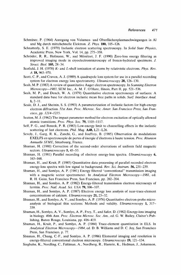
References 471
Schmiiser, P. (1964) Anregung von Volumen- und Oberfliichenplasmaschwingungen in Al und Mg durch mittelschnelle Elektron. Z. Phys. 180,105-126.
Schnatterly, S. E. (1979) Inelastic electron scattering spectroscopy. In Solid State Physics, Academic Press, New York, Vol. 14, pp. 275-358.
SchrOder, R. R., Hofmann, W., and Menetret, J. F. (1990) Zero-loss energy filtering as improved imaging mode in cryoelectronmicroscopy of frozen-hydrated specimens. 1. Struct. Bioi. 105, 28-34.
Scofield, J. H. (1978) K- and L-shell ionization of atoms by relativistic electrons. Phys. Rev. A 18, 963-970.
Scott, C. P., and Craven, A. J. (1989) A quadrupole lens system for use in a parallel recording system for electron energy loss spectrometry. Ultramicroscopy 28,126-130.
Seah, M. P. (1983) A review of quantitative Auger electron spectroscopy. In Scanning Electron Microscopy-1983, SEM Inc., A. M. F. O'Hare, Ilinois, Part II, pp. 521-536.
Seah, M. P., and Dench, W. A. (1979) Quantitative electron spectroscopy of surfaces: A standard data base for electron inelastic mean free paths in solids. Surf Interface Anal. 1,2-11.
Seale, D. J., and Sheinin, S. S. (1993) A parameterization of inelastic factors for high-energy electron diffraction. 51st Ann. Proc. Microsc. Soc. Amer. San Francisco Press, San Francisco, pp. 1214-1215.
Seaton, M. J. (1962) The impact parameter method for electron excitation of optically allowed atomic transitions. Proc. Phys. Soc. 79,1105-1117.
Self, P. G., and Buseck, P. R. (1983) Low-energy limit to channelling effects in the inelastic scattering of fast electrons. Phil. Mag. A48, L21-L26.
Sevely, J., Garg, R. K., Zanchi, G., and Jouffrey, B. (1985) Observation de modulations EXELFS en spectroscopie de pertes d'energie d'electrons a haute tension. Proc. Reunion Annuelle SFME, Strasbourg, France.
Shuman, H. (1980) Correction of the second-order aberrations of uniform field magnetic sectors. Ultramicroscopy 5, 45-53.
Shuman, H. (1981) Parallel recording of electron energy-loss spectra. Ultramicroscopy 6, 163-168.
Shuman, H., and Kruit, P. (1985) Quantitative data processing of parallel recorded electron energy-loss spectra with low signal to background. Rev. Sci. Instrum. 56,231-239.
Shuman, H., and Somlyo, A. P. (1981) Energy filtered "conventional" transmission imaging with a magnetic sector spectrometer. In Analytical Electron Microscopy-1981, ed. R. H. Geiss, San Francisco Press, San Francisco, pp. 202-204.
Shuman, H., and Somlyo, A. P. (1982) Energy-filtered transmission electron microscopy of ferritin. Proc. Natl. A cad. Sci. USA 79, 106-107.
Shuman, H., and Somlyo, A. P. (1987) Electron energy loss analysis of near-trace-element concentrations of calcium. Ultramicroscopy 21, 23-32.
Shuman, H., and Somlyo, A. V., and Somlyo, A. P. (1976) Quantitative electron-probe microanalysis of biological thin sections: Methods and validity. Ultramicroscopy 1, 317-339.
Shuman, H., Somlyo, A. V., Somlyo, A. P., Frey, T., and Safer, D. (1982) Energy-loss imaging in biology. 40th Ann. Proc. Electron Microsc. Soc. Am., ed. G. W. Bailey, Claitor's Publishing, Baton Rouge, Louisiana, pp. 416-419.
Shuman, H., Kruit, P., and Somlyo, A. P. (1984) Trace-element quantitation in ELS. In Analytical Electron Microscopy-1984, ed. D. B. Williams and D. C. Joy, San Francisco Press, San Francisco, p. 77.
Shuman, H., Chang, C.-F., and Somlyo, A. P. (1986) Elemental imaging and resolution in energy-filtered conventional electron microscopy. Ultramicroscopy 19, 121-134.
Siegbahn, K., Nordling, c., Fahlman, A., Nordberg, R., Hamrin, K., Hedman, J., Johansson,
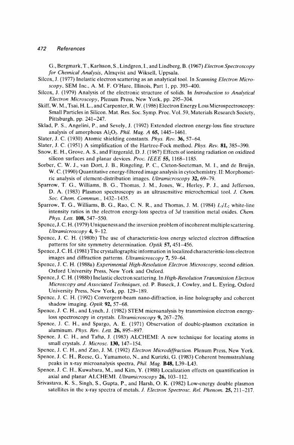
472 References
G., Bergmark, T., Karlsson, S., Lindgren, I., and Lindberg, B. (1967) Electron Spectroscopy for Chemical Analysis, Almqvist and Wiksell, Uppsala.
Silcox, J. (1977) Inelastic electron scattering as an analytical tool. In Scanning Electron Microscopy, SEM Inc., A. M. F. O'Hare, Illinois, Part 1, pp. 393-400.
Silcox, J. (1979) Analysis of the electronic structure of solids. In Introduction to Analytical Electron Microscopy, Plenum Press, New York, pp. 295-304.
Skiff, W. M., Tsai, H. L., and Carpenter, R. W. (1986) Electron Energy Loss Microspectroscopy: Small Particles in Silicon. Mat. Res. Soc. Symp. Proc. Vol. 59, Materials Research Society, Pittsburgh, pp. 241-247.
Sklad, P. S., Angelini, P., and Sevely, J. (1992) Extended electron energy-loss fine structure analysis of amorphous Al,03' Phil. Mag. A 65, 1445-1461.
Slater, J. C. (1930) Atomic shielding constants. Phys. Rev. 36,57-64. Slater, J. C. (1951) A simplification of the Hartree-Fock method. Phys. Rev. 81,385-390. Snow, E. H., Grove, A. S., and Fitzgerald, D. J. (1967) Effects of ionizing radiation on oxidized
silicon surfaces and planar devices. Proc. IEEE 55,1168-1185. Sorber, C. W. J., van Dort, J. B., Ringeling, P. c., Cleton-Soeteman, M. 1., and de Bruijn,
W. C. (1990) Quantitative energy-filtered image analysis in cytochemistry. II: Morphometric analysis of element-distribution images. Ultramicroscopy 32, 69-79.
Sparrow, T. G., Williams, B. G., Thomas, J. M., Jones, W., Herley, P. J., and Jefferson, D. A. (1983) Plasmon spectroscopy as an ultrasensitive microchemical tool. 1. Chern. Soc. Chern. Commun., 1432-1435.
Sparrow, T. G., Williams, B. G., Rao, C. N. R., and Thomas, J. M. (1984) L-JL, white-line intensity ratios in the electron energy-loss spectra of 3d transition metal oxides. Chern. Phys. Lett. 108,547-550.
Spence, J. C. H. (1979) Uniqueness and the inversion problem of incoherent multiple scattering. Ultramicroscopy 4, 9-12.
Spence, J. C. H. (1980b) The use of characteristic-loss energy selected electron diffraction patterns for site symmetry determination. Optik 57, 451-456.
Spence, J. C. H. (1981) The crystallographic information in localized characteristic-loss electron images and diffraction patterns. Ultramicroscopy 7, 59-64.
Spence, J. C. H. (1988a) Experimental High-Resolution Electron Microscopy, second edition. Oxford University Press, New York and Oxford.
Spence, J. C. H. (1988b) Inelastic electron scattering. In High-Resolution Transmission Electron Microscopy and Associated Techniques, ed. P. Buseck, J. Cowley, and L. Eyring, Oxford University Press, New York, pp. 129-189.
Spence, J. C. H. (1992) Convergent-beam nano-diffraction, in-line holography and coherent shadow imaging. Optik 92, 57-68.
Spence, J. C. H., and Lynch, J. (1982) STEM microanalysis by transmission electron energyloss spectroscopy in crystals. Ultramicroscopy 9, 267-276.
Spence, J. C. H., and Spargo, A. E. (1971) Observation of double-plasmon excitation in aluminum. Phys. Rev. Lett. 26,895-897.
Spence, J. C. H., and TafW, J. (1983) ALCHEMI: A new technique for locating atoms in small crystals. I. Microsc. 130,147-154.
Spence, J. C. H., and Zuo, J. M. (1992) Electron Microdiffraction. Plenum Press, New York. Spence, J. C. H., Reese, G., Yamamoto, N., and Kurizki, G. (1983) Coherent bremsstrahlung
peaks in x-ray microanalysis spectra, Phil. Mag. B48, L39-L43. Spence, J. C. H., Kuwabara, M., and Kim, Y. (1988) Localization effects on quantification in
axial and planar ALCHEM1. Ultramicroscopy 26, 103-112. Srivastava, K. S., Singh, S., Gupta, P., and Harsh, O. K. (1982) Low-energy double plasmon
satellites in the x-ray spectra of metals. I. Electron Spectrosc. ReI. Phenom. 25,211-217.
References 473
Steeds, J. W. (1984) Electron crystallography. In Quantitative Electron Microscopy, ed. J. N. Chapman and A J. Craven, SUSSP Publications, Edinburgh, pp. 49-96.
Steele, J. D., Titchmarsh, J. M., Chapman, J. N., and Paterson, J. H. (1985) A single stage process for quantifying electron energy-loss spectra. Ultramicroscopy 17, 273-276.
Stephens, A P. (1980) Quantitative microanalysis by electron energy-loss spectroscopy: Two corrections. Ultramicroscopy 5, 343-350.
Stephens, A P. (1981) Energy-loss spectroscopy in scanning transmission electron microscopy. Ph.D. thesis, University of Cambridge.
Stephens, A P., and Brown, L. M. (1980) Observation by scanning transmission electron microscopy of characteristic electron energy-losses due to hydrogen in transition metals. In Developments in Electron Microscopy and Analysis, Int. Phys. Conf. Ser. No. 52, pp.341-342.
Stephens, A P., and Brown, L. M. (1981) EXELFS in graphitic boron nitride. In Quantitative Microanalysis with High Spatial Resolution, The Metals Society, London, pp. 152-158.
Stern, E. A (1974) Theory of the extended x-ray-absorption fine structure. Phys. Rev. B 10, 3027-3037.
Stern, E. A. (1982) Comparison between electrons and x-rays for structure determination. Optik 61, 45-51.
Stern, E. A, and Ferrell, R. A. (1960) Surface plasma oscillations of a degenerate electron gas. Phys. Rev. 120, 130-136.
Stern, E. A., Bunker, B. A, and Heald, S. M. (1980) Many-body effects on extended x-ray absorption fine structure amplitudes. Phys. Rev. B 21, 5521-5539.
Stobbs, W. M., and Boothroyd, C. B. (1991) Approaches for energy loss and energy filtered imaging in TEM in relation to the materials problems to be solved. Microsc. Microanal. Microstruct. 2, 333-350.
Stobbs, W. M., and Saxton, W. O. (1988) Quantitative high resolution transmission electron microscopy: the need for energy filtering and the advantages of energy-loss imaging. J. Microsc. 151,171-184.
Stohr, J., and Outka, D. A (1987) Near edge x-ray absorption fine-structure studies of molecules and molecular chains bonded to surfaces. J. Vac. Sci. Technol. A 5, 919-926.
Strauss, M. G., Naday, I., Sherman, I. S., and Zaluzec, N. J. (1987) CCD-based parallel detection system for electron energy-loss spectroscopy and imaging. Ultramicroscopy 22,117-124.
Strutt,A J., and Williams, D. B. (1993) Chemical analysis of Cu-Be-Co alloys using quantitative parallel electron-energy-loss spectroscopy. Phil. Mag. A 67, 1007-1020.
Sturm, K. (1982) Electron energy loss in simple metals and semiconductors. Adv. Phys. 31,1-64.
Su, D. S., and Schattschneider, P. (1992a) Numerical aspects of the deconvolution of angleintegrated electron energy-loss spectra. J. Microsc. 167,63-75.
Su, D. S., and Schattschneider, P. (1992b) Deconvolution of angle-resolved electron energyloss spectra. Phil. Mag A 65, 1127-1140.
Su, D. S., Schattschneider, P., and Pongratz, P. (1992) Aperture effects and the multiplescattering problem of fast electrons in electron-energy-loss spectroscopy. Phys. Rev. B 46, 2775-2780.
Su, D. S., Wang, H. F., and Zeitler, E. (1995) The influence of plural scattering on EELS elemental analysis. Ultramicroscopy, 59. 181-190.
Sugar, J. (1972) Potential-barrier effects in photoabsorption. Phys. Rev. B 5, 1785-1793. Sun, S., Shi, S., and Leapman, R. (1993) Water distributions of hydrated biological specimens
by valence electron energy loss spectroscopy. Ultramicroscopy 50, 127-139.
474 References
Sun, S. Q., Shi, S-L., Hunt, 1. A., and Leapman, R. D. (1995) Quantitative water mapping of cryosectioned cells by electron energy loss spectroscopy. J. Microsc. 177,18-30.
Swyt, C. R., and Leapman, R. D. (1982) Plural scattering in electron energy-loss (EELS) microanalysis. In Scanning Electron Microscopyl1982, SEM Inc., A. M. F. O'Hare, Illinois, Part 1, pp. 73-82.
Swyt, C R., and Leapman, R. D. (1984) Removal of plural scattering in EELS: practical considerations. In Microbeam Analysis-1984, ed. A. D. Romig and 1. I. Goldstein, San Francisco Press, San Francisco, pp. 45-48.
Taft, E. A., and Philipp, H. R. (1965) Optical properties of graphite. Phys. Rev. A 138, 197-202. Tafti1l, 1. (1984) Absorption edge fine structure study with subunit cell spatial resolution. Nucl.
Instrum. Methods B2, 733-736. Tafti1l, I., and Krivanek, O. L. (1981) The combined effect of channelling and blocking in
electron energy-loss spectroscopy. 39th Ann. Proc. Electron Microsc. Soc. Am., ed. G. W. Bailey, Claitor's Publishing, Baton Rouge, Louisiana, pp. 190-191.
Tafti1l, I., and Krivanek, O. L. (1982a) Characteristic energy-loss from channeled 100 keV electrons. Nucl. Instrum. Methods 194, 153-158.
Tafti1l, I., and Krivanek, O. L. (1982b) Site-specific valence determination by electron energyloss spectroscopy. Phys. Rev. Lett. 48, 560-563.
Tafti1l, I., and Zhu, 1. (1982) Electron energy-loss near edge structure (ELNES), a potential technique in the studies of local atomic arrangements. Ultramicroscopy 9, 349-354.
Tafti1l, J., Krivanek, O. L., Spence, 1. C H., and Honig, J. M. (1982) Is your spinel normal or inverse? In Electron Microscopy-1982, 10th Int. Cong., Deutsche Gesellschaft flir Elektronenmikroskopie, Vol. I, pp. 615-616.
Takeda, S., Terauchi, M., Tanaka, M., and Kohyama, M. (1994) Line defect configuration incorporated with self-interstitials in Si: a combined study by HRTEM, EELS and electronic calculation. In Electron Microscopy 1994, Proc. 13th Int. Congo Electron Microsc., Paris, vol. 3, pp. 567-568.
Tang, T. T. (1982a) Design of an electron spectrometer for scanning transmission electron microscope (STEM). In Scanning Electron Microscopy, SEM In., A. M. F. O'Hare, Illinois, Part 1, pp. 39-50.
Tang, T. T. (1982b) Correction of aberrations in a magnetic spectrometer by electric multipole lenses. Ultramicroscopy 7, 305-309.
Tatlock, G.l., Baxter,A. G., Devenish, R. W., and Hurd, T.l. (1984) EELS analysis of extracted particles from steels. In Analytical Electron Microscopy-1984, ed. D. B. WIlliams and D. C Joy, San Francisco Press, San Francisco, pp. 227-230.
Tence, M., Colliex, C., Jeanguillaume, C, and Trebbia, P. (1984) Digital spectrum and image processing for EELS elemental analysis with a STEM. In Analytical Electron Microscopy-1984, ed. D. B. Williams and D. C loy, San Francisco Press, San Francisco, pp.21-23.
Tence, M., Quartuccio, M., and Colliex, C. (1995) PEELS compositional profiling and mapping at nanometer spatial resolution. Ultramicroscopy 58,42-54.
Teo, B.-K., and Lee, P. A. (1979) Ab initio calculations of amplitude and phase functions for extended x-ray absorption fine structure spectroscopy. J. Am. Chem. Soc. 101, 2815-2832.
Teo, B.-K., Lee, P. A., Simons, A. L., Eisenberger, P., and Kincaid, B. M. (1977) EXAFS: Approximation, parameterization and chemical transferability of amplitude functions. J. Am. Chem. Soc. 99,3854-3856.
Terauchi, M., Kuzuo, R., Tanaka, M., Tsuno, K., Saito, Y., and Shinohara, H. (1994) High resolution electron energy-loss study of solid C"h C70, Cs. and carbon nanotubes. Electron Microscopy 1994, Proc. 13th Int. Congo Electron Microsc., Paris, vol. 2a, pp. 333-334.
References 475
Thomas, G. J. (1981) Study of hydrogen and helium in metals by electron energy-loss spectroscopy. In Analytical Electron Microscopy-1981, ed. R. H. Geiss, San Francisco Press, San Francisco, pp. 195-197.
Thomas, L. E. (1982) High spatial resolution in STEM x-ray microanalysis. Ultramicroscopy 9,311-318.
Thomas, L. E. (1984) Microanalysis of light elements by simultaneous x-ray and electron spectrometry. In Analytical Electron Microscopy-1984, ed. D. B. Williams and D. C. Joy, San Francisco Press, San Francisco, pp. 358-362.
Timsit, R. S., Hutchinson, J. L., and Thornton, M. C. (1984) Preparation of metal specimens for HREM by ultramicrotomy. Ultramicroscopy 15, 371-374.
Titchmarsh, J. M. (1989) Comparison of high spatial resolution in EDX and EELS analysis. Ultramicroscopy 28, 347-351.
Titchmarsh, J. M., and Malis, T. (1989) On the effect of objective lens chromatic aberration on quantitative electron-energy-loss spectroscopy (EELS). Ultramicroscopy 28,277-282.
Treacy, M. M. J., Howie, A., and Wilson, C. J. (1978) Z contrast of platinum and palladium catalysts. Phil. Mag. A 38, 569-585.
Trebbia, P. (1988) Unbiased method for signal estimation in electron energy loss spectroscopy, concentration measurements and detection limits in quantitative microanalysis: methods and programs. Ultramicroscopy 24, 399-408.
Tremblay, S., and L'Esperance (1994) Volume fraction determination of secondary phase particles in aluminum thin foils with plasmon energy shift imaging. In Electron Microscopy 1994, Proc. ICEM-13, Paris, vol. 1, pp. 627-628.
Tucker, D. S., Jenkins, E. J., and Hren, J. J. (1985) Sectioning spherical aluminum oxide particles for transmission electron microscopy. J. Electron Microscope Tech. 2,29-33.
Tull, R. G. (1968) A comparison of photon counting and current measuring techniques in spectrophotometry of faint sources. Appl. Opt. 7, 2023-2029.
Tung, C. J., and Ritchie, R. (1977) Electron slowing-down spectra in aluminum metal. Phys. Rev. 16,4302-4313.
Turner, P. S., Bullough, T. J., Devenish, R. W., Maher, D. M., and Humphreys, C. J. (1990) Nanometer hole formation in MgO using electron beams. Phil. Mag. Letters 61, 181-193.
Turowski, M. A., and Kelly, T. F. (1992) Profiling of the dielectric function across AIISi02/
Si heterostructures with electron energy loss spectroscopy. Ultramicroscopy 41, 41-54. Tzou, Y., Bruley, J., Ernst, F., Ruhle, M., and Raj, R. (1994) TEM study of the structure and
chemistry of diamond/silicon interface. J. Mater. Res. 9, 1566-1572. Ueda, Y., and Saldin, D. K. (1992) Dipole approximation in electron-energy-loss spectroscopy:
M-shell excitations. Phys. Rev. B 46, 13697-13701. Ugarte, D., Colliex, c., and Trebbia, P. (1992) Surface- and interface-plasmon modes on small
semiconducting spheres. Phys. Rev. B 45, 4332-4343. Uhrich, M. L. (1969) Fast Fourier transforms without sorting. IEEE Trans. Audio Electro
acoust. AU-17, 170-172. Vasudevan, S., Rayment, T., Williams, B. G., and Holt, R. (1984) The electronic structure of
graphite from Compton profile measurements. Proc. R. Soc. London A391, 109-124. Veigele, W. J. (1973) Photon cross sections from 0.1 keY to 1 MeV for elements Z = 1 to
Z = 94. At. Data Tables 5, 51-111. Vogt, S. S., Tull, R. G., and Kelton, P. (1978) Self-scanned photodiode array: high performance
operation in high dispersion astronomical spectrophotometry. Appl. Opt. 17, 574-592. Vvedensky, D. D., Saldin, D. K., and Pendry, J. B. (1986) An update of DLXANES, the
calculation of x-ray absorption near-edge structure. Comput. Phys. Commun. 40,421-440. Waddington, W. G., Rez, P., Grant, I. P., and Humphreys, C. J. (1986) White lines in the L2~
electron-energy-loss and x-ray absorption spectra of the 3d transition metals. Phys. Rev. B 34, 1467-1473.
476 References
Wagner, H.-J. (1990) Contrast tuning by electron spectroscopic imaging of half-micrometerthick sections of nervous tissue. Ultramicroscopy 32, 42-47.
Walske, M. C. (1956) Stopping power of L-electrons. Phys. Rev. 101,940-944. Walther, J. P., and Cohen, M. L. (1972) Frequency- and wave-vector-dependent dielectric
function for Si. Phys. Rev. B 5, 3101-3110. Wang, Y-Y., Ho, R, Shao, Z., and Somlyo, A. P. (1992) Optimization of quantitative electron
energy-loss spectroscopy in the low loss region: phosphorus L-edge. Ultramicroscopy 41,11-31.
Wang, Y. Y., Zhang, H., Dravid, V. P., Shi, D., Hinks, D. G., Zheng, Y., and Jorgensen, J. D. (1993) Evolution of the low-energy excitations and dielectric function of Bat., K,BiOJ• Phys. Rev. B 47,14503-14507.
Wang, Y. Y., Zhang, H., and Dravid, V. P. (1995a) Transmission EELS of oxide superconductors with a cold field emission TEM. Microsc. Res. Technique 30, 208-217.
Wang, Y. Y., Cheng, S. c., Dravid, V. P., and Zhang, F. C. (1995b) Symmetry of electronic structure of BaTiOJ via momentum-transfer resolved electron energy loss spectroscopy. Ultramicroscopy 59, 109-119.
Wang, Z. L. (1993) Electron reflection, diffraction and imaging of bulk crystal surfaces in TEM and STEM. Rep. Prog. Phys. 56,997-1065.
Wang, Z. L. (1995) Elastic and Inelastic Scattering in Electron Diffraction and Imaging. Plenum Press, New York.
Wang, Z. L. (1996) Reflection Electron Microscopy and Spectroscopy for Surface Analysis. Cambridge University Press, U.K.
Wang, Z. L., and Bentley, J. (1992) Reflection energy-loss spectroscopy and imaging for surface studies in transmission electron microscopes. Microsc. Res. Technique 20, 390-405.
Wang, Z. L., and Cowley,J. M. (1987) Surface plasmon excitation for supported metal particles. Ultramicroscopy 21, 77-94.
Wang, Z. L., and Cowley, J. M. (1994) Electron channelling effects at high incident angles in convergent beam reflection diffraction. Ultramicroscopy 55, 228-240.
Wang, Z. L., and Egerton, R F. (1988) Absolute determination of surface atomic concentration by reflection electron energy-loss spectroscopy (REELS). Surf Sci. 205, 25-37.
Wang, Z. L., Colliex, c., Paul-Boncour, V., Percheron-Guegan, A., Archard, J. c., and Barrault, J. (1987) Electron microscopy characterization of lanthanum-cobalt intermetallic catalysts. J. Cataly. 105,120-143.
Wehenkel, C. (1975) Mise au point d'une nouvelle methode d'analyse quantitative des spectres depertes d'energie d'electrons diffuses dans la direction du faisceau incident: Application a l'etude des metaux nobles. J. Phys. (Paris) 36, 199-213.
Weiss, J. K., and Carpenter, R W. (1992) Factors limiting the spatial resolution and sensitivity of EELS microanalysis in a STEM. Ultramicroscopy 40, 339-351.
Weng, X., and Rez, P. (1988) Solid state effects on core electron cross-sections used in microanalysis. Ultramicroscopy 25, 345-348.
Weng, X., Rez, P., and Sankey, O. F. (1989) Pseudo-atomic-orbital band theory applied to electron-energy-loss near-edge structures. Phys. Rev. B 40, 5694-5704.
Wheatley, D. I., Howie, A., and McMullan, D. (1984) Surface microanalysis of Agla-Ah03 catalysts by STEM. In Developments in Electron Microscopy and Analysis 1983, ed. P. Doig, Inst. Phys. Conf. Ser. No. 68, LO.P., Bristol, pp. 245-248.
Whelan, M. J. (1976) On the energy-loss spectrum of fast electrons after plural inelastic scattering. J. Phys. C (Solid State Phys.) 9, L195-L197.
Whitlock, R R, and Sprague, J. A. (1982) TEM imaging and EELS measurement of massthickness variations in thick foils. 40th Ann. Proc. Electron Microsc. Soc. Am., ed. G. W. Bailey, Claitor's Publishing, Baton Rouge, Louisiana, pp. 504-505.
References 477
Wiggins, J. W. (1978) The use of scintillation detectors in the STEM. In Electron Microscopy-1978, 9th Int. Cong., ed. J. M. Sturgess, Microscopical Society of Canada, Toronto, Vol. 1, pp. 78-79.
Wilhelm, P., and Hofer, F. (1992) EELS-microanalysis of the elements Ca to Cu using Mw edges. In: Electron Microscopy, Proc. EUREM 92, Granada, Spain, vol. 1, pp. 281-282.
Williams, B. G., and Bourdillon, A. J. (1982) Localised Compton scattering using energy-loss spectroscopy. J. Phys. C (Solid-State Phys.) 15,6881-6890.
Williams, B. G., Parkinson, G. M., Eckhardt, C. J., and Thomas, J. M. (1981) A new approach to the measurement of the momentum densities in solids using an electron microscope. Chern. Phys. Lett. 78, 434-438.
Williams, B. G., Sparrow, T. G., and Thomas, J. M. (1983) Probing the structure of an amorphous solid: Proof from Compton scattering measurements that amorphous carbon is predominantly graphitic. J. Chern. Soc. Chern. Commun. 1434-1435.
Williams, B. G., Sparrow, T. G., and Egerton, R. F. (1984) Electron Compton scattering from solids. Proc. R. Soc. London A393, 409-422.
Williams, D. B. (1987) Practical Analytical Electron Microscopy in Materials Science, revised edition. Techbooks, Herndon, Virginia.
Williams, D. B., and Edington, J. W. (1976) High resolution microanalysis in materials science using electron energy-loss measurements. J. Microsc. 108, 113-145.
Williams, D. B., and Hunt, J. A. (1992) Applications of electron energy loss spectrum imaging. In Electron Microscopy 1992, Proc. EUREM 92, Granada, vol. 1, pp. 243-247.
Willis, R. F., ed. (1980) Vibrational Spectroscopy of Adsorbates. Springer Series in Chemical Physics, Springer-Verlag, New York, Vol. 15.
Wilson, C. J., Batson, P. E., Craven, A. J., and Brown, L. M. (1977) Differentiated energyloss spectroscopy in S.T.E.M. In Developments in Electron Microscopy and Analysis, Inst. Phys. Conf. Ser. No. 36, LO.P., pp. 365-368.
Wittry, D. B. (1969) An electron spectrometer for use with the transmission electron microscope. Brit. J. Appl. Phys. (1. Phys. D) 2,1757-1766.
Wittry, D. B. (1976) Use of coincidence techniques to improve the detection limits of electron spectroscopy in STEM. Ultramicroscopy 1, 297-300.
Wittry, D. B. (1980) Spectroscopy in microscopy and microanalysis: The search for an ultimate analytical technique. In Electron Microscopy-1980, 7th European Congress Foundation, The Hague, Vol. 3, pp. 14-21.
Wittry, D. B., Ferrier, R. P., and Cosslett, V. E. (1969) Selected-area electron spectrometry in the transmission electron microscope. Brit. J. Appl. Phys. (J. Phys. D) 2, 1767-1773.
Wong, K. (1994) EELS Study of Bulk Nickel Silicides and the NiS,/Si(I11) Interface. Ph.D. Thesis, Cornell, University.
Wong, K., and Egerton, R. F. (1995) Correction for the effects of elastic scattering in coreloss quantification. 1. Microsc. 178, 198-207.
Woo, T., and Carpenter, G. J. C. (1992) EELS characterization of zirconium hydrides. Microsc. Microanal. Microstruct. 3, 35-44.
Xu, P., Loane, R. F., and Silcox, J. (1991) Energy-filtered convergent-beam electron diffraction in STEM. Ultramicroscopy 38, 127-133.
Yamada, K., Sato, K., and Boothroyd, C. B. (1992) Quantification of nitrogen in solution in stainless steels using parallel EELS. Mater. Trans. 33,571-576.
Yamaguchi, T., Shibuya, S., Suga, S., and Shin, S. (1982) Inner-core excitation spectra of transit in-metal compounds. II: p-d absorption spectra. J. Phys. C 15, 2641-2650.
Yang, Y.-Y., and Egerton, R. F. (1992) The influence of lens chromatic aberration on electron energy-loss quantitative measurements. Microsc. Res. Tech. 21,361-367.
478 References
Yang, Y.-Y., and Egerton, R. F. (1995) Tests of two alternative methods for measuring specimen thickness in a transmission electron microscope. Micron 26,1-5.
Yoshida, K., Takaoka, A., and Ura, K. (1991) Channel mixing effect on SN-ratio of electron energy loss spectrum in parallel detector. 1. Electron Microsc. 40,319-324.
Yuan, J., Brown, L. M., and Liang, W. Y. (1988) Electron energy-loss spectroscopy of the high-temperature superconductor Ba2YCu,07.,. J. Phys. C 21,517-526.
Yuan, J., Brown L. M., Liang, W. Y., Liu, R. S., and Edwards, P. P. (1991) Electron-energyloss studies of core edges in Tlo,Pbo,Ca,.,Y,Sr,Cu,078' Phys. Rev. B 43, 8030-8037.
Yuan, J., Saeed, A., Brown, L. M., and Gaskell, P. H. (1992) The structure of highly tetrahedral amorphous diamond-like carbon. III: Study of inhomogeneity by high-resolution inelastic scanning transmission electron microscopy. Phil. Mag. B 66, 187-197.
Zabala, N., and Rivacoba, A. (1991) Support effects on the surface plasmon modes of small particles. Ultramicroscopy 35,145-150.
Zaluzec, N. J. (1980a) Materials science applications of analytical electron microscopy. 38th Ann. Proc. Electron Microsc. Soc. Am., ed. G. W. Bailey, Claitor's Publishing, Baton Rouge, Louisiana, pp. 98-101.
Zaluzec, N. J. (1980b) The influence of specimen thickness in quantitative electron energyloss spectroscopy. 38th Ann. Proc. Electron Microsc. Soc. Am., ed. G. W. Bailey, Claitor's Publishing, Baton Rouge, Louisiana, pp. 112-113.
Zaluzec, N. J. (1981) A reference library of electron energy-loss spectra. In Analytical Electron Microscopy-1981, ed. R. H. Geiss, San Francisco Press, San Francisco, pp. 193-194. Updated version available (free) from the author at: Materials Science Division, Argonne National Laboratory, Illinois 60439.
Zaluzec, N. J. (1982) An electron energy loss spectral library. Ultramicroscopy 9, 319-324. Zaluzec, N. J. (1983) The influence of specimen thickness in quantitative energy-loss spectro
scopy: (II). 41st Ann. Proc. Electron Microsc. Soc. Am .. ed. G. W. Bailey, San Francisco Press, San Francisco, pp. 388-389.
Zaluzec, N. J. (1984) K- and L-shell cross sections for x-ray microanalysis in an AEM. In Analytical Electron Microscopy-1984, ed. D. B. Williams, and D. C. Joy, San Francisco Press, San Francisco, pp. 279-284.
Zaluzec, N. J. (1985) Digital filters for application to data analysis in electron energy-loss spectroscopy. Ultramicroscopy 18, 185-190.
Zaluzec, N. J. (1988) A beginner's guide to electron energy loss spectroscopy. EMSA Bull. 16, 58-63, 72-80.
Zaluzec, N. J. (1992) Electron energy loss spectroscopy of advanced materials. In Transmission Electron Energy Loss Spectroscopy in Materials Science, ed. M. M. Disko, C. C. Ahn, and B. Fulz, The Metals Society, Warrendale, Pennsylvania, pp. 241-266.
Zaluzec, N. J., and Strauss. M. G. (1989) Two-dimensional CCD arrays as parallel detectors in electron-energy-loss and x-ray wavelength-dispersive spectroscopy. Ultramicroscopy 28,131-136.
Zaluzec, N. J., Hren, J., and Carpenter, R. W. (1980) The influence of diffracting conditions on quantitative electron energy-loss spectroscopy. 38th Ann. Proc. Electron Microsc. Soc. Am., ed. G. W. Bailey, Claitor's Publishing, Baton Rouge, Louisiana, pp. 114-115.
Zaluzec, N. J., Schober, T., and Westlake, D. G. (1981) Application of EELS to the study of metal-hydrogen systems. 39th Ann. Proc. Electron Microsc. Soc. Am., ed. G. W. Bailey, Claitor's Publishing, Baton Rouge, Louisiana, pp. 194-195.
Zanchi, G., Perez, J.-P., and Sevely, J. (1975) Adaptation of a magnetic filtering device on a one megavolt electron microscope. Optik 43,495-501.
Zanchi, G., Sevely, J., and Jouffrey, B. (1977a) An energy filter for high voltage electron microscopy. 1. Microsc. Spectrosc. Electron. 2,95-104.
References 479
Zanchi, G., Sevely, J., and Jouffrey, B. (1977b) Second-order image aberration of a one megavolt magnetic filter. Optik 48, 173-192.
Zanchi, G., Sevely, J., and Jouffrey, B. (1980) Filtered electron image contrast in amorphous objects: L Elastic scattering. 1. Phys. D (Appl. Phys.) 13, 1589-1604.
Zanchi, G., Kihn, Y., and Sevely, J. (1982) On aberration effects in the chromatic plane of the OMEGA filter. Optik 60, 427-436.
Zanetti, R., Bleloch, A. L., Grimshaw, M. P., and Jones (1994) The effect of grain size on fluorine gas bubble formation oin calcium fluoride during electron-beam irradiation. Philos. Mag. Lett. 69, 285-290.
Zeiss, G. D., Meath, M. J., MacDonald, J. C. F., and Dawson, D. J. (1977) Accurate evaluation of stopping and straggling mean excitation energies for N, 0, H2, N, using dipole oscillator strength distributions. Radiat. Res. 70, 284-303.
Zeitler, E. (1982) Radiation damage in beam-sensitive material. In Development in Electron Microscopy and Analysis 1979, ed. M. J. Goringe, Inst. Phys. Conf. Ser. No. 61, LO.P., Bristol, pp. 1-6.
Zeitler, E. (1992) The photographic emulsion as analog recorder for electrons. Ultramicroscopy 46,405-416.
Zener, C. (1930) Analytic atomic wave functions. Phys. Rev. 36,51-56. Zhao, L., Wang, Y. Y., Ho, R., Shao, Z., Somlyo, A. Y., and Somlyo, A. P. (1993) Thickness
determination of biological thin specimens by multiple-least-squares fitting of the carbon K-edge in the electron energy-loss spectrum. Ultramicroscopy 48, 290-296.
Zhu, Y., Wang, Z. L., and Suenaga, M. (1993) Grain-boundary studies by the coincident-site lattice model and electron-energy-Ioss spectroscopy of the oxygen K edge in YBa2CuJ 0 7." Phil. Mag. A 67, 11-28.
Zubin, J. A., and Wiggins, J. W. (1980) Image accumulation, storage and display system for a scanning transmission electron microscope. Rev. Sci. Instrum. 51,123-131.
Zuo, J. M. (1992) Automated lattice parameter measurement from HOLZ lines and their use for the measurement of oxygen content in YBa,Cu307.8 from nanometer-sized region. Ultramicroscopy 41, 211-223.
Index
Aberration correction, 56-68 Aberration figure, 35, 64--65 Aberrations
of magnetic-prism spectrometer, 56-61 of pre spectrometer lenses, 71-80
Absorption effect in EXAFS theory, 240 in TEM images, 140
ALCHEMI method, 140,357 Alkali halides, 169, 171,432 Alignment figure, 66-68,116 Alignment of a spectrometer, 63--68 Aloof excitation, 183 Alpha filter, 40 Angular distribution
of core-loss background, 195 of core-loss scattering, 214, 405, 270 of elastic scattering, 135, 137, 144 of inelastic scattering, 144, 428 of multiple scattering, 163 of phonon scattering, 142 of plasmon scattering, 162 of plural scattering, 188--192,428
Anisotropic crystals, 259, 431 Aperture aberrations, 35 Areal density, 280 Artifacts in spectra
serial recording, 82--84 parallel recording, 102, 108-112
Atom-probe analyzer, 18 Auger electron spectroscopy (AES), 20, 96
Background in spectra instrumental, 83,109,311 to ionization edges, 194--200
481
Background subtraction in core-loss spectroscopy, 269-277 in elemental mapping, 122,330 statistics of, 274--277
Backscattering amplitude, 240 Barber's rule, 33 Beam spreading, 346-347, 21 Beam stabilization, 45 Begrenzungs effect, 158, 180 Beryllium analysis, 339 Bethe
asymptotic cross section, 215, 405 ridge, 195,205,404, 160 sum rule, 151,310,261 surface, 204 theory, 147-149,201-215
Biological specimens, 388--391 Bloch waves, 138-141,357-361,363 Boersch effect, 43-44 Born approximation, 132, 136, 147 Boron analysis, 340 Borrmann effect, 140,357 Bragg angle, 138 Bragg's rule, 153 Bremsstrahlung energy losses, 196, 95
Calcium measurement, 344, 389 Carbon analysis, 341, 381 Castaing-Henry filter, 13,37, 116 Centrifugal barrier, 217 Cerenkovloss,172-174,404 Channeling of electrons, 138--141,357-361 Characteristic angle
for elastic scattering, 134 for inelastic scattering, 143, 149,209
482 Index
Characteristic electron dose, 392, 393 Charge-coupled (CCD) arrays, 97,101,122 Chromatic aberration
effect on elemental ratios, 74 effect on spatial resolution, 71, 346 effects on core-loss image, 335 point-spread function, 73, 163
Chemical shift, 225--227,373 Chi-square test, 274 Chromophores, 314 Coincidence measurements, 94 Collection angle
choice of, 336 effective, 286
Collision broadening, 44 Compton profile, 378 Contrast enhancement, 323-327 Contrast tuning, 329 Coordination of atoms, 363-368 Core excitons, 236 Critical (cutoff) angle, 161 Cross section, 9
core-loss, 210--215, 420--428 elastic scattering, 132-137 Lenz model, 134, 143,428 plasmon scattering, 163
Crystal-field splitting, 238, 366 Curve fitting,
for EXELFS data, 297 multiple least squares (MLS), 286 maximum-likelihood,274
Damping of pI as mons, 167,160 Deconvolution techniques
effect of collection aperture, 255, 268 for core-loss edges, 262-268, 410--419 for low-loss region, 245-256, 407-413 matrix method, 255, 407
Debye temperature, 376 Deexcitation, 4 Degeneracy of core levels, 436 Delocalization of scattering, 347, 358 Density of states
conduction-band, 227-233 joint, 167, 228, 315
Detection limits, 354, 345 Detective quantum efficiency (DQE)
of parallel-recording detector, 107 of a serial-recording system, 88-90
Diamond, 381 Diamondlike carbon (DLC), 384
Dielectric theory, 149 Difference spectra, 287, 110,337,314 Diffraction of electrons, 3, 138 Diffraction coupling of spectrometer, 69 Diode arrays, 96, 104 Dipole
approximation, 230, 426 region, 149,204 selection rule, 221, 228
Direct exposure of diode array, 104 Dispersion coefficient, 159,386 Dispersion compensation (matching), 45 Dispersion-plane tilt, 57, 61, 100 Dispersive power (magnetic prism), 51,56 Displacement damage 3, 395 Double-focussing condition, 34, 50, 59 Double plasmon (coherent), 193 Drift-tube scanning, 93, 119 Drude theory, 155, 168 Dynamic range
of electron detector, 108, 104 of energy-loss spectrum, 7, 212
Edge shapes and energies, 433-435 Effective charge, 225,373 Elastic scattering, 2, 131-140 Electron-Compton scattering (ECOSS), 378 Electron counting, 90, 87,104 Electron energy-loss spectrum, 6 Electron-probe microanalyzer (EPMA), 21 Electron spectrometer: see Spectrometer Elemental analysis,
of light elements, 337-344 procedure by EELS, 334, 277
Elemental mapping, 120,330,389 Energy calibration, 436 Energy-dispersive (EDX) analysis, 21, 356 Energy-filtered diffraction, 326, 361,118 Energy-filtered images, 31,36, 323-333 Energy gain of an electron, 181, 252 Energy-loss function, 150, 155 Energy-loss near-edge structure (ELNES),
227-238,363-369 Energy resolution
influence ofTEM lenses, 76 of spectrometer system, 43-48, 64-69 of a STEM system, 78
Energy-selected image, 31, 36, 330 Exchange effects, 135,203 Excitons, 171
core, 236
Extended fine structure (EXELFS), 238 analysis of, 288-300 examples of, 374--378
Extrapolation errors, 274, 286
Fano plot, 215, 406 Fermi golden rule, 227 Fibre optics, 103, 99 Field clamps, 52 Field-ion microscopy, 18 Fingerprint techniques,
for core-loss fine structure, 363 for low-loss region, 314, 391
Fitting to core-loss background, 269 maximum likelihood, 274 mUltiple least squares, 286, 339 ravine search, 274
Fixed-pattern noise, 101, 107, 109 Fluorine analysis, 344, ·389 Focussing by a magnetic prism, 33,49 Form factor, 132, 143, 148 Fourier analysis ofEXAFS data, 288 Fourier filtering of EXAFS data, 298 Fourier-log deconvolution, 246, 262 Fourier-ratio deconvolution, 264 Fourier transform, 247
discrete (DFT), 250 fast (FFT), 251, 413
Fringing fields, 51-53 Fullerenes (buckyballs), 387
Gain averaging, 111,112 Gain normalization, 109 Generalized oscillator strength (GOS), 148
inner-shell, 201-207 Gradient-field spectrometer, 33, 62 Graphite, 171,384,431,436
Hartret}-Fock method, 135,203 Hartree--Slater calculations, 203, 425 Helium detection, 338 History of EELS, 9-16 Hole count, 82 Hole drilling, 396 Hydrogen detection, 337 Hydrogenic model, 201,283,420-425
Icosahedral crystals, 314 Image coupling of a spectrometer, 69, 70 Image-spectrum, 32, 119
Index 483
Inelastic scattering of electrons, 4, 142 Instrumental background, 83, 109 Ionization edges, 6, 215
Jellium model, 155, 161, 166 Jump ratio of an edge, 83,198 Jump-ratio image, 331
Kramers--Kronig analysis, 256, 414,321 Kramers--Kronig sum rule, 258, 307 Kikuchi patterns, 140,362 Kunzl's law, 373
Landau distribution, 192, 330 Laser-microprobe analysis (LAMMA), 19 Lens coupling to spectrometer, 69-79 Lindhard model, 158-160 Linearity of energy-loss axis, 92, 58 Lithium, measurement of, 339, 318 Lithography, 396 Localization of inelastic scattering, 347, 358 Lonsdaleite, 382, 432
Magnetic moment, 372 Magnetic-prism spectrometer, 14,33,48-68 Mass loss during irradiation, 392, 354 Matrix calculations for spectrometer, 54--61 Maximum-entropy deconvolution, 267 Mean free path (MFP), 7
elastic, 137 inelastic, 188, 304 phonon, 142 plasmon, 165, 157
Mean inelastic-scattering angle, 162 Memory effects in detector, 94,108 Micelles, 373 Microanalysis
by EELS, 23-27, 334--344 with incident electrons, 19 with incident ions, 17 with incident photons, 18
Mirror plates, 52 Minimum detectable mass, 354 Minimum mass fraction (MMF), 354 Mixed scattering, 9, 225, 266,191 Modification function, 248, 266 M6llenstedt analyzer, 12 Monochromators, 43-47, 316 Monte Carlo calculations, 190,127 Mott cross section, 136 Multiple least squares (MLS) fitting, 286
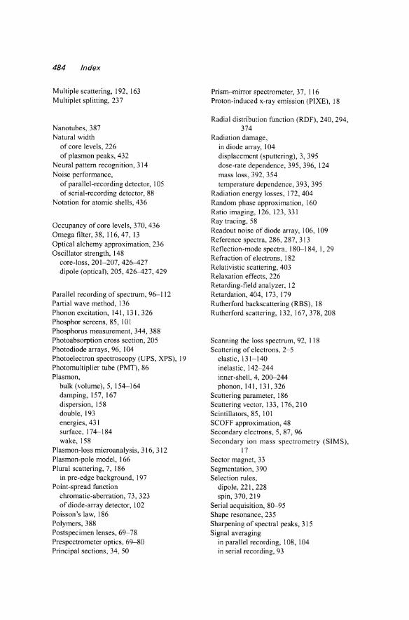
484 Index
Multiple scattering, 192, 163 Multiplet splitting, 237
Nanotubes, 387 Natural width
of core levels, 226 of plasmon peaks, 432
Neural pattern recognition, 314 Noise performance,
of parallel-recording detector, 105 of serial-recording detector, 88
Notation for atomic shells, 436
Occupancy of core levels, 370, 436 Omega filter, 38, 116,47, 13 Optical alchemy approximation, 236 Oscillator strength, 148
core-loss, 201-207, 426-427 dipole (optical), 205,426-427,429
Parallel recording of spectrum, 96--112 Partial wave method, 136 Phonon excitation, 141, 131, 326 Phosphor screens, 85,101 Phosphorus measurement, 344, 388 Photoabsorption cross section, 205 Photodiode arrays, 96, \04 Photoelectron spectroscopy (UPS, XPS), 19 Photomultiplier tube (PMT), 86 Plasmon,
bulk (volume), 5, 154--164 damping, 157, 167 dispersion, 158 double, 193 energies, 431 surface, 174--184 wake, 158
Plasmon-loss microanalysis, 316, 312 Plasmon-pole model, 166 Plural scattering, 7, 186
in pre-edge background, 197 Point-spread function
chromatic-aberration, 73, 323 of diode-array detector, 102
Poisson's law, 186 Polymers, 388 Postspecimen lenses, 69-78 Pre spectrometer optics, 69--80 Principal sections, 34, 50
Prism--mirror spectrometer, 37, 116 Proton-induced x-ray emission (PIXEl, 18
Radial distribution function (RDF), 240, 294, 374
Radiation damage, in diode array, 104 displacement (sputtering), 3, 395 dose-rate dependence, 395, 396, 124 mass loss, 392, 354 temperature dependence, 393, 395
Radiation energy losses, 172, 404 Random phase approximation, 160 Ratio imaging, 126, 123,331 Ray tracing, 58 Readout noise of diode array, 106, 109 Reference spectra, 286, 287, 313 Reflection-mode spectra, 180-184, I, 29 Refraction of electrons, 182 Relativistic scattering, 403 Relaxation effects, 226 Retarding-field analyzer, 12 Retardation, 404, 173, 179 Rutherford backscattering (RBS), 18 Rutherford scattering, 132, 167,378,208
Scanning the loss spectrum, 92, 118 Scattering of electrons, 2-5
elastic, 131-140 inelastic, 142-244 inner-shell, 4, 200-244 phonon, 141, 131,326
Scattering parameter, 186 Scattering vector, 133, 176,210 Scintillators, 85, 101 SCOFF approximation, 48 Secondary electrons, 5,87,96 Secondary ion mass spectrometry (SIMS),
17 Sector magnet, 33 Segmentation, 390 Selection rules,
dipole, 221, 228 spin, 370,219
Serial acquisition, 80-95 Shape resonance, 235 Sharpening of spectral peaks, 315 Signal averaging
in parallel recording, 108, 104 in serial recording, 93
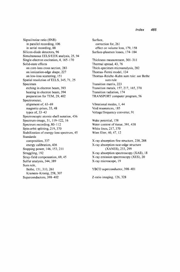
Signal/noise ratio (SNR) in parallel recording, 106 in serial recording, 88
Silicon-diode detectors, 96 Simultaneous EELS/ED X analysis, 25, 94 Single-electron excitation, 4, 165-170 Solid-state effects
on core-loss cross section, 283 on ionization-edge shape, 227 on low-loss scattering, 151
Spatial resolution of EELS, 345,71,25 Specimen
etching in electron beam, 393 heating in electron beam, 394 preparation for TEM, 29, 402
Spectrometer, alignment of, 63--69 magnetic-prism, 33,48 types of, 33--43
Spectroscopic atomic-shell notation, 436 Spectrum-image, 31,119-122,16 Spectrum recording, 80--112 Spin-orbit splitting, 219, 370 Stabilization of energy-loss spectrum, 45 Standards
composition, 337 energy calibration, 436
Stopping power, 146, 153,211 Straggling, 192 Stray-field compensation, 69,45 Sulfur analysis, 344, 389 Sum rule,
Bethe, 151,310,261 Kramers-Kronig, 258, 307
Superconductors, 398--402
Index 485
Surface, correction for, 261 effect on volume loss, 179, 158
Surface-plasmon losses, 174--184
Thickness measurement, 301-311 Thermal spread, 43, 76 Thick-specimen microanalysis, 282 Thomas-Fermi model, 134 Thornas-Reiche-Kuhn sum rule: see Bethe
sum rule Transition matrix, 223 Transition metals, 157,217,165,370 Transition radiation, 174 TRANSPORT computer program, 56
Vibrational modes, I, 44 Void resonances, 185 Voltage/frequency converter, 91
Wake potential, 158 Water content of tissue, 391, 438 White lines, 217, 370 Wien filter, 40, 47,12
X-ray absorption fine structure, 238, 288 X-ray absorption near-edge structure
(XANES), 233, 299 X-ray absorption spectroscopy (XAS), 18 X-ray emission spectroscopy (XES), 20 X-ray microscope, 19
YBCO superconductor, 398--401
Z-ratio imaging, 126,328
Related Documents