1998;115:200-209 J Thorac Cardiovasc Surg Yacoub M. Amrani, N. Latif, K. Morrison, Gray, J. Jayakumar, J. Corbett, Goodwin, Dunn and cardiomyocytes: Implications for myocardial protection Relative induction of heat shock protein in coronary endothelial cells and http://jtcs.ctsnetjournals.org/cgi/content/full/115/1/200 the World Wide Web at: The online version of this article, along with updated information and services, is located on American Association for Thoracic Surgery Association for Thoracic Surgery and the Western Thoracic Surgical Association. Copyright © 1998 is the official publication of the American The Journal of Thoracic and Cardiovascular Surgery on June 3, 2013 jtcs.ctsnetjournals.org Downloaded from

Welcome message from author
This document is posted to help you gain knowledge. Please leave a comment to let me know what you think about it! Share it to your friends and learn new things together.
Transcript
1998;115:200-209 J Thorac Cardiovasc SurgYacoub
M. Amrani, N. Latif, K. Morrison, Gray, J. Jayakumar, J. Corbett, Goodwin, Dunn and cardiomyocytes: Implications for myocardial protection
Relative induction of heat shock protein in coronary endothelial cells and
http://jtcs.ctsnetjournals.org/cgi/content/full/115/1/200the World Wide Web at:
The online version of this article, along with updated information and services, is located on
American Association for Thoracic Surgery Association for Thoracic Surgery and the Western Thoracic Surgical Association. Copyright © 1998
is the official publication of the AmericanThe Journal of Thoracic and Cardiovascular Surgery
on June 3, 2013 jtcs.ctsnetjournals.orgDownloaded from
RELATIVE INDUCTION OF HEAT SHOCK PROTEIN IN CORONARY ENDOTHELIAL CELLS ANDCARDIOMYOCYTES: IMPLICATIONS FOR MYOCARDIAL PROTECTION
M. Amrani, MDN. Latif, PhDK. Morrison, PhDC. C. Gray, MScJ. Jayakumar, MDJ. Corbett, PhDA. T. Goodwin, MDM. J. Dunn, PhDM. H. Yacoub, FRCS
Objectives: Induction of the 70 kd heat shock protein in the heart is knownto exert a protective effect against postischemic mechanical and endothelialdysfunction. However, the exact site of induction and the mechanismsinvolved remain unknown. The aim of this study was to investigate therelative capacity of endothelial and myocardial cells to express the 70 kdheat shock protein in response to heat stress, as well as their significance.Methods: (1) Postischemic recovery of cardiac mechanical and endothelialfunction was studied in isolated rat hearts with and without endothelialdenudation with saponin. (2) Semiquantitative determination of inductionof 70 kd heat shock protein by Western immunoblotting was performed inthe whole cardiac homogenate, in isolated cardiac myocytes, and incoronary endothelial cells. (3) Immunocytochemistry was used to visualizethe distribution of induction of 70 kd heat shock protein in both cell types.Results: Postischemic recovery (percent preischemic value 6 standard errorof the mean) of cardiac output in hearts from heat-stressed animals wassignificantly improved (66.7 6 6.9 vs 44.5 6 4.5 in the control group, p <0.01). In heat-stressed hearts treated with saponin no improvement in therecovery of cardiac output was noted (44.7 6 6.9 in heat-stressed hearts vs38.0 6 4.0 in heat-stressed, saponin-treated hearts, p 5 not significant).Endothelial function (as assessed by the vasodilatory response to theendothelium-dependent vasodilator 5-hydroxytryptamine) improved from31.0 6 5.2 in the control group to 65.8 6 7.1 in heat-stressed hearts (p <0.02 vs control) and dropped to –1.9 6 3.8 in heat-stressed hearts treatedwith saponin. Immunocytochemistry showed that only sections of heartsfrom heat-treated rats showed a strong specific reaction with heat shockprotein antibody. The positive staining was seen in endothelial cells.Induction of 70 kd heat shock protein content in the whole cardiachomogenate from heat-stressed rats as measured by Western immunoblot-ting was 5.2 6 1.9 (vs 0.0 in non-heat–stressed rats, p < 0.0001) anddropped to 0.0 in heat-stressed hearts treated with saponin. The tentativeamount of 70 kd heat shock protein was 18.1 6 7.8 in isolated endothelialcells from heat-stressed hearts and 2.3 6 2.3 in isolated cardiac myocytes(p < 0.01 vs endothelial cells). Conclusions: Coronary endothelial cells arethe main site of induction of 70 kd heat shock protein in the heart andappear to contribute to the protective effects of heat stress on the recoveryof mechanical and endothelial function. (J Thorac Cardiovasc Surg 1998;115:200-9)
From the Department of Cardiothoracic Surgery, Heart ScienceCentre, National Heart and Lung Institute at HarefieldHospital, Harefield, Middlesex, United Kingdom.
Received for publication April 7, 1997; revisions requested May 14,1997; revisions received July 9, 1997; accepted for publicationAugust 13, 1997.
Address for reprints: Professor Sir Magdi Yacoub, FRCS, De-partment of Cardiothoracic Surgery Heart Science Centre,National Heart and Lung Institute at Harefield Hospital,Harefield, Middlesex. UB9 6JH, United Kingdom.
Copyright © 1998 by Mosby, Inc.0022-5223/98 $5.00 1 0 12/1/85361
2 0 0
on June 3, 2013 jtcs.ctsnetjournals.orgDownloaded from
Heat-shock proteins (HSPs) are synthesized inmost cells in response to an increase in temper-
ature or after exposure to a variety of physical orchemical stimuli.1 Most are constitutively expressedin normal, unstressed cells, where they play animportant physiologic role by facilitating severalaspects of protein maturation in the cell, hence theyhave been called “molecular chaperones.”2 Theyalso play a role in cellular homeostasis in a variety ofclinically relevant conditions, including malignancy,immune responses, infection, and ischemia.3
Compelling evidence now exists that induction of70 kd HSP (HSP70), the most abundant and the beststudied HSP, is associated with an enhancement ofcardiac mechanical and endothelial function afterischemia.4 Endothelial function is known to be ofmajor importance in the maintenance of basal cor-onary flow and normal mechanical function,5 al-though the integrity of postischemic coronary endo-thelial cells has become an important issue in thegeneral strategy of myocardial protection.6 The con-tribution of cardiac myocytes and coronary endothe-lial cells in HSP70 expression has not been investi-gated. The aim of this study was to investigate therelative amount of HSP70 induced in these cellularcomponents.
Material and methods
Experimental time course. Five series of experimentswere performed in this study. Series 1: four groups ofhearts (n 5 6 in each group) were studied to evaluate thepreischemic and postischemic (4 hours of hypothermiccardioplegic arrest at 4° C) endothelial and mechanicalfunction in control hearts perfused with Krebs-Henseleitcarbonate buffer (KH) (group 1) or saponin (group 2) andin heat-shocked hearts perfused with KH (group 3) orsaponin (group 4). Series 2: four similar groups (n 5 6 ineach group) were used to evaluate the relative amount ofinduction of 70 kd HSP in the whole homogenate byWestern immunoblotting. Series 3: Hearts from all fourgroups (n 5 6 in each group) were used to assess thepreischemic and postischemic wet weight. Hearts weredisconnected from the Langendorff apparatus, weighed,and reconnected. The time taken for the disconnection,weighing, and reconnecting ranged from 21 to 33 seconds.Series 4: Hearts from heat-shocked (n 5 4) and control(n 5 4) animals were used to evaluate induction of 70 kdHSP (iHSP70) in isolated endothelial cells and myocytes.Series 5: Hearts from heat-shocked (n 5 6) and control(n 5 4) animals were used to visualize iHSP70 in isolatedendothelial and myocardial cells by immunocytochemis-try.
Animals. Sprague-Dawley rats weighing between 300and 330 gm were used in all experiments. Male rats wereused to avoid a potential lack of homogeneity related tohormonal cycle. Six hearts were studied in each group. In
all studies, animals received humane care in compliancewith the “Principles of Laboratory Animal Care” formu-lated by the National Society for Medical Research andthe “Guide for the Care and Use of Laboratory Animals”prepared by the Institute of Laboratory Animal Resourcesand published by the National Institutes of Health (NIHpublication 86-23, revised 1985).
Induction of heat stress. Rats were anesthetized withan intraperitoneal injection of sodium pentobarbitone (50mg/kg), then placed on a temperature-controlled heatingpad (IMS K-Temp control unit; Congleton, Cheshire,United Kingdom) set at 45° C until body temperaturereached 42° C. Body temperature was monitored with arectal temperature probe and maintained between 42° Cand 42.5° C for 15 minutes as previously described.1 Theanimals were left to recover for 24 hours before analysis ofmechanical function or determination of HSP70 content.
Isolated working rat heart preparation. The isolatedworking rat heart preparation, which has been describedin detail elsewhere,7 was used in this study. In this leftheart preparation oxygenated KH (NaCl, 118.5 mmol/L;NaHCO3, 25 mmol/L; KCl, 4.75 mmol/L; MgSO4, 1.19mmol/L; KH2PO4, 1.18 mmol/L; CaCl2, 2.5 mmol/L), pH7.4, containing glucose (11.1 mmol/L) and gassed with95% oxygen and 5% carbon dioxide at 37° C, enters thecannulated left atrium and passes into the left ventricle,from which it is spontaneously ejected through an aorticcannula against a hydrostatic pressure of 100 cm H2O.The heart continues to eject as long as the pressuregenerated in the left ventricle is greater than 100 cm H2O.Total cardiopulmonary bypass with maintained coronaryperfusion can be simulated by clamping the left atrialcannula and introducing perfusion fluid at 37° C into theaorta from a reservoir 100 cm above the heart. With thispreparation, which is essentially that described by Lange-ndorff, the heart will continue to beat but does notperform external work. Several parameters, includingcardiac output (CO)and coronary flow, are measured toassess cardiac function. Ischemic cardiac arrest may beproduced by clamping the aortic cannula. At this time acardioplegic solution is infused into a sidearm of the aorticcannula. During the ischemic period the heart is main-tained under hypothermic conditions (4° C) by a coolingcircuit.
Cardioplegic solution. St. Thomas’ Hospital cardiople-gic solution No. 1, supplied as concentrate (David BullLaboratories, Mulgrave, Victoria, Australia), was dilutedin Ringer’s solution (Travenol Laboratories, Thetford,Norfolk, United Kingdom) and passed through a 0.2 mmfilter (Pall Biomedical, Glen Cove, N.Y.).
Endothelial function. Endothelial function was as-sessed through observations of preischemic and postisch-emic coronary flow responses to 5-hydroxytryptamine(5-HT). This vasodilatory response is endothelium depen-dent. In the intact endothelium, 5-HT causes vasodilata-tion through the release of NO, whereas in the presence ofendothelial damage it causes vasoconstriction by a directeffect on smooth muscle. Our protocol for this test hasbeen described by us in earlier studies.8 We have previ-ously shown that perfusion of the isolated rat heart by5-HT leads to an increased secretion of nitric oxide in thecoronary effluent. Endothelium-independent vasodilata-
The Journal of Thoracic andCardiovascular SurgeryVolume 115, Number 1
Amrani et al. 2 0 1
on June 3, 2013 jtcs.ctsnetjournals.orgDownloaded from
tion, as assessed by the response to glyceryl trinitrate, hasbeen shown to be unchanged by a similar protocol (i.e., 4hours hypothermic [4° C] cardioplegic arrest).8 After ex-cision of the heart and aortic cannulation, Langendorffperfusion was initiated at 37° C. Coronary flow was mon-itored by an in-line electromagnetic flow probe (ECM2 20ml, Scalar, Delft, Holland) proximal to the aortic cannula,connected to its compatible flowmeter (MDL 140 l,Scalar). This provided an accurate (0.0 to 40.0 ml/min)digital readout of mean coronary flow and simultaneoushard-copy recording through a connection with a chartrecorder (series 3000, Gould Electronics, Hainault, Essex,United Kingdom) that allowed accurate monitoring ofsteady state conditions (,0.3 ml/min change in coronaryflow over 3 minutes). The calibration of the flow meter isperformed using the company manual and by collectingcoronary flow in a measuring cylinder over 1 minute. After9 to 13 minutes, the initial baseline coronary flow wasrecorded. The Langendorff infusion was switched to onecontaining additional 1025 mmol/L 5-HT (Sigma Chemi-cal Co., Poole, Dorset, United Kingdom). The ensuingvasodilator response was monitored and when the steadystate had been reached (between 5 and 7 minutes), thecoronary flow was recorded. After this period, 5-HT waswashed out by switching back to ordinary KH until asteady state had been reached (between 5 and 7 minutes).
The heart was then subjected to a 10 ml hypothermic(4° C) infusion with the cardioplegic solution and main-tained immersed in the same solution for 4 hours at 4° C.At the end of the ischemic period, the heart was reper-fused in the Langendorff mode at 37° C for at least 15minutes. When the baseline coronary flow had beenreestablished, the heart was again subjected to the sameprotocol of sequential infusion of 5-HT and KH as in thepreischemic period. The rationale of using the steadybaseline coronary flow has been detailed in our previousstudy.8
Endothelial cell removal with saponin. To assess theimportance of HSP70 induced in endothelial cells, endo-thelial function and mechanical function were assessedand compared in hearts perfused with or without saponin.This potent detergent agent has previously been shown toremove endothelial cells.9 Hearts were perfused withsaponin (30 mg/ml) dissolved in KH solution in threecycles, each consisting of a 2-minute perfusion period thatpreceded the ischemic interval.
Isolation of myocytes. This technique has been de-scribed in detail elsewhere.10 Hearts were perfused for 1minute in the Langendorff mode with Krebs buffer toremove all traces of blood, and then a piece of ventriclewas sliced quickly into pieces of approximately 1 mm3
using razor blades. The pieces of myocardium were incu-bated for a total of 12 minutes at 35° C in 25 ml oflow-calcium medium containing the following composi-tion (in mmol/L): NaCl, 120; KCl, 5.4; MgSO4, 5; pyru-vate, 5; glucose, 20; taurine, 20; HEPES solution, 10;nitrilotriacetic acid, 5; pH, 6.96; containing 1 to 2 mmcalcium. The medium was changed three times during thisincubation and was stirred by bubbling with 100% oxygen.The low-calcium medium was removed by filteringthrough 300 mm gauze. The pieces of myocardium werethen incubated at 35° C for 45 minutes in the same
solution but with nitrilotriacetic acid omitted, and 4 IU/mltype XXIV Protease (Sigma) and 30 mm calcium added,followed by two 45-minute incubations with the proteaseomitted and 400 IU/ml collagenase (BCL, BoehringerMannheim, United Kingdom Ltd.) added. The mediumwas shaken gently throughout the incubation and keptunder 100% oxygen. At the end of each 45-minute periodthe solution containing the dispersed cells was filteredthrough 300 mm gauze and centrifuged at 1000 rpm for 1to 2 minutes. The cells were then washed twice bycentrifugation in KH.10
Isolation of endothelial cells. The method used toisolate endothelial cells has been described in detailelsewhere.11 The fat, epicardium, and endocardium wereremoved from the ventricular tissue and diced up usingrazor blades. An amount of collagenase type II equivalentto twice the volume of tissue was added, and this wasincubated for 1 hour at 37° C with agitation. Hanksmedium with 5% FCS (fetal calf serum) was added andthe digest and spun at 1400 rpm for 10 minutes. Thesupernatant was gently poured off, and the pellet waswashed in 10% bovine serum albumin (BSA) and spun at1000 rpm for 7 minutes. The pellet was again washed inHanks medium with 5% FCS and spun at 1200 rpm for 6minutes. The pellet was then incubated with twice itsvolume of trypsin–ethylenediaminetetraacetic acid (EDTA)(0.25% in 1 mmol/L EDTA) at 37° C for 10 minutes withagitation. After this period, the tissue was washed twice inHanks medium with 5% FCS at 1200 rpm for 6 minutesand filtered through 100 mm gauze.11
Assessment of heat shock protein expression. The in-duction of HSP70 was assessed by sodium dodecyl sulfate(SDS), polyacrylamide gel electrophoresis, and Westernimmunoblotting as previously described.4 Whole hearthomogenate, isolated endothelial cells, and isolated myo-cytes were solubilized in 1% wt/vol SDS and assayed fortotal protein with the Bradford assay, denatured by heat-ing at 100° C in Laemmli buffer, and separated on 10%SDS gels until the bromophenol blue tracking dye reachedthe end of the gel. Gels were equilibrated for 30 minutesin transfer buffer, and transfer of the proteins was per-formed for 1 hour at 500 mA. Western blots were blockedusing 3% wt/vol nonfat dried milk (Marvel) in phosphate-buffered saline solution (PBS) containing 0.05% wt/volTween-20 (PBS-T) for 1 hour to block nonspecific bindingsites. The blots were then probed with mouse antibodiesspecific to inducible HSP70 (Bioquote Ltd, United King-dom) diluted to a final concentration of 1:1000 for 1 hour.Blots were washed three times and incubated with sec-ondary horseradish-peroxidase–conjugated rabbit anti-mouse antibody for 1 hour. The result was visualized usingan enhanced chemiluminescence (ECL) detection system(Amersham). Hyperfilm MP (myoperoxidase) was ex-posed to blots treated with ECL for 30 seconds anddeveloped in an automatic film processor. After ECLexposure, antibodies were removed from blots by incuba-tion in a solution of 2% wt/vol SDS, 6.25% vol/vol 1mmol/L Tris-HCl, pH 6.8, and 0.7% vol/vol 2-mercapto-ethanol. Proteins were then visualized by staining with0.01% Amido Black in a solution of methanol, water, andacetic acid (ratio of 45:45:10 vol/vol). Amido black–stained blots and ECL films were scanned with a Molec-
The Journal of Thoracic andCardiovascular Surgery
January 19982 0 2 Amrani et al.
on June 3, 2013 jtcs.ctsnetjournals.orgDownloaded from
ular Dynamics 300A laser densitometer and HSP70 levelsdetermined as a proportion of total protein loaded using theQuantity One software package (PDI, Huntington, N.Y.).
Immunocytochemistry. Rat hearts were sliced intopieces approximately 5 mm thick and snap frozen in liquidnitrogen. Six hearts from rats subjected to heat shock andfour hearts from control groups were analyzed. For eachheart, four serial frozen sections, each 6 mm thick, werecut onto glass slides, allowed to dry for 30 to 120 minutes,and subsequently fixed for 15 minutes in acetone beforethe immunohistochemical procedure. Sections were incu-bated for 16 hours at 4° C in either monoclonal mouseantibody to the inducible form of heat shock protein 72(iHSP72) (Bioquote Ltd., United Kingdom) at a dilutionof 2 mg/ml or to endothelial cells with the antibodyRECA-1 (Serotec, Oxford, United Kingdom) provided asa supernatant and diluted 1:150. Negative controls in-cluded incubation of sections in antibody diluent only orin an irrelevant IgG1 monoclonal mouse antibody used at2 mg/ml (Prod. no. X0931, Dako Ltd., High Wycombe,United Kingdom). This corresponds to the same immu-noglobulin subclass as the iHSP72 and RECA-1 antibod-ies. All sections were subsequently incubated for 30minutes at room temperature in biotinylated rabbit anti-mouse F(ab)2 at a concentration of 0.002 mg/ml andprepared at least 60 minutes before use in a 5% solutionof rat serum. All antibodies were diluted in 0.005 mmol/LTris-buffered saline solution, pH 7.6. After this, sectionswere immersed for 30 minutes at room temperature instreptavidin biotin-horseradish peroxidase complexes(Dako Ltd.), which had been prepared at least 30 minutesbefore use by diluting the components 1:200 in 0.05mmol/L Tris buffer, pH 7.6.12 Sections were then treated for5 minutes with a diaminobenzidine hydrochloride/hydrogenperoxide substrate, prepared according to the manufactur-er’s instructions (Prod. no. D5905, Sigma, Poole), to visualizethe antigenic sites. Each of the above stages was followed bythree 5-minute washes in 0.005 mmol/L Tris-buffered salinesolution, pH 7.6. The sections were then rinsed in coldrunning tap water, counterstained with Mayer’s hematoxylin,washed again in cold running tap water, and then dehydratedthrough graded alcohols cleared in CNP30 (Merck, Lutter-worth, United Kingdom) and mounted in DPX mountant(BDH, Poole, United Kingdom).
Statistical analysis. Postischemic recoveries of me-chanical function and coronary flows were comparedusing two-way analysis of variance (ANOVA) with Schef-fe’s correction factor. Statistical significance of differencesbetween groups was determined with a nonpaired Stu-dent’s t test, and significance was assumed when the pvalue was 0.05 or less. Values are given as means 6standard error of the mean (SEM). Preischemic andpostischemic wet weights were compared with a paired ttest.
Differences between postischemic wet weights werecompared using ANOVA with Scheffe’s correction. Levelsof HSP70 were compared using a nonpaired Student’s ttest.
Results
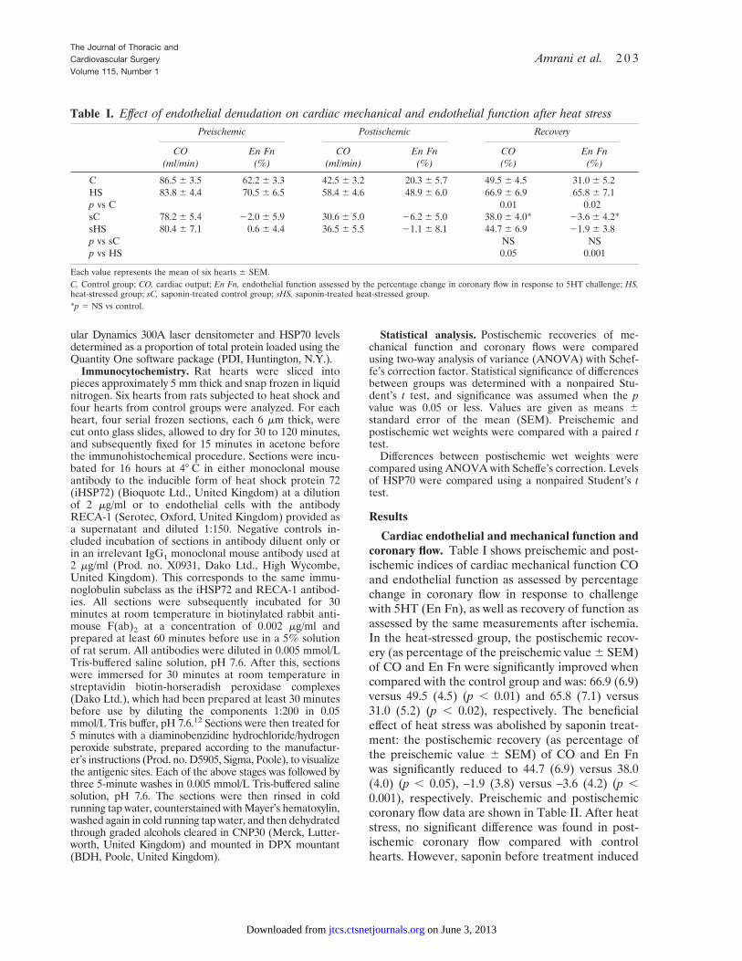
Cardiac endothelial and mechanical function andcoronary flow. Table I shows preischemic and post-ischemic indices of cardiac mechanical function COand endothelial function as assessed by percentagechange in coronary flow in response to challengewith 5HT (En Fn), as well as recovery of function asassessed by the same measurements after ischemia.In the heat-stressed group, the postischemic recov-ery (as percentage of the preischemic value 6 SEM)of CO and En Fn were significantly improved whencompared with the control group and was: 66.9 (6.9)versus 49.5 (4.5) (p , 0.01) and 65.8 (7.1) versus31.0 (5.2) (p , 0.02), respectively. The beneficialeffect of heat stress was abolished by saponin treat-ment: the postischemic recovery (as percentage ofthe preischemic value 6 SEM) of CO and En Fnwas significantly reduced to 44.7 (6.9) versus 38.0(4.0) (p , 0.05), –1.9 (3.8) versus –3.6 (4.2) (p ,0.001), respectively. Preischemic and postischemiccoronary flow data are shown in Table II. After heatstress, no significant difference was found in post-ischemic coronary flow compared with controlhearts. However, saponin before treatment induced
Table I. Effect of endothelial denudation on cardiac mechanical and endothelial function after heat stressPreischemic Postischemic Recovery
CO(ml/min)
En Fn(%)
CO(ml/min)
En Fn(%)
CO(%)
En Fn(%)
C 86.5 6 3.5 62.2 6 3.3 42.5 6 3.2 20.3 6 5.7 49.5 6 4.5 31.0 6 5.2HS 83.8 6 4.4 70.5 6 6.5 58.4 6 4.6 48.9 6 6.0 66.9 6 6.9 65.8 6 7.1p vs C 0.01 0.02sC 78.2 6 5.4 22.0 6 5.9 30.6 6 5.0 26.2 6 5.0 38.0 6 4.0* 23.6 6 4.2*sHS 80.4 6 7.1 0.6 6 4.4 36.5 6 5.5 21.1 6 8.1 44.7 6 6.9 21.9 6 3.8p vs sC NS NSp vs HS 0.05 0.001
Each value represents the mean of six hearts 6 SEM.C, Control group; CO, cardiac output; En Fn, endothelial function assessed by the percentage change in coronary flow in response to 5HT challenge; HS,heat-stressed group; sC, saponin-treated control group; sHS, saponin-treated heat-stressed group.*p 5 NS vs control.
The Journal of Thoracic andCardiovascular SurgeryVolume 115, Number 1
Amrani et al. 2 0 3
on June 3, 2013 jtcs.ctsnetjournals.orgDownloaded from
a significant reduction in postischemic coronary flowcompared with non-saponin–treated hearts, but nodifference was found between heat-stressed andcontrol hearts after saponin treatment.
Wet weight of heat-shocked and control heartssubjected to endothelial denudation. The preisch-emic and postischemic weights are reported in TableIII. In all groups the postischemic weights weresignificantly higher. In contrast, no statistically sig-nificant difference was found in the postischemic wetweights between the groups.
Immunocytochemistry. General background stain-ing in all heart sections was comparable. Onlysections of hearts from heat-treated rats showed anystrong specific reaction with the iHSP70 antibody(Fig. 1). The positive staining was seen in cellssurrounding myocytes rather than in the myocytesthemselves as originally expected. The distributionof iHSP70-positive cells appeared to have a similarpattern to that seen with endothelial cell markers incardiac tissue. Therefore serial sections of heartsfrom rats killed after heat treatment were used todemonstrate both endothelium and iHSP70. Theresults showed that positive iHSP70 cells were alsopositive with the endothelial cell marker (Fig. 2).
Determination of HSP70 by Western blotting inthe whole cardiac homogenate. HSP70 content andcardiac function were measured in the same cardiachomogenates of hearts. Whole cardiac tissue washomogenized, and nuclear and cytoplasmic fractionswere not distinguished. HSP levels were measuredas a fraction of total cell homogenate, which con-tained all soluble protein. Western blotting wasperformed immediately after the experiments toavoid any protein degradation with time. HSP70levels were determined as a proportion of total
protein loaded using the Quantity One softwarepackage.
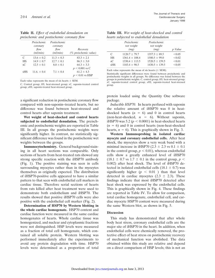
Inducible HSP70. In hearts perfused with saponinthe relative amount of iHSP70 was 0 in heat-shocked hearts (n 5 6) and 0 in control hearts(non-heat-shocked, n 5 6). Without saponin,iHSP70 was 5.2 (p , 0.0001) in heat-shocked hearts(n 5 6) and 0 in control hearts (non-heat-shockedhearts, n 5 6). This is graphically shown in Fig. 3.
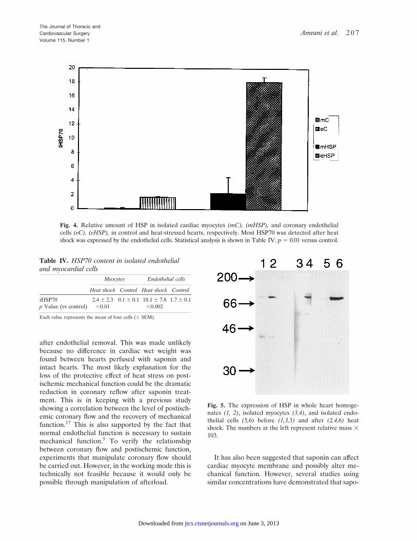
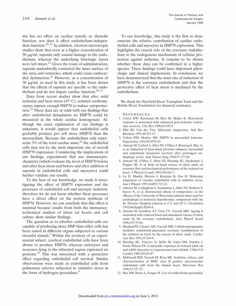
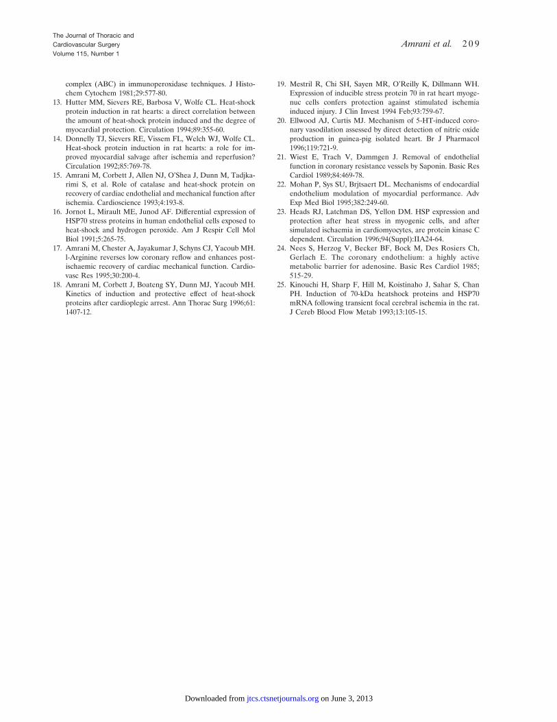
Western Immunoprobing in isolated cardiacmyocyte and coronary endothelial cells. After heatshock, the myocytes show a very weak band with aminimal increase in iHSP70 (2.3 6 2.3 vs 0.1 6 0.1in the control group, p , 0.01), whereas endothelialcells show a greatly enhanced level of iHSP70(18.1 6 0.7 vs 1.7 6 0.1 in the control group, p ,0.002) after heat shock. The level of iHSP70 de-tected in isolated endothelial cells (18.1 6 0.7) wassignificantly higher (p , 0.01 ) than that leveldetected in cardiac myocytes (2.3 6 2.3). Thesefindings indicate that most iHSP70 detected afterheat shock was expressed by the endothelial cells.This is graphically shown in Fig. 4. These findingsare reported in Table IV. To allow for comparison,total cardiac homogenate, endothelial cell, and car-diac myocyte HSP70 content were measured duringthe same Western blot, as shown in Fig. 5.
Discussion
This study has demonstrated that after wholebody heat stress, coronary endothelial cells are themajor site of iHSP70 in the heart. In addition, whenendothelial cells were chemically removed, the pro-tective effect of heat stress on postischemic recoveryof mechanical function was abolished. The dataobtained within this study are relative and dependon a direct comparison of HSP levels; this is not an
Table III. Wet weight of heat-shocked and controlhearts subjected to endothelial denudation
Preischemic Postischemicwet weight
(mg)wet weight
(mg) p Value
C 1120.7 6 79.7 1357.3 6 89.5 ,0.05HS 1361.4 6 108.4 1580.5 6 99.0 ,0.02sC 1330.4 6 113.5 1520.5 6 159.5 ,0.01sHS 1103.4 6 98.5 1430.5 6 139.5 ,0.05
Each value represents the mean of six hearts (6 SEM).Statistically significant differences were found between preischemic andpostischemic weights in all groups. No difference was found between thegroups in postischemic weights. C, control group; HS, heat-stressed group;sC, saponin-treated control group; sHS, saponin-treated heat-stressedgroup.
Table II. Effect of endothelial denudation onpreischemic and postischemic coronary flow
Preischemiccoronary
Postischemiccoronary
flow(ml/min)
flow(ml/min)
Recovery(% preischemic value)
C 13.6 6 0.2 11.3 6 0.3 83.3 6 2.1HS 14.9 6 0.7 12.7 6 0.1 86.3 6 3.0sC 12.5 6 0.1 8.0 6 0.1 64.3 6 3.3
p , 0.001 vs CsHS 11.6. 6 0.4 7.1 6 0.4 61.2 6 4.3
p , 0.01 vs HSP
Each value represents the mean of six hearts 6 SEM.C, Control group; HS, heat-stressed group; sC, saponin-treated controlgroup; sHS, saponin-treated heat-stressed group.
The Journal of Thoracic andCardiovascular Surgery
January 19982 0 4 Amrani et al.
on June 3, 2013 jtcs.ctsnetjournals.orgDownloaded from
attempt to give absolute quantification. When com-pared with myocytes, the induction of HSP70 is seento be predominant in endothelial cells. The very lowbasal level of HSP70 recorded in this study couldhave been due to the sensitivity of the antibody anddoes not affect the overall conclusion, which de-pends on comparison.
Over the past decade, a large number of in vivoand in vitro studies have established the beneficialeffect of HSP in myocardial ischemia in models ofglobal and regional ischemia.13, 14 In a clinicallyrelevant model of myocardial protection for cardiactransplantation, we have previously shown that bothcardiac mechanical and endothelial function wereimproved by heat stress.15 Endothelial function hasbeen shown to influence myocardial performance.12
Although endothelial cells are known to responddifferently to heat stress,16 and despite the fact thatendothelial function has become an important com-ponent of the general strategy in cardiac preserva-tion,17 to date no comparative data exist regardingiHSP in the different cellular components of theheart.
By the use of immunocytochemistry we havedemonstrated that 24 hours after heat stress, whenthe maximal level of cardiac iHSP70 is known to be
reached,18 staining for iHSP70 is only seen in coro-nary endothelial cells. Cardiac myocytes did notstain for iHSP70 in any of the hearts. Whereasisolated endothelial cells showed a high level ofiHSP70, isolated cardiac myocytes demonstrated avery weak concentration of the protein. The discretelocalization of HSP in endothelial cells is unlikely tobe the result of redistribution of the antigen acrossthe section after leakage from the myocyte duringimmunohistochemical analysis. The method andsubsequent detection of iHSP is similar to themethod used by Mestril and colleagues.19 By meansof this process of fixing, no suggestion of leakageregarding the antigen from the cells was made. Inaddition, we have measured and compared therelative amount of iHSP70 in homogenates of wholehearts subjected to chemical denudation of endothe-lial cells by saponin. No trace of iHSP70 could bedetected after this treatment.
To investigate the significance of these findingswe have assessed the effect of endothelial denuda-tion by saponin on the recovery of mechanical andendothelial function in control and heat-stressedhearts. Heat-stressed hearts displayed a better post-ischemic recovery of CO and endothelial function asassessed by the endothelium-dependent vasodila-
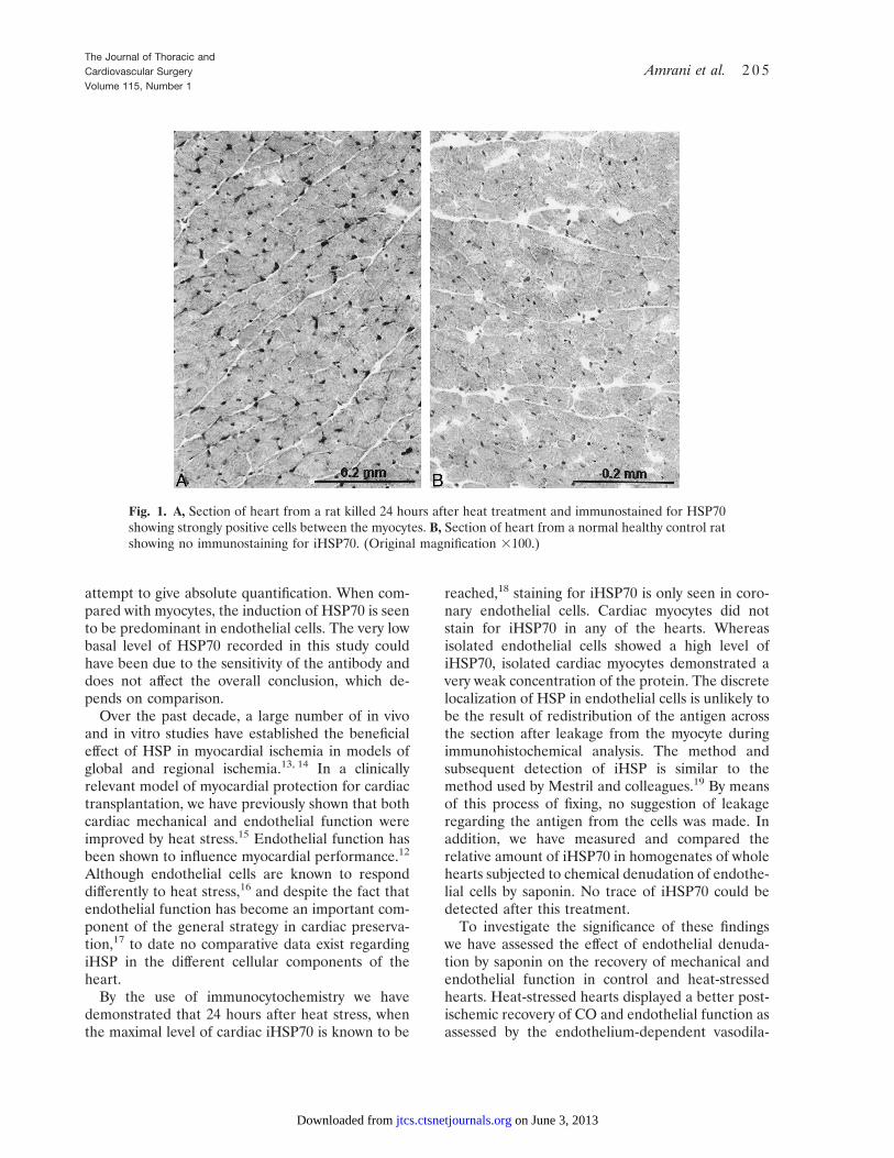
Fig. 1. A, Section of heart from a rat killed 24 hours after heat treatment and immunostained for HSP70showing strongly positive cells between the myocytes. B, Section of heart from a normal healthy control ratshowing no immunostaining for iHSP70. (Original magnification 3100.)
The Journal of Thoracic andCardiovascular SurgeryVolume 115, Number 1
Amrani et al. 2 0 5
on June 3, 2013 jtcs.ctsnetjournals.orgDownloaded from
tory effect of 5-HT. Not surprisingly, pretreatmentwith saponin suppressed this beneficial effect onendothelial function. However, we have demon-strated that the protective effect of heat stress on
postischemic mechanical function was lost in heat-stressed hearts whose endothelium was removed. Itcould be argued that the latter findings might berelated to the exacerbation of postischemic edema
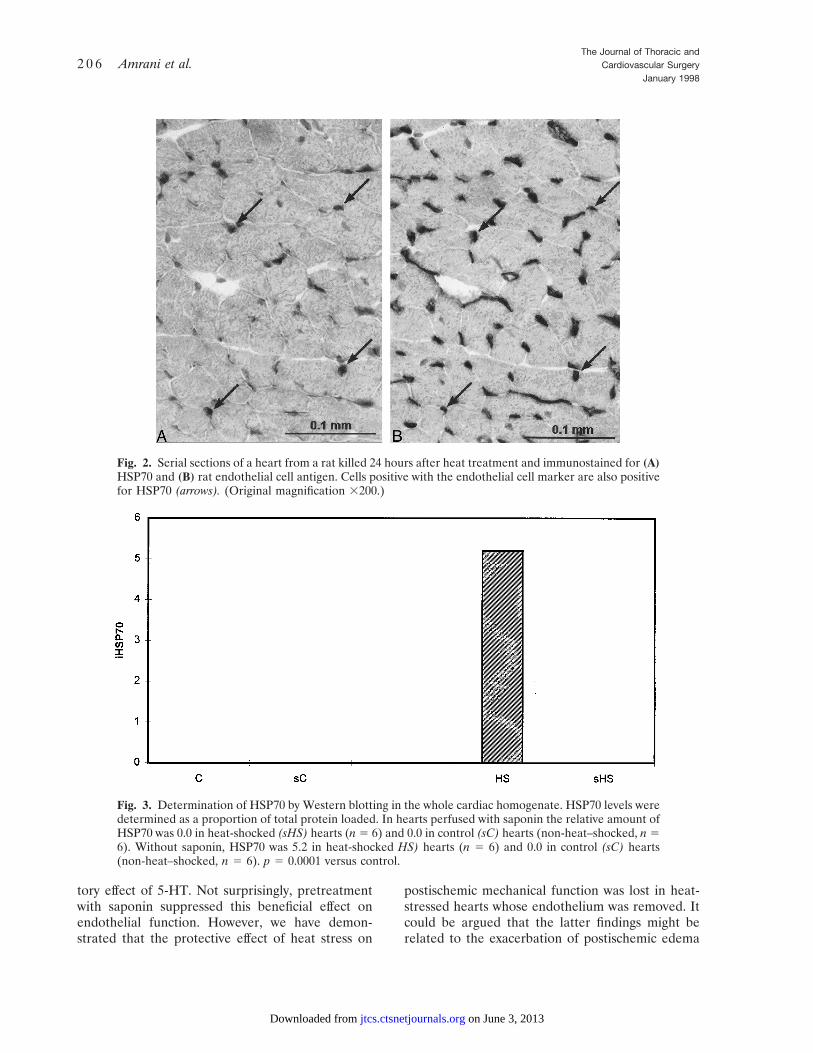
Fig. 2. Serial sections of a heart from a rat killed 24 hours after heat treatment and immunostained for (A)HSP70 and (B) rat endothelial cell antigen. Cells positive with the endothelial cell marker are also positivefor HSP70 (arrows). (Original magnification 3200.)
Fig. 3. Determination of HSP70 by Western blotting in the whole cardiac homogenate. HSP70 levels weredetermined as a proportion of total protein loaded. In hearts perfused with saponin the relative amount ofHSP70 was 0.0 in heat-shocked (sHS) hearts (n 5 6) and 0.0 in control (sC) hearts (non-heat–shocked, n 56). Without saponin, HSP70 was 5.2 in heat-shocked HS) hearts (n 5 6) and 0.0 in control (sC) hearts(non-heat–shocked, n 5 6). p 5 0.0001 versus control.
The Journal of Thoracic andCardiovascular Surgery
January 19982 0 6 Amrani et al.
on June 3, 2013 jtcs.ctsnetjournals.orgDownloaded from
after endothelial removal. This was made unlikelybecause no difference in cardiac wet weight wasfound between hearts perfused with saponin andintact hearts. The most likely explanation for theloss of the protective effect of heat stress on post-ischemic mechanical function could be the dramaticreduction in coronary reflow after saponin treat-ment. This is in keeping with a previous studyshowing a correlation between the level of postisch-emic coronary flow and the recovery of mechanicalfunction.17 This is also supported by the fact thatnormal endothelial function is necessary to sustainmechanical function.5 To verify the relationshipbetween coronary flow and postischemic function,experiments that manipulate coronary flow shouldbe carried out. However, in the working mode this istechnically not feasible because it would only bepossible through manipulation of afterload.
It has also been suggested that saponin can affectcardiac myocyte membrane and possibly alter me-chanical function. However, several studies usingsimilar concentrations have demonstrated that sapo-
Fig. 4. Relative amount of HSP in isolated cardiac myocytes (mC), (mHSP), and coronary endothelialcells (eC), (eHSP), in control and heat-stressed hearts, respectively. Most HSP70 was detected after heatshock was expressed by the endothelial cells. Statistical analysis is shown in Table IV. p 5 0.01 versus control.
Fig. 5. The expression of HSP in whole heart homoge-nates (1, 2), isolated myocytes (3,4), and isolated endo-thelial cells (5,6) before (1,3,5) and after (2,4,6) heatshock. The numbers at the left represent relative mass 3103.
Table IV. HSP70 content in isolated endothelialand myocardial cells
Myocytes Endothelial cells
Heat shock Control Heat shock Control
iHSP70 2.4 6 2.3 0.1 6 0.1 18.1 6 7.8 1.7 6 0.1p Value (vs control) ,0.01 ,0.002
Each value represents the mean of four cells (6 SEM).
The Journal of Thoracic andCardiovascular SurgeryVolume 115, Number 1
Amrani et al. 2 0 7
on June 3, 2013 jtcs.ctsnetjournals.orgDownloaded from
nin has no effect on cardiac systolic or diastolicfunction, nor does it affect endothelium-indepen-dent function.20, 21 In addition, electron microscopicstudies show that even at a higher concentration of50 mg/ml, saponin only caused damage to the endo-thelium, whereas the underlying histologic layerswere left intact.21 Given the route of administration,saponin undoubtedly contacted the inner surface ofthe atria and ventricles, which could cause endocar-dial dysfunction.22 However, at a concentration of30 mg/ml, as used in this study, it has been shownthat the effects of saponin are specific to the endo-thelium and do not impair cardiac function.20, 21
Data from recent studies show that after mildischemia and heat stress (43° C), isolated cardiomy-ocytes express enough HSP70 to induce cytoprotec-tion.23 These data are at odds with our findings thatafter endothelial denudation no HSP70 could bemeasured in the whole cardiac homogenate. Al-though the exact explanation of our findings isunknown, it would appear that endothelial cellsprobably produce per cell more iHSP70 than themyocardium. Because the endothelium only repre-sents 3% of the total cardiac mass,24 the endothelialcells may not be the most important site of overalliHSP70 expression. Considering the implications ofour findings, experiments that use immunocyto-chemistry (which evaluate the level of HSP70 beforeand after heat stress and before and after addition ofsaponin in endothelial cells and myocytes) couldfurther validate our results.
To the best of our knowledge, no study is inves-tigating the effect of HSP70 expression and theprocesses of endothelial cell and myocyte isolation;therefore we do not know whether these processeshave a direct effect on the protein synthesis ofHSP70. However, we can conclude that this effect isminimal because results from both the immunocy-tochemical analysis of intact rat hearts and cellculture show similar findings.
The question as to whether endothelial cells arecapable of producing more HSP than other cells hasbeen raised in different organs subjected to variousstressful stimuli.6 Within the territory of an experi-mental infarct, cerebral endothelial cells have beenshown to produce HSP70, whereas astrocytes andneurones lying in the infarcted region expressed noproteins.25 This was associated with a protectiveeffect regarding endothelial cell survival. Similarobservations were made in endothelial cells frompulmonary arteries subjected to oxidative stress inthe form of hydrogen peroxidase.6
To our knowledge, this study is the first to dem-onstrate the relative contribution of cardiac endo-thelial cells and myocytes in HSP70 expression. Thishighlights the crucial role of the coronary endothe-lium in the endogenous mechanism of cellular pro-tection against ischemia. It remains to be shownwhether these data can be confirmed in a higherspecies. These findings could have important physi-ologic and clinical implications. In conclusion, wehave demonstrated that the main site of induction ofiHSP70 is the coronary endothelium and that theprotective effect of heat stress is mediated by theendothelium.
We thank the Harefield Heart Transplant Trust and theBritish Heart Foundation for financial assistance.
R E F E R E N C E S1. Currie RW, Karmazyn M, Kloc M, Mailer K. Heat-shock
response is associated with enhanced post-ischemic ventric-ular recovery. Circ Res 1988;63:543-9.
2. Ellis RJ, Van der Vies. Molecular chaperones. Ann RevBiochem 1991;60:321-47.
3. Yellon DM, Marber MS. HSP70 in myocardial ischemia.Experientia 1994;50:1075-83.
4. Amrani M, Corbett J, Allen NJ, O’Shea J, Boateng S, May A,et al. Induction of heat-shock proteins enhances myocardialand endothelial functional recovery after prolonged car-dioplegic arrest. Ann Thorac Surg 1994;57:157-60.
5. Amrani M, O’Shea J, Allen NJ, Harding SE, Jayakumar J,Pepper JR, et al. Role of basal release of nitric oxide oncoronary flow and mechanical performance of the isolated ratheart. J Physiol (Lond) 1992;456:681-7.
6. Lu D, Maulik, Moraru I, Kreutzer K, Das D. Molecularadaptation of vascular endothelial cells to oxidative stress.Am J Physiol 1993;264P:C715-22.
7. Amrani M, Ledingham S, Jayakumar J, Allen NJ, Rothery S,Severs N, et al. Detrimental effects of temperature on theefficacy of the University of Wisconsin solution when used forcardioplegia at moderate hypothermia: comparison with theSt. Thomas’ Hospital solution at 4° C and 20° C. Circulation1992;86(Suppl):II286-8.
8. Amrani M, Goodwin AT, Gray CC, Yacoub MH. Ageing isassociated with reduced basal and stimulated release of nitricoxide by the coronary endothelium. Acta Physiol Scand1996;157:79-84.
9. Mankad PS, Chester AH, Yacoub MH. 5-Hydroxytryptaminemediates endothelial-dependent coronary vasodilatation inthe isolated rat heart by the release of nitric oxide. Cardio-vasc Res 1991;25:244-8.
10. Harding SE, Vescovo G, Kirby M, Jones SM, Gurden J,Poole-Wilson PA. Contractile responses of isolated adult ratand rabbit myocytes to isoproterenol and calcium. J Mol CelCardiol 1988;20:635-47.
11. McDouall RM, Yacoub M, Rose ML. Isolation, culture, andcharacterisation of MHC class II positive microvascularendothelial cells from the human heart. Microvasc Res1996:51:137-52.
12. Hsu SM, Raine L, Fanger H. Use of avidin-biotin-peroxidase
The Journal of Thoracic andCardiovascular Surgery
January 19982 0 8 Amrani et al.
on June 3, 2013 jtcs.ctsnetjournals.orgDownloaded from
complex (ABC) in immunoperoxidase techniques. J Histo-chem Cytochem 1981;29:577-80.
13. Hutter MM, Sievers RE, Barbosa V, Wolfe CL. Heat-shockprotein induction in rat hearts: a direct correlation betweenthe amount of heat-shock protein induced and the degree ofmyocardial protection. Circulation 1994;89:355-60.
14. Donnelly TJ, Sievers RE, Vissem FL, Welch WJ, Wolfe CL.Heat-shock protein induction in rat hearts: a role for im-proved myocardial salvage after ischemia and reperfusion?Circulation 1992;85:769-78.
15. Amrani M, Corbett J, Allen NJ, O’Shea J, Dunn M, Tadjka-rimi S, et al. Role of catalase and heat-shock protein onrecovery of cardiac endothelial and mechanical function afterischemia. Cardioscience 1993;4:193-8.
16. Jornot L, Mirault ME, Junod AF. Differential expression ofHSP70 stress proteins in human endothelial cells exposed toheat-shock and hydrogen peroxide. Am J Respir Cell MolBiol 1991;5:265-75.
17. Amrani M, Chester A, Jayakumar J, Schyns CJ, Yacoub MH.l-Arginine reverses low coronary reflow and enhances post-ischaemic recovery of cardiac mechanical function. Cardio-vasc Res 1995;30:200-4.
18. Amrani M, Corbett J, Boateng SY, Dunn MJ, Yacoub MH.Kinetics of induction and protective effect of heat-shockproteins after cardioplegic arrest. Ann Thorac Surg 1996;61:1407-12.
19. Mestril R, Chi SH, Sayen MR, O’Reilly K, Dillmann WH.Expression of inducible stress protein 70 in rat heart myoge-nuc cells confers protection against stimulated ischemiainduced injury. J Clin Invest 1994 Feb;93:759-67.
20. Ellwood AJ, Curtis MJ. Mechanism of 5-HT-induced coro-nary vasodilation assessed by direct detection of nitric oxideproduction in guinea-pig isolated heart. Br J Pharmacol1996;119:721-9.
21. Wiest E, Trach V, Dammgen J. Removal of endothelialfunction in coronary resistance vessels by Saponin. Basic ResCardiol 1989;84:469-78.
22. Mohan P, Sys SU, Brjtsaert DL. Mechanisms of endocardialendothelium modulation of myocardial performance. AdvExp Med Biol 1995;382:249-60.
23. Heads RJ, Latchman DS, Yellon DM. HSP expression andprotection after heat stress in myogenic cells, and aftersimulated ischaemia in cardiomyocytes, are protein kinase Cdependent. Circulation 1996;94(Suppl):IIA24-64.
24. Nees S, Herzog V, Becker BF, Bock M, Des Rosiers Ch,Gerlach E. The coronary endothelium: a highly activemetabolic barrier for adenosine. Basic Res Cardiol 1985;515-29.
25. Kinouchi H, Sharp F, Hill M, Koistinaho J, Sahar S, ChanPH. Induction of 70-kDa heatshock proteins and HSP70mRNA following transient focal cerebral ischemia in the rat.J Cereb Blood Flow Metab 1993;13:105-15.
The Journal of Thoracic andCardiovascular SurgeryVolume 115, Number 1
Amrani et al. 2 0 9
on June 3, 2013 jtcs.ctsnetjournals.orgDownloaded from
1998;115:200-209 J Thorac Cardiovasc SurgYacoub
M. Amrani, N. Latif, K. Morrison, Gray, J. Jayakumar, J. Corbett, Goodwin, Dunn and cardiomyocytes: Implications for myocardial protection
Relative induction of heat shock protein in coronary endothelial cells and
Continuing Medical Education Activities
http://cme.ctsnetjournals.org/cgi/hierarchy/ctsnetcme_node;JTCSSubscribers to the Journal can earn continuing medical education credits via the Web at
Subscription Information
http://jtcs.ctsnetjournals.org/cgi/content/full/115/1/200#BIBLThis article cites 20 articles, 8 of which you can access for free at:
Citations
http://jtcs.ctsnetjournals.org/cgi/content/full/115/1/200#otherarticlesThis article has been cited by 13 HighWire-hosted articles:
Permissions and Licensing
http://www.elsevier.com/wps/find/obtainpermissionform.cws_home/obtainpermissionformavailable at: An on-line permission request form, which should be fulfilled within 10 working days of receipt, is
. http://www.elsevier.com/wps/find/supportfaq.cws_home/permissionusematerialfound online at: General information about reproducing this article in parts (figures, tables) or in its entirety can be
on June 3, 2013 jtcs.ctsnetjournals.orgDownloaded from
Related Documents