Relationship between oxidative stress, lipid peroxidation, and ultrastructural damage in patients with coronary artery disease undergoing cardioplegic arrest/reperfusion ☆ José Milei a , Pedro Forcada a , César G. Fraga b,c , Daniel R. Grana a , Gabriele Iannelli d , Massimo Chiariello d , Isabella Tritto e , Giuseppe Ambrosio e, ⁎ a Instituto de Investigaciones Cardiológicas “Alberto C. Taquini” UBA-Conicet, Buenos Aires, Argentina b Physical Chemistry, School of Pharmacy and Biochemistry, University of Buenos Aires, Argentina c Department of Nutrition, University of California, Davis, USA d Department of Cardiology, University of Naples, Italy e Division of Cardiology, University of Perugia, Italy Received 6 September 2006; received in revised form 7 December 2006; accepted 8 December 2006 Available online 12 December 2006 Time for primary review 19 days Abstract Objective: In animal models, formation of oxidants during postischemic reperfusion may exert deleterious effects (“oxidative stress”). Cardioplegic arrest/reperfusion during cardiac surgery might similarly induce oxidative stress. However, the phenomenon has not been precisely characterized in patients, and therefore the role of antioxidant therapy at cardiac surgery is a matter of debate. Thus, we wanted to ascertain whether the relationship between oxidant formation and development of myocardial injury also translates to the situation of patients subjected to cardioplegic arrest. Methods: In 24 patients undergoing coronary artery bypass, trans-cardiac blood samples and myocardial biopsies were taken before cardioplegic arrest and again following reperfusion. Results: Cardiac glutathione release (marker of oxidant production) was negligible at baseline (0.02 ± 0.04 μmol/L), but it increased 15 min into reperfusion (1.10 ± 0.40 μmol/L; p b 0.05); concomitantly, myocardial concentration of the antioxidant ubiquinol decreased from 144.5 ± 52.0 to 97.6 ± 82.0 nmol/g (p b 0.05). Although these changes document cardiac exposure to oxidants, they were not accompanied by evidence of injury. Neither coronary sinus blood nor cardiac biopsies showed increased lipid peroxide concentrations. Furthermore, electron microscopy showed no major ultrastructural alterations. Finally, full recovery of left ventricular systolic and diastolic function was observed. Conclusions: Careful investigation reveals that while oxidant production does occur during cardiac surgery in patients with chronic ischemic heart disease, cardiac oxidative stress may not progress through membrane damage and irreversible injury. © 2006 European Society of Cardiology. Published by Elsevier B.V. All rights reserved. Keywords: Oxidative stress; Ischemia; Reperfusion; Coronary artery by-pass graft This article has been referred to in the Editorial by D.J. Chambers (pages 626–628) in this issue. Reintroduction of blood to hearts subjected to ischemia may induce a condition of “oxidative stress” due to formation of oxygen radicals and other “reactive oxygen species” (ROS), which might result in cell injury [1,2]. During cardiac surgery, the heart is kept arrested for a substantial amount of time and then subjected to Cardiovascular Research 73 (2007) 710 – 719 www.elsevier.com/locate/cardiores ☆ Presented in part, XX Congress of the European Society of Cardiology, Barcelona, September 2006. ⁎ Corresponding author. Cardiology, Ospedale Silvestrini, S. Andrea delle Fratte, 06156 Perugia, Italy. Tel.: +39 0755271509; fax: +39 0755271244. E-mail address: [email protected] (G. Ambrosio). 0008-6363/$ - see front matter © 2006 European Society of Cardiology. Published by Elsevier B.V. All rights reserved. doi:10.1016/j.cardiores.2006.12.007 by guest on May 7, 2016 Downloaded from

Welcome message from author
This document is posted to help you gain knowledge. Please leave a comment to let me know what you think about it! Share it to your friends and learn new things together.
Transcript

73 (2007) 710–719www.elsevier.com/locate/cardiores
Cardiovascular Research
D
Relationship between oxidative stress, lipid peroxidation, andultrastructural damage in patients with coronary arterydisease undergoing cardioplegic arrest/reperfusion☆
José Milei a, Pedro Forcada a, César G. Fraga b,c, Daniel R. Grana a, Gabriele Iannelli d,Massimo Chiariello d, Isabella Tritto e, Giuseppe Ambrosio e,⁎
a Instituto de Investigaciones Cardiológicas “Alberto C. Taquini” UBA-Conicet, Buenos Aires, Argentinab Physical Chemistry, School of Pharmacy and Biochemistry, University of Buenos Aires, Argentina
c Department of Nutrition, University of California, Davis, USAd Department of Cardiology, University of Naples, Italye Division of Cardiology, University of Perugia, Italy
Received 6 September 2006; received in revised form 7 December 2006; accepted 8 December 2006Available online 12 December 2006Time for primary review 19 days
by guest on May 7, 2016
ownloaded from
Abstract
Objective: In animal models, formation of oxidants during postischemic reperfusion may exert deleterious effects (“oxidative stress”).Cardioplegic arrest/reperfusion during cardiac surgery might similarly induce oxidative stress. However, the phenomenon has not beenprecisely characterized in patients, and therefore the role of antioxidant therapy at cardiac surgery is a matter of debate. Thus, we wanted toascertain whether the relationship between oxidant formation and development of myocardial injury also translates to the situation of patientssubjected to cardioplegic arrest.Methods: In 24 patients undergoing coronary artery bypass, trans-cardiac blood samples and myocardial biopsies were taken beforecardioplegic arrest and again following reperfusion.Results: Cardiac glutathione release (marker of oxidant production) was negligible at baseline (0.02±0.04 μmol/L), but it increased 15min intoreperfusion (1.10±0.40 μmol/L; pb0.05); concomitantly, myocardial concentration of the antioxidant ubiquinol decreased from 144.5±52.0 to97.6±82.0 nmol/g (pb0.05). Although these changes document cardiac exposure to oxidants, they were not accompanied by evidence of injury.Neither coronary sinus blood nor cardiac biopsies showed increased lipid peroxide concentrations. Furthermore, electron microscopy showedno major ultrastructural alterations. Finally, full recovery of left ventricular systolic and diastolic function was observed.Conclusions: Careful investigation reveals that while oxidant production does occur during cardiac surgery in patients with chronic ischemicheart disease, cardiac oxidative stress may not progress through membrane damage and irreversible injury.© 2006 European Society of Cardiology. Published by Elsevier B.V. All rights reserved.
Keywords: Oxidative stress; Ischemia; Reperfusion; Coronary artery by-pass graft
☆ Presented in part, XX Congress of the European Society of Cardiology,Barcelona, September 2006.⁎ Corresponding author. Cardiology, Ospedale Silvestrini, S. Andrea delle
Fratte, 06156 Perugia, Italy. Tel.: +39 0755271509; fax: +39 0755271244.E-mail address: [email protected] (G. Ambrosio).
0008-6363/$ - see front matter © 2006 European Society of Cardiology. Publishdoi:10.1016/j.cardiores.2006.12.007
This article has been referred to in the Editorial by D.J.Chambers (pages 626–628) in this issue.
Reintroduction of blood to hearts subjected toischemia may induce a condition of “oxidative stress”due to formation of oxygen radicals and other “reactiveoxygen species” (ROS), which might result in cell injury[1,2]. During cardiac surgery, the heart is kept arrestedfor a substantial amount of time and then subjected to
ed by Elsevier B.V. All rights reserved.

711J. Milei et al. / Cardiovascular Research 73 (2007) 710–719
by guest on May 7, 2016
Dow
nloaded from
reperfusion. Because this condition has evident analogieswith postischemic reperfusion, the possibility that ROS-mediated injury may develop during cardiac surgery hasattracted obvious interest. Preventing this phenomenonwould improve myocardial protection during surgery andpostoperative outcome. However, despite encouragingresults initially obtained in animal models [3–5], clinicaltrials of antioxidants in cardiac surgery have yieldedconflicting results [6–15]. This might stem fromincomplete characterization of the phenomenon inpatients, which might lead to non-optimal design ofantioxidant strategies.
Formation of ROS during postischemic reperfusiontriggers a complex chain of events [1,2]. During cardiacsurgery, ROS formation does occur during reperfusionafter cardioplegia, as shown by direct measurement ofradicals [16–18] and of cardiac release of glutathione (amarker of oxidant formation) [19]. However, while thesefindings clearly demonstrate exposure to oxidants, theydo not necessarily equate injury. Glutathione is anendogenous mechanism of oxidant inactivation [20,21];accordingly, once formed ROS oxidize glutathione (whichis then released outside the cells [22]) and are inactivatedin the process. Up to a substantial amount, this effecti-vely prevents further spreading of oxidative damage. Ifoxidant attack continues and/or intensifies, oxidation oflipid constituents of membranes ensues, which impairsthe function of cell organelles and eventually culminatesin ultrastructural injury [23]. While this sequence ofevents has been clearly documented in experimentalmodels [24–27], evidence for the occurrence of thisphenomenon in patients subjected to cardiac surgery isscanty. Furthermore, it has not been gathered in acomprehensive fashion so as to link the variousalterations together and to provide an encompassingpicture of the phenomenon.
In the present study, we investigated whether a preciserelationship exists between the occurrence of oxidative stress,peroxidation ofmembrane lipids, and ultrastructural evidence ofirreversible tissue injury in the heart of patients with chronicischemic heart disease undergoing ischemia/reperfusion atcardiac surgery.
Fig. 1. Study protocol. Blood sampling: venous and coronary blood sampling. B
1. Methods
1.1. Patient selection
Twenty-four consecutive patients scheduled for electivecoronary artery bypass surgery at Hospital Francés,Buenos Aires, were enrolled. Inclusion criteria were: a)stable (N3 months) exercise-induced angina; b) ≥70%stenosis of the left anterior descending coronary arteryand at least one other major vessel, suitable for bypasssurgery; c) ejection fraction N40%; d) no recent(N4 weeks) acute coronary syndrome; e) written informedconsent. Patients with valvular disease or treated withallopurinol, ACE-inhibitors, or antioxidant drugs wereexcluded. The study protocol was approved andcontrolled by the Ethics Committee of the ArgentineSociety of Cardiology. The investigation conformswith the principles outlined in the Declaration ofHelsinki.
1.2. Study protocol
1.2.1. Inclusion visitAt day 0, eligible patients underwent clinical examina-
tion, ECG, and evaluation of LV systolic and diastolicfunction by radionuclide angiography. Therapy was adjust-ed, if needed (Fig. 1).
1.2.2. Presurgery visit20 days after inclusion visit, patients were admitted.
Clinical examination, ECG, and radionuclide angiographywere repeated. Beta-blockers were discontinued.
1.2.3. Discharge visitTen days after surgery, i.e. at the time of, or immediately
after discharge, clinical examination and radionuclideangiography were repeated.
1.3. Radionuclide angiography
Autologous Tc99-labeled erythrocytes were adminis-tered. Acquisitions were performed with a gamma-camera
iopsies: TBARS, alpha-tocopherol, beta-carotene, ubiquinone, pathology.
712 J. Milei et al. / Cardiovascular Research 73 (2007) 710–719
by guest on May 7, 2016
Dow
nloaded from
interfaced to a computer using ECG gating. Sixteenframes/R–R interval were taken to assess LV wall motionand ejection fraction. Acquisitions were analyzed for peakfilling rate, time-to-peak filling, ejection fraction, andregional function, by an investigator unaware of clinicalconditions of patients.
1.4. Surgical protocol
Patients were sedated, a radial artery was cannulated, anda Swan-Ganz catheter advanced into the pulmonary artery.Arterial blood pressure, heart rate, right atrial pressure, pul-monary artery pressure, cardiac output, stroke work index,and ECG, were measured. Anesthesia was achieved withadministration of fentanyl, benzodiazepine, pancuroniumbromide. After sternotomy, cardiopulmonary bypass wasinstituted under mild hypothermia (32–33 °C). Followingaortic clamping, the heart was arrested by intraaortic infusionof 500 mL of cold (4 °C) St. Thomas cardioplegic solution,delivered at a pressure of 80 mm Hg. A perfusion cannulawas introduced through a purse-string suture in the rightatrium and advanced into the coronary sinus, for retrogradedelivery of 100 mL cardioplegic solution (at 4 °C) every30 min, and for sampling of cardiac venous effluent. The leftinternal mammary artery was used to revascularize the leftanterior descending coronary artery; saphenous vein graftswere used for lesions of the circumflex and/or the rightcoronary artery. After completion of the grafts, the aorticclamp was removed and the heart reperfused.
1.5. Blood and tissue sampling
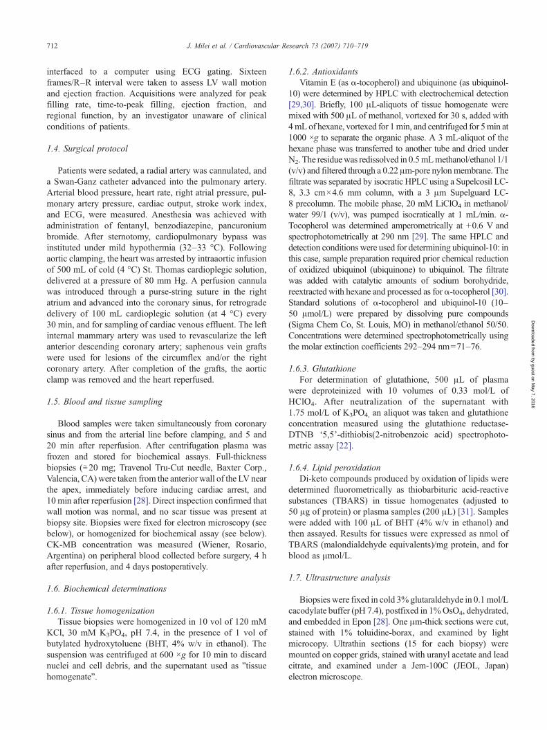
Blood samples were taken simultaneously from coronarysinus and from the arterial line before clamping, and 5 and20 min after reperfusion. After centrifugation plasma wasfrozen and stored for biochemical assays. Full-thicknessbiopsies (≅20 mg; Travenol Tru-Cut needle, Baxter Corp.,Valencia, CA) were taken from the anterior wall of the LV nearthe apex, immediately before inducing cardiac arrest, and10 min after reperfusion [28]. Direct inspection confirmed thatwall motion was normal, and no scar tissue was present atbiopsy site. Biopsies were fixed for electron microscopy (seebelow), or homogenized for biochemical assay (see below).CK-MB concentration was measured (Wiener, Rosario,Argentina) on peripheral blood collected before surgery, 4 hafter reperfusion, and 4 days postoperatively.
1.6. Biochemical determinations
1.6.1. Tissue homogenizationTissue biopsies were homogenized in 10 vol of 120 mM
KCl, 30 mM K3PO4, pH 7.4, in the presence of 1 vol ofbutylated hydroxytoluene (BHT, 4% w/v in ethanol). Thesuspension was centrifuged at 600 ×g for 10 min to discardnuclei and cell debris, and the supernatant used as ”tissuehomogenate”.
1.6.2. AntioxidantsVitamin E (as α-tocopherol) and ubiquinone (as ubiquinol-
10) were determined by HPLC with electrochemical detection[29,30]. Briefly, 100 μL-aliquots of tissue homogenate weremixed with 500 μL of methanol, vortexed for 30 s, added with4mLof hexane, vortexed for 1min, and centrifuged for 5min at1000 ×g to separate the organic phase. A 3 mL-aliquot of thehexane phase was transferred to another tube and dried underN2. The residuewas redissolved in 0.5mLmethanol/ethanol 1/1(v/v) and filtered through a 0.22 μm-pore nylonmembrane. Thefiltrate was separated by isocratic HPLC using a Supelcosil LC-8, 3.3 cm×4.6 mm column, with a 3 μm Supelguard LC-8 precolumn. The mobile phase, 20 mM LiClO4 in methanol/water 99/1 (v/v), was pumped isocratically at 1 mL/min. α-Tocopherol was determined amperometrically at +0.6 V andspectrophotometrically at 290 nm [29]. The same HPLC anddetection conditions were used for determining ubiquinol-10: inthis case, sample preparation required prior chemical reductionof oxidized ubiquinol (ubiquinone) to ubiquinol. The filtratewas added with catalytic amounts of sodium borohydride,reextracted with hexane and processed as forα-tocopherol [30].Standard solutions of α-tocopherol and ubiquinol-10 (10–50 μmol/L) were prepared by dissolving pure compounds(Sigma Chem Co, St. Louis, MO) in methanol/ethanol 50/50.Concentrations were determined spectrophotometrically usingthe molar extinction coefficients 292–294 nm=71–76.
1.6.3. GlutathioneFor determination of glutathione, 500 μL of plasma
were deproteinized with 10 volumes of 0.33 mol/L ofHClO4. After neutralization of the supernatant with1.75 mol/L of K3PO4, an aliquot was taken and glutathioneconcentration measured using the glutathione reductase-DTNB ‘5,5’-dithiobis(2-nitrobenzoic acid) spectrophoto-metric assay [22].
1.6.4. Lipid peroxidationDi-keto compounds produced by oxidation of lipids were
determined fluorometrically as thiobarbituric acid-reactivesubstances (TBARS) in tissue homogenates (adjusted to50 μg of protein) or plasma samples (200 μL) [31]. Sampleswere added with 100 μL of BHT (4% w/v in ethanol) andthen assayed. Results for tissues were expressed as nmol ofTBARS (malondialdehyde equivalents)/mg protein, and forblood as μmol/L.
1.7. Ultrastructure analysis
Biopsies were fixed in cold 3%glutaraldehyde in 0.1 mol/Lcacodylate buffer (pH 7.4), postfixed in 1%OsO4, dehydrated,and embedded in Epon [28]. One μm-thick sections were cut,stained with 1% toluidine-borax, and examined by lightmicrocopy. Ultrathin sections (15 for each biopsy) weremounted on copper grids, stained with uranyl acetate and leadcitrate, and examined under a Jem-100C (JEOL, Japan)electron microscope.
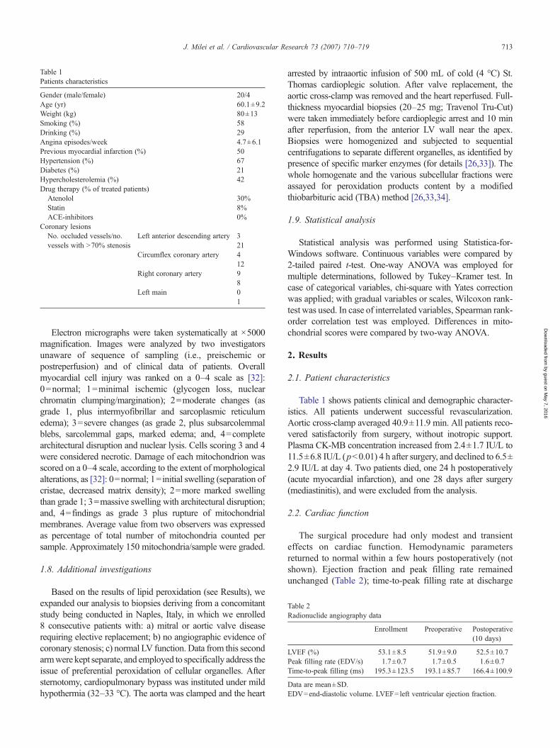
Table 1Patients characteristics
Gender (male/female) 20/4Age (yr) 60.1±9.2Weight (kg) 80±13Smoking (%) 58Drinking (%) 29Angina episodes/week 4.7±6.1Previous myocardial infarction (%) 50Hypertension (%) 67Diabetes (%) 21Hypercholesterolemia (%) 42Drug therapy (% of treated patients)
Atenolol 30%Statin 8%ACE-inhibitors 0%
Coronary lesionsNo. occluded vessels/no.vessels with N70% stenosis
Left anterior descending artery 321
Circumflex coronary artery 412
Right coronary artery 98
Left main 01
Table 2Radionuclide angiography data
Enrollment Preoperative Postoperative(10 days)
LVEF (%) 53.1±8.5 51.9±9.0 52.5±10.7Peak filling rate (EDV/s) 1.7±0.7 1.7±0.5 1.6±0.7Time-to-peak filling (ms) 195.3±123.5 193.1±85.7 166.4±100.9
Data are mean±SD.EDV=end-diastolic volume. LVEF=left ventricular ejection fraction.
713J. Milei et al. / Cardiovascular Research 73 (2007) 710–719
by guest on May 7, 2016
Dow
nloaded from
Electron micrographs were taken systematically at ×5000magnification. Images were analyzed by two investigatorsunaware of sequence of sampling (i.e., preischemic orpostreperfusion) and of clinical data of patients. Overallmyocardial cell injury was ranked on a 0–4 scale as [32]:0=normal; 1=minimal ischemic (glycogen loss, nuclearchromatin clumping/margination); 2=moderate changes (asgrade 1, plus intermyofibrillar and sarcoplasmic reticulumedema); 3=severe changes (as grade 2, plus subsarcolemmalblebs, sarcolemmal gaps, marked edema; and, 4=completearchitectural disruption and nuclear lysis. Cells scoring 3 and 4were considered necrotic. Damage of each mitochondrion wasscored on a 0–4 scale, according to the extent of morphologicalalterations, as [32]: 0=normal; 1=initial swelling (separation ofcristae, decreased matrix density); 2=more marked swellingthan grade 1; 3=massive swelling with architectural disruption;and, 4=findings as grade 3 plus rupture of mitochondrialmembranes. Average value from two observers was expressedas percentage of total number of mitochondria counted persample. Approximately 150 mitochondria/sample were graded.
1.8. Additional investigations
Based on the results of lipid peroxidation (see Results), weexpanded our analysis to biopsies deriving from a concomitantstudy being conducted in Naples, Italy, in which we enrolled8 consecutive patients with: a) mitral or aortic valve diseaserequiring elective replacement; b) no angiographic evidence ofcoronary stenosis; c) normal LV function. Data from this secondarmwere kept separate, and employed to specifically address theissue of preferential peroxidation of cellular organelles. Aftersternotomy, cardiopulmonary bypass was instituted under mildhypothermia (32–33 °C). The aorta was clamped and the heart
arrested by intraaortic infusion of 500 mL of cold (4 °C) St.Thomas cardioplegic solution. After valve replacement, theaortic cross-clamp was removed and the heart reperfused. Full-thickness myocardial biopsies (20–25 mg; Travenol Tru-Cut)were taken immediately before cardioplegic arrest and 10 minafter reperfusion, from the anterior LV wall near the apex.Biopsies were homogenized and subjected to sequentialcentrifugations to separate different organelles, as identified bypresence of specific marker enzymes (for details [26,33]). Thewhole homogenate and the various subcellular fractions wereassayed for peroxidation products content by a modifiedthiobarbituric acid (TBA) method [26,33,34].
1.9. Statistical analysis
Statistical analysis was performed using Statistica-for-Windows software. Continuous variables were compared by2-tailed paired t-test. One-way ANOVA was employed formultiple determinations, followed by Tukey–Kramer test. Incase of categorical variables, chi-square with Yates correctionwas applied; with gradual variables or scales, Wilcoxon rank-test was used. In case of interrelated variables, Spearman rank-order correlation test was employed. Differences in mito-chondrial scores were compared by two-way ANOVA.
2. Results
2.1. Patient characteristics
Table 1 shows patients clinical and demographic character-istics. All patients underwent successful revascularization.Aortic cross-clamp averaged 40.9±11.9 min. All patients reco-vered satisfactorily from surgery, without inotropic support.Plasma CK-MB concentration increased from 2.4±1.7 IU/L to11.5±6.8 IU/L (pb0.01) 4 h after surgery, and declined to 6.5±2.9 IU/L at day 4. Two patients died, one 24 h postoperatively(acute myocardial infarction), and one 28 days after surgery(mediastinitis), and were excluded from the analysis.
2.2. Cardiac function
The surgical procedure had only modest and transienteffects on cardiac function. Hemodynamic parametersreturned to normal within a few hours postoperatively (notshown). Ejection fraction and peak filling rate remainedunchanged (Table 2); time-to-peak filling rate at discharge

Fig. 3. Transcardiac release of lipid peroxides before cardioplegic arrest, andat 5 and 20 min reperfusion. Data are coronary sinus minus aortaconcentrations. p=NS.
714 J. Milei et al. / Cardiovascular Research 73 (2007) 710–719
by guest on May 7, 2016
Dow
nloaded from
was shorter than baseline, but the difference was notsignificant (Table 2).
2.3. Markers of oxidative stress in cardiac effluent
2.3.1. GlutathioneBefore ischemia, concentrations of glutathione were
similar in systemic arterial blood and in blood from thecoronary sinus (0.7±0.2 and 0.8±0.7 μmol/L, respectively).During reperfusion after cardioplegic arrest, glutathioneconcentration in arterial blood entering the heart remainedstable (peak 0.8±0.7 μmol/L); in contrast, glutathioneconcentration in blood drawn from the coronary sinussignificantly increased (1.8±0.2 μmol/L; pb0.01); thus, anet trans-cardiac release of glutathione (i.e., positivedifference between venous minus arterial concentrations)occurred at 5 min reperfusion, which persisted after 20 min(Fig. 2).
2.3.2. Lipid peroxidesPrior to cardiac arrest, thiobarbituric acid-reactive sub-
stances (TBARS) in systemic arterial blood averaged 3.7±0.6 μmol/L; concentration in venous blood from the coronarysinuswas slightly lower than in arterial blood (3.3±0.9μmol/L),indicating a tendency to cardiac extraction of lipid peroxides(Fig. 3) consistent with metabolism by mitochondria alde-hyde-dehydrogenase and aldose-reductase [35,36]. Five minafter reperfusion, TBARS concentrations were similar inarterial blood entering the heart (3.1±0.6 μmol/L), and incoronary sinus blood (3.1±0.7 μmol/L); therefore, no netcardiac release was observed (Fig. 3); this behavior persisted at20 min of reperfusion (Fig. 3).
2.4. Markers of oxidative stress in cardiac tissue
2.4.1. AntioxidantsCardiac concentration of ubiquinol decreased N30% as
an effect of ischemia/reperfusion, from 144.5±52.0 nmol/gin biopsies taken before cardioplegic arrest, to 97.6±82.0 nmol/g 10 min after reperfusion (pb0.05). Concen-tration of α-tocopherol did not change, averaging 42.6±
Fig. 2. Transcardiac glutathione release before cardioplegic arrest, and at 5and 20 min reperfusion. Data are coronary sinus minus aorta concentrations.⁎=pb0.05 vs pre-ischemia.
28 nmol/g in preischemic biopsies, and 46.7±25 nmol/g inbiopsies taken after postischemic reperfusion.
2.4.2. Lipid peroxidesTBARS averaged 0.99±0.42 nmol/mg protein in biopsies
taken before cardioplegic arrest, and 0.92±0.37 nmol/mgprotein in biopsies taken 10 min after reperfusion ( p=NS).Thus, no increase in membrane lipid peroxidation wasdocumented in the whole cardiac extract.
Previous animal studies of oxygen radical-mediatedreperfusion injury had shown that peroxidation of membranelipids can be prominent in specific subcellular organelles[25,26,33]. To investigate this specific point, we analyzedcardiac biopsies that were processed to isolate varioussubcellular fractions in a concomitant study. Also in thiscase, there was no increase in TBARS after postischemicreperfusion, neither in the whole homogenate, nor in anysubcellular fraction (Table 3).
2.5. Electron microscopy
2.5.1. Qualitative analysisIntracellular myocyte architecture was preserved in
preischemic biopsies (Fig. 4). Sarcomere alignment waspreserved; sarcolemmal membrane, T-tubules, intercalateddisks, and basement membranes were intact; nuclei andcytoplasmic glycogen were generally well preserved (Fig. 4,left panel); mild cytosolic and mild intermyofibrillar edema
Table 3Subcellular distribution of lipid peroxidation products
Specimen Buenos Aires Naples
Wholehomogenate
Wholehomogenate
Mitochondria Cytosol
Before arrest 0.99±0.42 1.07±0.20 1.04±0.16 1.27±0.30Reperfusion 0.92±0.37 1.06±0.20 0.70±0.19 1.04±0.28
Data are mean±SD of nmol/mg prot. p=NS.

Fig. 4. Typical electron microscopy appearance of myocardial biopsies before cardioplegic arrest. Left panel: normal mitochondria, mild cytosolic and mildintermyofibrillar edema, and modest T-tubules dilation can be observed. ×8000. Right panel: cross-section at higher magnification, showing mitochondria withintact membranes and tightly packed cristae. ×10,000.
715J. Milei et al. / Cardiovascular Research 73 (2007) 710–719
by guest D
ownloaded from
was occasionally seen; mitochondria showed tightly packedcristae, with no or minimal signs of edema (Fig. 4, rightpanel).
Cardioplegic arrest/reperfusion did not result in majormorphological alterations. Overall myocardial and vascularstructure was generally preserved (Fig. 5-A). Reperfusionbiopsies mostly showed areas with intermyofibrillar edemaand decreased or absent glycogen stores (Fig. 5-B); mito-chondria mostly showed mild-to-moderate damage (Fig. 5-B).In some areas, moderate mitochondrial swelling, withdecreased matrix density and separation of cristae, mildsarcomere hypercontraction, and dilatation of T-tubules, were
Fig. 5. Typical appearance of biopsies taken after 10 min reperfusion following cardsection. Myocardial architecture and vessel structure are well preserved. Mild intermTransmission electron micrograph of reperfusion biopsy. Intermyofibrillar edema wedema. Mild mitochondrial damage (degree 0–2); ×8000. Panel C. Sarcomintermyofibrillar edema; ×10,000. Panel D. Focal sarcomere fragmentation is shomyofibrillar disorganization, with glycogen loss; ×8000.
observed (Fig. 5-C). Massive mitochondrial swelling andmyofibrillar alterations were seen occasionally (Fig. 5-D).Severe changes (e.g., subsarcolemmal blebs, sarcolemmalgaps) were rare, and complete architectural disruption andnuclear lysis extremely scarce.
2.5.2. Quantitative analysisOverall injury score showed no differences between
preischemic and reperfusion biopsies (1.7±0.5 vs 1.8±0.4).In addition, since mitochondria can be a target of reperfusioninjury, systematic evaluation was performed scoring 150mitochondria/section (i.e., N2000 mitochondria/biopsy).
ioplegic arrest. Panel A. Large-field view by light microscopy of 1 μm-thickyofibrillar edema is observed. 1% Toluidine-borax staining; ×400. Panel B.ith mild sarcomere hypercontraction, glycogen decrease, and mild cytosolicere hypercontraction, mitochondrial swelling, damage degree 1–2, andwn. Mitochondrial damage degree 3, lipid vacuoles, T-tubules dilation and
on May 7, 2016

Fig. 6. Mitochondrial injury score on biopsies taken before arrest (whitecolumns), or 10 min after reperfusion (see Methods for details). Data arepercent of score distribution.
716 J. Milei et al. / Cardiovascular Research 73 (2007) 710–719
by guest on May 7, 2016
Dow
nloaded from
Before cardiac arrest, the great majority of mitochondriashowed no or minimal alterations (Fig. 6). After reperfusion,there was a decrease in the number of normal mitochondriawith concomitant increase in the proportion of mitochondriashowing ultrastructural alterations (Fig. 6). This tendency,however, did not reach statistical significance ( p=0.08).
3. Discussion
Experimental studies have documented ROS formationupon reperfusion of ischemic myocardium, with consequentoxidative injury to cell function and structure [1,2]. In thepresent study, in patients with coronary artery diseaseundergoing cardiac surgery we did find evidence of cardiacoxidant formation during reperfusion after cardioplegicarrest. However, this phenomenon did not progress intoattack to cardiac membranes, nor did it translate into full-fledged ultrastructural injury. Finally, no impairment ofcardiac function was observed. Together, these datademonstrate that no major oxidative injury occurs atreperfusion in the heart of patients with coronary arterydisease subjected to cardiac surgery under the conditions ofour study.
In most cardiac surgery procedures, the heart is keptarrested for a substantial amount of time and subsequentlyundergoes reperfusion: this condition is expected to favoroxidant formation. Indeed, ROS production occurs inpatients during reperfusion after cardioplegic arrest [16–18], and is reduced by antioxidants/scavengers [12,15,37].Yet, antioxidant strategies did not consistently translate intoclinically relevant improvement [9–15]. An explanation forthese controversial findings might be that the magnitude ofoxidant-mediated injury may actually be modest under theconditions encountered in cardiac surgery. The findings ofthe present study lend credence to this hypothesis.
Consistent with previous reports [19], we observed trans-cardiac release of glutathione during reperfusion. However,although cardiac formation and release of glutathione isinduced by oxidants [22,38,39], and consistently foundduring postischemic reperfusion [40,41], this may be simply
an indication of exposure to ROS. It is only when defensemechanisms are overwhelmed that oxidant attack progressesto injuring cells. In fact, oxidant concentrations capable toelicit cardiac glutathione release larger than what found inthis and previous clinical studies, have no detrimentaleffects, which nonetheless can be induced by up-titration ofoxidants [38,39,42].
Endogenous antioxidants are another defense mecha-nism. In our patients, cardiac concentrations of ubiquinoldecreased upon reperfusion, consistent with the notion thatendogenous antioxidants are “consumed” in the process ofoxidant detoxification [11,12,43]. This finding confirmsthat the hearts were exposed to ROS. However, sinceoxidation of glutathione and antioxidant vitamins occursupstream in the chain of events following ROS formation,in order to establish whether injury has occurred thesemeasurements must be complemented by other indices ofoxidant attack.
Lipid oxidation is a major harmful consequence of ROSformation [23–26], as it reflects irreversible oxidativechanges of membranes. Increased venous concentration ofmalonyldialdehyde has been found in patients subjected tocardiac surgery [10,11,44]. However, malonyldialdehydeconcentrations in systemic blood may reflect changesunrelated to cardiac oxidative stress (prostanoid synthesis[45], activity of aldehyde-dehydrogenase [35] and aldose-reductase [36]). Indeed, surgery itself (independent ofcardioplegic arrest/reperfusion) may induce systemic lipidperoxidation [18,44,46]. To specifically investigate lipidperoxidation at cardiac level, we simultaneously measuredlipid peroxides in the blood entering the heart and in thecoronary sinus effluent, and therefore could establish that nocardiac release of lipid peroxides occurred.
In experimental studies, lipid peroxidation of cardiacmembranes was found at postischemic reperfusion [24–26],which was preventable by scavenging of ROS [26]. It can betaken as a reliable “chemical signature” of oxidative injury[24]. However, we did not detect any appreciable increase inlipid peroxide content of cardiac tissue, in spite of biopsiesbeing taken 10 min after reinstitution of perfusion, whenlipid peroxidation peaks [24]. To ascertain whether perox-idation may had occurred only in specific organelles, whichmight have gone undetected because of possible “dilution”within the whole homogenates, we also analyzed cardiacmembranes isolated by subcellular fractionation. While thisprocedure does detect oxygen radical-mediated lipid perox-idation of specific organelles in ischemia/reperfusion models[25,26,37], it failed to document increased lipid peroxidationin our patients. In this study, lipid peroxidation wasevaluated by measuring thiobarbiturate-reactive aldehydes.This approach has potential limitations in terms of specificityand possible artifactual formation. However, this is unlikelyto have influenced our results since, if anything, it wouldhave resulted in lipoperoxide concentrations being spurious-ly higher, whereas we found no increase at all. Furthermore,we had previously shown that under our conditions of
717J. Milei et al. / Cardiovascular Research 73 (2007) 710–719
by guest on May 7, 2016
Dow
nloaded from
homogenization and assay, malonyldialdehyde measure-ments compare favorably with other markers of cardiacmembrane peroxidation [26,33].
Collectively, lack of lipid peroxide release, absence ofincreased membrane peroxidation, and preserved cardiacconcentration of membrane-bound antioxidant α-tocopherol,all coincide to indicate that oxidant formation in our patientsdid not translate into appreciable oxidative modification ofcell structures. To our knowledge, only one study haspreviously addressed this issue in a rigorous fashion: similarto what we have found, Janssen et al. [42] clearlydocumented that human hearts subjected to cardioplegicarrest/reperfusion release glutathione, but lipid peroxidationdid not increase. However, that study was performed onexplanted human hearts perfused ex vivo. Thus, our presentstudy for the first time provide a thorough assessment of thisissue in patients.
Ultrastructural alterations that characterize reperfusioninjury (contraction-band necrosis, massive mitochondrialswelling, sarcolemmal disruption) were mostly absent. Thefinding that myocyte ultrastructure was largely preservedhelps explaining why recovery of systolic and diastolicfunction was unimpeded, and it is internally consistent withabsence of biochemical “signature” of irreversible oxidativeinjury in these patients. This lack of major alterations needsto be confronted with the results of earlier observations[47,48]. In one study [47], reperfusion biopsies showeddecreased succinate-dehydrogenase activity (indicating lossof mitochondrial integrity), increased hydroperoxide-initiat-ed chemiluminescence (marker of enhanced susceptibility tolipoperoxidation), and ultrastructural alterations; similarly,Weisel et al. [48]. reported transcardiac release of conjugateddienes (a specific marker of lipid peroxidation), anddecreased myocardial concentrations of membrane-boundα-tocopherol. However, several features of the present studymay have contributed to the difference observed. First, sincethe '80s major advances have been made with respect tocardiopulmonary bypass technique and cardioplegia, allresulting in improved myocardial protection. For example,retrograde delivery of cardioplegic solution was not usedthen [47,48]. Furthermore, aortic cross clamp time averaged40.9±11.9 min in this study, whereas it ranged between 55±5 min and 75±6 min then [47,48]; this may be important,since oxidative stress increases with length of arrest [19].Finally, in the present study we investigated only patientsscheduled for elective surgery, in stable clinical conditions,with good LV function; care was also exercised to optimizepreoperative conditions, by adjusting patients' medicationsthree weeks before surgery.
In addition, it should be noted that in experimental studiesischemia/reperfusion is induced in healthy animals, while westudied patients with multivessel chronic ischemic heartdisease. It is expected that most such patients had gonethrough brief ischemia/reperfusion episode(s) before cardiacsurgery, and it is now established that sublethal ischemiamay paradoxically “precondition” the heart, i.e., render it
more tolerant toward a subsequent major insult of ischemia/reperfusion [49–52]. With preconditioning there may beupregulation of antiapoptotic proteins, activation of prosur-vival kinases, and improved coronary perfusion with bypassgrafting [49–52]. Thus, the possibility exists that hearts frompatients with chronic ischemic heart disease may be“preconditioned”, and hence more resistant to subsequentinjury by cardioplegic arrest/reperfusion than healthy,ischemia naive animals.
3.1. Implications
It is tempting to speculate that if the extent of oxidantinjury is modest, there might be more harm than good intrying to eradicate oxidant production. Small amounts ofoxidants may in fact precondition the heart, i.e., render itmore tolerant toward a subsequent ischemia/reperfusioninsult [50,53]. This phenomenon may occur during cardiacsurgery [17,49,51,52], and the majority of studies ofpreconditioning in cardiac surgery have shown that ischemicpreconditioning is an effective adjunct to myocardialprotection in cardiac surgery [49]. Patients receivingantioxidants prior to and/or during cardiac surgery mightthus be deprived of this potential benefit, while not gainingmuch from reduction of an otherwise small oxidant load atreperfusion.
3.2. Limitations
It is now appreciated that many pathological conditions,which are frequently present in patients with coronary arterydisease (e.g., obesity, hypertension, diabetes, and hyperlip-idemia) may induce oxidant stress and affect antioxidantdefense systems, thereby modulating resistance to ischemia/reperfusion injury via a preconditioning effect [48–54]. Onthe other side, some drugs, such as ACE-inhibitors andangiotensin II type-1 receptor blockers, may inhibit oxidantstress and exert cardioprotective effects through theactivation of eNOS [54]. Although we excluded patientstaking these drugs from the study, our data do not allow toevaluate whether and how the complex pathophysiologicalrelationship between oxidative stress and preconditioningwas altered by the pathological conditions and eventsoccurring in patients.
Because of protocol design centered around biochemicaland ultrastructural parameters, and not based on clinicalend-points, and because of the limited sample size, ourfindings cannot be taken as immediate evidence againstadministration of antioxidants in cardiac surgery. Further-more, because of the strict inclusion criteria, our observa-tions may not be applicable to all patients. Excluded arepatients hemodynamically or clinically unstable; similarly,severely impaired ventricles might be more vulnerable tocardioplegic arrest/reperfusion; finally, procedures requiringlonger cardioplegic arrest might be associated with greaterextent of oxidative stress.

718 J. Milei et al. / Cardiovascular Research 73 (2007) 710–719
In conclusion, patients subjected to elective bypass surgeryundergo oxidative stress upon reperfusion after cardioplegicarrest; the magnitude of the phenomenon, however, may besmall and without major consequences. Our data indicate thatdevising optimal antioxidant therapy in the setting of cardiacsurgery may require a more detailed understanding of thecomplex mechanisms of redox regulation in patients.
Acknowledgments
We gratefully acknowledge Luis Molteni, MD, for coordi-nating the surgical team. The experiments reported in this paperwere performed under a Framework Agreement between theUniversity of Buenos Aires (Buenos Aires, Argentina) and theUniversity of Perugia School of Medicine (Perugia, Italy).
by guest on May 7, 2016
Dow
nloaded from
References
[1] Ambrosio G, Tritto I. Reperfusion injury: experimental evidence andclinical implications. Am Heart J 1999;138:S69–75.
[2] Semenza GL. Cellular and molecular dissection of reperfusion injury.ROS within and without. Circ Res 2000;86:117–8.
[3] Menasche P, Grousset C, Braudel Y, Piwnica A. A comparative studyof free radical scavengers in cardioplegic solution. Improved protectionwith peroxidase. J Thorac Cardiovasc Surg 1986;92:264–71.
[4] Johnson DL, Horneffer PJ, Dinatale Jr JM, Gott VL, Gardner TJ. Freeradical scavengers improve functional recovery of stunned myocardi-um in a model of surgical coronary revascularization. Surgery1987;102:334–40.
[5] Ytrehus K, Gunnes S, Myklebust R, Mjos OD. Protection by superoxidedismutase and catalase in the isolated rat heart reperfused after prolongedcardioplegia: a combined study ofmetabolic, functional, andmorphometricultrastructural variables. Cardiovasc Res 1987;21:492–9.
[6] Ferreira R, Llesuy S, Molteni L, Milei J, Flecha BG, Boveris A.Reduction of reperfusion injury with mannitol cardioplegia. AnnThorac Surg 1989;48:77–83.
[7] Ferreira R, Burgos M, Milei J, Llesuy S, Molteni L, Hourquebie H, et al.Effect of supplementing cardioplegic solutionwith deferoxamine on humanreperfused myocardium. J Thorac Cardiovasc Surg 1990;100: 708–14.
[8] Milei J, Ferreira R, Llesuy S, Forcada P, Covarrubias J, Boveris A.Reduction of reperfusion injury with preoperative rapid intravenousinfusion of taurine during myocardial revascularization. Am Heart J1992;123:339–45.
[9] Butterworth J, Legault C, Stump DA, Coker L, Hammon JW, TroostBT, et al. A randomized, blinded trial of the antioxidant pegorgotein:no reduction in neuropsychological deficits, inotropic drug support, ormyocardial ischemia after coronary artery bypass surgery. J Cardi-othorac Vasc Anesth 1999;13:690–4.
[10] Paraskevaidis IA, Iliodromitis EK, Vlahakos D, Tsiapras DP,Nikolaidis A, Marathias A, et al. Deferoxamine infusion duringcoronary artery bypass grafting ameliorates lipid peroxidation andprotects the myocardium against reperfusion injury: immediate andlong-term significance. Eur Heart J 2005;26:263–70.
[11] Lassnigg A, Punz A, Barker R, Keznickl P, Manhart N, Roth E, et al.Influence of intravenous vitamin E supplementation in cardiac surgeryon oxidative stress: a double-blinded, randomized, controlled study. BrJ Anaest 2003;90:148–54.
[12] Westhuyzen J, Cochrane AD, Tesar PJ, Mau T, Cross DB, FrenneauxMP, et al. Effect of preoperative supplementation with alpha-tocopheroland ascorbic acid on myocardial injury in patients undergoing cardiacoperations. J Thorac Cardiovasc Surg 1997;113:942–8.
[13] Yau TM, Weisel RD, Mickle DA, Burton GW, Ingold KU, Ivanov J,et al. Vitamin E for coronary bypass operations. A prospective,double-blind, randomized trial. J Thorac Cardiovasc Surg 1994;108:302–10.
[14] Weimert NA, Tanke WF, Sims JJ. Allopurinol as a cardioprotectantduring coronary artery bypass graft surgery. Ann Pharmacother 2003;37:1708–11.
[15] Tossios P, Bloch W, Huebner A, Raji MR, Dodos F, Klass O, et al.N-acetylcysteine prevents reactive oxygen species-mediated myo-cardial stress in patients undergoing cardiac surgery: results of arandomized, double-blind, placebo-controlled clinical trial. J ThoracCardiovasc Surg 2003;126:1513–20.
[16] Tortolani AJ, Powell SR, Misik V, Weglicki WB, Pogo GJ, Kramer JH.Detection of alkoxyl and carbon-centered free radicals in coronarysinus blood from patients undergoing elective cardioplegia. Free RadicBiol Med 1993;14:421–6.
[17] Wu ZK, Tarkka MR, Eloranta J, Pehkonen E, Kaukinen L, HonkonenEL, et al. Effect of ischemic preconditioning on myocardial protectionin coronary artery bypass graft patients. Can the free radicals act as atrigger for ischemic preconditioning? Chest 2001;119:1061–8.
[18] Clermont G, Vergely C, Jazayeri S, Lahet JJ, Goudeau JJ, Lecour S, et al.Systemic free radical activation is a major event involved in myocardialoxidative stress related to cardiopulmonary bypass. Anesthesiology2002;96:80–7.
[19] Ferrari R, Alfieri O, Curello S, Ceconi C, Cargnoni A, Marzullo P, et al.Occurrence of oxidative stress during reperfusion of the human heart.Circulation 1990;81:201–11.
[20] Chance B, Sies H, Boveris A. Hydroperoxide metabolism inmammalian organs. Physiol Rev 1979;59:527–605.
[21] Blaustein A, Deneke SM, Stolz RI, Baxter D, Healy N, Fanburg BL.Myocardial glutathione depletion impairs recovery after short periodsof ischemia. Circulation 1989;80:1449–57.
[22] Sies H, Akerboom PMT. Glutathione disulfide efflux from cells andtissues. Methods Enzymol 1984;105:445–51.
[23] Lucas DT, Szweda LI. Cardiac reperfusion injury: aging, lipid peroxida-tion, and mitochondrial dysfunction. Proc Natl Acad Sci U S A1998;95:510–4.
[24] Romaschin AD, Rebeyka I, Wilson GJ, Mickle DAG. Conjugateddienes in ischemic and reperfused myocardium: an in vivo chemicalsignature of oxygen free radical mediated injury. J Mol Cell Cardiol1987;19:289–302.
[25] Romaschin AD, Wilson GJ, Thomas U, Feitler DA, Tumiati L, MickleDAG. Subcellular distribution of peroxidized lipids in myocardialreperfusion injury. Am J Physiol 1990;259:H116–23.
[26] Ambrosio G, Flaherty JT, Duilio C, Tritto I, Santoro G, Elia PP, et al.Oxygen radicals generated at reflow induce peroxidation of membranelipids in reperfused hearts. J Clin Invest 1991;87:2056–66.
[27] Ambrosio G, Zweier JL, Flaherty JT. The relationship between oxygenradical generation and impairment of myocardial energy metabolismfollowing post-ischemic reperfusion. J Mol Cell Cardiol 1991;23:1359–74.
[28] Milei J, Fraga C, Grana DR, Ferreira R, Ambrosio G. Ultrastructuralevidence of increased tolerance of hibernating myocardium to cardio-plegic ischemia–reperfusion injury. J Am Coll Cardiol 2004;43:2329–36.
[29] Motchnik PA, Frei B, Ames BNA. Measurement of antioxidants inhuman plasma. Methods Enzymol 1994;234:269–79.
[30] Actis-Goretta L, Carrasquedo F, Fraga CG. The regular supplemen-tation with an antioxidant mixture decreases oxidative stress in healthyhumans. Gender effect. Clin Chim Acta 2004;349:97–103.
[31] de Cavanagh EM, Ferder L, Carrasquedo F, Scrivo D, Wassermann A,Fraga CG, et al. Higher levels of antioxidant defenses in enalapril-treated versus non-enalapril-treated hemodialysis patients. Am JKidney Dis 1999;34:445–55.
[32] Kloner RA, Fishbein MC, Braunwald E, Maroko PR. Effect ofpropranolol on mitochondrial morphology during acute myocardialischemia. Am J Cardiol 1978;41:880–6.
719J. Milei et al. / Cardiovascular Research 73 (2007) 710–719
by guest onD
ownloaded from
[33] Ambrosio G, Zweier JL, Duilio C, Kuppusamy P, Santoro G, Elia PP,et al. Evidence that mitochondrial respiration is a source of potentiallytoxic oxygen free radicals in intact rabbit hearts subjected to ischemiaand reflow. J Biol Chem 1993;268:18532–41.
[34] Liedtke AJ, Mahar CQ, Ytrehus K, Mjos OD. Estimates of free radicalproduction in rat and swine hearts: methods and application ofmeasuring malonyldialdehyde levels in fresh and frozen myocardium.Basic Res Cardiol 1984;79:513–8.
[35] Siu GM, Draper HH. Metabolism of malondialdehyde in vivo and invitro. Lipids 1982;17:349–55.
[36] Shinmura K, Bolli R, Liu SQ, Tang XL, Kodani E, Xuan YT, et al.Aldose reductase is an obligatory mediator of the late phase ofischemic preconditioning. Circ Res 2002;91:240–6.
[37] TarkkaMR,VuolleM,KaukinenS, HolmP, Eloranta J, KaukinenU, et al.Effect of allopurinol on myocardial oxygen free radical production incoronary bypass surgery. Scand Cardiovasc J 2000;34:593–6.
[38] Ambrosio G, Santoro G, Tritto I, Elia PP, Duilio C, Basso A, et al.Effects of ischemia and reperfusion on cardiac tolerance to oxidativestress. Am J Physiol 1992;262:H23–30.
[39] Abete P, Napoli C, Santoro G, Ferrara N, Tritto I, Chiariello M, et al.Age-related decrease in cardiac tolerance to oxidative stress. J Mol CellCardiol 1999;31:227–36.
[40] Lesnefsky EJ, Repine JE, Horwitz LD. Oxidation and release ofglutathione from myocardium during early reperfusion. Free RadicBiol Med 1989;7:31–5.
[41] Tritto I, Duilio C, Santoro G, Elia PP, Cirillo P, De Simone C, et al. Ashort burst of oxygen radicals at reflow induces sustained release ofoxidized glutathione from postischemic hearts. Free Radic Biol Med1998;15:290–7.
[42] Janssen M, Koster JF, Bos E, de Jong JW. Malondialdehyde andglutathione production in isolated perfused human and rat hearts. CircRes 1993;73:681–8.
[43] Beyer RE. The participation of coenzyme Q in free radical productionand antioxidation. Free Radic Biol Med 1990;8:545–65.
[44] Hadjinikolaou L, Alexiou C, Cohen AS, Standbridge Rde L, CollMcAJ, et al. Early changes in plasma antioxidant and lipid peroxidationlevels following coronary artery bypass surgery: a complex response.Eur J Cardiothorac Surg 2003;23:969–75.
[45] Capdevila JH, Falck JR, Harris RC. Cytochrome P450 and arachidonicacid bioactivation: molecular and functional properties of thearachidonate monooxygenase. J Lipid Res 2000;41:163–81.
[46] Andreoni KA, Kazui M, Cameron DE, Nyhan D, Sehnert SS, RohdeCA, et al. Ethane: a marker of lipid peroxidation during cardiopulmo-nary bypass in humans. Free Radic Biol Med 1999;26:439–45.
[47] Ferreira R, Llesuy S, Milei J, Scordo D, Hourquebie H, Molteni L, et al.Assessment of myocardial oxidative stress in patients after myocardialrevascularization. Am Heart J 1988;115:307–12.
[48] Weisel RD, Mickle DA, Finkle CD, Tumiati LC, Madonik MM,Ivanov J, et al. Myocardial free-radical injury after cardioplegia.Circulation 1989;80:III14–8.
[49] Valen G, Vaage J. Pre- and postconditioning during cardiac surgery.Basic Res Cardiol 2005;100:179–86.
[50] Tritto I, D'Andrea D, Eramo N, Scognamiglio A, De Simone C,Violante A, et al. Oxygen radicals can induce preconditioning in rabbithearts. Circ Res 1997;80:743–8.
[51] Belhomme D, Peynet J, LouzyM, Launay JM, KitakazeM,Menasche P.Evidence for preconditioning by isoflurane in coronary artery bypassgraft surgery. Circulation 1999;100:II340–4.
[52] Stowe DF, Kevin LG. Cardiac preconditioning by volatile anestheticagents: A defining role for altered mitochondrial bioenergetics.Antioxid Redox Signal 2004;6:439–48.
[53] Baines CP, Goto M, Downey JM. Oxygen radicals released duringischemic preconditioning contribute to cardioprotection in the rabbitmyocardium. J Mol Cell Cardiol 1997;29:207–16.
[54] Kloner RA, Rezkalla SH. Preconditioning, postconditioning and theirapplication to clinical cardiology. Cardiovasc Res 2006;70:297–307.
Ma
y 7, 2016
Related Documents