Relating sequence encoded information to form and function of intrinsically disordered proteins Rahul K Das, Kiersten M Ruff and Rohit V Pappu Intrinsically disordered proteins (IDPs) showcase the importance of conformational plasticity and heterogeneity in protein function. We summarize recent advances that connect information encoded in IDP sequences to their conformational properties and functions. We focus on insights obtained through a combination of atomistic simulations and biophysical measurements that are synthesized into a coherent framework using polymer physics theories. Address Department of Biomedical Engineering and Center for Biological Systems Engineering, Washington University in St. Louis, One Brookings Drive, Campus Box 1097, St. Louis, MO 63130, USA Corresponding author: Pappu, Rohit V ([email protected]) Current Opinion in Structural Biology 2015, 32:102–112 This review comes from a themed issue on Sequences and Topology Edited by M Madan Babu and Anna R Panchenko http://dx.doi.org/10.1016/j.sbi.2015.03.008 0959-440X/# 2015 Elsevier Ltd. All rights reserved. Introduction Protein domains are modular building blocks of macro- molecular complexes and interaction networks [1]. The concept of domains can be generalized to include se- quence regions that fail to fold as autonomous units [2]. These intrinsically disordered regions/proteins, referred to collectively hereafter as IDPs, are distinct from struc- tured domains. Their sequences encode an intrinsic inability to fold into singular well-defined three-dimen- sional structures [3 ,4 ,5–7] although some IDPs do fold into well-ordered structures in the context of functional complexes. IDPs are implicated in important cellular processes that include cell division [8,9 ], cell signaling [3 ,10], intracellular transport [11,12 ], bacterial translo- cation [13 ], cell mechanics [14 ,15], protein degradation [16,17], posttranscriptional regulation [18], and cell cycle control [19]. IDPs can be classified into distinct conformational classes based on their amino acid compositions [20–41]. We summarize recent results that have identified composi- tion-to-conformation relationships (CCRs) through studies of archetypal IDPs. CCRs enable the assignments of conformational descriptors and inferences regarding the amplitudes of conformational fluctuations of IDPs. These insights are relevant because amino acid compositions are often well conserved among orthologs of IDPs even if their sequences are poorly conserved [42,43]. Compositional classes of IDPs Amino acid compositions of IDPs are characterized by distinct biases [5]. They are deficient in canonical hydro- phobic residues and enriched in polar and charged resi- dues. Accordingly, IDPs fall into three distinct compositional classes that reflect the fraction of charged versus polar residues. The distinct classes are polar tracts, polyampholytes, and polyelectrolytes [41] (see Figure 1). Polar tracts are deficient in charged, hydrophobic, and proline residues. They are enriched in polar amino acids such as Asn, Gly, Gln, His, Ser, and Thr. Polyampholytes and polyelectrolytes can either be weak or strong depending on the fraction of charged residues (FCR) that is quanti- fied as the sum of f + and f (see Figure 2). The latter two parameters quantify the fraction of positive and negative- ly charged residues in an IDP sequence. Polyelectrolytes have an excess of one type of charge, that is, f + > f or vice versa. Polyampholytes have roughly equivalent fractions of opposite charges, that is, f + f . The designation of weak versus strong polyampholytes/polyelectrolytes is governed by the value of FCR. In strong polyampho- lytes/polyelectrolytes, the high FCR values encode an intrinsic tendency for populating expanded coil-like con- formations because charged residues prefer to be solvated in aqueous milieus. A formal language for describing conformational preferences of IDPs Ensembles of conformations as opposed to singular rep- resentative structures are appropriate for describing IDPs. The balance between solvent-mediated intra-chain attractions versus repulsions determines the types of con- formations that make up the ensemble that is thermody- namically accessible to an IDP sequence. When attractions dominate, the conformations in the ensemble are, on aver- age, compact and spherical, that is, globular. Conversely, if intra-chain repulsions dominate over attractions or, stated differently, chain solvation is preferred over desolvation, then the conformations are, on average, expanded, prolate ellipsoidal, and coil-like. An intermediate scenario results if the strengths of intra-chain solvent mediated repulsions are counterbalanced by equivalent attractive interactions. Under such circumstances, the ensembles are characterized Available online at www.sciencedirect.com ScienceDirect Current Opinion in Structural Biology 2015, 32:102–112 www.sciencedirect.com

Welcome message from author
This document is posted to help you gain knowledge. Please leave a comment to let me know what you think about it! Share it to your friends and learn new things together.
Transcript

Relating sequence encoded information to formand function of intrinsically disordered proteinsRahul K Das, Kiersten M Ruff and Rohit V Pappu
Available online at www.sciencedirect.com
ScienceDirect
Intrinsically disordered proteins (IDPs) showcase the
importance of conformational plasticity and heterogeneity in
protein function. We summarize recent advances that connect
information encoded in IDP sequences to their conformational
properties and functions. We focus on insights obtained
through a combination of atomistic simulations and biophysical
measurements that are synthesized into a coherent framework
using polymer physics theories.
Address
Department of Biomedical Engineering and Center for Biological
Systems Engineering, Washington University in St. Louis, One Brookings
Drive, Campus Box 1097, St. Louis, MO 63130, USA
Corresponding author: Pappu, Rohit V ([email protected])
Current Opinion in Structural Biology 2015, 32:102–112
This review comes from a themed issue on Sequences and Topology
Edited by M Madan Babu and Anna R Panchenko
http://dx.doi.org/10.1016/j.sbi.2015.03.008
0959-440X/# 2015 Elsevier Ltd. All rights reserved.
IntroductionProtein domains are modular building blocks of macro-
molecular complexes and interaction networks [1]. The
concept of domains can be generalized to include se-
quence regions that fail to fold as autonomous units [2].
These intrinsically disordered regions/proteins, referred
to collectively hereafter as IDPs, are distinct from struc-
tured domains. Their sequences encode an intrinsic
inability to fold into singular well-defined three-dimen-
sional structures [3�,4�,5–7] although some IDPs do fold
into well-ordered structures in the context of functional
complexes. IDPs are implicated in important cellular
processes that include cell division [8,9�], cell signaling
[3�,10], intracellular transport [11,12�], bacterial translo-
cation [13��], cell mechanics [14�,15], protein degradation
[16,17], posttranscriptional regulation [18], and cell cycle
control [19].
IDPs can be classified into distinct conformational classes
based on their amino acid compositions [20–41]. We
summarize recent results that have identified composi-
tion-to-conformation relationships (CCRs) through studies
Current Opinion in Structural Biology 2015, 32:102–112
of archetypal IDPs. CCRs enable the assignments of
conformational descriptors and inferences regarding the
amplitudes of conformational fluctuations of IDPs. These
insights are relevant because amino acid compositions are
often well conserved among orthologs of IDPs even if their
sequences are poorly conserved [42,43].
Compositional classes of IDPsAmino acid compositions of IDPs are characterized by
distinct biases [5]. They are deficient in canonical hydro-
phobic residues and enriched in polar and charged resi-
dues. Accordingly, IDPs fall into three distinct
compositional classes that reflect the fraction of charged
versus polar residues. The distinct classes are polar tracts,polyampholytes, and polyelectrolytes [41] (see Figure 1). Polar
tracts are deficient in charged, hydrophobic, and proline
residues. They are enriched in polar amino acids such as
Asn, Gly, Gln, His, Ser, and Thr. Polyampholytes and
polyelectrolytes can either be weak or strong depending
on the fraction of charged residues (FCR) that is quanti-
fied as the sum of f+ and f� (see Figure 2). The latter two
parameters quantify the fraction of positive and negative-
ly charged residues in an IDP sequence. Polyelectrolytes
have an excess of one type of charge, that is, f+ > f� or viceversa. Polyampholytes have roughly equivalent fractions
of opposite charges, that is, f+ � f�. The designation of
weak versus strong polyampholytes/polyelectrolytes is
governed by the value of FCR. In strong polyampho-
lytes/polyelectrolytes, the high FCR values encode an
intrinsic tendency for populating expanded coil-like con-
formations because charged residues prefer to be solvated
in aqueous milieus.
A formal language for describingconformational preferences of IDPsEnsembles of conformations as opposed to singular rep-
resentative structures are appropriate for describing IDPs.
The balance between solvent-mediated intra-chain
attractions versus repulsions determines the types of con-
formations that make up the ensemble that is thermody-
namically accessible to an IDP sequence. When attractions
dominate, the conformations in the ensemble are, on aver-
age, compact and spherical, that is, globular. Conversely, if
intra-chain repulsions dominate over attractions or, stated
differently, chain solvation is preferred over desolvation,
then the conformations are, on average, expanded, prolate
ellipsoidal, and coil-like. An intermediate scenario results if
the strengths of intra-chain solvent mediated repulsions
are counterbalanced by equivalent attractive interactions.
Under such circumstances, the ensembles are characterized
www.sciencedirect.com

Encoding form and function of IDPs Das, Ruff and Pappu 103
Figure 1
POLAR TRACTS
PolyQ: …QQQQQQQQQ…QQQQQQQQQQ …Sup35: …SNQGNNQQNYQQ YSQNGNQQ…EcSSB: …QGGGAPAGGN IGGGQ PQGGW…Nup42: …TSPFGS LQQN ASQN ASSTSS …
POLYAMPHOLYTES
Nup60: …NAYKSENAPSASS KEFNFTN …PfSSB: …FM PLNSN DKIIED KEFTDRL… Nsp1: …AF SFGAKSDEKK DGDASKPA… PQBP1: …YDKVDRERERDRERDRDRGY…
POLYELECTROLYTESPRM2: …ACYPVNIRARGLGKNMGMKS…PDE6G: …DITVI CPWEAFNH LELHELA …NP1: …RARSRGRSVRRRRR GRSPGR…RAG2: …SFDGDDE FDTYNEDDEDDE S…
WEAK
STRONG
WEAK
STRONG
… ………………
……
……
…………
……
……
…………
……
Hydrophobic Polar Proline Positive Negative
Current Opinion in Structural Biology
Definitions of polar tracts, polyelectrolytes, and polyampholytes. Polar tracts shown here include polyQ (UniProt ID: P42858): Polyglutamine tracts
are found in at least ten proteins associated with human neurodegenerative disorders including Huntington’s disease; Sup35 (UniProt ID: P05453):
Residues 4–23 of S. cerevisiae Sup35 corresponding to a region of the N-terminal prion domain; EcSSB (UniProt ID: P0AGE0): Residues 117–136
of E. coli single stranded DNA binding protein corresponding to a region of the C-terminal tail; Nup42 (UniProt ID: P49686): Residues 181–200 of
S. cerevisiae nucleoporin Nup42 corresponding to a region of the FG domain, which modulates gating of the nuclear pore complex.
Polyampholytes shown here include: Nup60 (UniProt ID: P39705): Residues 412–431 of S. cerevisiae nucleoporin Nup60 corresponding to a region
of the FG domain which modulates gating of the nuclear pore complex; PfSSB (UniProt ID: Q8I415): Residues 232–251 of P. falciparum single
stranded DNA binding protein corresponding to a region of the C-terminal tail; Nsp1 (UniProt ID: P14907): Residues 359–378 of S. cerevisiae
nucleoporin Nsp1 corresponding to a region of the FG domain which modulates gating of the nuclear pore complex; PQBP1 (UniProt ID: O60828):
Residues 146–165 of H. sapiens polyglutamine-tract binding protein 1 corresponding to a region of the expanded linker, which connects the
N-terminal WW domain and the C-terminal U5 15 kDa binding region. Polyelectrolytes shown here include: PRM2 (UniProt ID: Q9EP54): Residues
2–21 of the C. griseus DNA packaging protein protamine 2, which is involved in the chromatin condensation process during spermatogenesis [6];
PDE6G (UniProt ID: P18545): Residues 63–82 of H. sapiens retinal rod rhodopsin-sensitive cGMP 30,50-cyclic phosphodiesterase subunit gamma
protein, which is involved in processing visual signal; NP1 (UniProt ID: O13030): Residues 5–24 of C. pyrrhogaster protamine 1 which is involved in
the chromatin condensation process during spermatogenesis; RAG2 (UniProt ID: P21784): Residues 392–411 of C. griseus V(D)J recombination-
activating protein 2 corresponding to a region of the ‘acidic hinge’ which modulates DNA repair mechanisms.
by maximal conformational heterogeneity and compact,
semi-compact, expanded, and chimeric conformations be-
come thermodynamically accessible [41]. Typical hetero-
polymeric IDP sequences can sample conformations that
are chimeras of globules, coils, rods, and semi-compact
hairpins. The preference is governed by the region-specific
amino acid compositions along the linear sequence.
Polymer physics theories provide access to formal
descriptors of conformational ensembles for heteroge-
neous systems such as IDPs and these have been
reviewed recently [41,44]. Analytical relationships predict
the scaling of parameters such as radii of gyration, mean
end-to-end distances, and hydrodynamic radii as func-
tions of chain length, amino acid composition, and intrin-
sic stiffness. Analytical relations are also available to relate
the scaling of inter-residue distances to the linear se-
quence separation between residues [41]. Finally, one can
www.sciencedirect.com
also classify the sequence-specific conformational prop-
erties by quantifying the amplitudes of conformational
fluctuations [39]. All of these classifiers and descriptors
rely on comparisons of measured or calculated values of
conformational fluctuations to expectations from analyti-
cal theories for flexible polymers in different types of
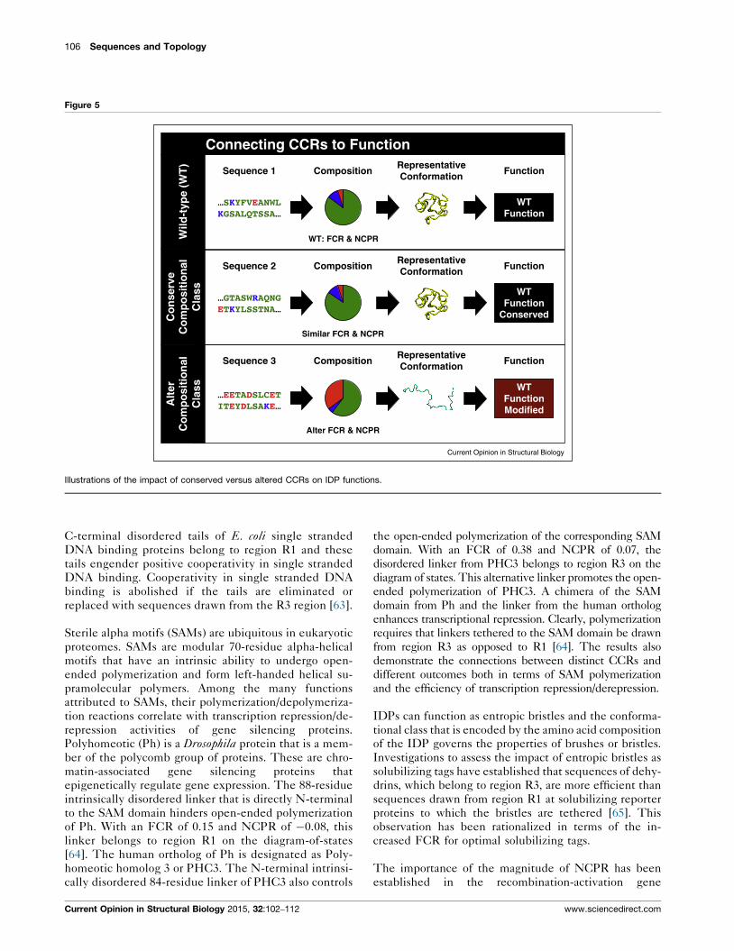
solvents. Figure 3 summarizes the typical workflow that
leads from analysis of results from computer simulations
or in vitro experiments to quantitative inferences regard-
ing CCRs and/or sequence-to-conformation relationships
(SCRs).
Distinct compositional classes can bemapped to distinct conformational classesResults from atomistic simulations obtained using explic-
it representations of solvent molecules [45,46] and studies
based on fluorescence correlation spectroscopy [46,47]
have shown that polyglycine chains, that is, polypeptide
Current Opinion in Structural Biology 2015, 32:102–112
104 Sequences and Topology
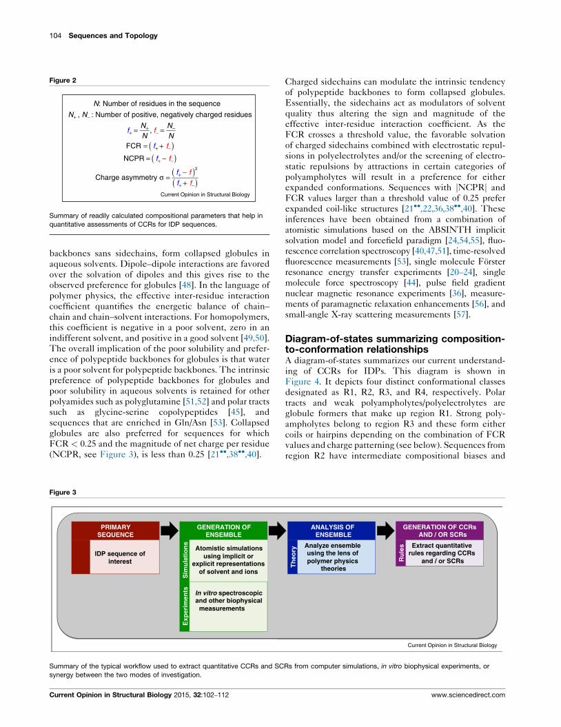
Figure 2
N: Number of residues in the sequence
N+ , N− : Number of positive, negatively charged residues
f+ = N+N
, f− = N−N
FCR = f+ + f−( )NCPR = f+ − f−( )
Charge asymmetry σ =f+ − f( )2
f+ + f−( )Current Opinion in Structural Biology
Summary of readily calculated compositional parameters that help in
quantitative assessments of CCRs for IDP sequences.
backbones sans sidechains, form collapsed globules in
aqueous solvents. Dipole–dipole interactions are favored
over the solvation of dipoles and this gives rise to the
observed preference for globules [48]. In the language of
polymer physics, the effective inter-residue interaction
coefficient quantifies the energetic balance of chain–chain and chain–solvent interactions. For homopolymers,
this coefficient is negative in a poor solvent, zero in an
indifferent solvent, and positive in a good solvent [49,50].
The overall implication of the poor solubility and prefer-
ence of polypeptide backbones for globules is that water
is a poor solvent for polypeptide backbones. The intrinsic
preference of polypeptide backbones for globules and
poor solubility in aqueous solvents is retained for other
polyamides such as polyglutamine [51,52] and polar tracts
such as glycine-serine copolypeptides [45], and
sequences that are enriched in Gln/Asn [53]. Collapsed
globules are also preferred for sequences for which
FCR < 0.25 and the magnitude of net charge per residue
(NCPR, see Figure 3), is less than 0.25 [21��,38��,40].
Figure 3
GENERATION OF ENSEMBLE
PRIMARY SEQUENCE
Sim
ula
tio
ns
Exp
erim
ents
Atomistic simulations using implicit or
explicit representations of solvent and ions
In vitro spectroscopic and other biophysical
measurements
IDP sequence ofinterest
Summary of the typical workflow used to extract quantitative CCRs and SC
synergy between the two modes of investigation.
Current Opinion in Structural Biology 2015, 32:102–112
Charged sidechains can modulate the intrinsic tendency
of polypeptide backbones to form collapsed globules.
Essentially, the sidechains act as modulators of solvent
quality thus altering the sign and magnitude of the
effective inter-residue interaction coefficient. As the
FCR crosses a threshold value, the favorable solvation
of charged sidechains combined with electrostatic repul-
sions in polyelectrolytes and/or the screening of electro-
static repulsions by attractions in certain categories of
polyampholytes will result in a preference for either
expanded conformations. Sequences with jNCPRj and
FCR values larger than a threshold value of 0.25 prefer
expanded coil-like structures [21��,22,36,38��,40]. These
inferences have been obtained from a combination of
atomistic simulations based on the ABSINTH implicit
solvation model and forcefield paradigm [24,54,55], fluo-
rescence correlation spectroscopy [40,47,51], time-resolved
fluorescence measurements [53], single molecule Forster
resonance energy transfer experiments [20–24], single
molecule force spectroscopy [44], pulse field gradient
nuclear magnetic resonance experiments [36], measure-
ments of paramagnetic relaxation enhancements [56], and
small-angle X-ray scattering measurements [57].
Diagram-of-states summarizing composition-to-conformation relationshipsA diagram-of-states summarizes our current understand-
ing of CCRs for IDPs. This diagram is shown in
Figure 4. It depicts four distinct conformational classes
designated as R1, R2, R3, and R4, respectively. Polar
tracts and weak polyampholytes/polyelectrolytes are
globule formers that make up region R1. Strong poly-
ampholytes belong to region R3 and these form either
coils or hairpins depending on the combination of FCR
values and charge patterning (see below). Sequences from
region R2 have intermediate compositional biases and
ANALYSIS OF ENSEMBLE
Th
eory
Analyze ensemble using the lens of polymer physics
theories
GENERATION OF CCRs AND / OR SCRs
Ru
les Extract quantitative
rules regarding CCRs and / or SCRs
Current Opinion in Structural Biology
Rs from computer simulations, in vitro biophysical experiments, or
www.sciencedirect.com

Encoding form and function of IDPs Das, Ruff and Pappu 105
Figure 4
0
0.2
0.4
0.6
0.8
1.0
0 0.2 0.4 0.6 0.8 1.0
R3
R4
R4R1
R2
R1 (25%): Globules, FCR < 0.25 & NCPR < 0.25 R2 (40%): Chimeras of globules & coils, 0.25 ≤ FCR ≤ 0.35 & NCPR ≤ 0.35 R3 (30%): Polyampholytic coils or hairpins, FCR > 0.35 & NCPR ≤ 0.35 R4 (5%): Polyelectrolytic semi-flexible rods or coils, FCR > 0.35 & NCPR > 0.35
f −
f+Current Opinion in Structural Biology
Diagram-of-states classification depicting the distinct conformational classes for IDP sequences. Statistics for different regions (percentages) are
from analysis of bona fide IDPs in DISPROT [61].
their conformations are likely to be chimeras of globules
and coils. IDPs that undergo folding upon binding pre-
dominantly populate region R2. This highlights the role
played by context dependent interactions as determi-
nants of conformational transitions for sequences drawn
from R2 [58]. It is worth emphasizing that the boundary
between R1 and R2 is rather ad hoc. The placement of this
boundary reflects the limited ‘titration’ of CCRs for
sequences drawn from these two regions. Region R4
spans two areas, one each for acid versus base rich poly-
electrolytes, respectively. For these sequences, the com-
bination of electrostatic repulsions between charged
sidechains and the favorable solvation free energies of
these sidechains gives rise to semi-flexible worm-like
conformations.
The diagram-of-states classification shown in Figure 4 is
valid for IDP sequences that have at least thirty residues,
low overall hydropathy, and low proline contents. The
physical principles underlying the conformational prop-
erties of weak polyampholytes and polyelectrolytes sug-
gest that the conformational transitions are likely to be
continuous functions of FCR and NCPR [59,60]. If this
expectation is borne out for longer sequences with low
FCR and NCPR values or sequences with equivalent
fractions of charged and polar residues, then the compo-
sition range spanned by R2 will be larger than what is
shown in Figure 4. Unpublished results suggest that the
classification of CCRs derived from the diagram-of-states,
particularly the assignment of a sequence to region R1 or
R2, might only be valid for IDP sequences within a
www.sciencedirect.com
certain length range and proline contents that fall below
a reasonably low threshold.
Statistics for different regions of the diagram-of-statesThe DISPROT database is an inventory of bona fide IDP
sequences [61]. Analysis of the compositional biases of
sequences from this database reveals that at least 70% of
known IDP sequences belong to regions R2 and R3.
These sequences are symmetric polyampholytes
( f+ � f�), asymmetric polyampholytes ( f+ 6¼ f�), or weak
polyelectrolytes. Based on their compositional biases,
sequences corresponding to regions R2 and R3 are
expected to adopt coil-like conformations, semi-compact
hairpins, or conformations that are chimeras of coils and
globules or coils and semi-compact hairpins. In addition,
their ensembles are expected to display significant con-
formational heterogeneity [39] as characterized by spon-
taneous conformational fluctuations whose amplitudes
are likely to be considerably larger than those of globular
proteins. Regions R1, R2, and R3 together encompass at
least 95% of the known sequences of IDPs.
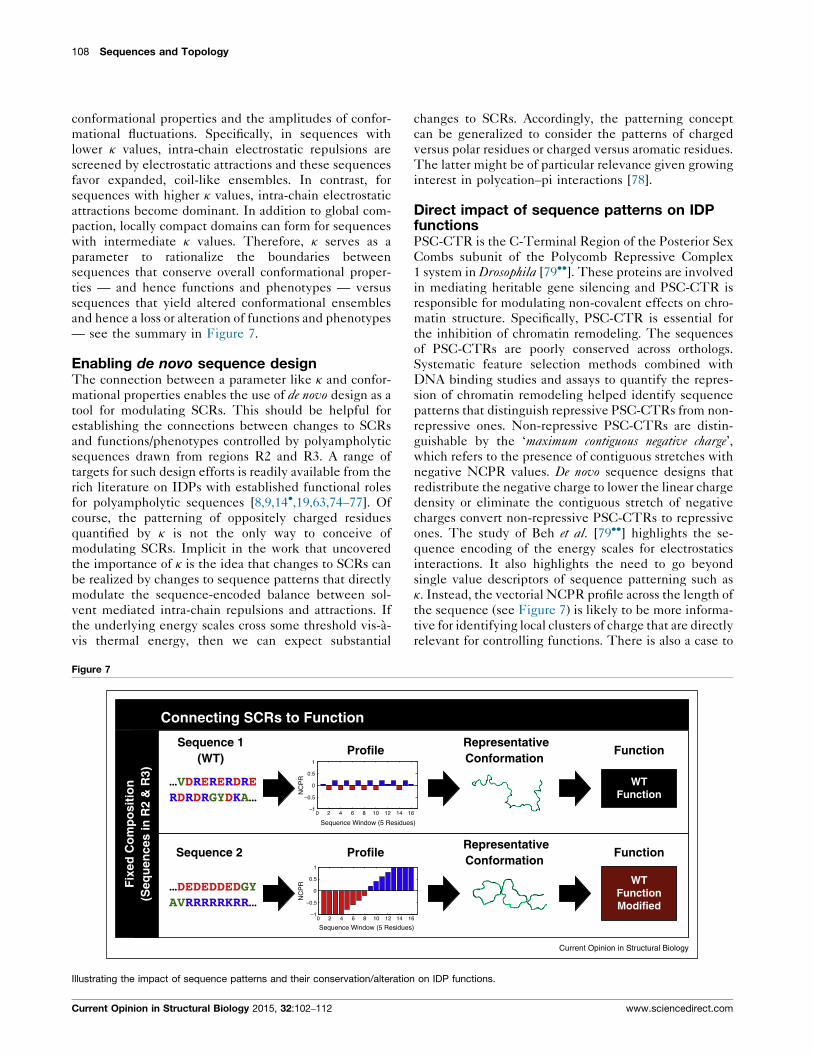
Connecting CCRs to functionWe present highlights from a growing body of data to
demonstrate the functional implications of CCRs. The
overall theme presented in this discussion is summa-
rized in Figure 5. Long disordered linkers that belong
either to the R2 or R3 region of the diagram-of-states can
help localize proteins to the junction between the
endoplasmic reticulum and plasma membrane [62].
Current Opinion in Structural Biology 2015, 32:102–112
106 Sequences and Topology
Figure 5
Connecting CCRs to Function
Composition
Composition
Sequence 1
Composition
…SKYFVEANWLKGSALQTSSA…
…GTASWRAQNGETKYLSSTNA…
Sequence 2
Sequence 3
…EETA DSLCETITEYDLSAKE…
Representative Conformation
Representative Conformation
Representative Conformation
Function
WTFunction
WTFunction
Conserved
WTFunction Modified
Wild
-typ
e (W
T)
Co
nse
rve
Co
mp
osi
tio
nal
C
lass
Alt
er
Co
mp
osi
tio
nal
C
lass
Function
Function
WT: FCR & NCPR
Similar FCR & NCPR
Alter FCR & NCPR
Current Opinion in Structural Biology
Illustrations of the impact of conserved versus altered CCRs on IDP functions.
C-terminal disordered tails of E. coli single stranded
DNA binding proteins belong to region R1 and these
tails engender positive cooperativity in single stranded
DNA binding. Cooperativity in single stranded DNA
binding is abolished if the tails are eliminated or
replaced with sequences drawn from the R3 region [63].
Sterile alpha motifs (SAMs) are ubiquitous in eukaryotic
proteomes. SAMs are modular 70-residue alpha-helical
motifs that have an intrinsic ability to undergo open-
ended polymerization and form left-handed helical su-
pramolecular polymers. Among the many functions
attributed to SAMs, their polymerization/depolymeriza-
tion reactions correlate with transcription repression/de-
repression activities of gene silencing proteins.
Polyhomeotic (Ph) is a Drosophila protein that is a mem-
ber of the polycomb group of proteins. These are chro-
matin-associated gene silencing proteins that
epigenetically regulate gene expression. The 88-residue
intrinsically disordered linker that is directly N-terminal
to the SAM domain hinders open-ended polymerization
of Ph. With an FCR of 0.15 and NCPR of �0.08, this
linker belongs to region R1 on the diagram-of-states
[64]. The human ortholog of Ph is designated as Poly-
homeotic homolog 3 or PHC3. The N-terminal intrinsi-
cally disordered 84-residue linker of PHC3 also controls
Current Opinion in Structural Biology 2015, 32:102–112
the open-ended polymerization of the corresponding SAM
domain. With an FCR of 0.38 and NCPR of 0.07, the
disordered linker from PHC3 belongs to region R3 on the
diagram of states. This alternative linker promotes the open-
ended polymerization of PHC3. A chimera of the SAM
domain from Ph and the linker from the human ortholog
enhances transcriptional repression. Clearly, polymerization
requires that linkers tethered to the SAM domain be drawn
from region R3 as opposed to R1 [64]. The results also
demonstrate the connections between distinct CCRs and
different outcomes both in terms of SAM polymerization
and the efficiency of transcription repression/derepression.
IDPs can function as entropic bristles and the conforma-
tional class that is encoded by the amino acid composition
of the IDP governs the properties of brushes or bristles.
Investigations to assess the impact of entropic bristles as
solubilizing tags have established that sequences of dehy-
drins, which belong to region R3, are more efficient than
sequences drawn from region R1 at solubilizing reporter
proteins to which the bristles are tethered [65]. This
observation has been rationalized in terms of the in-
creased FCR for optimal solubilizing tags.
The importance of the magnitude of NCPR has been
established in the recombination-activation gene
www.sciencedirect.com

Encoding form and function of IDPs Das, Ruff and Pappu 107
(RAG2). The sequence architecture of RAG2 is modular
and comprises a 60-residue ‘acidic hinge’ region that
connects the beta propeller core domain to a pleckstrin
homology domain [66]. The acidic hinge region is impor-
tant for the function of RAG2, which involves preventing
access to inappropriate repair mechanisms for DNA dou-
ble-stranded breaks such as alternative non-homologous
end joining. Key observations regarding the acidic hinge
highlight the importance of NCPR over details of the
primary sequence. Neutralization of charged residues
within the 31-residue N-terminal region of the acidic
hinge leads to increased alternative non-homologous
end joining whereas scrambling of the sequence that
maintains NCPR maintains the functionality of the wild
type sequence. Similarly, human sequence variants of
RAG2 that lead to changes in NCPR cause increased
alternative non-homologous end joining and impaired
genome stability [66].
FG nucleoporins or FG-Nups can have distinct composi-
tional biases and these are distinguished by their FCR
values. FG-Nups with low FCR values belong to region
R1 of the diagram-of-states and these are designated as
‘cohesive’ in contrast to sequences with higher FCR
values that belong to regions R2 and R3 and are desig-
nated as being ‘repulsive’ [67]. The two categories of
sequences are proposed to play distinct roles as modula-
tors of gating mechanisms in the nuclear pore complex.
Going beyond CCRs: connecting sequencepatterns to conformational propertiesThe diagram-of-states relies purely on the details of
amino acid compositions and provides a zeroth order
classification of relationships between IDP sequences
and conformational classes. The documented CCRs raise
an interesting question: Since the number of sequences
Figure 6
WPPDRGHDKSDRDRERGYDKVDRERERDRE
WPPYDDRSRHERRHKYRRRRARKRHKGDRE
WPPGGEDDEDDDDEEDDEGEDEDEDEAHYY
wtsv1sv2
=( )2
i=1
nw
nw;δseq
σi σ∑
Calculation of k and using it to distinguish the sequences with different linea
is calculated. The overall charge asymmetry s is determined by the amino a
sliding windows and the mean squared deviation d helps quantify the devia
vis the charge asymmetry encoded by the amino acid composition. The val
amino acid composition and this is used to evaluate the value of k, as show
of the patterning that is quantified using k, we show the sequence of the ‘p
tract binding protein PQB-P1. The bottom two rows show two de novo des
1 and 2. These two sequences were derived from alterations to the linear s
row, the values of k are shown to the right.
www.sciencedirect.com
that are compatible with a given amino acid composition
is astronomically large, do all conceivable sequences
encode similar conformational properties and impact
function in similar ways? Of course, since IDPs serve
as scaffolds for short linear motifs (SLiMs) [4,68–70], it
stands to reason that conserving the identities and posi-
tions of SLiMs will winnow down the number of func-
tionally relevant sequence alternatives for a given amino
acid composition. Are there additional constraints that
could have a direct impact on global conformational
properties and hence on function?
Quantitative studies of DNA binding proteins identified a
curious pattern of clustering of like-charged residues
[71,72]. Recent systematic studies of charge patterning
have revealed the importance of the linear segregation
versus mixing of oppositely charged residues as determi-
nants of conformational properties of polyampholytic
IDPs [38��,73]. The patterning of oppositely charged
residues is quantified in terms of a parameter designated
as k (see Figure 6). This parameter is bounded, 0 � k � 1,
and approaches zero if the oppositely charged residues are
well mixed in the linear sequence and approaches unity if
the oppositely charged residues are segregated [38��].The number of sequences n(k) that are conceivable for
a given value of k is governed by the combination of FCR
and the constraints placed by the presence of conserved
SLiMs. In general, n(k) is orders of magnitude higher for
low to intermediate k values when compared to high kvalues. This high sequence entropy provides a default
explanation for the observed preponderance of naturally
occurring sequences drawn from R2 and R3 for k values in
the range of 0.1–0.4 and a depletion of sequences with
higher k values [38��]. It is noteworthy that k also serves as
a single parameter surrogate for the strengths of intra-
chain electrostatic interactions that determine the overall
RDRDRGYDKADREEGKERRHHRREE
EGEEDVEDEDGDRRRRRKDDDDEGE
KSHGRRRRKKRRKRRRHRRRRRRVR
κ = 0.02
κ = 0.43
κ = 0.91
=κδseq
δmax
Current Opinion in Structural Biology
r patterns of oppositely charged residues. The top row shows how k
cid composition (see Figure 2). Each sequence is divided into nw
tion of the charge asymmetry across different sequence windows vis-a-
ue of d is calculated for all sequence variants that are realizable for the
n, thus ensuring that k is bounded between 0 and 1. As an illustration
olar rich domain’ extracted from the sequence of the polyglutamine
igned sequences designated as sv1 and sv2 for sequence variants
equence distribution of oppositely charged residues [38��]. On each
Current Opinion in Structural Biology 2015, 32:102–112
108 Sequences and Topology
conformational properties and the amplitudes of confor-
mational fluctuations. Specifically, in sequences with
lower k values, intra-chain electrostatic repulsions are
screened by electrostatic attractions and these sequences
favor expanded, coil-like ensembles. In contrast, for
sequences with higher k values, intra-chain electrostatic
attractions become dominant. In addition to global com-
paction, locally compact domains can form for sequences
with intermediate k values. Therefore, k serves as a
parameter to rationalize the boundaries between
sequences that conserve overall conformational proper-
ties — and hence functions and phenotypes — versus
sequences that yield altered conformational ensembles
and hence a loss or alteration of functions and phenotypes
— see the summary in Figure 7.
Enabling de novo sequence designThe connection between a parameter like k and confor-
mational properties enables the use of de novo design as a
tool for modulating SCRs. This should be helpful for
establishing the connections between changes to SCRs
and functions/phenotypes controlled by polyampholytic
sequences drawn from regions R2 and R3. A range of
targets for such design efforts is readily available from the
rich literature on IDPs with established functional roles
for polyampholytic sequences [8,9,14�,19,63,74–77]. Of
course, the patterning of oppositely charged residues
quantified by k is not the only way to conceive of
modulating SCRs. Implicit in the work that uncovered
the importance of k is the idea that changes to SCRs can
be realized by changes to sequence patterns that directly
modulate the sequence-encoded balance between sol-
vent mediated intra-chain repulsions and attractions. If
the underlying energy scales cross some threshold vis-a-
vis thermal energy, then we can expect substantial
Figure 7
Connecting SCRs to Function
ProfileSequence 1
(WT)
…VDRERERDRERDRDRGY DKA…
ProfileSequence 2
…DEDEDDEDGYAVRRRRRKRR…
Fix
ed C
om
po
siti
on
(Seq
uen
ces
in R
2 &
R3)
1
0.5
0
0 2 4 6 8 10 12 14 1–1
–0.5
NC
PR
1
0.5
0
–1
–0.5
NC
PR
Sequence Window (5 Residue
0 2 4 6 8 10 12 14 1
Sequence Window (5 Residues
Illustrating the impact of sequence patterns and their conservation/alteration
Current Opinion in Structural Biology 2015, 32:102–112
changes to SCRs. Accordingly, the patterning concept
can be generalized to consider the patterns of charged
versus polar residues or charged versus aromatic residues.
The latter might be of particular relevance given growing
interest in polycation–pi interactions [78].
Direct impact of sequence patterns on IDPfunctionsPSC-CTR is the C-Terminal Region of the Posterior Sex
Combs subunit of the Polycomb Repressive Complex
1 system in Drosophila [79��]. These proteins are involved
in mediating heritable gene silencing and PSC-CTR is
responsible for modulating non-covalent effects on chro-
matin structure. Specifically, PSC-CTR is essential for
the inhibition of chromatin remodeling. The sequences
of PSC-CTRs are poorly conserved across orthologs.
Systematic feature selection methods combined with
DNA binding studies and assays to quantify the repres-
sion of chromatin remodeling helped identify sequence
patterns that distinguish repressive PSC-CTRs from non-
repressive ones. Non-repressive PSC-CTRs are distin-
guishable by the ‘maximum contiguous negative charge’,which refers to the presence of contiguous stretches with
negative NCPR values. De novo sequence designs that
redistribute the negative charge to lower the linear charge
density or eliminate the contiguous stretch of negative
charges convert non-repressive PSC-CTRs to repressive
ones. The study of Beh et al. [79��] highlights the se-
quence encoding of the energy scales for electrostatics
interactions. It also highlights the need to go beyond
single value descriptors of sequence patterning such as
k. Instead, the vectorial NCPR profile across the length of
the sequence (see Figure 7) is likely to be more informa-
tive for identifying local clusters of charge that are directly
relevant for controlling functions. There is also a case to
RepresentativeConformation
Function
WTFunction
RepresentativeConformation
WTFunctionModified
Function
6
s)
6
)
Current Opinion in Structural Biology
on IDP functions.
www.sciencedirect.com

Encoding form and function of IDPs Das, Ruff and Pappu 109
be made for going beyond the identification of conserved
SLiMs to include the presence of clusters of like charges
in functional annotations of IDPs. Such clusters might
contribute either to attractive or repulsive long-range
interactions that engender specificity of functions
through disordered regions.
ConclusionsWe have summarized recent insights that help connect
the information encoded in IDP sequences to conforma-
tional properties and functions. Efforts to uncover syner-
gies among CCRs, SCRs, and SLiMs [69] as determinants
of conformational properties and functions of IDPs both
in vitro and in vivo are just burgeoning and several
questions remain open for investigation especially with
regard to the in vivo implications of CCRs and SCRs. The
impact of chain length on CCRs and SCRs remains
unexplored. Many IDP sequences have high proline
contents and a systematic investigation of this feature
is warranted. It is conceivable that different polar side-
chains will have different effects on the conformational
properties and solubility profiles of IDPs, that is, there is
good reason to conjecture that Ser-rich sequences might
behave differently than Gln-rich sequences and so on.
This conjecture has merits given published accounts of
differences between Gln versus Asn rich disordered
regions [80]. Targets for alternative splicing are enriched
in transcripts for IDPs [18]. This opens the door to the
possibility that posttranscriptional processing provides a
route to regulate CCRs and SCRs for tissue-specific
control and rewiring of protein interaction networks.
Many of the common cellular posttranslational modifica-
tions involve either addition (Ser/Thr/Tyr phosphoryla-
tion, Gln/Asn deamidation, Tyr, Trp, or hydroxy amino
acid sulfonation, and Tyr nitration) or neutralization of
charges (Lys acetylation, Glu/Asp amidation, and Arg
citrullination). N-linked and O-linked glycosylation can
either add or neutralize charge depending on the sugar
being added. These post-translational modifications can
lead to a change in conformational class. They can also
influence the sequence patterning of oppositely charged
residues or the linear charge density within contiguous
stretches of like charges. Therefore, altered sequence
patterns within IDPs and their functional consequences
are likely to be an emergent property of posttranslational
modifications. Finally, the connection between the time
scales for inter-conversions between distinct conforma-
tions and equilibrium descriptions of CCRs and SCRs
remains under explored. Preliminary work has focused on
the impact of sequence-specific contributions to internal
friction [20,81–84]. Advances in nuclear magnetic reso-
nance [85–89] and single molecule spectroscopies [90–92]
combined with novel computational and theoretical
methodologies [93–95] should pave the way for compre-
hensive characterization of IDP dynamics and assessing
their impact on the dynamical regulation of cellular
phenotypes [96,97]. Overall, it is clear that continued
www.sciencedirect.com
synergistic investigations must be brought to bear in order
to build on the insights that have been forthcoming with
regard to connecting information encoded in IDP
sequences to their form and function.
Conflict of interestNone declared.
AcknowledgementsWe are grateful to M. Madan Babu, Martin Blackledge, Doug Barrick,Ashok Deniz, Julie Forman-Kay, Tyler Harmon, Alex Holehouse, RichardKriwacki, Petra Levin, Timothy Lohman, Tanja Mittag, Anuradha Mittal,Michael Rosen, Benjamin Schuler, and Andrea Soranno for many insightfuldiscussions over the past two years. This work was supported by grants fromthe US National Science Foundation (MCB 1121867) and US NationalInstitutes of Health (5R01NS056114).
References and recommended readingPapers of particular interest, published within the period of review havebeen highlighted as:
� of special interest�� of outstanding interest
1. Chothia C, Gough J, Vogel C, Teichmann SA: Evolution of theprotein repertoire. Science 2003, 300:1701-1703.
2. Babu MM, Kriwacki RW, Pappu RV: Versatility from proteindisorder. Science 2012, 337:1460-1461.
3.�
Wright PE, Dyson HJ: Intrinsically disordered proteins incellular signalling and regulation. Nat Rev: Mol Cell Biol 2015,16:18-29.
An updated account of the importance of IDPs in cell signaling and thecontrol of cellular decisions and fates.
4.�
van der Lee R, Buljan M, Lang B, Weatheritt RJ, Daughdrill GW,Dunker AK, Fuxreiter M, Gough J, Gsponer J, Jones DT et al.:Classification of intrinsically disordered regions and proteins.Chem Rev 2014, 114:6589-6631.
A comprehensive review of informatics and physical considerations thathave enabled the classification of motifs and IDPs.
5. Uversky VN: Natively unfolded proteins: a point where biologywaits for physics. Protein Sci 2002, 11:739-756.
6. Uversky VN: A decade and a half of protein intrinsic disorder:biology still waits for physics. Protein Sci 2013, 22:693-724.
7. Dunker AK, Brown CJ, Lawson JD, Iakoucheva LM, Obradovic Z:Intrinsic disorder and protein function. Biochemistry 2002,41:6573-6582.
8. Buske PJ, Levin PA: Extreme C terminus of bacterialcytoskeletal protein FtsZ plays fundamental role in assemblyindependent of modulatory proteins. J Biol Chem 2012,287:10945-10957.
9.�
Buske PJ, Levin PA: A flexible C-terminal linker is required forproper FtsZ assembly in vitro and cytokinetic ring formation invivo. Mol Microbiol 2013, 89:249-263.
This paper demonstrates the central importance of the disorderedC-terminal linker in forming Z-rings that mediate bacterial cell divi-sion. The linker is a polyampholyte and it connects the core domainof FtsZ, which is a tubulin homolog, to the conserved C-terminalSLiM that mediates interactions with the network of FtsZ bindingproteins.
10. Tantos A, Han KH, Tompa P: Intrinsic disorder in cell signalingand gene transcription. Mol Cell Endocrinol 2012, 348:457-465.
11. Meinema AC, Laba JK, Hapsari RA, Otten R, Mulder FAA, Kralt A,van den Bogaart G, Lusk CP, Poolman B, Veenhoff LM: Longunfolded linkers facilitate membrane protein import throughthe nuclear pore complex. Science 2011, 333:90-93.
12.�
Meinema AC, Poolman B, Veenhoff LM: Quantitative analysis ofmembrane protein transport across the nuclear pore complex.Traffic 2013, 14:487-501.
Current Opinion in Structural Biology 2015, 32:102–112
110 Sequences and Topology
An elegant study showing the importance of amino acid composition as adeterminant of IDP function. The authors demonstrate that compositioncontrols the Stokes radius of disordered linkers that play a role infacilitating nuclear import of substrates.
13.��
Housden NG, Hopper JTS, Lukoyanova N, Rodriguez-Larrea D,Wojdyla JA, Klein A, Kaminska R, Bayley H, Saibil HR,Robinson CV et al.: Intrinsically disordered protein threadsthrough the bacterial outer-membrane Porin OmpF. Science2013, 340:1570-1574.
A multi-pronged investigation that highlights the role of the unstructuredN-terminal domain of colicin E9 in mediating the formation of bacterialtranslocons. The IDR threads through two of the pores of the trimericOmpF porin and does so spontaneously in a manner that remainsunexplained. The sequence of the unstructured IDR belongs to the R1region of the diagram-of-states raising intriguing questions aboutmechanisms.
14.�
Srinivasan N, Bhagawati M, Ananthanarayanan B, Kumar S:Stimuli-sensitive intrinsically disordered protein brushes. NatCommun 2014, 5:5145.
This study demonstrates the feasibility of using IDPs in engineeringapplications. The authors use sequences that mimic those of the poly-ampholytic heavy subunits of neurofilament sidearms and graft these tosurfaces to generate polymer brushes. They show that these well-mixedpolyampholytes characterized by low k values undergo dramatic con-formational transitions in response to changes in pH and solution con-ditions. This clearly highlights the possibility that solution conditionsmight have an impact that mimics the effect of increasing k.
15. Guharoy M, Szabo B, Martos SC, Kosol S, Tompa P: Intrinsicstructural disorder in cytoskeletal proteins. Cytoskeleton 2013,70:550-571.
16. van der Lee R, Lang B, Kruse K, Gsponer J, de Groot NS,Huynen MA, Matouschek A, Fuxreiter M, Babu MM: Intrinsicallydisordered segments affect protein half-life in the cell andduring evolution. Cell Rep 2014, 8:1832-1844.
17. Gsponer J, Futschik ME, Teichmann SA, Babu MM: Tightregulation of unstructured proteins: from transcript synthesisto protein degradation. Science 2008, 322:1365-1368.
18. Buljan M, Chalancon G, Eustermann S, Wagner GP, Fuxreiter M,Bateman A, Babu MM: Tissue-specific splicing of disorderedsegments that embed binding motifs rewires proteininteraction networks. Mol Cell 2012, 46:871-883.
19. Wang YF, Fisher JC, Mathew R, Ou L, Otieno S, Sublet J, Xiao LM,Chen JH, Roussel MF, Kriwacki RW: Intrinsic disorder mediatesthe diverse regulatory functions of the Cdk inhibitor p21. NatChem Biol 2011, 7:214-221.
20. Borgia A, Wensley BG, Soranno A, Nettels D, Borgia MB,Hoffmann A, Pfeil SH, Lipman EA, Clarke J, Schuler B: Localizinginternal friction along the reaction coordinate of proteinfolding by combining ensemble and single-moleculefluorescence spectroscopy. Nat Commun 2012, 3:1195.
21.��
Hofmann H, Soranno A, Borgia A, Gast K, Nettels D, Schuler B:Polymer scaling laws of unfolded and intrinsically disorderedproteins quantified with single-molecule spectroscopy. ProcNatl Acad Sci USA 2012, 109:16155-16160.
An important paper that quantifies the impact of charge on the dimen-sions of archetypal IDPs and unfolded proteins in aqueous solutions. Thiswork helps in identifying the connection between amino acid compositionand the effective theta point for different protein sequences. The combi-nation of innovative single molecule measurements, its comprehensivenature, and the use of updated adaptations of polymer physics theoriesmake this a very important paper.
22. Mueller-Spaeth S, Soranno A, Hirschfeld V, Hofmann H,Rueegger S, Reymond L, Nettels D, Schuler B: Chargeinteractions can dominate the dimensions of intrinsicallydisordered proteins. Proc Natl Acad Sci USA 2010, 107:14609-14614.
23. Soranno A, Koenig I, Borgia MB, Hofmann H, Zosel F, Nettels D,Schuler B: Single-molecule spectroscopy reveals polymereffects of disordered proteins in crowded environments. ProcNatl Acad Sci USA 2014, 111:4874-4879.
24. Wuttke R, Hofmann H, Nettels D, Borgia MB, Mittal J, Best RB,Schuler B: Temperature-dependent solvation modulates the
Current Opinion in Structural Biology 2015, 32:102–112
dimensions of disordered proteins. Proc Natl Acad Sci USA2014, 111:5213-5218.
25. Guerry P, Mollica L, Blackledge M: Mapping proteinconformational energy landscapes using NMR and molecularsimulation. Chemphyschem 2013, 14:3046-3058.
26. Jensen MR, Blackledge M: Testing the validity of ensembledescriptions of intrinsically disordered proteins. Proc NatlAcad Sci USA 2014, 111:E1557-E1558.
27. Jensen MR, Ruigrok RWH, Blackledge M: Describingintrinsically disordered proteins at atomic resolution by NMR.Curr Opin Struct Biol 2013, 23:426-435.
28. Ozenne V, Schneider R, Yao M, Huang J-R, Salmon L,Zweckstetter M, Jensen MR, Blackledge M: Mapping thepotential energy landscape of intrinsically disordered proteinsat amino acid resolution. J Am Chem Soc 2012, 134:15138-15148.
29. Parigi G, Rezaei-Ghaleh N, Giachetti A, Becker S, Fernandez C,Blackledge M, Griesinger C, Zweckstetter M, Luchinat C: Long-range correlated dynamics in intrinsically disordered proteins.J Am Chem Soc 2014, 136:16201-16209.
30. Schwalbe M, Ozenne V, Bibow S, Jaremko M, Jaremko L,Gajda M, Jensen MR, Biernat J, Becker S, Mandelkow E et al.:Predictive atomic resolution descriptions of intrinsicallydisordered hTau40 and alpha-synuclein in solution from NMRand small angle scattering. Structure 2014, 22:238-249.
31. Jain N, Bhattacharya M, Mukhopadhyay S: Chain collapse of anamyloidogenic intrinsically disordered protein. Biophys J 2011,101:1720-1729.
32. Forman-Kay JD, Mittag T: From sequence and forces tostructure function, and evolution of intrinsically disorderedproteins. Structure 2013, 21:1492-1499.
33. Krzeminski M, Marsh JA, Neale C, Choy W-Y, Forman-Kay JD:Characterization of disordered proteins with ENSEMBLE.Bioinformatics 2013, 29:398-399.
34. Liu B, Chia D, Csizmok V, Farber P, Forman-Kay JD, Gradinaru CC:The effect of intrachain electrostatic repulsion onconformational disorder and dynamics of the Sic1 protein.J Phys Chem B 2014, 118:4088-4097.
35. Marsh JA, Dancheck B, Ragusa MJ, Allaire M, Forman-Kay JD,Peti W: Structural diversity in free and bound states ofintrinsically disordered protein phosphatase 1 regulators.Structure 2010, 18:1094-1103.
36. Marsh JA, Forman-Kay JD: Sequence determinants ofcompaction in intrinsically disordered proteins. Biophys J2010, 98:2383-2390.
37. Marsh JA, Forman-Kay JD: Ensemble modeling of proteindisordered states: experimental restraint contributions andvalidation. Proteins – Struct Funct Bioinform 2012, 80:556-572.
38.��
Das RK, Pappu RV: Conformations of intrinsically disorderedproteins are influenced by linear sequence distributions ofoppositely charged residues. Proc Natl Acad Sci USA 2013,110:13392-13397.
This paper introduces the importance of charge patterning as a determi-nant of IDP conformations. It also introduces the diagram-of-statesclassification that forms the focus of the current review.
39. Lyle N, Das RK, Pappu RV: A quantitative measure for proteinconformational heterogeneity. J Chem Phys 2013, 139:121907.
40. Mao AH, Crick SL, Vitalis A, Chicoine CL, Pappu RV: Net chargeper residue modulates conformational ensembles ofintrinsically disordered proteins. Proc Natl Acad Sci USA 2010,107:8183-8188.
41. Mao AH, Lyle N, Pappu RV: Describing sequence–ensemblerelationships for intrinsically disordered proteins. Biochem J2013, 449:307-318.
42. Brown CJ, Johnson AK, Dunker AK, Daughdrill GW: Evolution anddisorder. Curr Opin Struct Biol 2011, 21:441-446.
43. Moesa HA, Wakabayashi S, Nakai K, Patil A: Chemicalcomposition is maintained in poorly conserved intrinsically
www.sciencedirect.com

Encoding form and function of IDPs Das, Ruff and Pappu 111
disordered regions and suggests a means for theirclassification. Mol BioSyst 2012, 8:3262-3273.
44. Brucale M, Schuler B, Samori B: Single-molecule studies ofintrinsically disordered proteins. Chem Rev 2014, 114:3281-3317.
45. Tran HT, Mao A, Pappu RV: Role of backbone – solventinteractions in determining conformational equilibria ofintrinsically disordered proteins. J Am Chem Soc 2008,130:7380-7392.
46. Holehouse AS, Garai K, Lyle N, Vitalis A, Pappu RV: Quantitativeassessments of the distinct contributions of polypeptidebackbone amides versus side chain groups to chainexpansion via chemical denaturation. J Am Chem Soc 2015,137:2984-2995.
47. Teufel DP, Johnson CM, Lum JK, Neuweiler H: Backbone-drivencollapse in unfolded protein chains. J Mol Biol 2011, 409:250-262.
48. Karandur D, Wong K-Y, Pettitt BM: Solubility and aggregation ofGly5 in water. J Phys Chem B 2014, 118:9565-9572.
49. Rubinstein M, Colby RH: Polymer Physics.. Oxford/New York:Oxford University Press; 2003.
50. Sanchez IC: Phase transition behavior of the isolated polymerchain. Macromolecules 1979, 12:980-988.
51. Crick SL, Jayaraman M, Frieden C, Wetzel R, Pappu RV:Fluorescence correlation spectroscopy shows thatmonomeric polyglutamine molecules form collapsedstructures in aqueous solutions. Proc Natl Acad Sci USA 2006,103:16764-16769.
52. Crick SL, Ruff KM, Garai K, Frieden C, Pappu RV: Unmasking theroles of N- and C-terminal flanking sequences from exon 1 ofhuntingtin as modulators of polyglutamine aggregation. ProcNatl Acad Sci USA 2013, 110:20075-20080.
53. Mukhopadhyay S, Krishnan R, Lemke EA, Lindquist S, Deniz AA: Anatively unfolded yeast prion monomer adopts an ensemble ofcollapsed and rapidly fluctuating structures. Proc Natl Acad SciUSA 2007, 104:2649-2654.
54. Vitalis A, Pappu RV: ABSINTH: a new continuum solvationmodel for simulations of polypeptides in aqueous solutions.J Comput Chem 2009, 30:673-699.
55. Radhakrishnan A, Vitalis A, Mao AH, Steffen AT, Pappu RV:Improved atomistic Monte Carlo simulations demonstratethat poly-L-proline adopts heterogeneous ensembles ofconformations of semi-rigid segments interrupted by kinks.J Phys Chem B 2012, 116:6862-6871.
56. Xue Y, Skrynnikov NR: Motion of a disordered polypeptidechain as studied by paramagnetic relaxation enhancements,15N relaxation, and molecular dynamics simulations: how fastis segmental diffusion in denatured ubiquitin? J Am Chem Soc2011, 133:14614-14628.
57. Bernado P, Svergun DI: Structural analysis of intrinsicallydisordered proteins by small-angle X-ray scattering. MolBioSyst 2012, 8:151-167.
58. Mittal A, Lyle N, Harmon TS, Pappu RV: Hamiltonian SwitchMetropolis Monte Carlo Simulations for improvedconformational sampling of intrinsically disordered regionstethered to ordered domains of proteins. J Chem TheoryComput 2014, 10:3550-3562.
59. Dobrynin AV, Colby RH, Rubinstein M: Scaling theory ofpolyelectrolyte solutions. Macromolecules 1995, 28:1859-1871.
60. Dobrynin AV, Rubinstein M: Flory theory of a polyampholytechain. J Phys II 1995, 5:677-695.
61. Sickmeier M, Hamilton JA, LeGall T, Vacic V, Cortese MS,Tantos A, Szabo B, Tompa P, Chen J, Uversky VN et al.: DisProt:the database of disordered proteins. Nucleic Acids Res 2007,35:D786-D793.
62. Kralt A, Carretta M, Mari M, Reggiori F, Steen A, Poolman B,Veenhoff LM: Intrinsically disordered linker and plasma
www.sciencedirect.com
membrane-binding motif sort Ist2 and Ssy1 to junctions.Traffic 2015, 16:135-147.
63. Kozlov AG, Weiland E, Mittal A, Waldman V, Antony E, Fazio N,Pappu RV, Lohman TM: Intrinsically disordered C-terminal tailsof E. coli single-stranded DNA binding protein regulatecooperative binding to single-stranded DNA. J Mol Biol 2015,427:763-774.
64. Robinson AK, Leal BZ, Nanyes DR, Kaur Y, Ilangovan U, Schirf V,Hinck AP, Demeler B, Kim CA: Human polyhomeotic homolog 3(PHC3) sterile alpha motif (SAM) linker allows open-endedpolymerization of PHC3 SAM. Biochemistry 2012, 51:5379-5386.
65. Santner AA, Croy CH, Vasanwala FH, Uversky VN, Van Y-YJ,Dunker AK: Sweeping away protein aggregation with entropicbristles: intrinsically disordered protein fusions enhancesoluble expression. Biochemistry 2012, 51:7250-7262.
66. Coussens MA, Wendland RL, Deriano L, Lindsay CR, Arnal SM,Roth DB: RAG2’s acidic hinge restricts repair-pathway choiceand promotes genomic stability. Cell Rep 2013, 4:870-878.
67. Yamada J, Phillips JL, Patel S, Goldfien G, Calestagne-Morelli A,Huang H, Reza R, Acheson J, Krishnan VV, Newsam S et al.: Abimodal distribution of two distinct categories of intrinsicallydisordered structures with separate functions in FGnucleoporins. Mol Cell Proteomics 2010, 9:2205-2224.
68. Davey NE, Van Roey K, Weatheritt RJ, Toedt G, Uyar B,Altenberg B, Budd A, Diella F, Dinkel H, Gibson TJ: Attributes ofshort linear motifs. Mol BioSyst 2012, 8:268-281.
69. Tompa P, Davey NE, Gibson TJ, Babu MM: A million peptidemotifs for the molecular biologist. Mol Cell 2014, 55:161-169.
70. Ba ANN, Yeh BJ, van Dyk D, Davidson AR, Andrews BJ, Weiss EL,Moses AM: Proteome-wide discovery of evolutionaryconserved sequences in disordered regions. Sci Signal 2012:5.
71. Potoyan DA, Papoian GA: Energy landscape analyses ofdisordered histone tails reveal special organization of theirconformational dynamics. J Am Chem Soc 2011, 133:7405-7415.
72. Vuzman D, Levy Y: DNA search efficiency is modulated bycharge composition and distribution in the intrinsicallydisordered tail. Proc Natl Acad Sci USA 2010, 107:21004-21009.
73. Srivastava D, Muthukumar M: Sequence dependence ofconformations of polyampholytes. Macromolecules 1996,29:2324-2326.
74. Mitrea DM, Yoon MK, Ou L, Kriwacki RW: Disorder-functionrelationships for the cell cycle regulatory proteins p21 andp27. Biol Chem 2012, 393:259-274.
75. Bertagna A, Toptygin D, Brand L, Barrick D: The effects ofconformational heterogeneity on the binding of the Notchintracellular domain to effector proteins: a case of biologicallytuned disorder. Biochem Soc Trans 2008, 36:157-166.
76. Johnson SE, Barrick D: Dissecting and circumventing therequirement for RAM in CSL-dependent notch signaling. PLoSONE 2012, 7:e39093.
77. Lai J, Koh CH, Tjota M, Pieuchot L, Raman V, Chandrababu KB,Yang D, Wong L, Jedd G: Intrinsically disordered proteinsaggregate at fungal cell-to-cell channels and regulateintercellular connectivity. Proc Natl Acad Sci USA 2012,109:15781-15786.
78. Song J, Ng SC, Tompa P, Lee KAW, Chan HS: Polycation–piinteractions are a driving force for molecular recognition by anintrinsically disordered oncoprotein family. PLoS Comput Biol2013, 9:e1003239.
79.��
Beh LY, Colwell LJ, Francis NJ: A core subunit of Polycombrepressive complex 1 is broadly conserved in function but notprimary sequence. Proc Natl Acad Sci USA 2012, 109:E1063-E1071.
This paper captures the essence of the connections between sequencepatterning and IDP functions. The focus on the evolution of coarse grainsequence patterns that defy ready recognition by naıve sequence com-parisons makes this a very appealing read.
Current Opinion in Structural Biology 2015, 32:102–112

112 Sequences and Topology
80. Halfmann R, Alberti S, Krishnan R, Lyle N, O’Donnell CW, King OD,Berger B, Pappu RV, Lindquist S: Opposing effects of glutamineand asparagine govern prion formation by intrinsicallydisordered proteins. Mol Cell 2011, 43:72-84.
81. Schulz JCF, Schmidt L, Best RB, Dzubiella J, Netz RR: Peptidechain dynamics in light and heavy water: zooming in oninternal friction. J Am Chem Soc 2012, 134:6273-6279.
82. Soranno A, Buchli B, Nettels D, Cheng RR, Mueller-Spaeth S,Pfeil SH, Hoffmann A, Lipman EA, Makarov DE, Schuler B:Quantifying internal friction in unfolded and intrinsicallydisordered proteins with single-molecule spectroscopy. ProcNatl Acad Sci USA 2012, 109:17800-17806.
83. de Sancho D, Sirur A, Best RB: Molecular origins of internalfriction effects on protein-folding rates. Nat Commun 2014,5:4307.
84. Echeverria I, Makarov DE, Papoian GA: Concerted dihedralrotations give rise to internal friction in unfolded proteins. J AmChem Soc 2014, 136:8708-8713.
85. Silvers R, Sziegat F, Tachibana H, Segawa S-I, Whittaker S,Guenther UL, Gabel F, Huang J-R, Blackledge M, Wirmer-Bartoschek J et al.: Modulation of structure and dynamics bydisulfide bond formation in unfolded states. J Am Chem Soc2012, 134:6846-6854.
86. Guerry P, Schneider R, Huang JR, Delaforge E, Maurin D,Ozenne V, Communie G, Mollica L, Jensen M, Blackledge M:Protein conformational dynamics and molecular recognitionin folded and unfolded proteins by NMR. Eur Biophys J BiophysLett 2013, 42:S61.
87. Markwick PRL, Bouvignies G, Salmon L, McCammon JA,Nilges M, Blackledge M: Toward a unified representation ofprotein structural dynamics in solution. J Am Chem Soc 2009,131:16968-16975.
88. Mittag T, Kay LE, Forman-Kay JD: Protein dynamics andconformational disorder in molecular recognition. J MolRecognit 2010, 23:105-116.
Current Opinion in Structural Biology 2015, 32:102–112
89. Andresen C, Helander S, Lemak A, Fares C, Csizmok V,Carlsson J, Penn LZ, Forman-Kay JD, Arrowsmith CH,Lundstrom P et al.: Transient structure and dynamics in thedisordered c-Myc transactivation domain affect Bin1 binding.Nucleic Acids Res 2012, 40:6353-6366.
90. Polinkovsky ME, Gambin Y, Banerjee PR, Erickstad MJ,Groisman A, Deniz AA: Ultrafast cooling reveals microsecond-scale biomolecular dynamics. Nat Commun 2014, 5:5737.
91. Kalinin S, Peulen T, Sindbert S, Rothwell PJ, Berger S, Restle T,Goody RS, Gohlke H, Seidel CAM: A toolkit and benchmarkstudy for FRET-restrained high-precision structural modeling.Nat Methods 2012, 9:U1129-U1218.
92. Olofsson L, Felekyan S, Doumazane E, Scholler P, Fabre L,Zwier JM, Rondard P, Seidel CAM, Pin J-P, Margeat E: Finetuning of sub-millisecond conformational dynamics controlsmetabotropic glutamate receptors agonist efficacy. NatCommun 2014, 5:5206.
93. Bolhuis PG, Chandler D, Dellago C, Geissler PL: Transition pathsampling: throwing ropes over rough mountain passes, in thedark. Annu Rev Phys Chem 2002, 53:291-318.
94. Borrero EE, Dellago C: Overcoming barriers in trajectory space:mechanism and kinetics of rare events via Wang–Landauenhanced transition path sampling. J Chem Phys 2010,133:134112.
95. Juraszek J, Vreede J, Bolhuis PG: Transition path samplingof protein conformational changes. Chem Phys 2012,396:30-44.
96. Borcherds W, Theillet F-X, Katzer A, Finzel A, Mishall KM,Powell AT, Wu H, Manieri W, Dieterich C, Selenko P et al.: Disorderand residual helicity alter p53-Mdm2 binding affinity andsignaling in cells. Nat Chem Biol 2014, 10:1000-1002.
97. Ferreon ACM, Ferreon JC, Wright PE, Deniz AA: Modulationof allostery by protein intrinsic disorder. Nature 2013, 498:390-394.
www.sciencedirect.com
Related Documents