Relacin, a Novel Antibacterial Agent Targeting the Stringent Response Ezequiel Wexselblatt 1. , Yaara Oppenheimer-Shaanan 2. , Ilana Kaspy 3. , Nir London 2 , Ora Schueler- Furman 2 , Eylon Yavin 1 , Gad Glaser 3 , Joshua Katzhendler 1 , Sigal Ben-Yehuda 2 * 1 Institute for Drug Research, School of Pharmacy, The Hebrew University of Jerusalem, Jerusalem, Israel, 2 Department of Microbiology and Molecular Genetics, Institute for Medical Research Israel-Canada (IMRIC), The Hebrew University of Jerusalem, Jerusalem, Israel, 3 Department of Developmental Biology and Cancer Research, Institute for Medical Research Israel-Canada (IMRIC), The Hebrew University of Jerusalem, Jerusalem, Israel Abstract Finding bacterial cellular targets for developing novel antibiotics has become a major challenge in fighting resistant pathogenic bacteria. We present a novel compound, Relacin, designed to inhibit (p)ppGpp production by the ubiquitous bacterial enzyme RelA that triggers the Stringent Response. Relacin inhibits RelA in vitro and reduces (p)ppGpp production in vivo. Moreover, Relacin affects entry into stationary phase in Gram positive bacteria, leading to a dramatic reduction in cell viability. When Relacin is added to sporulating Bacillus subtilis cells, it strongly perturbs spore formation regardless of the time of addition. Spore formation is also impeded in the pathogenic bacterium Bacillus anthracis that causes the acute anthrax disease. Finally, the formation of multicellular biofilms is markedly disrupted by Relacin. Thus, we establish that Relacin, a novel ppGpp analogue, interferes with bacterial long term survival strategies, placing it as an attractive new antibacterial agent. Citation: Wexselblatt E, Oppenheimer-Shaanan Y, Kaspy I, London N, Schueler-Furman O, et al. (2012) Relacin, a Novel Antibacterial Agent Targeting the Stringent Response. PLoS Pathog 8(9): e1002925. doi:10.1371/journal.ppat.1002925 Editor: Tomoko Kubori, Osaka University, Japan Received April 17, 2012; Accepted August 9, 2012; Published September 20, 2012 Copyright: ß 2012 Wexselblatt et al. This is an open-access article distributed under the terms of the Creative Commons Attribution License, which permits unrestricted use, distribution, and reproduction in any medium, provided the original author and source are credited. Funding: This work was supported by the European Research Council (ERC, erc.europa.eu) Starting Grant (209130) awarded to SBY, the Israeli National Academy of Science Foundation (ISF, www.isf.org.il) Grant (374/08) awarded to GG and the German-Israeli Foundation (GIF, www.gif.org.il) Grant (835/2004 and 943-334.9/ 2006) awarded to JK. The funders had no role in study design, data collection and analysis, decision to publish, or preparation of the manuscript. Competing Interests: The authors have declared that no competing interests exist. * E-mail: [email protected] . These authors contributed equally to this work. Introduction The emergence of multi drug resistant bacteria dictates the need to develop novel antibiotics that target key components of essential bacterial processes. The pleiotropic response to starvation, known as the Stringent Response, provides a potential target, as it is crucial for activation of survival strategies such as stationary phase, sporulation and biofilm formation [1–4]. Further, the Stringent Response has been recently shown to mediate antibiotic tolerance in nutrient-limited bacteria [5]. The Stringent Response is induced by the accumulation of the bacterial signaling molecules 59- triphosphate-39-diphosphate and 59-39-bis-diphosphate, collective- ly called (p)ppGpp [6]. Synthesis of (p)ppGpp has been charac- terized as a ribosome-dependent pyrophosphate transfer of the b and c phosphates from an ATP donor to the 39 hydroxyl group of GTP or GDP [7]. In Gram negative bacteria (p)ppGpp is mostly synthesized by RelA and hydrolyzed by SpoT, while in Gram positive bacteria a bifunctional enzyme, Rel/Spo, both synthesizes and hydrolyses (p)ppGpp [8,9]. Upon nutrient deprivation, Rel proteins bind to ribosomes blocked by uncharged tRNA and catalyze the synthesis of (p)ppGpp [10]. It has been proposed that Rel proteins hop between stalled ribosomes in order to achieve the (p)ppGpp concentration required to induce the Stringent Response [10]. A recent report, however, proposes that RelA actually synthesizes ppGpp only after it is dissociated from the ribosome [11]. The Rel proteins comprise two major domains: a catalytic domain located in the N-terminus and a regulatory domain in the C-terminus [12]. Crystal structure analysis of the N-terminal domain of Rel/ Spo from Streptococcus equisimilis (S. equisimilis) revealed two conformations with opposing hydrolase and synthetase states [13]. Further, the N-terminal domain was found to harbor two catalytic subdomains: N-terminal which hydrolyses (p)ppGpp and C-terminal that catalyzes its synthesis [12]. When ppGpp accumulates within the bacterial cell it affects transcription and a plethora of physiological activities [14–16]. Indeed, the activation of many stress-induced genes is partially or totally dependent on ppGpp [17,18]. The importance of (p)ppGpp as a regulator of bacterial survival prompted us to develop a series of non-hydrolyzable ppGpp analogues [19] potentially targeting Rel proteins. Here we present Relacin, a potent inhibitor of Rel proteins. We demonstrate that Relacin inhibits RelA and Rel/Spo in vitro and impairs growth, sporulation and biofilm formation in Gram positive bacteria. Results Relacin inhibits (p)ppGpp production by Rel proteins Based on the Rel/Spo crystal structure [13], we designed Relacin (Figure 1A), a 29-deoxyguanosine-based analogue of ppGpp, in which the original pyrophosphate moieties at positions 59 and 39 were replaced by glycyl-glycine dipeptides linked to the PLOS Pathogens | www.plospathogens.org 1 September 2012 | Volume 8 | Issue 9 | e1002925

Welcome message from author
This document is posted to help you gain knowledge. Please leave a comment to let me know what you think about it! Share it to your friends and learn new things together.
Transcript

Relacin, a Novel Antibacterial Agent Targeting theStringent ResponseEzequiel Wexselblatt1., Yaara Oppenheimer-Shaanan2., Ilana Kaspy3., Nir London2, Ora Schueler-
Furman2, Eylon Yavin1, Gad Glaser3, Joshua Katzhendler1, Sigal Ben-Yehuda2*
1 Institute for Drug Research, School of Pharmacy, The Hebrew University of Jerusalem, Jerusalem, Israel, 2 Department of Microbiology and Molecular Genetics, Institute
for Medical Research Israel-Canada (IMRIC), The Hebrew University of Jerusalem, Jerusalem, Israel, 3 Department of Developmental Biology and Cancer Research, Institute
for Medical Research Israel-Canada (IMRIC), The Hebrew University of Jerusalem, Jerusalem, Israel
Abstract
Finding bacterial cellular targets for developing novel antibiotics has become a major challenge in fighting resistantpathogenic bacteria. We present a novel compound, Relacin, designed to inhibit (p)ppGpp production by the ubiquitousbacterial enzyme RelA that triggers the Stringent Response. Relacin inhibits RelA in vitro and reduces (p)ppGpp productionin vivo. Moreover, Relacin affects entry into stationary phase in Gram positive bacteria, leading to a dramatic reduction incell viability. When Relacin is added to sporulating Bacillus subtilis cells, it strongly perturbs spore formation regardless ofthe time of addition. Spore formation is also impeded in the pathogenic bacterium Bacillus anthracis that causes the acuteanthrax disease. Finally, the formation of multicellular biofilms is markedly disrupted by Relacin. Thus, we establish thatRelacin, a novel ppGpp analogue, interferes with bacterial long term survival strategies, placing it as an attractive newantibacterial agent.
Citation: Wexselblatt E, Oppenheimer-Shaanan Y, Kaspy I, London N, Schueler-Furman O, et al. (2012) Relacin, a Novel Antibacterial Agent Targeting theStringent Response. PLoS Pathog 8(9): e1002925. doi:10.1371/journal.ppat.1002925
Editor: Tomoko Kubori, Osaka University, Japan
Received April 17, 2012; Accepted August 9, 2012; Published September 20, 2012
Copyright: � 2012 Wexselblatt et al. This is an open-access article distributed under the terms of the Creative Commons Attribution License, which permitsunrestricted use, distribution, and reproduction in any medium, provided the original author and source are credited.
Funding: This work was supported by the European Research Council (ERC, erc.europa.eu) Starting Grant (209130) awarded to SBY, the Israeli National Academyof Science Foundation (ISF, www.isf.org.il) Grant (374/08) awarded to GG and the German-Israeli Foundation (GIF, www.gif.org.il) Grant (835/2004 and 943-334.9/2006) awarded to JK. The funders had no role in study design, data collection and analysis, decision to publish, or preparation of the manuscript.
Competing Interests: The authors have declared that no competing interests exist.
* E-mail: [email protected]
. These authors contributed equally to this work.
Introduction
The emergence of multi drug resistant bacteria dictates the need
to develop novel antibiotics that target key components of essential
bacterial processes. The pleiotropic response to starvation, known
as the Stringent Response, provides a potential target, as it is
crucial for activation of survival strategies such as stationary phase,
sporulation and biofilm formation [1–4]. Further, the Stringent
Response has been recently shown to mediate antibiotic tolerance
in nutrient-limited bacteria [5]. The Stringent Response is induced
by the accumulation of the bacterial signaling molecules 59-
triphosphate-39-diphosphate and 59-39-bis-diphosphate, collective-
ly called (p)ppGpp [6]. Synthesis of (p)ppGpp has been charac-
terized as a ribosome-dependent pyrophosphate transfer of the band c phosphates from an ATP donor to the 39 hydroxyl group of
GTP or GDP [7].
In Gram negative bacteria (p)ppGpp is mostly synthesized by
RelA and hydrolyzed by SpoT, while in Gram positive bacteria a
bifunctional enzyme, Rel/Spo, both synthesizes and hydrolyses
(p)ppGpp [8,9]. Upon nutrient deprivation, Rel proteins bind to
ribosomes blocked by uncharged tRNA and catalyze the synthesis
of (p)ppGpp [10]. It has been proposed that Rel proteins hop
between stalled ribosomes in order to achieve the (p)ppGpp
concentration required to induce the Stringent Response [10]. A
recent report, however, proposes that RelA actually synthesizes
ppGpp only after it is dissociated from the ribosome [11]. The Rel
proteins comprise two major domains: a catalytic domain located
in the N-terminus and a regulatory domain in the C-terminus
[12]. Crystal structure analysis of the N-terminal domain of Rel/
Spo from Streptococcus equisimilis (S. equisimilis) revealed two
conformations with opposing hydrolase and synthetase states
[13]. Further, the N-terminal domain was found to harbor two
catalytic subdomains: N-terminal which hydrolyses (p)ppGpp and
C-terminal that catalyzes its synthesis [12].
When ppGpp accumulates within the bacterial cell it affects
transcription and a plethora of physiological activities [14–16].
Indeed, the activation of many stress-induced genes is partially or
totally dependent on ppGpp [17,18]. The importance of (p)ppGpp
as a regulator of bacterial survival prompted us to develop a series
of non-hydrolyzable ppGpp analogues [19] potentially targeting
Rel proteins. Here we present Relacin, a potent inhibitor of Rel
proteins. We demonstrate that Relacin inhibits RelA and Rel/Spo
in vitro and impairs growth, sporulation and biofilm formation in
Gram positive bacteria.
Results
Relacin inhibits (p)ppGpp production by Rel proteinsBased on the Rel/Spo crystal structure [13], we designed
Relacin (Figure 1A), a 29-deoxyguanosine-based analogue of
ppGpp, in which the original pyrophosphate moieties at positions
59 and 39 were replaced by glycyl-glycine dipeptides linked to the
PLOS Pathogens | www.plospathogens.org 1 September 2012 | Volume 8 | Issue 9 | e1002925

sugar ring by a carbamate bridge (see Text S1; Figure S1A and
S1B). Modeling the binding of Relacin to the Rel/Spo synthetase
site shows that it occupies a considerable volume of the binding
pocket and forms a range of hydrogen bonds and hydrophobic
interactions (Figure S1C), providing a structural basis for the
inhibitory effect of Relacin.
To investigate the biological activity of Relacin, we first
evaluated its inhibitory potential on the (p)ppGpp synthetase
activity of RelA and Rel/Spo purified from Escherichia coli (E. coli)
and Deinococcus radiodurans (D. radiodurans), respectively. Relacin was
shown to inhibit both Rel proteins in a dose-dependent manner.
Remarkably, at the highest Relacin concentration, the Rel
enzymes from Gram negative and positive bacteria were inhibited
by approximately 100% and 80%, respectively (Figure 1B and
1C). Notably, the synthesis of ppGpp and pppGpp by both Rel
proteins was similarly inhibited (Figure S2A and S2B).
Next, we examined the effect of Relacin on the interaction
between Rel/Spo and stalled ribosomes. Ribosomes purified from
D. radiodurans were incubated with Rel/Spo in the presence of
increasing concentrations of Relacin, and the relative amount of
Rel/Spo molecules associated with 70S complexes was examined.
Western blot analysis revealed that Relacin increases the levels of
Rel/Spo locked on the ribosomes (Figure 2A), suggesting that the
presence of Relacin reduces the pool of protein molecules available
for (p)ppGpp re-synthesis [10]. To further investigate whether
ribosomes are actually required for Relacin activity, we took
advantage of a RelA mutant protein (RelAC638F), which exerts its
activity in a ribosome-independent manner. Relacin was equally
able to inhibit the mutant protein (Figure 2B), indicating a direct
Relacin-RelA interaction.
We then examined the influence of Relacin on (p)ppGpp
production in living cells upon induction of the Stringent
Response. To this end, cells of the Gram positive spore forming
bacterium Bacillus subtilis (B. subtilis) were incubated with Relacin
and treated with serine hydroxamate (SHX) to simulate amino
acid starvation [20,21]. Subsequently, the accumulated levels of
(p)ppGpp were monitored from cell extracts. In line with the
inhibitory activity observed in vitro, Relacin markedly reduced
(p)ppGpp production in vivo (Figure 1D). Interestingly, although
Relacin was found to completely inhibit the activity of purified
RelA from the Gram negative bacterium E. coli (Figure 1B), no
obvious effect of the compound on bacterial (p)ppGpp synthesis
was observed (Figure S2C). This is most likely due to the inability
of Relacin to penetrate the E. coli cell and reach its target.
Relacin reduces survival of Gram positive bacteriaHaving ascertained that Relacin affects the production of
(p)ppGpp in vivo by B. subtilis cells and given the vital role of the
Stringent Response in bacterial survival, we investigated the
impact of Relacin on cell growth and viability. Interestingly, in the
presence of Relacin, cells exhibited an extended logarithmic phase
as indicated by the increase in OD600 values, implying that they
failed to properly enter into stationary phase (Figure 3A). Of note,
a similar phenomenon was observed for spoT null mutant of
Helicobacter pylori [22]. This failure led to substantial dose-
dependent cell death after 24 hours, with an estimated IC50 of
200 mM, as monitored by the reduction in colony forming units
(Figures 3B and S3). Moreover, after 48 hours the deleterious
effect of Relacin persisted, reducing the number of colonies by
approximately five orders of magnitude relative to untreated
cultures (Figure 3C). A similar viability pattern was observed in
untreated B. subtilis cells lacking Rel/Spo (Figure 3C), suggesting
that this enzyme is indeed the main target for Relacin action.
Consistent with this observation, the survival of the mutant strain
was not affected by the addition of Relacin (Figure 3C). Notably,
the effect of Relacin on survival is not likely to be dependent on
spore formation, as only few spores, if any, were present in
untreated cultures. On the other hand, the appearance of dead
cells as well as disintegrated cells was largely increased within the
treated population over time (Figure S4). Consistent with the
inability of Relacin to perturb (p)ppGpp production in E. coli
(Figure S2C), no effect on growth and viability was detected in
these cells.
The biological activity of Relacin was further explored in non-
spore-forming Gram positive bacteria. Treating the Group A
streptococcus (GAS) with Relacin revealed that, although growth
rate was only slightly affected, cell viability was largely reduced
after 24 hours (Figure S5A and S5B). This toxic effect was
enhanced after 48 hours (Figure 3D) and was associated with the
formation of very small colonies. Additionally, as observed for B.
subtilis, entering stationary phase was perturbed by Relacin in the
extremely slow growing bacterium D. radiodurans (Figure S5C).
Furthermore, the addition of Relacin to D. radiodurans cells
diminished bacterial viability, as indicated by plating efficiency
assay carried out after 56 and 72 hours of incubation (Figure S5D).
Thus, we established that Relacin functions as an antibacterial
agent that impairs entry into stationary phase and causes bacterial
death.
Relacin perturbs long term survival strategiesIn addition to switching into stationary phase some bacteria,
such as Bacilli, respond to nutrient limitation by producing highly
resilient dormant spores as a strategy for long term survival [23–
25]. Entry into sporulation is triggered by a decrease in the
intracellular GTP pools, in part due to conversion of GTP into
(p)ppGpp by RelA [26]. At the onset of sporulation, an
asymmetric septum is generated, dividing the cell into a nurturing
mother cell and a smaller forespore compartment that develops
into a spore. Subsequently, the forespore is engulfed by the mother
cell to form a fully mature spore. Remarkably, when nutrients
become available the spore can rapidly convert into an actively
growing cell [23–25]. To explore whether Relacin affects
Author Summary
The development of new antibacterial agents has becomethe major demand for fighting against pathogenicbacteria. The identification of new unexplored cellulartargets in this combat is essential to prevent a possiblereturn to the pre-antibiotic era. Traditional antibioticstarget essential cellular components such as ribosomesand cell wall constituents, making them effective mostlyduring bacterial growth. However, the ability of bacteria toreside in nature at durable stages sets the need to copewith these alternative survival strategies. In this report, wepresent a novel antibiotic, termed Relacin, which targetsthe Stringent Response, a process required for thetransition into stationary phase, crucial for bacterialsurvival. Relacin inhibits the abundant bacterial Relenzymes that synthesize the signaling molecules requiredto activate the Stringent Response. We found that Relacinperturbs the switch into stationary phase in Gram positivebacteria and leads to cell death. Further, Relacin inhibitssporulation and biofilm formation, additional bacteriallong term survival strategies. The ubiquity of Rel enzymesamong bacteria, combined with the absence of knownhomologues in mammalian cells, strengthen the potentialof Relacin to turn into a therapeutic antibiotic.
Novel Antibiotic Targeting the Stringent Response
PLOS Pathogens | www.plospathogens.org 2 September 2012 | Volume 8 | Issue 9 | e1002925
sporulation, B. subtilis cells were induced to sporulate in the
presence or absence of Relacin and sporulation progression was
monitored by observing polar septa formation. Indeed, sporulation
was largely inhibited, with asymmetric septa exhibited by only 8%
and 0.5% of the cells treated with 200 mM and 1 mM of Relacin,
respectively. In comparison, 47% of untreated cells displayed polar
septa at the same time point (Figure 4A). In line with these
observations, Relacin lowered the number of cells expressing early
(SpoIIE), middle (SpoIIQ) and late (SspE) sporulation-specific
proteins along the process [25] (Figures 4B and S6). Subsequently,
a fivefold drop in the formation of mature heat resistant spores was
measured at the highest Relacin concentration (Figure 4A and
4C). Remarkably, adding Relacin to sporulating cells at different
time points, up to six hours after the induction of sporulation,
strongly inhibited spore formation regardless of the time of
addition (Figure 4E). These findings indicate that the ppGpp signal
is crucial throughout the entire pathway of sporulation, and
demonstrate the potency of Relacin to impede this process.
Importantly, spore formation in the pathogenic bacterium Bacillus
anthracis, the causative agent of anthrax disease, was inhibited by
Relacin in a similar fashion (Figure 4D), establishing the
compound as a general inhibitor of the Bacilli sporulation process.
Since it has been reported that relA mutant cells fail to properly
form multicellular biofilm structures [2], the effect of Relacin on
the ability of B. subtilis cells to produce biofilms was evaluated.
Indeed, a disrupted pellicle was visualized at the air/liquid
interface of standing cell cultures grown in the presence of the
compound (Figure 5A). Importantly, the effect on biofilm
formation was found to be dose-dependent (Figure 5A). Consistent
with this observation, Relacin also inhibited the development of
biofilm on solid medium, leading to the formation of colonies with
altered morphology that were smaller in size than the untreated
ones (Figure 5B). To visualize cell assembly within the biofilm
pellicle in higher resolution upon Relacin treatment, we took
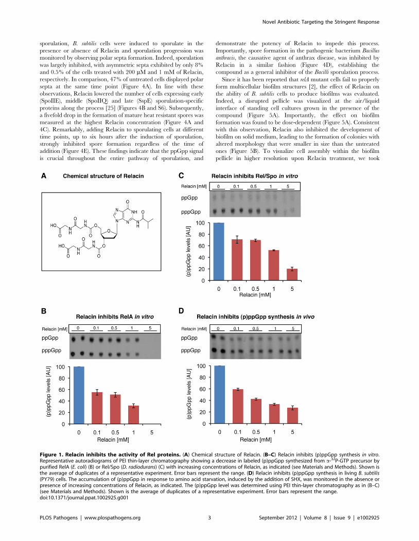
Figure 1. Relacin inhibits the activity of Rel proteins. (A) Chemical structure of Relacin. (B–C) Relacin inhibits (p)ppGpp synthesis in vitro.Representative autoradiograms of PEI thin-layer chromatography showing a decrease in labeled (p)ppGpp synthesized from a-32P-GTP precursor bypurified RelA (E. coli) (B) or Rel/Spo (D. radiodurans) (C) with increasing concentrations of Relacin, as indicated (see Materials and Methods). Shown isthe average of duplicates of a representative experiment. Error bars represent the range. (D) Relacin inhibits (p)ppGpp synthesis in living B. subtilis(PY79) cells. The accumulation of (p)ppGpp in response to amino acid starvation, induced by the addition of SHX, was monitored in the absence orpresence of increasing concentrations of Relacin, as indicated. The (p)ppGpp level was determined using PEI thin-layer chromatography as in (B–C)(see Materials and Methods). Shown is the average of duplicates of a representative experiment. Error bars represent the range.doi:10.1371/journal.ppat.1002925.g001
Novel Antibiotic Targeting the Stringent Response
PLOS Pathogens | www.plospathogens.org 3 September 2012 | Volume 8 | Issue 9 | e1002925

advantage of a strain harboring the rrnE promoter fused to gfp.
This promoter was found to be constitutively active [27], and
therefore reports cell viability and localization. Observing biofilm
pellicles by confocal laser scanning microscopy revealed that the
untreated cells formed homogeneous biofilm layers, while the
treated cell pellicles contained large gaps, indicating their
disintegrated state (Figure 5C). Moreover, staining the biofilm
with propidium iodide (PI), indicative of unviable cells, showed the
signal to be higher within the treated biofilm (Figure 5E). Finally,
quantifying GFP fluorescence from recovered pellicles revealed a
clear reduction in the viable biomass upon Relacin treatment, as
the measured fluorescence level was significantly reduced
(Figure 5D). Taken together, we conclude that Relacin interferes
with biofilm formation, an alternative bacterial developmental
pathway.
Discussion
In this report, we established Relacin as a novel antibacterial
agent. By specifically interfering with the activation of the
Stringent Response, Relacin perturbs the switch into stationary
phase in several tested Gram positive bacteria and leads to
bacterial death. Although Relacin did not affect growth and
survival of the Gram negative E. coli, it was found to effectively
inhibit the E. coli RelA in vitro, implying that improving the delivery
of Relacin to Gram negative bacteria may lead to an effective
outcome. Relacin was found to block every tested stage of B. subtilis
sporulation, proving the essentiality of the Stringent Response
throughout this process. Finally, we demonstrate that Relacin
affects the production of multicellular biofilm communities,
formed in response to challenging conditions. Taken together,
we present evidence that Relacin impedes bacterial long term
survival pathways, placing the compound as a new promising
antibacterial agent.
By utilizing the crystal structure of Rel/Spo from the S.
equisimilis, we were able to model the interaction of Relacin with
amino acid residues located within the Rel/Spo synthetase site.
This analysis yielded the identification of a putative binding mode
of Relacin, presumably adopting the conformation shown in
Figure S1C. In this conformation, Relacin forms a net of hydrogen
Figure 2. The effect of Relacin on Rel-ribosomes interaction. (A) Relacin inhibits dissociation of Rel/Spo from the ribosome. The relativeamount of Rel/Spo (D. radiodurans) bound to purified ribosomes was quantified following the addition of increasing levels of Relacin. Rel/Spomolecules associated with 70S complexes were detected by Western blot analysis (see Materials and Methods). Histogram indicates the average oftwo independent biological repeats. Error bars represent the range. (B) Ribosome independent inhibition of (p)ppGpp synthesis. The constitutivelyactive, ribosome-independent RelAC638F (E. coli) protein was treated with increasing concentrations of Relacin, as indicated (see Materials andMethods) in the presence or absence of isolated ribosomes. Shown is the average of duplicates of a representative experiment. Error bars representthe range.doi:10.1371/journal.ppat.1002925.g002
Novel Antibiotic Targeting the Stringent Response
PLOS Pathogens | www.plospathogens.org 4 September 2012 | Volume 8 | Issue 9 | e1002925
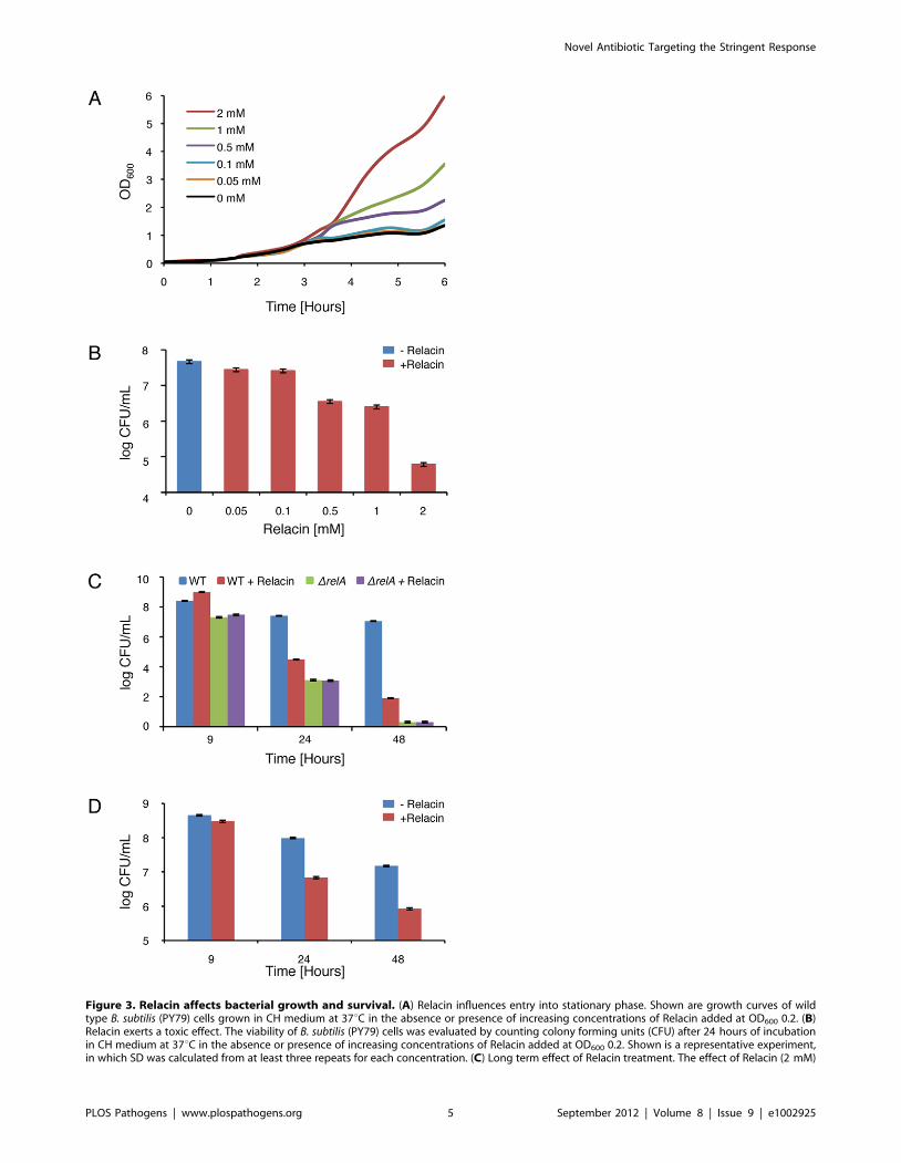
Figure 3. Relacin affects bacterial growth and survival. (A) Relacin influences entry into stationary phase. Shown are growth curves of wildtype B. subtilis (PY79) cells grown in CH medium at 37uC in the absence or presence of increasing concentrations of Relacin added at OD600 0.2. (B)Relacin exerts a toxic effect. The viability of B. subtilis (PY79) cells was evaluated by counting colony forming units (CFU) after 24 hours of incubationin CH medium at 37uC in the absence or presence of increasing concentrations of Relacin added at OD600 0.2. Shown is a representative experiment,in which SD was calculated from at least three repeats for each concentration. (C) Long term effect of Relacin treatment. The effect of Relacin (2 mM)
Novel Antibiotic Targeting the Stringent Response
PLOS Pathogens | www.plospathogens.org 5 September 2012 | Volume 8 | Issue 9 | e1002925

bonds and hydrophobic interactions that are most likely to provide
a more efficient binding in comparison to previously identified
inhibitors exhibiting lower activity [19].
Relacin appears to specifically target Rel proteins, as the effect of
the compound was nearly undetectable when tested on Rel/Spo
mutant cells. Consistently, Relacin activity in vivo resulted in a sharp
decrease in (p)ppGpp synthesis. Since ppGpp inhibits the enzyme
inosine monophosphate dehydrogenase, it causes the cellular GTP
levels to decrease [28]. The intracellular levels of GDP/GTP are
known to determine the initiation of several developmental
pathways such as sporulation and biofilm formation [26,29,30]
that were indeed shown to be influenced by Relacin. Interestingly,
we also observed that Relacin treatment resulted in a large decrease
in Rel/Spo ability to dissociate from ribosomes in vitro. This
deficiency could be explained by the model proposed by Wendrich
et al., [10] in which the rapid accumulation of ppGpp during amino
acid starvation is attributed to the ability of RelA to ‘hop’ between
ribosomes. This potential hopping is probably a consequence of the
synthesis of (p)ppGpp that releases RelA from the ribosome,
liberating it for another synthesis cycle.
The emergence of bacterial resistance to the current array of
antimicrobial agents demands the development of novel strategies
to eradicate pathogenic bacteria. The traditional cellular antibiotic
targets include ribosomes, cell wall constituents and components of
nucleic acids synthesis [31]. These cellular targets are mainly
active during the bacterial vegetative phase, making the available
antibiotics effective mostly during growth. However, the ability of
bacteria to reside in nature within biofilm communities or as
durable spores, as well as to become persistent to antibiotic
treatment [32], sets the need to tackle these alternative modes. In
this regard, Relacin affects specifically the Stringent Response, a
pathway crucial for the activation of bacterial survival strategies.
Since Relacin can persist for a relatively long period of time, and
exert its effect even a few days post addition, it might become a
valuable antagonist of these long term survival approaches. Taken
together, Relacin may be combined with antibiotics currently in
use, to eradicate non-homogenous bacterial populations with cells
residing in diverse life cycles.
Cellular components, which are conserved throughout the
bacterial kingdom and crucial for cellular survival, provide
attractive antimicrobial targets as long as they lack eukaryotic
counterparts. One of such targets is the highly conserved bacterial
tubulin-like cell division protein FtsZ, which provides the basis for
the assembly of the division machinery [33]. Indeed, a promising
inhibitor of FtsZ with potent and selective activity against
Staphylococci has been described [34]. In a similar fashion, the
ubiquity of Rel enzymes among bacteria, combined with the
absence of known (p)ppGpp synthetases in mammalian cells
[35,36], strengthen the potential of Relacin to turn into a
therapeutic antibiotic. The profound influence of Relacin on long
term bacterial survival makes it an attractive compound to serve as
a scaffold for generating an array of new antibacterial agents.
Materials and Methods
Synthesis and modeling of RelacinSynthesis of Relacin and a structural model for its interaction
with Rel/Spo (p)ppGpp synthetase binding pocket are described in
details Text S1.
Bacterial growth conditionsBacterial strains used in this study are described in Table S1.
Plasmid construction is described in Text S1. All general methods
for B. subtilis were carried out as described previously [37]. B.
subtilis cells were grown in hydrolyzed casein (CH) at 37uC [37],
unless indicated differently. GAS strain was grown at 37uCwithout shaking in Todd-Hewitt medium supplemented with 0.2%
yeast extract (THY) [38]. D. radiodurans R1 cells were grown in
TYG which contains: 0.5% tryptone, 0.3% yeast extract and 0.1%
glucose at 30uC with shaking. E. coli cells were grown at 37uC in
LB medium. Cultures were inoculated to an OD600 of 0.05 using
an overnight culture grown in the same medium, unless indicated
differently. Sporulation conditions and biofilm colony and pellicle
formation are described in Text S1.
Purification of Rel proteins and crude ribosomesPurification of RelA or RelA-C638F from E. coli (CF9467)
harboring DrelA and over-expressing pQE30-RelA or pQE30-
RelA-C638F respectively, was carried out as described previously
[19]. Purification of Rel/Spo from D. Radiodurans R1 was
performed under identical conditions; however, the protein was
expressed in E. coli BL21 CodonPlus (Stratagene) cells. Of note,
Rel/Spo from D. Radiodurans R1, is the only known full length
active protein purified from Gram positive bacteria. Isolation of
crude ribosomes (containing 70S, mRNA, tRNA) from E. coli
(CF9467) was carried out as described previously [19]. Isolation
of crude ribosomes from D. Radiodurans was carried out in a
similar fashion with the following modifications: D. radiodurans R1
cells were grown in LB(+) over night at 30uC, cells were diluted
1:100 in LB(+) medium and incubated at 30uC for additional
48 hours.
Measuring (p)ppGpp synthesis in vitroFor measuring (p)ppGpp synthesis by RelA, RelA-C638F or
Rel/Spo proteins in vitro: 1 mg of purified Rel protein, 20 mg of
isolated ribosomes and 10 mCi of a-32P labeled GTP, were mixed
in a buffer [0.5 mM GTP, 4 mM ATP, 50 mM Tris-HCl
(pH 7.4), 1 mM DTT, 10 mM MgCl2, 10 mM KCl and
27 mM (NH4)2SO4] to a final volume of 20 mL without or with
increasing amounts of Relacin as indicated. Reactions were
stopped by the addition of 5 mL formic acid. Each reaction was
loaded (5 mL) and separated on Cellulose PEI TLC plates (Merck)
using 1.5 M KH2PO4 as mobile phase. Plates were autoradio-
graphed using the Fijix Bas100 PhosphorImager (Japan).
(p)ppGpp signal was measured using TINA 2.0 software (Raytest,
Strauben-Hardt). The total amount of (p)ppGpp was the sum of
signals from ppGpp and pppGpp.
Measuring (p)ppGpp synthesis in vivoB. subtilis (PY79) or E. coli (W3110) cells were grown in MOPS
glucose minimal medium [39] supplemented with all amino
acids except glutamine and glutamate. At OD600 0.1, cells were
supplemented with H332PO4 and incubated for 45 minutes,
after which Relacin was added at the indicated concentrations.
Cells were incubated for additional 15 minutes. Next, amino
acid starvation was induced by adding serine-hydroxamate
(SHX, Sigma) 1 mg/mL [20]. Samples were withdrawn ten
minutes after addition of SHX and analyzed for their (p)ppGpp
on the viability of wild type B. subtilis (PY79) cells or DrelA (ME215) cells was measured. Cells were incubated in CH medium at 37uC, and viability wasdetermined by counting colony forming units (CFU). Relacin was added at OD600 0.2. Shown is a representative experiment, in which SD wascalculated from at least three repeats for each point. (D) The toxic effect of Relacin on GAS. The effect of Relacin (2 mM) on the viability of wild typeGAS (JRS4) cells, incubated in THY medium at 37uC, was evaluated as in (C).doi:10.1371/journal.ppat.1002925.g003
Novel Antibiotic Targeting the Stringent Response
PLOS Pathogens | www.plospathogens.org 6 September 2012 | Volume 8 | Issue 9 | e1002925

content as described above (Measuring (p)ppGpp synthesis in
vitro).
Measuring Rel/Spo- 70S associationThe reaction was carried out as described above for
measuring (p)ppGpp synthesis in vitro, without the addition of
radiolabeled GTP, with or without increasing amounts of
Relacin as indicated. Reactions were centrifuged for 4 hours at
35,000 g (4uC), ribosomal fractions were separated by 12%
SDS-polyacrylamide gel electrophoresis, transferred to PVDF
membrane (Millipore Bedford) and processed for immunoreac-
tion using mouse anti-His antibody (1:10,000; Amersham).
Immunoreactive proteins were detected using a chemilumines-
cence kit (Biological Industries) according to the manufacturer’s
protocol.
Fluorescence microscopyFluorescence microscopy was carried out as previously described
[40]. Samples (0.5 mL) of a given culture were removed, centrifuged
briefly, and resuspended in 10 mL of PBS61 (Phosphate-Buffered
Saline) supplemented with 1 mg/mL membrane stain FM1–43 or
FM4–64 (Molecular Probes, Invitrogen). Cells were visualized and
photographed using an Axioplan2 microscope (Zeiss) equipped with
CoolSnap HQ camera (Photometrics, Roper Scientific) or an
Axioobserver Z1 microscope (Zeiss) equipped with a CoolSnap
HQII camera (Photometrics, Roper Scientific). System control and
Figure 4. Relacin influences the sporulation process in Bacilli. (A) Relacin inhibits sporulation. Microscopy images of sporulating wild type B.subtilis (PY79) cells in the absence or presence of Relacin, added at time 0 of sporulation at the indicated concentrations. Upper panels: cells at t = 2 hrof sporulation stained with the fluorescent membrane dye FM1–43. Arrows indicate position of polar septa. Lower panels: phase contrast images ofcells at t = 24 hr of sporulation. Scale bars correspond to 1 mm. (B) Relacin inhibits expression of the mid-sporulation protein SpoIIQ. Fluorescencemicroscopy images of B. subtilis (PE128) cells harboring spoIIQ-gfp at t = 4 hr of sporulation, in the absence (upper panels) or presence (lower panels)of Relacin (1 mM), added at time 0 of sporulation. Shown are phase contrast (red), GFP fluorescence (green) and overlay images. Scale barcorresponds to 1 mm. (C–D) Relacin inhibits Bacilli spore formation. The formation of heat resistant B. subtilis (PY79) (C) and B. anthracis (Sterne) (D)spores was monitored in the absence or presence of Relacin, added at the indicated concentrations at time 0 of sporulation (see Text S1). Shown arerepresentative experiments, in which SD was calculated from at least three repeats for each concentration. (E) Relacin added at different time pointsduring sporulation inhibits spore formation. Inhibition of spore formation by wild type B. subtilis (PY79) cells was evaluated after addition of Relacin(1 mM) at the indicated time points of sporulation. Inhibition was determined using a heat resistance assay (see Text S1) and is expressed relative tountreated cultures. Shown is a representative experiment, in which SD was calculated from at least three repeats for each time point.doi:10.1371/journal.ppat.1002925.g004
Novel Antibiotic Targeting the Stringent Response
PLOS Pathogens | www.plospathogens.org 7 September 2012 | Volume 8 | Issue 9 | e1002925
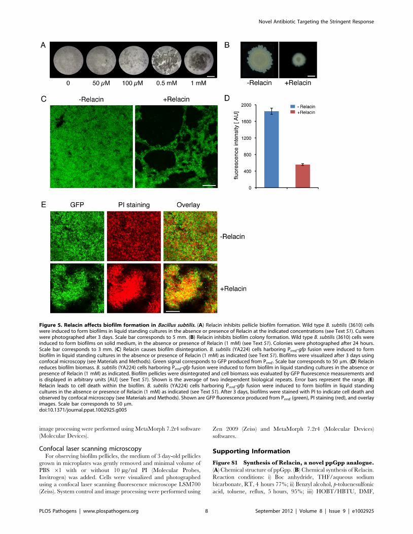
image processing were performed using MetaMorph 7.2r4 software
(Molecular Devices).
Confocal laser scanning microscopyFor observing biofilm pellicles, the medium of 3 day-old pellicles
grown in microplates was gently removed and minimal volume of
PBS 61 with or without 10 mg/ml PI (Molecular Probes,
Invitrogen) was added. Cells were visualized and photographed
using a confocal laser scanning fluorescence microscope LSM700
(Zeiss). System control and image processing were performed using
Zen 2009 (Zeiss) and MetaMorph 7.2r4 (Molecular Devices)
softwares.
Supporting Information
Figure S1 Synthesis of Relacin, a novel ppGpp analogue.(A) Chemical structure of ppGpp. (B) Chemical synthesis of Relacin.
Reaction conditions: i) Boc anhydride, THF/aqueous sodium
bicarbonate, RT, 4 hours 77%; ii) Benzyl alcohol, p-toluenesulfonic
acid, toluene, reflux, 5 hours, 95%; iii) HOBT/HBTU, DMF,
Figure 5. Relacin affects biofilm formation in Bacillus subtilis. (A) Relacin inhibits pellicle biofilm formation. Wild type B. subtilis (3610) cellswere induced to form biofilms in liquid standing cultures in the absence or presence of Relacin at the indicated concentrations (see Text S1). Cultureswere photographed after 3 days. Scale bar corresponds to 5 mm. (B) Relacin inhibits biofilm colony formation. Wild type B. subtilis (3610) cells wereinduced to form biofilms on solid medium, in the absence or presence of Relacin (1 mM) (see Text S1). Colonies were photographed after 24 hours.Scale bar corresponds to 3 mm. (C) Relacin causes biofilm disintegration. B. subtilis (YA224) cells harboring PrrnE-gfp fusion were induced to formbiofilm in liquid standing cultures in the absence or presence of Relacin (1 mM) as indicated (see Text S1). Biofilms were visualized after 3 days usingconfocal microscopy (see Materials and Methods). Green signal corresponds to GFP produced from PrrnE. Scale bar corresponds to 50 mm. (D) Relacinreduces biofilm biomass. B. subtilis (YA224) cells harboring PrrnE-gfp fusion were induced to form biofilm in liquid standing cultures in the absence orpresence of Relacin (1 mM) as indicated. Biofilm pellicles were disintegrated and cell biomass was evaluated by GFP fluorescence measurements andis displayed in arbitrary units [AU] (see Text S1). Shown is the average of two independent biological repeats. Error bars represent the range. (E)Relacin leads to cell death within the biofilm. B. subtilis (YA224) cells harboring PrrnE-gfp fusion were induced to form biofilm in liquid standingcultures in the absence or presence of Relacin (1 mM) as indicated (see Text S1). After 3 days, biofilms were stained with PI to indicate cell death andobserved by confocal microscopy (see Materials and Methods). Shown are GFP fluorescence produced from PrrnE (green), PI staining (red), and overlayimages. Scale bar corresponds to 50 mm.doi:10.1371/journal.ppat.1002925.g005
Novel Antibiotic Targeting the Stringent Response
PLOS Pathogens | www.plospathogens.org 8 September 2012 | Volume 8 | Issue 9 | e1002925
DIEA, RT, overnight. 98%; iv) 50% TFA in DCM, RT,
30 minutes, 86%; v) a) trimethylsilyl chloride, pyridine, 0uC,
1 hour, b) Isobutyric anhydride, RT, 4 hours, 87%; vi) a) CDI,
acetonitrile, RT, overnight, b) (4), DCM, DIEA, RT, 20 hours,
48%; vii) H2, 10% Pd/C, methanol, 3 hours, RT, 30 psi, 80%. (C)
Structural basis of binding and inhibition of Rel/Spo by Relacin. A
putative model describing how Relacin (sticks, colored according to
the cpk scheme) binds in the known GDP binding site of Rel/Spo
protein from Streptococcus equisimilis (shown as transparent white
surface and cartoon). Relacin also forms additional contacts with
Rel/Spo within the active site. GDP is shown in black lines for
comparison, and residues of Rel/Spo that form hydrogen bonding
contacts to GDP are shown in stick representation (see Text S1).
The high affinity of Relacin can be explained by the extensive
contacts formed between the ligand and the receptor. The ligand
occupies a considerable volume of the binding pocket, including
both the GDP binding sites, as well as additional regions. In addition
to a range of hydrogen bonds mediated by the overall very polar
pocket, the hydrophobic isobutyryl group contacts a defined
hydrophobic patch shown as black dots.
(TIF)
Figure S2 The effect of Relacin on (p)ppGpp synthesis.(A) Relacin inhibits RelA in vitro. The relative amount of ppGpp
and pppGpp produced by purified RelA (E. coli) in the absence or
presence of Relacin at the indicated concentrations was calculated
from autoradiograms of PEI thin-layer chromatography, corre-
sponding to Figure 1B. Shown is the average of duplicates of a
representative experiment. Error bars represent the range. (B)
Relacin inhibits Rel/Spo in vitro. The relative amount of ppGpp
and pppGpp produced by purified Rel/Spo (D. radiodurans) in the
absence or presence of Relacin at the indicated concentrations was
calculated from autoradiograms of PEI thin-layer chromatogra-
phy, corresponding to Figure 1C. Shown is the average of
duplicates of a representative experiment. Error bars represent the
range. (C) Relacin does not inhibit (p)ppGpp synthesis in living E.
coli cells. The accumulation of (p)ppGpp in response to amino acid
starvation, induced by SHX, was monitored in the absence or
presence of increasing concentrations of Relacin. The (p)ppGpp
level was determined using PEI thin-layer chromatography of
radiolabeled (p)ppGpp (see Materials and Methods). Histogram
indicates the average of two independent biological repeats. Error
bars represent the range.
(TIF)
Figure S3 Effect of Relacin on survival of B. subtilisgrown in minimal medium. Survival of wild type B. subtilis
(PY79) cells grown in S7 minimal medium was determined by
CFU counting after 9 and 24 hours of incubation in the absence
or presence of Relacin (2 mM), as indicated. Relacin was added at
OD600 0.2. Shown is a representative experiment, in which SD
was calculated from at least three repeats for each point.
(TIF)
Figure S4 The toxic effect of Relacin is visible. (A–B) B.
subtilis (PY79) cells were grown in CH medium at 37uC in the absence
(upper panels) and presence (lower panels) of Relacin (1 mM), added
at OD600 0.2. Cells were stained with viability indicators SYTO9
(green, highlights live cells) and PI (red, highlights dead cells) at
9 hours (A) and 24 hours (B) of incubation. Shown are phase contrast
images (left panels) and Live/Dead overlay fluorescence images (right
panels). Of note, some of the disintegrated cells were not stained with
any of the dyes. Scale bar corresponds to 1 mm.
(TIF)
Figure S5 Relacin affects growth and survival ofGram positive bacteria. (A) Effect of Relacin on growth
of GAS. Shown are growth curves of wild type GAS (JRS4)
cells grown at 37uC without shaking in THY medium in the
absence or presence of increasing concentrations of Relacin
added at OD600 0.2. (B) Effect of Relacin on survival of GAS.
The effect of Relacin, at the indicated concentrations, on
survival of GAS (JRS4) cells grown at 37uC without shaking in
THY medium was determined by CFU counting of treated
and untreated cultures. Relacin was added at OD600 0.2.
Shown is a representative experiment, in which SD was
calculated from at least three repeats for each concentration.
(C) Effect of Relacin on growth of D. radiodurans. Shown are
growth curves of wild type D. radiodurans R1 grown in TYG
medium at 30uC in the absence or presence of Relacin at the
indicated concentrations added at OD600 0.2. (D) Effect of
Relacin on D. radiodurans survival. The effect of Relacin, at the
indicated concentrations, on survival of wild type D. radiodurans
R1 cells grown at 30uC in TYG medium was determined by
CFU counting of treated and untreated cultures. Relacin was
added at OD600 0.2. Shown is a representative experiment, in
which SD was calculated from at least three repeats for each
point.
(TIF)
Figure S6 Effect of Relacin on the expression of earlyand late sporulation-specific proteins. (A) Fluorescence
microscopy images of B. subtilis (SB201) cells harboring spoIIE-gfp
fusion at t = 2 hr of sporulation, in the absence (upper panels)
and presence (lower panels) of Relacin (1 mM), added at time 0
of sporulation. Shown are cells stained with FM4–64 membrane
dye (red), SpoIIE-GFP fluorescence (green) and overlay images.
Scale bar corresponds to 1 mm. (B) Fluorescence microscopy
images of B. subtilis (ES7) cells harboring sspE-gfp fusion at
t = 5 hr of sporulation, in the absence (upper panels) and
presence (lower panels) of Relacin (1 mM), added at time 0 of
sporulation. Shown are phase contrast (red), SspE-GFP fluores-
cence (green) and overlay images. Scale bar corresponds to
1 mm.
(TIF)
Text S1 Supplemental information, including one tableas well as additional experimental procedures andreferences.
(DOCX)
Acknowledgments
We thank R. Losick (Harvard U), A. Rouvinski (Hebrew U) and B. Traag
(Harvard U) for comments on the manuscripts. We are grateful to M.
Ravins (Hebrew U), C. Block and R. Nir-Paz (Hadassah Medical Center)
for technical help. We thank C. Block (Hadassah Medical Center), M.
Cashel (NIH), E. Hanski (Hebrew U) and R. Rudner (Hunter College) for
providing strains. We are indebted to members of the Ben-Yehuda
laboratory, for valuable help and discussions, especially to A. Rosenberg
and E. Segev for contributing strains and G. Mamou for technical help.
Relacin is the subject of a provisional application for patent: application
No. 61/431,902 01/2011.
Author Contributions
Conceived and designed the experiments: EW YOS IK NL OSF EY GG
JK SBY. Performed the experiments: EW YOS IK NL OSF. Analyzed the
data: EW YOS IK NL OSF EY GG JK SBY. Wrote the paper: EW YOS
SBY.
Novel Antibiotic Targeting the Stringent Response
PLOS Pathogens | www.plospathogens.org 9 September 2012 | Volume 8 | Issue 9 | e1002925

References
1. Jain V, Kumar M, Chatterji D (2006) ppGpp: Stringent response and survival.
J Microbiol 44: 1–10.2. Lemos JAC, Brown TA, Burne RA (2004) Effects of RelA on key virulence
properties of planktonic and biofilm populations of Streptococcus mutans. InfectImmun 72: 1431–1440.
3. Ochi K, Kandala JC, Freese E (1981) Initiation of Bacillus subtilis sporulation by
the stringent response to partial amino-acid deprivation. J Biol Chem 256: 6866–6875.
4. Potrykus K, Cashel M (2008) (p)ppGpp: Still Magical? Annu Rev Microbiol pp.35–51.
5. Nguyen D, Joshi-Datar A, Lepine F, Bauerle E, Olakanmi O, et al. (2011) Active
starvation responses mediate antibiotic tolerance in biofilms and nutrient-limitedbacteria. Science 334: 982–986.
6. Cashel M, Gallant J (1969) Two compounds implicated in the function of theRC gene of Escherichia coli. Nature 221: 838–841.
7. Cashel M GD, Hernandez VH, Vinella D (1996) The stringent response. In:Neidhardt, FC et al, editors. Escherichia coli and Salmonella typhimurium: Cellular and
Molecular Biology (2nd edn). ASM Press. pp. 1458–1496.
8. Metzger S, Sarubbi E, Glaser G, Cashel M (1989) Protein sequences encoded bythe relA and the spoT genes of Escherichia coli are interrelated. J Biol Chem 264:
9122–9125.9. Wendrich TM, Marahiel MA (1997) Cloning and characterization of a relA/spoT
homologue from Bacillus subtilis. Mol Microbiol 26: 65–79.
10. Wendrich TM, Blaha G, Wilson DN, Marahiel MA, Nierhaus KH (2002)Dissection of the mechanism for the stringent factor RelA. Mol Cell 10: 779–
788.11. English BP, Hauryliuk V, Sanamrad A, Tankov S, Dekker NH, et al. (2011)
Single-molecule investigations of the stringent response machinery in livingbacterial cells. Proc Natl Acad Sci U S A 108: E365–373.
12. Mechold U, Murphy H, Brown L, Cashel M (2002) Intramolecular regulation of
the opposing (p)ppGpp catalytic activities of Rel(Seq), the Rel/Spo enzyme fromStreptococcus equisimilis. J Bacteriol 184: 2878–2888.
13. Hogg T, Mechold U, Malke H, Cashel M, Hilgenfeld R (2004) Conformationalantagonism between opposing active sites in a bifunctional RelA/SpoT homolog
modulates (p)ppGpp metabolism during the stringent response. Cell 117: 57–68.
14. Barker MM, Gaal T, Josaitis CA, Gourse RL (2001) Mechanism of regulation oftranscription initiation by ppGpp. I. Effects of ppGpp on transcription initiation
in vivo and in vitro. J Mol Biol 305: 673–688.15. Krasny L, Gourse RL (2004) An alternative strategy for bacterial ribosome
synthesis: Bacillus subtilis rRNA transcription regulation. EMBO J 23: 4473–4483.
16. Vrentas CE, Gaal T, Berkmen MB, Rutherford ST, Haugen SP, et al. (2008)
Still looking for the magic spot: The crystallographically defined binding site forppGpp on RNA polymerase is unlikely to be responsible for rRNA transcription
regulation (vol 377, pg 551, 2008). J Mol Biol 379: 1130–1130.17. Gruber TM, Gross CA (2003) Multiple sigma subunits and the partitioning of
bacterial transcription space. In: Ornston LN, editor. Annu Rev Microbiol
Volume 57. pp. 441–466.18. Magnusson LU, Farewell A, Nystrom T (2005) ppGpp: a global regulator in
Escherichia coli. Trends Microbiol 13: 236–242.19. Wexselblatt E, Katzhendler J, Saleem-Batcha R, Hansen G, Hilgenfeld R, et al.
(2010) ppGpp analogues inhibit synthetase activity of Rel proteins from Gramnegative and Gram positive bacteria. Bioorg Med Chem 18: 4485–4497.
20. Gropp M, Strausz Y, Gross M, Glaser G (2001) Regulation of Escherichia coli
RelA requires oligomerization of the C-terminal domain. J Bacteriol 183: 570–
579.
21. Tosa T, Pizer LI (1971) Effect of serine hydroxamate on the growth of Escherichia
coli. J Bacteriol 106: 966–971.
22. Zhou YN, Coleman WG, Yang ZX, Yang Y, Hodgson N, et al. (2008)
Regulation of Cell Growth during Serum Starvation and Bacterial Survival inMacrophages by the Bifunctional Enzyme SpoT in Helicobacter pylori. J Bacteriol
190: 8025–8032.
23. Errington J (2003) Regulation of endospore formation in Bacillus subtilis. Nat Rev
Microbiol 1: 117–126.
24. Piggot PJ, Hilbert DW (2004) Sporulation of Bacillus subtilis. Curr Opin
Microbiol 7: 579–586.
25. Stragier P, Losick R (1996) Molecular genetics of sporulation in Bacillus subtilis.
Annu Rev Genet 30: 297–341.
26. Lopez JM, Marks CL, Freese E (1979) Decrease of guanine-nucleotides initiates
sporulation of Bacillus subtilis. Biochim Biophys Acta 587: 238–252.
27. Rosenberg A, Sinai L, Smith Y, Ben-Yehuda S (2012) Dynamic Expression of
the Translational Machinery during Bacillus subtilis Life Cycle at a Single Cell
Level. PLoS One 7: e41921.
28. Ochi K, Kandala J, Freese E (1982) Evidence that Bacillus subtilis sporulation
induced by the stringent response is caused by the decrease in GTP or GDP.J Bacteriol 151: 1062–1065.
29. Hsueh YH, Somers EB, Wong AC (2008) Characterization of the codY gene andits influence on biofilm formation in Bacillus cereus. Arch Microbiol 189: 557–568.
30. Ratnayake-Lecamwasam M, Serror P, Wong KW, Sonenshein AL (2001)Bacillus subtilis CodY represses early-stationary-phase genes by sensing GTP
levels. Genes Dev 15: 1093–1103.
31. Kohanski MA, Dwyer DJ, Collins JJ (2010) How antibiotics kill bacteria: from
targets to networks. Nat Rev Microbiol 8: 423–435.
32. Costerton JW, Stewart PS, Greenberg EP (1999) Bacterial biofilms: a common
cause of persistent infections. Science 284: 1318–1322.
33. Goehring NW, Beckwith J (2005) Diverse paths to midcell: assembly of the
bacterial cell division machinery. Curr Biol 15: R514–526.
34. Haydon DJ, Stokes NR, Ure R, Galbraith G, Bennett JM, et al. (2008) An
inhibitor of FtsZ with potent and selective anti-staphylococcal activity. Science321: 1673–1675.
35. Mittenhuber G (2001) Comparative genomics and evolution of genes encodingbacterial (p)ppGpp synthetases/hydrolases (the Rel, RelA and SpoT proteins).
J Mol Microbiol Biotech 3: 585–600.
36. Sun D, Lee G, Lee JH, Kim H-Y, Rhee H-W, et al. (2010) A metazoan orthologof SpoT hydrolyzes ppGpp and functions in starvation responses. Nat Struct
Mol Biol 17: 1188–1194.
37. Harwood CR, Cutting SM (1990) Modern microbiological methods molecular
biological methods for Bacillus. John Wiley & Sons Press. 618 p.
38. Vanderijn I, Kessler RE (1980) Growth-characteristics of Group-A streptococci in
a new chemically defined medium. Infect Immun 27: 444–448.
39. Neidhard F, Bloch PL, Smith DF (1974) Culture medium for enterobacteria.
J Bacteriol 119: 736–747.
40. Bejerano-Sagie M, Oppenheimer-Shaanan Y, Berlatzky I, Rouvinski A,
Meyerovich M, et al. (2006) A checkpoint protein that scans the chromosomefor damage at the start of sporulation in Bacillus subtilis. Cell 125: 679–690.
Novel Antibiotic Targeting the Stringent Response
PLOS Pathogens | www.plospathogens.org 10 September 2012 | Volume 8 | Issue 9 | e1002925
Related Documents