Regulatory T-Cell (Treg) Hybridoma as a Novel Tool to Study Foxp3 Regulation and Treg Fate 1 Rahul Sharma * , Sun-Sang J. Sung * , Chiao-Ying A. Ju * , S. Deshmukh Umesh * , Shu Man Fu *,§ , and Shyr-Te Ju *,§ * Center for Inflammation, Immunity and Regenerative Medicine, Department of Medicine University of Virginia, Charlottesville, VA 22908-0412 § Department of Microbiology, University of Virginia, Charlottesville, VA 22908-0412 Abstract The CD25 + Foxp3 + regulatory T-cells (Treg) that had lost CD25 and Foxp3 in vivo (ex-Treg) exist but are difficult to study. We generated antigen (Ag)-specific Treg hybridomas from iTreg clones (iTreg-hyb) using iTreg of DO11.10.Foxp3-GFP mice and presented evidence that they behave like ex-Treg. The iTreg-hyb displayed little CD25 and Foxp3-GFP but strong expression could be induced with OVA 323–339 in the presence of Ag-presenting cells, rIL-2 and rTGF-β1. They displayed all of the iTreg-associated markers examined except CTLA-4, the latter was also absent in the ex-Treg. They lacked the Helios transcription factor, suggesting they were derived from iTreg. Similar to ex-Treg, the iTreg-hyb produced high level of IL-2 and Foxp3 under specific activation conditions. Two unusual properties were observed. First, the ability to induce Foxp3- GFP upon activation is progressively lost in culture over a period of 2 to 4 weeks. Second, Rag2 −/− spleen cells alone selectively induced Foxp3-GFP expression albeit 30 times less efficient than Ag-specific activation. We identified cell-free supernatant, IL-6, IL-9, and IL-27 as Foxp3- inducing factors. Our study has significant implications to the stability, plasticity and fate of Treg. The usefulness and limitation of iTreg-hyb as a novel tool to study Foxp3 regulation and the fate of specific Treg subsets are discussed. Keywords Foxp3; Hybridoma; Cytokines; Regulatory T-cell fate Introduction The CD4 + T-cells can be classified into subsets based on functions, cytokine patterns, and expression of subset-specific transcription factors (reviewed in 1). The presence of the X- linked transcription factor Foxp3 and/or CD25 in the CD4 + T-cells defines the naturally- 1 Supported by NIH grants AR-051203 (STJ), DE-017579 (STJ), AR-051203 (SMF), AR-047988 (SMF), AR-049449 (SMF), AI-079621 (USD), DE-109883 (USD), and AI-079906 (SJS). We thank Dr. C. David for providing the class-II −/− mice and Mr. C. Abaya for technical help. © 2011 Elsevier Ltd. All rights reserved. Correspondence: Shu Man Fu, MD. Ph.D., or Shyr-Te Ju, Ph.D. Rm5777, 5 th floor, Old Medical School Building, Hospital Drive, University of Virginia, Charlottesville, VA 22908-0412. (Tel: 434-924-9627 or 434-243-6358; Fax: 434-924-9578), [email protected]. Publisher's Disclaimer: This is a PDF file of an unedited manuscript that has been accepted for publication. As a service to our customers we are providing this early version of the manuscript. The manuscript will undergo copyediting, typesetting, and review of the resulting proof before it is published in its final citable form. Please note that during the production process errors may be discovered which could affect the content, and all legal disclaimers that apply to the journal pertain. NIH Public Access Author Manuscript J Autoimmun. Author manuscript; available in PMC 2012 September 1. Published in final edited form as: J Autoimmun. 2011 September ; 37(2): 113–121. doi:10.1016/j.jaut.2011.05.008. NIH-PA Author Manuscript NIH-PA Author Manuscript NIH-PA Author Manuscript

Welcome message from author
This document is posted to help you gain knowledge. Please leave a comment to let me know what you think about it! Share it to your friends and learn new things together.
Transcript

Regulatory T-Cell (Treg) Hybridoma as a Novel Tool to StudyFoxp3 Regulation and Treg Fate1
Rahul Sharma*, Sun-Sang J. Sung*, Chiao-Ying A. Ju*, S. Deshmukh Umesh*, Shu ManFu*,§, and Shyr-Te Ju*,§
* Center for Inflammation, Immunity and Regenerative Medicine, Department of MedicineUniversity of Virginia, Charlottesville, VA 22908-0412§ Department of Microbiology, University of Virginia, Charlottesville, VA 22908-0412
AbstractThe CD25+Foxp3+ regulatory T-cells (Treg) that had lost CD25 and Foxp3 in vivo (ex-Treg) existbut are difficult to study. We generated antigen (Ag)-specific Treg hybridomas from iTreg clones(iTreg-hyb) using iTreg of DO11.10.Foxp3-GFP mice and presented evidence that they behavelike ex-Treg. The iTreg-hyb displayed little CD25 and Foxp3-GFP but strong expression could beinduced with OVA323–339 in the presence of Ag-presenting cells, rIL-2 and rTGF-β1. Theydisplayed all of the iTreg-associated markers examined except CTLA-4, the latter was also absentin the ex-Treg. They lacked the Helios transcription factor, suggesting they were derived fromiTreg. Similar to ex-Treg, the iTreg-hyb produced high level of IL-2 and Foxp3 under specificactivation conditions. Two unusual properties were observed. First, the ability to induce Foxp3-GFP upon activation is progressively lost in culture over a period of 2 to 4 weeks. Second,Rag2−/− spleen cells alone selectively induced Foxp3-GFP expression albeit 30 times less efficientthan Ag-specific activation. We identified cell-free supernatant, IL-6, IL-9, and IL-27 as Foxp3-inducing factors. Our study has significant implications to the stability, plasticity and fate of Treg.The usefulness and limitation of iTreg-hyb as a novel tool to study Foxp3 regulation and the fateof specific Treg subsets are discussed.
KeywordsFoxp3; Hybridoma; Cytokines; Regulatory T-cell fate
IntroductionThe CD4+ T-cells can be classified into subsets based on functions, cytokine patterns, andexpression of subset-specific transcription factors (reviewed in 1). The presence of the X-linked transcription factor Foxp3 and/or CD25 in the CD4+ T-cells defines the naturally-
1Supported by NIH grants AR-051203 (STJ), DE-017579 (STJ), AR-051203 (SMF), AR-047988 (SMF), AR-049449 (SMF),AI-079621 (USD), DE-109883 (USD), and AI-079906 (SJS). We thank Dr. C. David for providing the class-II−/− mice and Mr. C.Abaya for technical help.© 2011 Elsevier Ltd. All rights reserved.Correspondence: Shu Man Fu, MD. Ph.D., or Shyr-Te Ju, Ph.D. Rm5777, 5th floor, Old Medical School Building, Hospital Drive,University of Virginia, Charlottesville, VA 22908-0412. (Tel: 434-924-9627 or 434-243-6358; Fax: 434-924-9578),[email protected]'s Disclaimer: This is a PDF file of an unedited manuscript that has been accepted for publication. As a service to ourcustomers we are providing this early version of the manuscript. The manuscript will undergo copyediting, typesetting, and review ofthe resulting proof before it is published in its final citable form. Please note that during the production process errors may bediscovered which could affect the content, and all legal disclaimers that apply to the journal pertain.
NIH Public AccessAuthor ManuscriptJ Autoimmun. Author manuscript; available in PMC 2012 September 1.
Published in final edited form as:J Autoimmun. 2011 September ; 37(2): 113–121. doi:10.1016/j.jaut.2011.05.008.
NIH
-PA Author Manuscript
NIH
-PA Author Manuscript
NIH
-PA Author Manuscript

occurring regulatory T cells (nTreg)2(2, 3). The Foxp3-expressing Treg is critical to selftolerance. Neonatal mice and newborn boys lacking functional Foxp3 invariably developfatal multi-organ inflammatory response (4–6).
Treg are generated in the thymus during T-cell development. They express CD25, Foxp3and the Helios transcription factor (7). Treg can also be induced (iTreg) when naïveCD4+CD44lowFoxp3− T cells in the periphery are activated with anti-CD3 mAb in thepresence of rIL-2 and rTGF-β1 (7, 8). Such induced Treg express CD25 and Foxp3 but notHelios (7). There are similarities and differences between the two populations (9). Studiesusing CD25+ and/or Foxp3+ CD4+ T-cells as the source of nTreg invariably contained boththe thymic-derived Treg and the peripheral-derived iTreg and as such the results andinterpretations were unavoidably complicated by this heterogeneity, making it difficult toassign the specific functions, Foxp3 regulation properties, and cell fate to a defined Tregsubset.
Under homeostatic condition, newly derived Treg from the thymus and peripherycontinuously enter the Treg pool and some of the existing Treg must lose their Foxp3expression (ex-Treg) and exit the pool. Several studies have demonstrated the existence ofex-Treg (10–12). It is important to understand the fate of the Foxp3+ T-cells because it hasimplication toward lineage specification, stability of the Treg, the fate of Treg and howperipheral tolerance is maintained (3). Transfer of the in vitro-generated iTreg into one-dayold normal recipients resulted in 50–75% loss of their Foxp3 expression at 3 weeks aftertransfer (11). Transfer of FACS-purified Foxp3-GFP+ nTreg into adult lymphopenic micerevealed that a large fraction of the transferred cells became CD25−Foxp3− ex-Treg (10). Afraction of the ex-Treg isolated from these mice could be induced to produce IL-2, IFN-γ,Foxp3, and IL-17 (10). In another study, the ex-Treg isolated from the in vivo-labeled ex-Treg induced autoimmune disease upon adoptive transfer (12). These observations wereinterpreted as evidence for the presence of an uncommitted Treg subset that became ex-Tregbut could further progress into distinct T-cell subsets under specific conditions. In contrast,using an in vivo Tamoxifen-induced labeling of Foxp3+ T-cells to track Treg fate, it wasdemonstrated that the great majority of the Foxp3+ Treg was stable over 5 to 8 months (13).Interestingly, approximately 5% of the labeled cells were Foxp3−. Although they could beex-Treg, further characterization of this population was not reported (13). Finally, someTreg and ex-Treg must also die in order to maintain the steady-state of the Treg pool.
To date, long-term in vitro maintained nTreg, iTreg or ex-Treg has not been achieved. Sucha condition could be useful to investigate Foxp3 regulation and Treg fate in the absence ofextrinsic stimulating signals. As an alternative, we attempted to derive Treg-hyb for such astudy. An early report claimed that Foxp3-expressing Treg-hyb could not be generated (14).Treg-hyb that expressed Foxp3 either constitutively or induced was not found. In that study,the critical Foxp3-inducing factors, rIL-2 and rTGF-β1, were not added during anti-CD3activation of the hybridomas. Intuitively, iTreg-hyb must maintain the ability to expressTCR, IL-2R, TGF-βR and their signaling components for Foxp3 induction.
Here, we report the first successful generation of Ag-specific iTreg-hyb derived fromDO11.10.Foxp3-GFP mice. We did not obtain Treg-hyb that constitutively express Foxp3-GFP. However, upon stimulation with immobilized anti-CD3 mAb and in the presence ofrIL-2 and rTGF-β1, Foxp3-GFP-expressing Treg-hyb were identified. Evidence that theiTreg-hyb were derived from iTreg but not thymic-derived Treg is presented. Similar to theCD25−Foxp3− ex-Treg, the iTreg-hyb could be induced to express CD25, IL-2, and Foxp3.They also express many of the Treg-associated markers some of which are constitutive
2Abbreviations: CFS: Cell-free supernatant; Hyb: Hybridomas; SFS: Serum-free supernatant; Treg: Regulatory T cells
Sharma et al. Page 2
J Autoimmun. Author manuscript; available in PMC 2012 September 1.
NIH
-PA Author Manuscript
NIH
-PA Author Manuscript
NIH
-PA Author Manuscript

whereas others are inducible. However, two unusual properties were also observed. One isthe spontaneous loss of Foxp3-GFP induction in the iTreg-hyb that has been maintained inculture for 2 to 4 weeks, a property that is also observed during repetitive stimulation ofhuman iTreg in vitro (15). The ability of the iTreg-hyb to express Foxp3-GFP uponinduction can be retrieved, however, from frozen aliquot and by sub-cloning. The secondunusual property is that the Foxp3-GFP expression can be induced by cytokines alone in aTCR-independent manner, a finding that can be satisfactorily explained by the uniquefeature of long-term culture of iTreg-hyb in which the STAT binding sites of the foxp3promoter/enhance region become available to STAT-activating cytokines. Our studydemonstrates that iTreg-hyb can be generated and that the iTreg-hyb behaves like ex-Treg.Our study confirms the plasticity of such population to further progress into T-cells withdifferent effector functions and provides mechanisms that can account for these properties.The iTreg-hyb, despite of its limitation inherently associated with fusion, offers a novel wayto study the regulation of Foxp3, cytokines and the fate of the Treg.
Materials and MethodsMice
Balb/C, B6, C.Cg-Foxp3tm2Tch/J (abbreviated as Balb.Foxp3-GFP) and DO11.10 TCRtransgenic mice were obtained from The Jackson Laboratories. DO11.10.Foxp3-GFP micewere generated by breeding. Class-II−/− (Aβ0/0 in B6xS129 background) (16) mice wereprovided by Dr. C. David at Mayo Clinics. Rag2−/− mice were obtained from Taconic.
Generation of Treg-hybSingle cell suspension was prepared from lymph nodes of male DO11.10.Foxp3-GFP mice.CD8+ cells were depleted by anti-CD8-magnetic beads. The CD4+ T cell-enrichedpopulation was cultured with OVA323–339 (3μM) peptide in the presence of rIL-2 (2000IU/ml) and rTGF-β1 (5ng/ml) for 4 days. Viable cells were fused with TCRα−TCRβ− BW5147cells using polyethylene glycol-1400 (Sigma-Aldrich). Hybrids were selected by HAT(Hypoxanthine, Aminopterine, Thymidine) (Sigma-Aldrich). They were expanded andanalyzed. Some were cloned by limiting dilution.
Maintenance and retrieving Treg-hyb that display Foxp3-GFP expression upon inductionTreg-hyb clones were maintained in culture in normal culture medium. Their ability toexpress Foxp3-GFP upon induction was gradually lost within 2 to 4 weeks. We kept largealiquot of iTreg-hyb when they responded well to the induction of Foxp3-GFP uponactivation and from them the induction of Foxp3-GFP expression can be successfullyretrieved. The second method is to sub-clone the iTreg-hyb by limiting dilution and selectindividual clones that express high Foxp3-GFP upon induction. Despite many attempts, theability of iTreg-hyb to express Foxp3-GFP upon induction still lost within 2 to 4 weeks ofculture.
Cell free culture supernatants (CFS) and recombinant cytokinesCFS or serum-free supernatants (SFS) were prepared by culturing Rag2−/− spleen cells(2×10⁶/well) in a 48-well plate for 2 to 3 days in the presence or absence of 2μg/ml LPS.SFS were generated using serum-free culture medium (Invitrogen). SFS were fractionatedwith Centricon filters (Millipore); one that retains molecules with ≥5-kd size and the otherretains molecules with ≥3-kd size. Fractions were supplemented with FCS to 10% (v/v).IL-35-Fc fusion protein (Enzo Sciences), rIL-27 (R&D System), LIF (Millipore), and allother cytokines (PeproTech) were obtained commercially.
Sharma et al. Page 3
J Autoimmun. Author manuscript; available in PMC 2012 September 1.
NIH
-PA Author Manuscript
NIH
-PA Author Manuscript
NIH
-PA Author Manuscript

Induction of Foxp3-GFPWells (48-well plate) were coated with ant-CD3 mAb (1.5μg/150μl bicarbonate buffer,0.1M, pH 9) for 20 hours and washed before use. For Ag-specific activation, Treg-hybclones were cultured with various numbers of Rag2−/− spleen cells or CD11c+ cells in thepresence or absence of OVA323–339 (3μM). CD11c+ cells were purified using anti-CD11cmAb and magnetic beads. For cytokine induction of Foxp3-GFP, 25% (v/v) CFS, SFS, or50–100ng/ml recombinant cytokines were used. In all cases, Treg-hyb cells (8×10⁴/0.4ml/well) were cultured in a 37°C, 10% CO2 incubator for 20 hours. Cells were stained andCD3+ cells were analyzed by flow cytometry for Foxp3-GFP and various surface markersusing reagents from eBiosciences or BD Biosciences. In some cases, cells were stained withAlexa Fluor A488-labeled anti-Foxp3 mAb (eBioscience) to correlate with Foxp3-GFPexpression. In other cases, cells were analyzed for activation of STAT by Western blot usingantibodies against pSTAT1, pSTAT3 and pSTAT5 (Cell Signaling Technologies, MA).
Induction of Th17The iTreg-hyb cells (8×10⁴/well) were cultured in anti-CD3-coated wells in the presence ofrIL-2 and rTGF-β1 with or without rIL-6 (50ng/ml). Apoptosis was prevented by theaddition of Fas-Fc fusion protein (20μg/ml). Naïve CD4+CD25−CD44low T cells werepurified from Balb/c lymph nodes using PE-conjugated anti-CD8, anti-CD25, and anti-CD44 mAb, followed by anti-PE-magnetic beads. They (2×10⁵/well) were used as positivecontrol. At 5 days after culture which is the optimal time for Th17 induction, cells werestained with anti-Foxp3 and anti-IL-17A and analyzed. For the iTreg-hyb, Foxp3 expressionwas determined by flow cytometry analysis of GFP intensity.
ResultsGeneration of Treg-hyb
We generated 96 hybrids and expanded them. Only one was weakly fluorescent. We thenactivated 20 hybrids with immobilized anti-CD3 mAb in the presence of rIL-2 and rTGF-β1for 20 hours. Among them, 16 expressed CD25 but only 4 of the 16 expressed Foxp3-GFP.Longer activation time did not increase the frequency or the intensity of Foxp3-GFPexpression. By limiting dilution, we cloned 3 hybridomas (10G11, 7F8, and 3C3) for furtherstudy. They represent the various spectrum of Foxp3-GFP expression following activation(Fig. 1A). Optimal induction of Foxp3-GFP required TCR engagement and the presence ofrIL-2 and rTGF-β1. In contrast, induction of CD25 was observed in all cases andimmobilized anti-CD3 mAb alone was sufficient to do so. Under the optimal inductioncondition, the percent of Foxp3-GFP+ cells, although varied among experiments, was highfor 10G11 (net increase 30–65%), moderate for 7F8 (net increase 20–35%), and nearlyundetectable for 3C3 (net increase 0–10%). In addition, a significant fraction ofCD25+Foxp3-GFP− cells and a minor fraction of CD25−Foxp3-GFP+ cells were observedfor 10G11 and 7F8. How the latter population was developed was unclear. Hybridoma 3C3was included in this study as a negative control.
The inducible rather than constitutive nature of Foxp3-GFP expression in the Treg-hybsuggests that without exogenous stimulating signals, CD25 and Foxp3 expression rapidlyrescinds to background level. Since there is no precedent of this in vitro condition for anyTreg subset, we consider them as iTreg-hyb even though it resembles the ex-Treg ortransferred Treg that had lost CD25 and Foxp3 expression in vivo in adult lymphopenicrecipients (10). Therefore, we first determined and compared several properties known toassociate with iTreg and ex-Treg.
Sharma et al. Page 4
J Autoimmun. Author manuscript; available in PMC 2012 September 1.
NIH
-PA Author Manuscript
NIH
-PA Author Manuscript
NIH
-PA Author Manuscript
Ag-specific induction of Foxp3-GFP in the iTreg-hybNearly all Treg in DO11.10 mice contain dual TCR (17). All three hybridomas werestrongly stained with the anti-clonotypic mAb KJ1–26 (data not shown), consistent with theobservation that they could be activated with OVA323–339 (Fig. 1B). In addition to thetransgenic TCR genes (Vβ8, Vα13), RT-PCR typing identified an endogenous Vα2 for 7F8and endogenous Vβ6 and Vα3 for 3C3.
The TCR responsible for the induction of Foxp3-GFP was determined. In the presence ofrIL-2 and rTGF-β1, OVA323–339 presented by Balb/c CD11c+ cells strongly induced CD25and Foxp3-GFP in 10G11 and 7F8 whereas only CD25 was induced in 3C3 (Fig. 1B). In theabsence of OVA323–339, a moderate but significant level of induction of Foxp3-GFP wasobserved for 10G11 (43%) and 7F8 (15%). In contrast to TCR-dependent induction, CD25expression was not induced. Titration of Rag2−/− spleen cells indicated that this inductionwas 30 times less effective than the TCR-mediated induction of Foxp3-GFP (Fig. 1C). Thisproperty has not been addressed for the ex-Treg because long term in vitro culture of thispopulation has yet to be developed.
To firmly establish that the GFP expression is a valid marker for Foxp3, we conductedintracellular staining of Foxp3 with Alexa Fluor A488-labeled anti-Foxp3 mAb. The resultshowed that authentic Foxp3 was induced upon Ag-specific activation of the iTreg-hyb inthe presence of rIL-2 and rTGF-β1 and that this induction coincided with the expression ofGFP (Fig. 1D).
Additional phenotypic characterization of iTreg-hybIn addition to the induced expression of Foxp3, the iTreg-hyb also expressed several Treg-associated markers (Fig. 2). CD73, GITR, and Folic acid receptor-4 (FR4) wereconstitutively expressed on 10G11. By contrast, CD25, CD39 and CD103 were stronglyinduced upon stimulation with OVA323–339 presented by CD11c+ cells and in the presenceof rIL-2 and rTGF-β1 (Fig. 2A). Intracellular IL-10 was detected in iTreg-hyb and a strongerexpression was observed upon activation. In contrast, expression of CTLA-4 was notexpressed nor induced upon activation (Fig. 2B and 2C). Expression of IL-10 in ex-Tregwas not reported but CTLA-4 was not expressed in ex-Treg either before or after activation(10). Both 10G11 and 7F8, like BW5147, did not express Helios. As a positive control,expression of Helios and Foxp3-GFP was observed in 1B6 Treg-hyb generated in a differentfusion. The absence of Helios transcription factor strongly suggests that 10G11 and 7F8were generated from two different peripheral-derived iTreg clones.
A well-established property of Treg is its ability to produce TGF-β1 but not IL-2. The iTreg-hyb 10G11 and 7F8, and the control 3C3 were found to produce undetectable level of “free”TGF-β1 in the culture supernatant upon activation (data not shown). However, latent form ofTGF-β1 was detected in untreated cells but only a less than 2-fold increase was observedfollowing anti-CD3 activation (Fig. 2D). In contrast, iTreg-hyb secreted high levels of IL-2into the culture medium at 20 hours after activation (Fig. 2E). IL-2 production was alsodemonstrated for ex-Treg following activation (10).
Under Th17 induction condition, IL-6 inhibits iTreg development and favors Th17generation from naïve CD4+CD44lowFoxp3− T-cells (18–20). In addition, a fraction of theex-Treg could produce IL-17 upon activation (10). Under optimal Th17 induction condition,however, IL-6 failed to inhibit Foxp3-GFP expression and did not induce IL-17A in 10G11.Under identical condition, IL-6 inhibited Foxp3 expression and induced IL-17A in the naïveCD4+CD44low precursors (Fig. 3). This observation is consistent with the interpretation thatFoxp3 regulation in the iTreg-hyb, derived from fusion with an iTreg, is different from thatin the naïve CD4+CD44lowFoxp3− T-cells. The inability to express IL-17A, among other
Sharma et al. Page 5
J Autoimmun. Author manuscript; available in PMC 2012 September 1.
NIH
-PA Author Manuscript
NIH
-PA Author Manuscript
NIH
-PA Author Manuscript
possible reasons, could be due to fusion-induced loss of critical components needed forIL-17A induction.
Taken together, this study demonstrated a high degree of similarity between iTreg-hyb andthe ex-Treg derived from Treg that had lost CD25 and Foxp3 in vivo. Among all theproperties examined, the ability of iTreg-hyb to produce IL-17A under specific Th17induction condition was the only one that was not observed.
TCR-independent induction of Foxp3-GFPThe interesting property of Foxp3-GFP induction in the iTreg-hyb by CD11c+ cells alonehas not been reported for ex-Treg (10). The lack of CD25 expression suggests that theinduction of Foxp3-GFP by CD11c+ cells or Rag2−/− spleen cells was not mediated throughTCR/Ag-APC interaction. Indeed, a high dose of spleen cells (2×10⁶/well) from B6.Rag1−/−
(allogeneic) and class-II−/− (APC lacking class-II Ag) mice also induced Foxp3-GFPexpression in 10G11 (Fig. 4A). Under this condition, optimal Foxp3-GFP induction byexcess Rag2−/− spleen cells was moderate for 10G11 (increase up to 52%) and 7F8 (40%),but nearly undetectable for 3C3 (0–5%, not shown). Similar to CD11c+ cells, spleen cellsfrom these mice induced little or undetectable levels of CD25. Importantly, CFS from highdensity culture of Rag2−/− spleen cells induced Foxp3-GFP expression (Fig. 4B). Theseobservations indicate that the Foxp3-GFP induction by Rag2−/− spleen cells was mediatedby soluble factors.
Identification of Foxp3-inducing cytokinesTo characterize the factors in the CFS, we prepared SFS with high Foxp3-GFP-inducingactivity from LPS-treated Rag2−/− spleen cells (Fig. 4B). LPS alone did not induce Foxp3-GFP expression (see Table 1 below). We fractionated the SFS using membrane filtration andreconstituted the fractions with FCS (10% final concentration). Both the ≥5-kd and the ≤5-kd fractions displayed significant Foxp3-GFP-inducing activity whereas the ≥3-kd fractionretained all activity and the ≤3-kd fraction had none (Fig. 4C). Under the same condition,the Foxp3-GFP-inducing activity of rIL-6 was retained in the ≥5-kd fraction. The identity ofthese factors is unknown but the ≥5-kd factor(s) appears to be related to STAT1 and/orSTAT3-activating cytokines because they induced the expression of pSTAT1 and pSTAT3(Fig. 4D). STAT5 was not activated under the condition.
We also examined 57 recombinant cytokines (50–100 ng/ml) for Foxp3-inducing activity(Table 1). Only rIL-6, rIL-9 and rIL-27 induced Foxp3-GFP expression. IL-2, TGF-β1 andall chemokines examined did not induce Foxp3-GFP expression. The result is consistentwith the observation that certain STAT1 and STAT3-activating cytokines produced fromRag2−/− spleen cells induce Foxp3-GFP expression in iTreg-hyb. It also indicates that asingle cytokine can induce Foxp3-GFP expression in the iTreg-hyb. A few STAT1 andSTAT3-activating cytokines that failed to induce Foxp3-GFP could be due to the absence ofthe corresponding receptors in the iTreg-hyb.
Loss of Foxp3 induction when iTreg-hyb was cultured for 2 to 4 weeksWe noticed a significant drop of the level of Foxp3-GFP induction when the iTreg-hyb wasmaintained in culture for 2 to 4 weeks (Fig. 5A). The ability of cytokines to induce Foxp3-GFP was also reduced in parallel (Fig. 5B). The loss of Foxp3-GFP induction was observedfor both 10G11 and 7F8, suggesting this instability is not by chance but rather a predictableevent. Once we realized this property, we froze down many aliquot of iTreg-hyb when theirinduced Foxp3-GFP expression level was high. Indeed, responsive iTreg-hyb could beretrieved. We also conducted sub-cloning and we were able to select clones that remained
Sharma et al. Page 6
J Autoimmun. Author manuscript; available in PMC 2012 September 1.
NIH
-PA Author Manuscript
NIH
-PA Author Manuscript
NIH
-PA Author Manuscript

responsive to Foxp3 induction. However, this interesting property of losing Foxp3-GFPinduction with time in culture still remains with the newly derived sub-clones.
DiscussionThe present study demonstrated for the first time that cloned iTreg-hyb can be establishedbased on the criteria of Foxp3-GFP induction upon TCR engagement and in the presence ofrIL-2 and rTGF-β1. Our characterization of the iTreg-hyb strongly suggests that theyresemble ex-Treg, the in vivo iTreg that had lost their CD25 and Foxp3 expression (10–12).Although a few properties that had not been described for iTreg or ex-Treg were observed,the majority of the characters and properties associated with iTreg and ex-Treg weredemonstrated with the iTreg-hyb and thus validated the authenticity of the iTreg-hyb.Because there was no stimulating signal for Foxp3-GFP induction during the maintenance ofiTreg-hyb in culture, the iTreg-hyb reversed to a state in which the CD25 and Foxp3-GFPwas no longer expressed but remained inducible upon receiving signals from TCR, rIL-2and rTGF-β1. Such a “clean” way to remove stimulating signals cannot be achieved in vivoeven with the adoptive transfer of FACS-purified Treg into Rag−/− recipients (10). Thiscould be a major reason why new properties were identified with the iTreg-hyb. With this inmind, our study validated the use of this technology as a tool to study Foxp3 regulation andthe fate of iTreg.
The observation that the long-term cultured iTreg-hyb loses its ability to express Foxp3-GFPupon induction is interesting. This is particularly relevant to Foxp3 expression because thereis no consensus on the stability of Foxp3 and Treg plasticity of Treg (10–13). As a result offusion and spontaneous loss of chromosomes, the stability of a specific phenotype is alwaysan issue for hybridomas. With respect to Foxp3-GFP induction, this is a likely event becausethe induction of Foxp3-GFP requires numerous components including those of the signalingpathways of TCR, IL-2, and TGF-β1 and missing any one of these components could resultin loss of Foxp3-GFP induction. However, several observations strongly suggest that theloss of Foxp3-GFP induction in iTreg-hyb is a real property of Foxp3 regulation. First, lossof the donor’s X-chromosome that contains the foxp3-gfp gene was unlikely because theiTreg-hyb remained resistant to HAT, a property controlled by the HGPRT linked to thedonor’s X-chromosome. Second, loss of Foxp3-GFP induction was consistently observedwithin 2 to 4 weeks of culture and it occurred in two independently derived clones in apredictable time course. Third, this property remained despite further sub-cloning. Fourth,Foxp3-GFP induction by cytokines was lost in parallel. Fifth, loss of Foxp3 induction wasobserved within 3 weeks when human iTreg was subject to repetitive stimulation under thestandard in vitro Foxp3 induction condition (15). This loss of Foxp3 induction correlatedwith a selective methylation of the CpG moieties of foxp3 gene in the iTreg (15, 21).
Another unexpected observation is the TCR-independent induction of Foxp3-GFP bycytokines. We identified rIL-6, rIL-9, rIL-27, and soluble factors from Rag2−/− spleen cellsas the Foxp3-GFP-inducing agents. It is unlikely that this property is an artifact of fusionbecause different cytokines could induce Foxp3-GFP in two independently derived iTreg-hyb clones. In addition, the cytokine-mediated induction is specific in that CD25 inductionwas minimal. IL-6, IL-9, and IL-27 are known to activate STAT3 or STAT1 and the solublefactors induced the expression of pSTAT1 and pSTAT3 but not pSTAT5 (Fig. 4D). It isimportant to emphasize that the TCR-dependent, Ag-specific induction of Foxp3-GFP is 30times stronger than these cytokines could have provided under TCR/Ag-APC interaction(Fig. 1C). Strong induction of Foxp3-GFP was also obtained with plate-bound anti-CD3mAb in the absence of these cytokines (Fig. 1A). The signals provided by TCR, rIL-2, andrTGF-β1 recruit pSTAT5, NFAT, AP1, Sp1, CREB-ATF and Smad3 to the Foxp3 promoter/enhancer region for a much stronger Foxp3 induction (22). The weak Foxp3 induction by
Sharma et al. Page 7
J Autoimmun. Author manuscript; available in PMC 2012 September 1.
NIH
-PA Author Manuscript
NIH
-PA Author Manuscript
NIH
-PA Author Manuscript

cytokines could have been easily masked under such a dominant induction force. Thecytokine-induced Foxp3 expression could account for the in vivo presence of theCD25−Foxp3+ Treg (3).
Our result is by no means contradictory to the inhibitory role of IL-6 in the development ofiTreg from the naïve CD4+CD44lowFoxp3− T-cells (18–20). Foxp3 promoter/enhancerregion contains several pSTAT binding sequences critical to Foxp3 induction. The naïveCD4+ T-cells express IL-6Rα/GP130 but not CD25 (23). Under the Th17 inductioncondition, rIL-6 rapidly induces pSTAT3 whereas STAT5 activation by rIL-2 is delayedpending TCR-dependent induction of CD25, thus giving the exogenous rIL-6 a competitiveedge to induce pSTAT3 and a head start for pSTAT3-dependent induction of RORγt whichoverwhelmingly provokes and shifts the precursor cells to Th17 development (18, 19). ThatIL-6 alone could not induce Foxp3 in naïve CD4+ T-cells agrees well with the fact that theiTreg-hyb is derived from iTreg clones and it behaves like ex-Treg. The iTreg-hybmaintained in culture had no pSTAT (Fig. 4D), leaving the pSTAT binding sites of foxp3gene readily available for the cytokine-induced pSTAT3. This interpretation satisfactorilyexplains why IL-6 as an inducer for pSTAT3, which is a Signal Transducer and Activator ofTranscription but not an inhibitor of transcription, induces rather than inhibits Foxp3-GFPexpression in iTreg-hyb. Despite positive IL-6 signaling, we did not observe IL-17Aexpression under Th17 induction condition (Fig. 3). Thus, either a critical componentinvolved in Th17 generation (such as Il17a, Il23, Il23r, or RORγt) was lost in the iTreg-hyb,or that some of the iTreg or ex-Treg were not reprogrammable to Th17 (10, 19).
Due to the rapid proliferation and medium consumption rate, the ability of the iTreg-hyb toexpress suppressive function cannot be examined under the standard Treg suppression assay.In addition, the Foxp3-GFP expression in the iTreg-hyb required an extra one day ofactivation to express (Fig. 1). Although functional suppressive activity of iTreg-hyb isdifficult, a recent study has demonstrated that IL-6 transgenic mice over-express Treg thatare functionally equal to the conventional Treg in terms of suppressive power (24). Due torapid lympho-proliferation, both the Helios+ thymic-derived Treg and the Helios− iTregwere significantly higher than wild type control (24). Our result that IL-6 directly inducesFoxp3-GFP expression in iTreg-hyb through pSTAT3 provides strong molecular evidencefor their observation.
The adoptive transfer of FACS-purified CD4+Foxp3-GFP+ nTreg into lymphopenicrecipients could not determine whether it was the thymic-derived Treg or the iTreg thatprogressed into ex-Treg (10). It was proposed that the nTreg contains two subsets; one islineage-committed and the other that could progress into other effector T-cells is theuncommitted subset (10). Our results demonstrated that the iTreg is plastic enough toprogress into different T-cell subsets. Thus, it is possible that the thymic-derived Treg mayrepresent the committed Treg subset in the adoptive transfer experiment (10) and the stableTreg demonstrated by Rubtsov et al, who used the in vivo Tamoxifen-induced labeling of theFoxp3-expressing Treg to track the fate of nTreg (13). Still, in the latter approach, if theeffect of Tamoxifen treatment on the thymic-derived and peripheral-derived Treg could bedetermined separately, a clearer picture of the fate of the nTreg might have emerged.
Based on published reports and the present study, we propose the following schemeregarding the Treg subsets, their responses to extrinsic cues, and their fates in vivo (Fig. 6).The development of CD4+ T-cells in the thymus generates the thymic-derivedCD4+CD25+Foxp3+ Treg and CD4+CD25−CD44lowFoxp3− naïve T-cells. In the periphery,some naïve T-cells could develop into iTreg upon receiving stimulating signals from Ag-APC, IL-2, and TGF-β1. The thymic-derived Treg and the iTreg constitute the nTreg pool.As a whole, the nTreg appears to adopt a stable lineage in vivo according to the work of
Sharma et al. Page 8
J Autoimmun. Author manuscript; available in PMC 2012 September 1.
NIH
-PA Author Manuscript
NIH
-PA Author Manuscript
NIH
-PA Author Manuscript

Rubtsov et al (13). On the other hand, the fate of iTreg is unstable because theenvironmental cues for Ag-specific activation wax and wane. Our iTreg-hyb presented aspecific condition in which both CD25 and Foxp3 expression disappeared in the absence ofany extrinsic cues. The iTreg that had lost CD25 and Foxp3 expression can progress intovarious effector cells depending on the stimulating signals of the microenvironment. Someof the CD25−Foxp3+ iTreg could be the result of cytokine-mediated Foxp3 induction in theex-Treg. To reconcile the absence of unstable Treg in the study by Rubtsov et al (13), wepropose that it is the thymic-derived Treg that adopts a stable Treg phenotype and either thatthe Tamoxifen treatment preferentially labeled this Treg subset or that rapid cycling of thelabeled nTreg preferentially maintained the thymic-derived Treg and its Foxp3 expression(Fig. 6).
In spite of the limitation of hybridoma technology, there are advantages to derive Treg-hybto study Foxp3 regulation and Treg fate. For example, the question whether the stability ofthe Foxp3-expressing, thymic-derived Treg is an intrinsic property of the cells or depends onconstant extrinsic cues remains unanswered. This is because the fate of the thymic-derivedFoxp3-expressing Treg cells has yet to be studied in the absence of any exogenousstimulating signals, either in vivo or in vitro. Our study of the iTreg-hyb not only providesstrong evidence that the iTreg rapidly loses its Foxp3 expression in the absence of extrinsiccues but also supports the presence of ex-Treg in vivo as a legitimate fate of iTreg (10–12).In this respect, we hope that the Treg-hyb technology can be used to provide potentialreference for future characterization of Treg subsets in vivo. One of the goals of this firstreport of authentic iTreg-hyb is to inform both the difficulty and usefulness of thistechnology in its application for Treg research.
It is particularly appropriate that this manuscript be published in the dedicated issue toChella David; Chella provided the class II−/− mice used herein, reflecting his continuedeagerness to collaborate and work with investigators throughout the world. As noted byother authors in this issue, this issue is part of a special series of the Journal ofAutoimmunity/Autoimmunity Reviews that recognizes key figures in autoimmune diseaseresearch and including special issues that focus on topics such as geoepidemiology, animalmodels and reviews of difficult and esoteric areas of patient care that are not often found inthe literature (25–34).
References1. Basso A, Cheroutre SH, Mucida D. More stories on Th17 cells. Cell Res. 2009; 19:399–411.
[PubMed: 19255592]2. Sakaguchi S. Naturally arising Foxp3-expressing CD25+ CD4+ regulatory T cells in immunological
tolerance to self and non-self. Nat. Immunol. 2005; 6:346–52.3. Fontenot JD, Rasmussen JP, Williams LM, Dooley L, Farr AG, Rudensky AY. Regulatory T cell
lineage specification by the forkhead transcription factor Foxp3. Immunity. 2005; 22:329–41.[PubMed: 15780990]
4. Brunkow ME, Jeffery EW, Hjerrild KA, Paeper B, Clark LB, Yasayko SA, et al. Disruption of anew forkhead/winged-helix protein, scurfin, results in the fatal lymphoproliferative disorder of thescurfy mouse. Nat Genet. 2001; 27:68–73. [PubMed: 11138001]
5. Wildin RS, Smyk-Pearson S, Filipovich AH. Clinical and molecular features of theimmunodysregulation, polyendocrinopathy, enteropathy, X-linked (IPEX) syndrome. J Med Genet.2002; 39:537–45. [PubMed: 12161590]
6. Sharma R, Sung SSJ, Fu SM, Ju ST. Regulation of multi-organ inflammation in regulatory T cell-deficient scurfy mice. J Biomed Sci. 2009; 16:20. (public access). [PubMed: 19272184]
7. Thornton AM, Korty PE, Tran DQ, Wohlfert EA, Murray PE, Belkaid Y, et al. Expression ofHelios, an Ikaros transcription factor family member, differentiates thymic-derived from
Sharma et al. Page 9
J Autoimmun. Author manuscript; available in PMC 2012 September 1.
NIH
-PA Author Manuscript
NIH
-PA Author Manuscript
NIH
-PA Author Manuscript
peripherally induced Foxp3+ T regulatory cells. J Immunol. 2010; 184:3433–41. [PubMed:20181882]
8. Chen W, Jin W, Hardegen N, Lei K, Li L, Marinos N, et al. Conversion of peripheral CD4+CD25−naïve T cells to CD4+CD25+ regulatory T cells by TGF-β induction of transcription factor foxp3. J.Exp. Med. 2003; 198:1875–86.
9. Curotto de Lafaille MA, Lafaille JL. Natural and adaptive Foxp3+ regulatory T cells: More of thesame or a division of labor? Immunity. 2009; 30:626–35. [PubMed: 19464985]
10. Komatsu N, Mariotti-Ferrandiz ME, Wanga Y, Malissen B, Waldmann H, Horia S. Heterogeneityof natural Foxp3+ T cells: A committed regulatory T-cell lineage and an uncommitted minorpopulation retaining plasticity. Proc. Nat. Acad. Sci USA. 2009; 106:1905–8.
11. Huter EN, Punkosdy GA, Glass DD, Cheng LI, Ward JM, Shevach EM. TGFβ-induced Foxp3+
regulatory T cells rescue Scurfy mice. Eur J Immunol. 2008; 38:1814–21. [PubMed: 18546144]12. Zhou X, Bailey-Bucktrout SL, Jeker LT, Penaranda C, Martinez-Llordella M, Ashby M, et al.
Instability of the transcription factor Foxp3 leads to the generation of pathogenic memory T cellsin vivo. Nat. Immunol. 2009; 10:1000–7.
13. Rubtsov YP, Niec RE, Josefowicz S, Li L, Darce J, Mathis D, et al. Stability of the regulatory Tcell lineage in vivo. Science. 2010; 329:1667–71. [PubMed: 20929851]
14. Pacholczyk R, Kern J, Singh N, Iwashima M, Kraj P, Ignatowicz L. Nonself-antigens are thecognate specificities of Foxp3+ regulatory T cells. Immunity. 2007; 27:493–504. [PubMed:17869133]
15. Hoffmann P, Boeld TJ, Eder R, Huehn J, Floess S, Wieczorek G, et al. Loss of FOXP3 expressionin natural human CD4+CD25+ regulatory T cells upon repetitive in vitro stimulation. Eur JImmunol. 2009; 39:1088–97. [PubMed: 19283780]
16. Cosgrove D, Gray D, Dierich A, Kaufman J, Lemeur M, Benoist C, et al. Mice lacking MHC classII molecules. Cell. 1991; 66:1051–66. [PubMed: 1909605]
17. Zhou P, Borojevic R, Streutker C, Snider D, Liang H, Croitoru K. Expression of dual TCR onDO11.10 T cells allows for ovalbumin-induced oral tolerance to prevent T cell-mediated colitisdirected against unrelated enteric bacterial antigens. J Immunol. 2004; 172:1515–23. [PubMed:14734729]
18. Yang XO, Panopoulos AD, Nurieva R, Chang SH, Wang D, Watowich SS, et al. STAT3 regulatescytokine-mediated generation of inflammatory helper T cells. Proc Nat Acad Sci USA. 2007;282:9358–63.
19. Yang XO, Nurieva R, Martinez GJ, Kang HS, Chung Y, Pappu BP, et al. Molecular antagonismand plasticity of regulatory and inflammatory T cell programs. Immunity. 2008; 29:44–56.[PubMed: 18585065]
20. Bettelli E, Carrier Y, Gao W, Korn T, Strom TB, Oukka M, et al. Reciprocal developmentpathways for the generation of pathogenic TH17 and regulatory T cells. Nature. 2006; 441:235–8.[PubMed: 16648838]
21. Floess S, Freyer J, Siewert C, Baron U, Olek S, Polansky J, et al. Epigenetic control of the foxp3locus in regulatory T cells. PLoS Biol. 2007; 5:e38. [PubMed: 17298177]
22. Von Boehmer H, Nolting J. What turns on Foxp3? Nat. Immunol. 2008; 9:121–2.23. Betz UAK, Muller W. Regulated expression of gp130 and IL-6 receptor α chain in T cell
maturation and activation. Int Immunol. 1998; 10:1175–84. [PubMed: 9723704]24. Fujimoto M, Nakano M, Terabe F, Kawahata H, Ohkawara T, Han Y, et al. The influence of
excessive IL-6 production in vivo on the development and function of Foxp3+ regulatory T-cells. JImmunol. 2011; 186:32–40. [PubMed: 21106853]
25. Ansari AA, Gershwin ME. Navigating the passage between Charybdis and Scylla: Recognizing theachievements of Noel Rose. J Autoimmun. 2009; 33:165–169. [PubMed: 19682857]
26. Mackay IR. Clustering and commonalities among autoimmune diseases. J Autoimmun. 2009;33:170–177. [PubMed: 19837564]
27. Cooper GS, Bynum MLK, Somers EC. Recent insights in the epidemiology of autoimmunediseases: Improved prevalence estimates and understanding of clustering of diseases. JAutoimmun. 2009; 33:197–207. [PubMed: 19819109]
Sharma et al. Page 10
J Autoimmun. Author manuscript; available in PMC 2012 September 1.
NIH
-PA Author Manuscript
NIH
-PA Author Manuscript
NIH
-PA Author Manuscript

28. Leuschner F, Katus HA, Kaya Z. Autoimmune myocarditis: Past, present and future. JAutoimmun. 2009; 33:282–289. [PubMed: 19679447]
29. Schilder AM. Wegener’s granulomatosis vasculitis and granuloma. Autoimmun Rev. 2010; 9:483–487. [PubMed: 20156603]
30. Meroni PL, Shoenfeld Y. Systemic lupus erythematosus and the SLE galaxy. Autoimmun Rev.2010; 10:1–2. [PubMed: 20863904]
31. Katz U, Zandman-Goddard G. Drug-induced lupus: An update. Autoimmun Rev. 2010; 10:46–50.[PubMed: 20656071]
32. Youinou P, Haralampos M. Moutsopoulos: A lifetime in autoimmunity. J Autoimmun. 2010;35:171–175. [PubMed: 20673706]
33. Youinou P, Pers J-O. The international symposium on Sjogren’s syndrome in Brest: The “top ofthe tops” at the “tip of the tips”. Autoimmun Rev. 2010; 9:589–590. [PubMed: 20493281]
34. Youinou P, Pers J-O, Gershwin ME, Shoenfeld Y. Geo-epidemiology and autoimmunity. JAutoimmun. 2010; 34:J163–J167. [PubMed: 20056534]
Sharma et al. Page 11
J Autoimmun. Author manuscript; available in PMC 2012 September 1.
NIH
-PA Author Manuscript
NIH
-PA Author Manuscript
NIH
-PA Author Manuscript

Figure 1.Induction of Foxp3-GFP in Treg-hyb. (A) 10G11, 7F8, and 3C3 were activated with plate-bound anti-CD3 and in the presence or absence of rIL-2 and rTGF-β1 as described in“Materials and Methods”. (B) Induction of Foxp3-GFP by engagement of the transgenicTCR. Treg-hyb cells were cultured with purified Balb/c CD11c+ cells (5×10⁵/well) in thepresence of rIL-2 and rTGF-β1 with or without OVA323–339. (C) Dose-dependent inductionof Foxp3-GFP by Rag2−/− spleen cells (as APC). 10G11 cells (8×10⁴) were cultured in a 48-well plate with various numbers of Rag2−/− spleen cells in the presence of rIL-2 and rTGF-β1 and with or without OVA323–339. Expression of CD25 and Foxp3-GFP was determinedby flow cytometry 20 hours later. (D) Ag-activated and control hybridoma cells were stainedwith PE-anti-CD3 mAb and analyzed for GFP expression (top row) on CD3-gated cells. An
Sharma et al. Page 12
J Autoimmun. Author manuscript; available in PMC 2012 September 1.
NIH
-PA Author Manuscript
NIH
-PA Author Manuscript
NIH
-PA Author Manuscript
aliquot of cells was stained for intracellular Foxp3 expression using either Alexa FluorA488-labeled anti-Foxp3 mAb (bottom row) or isotype control antibody (middle row). Onlytreated iTreg-hyb 7F8 and 10G11 expressed both GFP and Ab-stained Foxp3. Allexperiments were conducted two times or more with similar results.
Sharma et al. Page 13
J Autoimmun. Author manuscript; available in PMC 2012 September 1.
NIH
-PA Author Manuscript
NIH
-PA Author Manuscript
NIH
-PA Author Manuscript

Figure 2.Phenotypic characterization of iTreg-hyb. (A) 10G11 cells were stimulated withOVA323–339 presented by CD11c+ cells and in the presence of rIL-2 and rTGF-β1. Cellsurface expression of CD25, CD39, CD73, CD103, GITR, and FR4 was determined 20hours later. Untreated cells were included as control. (B) 10G11 cells were treated asdescribed above, permeabilized and then examined for intracellular staining of IL-10 andtotal CTLA-4. (C) 10G11 and 7F8 cells were stained for Helios. BW5147 (Helios−) andTreg-hyb 1B6 (unpublished) that constitutively expresses Helios were included as controls.(D) & (E) Hybridoma cells either untreated or activated with Immobilized anti-CD3 mAbfor 20 hours were determined for the expression of TGF-β1 (D) and IL-2 (E) as described in“Materials and Methods”. All experiments have been conducted at least three times andrepresentative ones were shown.
Sharma et al. Page 14
J Autoimmun. Author manuscript; available in PMC 2012 September 1.
NIH
-PA Author Manuscript
NIH
-PA Author Manuscript
NIH
-PA Author Manuscript
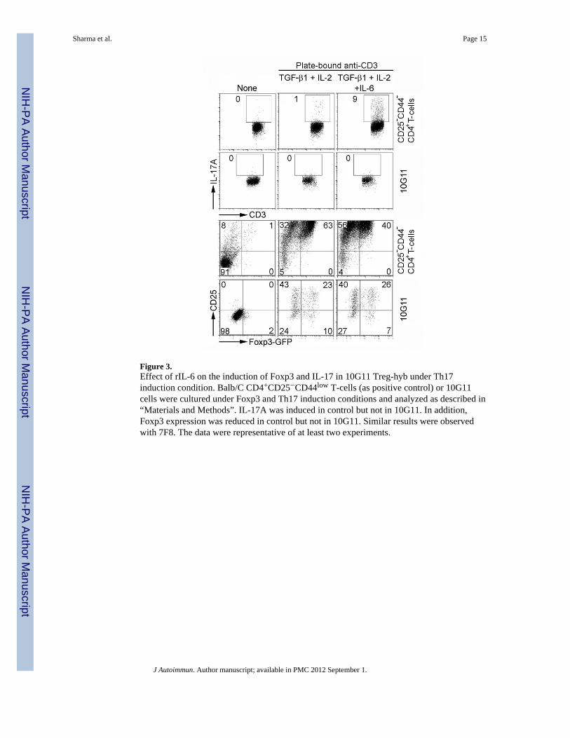
Figure 3.Effect of rIL-6 on the induction of Foxp3 and IL-17 in 10G11 Treg-hyb under Th17induction condition. Balb/C CD4+CD25−CD44low T-cells (as positive control) or 10G11cells were cultured under Foxp3 and Th17 induction conditions and analyzed as described in“Materials and Methods”. IL-17A was induced in control but not in 10G11. In addition,Foxp3 expression was reduced in control but not in 10G11. Similar results were observedwith 7F8. The data were representative of at least two experiments.
Sharma et al. Page 15
J Autoimmun. Author manuscript; available in PMC 2012 September 1.
NIH
-PA Author Manuscript
NIH
-PA Author Manuscript
NIH
-PA Author Manuscript

Figure 4.TCR-independent induction of Foxp3-GFP in iTreg-hyb. (A) 10G11 cells (8×10⁴) werecultured with 2×10⁶ spleen cells from Balb.Rag2−/−, B6.Rag1−/−, and class-II−/− mice for20 hours and analyzed by flow cytometry. (B) Cell free supernatants were prepared fromRag2−/− spleen cells cultured for 2 to 3 days in regular medium or serum free medium(Invitrogen) in the presence of 2μg/ml LPS. 10G11 cells were cultured overnight withvarious concentrations (v/v) of the prepared supernatants. Cells were analyzed by flowcytometry for CD25 and Foxp3-GFP expression. (C) SFS and rIL-6 (50ng/ml) werefractionated with a Centricon membrane filter. Fractions retained and passed through themembrane were adjusted to original volume and 10% final concentration of FCS. Variousdilutions of the fractions were cultured with 10G11 cells for 20 hours. Foxp3-GFPexpression was determined by flow cytometry. The result identified two Foxp3-inducingfactors; one with a molecular size larger than 5-kd and one with a molecular size between 3-kd and 5-kd. (D) The >5-kd but not <5-kd factor activates STAT1 and STAT3. STAT5 wasnot activated. 10G11 was used in this experiment.
Sharma et al. Page 16
J Autoimmun. Author manuscript; available in PMC 2012 September 1.
NIH
-PA Author Manuscript
NIH
-PA Author Manuscript
NIH
-PA Author Manuscript

Figure 5.Loss of Foxp3-GFP induction in iTreg-hyb maintained in culture for 2 to 4 weeks. 10G11and 7F8 that were highly responsive to induction on the day analyzed were used in theexperiments. The untreated Treg-hyb were maintained in culture for various times (2 to 4weeks) and then determined for their ability to express Foxp3 upon induction with anti-CD3mAb and in the presence of rIL-2 and rTGF-β1 or with 30% CFS (v/v). Foxp3-GFPexpression was determined 20 hours after treatment. The time point at which the optimalinduction of Foxp3-GFP was observed was used as d1.
Sharma et al. Page 17
J Autoimmun. Author manuscript; available in PMC 2012 September 1.
NIH
-PA Author Manuscript
NIH
-PA Author Manuscript
NIH
-PA Author Manuscript

Figure 6.A schematic presentation showing the development, Foxp3 regulation, and fate of Tregsubsets. The nTreg pool in the periphery is consisted of the thymic-derived Treg and theiTreg generated during Ag-specific activation. The former stably expresses CD25 and Foxp3and this stable phenotype may depend on the constant presence of extrinsic cues. The iTregadopts an uncommitted phenotype. They lose their CD25 and Foxp3 and became ex-Tregwhen the stimulating signals wane as is the case of iTreg-hyb derived from iTreg clones(***). The ex-Treg possesses the plastic ability to progress into various effector T-cells(IL-2-producing, IL-17-producing, or Foxp3-expressing) depending on the stimulatingsignals received. Dying Treg cells are not depicted here but are presumably replaced tomaintain the homeostasis of Treg pool by new Treg derived from thymus and periphery andthe renewed Treg from the existing nTreg that received extrinsic cues to proliferate asevidenced by its spontaneous cycling in vivo (13).
Sharma et al. Page 18
J Autoimmun. Author manuscript; available in PMC 2012 September 1.
NIH
-PA Author Manuscript
NIH
-PA Author Manuscript
NIH
-PA Author Manuscript

NIH
-PA Author Manuscript
NIH
-PA Author Manuscript
NIH
-PA Author Manuscript
Sharma et al. Page 19
Table 1
Foxp3-GFP-inducing factors*
Medium (7%) BLC (7%) C-10 (7%) KC (7%) CXCL15 (6%)
Eotaxin (7%) Eotaxin-2 (7%) Exodus-2 (6%) IP-10 (7%) I-TAC (7%)
C-TACK (7%) Lix70aa (6%) Lix93aa (7%) lungkine (8%) MCP-1 (7%)
MCP-2 (7%) MCP-3 (7%) MCP-5 (7%) MDC (7%) MEC (6%)
MIG (7%) MIP-1α (7%) MIP-1β (6%) MIP-1γ (7%) MIP-2 (7%)
MIP-3α (7%) MIP-3β (7%) PF-4 (7%) RANTES (7%) SDF-1α (7%)
SDF-1α (7%) IL-1α (11%) IL-1β (10%) IL-2 (9%) IL-3 (9%)
IL-4 (8%) IL-5 (9%) IL-6 (47%) IL-7 (10%) IL-9 (45%)
IL-10 (10%) IL-11 (10%) IL-12(11%) IL-13 (13%) IL-15 (9%)
IL-17 (10%) IL-17F (9%) IL-22 (8%) IL-27 (36%) IL-31 (9%)
IL-33 (9%) IL-35 (5%)* IFN-γ (9%) TNF-α (12%) GM-CSF (8%)
TGF-β1 (11%) LIF (5%) Oncostatin-M (5%) CFS (45%) LPS (10%)*
*A total of 57 cytokines were examined. IL-35 as Fc fusion protein was tested at 100ng/ml. Other cytokines were examined at 50ng/ml. LPS was
tested at 2μg/ml. Please note that IL-2 and TGF-β1 did not induce Foxp3-GFP expression in the absence of TCR engagement.
J Autoimmun. Author manuscript; available in PMC 2012 September 1.
Related Documents