doi:10.1152/ajprenal.00162.2010 299:167-177, 2010. First published May 12, 2010; Am J Physiol Renal Physiol Kenneth R. Hallows Wallimann, Roland Scholz, Núria M. Pastor-Soler, Dietbert Neumann and Hui Li, Ramon F. Thali, Christy Smolak, Fan Gong, Rodrigo Alzamora, Theo You might find this additional information useful... 47 articles, 25 of which you can access free at: This article cites http://ajprenal.physiology.org/cgi/content/full/299/1/F167#BIBL including high-resolution figures, can be found at: Updated information and services http://ajprenal.physiology.org/cgi/content/full/299/1/F167 can be found at: AJP - Renal Physiology about Additional material and information http://www.the-aps.org/publications/ajprenal This information is current as of August 6, 2010 . http://www.the-aps.org/. American Physiological Society. ISSN: 0363-6127, ESSN: 1522-1466. Visit our website at (monthly) by the American Physiological Society, 9650 Rockville Pike, Bethesda MD 20814-3991. Copyright © 2010 by the respective cells and vasculature, as well as to the control of body fluid volume and composition. It is published 12 times a year publishes original manuscripts on a broad range of subjects relating to the kidney, urinary tract, and their AJP - Renal Physiology on August 6, 2010 ajprenal.physiology.org Downloaded from

Welcome message from author
This document is posted to help you gain knowledge. Please leave a comment to let me know what you think about it! Share it to your friends and learn new things together.
Transcript

doi:10.1152/ajprenal.00162.2010 299:167-177, 2010. First published May 12, 2010;Am J Physiol Renal Physiol
Kenneth R. Hallows Wallimann, Roland Scholz, Núria M. Pastor-Soler, Dietbert Neumann and Hui Li, Ramon F. Thali, Christy Smolak, Fan Gong, Rodrigo Alzamora, Theo
You might find this additional information useful...
47 articles, 25 of which you can access free at: This article cites http://ajprenal.physiology.org/cgi/content/full/299/1/F167#BIBL
including high-resolution figures, can be found at: Updated information and services http://ajprenal.physiology.org/cgi/content/full/299/1/F167
can be found at: AJP - Renal Physiologyabout Additional material and information http://www.the-aps.org/publications/ajprenal
This information is current as of August 6, 2010 .
http://www.the-aps.org/.American Physiological Society. ISSN: 0363-6127, ESSN: 1522-1466. Visit our website at (monthly) by the American Physiological Society, 9650 Rockville Pike, Bethesda MD 20814-3991. Copyright © 2010 by therespective cells and vasculature, as well as to the control of body fluid volume and composition. It is published 12 times a year
publishes original manuscripts on a broad range of subjects relating to the kidney, urinary tract, and theirAJP - Renal Physiology
on August 6, 2010
ajprenal.physiology.orgD
ownloaded from
Regulation of the creatine transporter by AMP-activated protein kinasein kidney epithelial cells
Hui Li,1 Ramon F. Thali,3 Christy Smolak,1 Fan Gong,1 Rodrigo Alzamora,1 Theo Wallimann,3
Roland Scholz,3 Núria M. Pastor-Soler,1,2 Dietbert Neumann,3 and Kenneth R. Hallows1,2
1Renal-Electrolyte Division, Department of Medicine and 2Department of Cell Biology and Physiology, University ofPittsburgh School of Medicine, Pittsburgh, Pennsylvania; and 3Department of Biology, Institute of Cell Biology, ETH Zurich,Zurich, Switzerland
Submitted 18 March 2010; accepted in final form 6 May 2010
Li H, Thali RF, Smolak C, Gong F, Alzamora R, Wallimann T,Scholz R, Pastor-Soler NM, Neumann D, Hallows KR. Regulationof the creatine transporter by AMP-activated protein kinase in kidneyepithelial cells. Am J Physiol Renal Physiol 299: F167–F177, 2010.First published May 12, 2010; doi:10.1152/ajprenal.00162.2010.—The metabolic sensor AMP-activated protein kinase (AMPK) regu-lates several transport proteins, potentially coupling transport activityto cellular stress and energy levels. The creatine transporter (CRT;SLC6A8) mediates creatine uptake into several cell types, includingkidney epithelial cells, where it has been proposed that CRT isimportant for reclamation of filtered creatine, a process critical fortotal body creatine homeostasis. Creatine and phosphocreatine pro-vide an intracellular, high-energy phosphate-buffering system essen-tial for maintaining ATP supply in tissues with high energy demands.To test our hypothesis that CRT is regulated by AMPK in the kidney,we examined CRT and AMPK distribution in the kidney and theregulation of CRT by AMPK in cells. By immunofluorescence stain-ing, we detected CRT at the apical pole in a polarized mouse S3proximal tubule cell line and in native rat kidney proximal tubules, adistribution overlapping with AMPK. Two-electrode voltage-clamp(TEV) measurements of Na�-dependent creatine uptake into CRT-expressing Xenopus laevis oocytes demonstrated that AMPK inhibitedCRT via a reduction in its Michaelis-Menten Vmax parameter.[14C]creatine uptake and apical surface biotinylation measurements inpolarized S3 cells demonstrated parallel reductions in creatine influxand CRT apical membrane expression after AMPK activation with theAMP-mimetic compound 5-aminoimidazole-4-carboxamide-1-�-D-ribofuranoside. In oocyte TEV experiments, rapamycin and theAMPK activator 5-aminoimidazole-4-carboxamide-1-�-D-ribofurano-syl 5=-monophosphate (ZMP) inhibited CRT currents, but there wasno additive inhibition of CRT by ZMP, suggesting that AMPK mayinhibit CRT indirectly via the mammalian target of rapamycin path-way. We conclude that AMPK inhibits apical membrane CRT expres-sion in kidney proximal tubule cells, which could be important inreducing cellular energy expenditure and unnecessary creatine reab-sorption under conditions of local and whole body metabolic stress.
proximal tubule; metabolism; Xenopus oocytes; target of rapamycin;SLC6A8
AMP-ACTIVATED PROTEIN KINASE (AMPK) is a ubiquitous meta-bolic-sensing kinase that exists as an �,�,� heterotrimer and isactivated by cellular energy depletion (elevated ratios of intra-cellular AMP to ATP concentration) and other cellular stresses(e.g., Ca2� stress). AMPK activation involves phosphorylationof the catalytic �-subunit at Thr172 (pThr172) by upstreamAMPK kinases that include the LKB1 complex and the Ca2�/
calmodulin-dependent kinase kinase-� (24). AMPK regulates awide variety of cellular processes, including metabolic path-ways, cell growth [via tuberous sclerosis complex (TSC2) inthe mammalian target of rapamycin (mTOR) pathway], inflam-mation, protein synthesis, gene transcription, and membranetransport (16, 23, 37). We and others have shown that AMPKregulates a growing number of epithelial ion transport proteins,including the CFTR Cl� channel, the epithelial Na� channel(ENaC), the Na�-K�-2Cl� cotransporter (NKCC2), theKCa3.1 channel, and the vacuolar H�-ATPase (4, 6, 10, 13, 17,19–21, 29, 48). AMPK-dependent inhibition of these and otherdiffusive ion transport pathways may serve to minimize thedissipation of ionic gradients under conditions of cellularenergy depletion. Concomitantly, AMPK activation enhancescellular nutrient uptake through increased plasma membraneexpression and transcription of glucose and fatty acid transportproteins (16). Thus, recent studies suggest an emerging para-digm whereby AMPK couples membrane transport to cellularmetabolism, preserving the intracellular ionic environment,limiting ATP consumption, and defending the ability of cells togenerate ATP and, thereby, continue vital processes in the faceof metabolic stress. AMPK-dependent regulation of transportproteins may be indirect and mediated through common cel-lular signaling pathways and trafficking regulators (e.g., theubiquitin ligase Nedd4-2, which regulates the ENaC) (4). Wehave thus considered that AMPK may regulate other importantmembrane transport proteins, the expression or activities ofwhich are modulated by cellular energy status or other cellularstresses.
The creatine transporter (CRT; SLC6A8) is a 635-amino acidNa�-Cl�-coupled electrogenic cotransporter (with a 2 Na�:1Cl�:1 creatine stoichiometry) (9, 12) that mediates creatineuptake into a variety of cells, including neuronal cells, skeletaland cardiac muscle cells, and intestinal and kidney epithelialcells (15, 34). Like AMPK, the creatine kinase (CK)-phospho-creatine (PCr) system is important in the maintenance of ATPhomeostasis in tissues that have a high and rapidly fluctuatingenergy requirement (32, 47). Specifically, PCr, formed byCK-dependent phosphorylation of creatine, serves as an im-portant energy reservoir for ATP production. The CK-PCrsystem also serves as a mechanism for the transport of high-energy phosphate from intracellular sites of energy production(e.g., glycolysis or mitochondrial respiration) to sites of energyconsumption (e.g., membrane pump ATPases) (3, 36, 47). Inaddition, it appears that CRT expression is required to providecreatine for its conversion to PCr in cells that cannot manu-facture creatine, such as brain and muscle cells (8). Its impor-tance in these cells is underscored by naturally occurring
Address for reprint requests and other correspondence: K. R. Hallows, Univ.of Pittsburgh, Renal-Electrolyte Division, S976 Scaife Hall, 3550 Terrace St.,Pittsburgh, PA 15261 (e-mail: [email protected]).
Am J Physiol Renal Physiol 299: F167–F177, 2010.First published May 12, 2010; doi:10.1152/ajprenal.00162.2010.
0363-6127/10 Copyright © 2010 the American Physiological Societyhttp://www.ajprenal.org F167
on August 6, 2010
ajprenal.physiology.orgD
ownloaded from

mutations in SLC6A8 that can cause developmental delays,mental retardation, severe language problems, skeletoneuro-muscular disorders, and cardiomyopathy (42).
In intestinal and renal epithelial cells, CRT expression at theapical membrane appears to have a different main role. In theintestine, CRT mediates alimentary creatine uptake and trans-epithelial creatine transport (34). In the kidney, it mediates thefirst step of creatine reclamation in the nephron, a process thatis critical for total body creatine homeostasis, thereby limitingthe need for de novo creatine synthesis in the liver and otherorgans (12). De novo creatine synthesis is energetically costlyand requires a significant fraction of the total body methylgroup donor potential in the form of S-adenosylmethionine(41). Under normal conditions, the kidney effectively salvagescreatine from the urine, and adult men on a creatine-free dietexcrete little creatine. However, significant amounts of creatineare excreted in the urine following toxic exposures to doxoru-bicin (33) or lithium (27). Similarly, it has long been recog-nized that urinary creatine excretion is dramatically increasedunder an inadequate diet, fasting, and other muscle mass-reducing conditions (26, 45), where AMPK activity is expectedto be elevated in cells throughout the body (24). Indeed,creatine import for energy storage into cells would be unnec-essary under conditions of starvation, when the body is in acatabolic state, and it would be energetically wasteful, as itwould tend to dissipate cellular ionic gradients and necessitategreater Na�-K�-ATPase activity. Specifically, the CRT usesan inward Na� gradient to move creatine into the cell. Al-though this process does not expend energy directly, the influxof Na� enhances the activity of the Na�-K�-ATPase on thebasolateral membrane of the cell, and this pump consumesATP in this process.
A detailed understanding of the regulation of CRT is lack-ing, but it has been recently reported that CRT is stimulated bymTOR (38, 39). Interestingly, AMPK is an upstream inhibitorof mTOR via its activation of tuberin (TSC2) in the tuberoussclerosis complex (37), providing a potential mechanism forAMPK-dependent regulation of CRT. Furthermore, it has beenrecently demonstrated that significant AMPK activation occursin rat kidney following induction of ischemia through ligationof the renal artery (31). Given the common signaling pathways,the central role of the creatine-PCr system in cellular energyhomeostasis (46), and the previous observation that metabolicdepletion inhibits creatine reabsorption by the kidney, wehypothesized that AMPK may inhibit CRT activity at theplasma membrane of cells in the kidney and other tissues.
In this study, we examined the localization of CRT andAMPK in rat kidney tissue and in a mouse S3 proximal tubulecell line. We also measured CRT-dependent transport in thepresence or absence of the AMPK activator 5-aminoimidazole-4-carboxamide-1-�-D-ribofuranoside (AICAR) in the Xenopusoocyte expression system using two-electrode voltage clamp(TEV) and in polarized S3 cells through [14C]creatine uptakefluxes. We also measured changes in apical membrane expres-sion of CRT in S3 cells by cell surface biotinylation as afunction of AMPK activation. Finally, we tested the role of themTOR pathway in the AMPK-dependent regulation of CRT inoocyte TEV studies. We found that AMPK regulates CRTplasma membrane expression in oocytes and kidney proximaltubule cells, potentially through a mechanism involving themTOR pathway.
MATERIALS AND METHODS
Reagents and chemicals. All chemical compounds were purchasedfrom Sigma unless otherwise stated. AICAR and 5-aminoimidazole-4-carboxamide-1-�-D-ribofuranosyl 5=-monophosphate (ZMP) werepurchased from Toronto Research Chemicals. [14C]creatine was ob-tained from Moravek Biochemicals. EZ-Link sulfo-NHS-SS-biotinwas obtained from Pierce.
Antibodies. Custom-made immunopurified anti-CRT antibodiesrecognizing the NH2 terminus of the CRT were obtained by immu-nization of rabbits with a synthetic peptide comprising amino acids14–27 of rat SLC6A8 (GenBank accession no. NM_017348.2, Anawa,Switzerland). In addition, the following antibodies were obtained: anti-hemagglutinin (HA; HA.11, Covance); anti-AMPK-� and anti-pThr172
AMPK-� (AMPK-�-pThr172; Cell Signaling); �-actin (Sigma); mouseanti-ZO-1 and Cy3-coupled phalloidin (Invitrogen); horseradish per-oxidase-conjugated secondary IgGs (Amersham Biosciences); andbiotin-conjugated Cy3-coupled goat anti-mouse and Cy5-coupledgoat anti-rabbit antibodies (Jackson Immunologicals).
Kidney tissue preparation and confocal immunofluorescencemicroscopy. All animals were treated according to approved protocolsby the Institutional Animal Care and Use Committee at the Universityof Pittsburgh. Adult male rats were perfused via the left ventricle withPBS at pH 7.4 at 37°C and then with paraformaldehyde (4%)-lysine-periodate buffer, as described previously (22). The kidneys wereharvested, cut in transverse sections, and further fixed overnight at4°C in paraformaldehyde (4%)-lysine-periodate. After fixation, tis-sues were washed, quenched in NH4Cl, and cryoprotected in 30%sucrose in PBS-azide. Tissues were embedded in Tissue-Tek, and4-�m cryosections were obtained using a Reichert-Frigocut cryostat(22). The sections were rehydrated in PBS and used for immunola-beling experiments employing the anti-CRT antibody following anantigen retrieval technique (5, 22). For colabeling of CRT and AMPK,the tissue was immunolabeled with the anti-CRT antibody (1:4,000dilution) and then with biotin-conjugated secondary goat anti-rabbitantibody (1:1,000 dilution). For amplification of the CRT antibodysignal, we used a tyramide signal amplification (TSA) kit (PerkinElmer) according to the manufacturer’s instructions; then the sametissue sections were immunolabeled with an anti-AMPK-� antibody(1:50 dilution) in DAKO diluent followed by goat anti-rabbit second-ary antibody coupled to Cy5 (1:100 dilution). For immunolabeling ofCRT alone, anti-CRT antibody (1:100 dilution) was followed byCy5-coupled goat anti-rabbit antibody (1:100 dilution). Immunola-beled tissues were imaged using a laser confocal microscope (Leica)at identical settings for all samples, and images were imported intoAdobe Photoshop, as described elsewhere (22).
Constructs. For generation of cRNA, the pHAC1 CRT-C-NN(wild-type rat CRT HA-tagged at the NH2 terminus) (43) was sub-cloned by PCR into the dual mammalian-oocyte expression vectorpMO (4) using its NotI and XbaI restriction sites to generate thepMO-HA-CRT plasmid. The forward primer was 5=-AGT-CAGTCGCGGCCGCACCATGTACCCATACGATGTTCC-3=, andthe reverse primer was 5=-TTCCTCTAGATCACATGACACTCTC-CACC-3=. All clones were confirmed by DNA sequencing.
Oocyte TEV measurements. Xenopus laevis oocytes were har-vested, treated with collagenase, and maintained as described previ-ously (6). Oocytes were injected with 30 ng of cRNA encodingHA-tagged CRT. Experiments were performed at room temperature5–6 days after the injection. TEV recordings were performed at aholding potential of �60 mV. The control (superfusate-ND96) solu-tion contained 96 mM NaCl, 2 mM KCl, 1.8 mM CaCl2, 1 mMMgCl2, and 5 mM HEPES, pH 7.4. Creatine was added to the solutionat the indicated concentrations. NaOH was used to titrate the finalsolutions to pH 7.4. TEV recordings were performed 6 h aftermicroinjection of 40 mM potassium-ZMP (K-ZMP, an AMPK acti-vator) or K-gluconate (control) into the oocytes (32 nl/oocyte) atvarious concentrations of external creatine. Currents were then fitted
F168 CREATINE TRANSPORTER REGULATION BY AMPK IN KIDNEY CELLS
AJP-Renal Physiol • VOL 299 • JULY 2010 • www.ajprenal.org
on August 6, 2010
ajprenal.physiology.orgD
ownloaded from

by nonlinear least-squares regression to the Michaelis-Menten equa-tion (V � Vmax/{1 � (Km/[creatine])}) using Igor Pro software(Wavemetrics).
Cell culture. Mouse S3 proximal tubule cells, originally derived bydissection of cells from S3 proximal tubule segments from a Brinstertransgenic mouse [Tg(SV4OE)Bri7] carrying the large-T antigen ofthe SV40 virus, as described elsewhere (28), were a kind gift of Dr.Simon Atkinson. Cells were used at passage 82–90 and cultured in thesame medium used to culture the mpkCCDc14 cell line (2). Thismedium is composed of equal volumes of DMEM and Ham’s F-12plus 60 nM sodium selenate, 5 �g/ml transferrin, 2 mM glutamine, 50nM dexamethasone, 1 nM triiodothyronine, 10 ng/ml epidermalgrowth factor, 5 �g/ml insulin, 20 mM D-glucose, 2% (vol/vol) FBS,and 20 mM HEPES, pH 7.4 (reagents from Invitrogen and Sigma).Cells were maintained at 37°C in a humidified 5% CO2-95% airincubator with medium changes every other day and passaged ap-proximately twice weekly. S3 cells were then subcultured onto dif-ferent-sized Transwell permeable filter supports (0.4-�m pore size;Corning Costar) and grown 5–7 days prior to use in experiments toallow for cell polarization (polarized S3 cells).
Immunofluorescence labeling and confocal microscopy of polar-ized S3 cells. For immunofluorescence (IF) staining, polarized S3 cellson Transwell filters (0.33-cm2 surface area) were pretreated with 1mM AICAR or vehicle at 37°C for 2–3 h prior to fixation in 2%paraformaldehyde in PBS buffer for 30 min and then permeabilizedby the addition of 1% PBS � 1% BSA � 0.1% Triton X-100 for 10min, as described elsewhere (18). After an additional wash with PBS,filters were immunolabeled with primary anti-CRT antibody (1:50dilution) in DAKO diluent, along with mouse anti-ZO-1 (1:100dilution), for 75 min and then with secondary goat anti-rabbit antibodyconjugated with Cy5 (1:100 dilution) and secondary goat anti-mouseantibody conjugated with Cy3 (1:800 dilution). In additional experi-ments, cells were labeled with Cy3-coupled phalloidin (1:1,000 dilu-tion) to stain F-actin. Filters were mounted in Poly-Mount (Poly-sciences) and imaged in a Leica confocal microscope using a �40objective with identical laser and stack acquisition and x-z reconstruc-tion settings for all samples. Images were imported into AdobePhotoshop for further analysis and presentation.
Transfection and immunoblotting. Mouse S3 proximal tubule cellswere cultured as described above. HEK-293 cells were cultured inDMEM (Sigma) supplemented with 10% FBS, 1% (vol/vol) penicillin(10,000 U/ml), 1% (vol/vol) streptomycin (10,000 �g/ml), and 2 mML-glutamine (Invitrogen). For overexpression of CRT, cells weregrown to 60–80% confluency on dishes prior to transfection (Lipo-fectamine 2000, Invitrogen) with the empty vector pMO (Con) orpMO-HA-CRT. At 24 h after transfection, cells were washed withice-cold PBS prior to lysis in PBS containing 1% Triton X-100, 2 mMEDTA, 1 mM phenylmethylsulfonyl fluoride, 1 mM DTT, and 1�complete protease inhibitor cocktail (Roche). After incubation on icefor 15 min, lysates were centrifuged at 17,000 g for 15 min at 4°C, andthe supernatants were supplemented with 4� SDS Laemmli samplebuffer. Subsequent to heating at 65°C for 15 min, 30 �g of total lysatewere separated by SDS-PAGE on a 4–12% gradient gel (Nu-PAGE,Invitrogen), transferred to a nitrocellulose membrane, and immuno-blotted with primary antibody [anti-HA (1:500 dilution) or anti-CRT(1:1,000 dilution)] overnight at 4°C and then with goat anti-mouse oranti-rabbit secondary antibody for 1 h at room temperature.
Surface biotinylation assays. Polarized S3 cells grown on Trans-well filters (4.67-cm2 surface area) were treated with 1 mM AICAR orvehicle for 2 h prior to apical surface biotinylation assays. Cells werewashed three times for 5 min with ice-cold PBS containing Mg2� andCa2� with agitation on ice to remove medium. The apical membranewas biotinylated using 1 mg/ml EZ-Link sulfo-NHS-SS-biotin in PBSfor 30 min. The basolateral surface was incubated in PBS. Thebiotinylation reaction was then quenched by addition of ice-cold 10%FBS-containing medium to the apical surface. Monolayers werewashed three times with ice-cold PBS prior to lysis in PBS containing
1% Triton X-100, 2 mM EDTA, 1 mM phenylmethylsulfonyl fluoride,and 1� complete protease inhibitor cocktail (Roche) on ice for 15min. Protein concentration of the postnuclear supernatant was deter-mined, and 1 mg of protein was combined with streptavidin-Sepha-rose beads (Pierce) and incubated on a rotator overnight at 4°C.Samples from the streptavidin beads were washed three times in lysisbuffer and collected in 2� Laemmli sample buffer containing 10%DTT. The proteins were heated to 65°C for 15 min, separated bySDS-PAGE on a 4–12% gradient gel (Nu-PAGE), and subjected toWestern blot analysis using anti-AMPK-�-pThr172, anti-CRT, andanti-�-actin antibodies.
[14C]creatine uptake flux measurements. Polarized S3 cells grownon Transwell filters (1.12-cm2 surface area) were treated with AICAR(1 mM) for 2 h prior to and during uptake assays, which wereperformed on the basis of previously described methods (34). Cellswere serum-starved for 30 min at 37°C in Hanks’ buffer and thenwashed twice with Krebs-Ringer-HEPES (KRH) buffer [10 mMHEPES (pH 7.4), 4.7 mM KCl, 2.2 mM CaCl2, 1.2 mM MgSO4, 1.2mM KH2PO4, 10 mM glucose, and 120 mM NaCl]. Uptake assayswere performed by apical application of KRH buffer (� AICAR ifappropriate) containing 10 �M [14C]creatine (10 mCi/mmol; MoravecBiochemicals) for 45 min at 37°C (n � 3 for each condition). Thecompetitive CRT substrate �-guanidinopropionic acid (GPA, 1 mM)was applied in some fluxes so that background non-CRT-mediateduptake, which amounted to 10% of the total uptake, could besubtracted. For termination of creatine uptake, the uptake medium wasaspirated and the cells were washed twice with 0.5 ml of ice-coldKRH buffer in which 120 mM LiCl was substituted for NaCl. S3 cellswere solubilized with 80 �l of lysis buffer. First, samples were takento determine protein concentrations using the Bradford technique(Bio-Rad). Then aliquots from each sample were analyzed by Westernblotting to assess AMPK phosphorylation (anti-AMPK-�-pThr172).Fifty microliters of lysate was counted in 3 ml of advanced safety LSCscintillation cocktail (Fisher Scientific) in a liquid scintillationcounter. Results were normalized by protein concentration and sub-tracted by background (GPA-sensitive counts).
Coprecipitation assays. Glutathione S-transferase (GST) pull-downassays were performed using methods described previously (6, 21).Briefly, NH2-terminal HA-tagged CRT (pMO-HA-CRT), AMPK-�1
(pMT2-HA-AMPK-�1), and AMPK-�1 (pMT2-HA-AMPK-�1),along with NH2-terminal GST-tagged AMPK-�1 (pEBG-AMPK-�1)or GST alone (pEBG vector), were coexpressed through transienttransfection into HEK-293 cells 1 day prior to experimentation.Monolayers were washed with ice-cold PBS prior to lysis in PBScontaining 1% Triton X-100, 2 mM EDTA, 1 mM phenylmethylsul-fonyl fluoride, and 1� complete protease inhibitor cocktail on ice for15 min. Protein concentration of the postnuclear supernatant wasdetermined, and 1 mg of protein was combined with glutathione(GSH)-agarose beads (Pierce) and incubated overnight at 4°C. Sam-ples from the GSH-agarose beads were washed three times in lysisbuffer and collected in 2� Laemmli sample buffer containing 10%DTT. The proteins were heated to 65°C for 15 min, separated bySDS-PAGE (Nu-PAGE 4–12% gradient gel, Invitrogen), and sub-jected to Western blot analysis using anti-HA and anti-GST antibod-ies.
Statistical analysis. Statistical analyses were performed using Stat-View (SAS) or SigmaPlot (Jandel Scientific) software. ANOVA wasused to compare data obtained from different batches of oocytes forTEV experiments. For other biochemical experiments, statistics wereperformed using unpaired or paired Student’s t-tests. P 0.05 wasconsidered significant.
RESULTS
CRT and AMPK localize at the apical pole in rat kidneyproximal tubules. First, the cellular and subcellular distributionof CRT and AMPK in fixed rat kidney tissue was examined
F169CREATINE TRANSPORTER REGULATION BY AMPK IN KIDNEY CELLS
AJP-Renal Physiol • VOL 299 • JULY 2010 • www.ajprenal.org
on August 6, 2010
ajprenal.physiology.orgD
ownloaded from
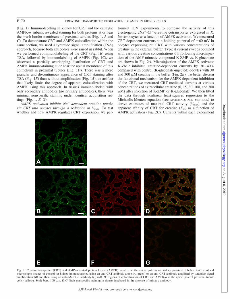
(Fig. 1). Immunolabeling in kidney for CRT and the catalyticAMPK-� subunit revealed staining for both proteins at or nearthe brush border membrane of proximal tubules (Fig. 1, A andC). To demonstrate CRT and AMPK colocalization within thesame section, we used a tyramide signal amplification (TSA)approach, because both antibodies were raised in rabbit. Whenwe performed coimmunolabeling of the CRT (Fig. 1B) usingTSA, followed by immunolabeling of AMPK (Fig. 1C), weobserved a partially overlapping distribution of CRT andAMPK immunostaining at or near the apical membrane of thisepithelium in proximal tubules (Fig. 1D). There was a moregranular and discontinuous appearance of CRT staining afterTSA (Fig. 1B) than without amplification (Fig. 1A), an artifactthat likely limits the degree of apparent colocalization withAMPK using this approach. In tissues immunolabeled withonly secondary antibodies (no primary antibodies), there wasminimal nonspecific staining under identical acquisition set-tings (Fig. 1, E–G).
AMPK activation inhibits Na�-dependent creatine uptakevia CRT into oocytes through a reduction in Vmax. To testwhether and how AMPK regulates CRT expression, we per-
formed TEV experiments to compare the activity of thiselectrogenic 2Na�-Cl�-creatine cotransporter expressed in X.laevis oocytes as a function of AMPK activation. We measuredCRT-dependent currents at a holding potential of �60 mV inoocytes expressing rat CRT with various concentrations ofcreatine in the external buffer. Typical current sweeps obtainedwith various creatine concentrations 6 h following microinjec-tion of the AMP-mimetic compound K-ZMP vs. K-gluconateare shown in Fig. 2A. Microinjection of the AMPK activatorK-ZMP inhibited creatine-dependent currents by 30–40%compared with control (K-gluconate-injected) oocytes with 30and 300 �M creatine in the buffer (Fig. 2B). To better discernthe functional mechanism for the AMPK-dependent inhibitionof the CRT, we measured CRT-mediated currents at variousconcentrations of extracellular creatine (0, 15, 30, 100, and 300�M) after injection of K-ZMP or K-gluconate. We then fittedthe data through nonlinear least-squares regression to theMichaelis-Menten equation (see MATERIALS AND METHODS) toderive estimates of maximal CRT activity (Vmax) and theapparent affinity of CRT for creatine (Km) as a function ofAMPK activation (Fig. 2C). Currents within each experiment
Fig. 1. Creatine transporter (CRT) and AMP-activated protein kinase (AMPK) localize at the apical pole in rat kidney proximal tubules. A–C: confocalmicroscopic images of control rat kidney immunolabeled using an anti-CRT antibody alone (A, green) or an anti-CRT antibody amplified by tyramide signalamplification (B) and then using an anti-AMPK-� antibody (C, red). D: regions of colocalization of CRT and AMPK-� at the apical pole of proximal tubulecells (yellow). Scale bars, 100 �m. E–G: little nonspecific staining in tissues incubated in the absence of primary antibody.
F170 CREATINE TRANSPORTER REGULATION BY AMPK IN KIDNEY CELLS
AJP-Renal Physiol • VOL 299 • JULY 2010 • www.ajprenal.org
on August 6, 2010
ajprenal.physiology.orgD
ownloaded from

were normalized to the mean calculated Vmax parameter forK-gluconate-injected oocytes. Injection with the AMPK acti-vator K-ZMP significantly reduced the Vmax of the CRT by38 � 2% compared with that of K-gluconate control-injectedoocytes. However, the apparent affinity of CRT for creatinewas not affected by the AMPK activator (Km � 30.2 � 3.5 �Mfor K-ZMP treatment vs. 31.3 � 2.3 �M for K-gluconatetreatment). These findings imply that inhibition of CRT activ-ity by AMPK may occur through a reduction in plasmamembrane expression of CRT in oocytes.
S3 proximal tubule cells redistribute CRT from the apicalpole to the cytoplasm following incubation with the AMPKactivator AICAR. To more directly test whether AMPK inhibitsCRT activity and apical membrane expression in a morephysiologically relevant cell system, we used immortalizedcells derived from the S3 segment of the kidney proximaltubule of an SV40 transgenic mouse (28), which were grownon Transwell filters to allow for polarization. We first per-formed time-course experiments to check for AMPK activationby treatment of S3 cells with 1 mM AICAR (Fig. 3A). Byimmunoblotting for the activated form of AMPK using aphosphospecific antibody (anti-AMPK-�-pThr172), we foundthat AMPK was well activated by 1 mM AICAR treatment for2 h in S3 cells, and further activation occurred at later timepoints (Fig. 3A). Because of potential compensatory cellularresponses occurring over longer treatment times with theAMPK activator, we focused on the acute effects of AICARtreatment on CRT distribution and activity in these cells at 2–3h. Immunofluorescence labeling of polarized S3 cells grown onfilters with or without AICAR for 2 h prior to fixation andstaining is shown in Fig. 3B. CRT apical immunolabeling wasqualitatively less in cells treated with the AMPK activator(AICAR) than in cells treated with vehicle alone. Specifically,the proportion of total cellular CRT staining apical to (above)the tight junction marker (ZO-1) was substantially greater invehicle-treated S3 cells than in the AICAR-treated cells (Fig.3B). To account for possible changes in cellular architecture asa function of AICAR treatment, we performed additionalstaining with phalloidin to delineate the actin cytoskeletalnetwork of polarized S3 cells under both treatment conditions(Fig. 3C). AICAR- and vehicle-treated cell monolayers had asimilar cytoskeletal actin cellular distribution with a preservedcortical actin web (Fig. 3C). Therefore, the decrease in appar-ent CRT apical pole localization with AICAR treatment ap-pears to be due to a true change in subcellular distribution,rather than a loss of cellular polarity or a disruption of thecortical actin web.
Immunoblotting of CRT-expressing HEK-293 cells. Multipleimmunoreactive bands for the CRT on Western blot have beenpreviously described and may vary greatly depending on tissueor cell type (35, 43). To demonstrate the specificity of the CRTantibody used for confocal IF staining and immunoblotting,cell lysates from vector-alone or HA-tagged CRT-transfectedHEK-293 cells were analyzed by immunoblotting usinganti-HA and anti-CRT antibodies (Fig. 4, lanes 1–4). Bothantibodies revealed similar signal patterns in CRT-overex-pressing HEK-293 cells: very prominent bands at 120 kDa,more strong bands at 45–50 and 55–60 kDa, and an additionalweaker band at 65–75 kDa. These findings suggest that theCRT antibody specifically recognizes CRT. The vector-alone-transfected cells probed with anti-CRT antibody (lane 1) dem-
Fig. 2. AMPK activation inhibits CRT-dependent currents in Xenopus laevisoocytes. A: representative traces of CRT-dependent inward (downward) cur-rents at different concentrations of buffer creatine in the presence of potassium5-aminoimidazole-4-carboxamide-1-�-D-ribofuranosyl 5=-monophosphate (ZMP)or potassium gluconate (KG). B: CRT-dependent currents (means � SE) weresignificantly inhibited (30–40%) by the AMPK activator ZMP compared withKG control after 6 h of treatment in the presence of 30 and 300 �M creatine.*P 0.05 relative to KG (2-tailed, unpaired t-tests; n � 24 oocytes, N � 3batches per condition). C: relative CRT-dependent currents measured atvarious creatine concentrations 6 h after injection of ZMP or KG and thenfitted by nonlinear least-squares regression to the Michaelis-Menten equation.Vmax for each experiment was normalized by the mean Vmax for KG (n � 24oocytes, N � 3 batches per condition). The AMPK activator ZMP caused a 38� 2% reduction in Vmax but had no effect on Km.
F171CREATINE TRANSPORTER REGULATION BY AMPK IN KIDNEY CELLS
AJP-Renal Physiol • VOL 299 • JULY 2010 • www.ajprenal.org
on August 6, 2010
ajprenal.physiology.orgD
ownloaded from

onstrated very weak signals corresponding to the lower threebands in the HA-CRT-overexpressing cells, suggesting thatthere may be low levels of endogenous CRT expression inthese cells. To establish the CRT immunoblot signal pattern in
S3 proximal tubule cells and test whether cellular host factorsmay modulate CRT processing, we compared immunoblots ofHEK-293 cells and S3 cells transfected with HA-tagged CRTand then immunoblotted using anti-HA antibody (Fig. 4, lanes3–6). The signal pattern observed with expression of HA-CRTin S3 cells differed significantly from that in HEK-293 cells: inS3 cells the uppermost band occurred at 90–95 kDa, and themiddle 65- to 75-kDa band was absent (Fig. 4, lane 6). Thesefindings indicate that cellular processing of the CRT at themRNA and/or protein level may differ as a function of host celltype.
Surface biotinylation and [14C]creatine uptake flux mea-surements in mouse S3 proximal tubule cells following treat-ment with the AMPK activator AICAR. To better quantifychanges in apical plasma membrane CRT expression followingAMPK activation in polarized S3 proximal tubule cells, weperformed apical domain-specific surface biotinylation studiesto label CRT present in the apical membrane using sulfo-NHS-SS-biotin, which covalently reacts with extracellular Lys res-idues in proteins. Polarized S3 cells were treated with 1 mMAICAR or vehicle for 2 h prior to apical surface biotin labeling.AICAR treatment enhanced mean AMPK cellular activity toalmost four times that of control-treated filters (Fig. 5A), whileCRT apical plasma membrane expression decreased by 30–40% with AICAR treatment relative to that of control-treatedfilters (Fig. 5B). Interestingly, only the uppermost CRT band at90–95 kDa appears to be present in the biotinylated apicalmembrane pool. Together, the results from Figs. 3 and 5demonstrate that AMPK activation in polarized kidney S3
Fig. 3. AMPK activation inhibits apical polelocalization of CRT in mouse S3 proximaltubule cells, as determined by immunofluores-cence confocal microscopy. A: time course of5-aminoimidazole-4-carboxamide-1-�-D-ribo-furanoside (AICAR)-mediated AMPK activa-tion, as measured by immunoblotting of phos-phorylated (Thr172) AMPK-� (pT172) in S3cell lysates. AICAR treatment (1 mM) for2–16 h induced a significant upregulation ofphosphorylated (Thr172) AMPK-� comparedwith untreated cells at time 0 under condi-tions of equivalent protein loading (�-actincontrol). B: AICAR-induced cytoplasmic re-distribution of CRT in polarized S3 cellsgrown on Transwell filters, as shown by x-zreconstructions of confocal microscope im-age stacks of S3 cell monolayers immunola-beled using anti-CRT antibody (green) aloneor anti-CRT antibody and an anti-ZO-1 an-tibody (red) grown under control conditions(Con) or in the presence of 1 mM AICAR for2 h. C: Cy3-coupled phalloidin staining ofS3 cell monolayers treated with vehicle(Con) or 1 mM AICAR for 2 h. Images arerepresentative of �3 different filter sets andtreatments. Scale bar, 10 �m.
Fig. 4. Complex signal pattern of CRT revealed by immunoblotting. HEK-293(lanes 1–4) or S3 (lanes 5–6) cells were transiently transfected with emptyvector (Con) or a plasmid encoding the hemagglutinin (HA)-tagged CRT.One-day-posttransfection cell lysates were separated by SDS-PAGE and ana-lyzed by immunoblotting using the anti-CRT (lanes 1 and 2) or anti-HA (lanes3–6) antibody. Both antibodies revealed similar immunoblot signal patternsafter transfection with HA-CRT in HEK-293 cells, identifying 4 major bands(short arrows on left). However, the signal pattern observed in HA-CRT-transfected S3 cells (lane 6; long arrows on right) differed from that inHA-CRT-transfected HEK-293 cells (lane 4), suggesting that cellular process-ing of the CRT at the mRNA and/or protein level differs as a function of hostcell type.
F172 CREATINE TRANSPORTER REGULATION BY AMPK IN KIDNEY CELLS
AJP-Renal Physiol • VOL 299 • JULY 2010 • www.ajprenal.org
on August 6, 2010
ajprenal.physiology.orgD
ownloaded from

proximal tubule cells is associated with an inhibition of apicalmembrane expression of the CRT, which is consistent with ourobservation that AMPK activation decreased the Vmax of ex-ogenously expressed CRT in Xenopus oocytes (Fig. 2).
To test whether AMPK inhibits CRT-mediated creatineuptake in polarized S3 proximal tubule cells, [14C]creatineuptake assays from the apical surface were performed underthese conditions in the presence or absence of 1 mM AICAR.Uptake assays under both treatment conditions were performed inthe presence or absence of 1 mM GPA, a competitive substrate ofthe CRT, to derive CRT-specific influx rates (34). Additionalcontrol experiments confirmed that CRT-specific creatine influxrates were linear during the 45-min uptake flux period used forthese measurements (data not shown). In this series of experi-
ments, 1 mM AICAR treatment enhanced mean AMPK cellularactivity to almost three times that of control-treated filters(Fig. 6A), while CRT-mediated [14C]creatine influx was inhib-ited by 20% with AICAR treatment relative to control filters(Fig. 6B). Therefore, the loss of apical plasma membrane CRTexpression following treatment with the AMPK activatorAICAR was associated with a parallel loss of CRT functionaltransport activity.
AMPK interacts with CRT in cells. To investigate potentialmechanisms for the regulation of CRT by AMPK, we firsttested whether AMPK interacted with CRT when epitope-tagged versions of these proteins were expressed in HEK-293cells. GST pull-down assays were performed on lysates fromcells that were transfected to express HA-tagged CRT, along
Fig. 5. AMPK activation inhibits apical surfaceexpression of CRT in mouse S3 proximal tubulecells, as measured by biotinylation. A: AMPKactivity as measured by immunoblotting ofphosphorylated (Thr172) AMPK-� (pThr-172) inpolarized S3 cell lysates treated with vehicle(Con) or 1 mM AICAR for 2 h under conditionsof equal protein loading (�-actin). Quantifica-tion (means � SE) of phosphorylated (Thr172)AMPK-� signal normalized to �-actin signal isshown relative to control (bottom). *P 0.05(n � 4 replicate experiments). B: immunoblot ofCRT in 5% of whole cell lysates or biotinylatedprotein samples from polarized S3 cell monolayersgrown under control conditions (Con) or treatedwith the AMPK activator AICAR. AICAR-medi-ated AMPK activation inhibited surface CRT ex-pression (biotinylated CRT) by 30–40% relativeto vehicle control, as measured by quantification(means � SE) of CRT band intensity in the bio-tinylated fraction divided by that in the cell lysateand expressed relative to that in control cells (bot-tom). *P 0.05 (n � 4 experiments). Relativelysmall amount of �-actin present in the biotinylatedfraction (2% of total cellular �-actin) likely rep-resents binding of the cortical actin cytoskeleton tointrinsic membrane proteins (e.g., Na�/H� ex-changer isoform 3) present at the apical membraneof these proximal tubule-derived S3 cells. A vari-ety of apical membrane transport proteins havebeen shown to interact directly or indirectly withthe actin cytoskeleton (30).
Fig. 6. AMPK activation inhibits CRT-mediated[14C]creatine uptake in mouse S3 proximal tu-bule cells. Polarized mouse proximal tubule S3segment cells were grown on Transwell filters.A: relative AMPK activity as measured by phos-phorylated (Thr172) AMPK-� immunoblottingfollowing treatment with AICAR (1 mM, 2 h)vs. vehicle. Values are means � SE. *P 0.05(n � 3). B: relative CRT uptake assays. Krebs-Ringer-HEPES buffer was applied in the pres-ence or absence of AICAR containing 10 �M[14C]creatine on the apical side for 45 min at37°C (n � 3 per condition). The competitive CRTsubstrate �-guanidinopropionic acid (1 mM) wasadded in parallel CRT uptake flux measurementsas a control (not shown). Resulting non-CRT-mediated [14C]creatine uptake (generally 10% oftotal) was considered background, and this valuewas subtracted from total uptake values. AICARinduced a 20% inhibition in CRT-dependent[14C]creatine uptake. *P 0.05 (n � 3).
F173CREATINE TRANSPORTER REGULATION BY AMPK IN KIDNEY CELLS
AJP-Renal Physiol • VOL 299 • JULY 2010 • www.ajprenal.org
on August 6, 2010
ajprenal.physiology.orgD
ownloaded from

with GST-tagged AMPK-�1 and HA-tagged AMPK-�1
and -�1 or GST alone (Con, Fig. 7). Samples of cell lysates(Input) or of the proteins affinity-purified from the lysates onGSH-agarose beads (GST pull-down) were immunoblottedusing anti-HA (Fig. 7A) or anti-GST (Fig. 7B) antibodies. As apositive control, the HA-tagged AMPK-�1 and -�1 subunits ofthe AMPK holoenzyme complex were efficiently pulled downby the GST-AMPK-�1 bound to GSH-agarose beads, but notby GST alone. Similarly, pull down of HA-tagged CRT byGST-AMPK-�1 was significant. These results suggest thatAMPK-�1 binds to CRT when expressed in HEK-293 cells,either directly or indirectly. However, additional studies failedto detect any significant in vitro phosphorylation of CRTexpressed in and then immunoprecipitated from HEK-293 celllysates and subsequently exposed to purified AMPK holoen-zyme (data not shown). These findings, along with localizationstudies in kidney tissue (Fig. 1), suggest that although AMPKand CRT may coexist within a regulatory complex in cells,CRT regulation by AMPK may be occurring by a mechanismthat does not involve direct phosphorylation of CRT byAMPK.
AMPK may inhibit CRT via an indirect mechanism involvingthe mTOR pathway. It has been recently reported that CRT isstimulated by mTOR (38), and AMPK is a well-characterizedupstream inhibitor of mTOR (37). To investigate whetherAMPK may inhibit CRT via an indirect mechanism involvingthe mTOR pathway, we injected cRNAs to express CRT andmTOR into Xenopus oocytes, which were used for TEVmeasurements of CRT currents 5–6 days after microinjection.An approach described previously to demonstrate regulation ofCRT by the mTOR pathway (38) was used to treat oocytes withvehicle (DMSO) or 50 nM rapamycin, an inhibitor of mTOR,3 days before experimentation. Oocytes were then microin-jected with K-ZMP (to activate AMPK) or K-gluconate (con-trol) 5–6 h before TEV measurements, as described above. Asobserved in oocytes expressing CRT alone (Fig. 2), the AMPKactivator ZMP inhibited CRT-dependent currents in oocytesexpressing CRT and mTOR together by 30% (Fig. 8, left).Rapamycin pretreatment inhibited CRT-dependent currentsrelative to vehicle to a similar extent as ZMP (Fig. 8). How-ever, there was no additive inhibitory effect of ZMP microin-jection in the rapamycin-pretreated oocytes (Fig. 8, right),suggesting that the ZMP-dependent inhibition of CRT may actthrough the mTOR pathway.
DISCUSSION
The kidney is the principal organ for the first step ofendogenous creatine synthesis, where guanidinoacetic acid issynthesized by arginine-glycine aminotransferase, an enzymehighly expressed in this organ. Guanidinoacetic acid synthe-sized by the kidney is then released into the bloodstream andtaken up by the liver, where it is methylated to form creatine.Subsequent release from the liver provides creatine to thevarious target organs (e.g., skeletal and cardiac muscle, brainand neuronal tissue, testis, and others) (for review see Ref. 49).Besides being involved in the endogenous synthesis of creat-ine, some kidney epithelial cells, especially those in the prox-imal tubule that are highly active metabolically due to largetranscellular ion fluxes driven by ion-pump ATPases, expresshigh levels of CK (14). Similar to muscle and brain cells, theydepend on energy-rich PCr and a high PCr-to-ATP ratio fortemporal and spatial energy buffering (47). Thus, efficient
Fig. 7. Glutathione S-transferase (GST) pull-down assays detect interaction of CRT withthe AMPK-�1 catalytic subunit. HEK-293cells were cotransfected with HA-CRT, HA-AMPK-�1, HA-AMPK-�1, and either GST-AMPK-�1 (AMPK) or GST alone (Con).Cell lysates were used for immunoblot (IB)directly (Input) or for affinity purificationusing glutathione-agarose beads (GST pull-down). A: immunoblot with HA-tagged an-tibodies to check for interaction of HA-CRTwith GST-AMPK-�1. B: Western blot anal-ysis of samples in A, with anti-GST antibod-ies used to verify GST or GST-AMPK-�1
expression, as well as enrichment by GSTpull-down. Images are representative of 3repeat experiments.
Fig. 8. AMPK regulation of CRT may involve the mammalian target ofrapamycin (mTOR) pathway. Xenopus oocytes were injected with CRT andmTOR cRNAs 5–6 days before 2-electrode voltage-clamp (TEV) measure-ments and treated with DMSO (vehicle) or 50 nM rapamycin for 3 days beforeTEV measurements. Oocytes from control and rapamycin-treated groups werethen injected with K-ZMP (ZMP) or K-gluconate (KG) 5–8 h before TEVmeasurements. CRT-dependent current decreased significantly in mTOR-expressing oocytes treated with ZMP compared with control oocytes treatedwith KG (left). ZMP did not have a significant effect in oocytes expressingmTOR and treated with rapamycin (right). *P 0.05 and #P 0.05 vs.mTOR-alone control (n � 17–21 oocytes per condition, N � 3 batches).
F174 CREATINE TRANSPORTER REGULATION BY AMPK IN KIDNEY CELLS
AJP-Renal Physiol • VOL 299 • JULY 2010 • www.ajprenal.org
on August 6, 2010
ajprenal.physiology.orgD
ownloaded from

proximal tubular epithelial cell function requires an optimalsupply of creatine as a precursor for PCr, which is afforded byexpression of the CRT SCL6A8 at the apical membrane.Indeed, it has been shown that preparations of kidney brushborders and apical membrane vesicles express an apical mem-brane CRT that is already active in rat fetuses and is develop-mentally regulated by increase in the density and/or turnover ofthe transporters (11). However, under conditions of ischemia ormetabolic depletion, unabated expression or activity of theCRT at the apical membrane could aggravate cellular meta-bolic demands by allowing apical Na� entry. Ongoing CRTactivity would then necessitate greater basolateral Na�-K�-ATPase activity and ATP consumption to maintain transcellu-lar ionic gradients. We have thus considered that AMPK, theactivity of which is exquisitely sensitive to changes in cellularmetabolic status, might be a relevant and important regulator ofthis transporter.
In this study, we have confirmed that CRT has a prominentapical distribution in kidney proximal tubule and in polarizedproximal tubule-derived S3 cells. In rat proximal tubule, im-munolabeling for the CRT revealed overlap in some apicalregions with the AMPK-� subunit. Moreover, cellular GSTpull-down studies showed significant binding of the CRT toAMPK, suggesting that these proteins exist in a complex. Thiscomplex could potentially include other proteins, such as theactin cytoskeleton (cf. Fig. 6), which is known to be involvedin apical membrane recycling and sorting of a variety oftransport proteins (30), and other signaling mediators that areinvolved in CRT regulation. Our results also suggest that theloss of apical plasma membrane CRT expression followingacute treatment with the AMPK activator AICAR was associ-ated with a parallel loss in CRT functional transport activityand that AMPK activation decreased Vmax and redistributed thetransporter from an apical to a more cytosolic distribution in S3proximal tubule epithelial cells. These findings are consistentwith a recent study that demonstrates AMPK-dependent down-regulation of the plasma membrane abundance of other Na�-coupled cotransporters, the glutamate transporters [excitatoryamino acid transporters (EAAT3 and EAAT4)] (40). More-over, consistent with a role for AMPK in the more generalizedinhibition of Na� reabsorption in the kidney proximal tubule,activation of AMPK in vivo has been recently shown toenhance the tubuloglomerular feedback mechanism and in-crease the fractional delivery of fluid and Na� to the end of theproximal tubule in rats with high dietary NaCl intake (25).AMPK-dependent regulation of transport proteins may beindirect and mediated through common cellular signaling path-ways and trafficking regulators. For example, we and othersrecently showed that ENaC regulation by AMPK is mediatedthrough the ubiquitin ligase Nedd4-2 (1, 4), which regulates avariety of membrane transport proteins (50). When expressedin oocytes, rapamycin and the AMPK activator ZMP inhibitedCRT currents, but there was no additive inhibition of CRT byZMP, suggesting that AMPK may inhibit CRT indirectly viainhibition of the mTOR pathway in cells. The potential path-ways downstream of mTOR that are important for AMPK-dependent regulation of CRT are unknown. However, it hasbeen reported that mTOR may act upstream of the serum- andglucocorticoid-regulated kinase isoforms SGK1 and SGK3 inthe stimulation of CRT (44). It is also known that CRT isdownregulated by activators of protein kinase C (PKC) (9),
which is another potential regulatory target of AMPK (7).Further studies to explore the downstream and upstream sig-naling pathways involved in the AMPK-dependent regulationof CRT in relevant cell model systems and in vivo are war-ranted.
We envision that the presence of CRT at the apical pole ofkidney proximal tubule cells is essential to modulate acutelythe metabolic needs of these epithelial cells, in conjunctionwith AMPK. This study examined acute effects of AMPKactivation, which could be relevant under physiological (e.g.,with minor fluctuations in cellular energy supply and demand)and pathological (e.g., ischemia) conditions. However, chronicAMPK activation, which may occur on a cellular and a wholebody level with chronic starvation or malnutrition, could con-ceivably have distinct effects on CRT function and may inhibittransepithelial reclamation of creatine in the kidney for theregulation of whole body creatine pools. Under normal condi-tions, the kidney effectively salvages creatine from the urine,but significant amounts of creatine are excreted in the urinewith fasting and other muscle mass-reducing conditions (26,45), where AMPK activity is expected to be high throughoutthe body (24). Indeed, creatine uptake into cells as an energystorage buffer could be considered unnecessary under condi-tions of starvation when the body is in a catabolic state.
A certain fraction of creatine that is reabsorbed from thetubular lumen into kidney epithelial cells is likely to betransported back into the blood, similar to transepithelial cre-atine transport in gastrointestinal epithelia. Alimentary creatineis taken up from the gut lumen by intestinal epithelial cells anddelivered by transepithelial transport into the bloodstream (34).Thus, for creatine reclamation from proximal tubular fluid, itseems likely that a creatine transport system should also existat the basolateral side of these cells for transepithelial transportof creatine into the bloodstream. However, as observed inintestinal epithelial cells, very little CRT appears to be local-ized at the basolateral membrane of kidney epithelia cells(Figs. 1 and 3). Therefore, such a transporter is unlikely to beCRT. This assertion is supported by the fact that, if CRT wereinvolved, the combined extracellular Na� and Cl� gradientswould move creatine in the opposite direction (i.e., from theblood into the kidney epithelial cell). Thus the nature of thisbasolateral transporter is still unknown. The function of CRTstudied in this setting would therefore primarily representreabsorption of urinary creatine to preserve whole body crea-tine metabolism. This notion is supported by the fact that, inmodels of nephrotoxicity or kidney disease, urinary creatineexcretion, which is very low in normal individuals, is highlyelevated in doxorubicin- or lithium-induced nephrotoxicity(27, 33).
In summary, this study presents evidence, for the first time,that AMPK regulates CRT in kidney proximal tubule epithelialcells. It appears that AMPK associates with CRT but may notdirectly phosphorylate the transporter. Further studies are nec-essary to elucidate the indirect mechanism for AMPK-medi-ated acute CRT regulation in the kidney.
ACKNOWLEDGMENTS
We thank Dr. Simon Atkinson for mouse S3 cells and Dr. Yu Jiang for themTOR plasmid. We also thank Christoph Gfeller for dedication to the project.
F175CREATINE TRANSPORTER REGULATION BY AMPK IN KIDNEY CELLS
AJP-Renal Physiol • VOL 299 • JULY 2010 • www.ajprenal.org
on August 6, 2010
ajprenal.physiology.orgD
ownloaded from
GRANTS
This study was supported by National Institute of Diabetes and Digestiveand Kidney Diseases Grants P30 DK-079307 “Pittsburgh Kidney ResearchCenter,” R01-DK-075048 (to K. R. Hallows), and R01-DK-084184 (to N. M.Pastor-Soler), Swiss National Science Foundation Grant 3100A0-114137, andEuropean Union FP6 contract LSHM-CT-2004-005272 (EXGENESIS) andETHIIRA Graduate Training Fellowships 32/05-3 and 36/05-3.
DISCLOSURES
No conflicts of interest, financial or otherwise, are declared by the authors.
REFERENCES
1. Almaca J, Kongsuphol P, Hieke B, Ousingsawat J, Viollet B, Schre-iber R, Amaral MD, Kunzelmann K. AMPK controls epithelial Na�
channels through Nedd4-2 and causes an epithelial phenotype whenmutated. Pflügers Arch 458: 713–721, 2009.
2. Bens M, Vallet V, Cluzeaud F, Pascual-Letallec L, Kahn A, Rafestin-Oblin ME, Rossier BC, Vandewalle A. Corticosteroid-dependent sodiumtransport in a novel immortalized mouse collecting duct principal cell line.J Am Soc Nephrol 10: 923–934, 1999.
3. Bessman SP, Carpenter CL. The creatine-creatine phosphate energyshuttle. Annu Rev Biochem 54: 831–862, 1985.
4. Bhalla V, Oyster NM, Fitch AC, Wijngaarden MA, Neumann D,Schlattner U, Pearce D, Hallows KR. AMP-activated kinase inhibits theepithelial Na channel through functional regulation of the ubiquitin ligaseNedd4-2. J Biol Chem 281: 26159–26169, 2006.
5. Brown D, Lydon J, McLaughlin M, Stuart-Tilley A, Tyszkowski R,Alper S. Antigen retrieval in cryostat tissue sections and cultured cells bytreatment with sodium dodecyl sulfate (SDS). Histochem Cell Biol 105:261–267, 1996.
6. Carattino MD, Edinger RS, Grieser HJ, Wise R, Neumann D, Schlat-tner U, Johnson JP, Kleyman TR, Hallows KR. Epithelial sodiumchannel inhibition by AMP-activated protein kinase in oocytes and polar-ized renal epithelial cells. J Biol Chem 280: 17608–17616, 2005.
7. Chen HC, Bandyopadhyay G, Sajan MP, Kanoh Y, Standaert M,Farese RV Jr, Farese RV. Activation of the ERK pathway and atypicalprotein kinase C isoforms in exercise- and aminoimidazole-4-carboxam-ide-1-�-D-riboside (AICAR)-stimulated glucose transport. J Biol Chem277: 23554–23562, 2002.
8. Chen NH, Reith ME, Quick MW. Synaptic uptake and beyond: thesodium- and chloride-dependent neurotransmitter transporter familySLC6. Pflügers Arch 447: 519–531, 2004.
9. Dai W, Vinnakota S, Qian X, Kunze DL, Sarkar HK. Molecularcharacterization of the human CRT-1 creatine transporter expressed inXenopus oocytes. Arch Biochem Biophys 361: 75–84, 1999.
10. Fraser SA, Gimenez I, Cook N, Jennings I, Katerelos M, Katsis F,Levidiotis V, Kemp BE, Power DA. Regulation of the renal-specificNa�-K�-2Cl� co-transporter NKCC2 by AMP-activated protein kinase(AMPK). Biochem J 405: 85–93, 2007.
11. Garcia-Delgado M, Garcia-Miranda P, Peral MJ, Calonge ML, Ilun-dain AA. Ontogeny up-regulates renal Na�/Cl�/creatine transporter in rat.Biochim Biophys Acta 1768: 2841–2848, 2007.
12. Garcia-Delgado M, Peral MJ, Cano M, Calonge ML, Ilundain AA.Creatine transport in brush-border membrane vesicles isolated from ratkidney cortex. J Am Soc Nephrol 12: 1819–1825, 2001.
13. Gong F, Alzamora R, Smolak C, Li H, Naveed S, Neumann D, HallowsKR, Pastor-Soler NM. Vacuolar H�-ATPase apical accumulation inkidney intercalated cells is regulated by PKA and AMP-activated proteinkinase. Am J Physiol Renal Physiol 298: F1162–F1169, 2010.
14. Guerrero ML, Beron J, Spindler B, Groscurth P, Wallimann T,Verrey F. Metabolic support of Na� pump in apically permeabilized A6kidney cell epithelia: role of creatine kinase. Am J Physiol Cell Physiol272: C697–C706, 1997.
15. Guimbal C, Kilimann MW. A Na�-dependent creatine transporter inrabbit brain, muscle, heart, and kidney. cDNA cloning and functionalexpression. J Biol Chem 268: 8418–8421, 1993.
16. Hallows KR. Emerging role of AMP-activated protein kinase in couplingmembrane transport to cellular metabolism. Curr Opin Nephrol Hypertens14: 464–471, 2005.
17. Hallows KR, Alzamora R, Li H, Gong F, Smolak C, Neumann D,Pastor-Soler NM. AMP-activated protein kinase inhibits alkaline pH- andPKA-induced apical vacuolar H�-ATPase accumulation in epididymalclear cells. Am J Physiol Cell Physiol 296: C672–C681, 2009.
18. Hallows KR, Fitch AC, Richardson CA, Reynolds PR, Clancy JP,Dagher PC, Witters LA, Kolls JK, Pilewski JM. Up-regulation ofAMP-activated kinase by dysfunctional cystic fibrosis transmembraneconductance regulator in cystic fibrosis airway epithelial cells mitigatesexcessive inflammation. J Biol Chem 281: 4231–4241, 2006.
19. Hallows KR, Kobinger GP, Wilson JM, Witters LA, Foskett JK.Physiological modulation of CFTR activity by AMP-activated proteinkinase in polarized T84 cells. Am J Physiol Cell Physiol 284: C1297–C1308, 2003.
20. Hallows KR, McCane JE, Kemp BE, Witters LA, Foskett JK. Regu-lation of channel gating by AMP-activated protein kinase modulates cysticfibrosis transmembrane conductance regulator activity in lung submucosalcells. J Biol Chem 278: 998–1004, 2003.
21. Hallows KR, Raghuram V, Kemp BE, Witters LA, Foskett JK.Inhibition of cystic fibrosis transmembrane conductance regulator bynovel interaction with the metabolic sensor AMP-activated protein kinase.J Clin Invest 105: 1711–1721, 2000.
22. Hallows KR, Wang H, Edinger RS, Butterworth MB, Oyster NM, LiH, Buck J, Levin LR, Johnson JP, Pastor-Soler NM. Regulation ofepithelial Na� transport by soluble adenylyl cyclase in kidney collectingduct cells. J Biol Chem 284: 5774–5783, 2009.
23. Hardie DG. The AMP-activated protein kinase pathway—new playersupstream and downstream. J Cell Sci 117: 5479–5487, 2004.
24. Hardie DG, Hawley SA, Scott JW. AMP-activated protein kinase—development of the energy sensor concept. J Physiol 574: 7–15, 2006.
25. Huang DY, Gao H, Boini KM, Osswald H, Nurnberg B, Lang F.In vivo stimulation of AMP-activated protein kinase enhanced tubuloglo-merular feedback but reduced tubular sodium transport during high dietaryNaCl intake. Pflügers Arch. In press.
26. Hunter A. The physiology of creatine and creatinine. Physiol Rev 2:586–626, 1922.
27. Hwang GS, Yang JY, Ryu do H, Kwon TH. Metabolic profiling ofkidney and urine in rats with lithium-induced nephrogenic diabetes insip-idus by 1H-NMR-based metabonomics. Am J Physiol Renal Physiol 298:F461–F470, 2010.
28. Kaunitz JD, Cummins VP, Mishler D, Nagami GT. Inhibition ofgentamicin uptake into cultured mouse proximal tubule epithelial cells byL-lysine. J Clin Pharmacol 33: 63–69, 1993.
29. Klein H, Garneau L, Trinh NT, Prive A, Dionne F, Goupil E,Thuringer D, Parent L, Brochiero E, Sauve R. Inhibition of the KCa3.1channels by AMP-activated protein kinase in human airway epithelialcells. Am J Physiol Cell Physiol 296: C285–C295, 2009.
30. Mazzochi C, Benos DJ, Smith PR. Interaction of epithelial ion channelswith the actin-based cytoskeleton. Am J Physiol Renal Physiol 291:F1113–F1122, 2006.
31. Mount PF, Hill RE, Fraser SA, Levidiotis V, Katsis F, Kemp BE,Power DA. Acute renal ischemia rapidly activates the energy sensorAMPK but does not increase phosphorylation of eNOS-Ser1177. Am JPhysiol Renal Physiol 289: F1103–F1115, 2005.
32. Neumann D, Schlattner U, Wallimann T. A molecular approach to theconcerted action of kinases involved in energy homoeostasis. Biochem SocTrans 31: 169–174, 2003.
33. Park JC, Hong YS, Kim YJ, Yang JY, Kim EY, Kwack SJ, Ryu do H,Hwang GS, Lee BM. A metabonomic study on the biochemical effects ofdoxorubicin in rats using 1H-NMR spectroscopy. J Toxicol EnvironHealth A 72: 374–384, 2009.
34. Peral MJ, Garcia-Delgado M, Calonge ML, Duran JM, De La HorraMC, Wallimann T, Speer O, Ilundain A. Human, rat and chicken smallintestinal Na�-Cl�-creatine transporter: functional, molecular character-ization and localization. J Physiol 545: 133–144, 2002.
35. Peral MJ, Vazquez-Carretero MD, Ilundain AA. Na�/Cl�/creatinetransporter activity and expression in rat brain synaptosomes. Neuro-science 165: 53–60, 2010.
36. Schlattner U, Tokarska-Schlattner M, Wallimann T. Mitochondrialcreatine kinase in human health and disease. Biochim Biophys Acta 1762:164–180, 2006.
37. Shaw RJ, Bardeesy N, Manning BD, Lopez L, Kosmatka M, DePinhoRA, Cantley LC. The LKB1 tumor suppressor negatively regulatesmTOR signaling. Cancer Cell 6: 91–99, 2004.
38. Shojaiefard M, Christie DL, Lang F. Stimulation of the creatine trans-porter SLC6A8 by the protein kinase mTOR. Biochem Biophys ResCommun 341: 945–949, 2006.
F176 CREATINE TRANSPORTER REGULATION BY AMPK IN KIDNEY CELLS
AJP-Renal Physiol • VOL 299 • JULY 2010 • www.ajprenal.org
on August 6, 2010
ajprenal.physiology.orgD
ownloaded from

39. Shojaiefard M, Christie DL, Lang F. Stimulation of the creatine trans-porter SLC6A8 by the protein kinases SGK1 and SGK3. Biochem BiophysRes Commun 334: 742–746, 2005.
40. Sopjani M, Alesutan I, Dermaku-Sopjani M, Fraser S, Kemp BE,Foller M, Lang F. Downregulation of Na�-coupled glutamate transporterEAAT3 and EAAT4 by AMP-activated protein kinase. J Neurochem. Inpress.
41. Stead LM, Brosnan JT, Brosnan ME, Vance DE, Jacobs RL. Is it timeto reevaluate methyl balance in humans? Am J Clin Nutr 83: 5–10, 2006.
42. Stockler S, Schutz PW, Salomons GS. Cerebral creatine deficiencysyndromes: clinical aspects, treatment and pathophysiology. Subcell Bio-chem 46: 149–166, 2007.
43. Straumann N, Wind A, Leuenberger T, Wallimann T. Effects ofN-linked glycosylation on the creatine transporter. Biochem J 393: 459–469, 2006.
44. Strutz-Seebohm N, Shojaiefard M, Christie D, Tavare J, Seebohm G,Lang F. PIKfyve in the SGK1 mediated regulation of the creatinetransporter SLC6A8. Cell Physiol Biochem 20: 729–734, 2007.
45. Walker JB. Creatine: biosynthesis, regulation, function. Adv EnzymolRelat Areas Mol Biol 50: 177–242, 1979.
46. Wallimann T, Tokarska-Schlattner M, Neumann D, Epand RM,Epand RF, Andres RH, Widmer HR, Hornemann T, Saks V, Agar-kova I, Schlattner U. The phosphocreatine circuit: molecular and cellularphysiology of creatine kinases, sensitivity to free radicals, and enhance-ment by creatine supplementation. In: Molecular System Bioenergetics,edited by Saks V. Weinheim, Germany: Wiley-VCH Verlag, 2007, p.195–264.
47. Wallimann T, Wyss M, Brdiczka D, Nicolay K, Eppenberger HM.Intracellular compartmentation, structure and function of creatine kinaseisoenzymes in tissues with high and fluctuating energy demands: the“phosphocreatine circuit” for cellular energy homeostasis. Biochem J 281:21–40, 1992.
48. Woollhead AM, Scott JW, Hardie DG, Baines DL. Phenformin and5-aminoimidazole-4-carboxamide-1-�-D-ribofuranoside (AICAR) activa-tion of AMP-activated protein kinase inhibits transepithelial Na� transportacross H441 lung cells. J Physiol 566: 781–792, 2005.
49. Wyss M, Kaddurah-Daouk R. Creatine and creatinine metabolism.Physiol Rev 80: 1107–1213, 2000.
50. Yang B, Kumar S. Nedd4 and Nedd4-2: closely related ubiquitin-proteinligases with distinct physiological functions. Cell Death Differ 17: 68–77,2010.
F177CREATINE TRANSPORTER REGULATION BY AMPK IN KIDNEY CELLS
AJP-Renal Physiol • VOL 299 • JULY 2010 • www.ajprenal.org
on August 6, 2010
ajprenal.physiology.orgD
ownloaded from
Related Documents