Regulation of SIRT6 protein levels by nutrient availability Yariv Kanfi a,1 , Ronnie Shalman a,1 , Victoria Peshti a , Shmuel N. Pilosof a , Yosi M. Gozlan a , Kevin J. Pearson b , Batya Lerrer a , Danesh Moazed c , Jean-Christophe Marine d , Rafael de Cabo b , Haim Y. Cohen a, * a The Mina and Everard Goodman Faculty of Life Sciences, Bar-Ilan University, Ramat-Gan 52900, Israel b Laboratory of Experimental Gerontology, NIA, NIH, Baltimore, MD 21224-6825, USA c Department of Cell Biology, Harvard Medical School, Boston, MA 02115, USA d Flanders Interuniversity Institute for Biotechnology, University of Ghent, Ghent, Belgium Received 5 December 2007; revised 16 January 2008; accepted 17 January 2008 Available online 31 January 2008 Edited by Noboru Mizushima Abstract Sirtuins have been shown to regulate life-span in response to nutritional availability. We show here that levels of the mammalian sirtuin, SIRT6, increased upon nutrient depriva- tion in cultured cells, in mice after fasting, and in rats fed a calorie-restricted diet. The increase in SIRT6 levels is due to stabilization of SIRT6 protein, and not via an increase in SIRT6 transcription. In addition, p53 positively regulates SIRT6 pro- tein levels under standard growth conditions but has no role in the nutrient-dependent regulation of SIRT6. These observations imply that at least two sirtuins are involved in regulation of life- span by nutrient availability. Ó 2008 Federation of European Biochemical Societies. Published by Elsevier B.V. All rights reserved. Keywords: SIRT6; Sirtuin; Calorie restriction; Nutrient availability 1. Introduction The sirtuins are highly conserved enzymes that utilize NAD + to modify other proteins [1]. The founder member of the sir- tuin family, yeast Sir2, displays both protein deacetylase and protein mono-ADP ribosyltransferase activities. However, de- spite their conservation from bacteria to humans, some sirtuins exhibit only one of these enzymatic activities [2]. Studies of several organisms indicate that sirtuins are pivotal in the regulation of longevity. Mutation of Saccharomyces cerevisiae Sir2 (ySir2) shortens yeast replicative life-span by 40%, whereas increasing the activity of sirtuins in S. cerevisiae, Caenorhabditis elegans and Drosophila melanogaster through either genetic or chemical means, extends life-span by at least 30% [3–6]. In addition, observations of yeast and drosophila suggested that sirtuins are required for mediating the beneficial effect of a calorie-restricted (CR) diet on life-span [7]. CR slows the rate of aging, delays the appearance of many age-related disorders and extends the maximum life-span of various organisms, including yeast, nematodes, drosophila and rodents [8–10]. However, the molecular mechanisms of CR are still poorly understood. Of the seven mammalian ySir2 homologues, SIRT1 to 7, only SIRT1 was implicated to date in the CR response. SIRT1 is induced upon nutrient deprivation in vitro in a p53-depen- dent manner [11] and after long-term CR [12], and mice over-expressing SIRT1 exhibit some physiological properties similar to those of mice on a CR regimen [13]. Another mam- malian sirtuin recently implicated in the regulation of aging is SIRT6, a nuclear protein that fails to deacetylate acetylated lysine in vitro, but instead catalyzes auto-ADP-ribosylation [2]. SIRT6-deficient mice are small, and by 2–3 weeks of age, develop abnormalities usually associated with aging [14]. These abnormalities include profound lymphopenia, loss of subcuta- neous fat, lordokyphosis, severe metabolic defects, and, even- tually, death at about 4 weeks. Notably, cells deficient in SIRT6 exhibit high levels of genomic instability that are likely due to defects in base excision repair (BER) [14]. Yet, the asso- ciation between defects in BER and aging remains tenuous, as mutation of other BER factors has yet to be demonstrated to display a similar aging phenotype [15]. In order to determine if SIRT6 was regulated by nutrient levels and was involved in CR response in a manner similar to SIRT1, we studied the effects of nutrient depletion both in vivo in rodents and in vitro in tissue culture cells, and observed elevated SIRT6 levels in both model systems. We demonstrate that the elevated expression of SIRT6 protein is not due to increased SIRT6 transcription or translation, but rather to stabilization of SIRT6 protein. Moreover, studies using inhibitors indicate that the proteosome degradation pathway is largely responsible for regulating SIRT6 levels. Taken together, these results suggest the involvement of SIRT6 in the response to nutrient levels, and raise the possi- bility that part of the influence of nutrient availability on life-span is mediated by increasing SIRT6 stability. 2. Results Nutrient availability was shown to regulate life-span, sug- gesting that proteins such as SIRT6, which are involved in aging and metabolism, might be regulated by nutrient levels. In order to investigate whether SIRT6 was involved in the response to nutrient levels, SIRT6 protein levels were moni- tored in mammalian models of nutrient deprivation. In human * Corresponding author. Fax: +972 3 738 4058. E-mail address: [email protected] (H.Y. Cohen). 1 These authors contributed equally to this work. 0014-5793/$34.00 Ó 2008 Federation of European Biochemical Societies. Published by Elsevier B.V. All rights reserved. doi:10.1016/j.febslet.2008.01.019 FEBS Letters 582 (2008) 543–548

Welcome message from author
This document is posted to help you gain knowledge. Please leave a comment to let me know what you think about it! Share it to your friends and learn new things together.
Transcript

FEBS Letters 582 (2008) 543–548
Regulation of SIRT6 protein levels by nutrient availability
Yariv Kanfia,1, Ronnie Shalmana,1, Victoria Peshtia, Shmuel N. Pilosofa, Yosi M. Gozlana,Kevin J. Pearsonb, Batya Lerrera, Danesh Moazedc, Jean-Christophe Marined,
Rafael de Cabob, Haim Y. Cohena,*
a The Mina and Everard Goodman Faculty of Life Sciences, Bar-Ilan University, Ramat-Gan 52900, Israelb Laboratory of Experimental Gerontology, NIA, NIH, Baltimore, MD 21224-6825, USA
c Department of Cell Biology, Harvard Medical School, Boston, MA 02115, USAd Flanders Interuniversity Institute for Biotechnology, University of Ghent, Ghent, Belgium
Received 5 December 2007; revised 16 January 2008; accepted 17 January 2008
Available online 31 January 2008
Edited by Noboru Mizushima
Abstract Sirtuins have been shown to regulate life-span inresponse to nutritional availability. We show here that levels ofthe mammalian sirtuin, SIRT6, increased upon nutrient depriva-tion in cultured cells, in mice after fasting, and in rats fed acalorie-restricted diet. The increase in SIRT6 levels is due tostabilization of SIRT6 protein, and not via an increase in SIRT6transcription. In addition, p53 positively regulates SIRT6 pro-tein levels under standard growth conditions but has no role inthe nutrient-dependent regulation of SIRT6. These observationsimply that at least two sirtuins are involved in regulation of life-span by nutrient availability.� 2008 Federation of European Biochemical Societies. Publishedby Elsevier B.V. All rights reserved.
Keywords: SIRT6; Sirtuin; Calorie restriction; Nutrientavailability
1. Introduction
The sirtuins are highly conserved enzymes that utilize NAD+
to modify other proteins [1]. The founder member of the sir-
tuin family, yeast Sir2, displays both protein deacetylase and
protein mono-ADP ribosyltransferase activities. However, de-
spite their conservation from bacteria to humans, some sirtuins
exhibit only one of these enzymatic activities [2].
Studies of several organisms indicate that sirtuins are pivotal
in the regulation of longevity. Mutation of Saccharomyces
cerevisiae Sir2 (ySir2) shortens yeast replicative life-span by
40%, whereas increasing the activity of sirtuins in S. cerevisiae,
Caenorhabditis elegans and Drosophila melanogaster through
either genetic or chemical means, extends life-span by at least
30% [3–6]. In addition, observations of yeast and drosophila
suggested that sirtuins are required for mediating the beneficial
effect of a calorie-restricted (CR) diet on life-span [7]. CR slows
the rate of aging, delays the appearance of many age-related
disorders and extends the maximum life-span of various
organisms, including yeast, nematodes, drosophila and rodents
*Corresponding author. Fax: +972 3 738 4058.E-mail address: [email protected] (H.Y. Cohen).
1These authors contributed equally to this work.
0014-5793/$34.00 � 2008 Federation of European Biochemical Societies. Pu
doi:10.1016/j.febslet.2008.01.019
[8–10]. However, the molecular mechanisms of CR are still
poorly understood.
Of the seven mammalian ySir2 homologues, SIRT1 to 7,
only SIRT1 was implicated to date in the CR response. SIRT1
is induced upon nutrient deprivation in vitro in a p53-depen-
dent manner [11] and after long-term CR [12], and mice
over-expressing SIRT1 exhibit some physiological properties
similar to those of mice on a CR regimen [13]. Another mam-
malian sirtuin recently implicated in the regulation of aging is
SIRT6, a nuclear protein that fails to deacetylate acetylated
lysine in vitro, but instead catalyzes auto-ADP-ribosylation
[2]. SIRT6-deficient mice are small, and by 2–3 weeks of age,
develop abnormalities usually associated with aging [14]. These
abnormalities include profound lymphopenia, loss of subcuta-
neous fat, lordokyphosis, severe metabolic defects, and, even-
tually, death at about 4 weeks. Notably, cells deficient in
SIRT6 exhibit high levels of genomic instability that are likely
due to defects in base excision repair (BER) [14]. Yet, the asso-
ciation between defects in BER and aging remains tenuous, as
mutation of other BER factors has yet to be demonstrated to
display a similar aging phenotype [15].
In order to determine if SIRT6 was regulated by nutrient
levels and was involved in CR response in a manner similar
to SIRT1, we studied the effects of nutrient depletion both
in vivo in rodents and in vitro in tissue culture cells, and
observed elevated SIRT6 levels in both model systems. We
demonstrate that the elevated expression of SIRT6 protein is
not due to increased SIRT6 transcription or translation, but
rather to stabilization of SIRT6 protein. Moreover, studies
using inhibitors indicate that the proteosome degradation
pathway is largely responsible for regulating SIRT6 levels.
Taken together, these results suggest the involvement of
SIRT6 in the response to nutrient levels, and raise the possi-
bility that part of the influence of nutrient availability on
life-span is mediated by increasing SIRT6 stability.
2. Results
Nutrient availability was shown to regulate life-span, sug-
gesting that proteins such as SIRT6, which are involved in
aging and metabolism, might be regulated by nutrient levels.
In order to investigate whether SIRT6 was involved in the
response to nutrient levels, SIRT6 protein levels were moni-
tored in mammalian models of nutrient deprivation. In human
blished by Elsevier B.V. All rights reserved.
544 Y. Kanfi et al. / FEBS Letters 582 (2008) 543–548
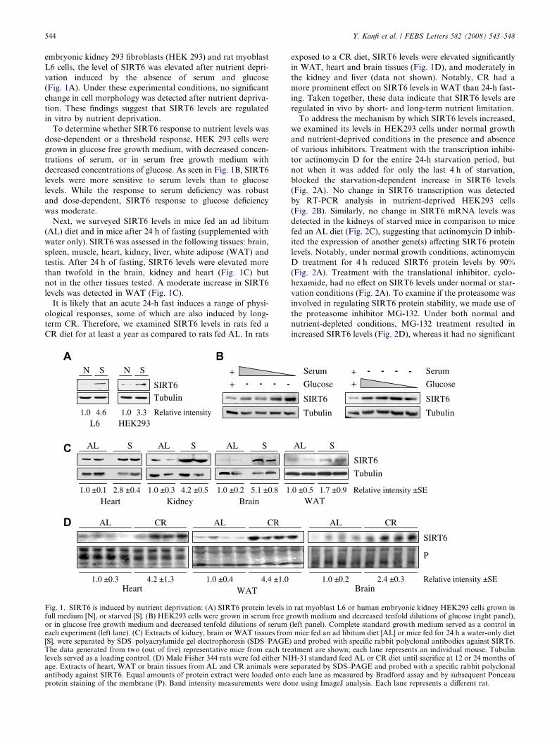
embryonic kidney 293 fibroblasts (HEK 293) and rat myoblast
L6 cells, the level of SIRT6 was elevated after nutrient depri-
vation induced by the absence of serum and glucose
(Fig. 1A). Under these experimental conditions, no significant
change in cell morphology was detected after nutrient depriva-
tion. These findings suggest that SIRT6 levels are regulated
in vitro by nutrient deprivation.
To determine whether SIRT6 response to nutrient levels was
dose-dependent or a threshold response, HEK 293 cells were
grown in glucose free growth medium, with decreased concen-
trations of serum, or in serum free growth medium with
decreased concentrations of glucose. As seen in Fig. 1B, SIRT6
levels were more sensitive to serum levels than to glucose
levels. While the response to serum deficiency was robust
and dose-dependent, SIRT6 response to glucose deficiency
was moderate.
Next, we surveyed SIRT6 levels in mice fed an ad libitum
(AL) diet and in mice after 24 h of fasting (supplemented with
water only). SIRT6 was assessed in the following tissues: brain,
spleen, muscle, heart, kidney, liver, white adipose (WAT) and
testis. After 24 h of fasting, SIRT6 levels were elevated more
than twofold in the brain, kidney and heart (Fig. 1C) but
not in the other tissues tested. A moderate increase in SIRT6
levels was detected in WAT (Fig. 1C).
It is likely that an acute 24-h fast induces a range of physi-
ological responses, some of which are also induced by long-
term CR. Therefore, we examined SIRT6 levels in rats fed a
CR diet for at least a year as compared to rats fed AL. In rats
Relative intensity
N S
L6
SIRT6Tubulin
HEK293
N S
1.0 4.6 1.0 3.3
WATHeart
AL CR
1.0 ±0.3 4.2 ±1.3 1.0 ±0.4 4.4 ±1.0
AL CR
+
+ - - - -
AL S
Heart Kidney Brain1.0 ±0.1 2.8 ±0.4 1.0 ±0.3 4.2 ±0.5 1.0 ±0.2 5.1 ±0.8
AL S SAL
1
Fig. 1. SIRT6 is induced by nutrient deprivation: (A) SIRT6 protein levels infull medium [N], or starved [S]. (B) HEK293 cells were grown in serum free gor in glucose free growth medium and decreased tenfold dilutions of serumeach experiment (left lane). (C) Extracts of kidney, brain or WAT tissues from[S], were separated by SDS–polyacrylamide gel electrophoresis (SDS–PAGEThe data generated from two (out of five) representative mice from each trelevels served as a loading control. (D) Male Fisher 344 rats were fed either NIage. Extracts of heart, WAT or brain tissues from AL and CR animals wereantibody against SIRT6. Equal amounts of protein extract were loaded ontoprotein staining of the membrane (P). Band intensity measurements were do
exposed to a CR diet, SIRT6 levels were elevated significantly
in WAT, heart and brain tissues (Fig. 1D), and moderately in
the kidney and liver (data not shown). Notably, CR had a
more prominent effect on SIRT6 levels in WAT than 24-h fast-
ing. Taken together, these data indicate that SIRT6 levels are
regulated in vivo by short- and long-term nutrient limitation.
To address the mechanism by which SIRT6 levels increased,
we examined its levels in HEK293 cells under normal growth
and nutrient-deprived conditions in the presence and absence
of various inhibitors. Treatment with the transcription inhibi-
tor actinomycin D for the entire 24-h starvation period, but
not when it was added for only the last 4 h of starvation,
blocked the starvation-dependent increase in SIRT6 levels
(Fig. 2A). No change in SIRT6 transcription was detected
by RT-PCR analysis in nutrient-deprived HEK293 cells
(Fig. 2B). Similarly, no change in SIRT6 mRNA levels was
detected in the kidneys of starved mice in comparison to mice
fed an AL diet (Fig. 2C), suggesting that actinomycin D inhib-
ited the expression of another gene(s) affecting SIRT6 protein
levels. Notably, under normal growth conditions, actinomycin
D treatment for 4 h reduced SIRT6 protein levels by 90%
(Fig. 2A). Treatment with the translational inhibitor, cyclo-
hexamide, had no effect on SIRT6 levels under normal or star-
vation conditions (Fig. 2A). To examine if the proteasome was
involved in regulating SIRT6 protein stability, we made use of
the proteasome inhibitor MG-132. Under both normal and
nutrient-depleted conditions, MG-132 treatment resulted in
increased SIRT6 levels (Fig. 2D), whereas it had no significant
SIRT6
P
BrainRelative intensity ±SE1.0 ±0.2 2.4 ±0.3
AL CR
Serum - - - - Serum
Tubulin
Glucose +
SIRT6
+
Tubulin
Glucose
SIRT6
SIRT6
Tubulin
Relative intensity ±SEWAT
SAL
.0 ±0.5 1.7 ±0.9
rat myoblast L6 or human embryonic kidney HEK293 cells grown inrowth medium and decreased tenfold dilutions of glucose (right panel),(left panel). Complete standard growth medium served as a control in
mice fed an ad libitum diet [AL] or mice fed for 24 h a water-only diet) and probed with specific rabbit polyclonal antibodies against SIRT6.atment are shown; each lane represents an individual mouse. TubulinH-31 standard feed AL or CR diet until sacrifice at 12 or 24 months ofseparated by SDS–PAGE and probed with a specific rabbit polyclonaleach lane as measured by Bradford assay and by subsequent Ponceaune using ImageJ analysis. Each lane represents a different rat.

1.50.750.375
3
GAPDH
SIRT6
N S
1.0 1.0 1.0 1.0 1.1 1.1 1.0 1.1
1.50.750.375
3 μg of RNA
Relative intensity
GAPDH
SIRT6
SAL
1.50.750.375
1.50.750.375
1.0 1.0 1.0 1.1 1.0 0.9
μg of RNA
Relative intensity
Relative intensity
ActD
4h 24h
SIRT6
β actin
1.0 0.8 0.1 0.5 1.7 1.7 2.2 0.9
1 2 3 4 5 6 7 8
N
CHXActD
4h 24hDMSO
S
CHXDMSO
N S
- + - + MG132
1.0 6.2 2.4 6.9 Relative intensity
SIRT6
Tubulin
1.0 0.7 2.4 3.1
SIRT1
Relative intensity
Fig. 2. The increase in SIRT6 upon nutrient deprivation is due to an increase in its protein stability: (A) SDS–PAGE analysis of SIRT6 in HEK293cells supplemented with normal levels of nutrients [N], or starved [S], in the presence or absence of the following agents: The translation inhibitorcyclohexamide (CHX) for 4 h; RNA transcription inhibitor actinomycin D (ActD) for 4 or 24 h; or dimethyl sulfoxide (DMSO), which served as asolvent control. (B) Semi-quantitative RT-PCR analysis of SIRT6 gene transcription of HEK293 cells under normal [N] or starvation [S] conditions.(C) Semi-quantitative RT-PCR analysis of SIRT6 gene transcription of kidney tissues from mice fed an ad libitum diet [AL], or mice fed for 24 h awater-only diet [S]. (D) SDS–PAGE analysis of SIRT1 and SIRT6 in HEK293 cells supplemented with normal levels of nutrients [N] or starved [S], inthe presence or absence of a proteasomal degradation inhibitor, MG132. In each panel, the intensity of a given band relative to the relevant loadingcontrol (beta-actin or tubulin for proteins and GAPDH for RNA) is indicated below each lane. Band intensity measurements were done usingImageJ analysis. For RT-PCR, the RNA templates were serially diluted (the amount of RNA is indicated above the panel).
Y. Kanfi et al. / FEBS Letters 582 (2008) 543–548 545
effect on the protein levels of SIRT1, the other mammalian
sirtuin reported to be induced by long-term CR [12] or nutrient
depletion [11]. In summary, SIRT6 levels are regulated by the
proteosome, and the increase in SIRT6 protein levels induced
by nutrient depletion is due to an increase in SIRT6 protein
stability, probably because of reduced proteasomal degrada-
tion of SIRT6.
The tumor suppressor p53 was shown to regulate SIRT1
transcription levels in a manner dependent on nutrient avail-
ability [11]. Thus, the levels of SIRT6 might also be subject
0.0
0.2
0.4
0.6
0.8
1.0
1.2
1.4
1 2 3 4p53 -/- mice
SIR
T6/
GA
PDH
(rel
ativ
e to
p53
+/+
mic
e)
Heart
SIRT
Tubu
Brain
p53-/-p53+/+
SIRT6
Tubulin
p53-/-p53+/+
Fig. 3. p53 regulates SIRT6 levels: (A) Protein extracts of brain or heart tisseparated by SDS–PAGE. SIRT6 protein levels were measured with specifiquantitative RT-PCR of SIRT6 RNA levels in the brain of wild type or p53GAPDH mRNA in each p53�/�mice versus wild type mice, as measured by dmeasurements were done using ImageJ analysis. (D) SIRT6 protein levels in twater only for 24 h [S].
to p53 regulation. To test this possibility, SIRT6 protein levels
were evaluated in brain and heart tissues of wild type or p53-
deficient (p53�/�) mice, and were found to be lower in the ab-
sence of p53 (Fig. 3A). RT-PCR analysis with SIRT6-specific
primers revealed similar SIRT6 mRNA levels in the brain of
wild type and p53�/�mice (Fig. 3B and C). Thus, p53 regulates
SIRT6 post-transcriptionally under normal growth conditions
of AL diet. Surprisingly, even in p53-deficient mice, SIRT6
levels still increased significantly after 24-h of fasting
(Fig. 3D). Taken together, our results demonstrate that p53
p53+/+ p53-/-
0.375
SIRT6
GAPDH
0.751.5 0.375
0.751.5 μg of RNA
6
lin
p53-/-
SAL
SIRT6
Tubulin
Brain
sues from wild type or p53�/� mice maintained on a normal diet werec rabbit polyclonal antibody against SIRT6. (B) Representative semi-�/� mice fed with AL diet. (C) The ratio between SIRT6 mRNA andensitometric analysis, was plotted and shown in a graph. Band intensityhe brain were also measured in p53�/� mice fed normally [AL], or with

546 Y. Kanfi et al. / FEBS Letters 582 (2008) 543–548
is a positive regulator of SIRT6 protein levels under normal
growth conditions, but does not participate in the stabilization
of SIRT6 under conditions of nutrient limitation.
3. Discussion
The yeast and fly sirtuins were demonstrated to control life-
span and to influence the CR response [16]. However, the role
of mammalian sirtuins in this process is still elusive. Here, we
follow the effect of nutrient depletion and CR on SIRT6, a
mammalian sirtuin whose absence promotes degenerative
abnormalities associated with aging [14]. We show that: (1) ele-
vated SIRT6 levels are induced in multiple organisms by 24 h
of nutrient depletion in vitro or in vivo, and by long-term CR;
(2) SIRT6 induction is tissue specific, being most pronounced
in the brain, kidney, heart and adipose tissues; (3) elevated
SIRT6 levels after nutrient deprivation are due to increased
SIRT6 protein stability, and SIRT6 levels were regulated sig-
nificantly by proteasomal degradation; and (4) p53 positively
regulates SIRT6 protein levels. Taken together, these results
suggest that SIRT6 protein levels are regulated by nutrient
levels, and that one way by which nutrient limitation acts to
extend life-span is by increasing SIRT6 levels. This increase
has the potential to increase SIRT6 activity, resulting in proper
glucose homeostasis and genome stability (Fig. 4).
The observation that CR regulates at least two mammalian
sirtuins, affirms the critical role of sirtuins in the regulation of
aging, despite differences in their enzymatic functions and
substrates. Furthermore, our results suggest that the beneficial
effect of CR on aging is likely to represent a combinatorial
outcome mediated by several sirtuins in various organs
(Fig. 4). For example, in rats fed a CR diet, SIRT1 [12] and
SIRT6 levels increase in the brain, kidney and WAT, whereas
only SIRT6 but not SIRT1 levels increase in the heart. Thus,
we propose that in order to delineate the key regulators of
life-span, one must search for master factors that are common
to the regulation of different sirtuins. One promising candidate
is the nutrient level regulated nicotinamide phosphoribosyl-
transferase (Nampt), which recycles nicotinamide in the
Calorie restricdecreased nutr
Stabilization of SIRTand increased SIR
Increased life
IncreaseSIRT1 levels
ApoptosisAxonal
degradationImproved
glucose homeos
Fig. 4. Combinatorial regulation of sirtuin levels under decreased nutrient grvarious sirtuins (SIRT1, SIRT6 and possibly others) increased in differentcombinatorial outcome is expressed as the beneficial effect of CR on age-rel
NAD+ de novo cycle, and was shown to regulate SIRT1 activ-
ity [17] and exhibit increased expression upon nutrient deple-
tion [18].
We could not detect significant changes in SIRT6 transcrip-
tion after nutrient deprivation in tissue culture (Fig. 2B) or in
starved mice (Fig. 2C). Thus, our results indicate that SIRT6 is
regulated mainly at the level of protein stability. Similarly, in
HEK 293 cells, we could not detect changes in SIRT1 mRNA
upon nutrient depletion, suggesting that this mode of regula-
tion is common to several members of the mammalian sirtuin
family (data not shown). Nevertheless, we found that treat-
ment with the transcription inhibitor actinomycin D blocked
the starvation-dependent increase in SIRT6 protein. These
results suggest the existence of another protein(s) that is regu-
lated at the level of transcription in response to nutrient
deprivation and influences SIRT6 protein levels. This putative
protein may either bind and stabilize SIRT6, or modify it post-
translationally under nutrient-depleted conditions. This func-
tion is absent under normal conditions, and SIRT6 half life
would be relatively short. Indeed, treatment for 4 h with acti-
nomycin D reduced the levels of SIRT6 by 90% under normal
conditions but had no effect under starvation (Fig. 2A). Inhi-
bition of proteosomal degradation by MG-132 significantly
increased SIRT6 but not SIRT1 levels to an extent similar to
that observed after nutrient depletion. Therefore, we suggest
that nutrient deprivation might exert its effect on SIRT6 levels
by regulating the labeling of SIRT6 by poly-ubiquitylation.
We observed that, under normal conditions, inhibition of
transcription reduced SIRT6 levels dramatically (Fig. 2A),
suggesting that SIRT6 protein has a short half life. Thus, the
main challenge of the cell under nutrient depletion is to stabi-
lize SIRT6 protein rather than increase the rate of its transcrip-
tion. Indeed, our results strongly support such a mode of
mechanism. This scenario is reminiscent of the mode of regu-
lation documented for another critical mediator of cellular
response to its environment, namely p53. It would be of great
interest to follow whether a similar mechanism stabilized
SIRT6 upon the induction of other pathways in which SIRT6
was demonstrated to be involved, such as the base excision
repair of DNA.
tion /ients
6 protein T6 levels
span
Increase other mammaliansirtuins levels
?
tasis Improved
genome stability ? ?
owth conditions. In response to CR or nutrient deprivation, the level oftissues. As a result, sirtuin-dependent pathways are affected and the
ated pathologies.

Y. Kanfi et al. / FEBS Letters 582 (2008) 543–548 547
p53 exhibited opposite effects on SIRT1 and SIRT6 levels.
Compared to wild type mice, p53�/� mice exhibited higher
SIRT1 levels [11] but lower SIRT6 levels (Fig. 3D). SIRT6
levels still increased in p53�/� mice after fasting. Thus,
although p53 positively regulated the levels of SIRT6 under
normal growth conditions, p53 could not have been the main
protein which stabilized SIRT6 protein upon nutrient depriva-
tion. Why would p53 have positive effects on SIRT6? Mice
with a SIRT6 knockout demonstrate a spectrum of pheno-
types, including an increase in lymphocyte apoptosis, meta-
bolic defects and deficiency in base excision repair (BER) of
DNA lesions. Thus, p53 might induce SIRT6 protein levels
as part of the involvement of p53 in the BER pathway [11].
Further analysis is required to determine whether p53 stabi-
lizes SIRT6 directly by physical interaction with SIRT6 or
via its effect on a third, unknown protein.
3.1. Perspective
We show that SIRT6, similar to SIRT1, is influenced by CR
and nutrient availability. SIRT1 and SIRT6 possess different
enzymatic activities and are induced in an overlapping subset
of organs. These findings raise the possibility that, in mam-
mals, several sirtuins mediate the beneficial effects of CR on
life span in a combinatorial manner. Hence, a systematic
approach is required when studying the role of sirtuins in aging
and CR. Furthermore, we propose that in order to develop
small molecules which could mimic the ability of CR to pro-
long healthy life-span, one should search for master regulators
with the ability to promote the activities of multiple sirtuins.
4. Materials and methods
4.1. Cell cultureAll cell lines were maintained as previously described [12]. For
in vitro nutrient starvation, the cells were grown without serum andglucose for 18–24 h.
4.2. RodentsMale C57BL mice were either fed AL, or given only water for 24 h
(starvation) prior to analysis. Male Fisher 344 rats were grown for 12months on an AL diet or 60% (CR) food supply, as previously de-scribed [12]. Protein extraction from the tissues was done as describedpreviously [12]. All experiments were approved by the InstitutionalAnimal Care and Use Committee.
4.3. Antibodies and western blotWhole cell extracts were prepared using lysis buffer (50 mM Tris [pH
8], 1% NP-40, 150 mM NaCl, 1 mM MgCl2, 10% glycerol, 1 mM DTTand 1X EDTA-free protease inhibitor cocktail [Roche Diagnostics]).The following antibodies were used: Mouse monoclonal anti-a-actin(A4700 Sigma), mouse monoclonal anti-b-tubulin (12G10 HybridomaBank, University of Iowa), rabbit polyclonal anti-SIRT1 (07-131 Milli-pore), rabbit polyclonal anti-SIRT6 (kindly provided by Sigma–AldrichIsrael, Ltd.), and rabbit polyclonal anti-SIRT6 antibody (Supp. Fig. 1).
4.4. PrimersFor PCR analysis, the following SIRT6 specific primers were used: S6
Fwd - 5� CCA AGT TCG ACA CCA CCT TT 3� and S6 Rev - 5� CGGACG TAC TGC GTC TTA CA 3�.
4.5. Inhibition of transcription or translationFor inhibition of transcription, the culture medium was supple-
mented with actinomycin D (5 lg/ml) for the entire 24 h or for the last4 h of the experiment. For inhibition of translation, the medium wassupplemented with cyclohexamide (50 lg/ml) 4 h before harvestingthe cells.
Acknowledgements: We thank Doron Ginsberg (Bar-Ilan University,Israel) and members of the Cohen lab for helpful comments on themanuscript; Fred Alt (HMS) for the SIRT6�/� mice; Moshe Oren(Weizmann Institute, Israel) for the p53�/� mice; and Izumi Horikawa(NIH) for pcDNA-DEST40-SIRT6. This study was supported bygrants from the Israeli Academy of Sciences, German–Israeli Founda-tion, Binational US–Israel Foundation, Israel Cancer Association,Koret Foundation, and the Israel Cancer Research Foundation.H.C. is funded by the Alon Foundation.
Appendix A. Supplementary data
Supplementary data associated with this article can be
found, in the online version, at doi:10.1016/j.febslet.2008.
01.019.
References
[1] Blander, G. and Guarente, L. (2004) The Sir2 family of proteindeacetylases. Annu. Rev. Biochem. 73, 417–435.
[2] Liszt, G., Ford, E., Kurtev, M. and Guarente, L. (2005) MouseSir2 homolog SIRT6 is a nuclear ADP-ribosyltransferase. J. Biol.Chem. 280, 21313–21320.
[3] Kaeberlein, M., McVey, M. and Guarente, L. (1999) The SIR2/3/4 complex and SIR2 alone promote longevity in Saccharomycescerevisiae by two different mechanisms. Genes. Dev. 13, 2570–2580.
[4] Howitz, K.T., Bitterman, K.J., Cohen, H.Y., Lamming, D.W.,Lavu, S., Wood, J.G., Zipkin, R.E., Chung, P., Kisielewski, A.,Zhang, L.L., Scherer, B. and Sinclair, D.A. (2003) Small moleculeactivators of sirtuins extend Saccharomyces cerevisiae lifespan.Nature 425, 191–196.
[5] Tissenbaum, H.A. and Guarente, L. (2001) Increased dosage of asir-2 gene extends lifespan in Caenorhabditis elegans. Nature 410,227–230.
[6] Rogina, B. and Helfand, S.L. (2004) Sir2 mediates longevity in thefly through a pathway related to calorie restriction. Proc. Natl.Acad. Sci. USA 101, 15998–16003.
[7] Blander, G., Olejnik, J., Krzymanska-Olejnik, E., McDonagh, T.,Haigis, M., Yaffe, M.B. and Guarente, L. (2005) SIRT1 Shows nosubstrate specificity in vitro. J. Biol. Chem. 280, 9780–9785.
[8] Weindruch, R. (2003) Caloric restriction, gene expression, andaging. Alzheimer Dis. Assoc. Disord. 17 (Suppl. 2), S58–S59.
[9] Lane, M.A., Black, A., Handy, A., Tilmont, E.M., Ingram, D.K.and Roth, G.S. (2001) Caloric restriction in primates. Ann. NYAcad. Sci. 928, 287–295.
[10] Weindruch, R. (1996) The retardation of aging by caloricrestriction: studies in rodents and primates. Toxicol. Pathol. 24,742–745.
[11] Nemoto, S., Fergusson, M.M. and Finkel, T. (2004) Nutrientavailability regulates SIRT1 through a forkhead-dependentpathway. Science 306, 2105–2108.
[12] Cohen, H.Y., Miller, C., Bitterman, K.J., Wall, N.R., Hekking,B., Kessler, B., Howitz, K.T., Gorospe, M., de Cabo, R. andSinclair, D.A. (2004) Calorie restriction promotes mammalian cellsurvival by inducing the SIRT1 deacetylase. Science 305, 390–392.
[13] Bordone, L., Cohen, D., Robinson, A., Motta, M.C., van Veen,E., Czopik, A., Steele, A.D., Crowe, H., Marmor, S., Luo, J., Gu,W. and Guarente, L. (2007) SIRT1 transgenic mice showphenotypes resembling calorie restriction. Aging Cell 6, 759–767.
[14] Mostoslavsky, R., Chua, K.F., Lombard, D.B., Pang, W.W.,Fischer, M.R., Gellon, L., Liu, P., Mostoslavsky, G., Franco, S.,Murphy, M.M., Mills, K.D., Patel, P., Hsu, J.T., Hong, A.L.,Ford, E., Cheng, H.L., Kennedy, C., Nunez, N., Bronson, R.,Frendewey, D., Auerbach, W., Valenzuela, D., Karow, M.,Hottiger, M.O., Hursting, S., Barrett, J.C., Guarente, L., Mul-ligan, R., Demple, B., Yancopoulos, G.D. and Alt, F.W. (2006)Genomic instability and aging-like phenotype in the absence ofmammalian SIRT6. Cell 124, 315–329.

548 Y. Kanfi et al. / FEBS Letters 582 (2008) 543–548
[15] Lombard, D.B., Chua, K.F., Mostoslavsky, R., Franco, S.,Gostissa, M. and Alt, F.W. (2005) DNA repair, genome stability,and aging. Cell 120, 497–512.
[16] Guarente, L. (2005) Calorie restriction and SIR2 genes – towardsa mechanism. Mech. Ageing Dev. 126, 923–928.
[17] Revollo, J.R., Grimm, A.A. and Imai, S. (2004) The NADbiosynthesis pathway mediated by nicotinamide phosphoribosyl-
transferase regulates Sir2 activity in mammalian cells. J. Biol.Chem. 279, 50754–50763.
[18] Yang, H., Yang, T., Baur, J.A., Perez, E., Matsui, T., Carmona,J.J., Lamming, D.W., Souza-Pinto, N.C., Bohr, V.A., Rosen-zweig, A., de Cabo, R., Sauve, A.A. and Sinclair, D.A. (2007)Nutrient-sensitive mitochondrial NAD+ levels dictate cellsurvival. Cell 130, 1095–1107.
Related Documents