Regulation of GIP and GLP1 Receptor Cell Surface Expression by N-Glycosylation and Receptor Heteromerization Gina M. Whitaker 1,2 , Francis C. Lynn 2,3 , Christopher H. S. McIntosh 2,4 , Eric A. Accili 1,2 * 1 Cardiovascular Research Group, University of British Columbia, Vancouver, British Columbia, Canada, 2 Life Sciences Institute, Department of Cellular and Physiological Sciences, University of British Columbia, Vancouver, British Columbia, Canada, 3 Department of Surgery, University of British Columbia, Vancouver, British Columbia, Canada, 4 Diabetes Research Group, University of British Columbia, Vancouver, British Columbia, Canada Abstract In response to a meal, Glucose-dependent Insulinotropic Polypeptide (GIP) and Glucagon-like Peptide-1 (GLP-1) are released from gut endocrine cells into the circulation and interact with their cognate G-protein coupled receptors (GPCRs). Receptor activation results in tissue-selective pleiotropic responses that include augmentation of glucose-induced insulin secretion from pancreatic beta cells. N-glycosylation and receptor oligomerization are co-translational processes that are thought to regulate the exit of functional GPCRs from the ER and their maintenance at the plasma membrane. Despite the importance of these regulatory processes, their impact on functional expression of GIP and GLP-1 receptors has not been well studied. Like many family B GPCRs, both the GIP and GLP-1 receptors possess a large extracellular N-terminus with multiple consensus sites for Asn-linked (N)-glycosylation. Here, we show that each of these Asn residues is glycosylated when either human receptor is expressed in Chinese hamster ovary cells. N-glycosylation enhances cell surface expression and function in parallel but exerts stronger control over the GIP receptor than the GLP-1 receptor. N-glycosylation mainly lengthens receptor half-life by reducing degradation in the endoplasmic reticulum. N-glycosylation is also required for expression of the GIP receptor at the plasma membrane and efficient GIP potentiation of glucose-induced insulin secretion from the INS-1 pancreatic beta cell line. Functional expression of a GIP receptor mutant lacking N-glycosylation is rescued by co-expressed wild type GLP1 receptor, which, together with data obtained using Bioluminescence Resonance Energy Transfer, suggests formation of a GIP-GLP1 receptor heteromer. Citation: Whitaker GM, Lynn FC, McIntosh CHS, Accili EA (2012) Regulation of GIP and GLP1 Receptor Cell Surface Expression by N-Glycosylation and Receptor Heteromerization. PLoS ONE 7(3): e32675. doi:10.1371/journal.pone.0032675 Editor: Nigel Irwin, University of Ulster, United Kingdom Received November 21, 2011; Accepted February 2, 2012; Published March 7, 2012 Copyright: ß 2012 Whitaker et al. This is an open-access article distributed under the terms of the Creative Commons Attribution License, which permits unrestricted use, distribution, and reproduction in any medium, provided the original author and source are credited. Funding: G.M.W. was supported by a Doctoral Research Award from the Canadian Institutes of Health Research. This study was also supported by operating funds from the Canadian Institutes for Health Research (C.H.M.) and the Heart and Stroke Foundation of British Columbia and Yukon (E.A.A.). E.A.A. is also the recipient of a Tier II Canada Research Chair. F.C.L. is supported by operating funds from the Juvenile Diabetes Research Foundation and the Canadian Institutes of Health Research. The funders had no role in study design, data collection and analysis, decision to publish, or preparation of the manuscript. Competing Interests: The authors have declared that no competing interests exist. * E-mail: [email protected] Introduction The hormones Glucose-dependent Insulinotropic Polypeptide (GIP) and Glucagon-like Peptide-1 (GLP-1) are released from gut endocrine cells into the circulation, in response to food ingestion. These peptide hormones act on specific G-protein coupled receptors (GPCRs), located in multiple tissues [1,2], including the pancreatic b cell where both GIP and GLP-1 exert their actions by augmenting glucose-induced insulin secretion. As for other intrinsic cell surface proteins and GPCRs [3,4], the GIP and GLP-1 receptors (GIPR; GLP-1R) are synthesized in the rough endoplasmic reticulum and likely pass through various steps of post-translational modifications and quality control to ensure delivery of a correctly folded form to the cell surface. N- glycosylation is a key process that regulates exit of many GPCRs from the ER and delivery to the plasma membrane [4,5,6]. However, the influence of these processes on GIPR and GLP-1R expression and function has not been comprehensively studied. Both GIPR and GLP-1R are expressed as glycoproteins in native tissues [7,8,9] implying that N-glycosylation plays a role in their function and/or cell surface expression. Indeed, treatment with tunicamycin, a fungicide that inhibits N-glycosylation, concentration-dependently reduced the number of GLP-1 binding sites and GLP-1-induced cAMP production in the RINm5F cell line, suggesting that N-glycosylation is important for functional surface expression [10]. The impact of N-glycosylation on GIPR surface expression or on GIP and GLP-1 potentiation of glucose- induced insulin secretion remains unexplored. Like all family B GPCRs, both GIPR and GLP-1R possess a large leucine-rich extracellular N-terminus with several potential sites for N- glycosylation [11,12], but the extent to which each site is used and their individual impact on receptor function is not known. Although able to function as monomers [13,14,15], GPCRs have been suggested to exist as homo- or hetero-oligomeric structures that influence cell surface expression and function [3,5,16]. However, whether oligomerization occurs among all GPCRs is unclear and has been intensely debated [5,6,17]. Studies using Bioluminescence Resonance Energy Transfer (BRET) support homomeric association of the GIPR [18] as well as heteromerization of the GLP1 and secretin receptors [19]. PLoS ONE | www.plosone.org 1 March 2012 | Volume 7 | Issue 3 | e32675

Welcome message from author
This document is posted to help you gain knowledge. Please leave a comment to let me know what you think about it! Share it to your friends and learn new things together.
Transcript

Regulation of GIP and GLP1 Receptor Cell SurfaceExpression by N-Glycosylation and ReceptorHeteromerizationGina M. Whitaker1,2, Francis C. Lynn2,3, Christopher H. S. McIntosh2,4, Eric A. Accili1,2*
1 Cardiovascular Research Group, University of British Columbia, Vancouver, British Columbia, Canada, 2 Life Sciences Institute, Department of Cellular and Physiological
Sciences, University of British Columbia, Vancouver, British Columbia, Canada, 3 Department of Surgery, University of British Columbia, Vancouver, British Columbia,
Canada, 4 Diabetes Research Group, University of British Columbia, Vancouver, British Columbia, Canada
Abstract
In response to a meal, Glucose-dependent Insulinotropic Polypeptide (GIP) and Glucagon-like Peptide-1 (GLP-1) are releasedfrom gut endocrine cells into the circulation and interact with their cognate G-protein coupled receptors (GPCRs). Receptoractivation results in tissue-selective pleiotropic responses that include augmentation of glucose-induced insulin secretionfrom pancreatic beta cells. N-glycosylation and receptor oligomerization are co-translational processes that are thought toregulate the exit of functional GPCRs from the ER and their maintenance at the plasma membrane. Despite the importanceof these regulatory processes, their impact on functional expression of GIP and GLP-1 receptors has not been well studied.Like many family B GPCRs, both the GIP and GLP-1 receptors possess a large extracellular N-terminus with multipleconsensus sites for Asn-linked (N)-glycosylation. Here, we show that each of these Asn residues is glycosylated when eitherhuman receptor is expressed in Chinese hamster ovary cells. N-glycosylation enhances cell surface expression and functionin parallel but exerts stronger control over the GIP receptor than the GLP-1 receptor. N-glycosylation mainly lengthensreceptor half-life by reducing degradation in the endoplasmic reticulum. N-glycosylation is also required for expression ofthe GIP receptor at the plasma membrane and efficient GIP potentiation of glucose-induced insulin secretion from the INS-1pancreatic beta cell line. Functional expression of a GIP receptor mutant lacking N-glycosylation is rescued by co-expressedwild type GLP1 receptor, which, together with data obtained using Bioluminescence Resonance Energy Transfer, suggestsformation of a GIP-GLP1 receptor heteromer.
Citation: Whitaker GM, Lynn FC, McIntosh CHS, Accili EA (2012) Regulation of GIP and GLP1 Receptor Cell Surface Expression by N-Glycosylation and ReceptorHeteromerization. PLoS ONE 7(3): e32675. doi:10.1371/journal.pone.0032675
Editor: Nigel Irwin, University of Ulster, United Kingdom
Received November 21, 2011; Accepted February 2, 2012; Published March 7, 2012
Copyright: � 2012 Whitaker et al. This is an open-access article distributed under the terms of the Creative Commons Attribution License, which permitsunrestricted use, distribution, and reproduction in any medium, provided the original author and source are credited.
Funding: G.M.W. was supported by a Doctoral Research Award from the Canadian Institutes of Health Research. This study was also supported by operatingfunds from the Canadian Institutes for Health Research (C.H.M.) and the Heart and Stroke Foundation of British Columbia and Yukon (E.A.A.). E.A.A. is also therecipient of a Tier II Canada Research Chair. F.C.L. is supported by operating funds from the Juvenile Diabetes Research Foundation and the Canadian Institutes ofHealth Research. The funders had no role in study design, data collection and analysis, decision to publish, or preparation of the manuscript.
Competing Interests: The authors have declared that no competing interests exist.
* E-mail: [email protected]
Introduction
The hormones Glucose-dependent Insulinotropic Polypeptide
(GIP) and Glucagon-like Peptide-1 (GLP-1) are released from gut
endocrine cells into the circulation, in response to food ingestion.
These peptide hormones act on specific G-protein coupled
receptors (GPCRs), located in multiple tissues [1,2], including
the pancreatic b cell where both GIP and GLP-1 exert their
actions by augmenting glucose-induced insulin secretion.
As for other intrinsic cell surface proteins and GPCRs [3,4], the
GIP and GLP-1 receptors (GIPR; GLP-1R) are synthesized in the
rough endoplasmic reticulum and likely pass through various steps
of post-translational modifications and quality control to ensure
delivery of a correctly folded form to the cell surface. N-
glycosylation is a key process that regulates exit of many GPCRs
from the ER and delivery to the plasma membrane [4,5,6].
However, the influence of these processes on GIPR and GLP-1R
expression and function has not been comprehensively studied.
Both GIPR and GLP-1R are expressed as glycoproteins in
native tissues [7,8,9] implying that N-glycosylation plays a role in
their function and/or cell surface expression. Indeed, treatment
with tunicamycin, a fungicide that inhibits N-glycosylation,
concentration-dependently reduced the number of GLP-1 binding
sites and GLP-1-induced cAMP production in the RINm5F cell
line, suggesting that N-glycosylation is important for functional
surface expression [10]. The impact of N-glycosylation on GIPR
surface expression or on GIP and GLP-1 potentiation of glucose-
induced insulin secretion remains unexplored. Like all family B
GPCRs, both GIPR and GLP-1R possess a large leucine-rich
extracellular N-terminus with several potential sites for N-
glycosylation [11,12], but the extent to which each site is used
and their individual impact on receptor function is not known.
Although able to function as monomers [13,14,15], GPCRs
have been suggested to exist as homo- or hetero-oligomeric
structures that influence cell surface expression and function
[3,5,16]. However, whether oligomerization occurs among all
GPCRs is unclear and has been intensely debated [5,6,17]. Studies
using Bioluminescence Resonance Energy Transfer (BRET)
support homomeric association of the GIPR [18] as well as
heteromerization of the GLP1 and secretin receptors [19].
PLoS ONE | www.plosone.org 1 March 2012 | Volume 7 | Issue 3 | e32675

However, self-association of the GLP1R or close associations
between the structurally-related GIPR and GLP1R have not been
demonstrated; this is potentially critical given the overlap of GIPR
and GLP1R expression and function in tissues such as the
endocrine pancreas.
In this study, we examined N-glycosylation of the incretin
receptors, GIPR and GLP-1R. To establish the extent to which
each of the putative sites are N-glycosylated and their impact on
function, we have carried out a mutational analysis of the N-
terminus of the human GIPR and GLP-1R and examined cell
signaling and surface expression using various approaches. Our
data support a critical and, in the case of the GIPR, essential role
for N-glycosylation in functional cell surface expression. Further-
more, we show that N-glycosylation is required for efficient GIP
potentiation of glucose-induced insulin secretion from the
pancreatic b-cell line, INS-1. Finally, we demonstrate that close
associations of co-expressed GIPR and GLP1R occur, which act to
restore functional expression of the GIPR that is otherwise
abolished by the lack of N-glycosylation, suggesting the formation
of receptor heteromers.
Materials and Methods
Plasmids and MutagenesisHuman GLP1R cDNA was purchased from GeneCopoeia
(OmicsLink Expression Clone EX-A0510-M02). Overlapping
PCR mutagenesis was used to remove the stop codon and add
common restriction sites in order to insert GLP1R cDNA into
pcDNA3.1-V5 (Invitrogen Corp) such that the V5 epitope was
expressed on the C terminal end of the receptor (GLP1R-V5). The
human GIPR was purchased from Addgene (plasmid 14942,
kindly donated by B Thorens) [20]. Overlapping PCR mutagen-
esis was used to remove the short form of the receptor and its stop
codon from this plasmid as well as insert common restriction sites
such that the cDNA could be inserted into pcDNA3.1-V5 with the
V5 epitope expressed on the C terminal end of the receptor
(GIPR-V5). For N-terminally HA-tagged constructs, overlapping
PCR mutagenesis was used to insert the HA epitope (YPYDVP-
DYA) directly downstream of the GIPR and GLP1R putative
signal peptide sequences on their N-termini. Quickchange PCR
mutagenesis (Stratagene) was used to make all glycosylation point
mutations in both V5-tagged and HA-tagged GIPR and GLP1R
constructs. For BRET constructs, pGFP-N and pRLuc-N vectors
(PerkinElmer), along with GIPR-V5 and GLP1R-V5 were
digested using common restriction sites such that either GFP or
Renilla Luciferase (RLuc) fusion proteins were expressed in frame
on the C-termini of both receptors. All tagged constructs were
tested for functionality using cAMP assays (data not shown). The
ion channel HCN2-GFP was utilized as a control, having been
tested functionally in a previous study [21].
Cell Culture and ExpressionCHO-KI cells (American Type Culture Collection) were
maintained in Ham’s F-12 medium (Invitrogen Corp) supple-
mented with 50 ug/ml penicillin/streptomycin (Invitrogen Corp)
and 10% fetal bovine serum (Sigma-Aldrich) and incubated at
37uC with 5% CO2. After 24 h, once cells were approximately
80% confluent, they were transiently transfected with mammalian
expression vectors encoding tagged wild type or mutant receptors
using FuGene6 transfection reagent (Roche Diagnostics).
Tunicamycin treatmentThree mg of cDNA were used for transfection in 50 mm dishes.
Six hours after transfection, tunicamycin (5 mg/ml) (Santa Cruz
Biotechnology Inc) or DMSO was added to cells, which were then
incubated overnight. Cells were then lysed for 30 minutes on ice
with radioimmunoprecipitation assay buffer (RIPA buffer: 50 mM
Tris at pH 8.0, 1% Nonidet P-40, 150 mM NaCl, 1 mM EDTA,
1 mM PMSF, 2 mM each Na3VO4 and NaF, and 10 mg/ml each
aprotinin, pepstatin, and leupeptin). 20 mg of protein was loaded in
10% SDS-PAGE gels.
Cell Surface BiotinylationThree mg of cDNA were used for transfections in 50 mm dishes.
24 hours after transfection, cells were washed twice with 16PBS
(pH 8.0), then 1 ml Versene (Invitrogen Corp) was added to
detach cells. Cells were collected and washed twice with 16PBS.
300 ml of 5 mM Sulfo-NHS-SS-Biotin (Thermo Scientific) was
added to cells for 45 minutes at room temperature. Cells were
washed with 16PBS supplemented with 100 mM glycine to
quench biotin reaction. Cells were then resuspended in RIPA
buffer and lysed on ice for 30 minutes followed by centrifugation
to collect supernatant. 100 mg of biotinylated lysate were added to
100 ml of 50% streptavidin-conjugated agarose beads (Sigma-
Aldrich), and bead-lysate mix was incubated overnight at 4uC with
rotation. Beads were washed with 16 PBS supplemented with
0.1% NP-40, and incubated with Laemmli sample buffer
supplemented with 1 mM DTT for 1 hour at room temperature,
then loaded in 10% SDS-PAGE gels.
Western BlottingProteins separated on SDS-PAGE gels were transferred onto
PVDF membranes using the i-Blot transfer device (Invitrogen
Corp). Membranes were then blocked with 5% non-fat milk in
TBST overnight at 4uC. Mouse-anti V5 (1:1000, Invitrogen Corp)
was added to the membrane and incubated for 2.5 hours at room
temperature. Blots were washed three times in TBST, followed by
addition of 1:3000 anti-mouse secondary antibody conjugated to
horseradish peroxidase (Santa Cruz Biotechnology Inc) for 1 hour
at room temperature. Blots were washed in TBST, followed by
chemiluminescence detection.
ELISA AssaysThree mg of cDNA were used for transfections in 50 mm dishes.
24 hours after transfection, cells were seeded into 96-well assay
plates (Corning Inc) at a density of approximately 100,000 cells per
well. After 24 hours, cells were fixed with 4% paraformaldehyde
and antigen detected with mouse-anti HA primary antibody
(1:1000, Sigma-Aldrich), followed by anti-mouse-HRP conjugated
secondary antibody (1:1000 Santa Cruz Biotechnology Inc) and
SuperSignal ELISA Femto Maximum Sensitivity Substrate
(Thermo Scientific). Signals were detected using the Victor 3 V
plate reader (PerkinElmer). Each condition was carried out in
triplicate, and data was collected from a minimum of 5 independent
transfections. Mutants and wild type receptor data were compared
using one-way ANOVA followed by Dunnett’s multiple comparison
tests (Graph Pad Prism).
TR-FRET cAMP assaysFor transfections in 50 mm dishes, 0.5 mg of cDNA was used.
24 hours after transfection, cells were rinsed with 16 HBSS
(Invitrogen Corp), detached with 1 mL Versene (Invitrogen Corp),
then resuspended at a density of 6000 cells per 5 ml in stimulation
buffer, pH 7.4 (16HBSS supplemented with 5 mM HEPES buffer,
0.1% BSA and 0.5 mM IBMX). Both GLP-1 7–36 amide and GIP
peptides (Sigma-Aldrich) were diluted in stimulation buffer. Assay
was then performed according to LANCE TR-FRET cAMP assay
Cell Surface Expression of Incretin Receptors
PLoS ONE | www.plosone.org 2 March 2012 | Volume 7 | Issue 3 | e32675

Figure 1. Consensus sites for N-glycosylation are found in the N-termini of the closely related human GIPR and GLP-1R. The aminoacid sequences for human GIPR and GLP-1R (A) and three mammalian species for each receptor (B) were aligned by ClustalW. Shading was carried outusing Boxshade on the Swiss EMBnet node web site (www.ch.embnet.org/software/BOX_form.html). Amino acids highlighted in black representcomplete identities, whereas those highlighted in gray represent conserved identities. Arrows are placed above or below putative N-glycosylation sitesand the numbers correspond to their location in the human sequences. In ‘‘A’’, the black and gray arrows correspond to the Asn (N) residues in thehuman GIPR and GLP-1R, respectively. In ‘‘B’’, the gray arrow corresponds to an Asn site found in the rodent sequences but not the human sequence.doi:10.1371/journal.pone.0032675.g001
Cell Surface Expression of Incretin Receptors
PLoS ONE | www.plosone.org 3 March 2012 | Volume 7 | Issue 3 | e32675

kit protocol (PerkinElmer), in 96-well white optiplates (PerkinElmer).
Peptide stimulation of cells was carried out for 30 minutes and
detection for 1 hour at room temperature, followed by fluorescence
detection on a Victor 3 V plate reader (PerkinElmer), according to
LANCE protocol parameters. RFU values generated at each dose
were subtracted from values obtained using untransfected cells. Data
for cAMP curves were fitted to the sigmoidal dose-response equation
(Graph Pad Prism) and EC50 values for individual experiments were
calculated. Each curve was generated from at least 5 independent
transfections and EC50 values were compared to wild type using one-
way ANOVA, followed by Dunnett’s multiple comparison tests.
Half-life AssayTwenty-four hours after transfection of V5-tagged constructs,
cycloheximide (Sigma-Aldrich) was added to cells in F-12 media
with serum to a final concentration of 100 ug/ml. Cells were then
lysed at times 0 h, 2 h, 4 h and 8 h after addition of cycloheximide.
DMSO was added to transfected cells as a negative control. 30 ug of
protein lysate were loaded into SDS-PAGE gels and western blots
were performed as described.
Immunocytochemistry, Imaging and PearsonCorrelations
Forty-eight hours after transfection, CHO cells were rinsed with
PBS and fixed with 2% paraformaldehyde in PBS for 5 min. Fixed
cells were washed twice with PBS, permeabilized for 10 min using
0.2% Triton X-100, then blocked with 10% normal goat serum
(NGS) for 10 min. After one wash with PBS containing 1% NGS,
cells were incubated with primary antibodies for 1 h at room
temperature. Anti-HA (Sigma-Aldrich) mouse monoclonal anti-
body was used at a dilution of 1:500 and rabbit anti-calnexin
(Sigma-Aldrich) at a dilution of 1:100. Cells were subsequently
washed with PBS three times and incubated with Alexa-555-
tagged anti-mouse and Alexa-488-tagged anti-rabbit secondary
antibodies (Molecular Probes, Inc.) at a dilution of 1:1500 in PBS
with 1% NGS for 1 h at room temperature in the dark. After
washing three times in PBS, coverslips were rinsed in H2O and
mounted on slides using Gel Mount (Sigma-Aldrich). Cells were
visualized using a Zeiss Axiovert 200 fluorescence microscopy with
an Apotome structured illumination module and with a 663 oil
immersion objective lens. Results reported represent three
transfections for each set of the imaging experiments described.
To correlate intensities of fluorescence for each pair of proteins,
Pearson correlation coefficients were calculated from captured
images of individual cells as determined by the following equation
(Axiovision User Guide),
((GVC1{MVC1)|(GVC2{MVC2))7ffiffiffiffiffiffiffiffiffiffiffiffiffiffiffiffiffiffiffiffiffiffiffiffiffiffiffiffiffiffiffiffiffiffiffiffiffiffiffiffiffiffiffiffiffiffiffiffiffiffiffiffiffiffiffiffiffiffiffiffiffiffiffiffiffiffiffiffiffiffiffiffiffiffiffiffiffiffiffiffiffiffiffiffiffiffiffiffiffiffiffiffiffiffiffiffiX
(GVC1{MVC1)2|X
(GVC2{MVC2)2q
where GV represents Gray Value, MV is Mean Value, and C is
channel.
The values range from 21 to +1, representing an increasing
correlation of the intensities measured in two channels. In other
words, the Pearson correlation coefficient describes the interde-
Figure 2. All putative N- glycosylation sites found on the N-termini of human GIPR and GLP-1R are used. Western Blots of C-terminallyV5-tagged GIPR (A) and GLP-1R (B) and respective tagged mutant receptors in which putative N-glycosylation sites were substituted by glutamine, asindicated. Blots were probed with mouse monoclonal antibody against the V5 epitope. For both receptors, blots were also probed with a rabbitpolyclonal antibody for actin to control for loading. Lanes marked with ‘‘+’’ were treated with tunicamycin, and ‘‘2’’ with DMSO control. UntaggedGIPR and GLP-1R are used as negative controls for non-specific antibody binding. Upper and lower arrows indicate the molecular weights for theheaviest bands and lightest bands. The predicted molecular weight for the immature form of GIPRV5 is 57.7 kDa and of GLP-1RV5 is 57.4 kDa.doi:10.1371/journal.pone.0032675.g002
Cell Surface Expression of Incretin Receptors
PLoS ONE | www.plosone.org 4 March 2012 | Volume 7 | Issue 3 | e32675

pendence of varying intensities of fluorescence between two
proteins of interest throughout a cell.
Culture of INS-1 CellsINS-1 (clone 831/12) cells were obtained from Dr. C.B.
Newgard (Duke University) [22]. Cell lines were maintained at
37uC with 5% CO2. Cells were grown in RPMI-1640 medium
containing 11 mM glucose, supplemented with 10% fetal bovine
serum (Cansera, Rexdale ON), and penicillin/streptomycin,
supplemented with 10 mM HEPES (pH 7.4), 1 mM sodium
pyruvate, 2 mM glutamine and 50 mM b-mercaptoethanol.
Saturation Binding Analysis in INS-1 CellsINS-1 cells were plated in 24 well plates at a density of 56105
cells/well and allowed to grow for 24 hours, then treated with
1 mg/ml tunicamycin for 24 hours. Saturation binding experi-
Figure 3. N-glycosylation more strongly impacts cell surface expression of the GIPR than the GLP-1R. Plots of cell surface luminescence(A), whole cell luminescence (B) and their ratio (C) from cells expressing N-terminally HA-tagged GIPR (left) and GLP-1R (right). All constructs wereassayed in triplicate from 4–5 separate transfections. Each group of triplicates was corrected for background luminescence from untransfected cellsand from cells treated with only secondary HRP-conjugated antibody for each transfected condition. Asterisks represent significance (P,0.05) asdetermined using a one-way ANOVA, followed by Dunnett’s multiple comparison tests, comparing mutant constructs to tagged wild type receptors.doi:10.1371/journal.pone.0032675.g003
Cell Surface Expression of Incretin Receptors
PLoS ONE | www.plosone.org 5 March 2012 | Volume 7 | Issue 3 | e32675

ments were carried out as previously described [23]. Data were
analyzed using a one site model for GIP binding and then fitted to
a curve with the equation:
Y~Bmax|X=(Kd+X )
where Bmax is the binding obtained when cells are saturated with125I-GIP, and Kd is the concentration of 125I-GIP required to
reach half-maximal binding. The number of receptors on each cell
was determined using the specific activity of the radiolabel and
Avogadro’s number.
Insulin Release from INS-1 CellsCells were plated into 24 well plates (56105 cells/well) and
grown for 24 hours. Cells were treated with 1 mg/ml tunicamycin
for 24 hours, followed by incubation with either 5.5 mM or
11 mM glucose with or without 50 nM GIP for 30 minutes at
37uC, then assayed for insulin content using RIA. The means were
compared using two-tailed ANOVA followed by Dunnett’s
multiple comparison tests.
BRET2 assaysHere, 0.5 mg of RLuc-tagged construct and 0.5–3 mg of GFP-
tagged construct cDNA were used for transfections in 50 mm
dishes. BRET2 experimental methods were carried out according
to our previously published protocol [24]. 24 hours after
transfection, cells were rinsed with DPBS (Invitrogen Corp) and
treated with 1 mL of 0.5% trypsin-EDTA (Invitrogen Corp). 3 ml
of HAM’s F12 media containing FBS was added; cells were
centrifuged at 8006g and resuspended in BRET2 Buffer (DPBS
supplemented with 2 mg/ml Aprotinin). Approximately 100,000
cells were distributed into 96-well white optiplates (PerkinElmer).
Using a Victor 3 V plate reader (PerkinElmer), expression of GFP-
tagged constructs was assessed by directly exciting GFP with a
400–410 nm excitation filter. Expression of Rluc-tagged constructs
was assessed using luminescence values obtained in the BRET2
assay. For BRET2 measurements, DeepBlueC substrate (Perki-
nElmer) was added to the cells at a final concentration of 5 mM,
and Rluc emission was measured through a 370–450 nm filter.
Resulting GFP emission was in turn measured with a 500–530 nm
filter. All raw data were corrected by subtracting the BRET2 ratio
and GFP/Rluc values determined from cells transfected with only
RLuc-tagged construct (plotted at 0,0). Data were fitted to a single
binding site equation by non-linear regression (GraphPad Prism).
Results
Multiple N-glycosylation consensus sites are present andutilized on the N-termini of the human GIPR and GLP-1R
The human GIPR and GLP-1R possess large N-terminal
domains containing 2 and 3 putative N-glycosylation sites,
respectively, as predicted by the NXS/T consensus sequences
for N-glycosylation where X?Proline. The locations of the two
putative sites in the GIPR correspond closely to those of the GLP-
1R (Fig. 1A), suggesting some evolutionary conservation of N-
glycosylation between them. The human GLP-1R has an
additional N-glycosylation site (N115), the closest to the first
transmembrane domain, which appears as an insertion when
aligned with the GIPR sequence. As for the human GLP-1R, the
mouse and rat GIPR have three putative N-glycosylation sites on
their N termini (Fig. 1B). However, the location of the third site in
the rodent GIPR does not correspond to that in the GLP-1R, but
is instead found between the two conserved sites (N62 and N77).
Here, we focus on N-glycosylation of sites within the N-termini of
the human GIPR and GLP-1R.
To determine whether each of the putative N-glycosylation sites
is utilized, we individually and progressively mutated all sites in the
human GIPR and GLP-1R from Asn to Gln and performed
western blotting to determine whether corresponding shifts in
molecular weight were produced. Both wild type receptors
migrated at higher molecular weights than when treated with
tunicamycin, an antibiotic that selectively inhibits oligosaccharyl-
transferase to block the addition of N-acetylglucosamine onto
dolicho-phosphate (Fig. 2A,B). Furthermore, mutation of all
putative N-glycosylation sites yielded single bands that migrated
at lower weights, unaltered by tunicamycin. The molecular
weights of receptors with single site mutations were reduced
compared to wild type GIPR and GLP-1R, but were still greater
than those following treatment with tunicamycin. Together, these
data suggest that all putative sites in both receptors are
glycosylated when expressed in CHO cells.
Figure 4. N-glycosylation increases the presence of cell surface isoforms of GIPR and GLP-1R protein. Western Blots of biotinylated cellsurface protein (upper) and total protein lysate (middle) for V5-tagged GIPR (left) and GLP-1R (right). Upper and middle blots were probed with amouse monoclonal antibody to the V5 epitope while the lower blots were probed with a rabbit polyclonal antibody to actin as a loading control.Note the absence of biotinylated protein in the lane containing N62,77Q-GIPR but not in the lane containing N63,82,115Q-GLP-1R.doi:10.1371/journal.pone.0032675.g004
Cell Surface Expression of Incretin Receptors
PLoS ONE | www.plosone.org 6 March 2012 | Volume 7 | Issue 3 | e32675

Two bands migrating at different weights can be seen in the
absence of tunicamycin for most constructs, likely representing the
presence of both core and complex glycosylated species. A second
band of higher molecular weight is present in tunicamycin-treated
GIPRs, and is also present in N62,77Q-GIPR lanes on blots with
longer exposure times (data not shown), suggesting the presence of
a second unglycosylated form. Treatment of lysates with PNGase,
which cleaves sugar moieties from Asn residues, produced band
patterns identical to those using tunicamycin (data not shown), as
expected.
N-glycosylation promotes cell surface expression of GLP-1R and is required for surface expression of GIPR
To examine the influence of N-glycosylation on cell surface
expression, GIPR and GLP-1R were quantified at the plasma
Figure 5. Sensitivity and efficacy of GIP and GLP-1 actions on cAMP production in CHO cells are enhanced by N-glycosylation oftheir receptors. Plots of Relative Fluorescence Units (RFU) versus log of concentration of GIP (A) or GLP-1 (B). RFU values were determined by FRETassay which was carried out using CHO cells expressing GIPR or GLP-1R constructs as indicated. Forskolin was used to control for variations in totalcell number. Mean data 6 SE (performed in duplicate for an n = 5 separate transfections) at each ligand concentration were plotted, converted to alogarithmic scale, and fitted using a sigmoidal dose-response curve.doi:10.1371/journal.pone.0032675.g005
Cell Surface Expression of Incretin Receptors
PLoS ONE | www.plosone.org 7 March 2012 | Volume 7 | Issue 3 | e32675

membrane by ELISA, using externally HA-tagged receptor
constructs. When individual sites were mutated, a significant
decrease in GIPR but not in GLP-1R cell surface expression was
observed when compared to wild type receptors (Fig. 3A). With all
N-glycosylation sites missing, surface expression of GLP-1R was
still detected, whereas GIPR cell surface expression was virtually
abolished. This trend was maintained when cell surface protein
was calculated as a fraction of whole cell protein (Fig. 3C). Thus,
the relative reduction in mutant surface protein expression was not
predominantly a result of the observed decreases in intracellular
protein (Fig. 3B).
To complement our findings from cell surface ELISA, we
examined receptor surface protein using biotinylation assays. Not
surprisingly, the intensities of the bands in western blots for both
receptors at the cell surface and total lysates showed patterns that
reflected those obtained by cell surface ELISA (Fig. 4). For both
GIPR and GLP-1R lacking any N-glycosylation sites, the total
protein (Fig. 4, lower protein lysates) was somewhat reduced
whereas the cell surface protein (Fig. 4, upper blot) was more
strongly decreased (N63,82,115Q-GLP-1RV5) or absent
(N62,77Q-GIPRV5). Interestingly, there was a more pronounced
reduction in amount of cell surface protein for N77Q-GIPR when
compared to the N62Q-GIPR, again consistent with the findings
from ELISA (Fig. 3A).
The blots of biotinylated protein also reveal multiple forms of
wild type incretin receptors at the cell surface, likely representing
variably N-glycosylated proteins. Single and double mutations of
the GLP-1R, as well as the N62Q-GIPR, were also found at the
cell surface in multiple forms. In contrast, only one immature form
was observed for the N77Q-GIPR and the GLP-1R lacking all N-
glycosylation sites.
Sensitivity and efficacy of the cAMP response of the GIPRand GLP-1R to their natural ligands are augmented by N-glycosylation at a single site
To investigate the role of N-glycosylation in receptor function,
cAMP production was measured in CHO cells expressing GIPR
or GLP-1R using a FRET-based assay. For both wild type
receptors, increases in cAMP levels were observed in response
to increasing concentrations of their respective ligands (GIP or
GLP-1; Fig. 5). Elimination of all receptor N-glycosylated sites
abolished cAMP production by GIP but not by GLP-1. This is not
surprising given the observed absence of GIPR but not GLP-1R at
the cell surface.
In all instances, with the exception of N77Q-GIPR, removal of
a single glycosylation site on either receptor resulted in
concentration-response curves similar to those of corresponding
wild type receptors (Fig. 5A and B, upper). Furthermore,
N63,82Q-GLP-1R displayed concentration-response curves that
did not differ from wild type GLP-1R (Fig. 5B, lower). In contrast,
the EC50 values for N63,82,115Q-GLP-1R and N77Q-GIPR
were significantly greater than those determined for the wild type
receptors (Table 1). The amplitude of the cAMP response was also
reduced for N63,82,115Q-GLP-1R.
The rightward shift in concentration-response curves is
observed in the same two mutants that displayed the lowest
protein expression on the cell surface (see Fig. 3), suggesting a
connection between the quantity of receptors expressed at the
Table 1. Mean EC50 values and amplitude of responses for cAMP concentration-response curves in Figure 5.
A.
EC50 (M) Amplitude (RFU)
Mean S.E. P Value Mean S.E. P Value
GIPR-V5 8.2610211 1.7610211 - 2.26104 1.36103 -
N62Q-V5 1.0610210 1.5610211 P.0.05 2.76104 3.36103 P.0.05
N77Q-V5 3.161029 1.161029 P,0.01 2.26104 6.56102 P.0.05
N62,N77Q - - - 8.06103 1.46103 P,0.01
B.
EC50 (M) Amplitude RFU
Mean S.E. P Value Mean S.E. P Value
GLP1R-V5 4.1610210 1.2610210 - 1.76104 3.76103 -
N63Q-V5 6.3610210 1.4610210 P.0.05 1.96104 3.96103 P.0.05
N82Q-V5 7.1610210 2.6610210 P.0.05 1.96104 2.66103 P.0.05
N115Q-V5 7.2610210 1.1610210 P.0.05 2.06104 4.26103 P.0.05
EC50 (M) Amplitude RFU
Mean S.E. P Value Mean S.E. P Value
GLP1R-V5 2.2610210 1.1610210 - 2.96104 9.06102 -
N63,82Q-V5 5.8610210 2.9610210 P.0.05 2.86104 1.96103 P.0.05
N63,82,115Q-V5 2.761029 6.8610210 P,0.01 2.06104 2.06103 P,0.01
EC50 values were determined for each individual experiment, using a sigmoidal dose-response equation. Compared to wild-type receptors, the EC50 values for N77Q-GIPR and N63,82,115Q-GLP-1R were significantly right shifted, as shown by a one-way ANOVA analysis followed by Dunnett’s multiple comparison test. The changes inRFU values for the N62,77Q-GIPR were small and could not be accurately fitted. The amplitude of response was calculated by subtracting the lowest RFU values fromthe highest for each individual experiment.doi:10.1371/journal.pone.0032675.t001
Cell Surface Expression of Incretin Receptors
PLoS ONE | www.plosone.org 8 March 2012 | Volume 7 | Issue 3 | e32675
surface and the sensitivity of the cAMP response. The rightward
shift could, however, be reflective of impaired function, and/or
coupling to adenylyl cyclase, of misfolded surface-localized
receptors.
The right-shift of the N77Q-GIPR, but not the N62Q-GIPR
concentration-response curve suggests that the former site exerts
functional dominance. Interestingly, cell surface ELISA data also
suggests that site N77 plays a stronger role in regulating cell
surface expression (see Fig. 3). In other intrinsic membrane
proteins of the plasma membrane, non-uniformity among multiple
sites has been noted [25,26,27].
N-glycosylation of incretin receptors preventsdegradation in the endoplasmic reticulum
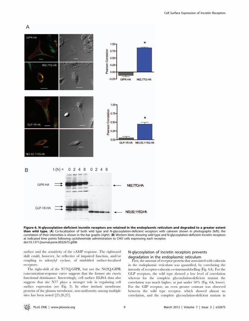
First, the amount of receptor protein that associated with calnexin
in the endoplasmic reticulum was quantified, by correlating the
intensity of receptor-calnexin co-immunolabelling (Fig. 6A). For the
GLP receptors, the wild type showed a low level of correlation
whereas for the complete glycosylation-deficient mutant the
correlation was much higher, at just under 50% (Fig. 6A, lower).
For the GIP receptor, an even greater contrast was observed
between the wild type receptor, which showed almost no
correlation, and the complete glycosylation-deficient mutant in
Figure 6. N-glycosylation-deficient incretin receptors are retained in the endoplasmic reticulum and degraded to a greater extentthan wild type. (A) Co-localization of both wild type and N-glycosylation-deficient receptors with calnexin shown in photographs (left); thecorrelation of their intensities is shown in the bar graphs (right). (B) Western blots showing wild type and N-glycosylation-deficient incretin receptorsat indicated time points following cycloheximide administration to CHO cells expressing each receptor.doi:10.1371/journal.pone.0032675.g006
Cell Surface Expression of Incretin Receptors
PLoS ONE | www.plosone.org 9 March 2012 | Volume 7 | Issue 3 | e32675

which correlation is about 80% (Fig. 6A, upper) Together, the data
suggest that the reduction in glycosylation- deficient mutants at the
cell surface is mainly due to an increase in ER retention and
degradation. These data are also consistent with a larger role for N-
glycosylation of the GIP receptor than for the GLP-1 receptor.
To address whether mis-folding and ER degradation is reduced
by N-glycosylation, we used western blotting to determine the
stability of the glycosylation deficient receptors following cyclo-
heximide application. The rate of disappearance of glycosylation
deficient receptors over time was increased compared to that of
wild type receptors (Fig. 6B). Moreover, even at time = 0, the
overall level of glycosylation-deficient receptors is much lower than
that of wild-type receptors; this is especially apparent for the GIP
mutant receptor.
N-glycosylation maintains cell surface GIP receptornumber and GIP-potentiated insulin secretion in INS-1cells
We next examined the role of GIPR N-glycosylation in
regulating ligand binding, surface receptor number and insulin
secretion. We chose the INS-1 b-cell line, which reflects a similar
physiology to that of native b-cells, including GIP-induced
potentiation of insulin secretion in the presence of glucose [22].
INS-1 cells were treated with tunicamycin and specific binding of
GIP was measured using a radioactive binding assay. Tunicamy-
cin lowered cell surface 125I-GIP binding, which, at saturating
levels, corresponded to a 70% decrease in cell surface GIPR
number (Fig. 7A). The dissociation constants (Kd) of GIP from the
surface of these cells did not significantly differ between the control
(455650 pM) and tunicamycin (3456100 pM) treated cells,
suggesting that ligand binding affinity was not impaired by
removal of N-glycosylation.
To investigate the impact of N-glycosylation on GIPR incretin
response, GIP-stimulated insulin secretion was measured in
tunicamycin-treated and untreated INS-1 cells. Without tunicamy-
cin, GIP (50 nM) significantly and strongly potentiated insulin
secretion, when increasing glucose from 5.5 mM to 11 mM (Fig. 7B).
Tunicamycin treatment, however, blunted the incretin effect of GIP.
Tunicamycin did not significantly modify insulin secretion in
response to glucose alone, suggesting that GIP potentiation was
eliminated specifically by the inhibition of N-glycosylation. These
Figure 7. N-glycosylation regulates GIP receptor number and GIP-potentiated insulin secretion in INS-1 cells. (A) Plot of specificbinding versus concentration of 125I-GIP saturation carried out in INS-1 cells with or without 1 mg/ml tunicamycin. Data were fitted with a non-linearregression equation assuming a single binding site (GraphPad Prism). Control cells expressed an average of 24436400 GIP receptors on the cellsurface (Kd 455650 pM), whereas tunicamycin-treated cells expressed 760670 GIP receptors on the cell surface (Kd 3456100 pM). (B) Plot of insulinrelease in response to glucose and/or GIP, from INS-1 cells with or without 1 mg/ml tunicamycin. Data are expressed as mean 6 SEM, n = 4 separateexperiments. Asterisk indicates a significant increase in insulin release compared to basal conditions without addition of 50 nM GIP (P,0.05), asdetermined using two-way ANOVA followed by Bonferroni post-tests.doi:10.1371/journal.pone.0032675.g007
Cell Surface Expression of Incretin Receptors
PLoS ONE | www.plosone.org 10 March 2012 | Volume 7 | Issue 3 | e32675

Figure 8. Function but not cell surface expression of N62,77Q-GIPR is rescued by co-expression of wild type GLP1R. (A) Plots ofRelative Fluorescence Units (RFU, indicative of cAMP production) versus log of concentration of GIP. RFU values were determined by FRET assaywhich was carried out using cells expressing GIPR and GLP1R as indicated. Forskolin was used to control for variations in total cell number. Mean data6 SE (n = 5 transfections) at each ligand concentration were fit using a sigmoidal dose-response curve. (B) Plots of cell surface (left) and whole cell(right) luminescence from cells co-expressing N-terminally HA-tagged GIPR and/or GLP1R, as indicated. All constructs were assayed in triplicate from4–5 separate transfections. Each group of triplicates was corrected for background luminescence from untransfected cells and from cells treated withonly secondary HRP-conjugated antibody for each transfected condition. Pairs of receptors (grouped in brackets), consisting of one HA-taggedconstruct and a second untagged construct, were statistically compared by a two-tailed unpaired t-test (p.0.05 for each comparison made).doi:10.1371/journal.pone.0032675.g008
Cell Surface Expression of Incretin Receptors
PLoS ONE | www.plosone.org 11 March 2012 | Volume 7 | Issue 3 | e32675

results are consistent with a reduction in GIP receptor number, but
do not discount a potential impairment in GIPR function and/or
signal transduction by the deficiency in N-glycosylation.
The function and cell surface expression of a GIPRmutant completely lacking N-glycosylation is rescued byclose association with the wild type GLP1R
It has been suggested that many GPCRs exist as homo- or
heterodimers and/or higher order oligomers at the cell surface
and, in some cases, this interaction may be initiated during
translation [28,29]. Because the GIPR and GLP1R have a
relatively high level of sequence identity (Fig. 1) and have similar
expression profiles and physiological function, we reasoned that
the GLP1R might form a functional complex with the GIPR. To
test this, we first examined whether cell surface expression and
function of the GIPR mutant lacking N-glycosylation could be
rescued by co-expression with wild type GLP1R. Cyclic AMP
levels were again measured in CHO cells expressing each receptor
construct. In cells expressing N62,77Q-GIPR only, the amount of
cAMP was unchanged from baseline in response to GIP. In
contrast, the levels of cAMP increased in a concentration-
dependent manner by GIP in cells co-expressing N62,77Q-GIPR
along with the wild type GLP1R, although the EC50 value was
right shifted compared to wild type GIPR (Fig. 8A, Table 2).
These results show that functional expression of N62,77Q-GIPR
was rescued by co-expressed wild type GLP1R.
In an attempt to quantify the extent of N62,77Q-GIPR cell
surface rescue by wild type GLP1R, cell surface ELISA was again
used. When the HA-tagged mutant GIPR was co-expressed with
wild type GLP1R, the measured cell surface luminescence was
very low and not significantly altered as compared to co-
expression with the untagged version of itself (Fig. 8B, upper).
Since the amplitude of cAMP responses to GIP were identical for
both wild type GIPR, and mutant GIPR in the presence of
GLP1R, it appears that rescue of a very small amount of the
N62,77Q-GIPR, below the level of detection by cell surface
ELISA, suffices to fully restore activation of adenylate cyclase in
the CHO cell system.
The rescue of only a small fraction of N62,77Q-GIPR to the cell
surface could be explained by a corresponding retention of wild type
GLP1R by the GIPR mutant. However, this was not the case since
the amount of HA-tagged GLP1R at the cell surface was unchanged
by co-expression with N62,77Q-GIPR (Fig. 8B, upper). Moreover,
when co-expressed, the total level of mutant GIPR and wild type
GLP1R were no different than when each was co-expressed with
non-HA tagged versions (Fig. 8B, lower). Therefore, it seems more
likely that functional rescue of the mutant GIPR is due to a low level
of association with the wild type GLP1R, which is below the
detection sensitivity of the ELISA assay.
The low level of association between N62,77Q-GIPR and wild
type GLP1R could be due to the small differences in their primary
sequences, thus potentially limiting interaction between key
regions. To test this, we co-expressed N62,77Q-GIPR with wild
type GIPR and measured cell surface and total protein expression
of both receptor constructs by ELISA. Co-expression of wild type
GIPR did not rescue cell surface expression of its mutant
Figure 9. Cell surface expression of N62,77Q-GIPR is not rescued by co-expression with the wild type GIPR. Plots of cell surface (left)and whole cell (right) luminescence from cells co-expressing HA-tagged GIPR as indicated. All constructs were assayed in triplicate from 4–5 separatetransfections. Each group of triplicates was corrected for background luminescence from untransfected cells and from cells treated with onlysecondary HRP-conjugated antibody for each transfected condition. Pairs of receptors (grouped in brackets), consisting of one HA-tagged constructand a second untagged construct, were statistically compared by a two-tailed unpaired t-test (p.0.05 for each comparison made).doi:10.1371/journal.pone.0032675.g009
Table 2. Mean EC50 values and amplitude of response forcAMP concentration-response curves in Figure 8A.
EC50 (M) Amplitude RFU
Mean S.E. P Value Mean S.E. P Value
GIPR-V5 3.4610211 1.1610211 - 2.36104 2.86103 -
N62,N77Q-V5 - - - 5.06103 7.16102 P,0.01
GLP1R-V5 - - - 6.36103 2.06103 P,0.01
GLP1R-V5+N62,N77Q-V5
1.161028 5.261029 P,0.0001 1.96104 2.36103 P.0.05
EC50 values were determined for each individual experiment, using a sigmoidalconcentration-response equation. Compared to wild-type GIPR, the EC50 valuesfor N62,77Q-GIPR when co-expressed with the wild type GLP1R wassignificantly right shifted, as shown by a one-way ANOVA analysis followed byDunnett’s multiple comparison test. The changes in RFU values for N62,77Q-GIPR or the GLP1R were small and could not be accurately fitted. The amplitudeof response was calculated by subtracting the lowest RFU values from thehighest for each individual experiment.doi:10.1371/journal.pone.0032675.t002
Cell Surface Expression of Incretin Receptors
PLoS ONE | www.plosone.org 12 March 2012 | Volume 7 | Issue 3 | e32675

counterpart, nor was its own level at the cell surface altered (Fig. 9,
left). Thus, the extent of receptor homology is not likely a factor for
limiting the amount of cell surface rescue of the mutant GIPR.
The functional rescue of the N62,77Q-GIPR by the wild type
GLP1R nevertheless suggests some form of physical association.
To test for this, we utilized BRET to measure the effects of co-
expressing GFP-labeled forms of GLP1R with RLuc-tagged GIPR
(Fig. 10A). For a negative control, we used GFP-tagged
hyperpolarization-activated channel (HCN2-GFP), a structurally
similar membrane protein that has been shown to localize to the
cell surface and function normally when expressed in CHO cells
[21], but which would not be expected to assemble with either
receptor. We also tested the effects of co-expressing GFP- and
RLuc- labeled GLP1R (Fig. 10B), as well as GFP- and RLuc-
labeled GIPR (Fig. 10C). For all three receptor combinations, the
BRET saturation curves reached higher maximal values than their
respective negative controls. The high values of BRET obtained,
coupled with the rescue of N62,77Q-GIPR function by the wild
type GLP1R, support heteromeric and homomeric associations of
the GIPR and GLP1R.
Discussion
Upon translation of the GIPR or GLP-1R nascent chains in the
endoplasmic reticulum, they become subject to glycosylation at the
Asn residues located in the extracellular N-terminus. We have
Figure 10. BRET values for cells expressing GIPR and GLP1R suggest heteromeric and homomeric associations. Plots of BRET2 ratioversus the ratio of GFP/Luc from cells co-expressing RLuc- and GFP-tagged combinations of GIPR, GLP1R and HCN2 channels. (A) Curves fromGIPR+GLP1R (Bmax = 0.07360.003) compared to GIPR+HCN2 negative control (Bmax = 0.02860.004). (B) Curves from GLP1R+GLP1R(Bmax = 0.1560.03), compared to GLP1R+HCN2 negative control (Bmax = 0.02960.006). (C) Curves from GIPR+GIPR (Bmax = 0.1360.02), comparedto GIPR+HCN2 negative control (Bmax = 0.02860.004). Data were collected from at least separate 5 transfections and fit with a non-linear regressionequation assuming a single binding site; they were statistically analyzed using a two-tailed unpaired t test and for all conditions, Bmax wassignificantly greater than negative controls, p,0.001.doi:10.1371/journal.pone.0032675.g010
Cell Surface Expression of Incretin Receptors
PLoS ONE | www.plosone.org 13 March 2012 | Volume 7 | Issue 3 | e32675
shown that each of the putative Asn residues is glycosylated when
either the human GIPR or GLP-1R is expressed in CHO cells. For
both receptors, cell surface expression and/or function were
impaired by removal of N-glycosylation, although the GIPR was
more strongly affected and possessed a single site (N77) that was
dominant. Our results from ELISA experiments suggest that the
decrease in cell surface receptor expression can be attributed to
factors independent of variations in total expression. Our data also
show enhanced ER retention and degredation in the absence of N-
glycosylation, in parallel with disrupted cell surface expression.
Our findings come from expression of CHO cells, which is a
limitation as primary beta-cells are very difficult to transfect and
the low level of GIP and GLP-1 expression makes studies on
endogenous glycosylation very difficult.
Importantly, we found that N-glycosylation regulates GIPR cell
surface expression and the potentiation of glucose-induced insulin
secretion by nanomolar levels of GIP in INS-1 cells. These data
are similar to those obtained with the GLP-1R in RINm5F cells,
where cell surface receptor number and GLP-1-induced cAMP
production are reduced by tunicamycin treatment [10]. Thus, N-
glycosylation maintains a level of GIPR and GLP-1R surface
expression that is critical for downstream signaling and regulation
of insulin secretion. Notably, 30% of the normal total number of
GIP receptors remained after tunicamycin treatment despite an
absence of GIP-potentiation of glucose-induced insulin secretion.
This implies that a threshold number of GIP receptors exist, which
must be surpassed in order to potentiate insulin secretion to a level
that could be measured by our assay. Our findings in CHO cells
suggest that the reduction in cell surface expression in the absence
of N-glycosylation in the INS-1 cells is also due to enhanced
retention and degredation of GIPR in the endoplasmic reticulum.
Basic mechanisms of cell surface trafficking and ER quality control
are conserved among most cells [30,31], but studies in beta cells
are required to determine their precise impact on incretin receptor
cell surface expression and lifespan.
When cAMP was assayed in response to increasing concentra-
tions of ligand, cells containing glycosylation-deficient GLP-1R or
N77Q-GIPR exhibited a right-shifted concentration-response
curve. This could have reflected a direct defect in GIP or GLP-
1 binding to their respective receptors, or to a coupling problem
with the signal transduction machinery. However, our data of
GIP-binding to INS-1 cells, and previous data on GLP-1 binding
to RINm5F cells [10], showed that binding affinity was unaffected
by tunicamycin treatment, whereas the total surface receptor
number was significantly decreased along with potentiation of
insulin secretion. These data are consistent with those in other
studies of bradykinin B2, P2Y12 ADP, and the type 1ametabotropic glutamate receptors, which also found that ligand-
binding affinity was unaltered by inhibition of N-glycosylation
[32,33,34]. Thus, the reduction in incretin sensitivity that we
observed is probably due impaired function, and/or coupling to
adenylyl cyclase, of misfolded surface-localized receptors. Alter-
natively, since cell surface expression and, in the case of the
glycosylation-deficient GLP-1R, the maximum cAMP response
were also reduced in concert with sensitivity, it is possible that the
efficiency of coupling to downstream effector molecules such as
adenylyl cyclase correlates with cell surface receptor number. Such
a relationship between EC50 and cell surface number has been
reported for the b2-adrenergic receptor, but the mechanism
underlying this association remains unknown [35,36]. In these
studies, the b2-adrenergic receptor was localized to caveolae to a
lesser extent when cell surface number was reduced; this suggests
that efficient signal transduction requires localization of receptors
to these structures, which contain the necessary machinery for
cAMP production.
The rescue of mutant GIPR function by the wild type GLP1R,
and the close association between the two receptors as measured
by BRET, suggests that they are able to associate in a receptor
complex. According to a recent definition, receptor heteromers are
‘‘composed of at least two functional receptor units with
biochemical properties that are demonstrably different from those
of its individual components’’ [37]. Functional co-assembly is
supported by reduced sensitivity of the rescued GIPR mutant to
GIP when compared to the wild type GIPR, as seen by the right
shift in EC50 for cAMP formation; however, this right shift could
also be explained by a corresponding reduction in number of
mutant receptors at the cell surface, compared to wild type.
Therefore, it remains uncertain as to whether oligomerization of
GIPR and GLP1R produces a receptor complex with unique
properties and, thus, whether a GIP-GLP1 receptor heteromer
exists in our system. We also found that oligomerization may have
been limited between glycosylation-deficient GIPR and the wild
type GLP1R, based on the lack of cell surface rescue. Indeed,
various studies have shown that when N-glycosylation is removed,
regular receptor dimer formation and stability is impaired, thereby
impacting the receptor heteromer’s ability to express at the cell
surface and/or correctly function [38,39,40,41,42]. Studies that
further examine the functional and structural nature of the
association between GLP-1R and GIPR will be required to
determine the complete nature of this association and whether a
true receptor heteromer is formed by them.
Regulation of GIPR and GLP-1R by N-glycosylation may have
important implications for type 2 diabetes (T2DM). There is a
reduced incretin effect in human T2DM patients [43], which has
been attributed to reduced ß-cell responsiveness to GIP [44] and,
to a lesser extent, GLP-1 [45]. Moreover, reduced incretin
receptor expression has been observed in animal models of T2DM
[2]. It will be important to determine if post-translational
modifications such as N-glycosylation are impaired in Type II
diabetes, and whether this results in a reduction in cell surface
GIPR and GLP-1R number along with the decreased incretin
response of the ß-cell. The finding that heteromerization between
GIPR and GLP1R can occur suggests that the much milder
reduction in GLP1 responsiveness could be a direct consequence
of the more profound and fundamental observed decrease in
GIPR expression. This possibility could explain the restoration of
responses to both incretins in diabetic patients in whom glucose
was almost normalized by insulin treatment [45]. Direct evidence
for heteromerization of GIPR and GLP1R in tissues such as the
pancreas would be an essential next step in determining the
potential physiological consequences, as well as the therapeutic
implications of such an interaction.
Author Contributions
Conceived and designed the experiments: GW FL CM EA. Performed the
experiments: GW FL. Analyzed the data: GW FL CM EA. Wrote the
paper: GW FL CM EA.
References
1. Baggio LL, Drucker DJ (2007) Biology of incretins: GLP-1 and GIP.
Gastroenterology 132: 2131–2157.
2. McIntosh CH, Widenmaier S, Kim SJ (2009) Glucose-dependent insulinotropic
polypeptide (Gastric Inhibitory Polypeptide; GIP). Vitam Horm 80: 409–471.
Cell Surface Expression of Incretin Receptors
PLoS ONE | www.plosone.org 14 March 2012 | Volume 7 | Issue 3 | e32675

3. Gurevich VV, Gurevich EV (2008) GPCR monomers and oligomers: it takes all
kinds. Trends Neurosci 31: 74–81.
4. Dong C, Filipeanu CM, Duvernay MT, Wu G (2007) Regulation of G protein-
coupled receptor export trafficking. Biochim Biophys Acta 1768: 853–870.
5. Bulenger S, Marullo S, Bouvier M (2005) Emerging role of homo- and
heterodimerization in G-protein-coupled receptor biosynthesis and maturation.
Trends Pharmacol Sci 26: 131–137.
6. Gurevich VV, Gurevich EV (2008) How and why do GPCRs dimerize? Trends
Pharmacol Sci 29: 234–240.
7. Amiranoff B, Couvineau A, Vauclin-Jacques N, Laburthe M (1986) Gastric
inhibitory polypeptide receptor in hamster pancreatic beta cells. Direct cross-
linking, solubilization and characterization as a glycoprotein. European journal
of biochemistry/FEBS 159: 353.
8. Widmann C, Dolci W, Thorens B (1996) Desensitization and phosphorylation of
the glucagon-like peptide-1 (GLP-1) receptor by GLP-1 and 4-phorbol 12-
myristate 13-acetate. Molecular endocrinology (Baltimore, Md) 10: 62.
9. Moens K, Heimberg H, Flamez D, Huypens P, Quartier E, et al. (1996)
Expression and functional activity of glucagon, glucagon-like peptide I, and
glucose-dependent insulinotropic peptide receptors in rat pancreatic islet cells.
Diabetes 45: 257.
10. Goke R, Just R, Lankat-Buttgereit B, Goke B (1994) Glycosylation of the GLP-1
receptor is a prerequisite for regular receptor function. Peptides 15: 675.
11. Mayo KE, Miller LJ, Bataille D, Dalle S, Goke B, et al. (2003) International
Union of Pharmacology. XXXV. The glucagon receptor family. Pharmacol Rev
55: 167–194.
12. Usdin TB, Mezey E, Button DC, Brownstein MJ, Bonner TI (1993) Gastric
inhibitory polypeptide receptor, a member of the secretin-vasoactive intestinal
peptide receptor family, is widely distributed in peripheral organs and the brain.
Endocrinology 133: 2861–2870.
13. Whorton MR, Bokoch MP, Rasmussen SG, Huang B, Zare RN, et al. (2007) A
monomeric G protein-coupled receptor isolated in a high-density lipoprotein
particle efficiently activates its G protein. Proc Natl Acad Sci U S A 104:
7682–7687.
14. Chabre M, le Maire M (2005) Monomeric G-protein-coupled receptor as a
functional unit. Biochemistry 44: 9395–9403.
15. Meyer BH, Segura JM, Martinez KL, Hovius R, George N, et al. (2006) FRET
imaging reveals that functional neurokinin-1 receptors are monomeric and
reside in membrane microdomains of live cells. Proc Natl Acad Sci U S A 103:
2138–2143.
16. Milligan G (2009) G protein-coupled receptor hetero-dimerization: contribution
to pharmacology and function. Br J Pharmacol.
17. Chabre M, Deterre P, Antonny B (2009) The apparent cooperativity of some
GPCRs does not necessarily imply dimerization. Trends Pharmacol Sci 30:
182–187.
18. Vrecl M, Drinovec L, Elling C, Heding A (2006) Opsin oligomerization in a
heterologous cell system. Journal of receptor and signal transduction research
26: 505.
19. Harikumar KG, Morfis MM, Sexton PM, Miller LJ (2008) Pattern of intra-
family hetero-oligomerization involving the G-protein-coupled secretin receptor.
Journal of molecular neuroscience: MN 36: 279.
20. Gremlich S, Porret A, Hani EH, Cherif D, Vionnet N, et al. (1995) Cloning,
functional expression, and chromosomal localization of the human pancreatic
islet glucose-dependent insulinotropic polypeptide receptor. Diabetes 44: 1202.
21. Whitaker GM, Angoli D, Nazzari H, Shigemoto R, Accili EA (2007) HCN2 and
HCN4 isoforms self-assemble and co-assemble with equal preference to form
functional pacemaker channels. J Biol Chem 282: 22900–22909.
22. Hohmeier HE, Mulder H, Chen G, Henkel-Rieger R, Prentki M, et al. (2000)
Isolation of INS-1-derived cell lines with robust ATP-sensitive K+ channel-
dependent and -independent glucose-stimulated insulin secretion. Diabetes 49:
424–430.
23. Lynn FC, Thompson SA, Pospisilik JA, Ehses JA, Hinke SA, et al. (2003) A
novel pathway for regulation of glucose-dependent insulinotropic polypeptide
(GIP) receptor expression in beta cells. Faseb J 17: 91–93.
24. Whitaker GM, Accili EA (2008) Using Bioluminescence Resonance Energy
Transfer to measure ion channel assembly. Methods in molecular biology(Clifton, NJ) 491: 189.
25. Hebert DN, Zhang JX, Chen W, Foellmer B, Helenius A (1997) The number
and location of glycans on influenza hemagglutinin determine folding andassociation with calnexin and calreticulin. J Cell Biol 139: 613–623.
26. Chang XB, Mengos A, Hou YX, Cui L, Jensen TJ, et al. (2008) Role of N-linkedoligosaccharides in the biosynthetic processing of the cystic fibrosis membrane
conductance regulator. Journal of cell science 121: 2814.
27. Mialet-Perez J, Green SA, Miller WE, Liggett SB (2004) A primate-dominantthird glycosylation site of the beta2-adrenergic receptor routes receptors to
degradation during agonist regulation. J Biol Chem 279: 38603–38607.28. Milligan G (2007) G protein-coupled receptor dimerisation: molecular basis and
relevance to function. Biochimica et biophysica acta 1768: 825.29. Bulenger S, Marullo S, Bouvier M (2005) Emerging role of homo- and
heterodimerization in G-protein-coupled receptor biosynthesis and maturation.
Trends in pharmacological sciences 26: 131.30. Vembar SS, Brodsky JL (2008) One step at a time: endoplasmic reticulum-
associated degradation. Nat Rev Mol Cell Biol 9: 944–957.31. Xie W, Ng DT (2010) ERAD substrate recognition in budding yeast. Semin Cell
Dev Biol 21: 533–539.
32. Mody N, Hermans E, Nahorski SR, Challiss RA (1999) Inhibition of N-linkedglycosylation of the human type 1alpha metabotropic glutamate receptor by
tunicamycin: effects on cell-surface receptor expression and function. Neuro-pharmacology 38: 1485.
33. Michineau S, Muller L, Pizard A, Alhenc-Gelas F, Rajerison RM (2004) N-linked glycosylation of the human bradykinin B2 receptor is required for optimal
cell-surface expression and coupling. Biological chemistry 385: 49.
34. Zhong X, Kriz R, Seehra J, Kumar R (2004) N-linked glycosylation of plateletP2Y12 ADP receptor is essential for signal transduction but not for ligand
binding or cell surface expression. FEBS letters 562: 111.35. Ostrom RS, Gregorian C, Drenan RM, Xiang Y, Regan JW, et al. (2001)
Receptor number and caveolar co-localization determine receptor coupling
efficiency to adenylyl cyclase. J Biol Chem 276: 42063–42069.36. Patel HH, Murray F, Insel PA (2008) Caveolae as organizers of pharmacolog-
ically relevant signal transduction molecules. Annu Rev Pharmacol Toxicol 48:359–391.
37. Ferre S, Baler R, Bouvier M, Caron MG, Devi LA, et al. (2009) Building a newconceptual framework for receptor heteromers. Nat Chem Biol 5: 131–134.
38. Fernandes H, Cohen S, Bishayee S (2001) Glycosylation-induced conforma-
tional modification positively regulates receptor-receptor association: a studywith an aberrant epidermal growth factor receptor (EGFRvIII/DeltaEGFR)
expressed in cancer cells. The Journal of biological chemistry 276: 5375.39. He J, Xu J, Castleberry AM, Lau AG, Hall RA (2002) Glycosylation of beta(1)-
adrenergic receptors regulates receptor surface expression and dimerization.
Biochemical and biophysical research communications 297: 565.40. Langer I, Leroy K, Gaspard N, Brion JP, Robberecht P (2008) Cell surface
targeting of VPAC1 receptors: evidence for implication of a quality controlsystem and the proteasome. Biochimica et biophysica acta 1783: 1663.
41. Michineau S, Alhenc-Gelas F, Rajerison RM (2006) Human bradykinin B2receptor sialylation and N-glycosylation participate with disulfide bonding in
surface receptor dimerization. Biochemistry 45: 2699.
42. Wanamaker CP, Green WN (2005) N-linked glycosylation is required fornicotinic receptor assembly but not for subunit associations with calnexin. J Biol
Chem 280: 33800–33810.43. Nauck M, Stockmann F, Ebert R, Creutzfeldt W (1986) Reduced incretin effect
in type 2 (non-insulin-dependent) diabetes. Diabetologia 29: 46–52.
44. Nauck MA, Heimesaat MM, Orskov C, Holst JJ, Ebert R, et al. (1993) Preservedincretin activity of glucagon-like peptide 1 [7–36 amide] but not of synthetic
human gastric inhibitory polypeptide in patients with type-2 diabetes mellitus.J Clin Invest 91: 301–307.
45. Hojberg PV, Vilsboll T, Rabol R, Knop FK, Bache M, et al. (2009) Four weeks
of near-normalisation of blood glucose improves the insulin response toglucagon-like peptide-1 and glucose-dependent insulinotropic polypeptide in
patients with type 2 diabetes. Diabetologia 52: 199–207.
Cell Surface Expression of Incretin Receptors
PLoS ONE | www.plosone.org 15 March 2012 | Volume 7 | Issue 3 | e32675
Related Documents