Regulation of Amyloid Precursor Protein Processing by Serotonin Signaling Anna A. Pimenova 1,2 , Amantha Thathiah 1,2 , Bart De Strooper 1,2 *, Ina Tesseur 1,2 * 1 KU Leuven Center for Human Genetics and Leuven research Institute for Neuroscience and Disease (LIND), Leuven, Belgium, 2 VIB Center for the Biology of Disease, Leuven, Belgium Abstract Proteolytic processing of the amyloid precursor protein (APP) by the b- and c-secretases releases the amyloid-b peptide (Ab), which deposits in senile plaques and contributes to the etiology of Alzheimer’s disease (AD). The a-secretase cleaves APP in the Ab peptide sequence to generate soluble APPa (sAPPa). Upregulation of a-secretase activity through the 5- hydroxytryptamine 4 (5-HT 4 ) receptor has been shown to reduce Ab production, amyloid plaque load and to improve cognitive impairment in transgenic mouse models of AD. Consequently, activation of 5-HT 4 receptors following agonist stimulation is considered to be a therapeutic strategy for AD treatment; however, the signaling cascade involved in 5-HT 4 receptor-stimulated proteolysis of APP remains to be determined. Here we used chemical and siRNA inhibition to identify the proteins which mediate 5-HT 4d receptor-stimulated a-secretase activity in the SH-SY5Y human neuronal cell line. We show that G protein and Src dependent activation of phospholipase C are required for a-secretase activity, while, unexpectedly, adenylyl cyclase and cAMP are not involved. Further elucidation of the signaling pathway indicates that inositol triphosphate phosphorylation and casein kinase 2 activation is also a prerequisite for a-secretase activity. Our findings provide a novel route to explore the treatment of AD through 5-HT 4 receptor-induced a-secretase activation. Citation: Pimenova AA, Thathiah A, De Strooper B, Tesseur I (2014) Regulation of Amyloid Precursor Protein Processing by Serotonin Signaling. PLoS ONE 9(1): e87014. doi:10.1371/journal.pone.0087014 Editor: Bart Dermaut, Pasteur Institute of Lille, France Received September 16, 2013; Accepted December 16, 2013; Published January 21, 2014 Copyright: ß 2014 Pimenova et al. This is an open-access article distributed under the terms of the Creative Commons Attribution License, which permits unrestricted use, distribution, and reproduction in any medium, provided the original author and source are credited. Funding: This work was supported by the Fund for Scientific Research Flanders, KU Leuven, Federal Office for Scientific Affairs (IAP P7/16), a Methusalem grant of the Flemish Government, VIB, IWT. BDS is the Arthur Bax and Anna Vanluffelen chair for Alzheimer’s disease. The funders had no role in study design, data collection and analysis, decision to publish, or preparation of the manuscript. Competing Interests: Bart De Strooper is a consultant for Janssen Pharmaceutica (Beerse, Belgium), Remynd NV (Leuven, Belgium) and Envivo Inc (Boston, USA). The authors have declared that this does not alter their adherence to all the PLOS ONE policies on sharing data and materials. The authors have no direct or indirect financial interest in the current study. The authors have declared that no competing interests exist. * E-mail: [email protected] (IT); [email protected] (BDS) Introduction The most common form of dementia in elderly people is Alzheimer’s disease (AD), which is pathologically characterized by progressive neuronal loss and deposition of the amyloid-b peptide (Ab) in amyloid plaques. Current therapeutic targets are the b- and c-secretases, which generate Ab from amyloid precursor protein (APP). Several drugs inhibiting or modulating the activity of these secretases have failed in clinical trials due to severe side effects or to difficulty in delivery through the blood brain barrier [1]. APP is also cleaved by a third secretase called a-secretase. The a-secretase cleaves APP within the Ab peptide sequence, producing a soluble APPa fragment (sAPPa), which precludes Ab generation. Indeed, in vivo overexpression or upregulation of a- secretase activity in mice indicate that a-secretase activation leads to a decrease in Ab production and a reduction in the amyloid plaque load in AD mouse models [2,3]. These effects were accompanied by an improvement in the cognitive deficits, providing considerable support for modulation of a-secretase activity as a viable strategy in the fight against AD [2,3]. To specifically target the non-amyloidogenic pathway of APP processing, a fundamental consideration would be to understand the mechanism of a-secretase activation and to determine the signaling cascade of kinases and second messengers that directly regulate a-secretase-mediated proteolysis of APP. These molecules can be directly targeted pharmacologically, but also indirectly via G protein-coupled receptors (GPCR), such as the muscarinic, glutamatergic and serotonergic receptors. In particular, the G protein coupled 5-hydroxytryptamine 4 (5-HT 4 ) receptor is gaining considerable interest as a modulator of a-secretase activity due to its role in memory and learning and regulation of APP processing [4]. Activation of the 5-HT 4 receptor leads to an increase in the population spike amplitude in the hippocampal CA1 region, and this effect persists in a transgenic mouse model of AD [5,6], suggesting that 5-HT 4 receptor-mediated signaling remains functional under these pathological conditions. On the other hand, agonist stimulation of the 5-HT 4 receptor results in increased sAPPa secretion with a concomitant decrease in Ab peptide levels in primary neuronal cultures and an alleviation of amyloid plaque load in AD mouse models [7–9]. Such amelio- ration of disease pathology is correlated with improvements in memory and learning in behavioral paradigms and scopolamine- induced models of cognitive deficit [10–12]. Additionally, an increase in acetylcholine release is observed after 5-HT 4 receptor agonist application in vivo [13]. This could be a valuable property when considering 5-HT 4 receptor agonists for AD treatment, which could complement the currently licensed therapy of cholinesterase inhibition for partial symptomatic relief [14]. Despite numerous reports on 5-HT 4 receptor function in memory and learning and its effect on APP processing, the PLOS ONE | www.plosone.org 1 January 2014 | Volume 9 | Issue 1 | e87014

Welcome message from author
This document is posted to help you gain knowledge. Please leave a comment to let me know what you think about it! Share it to your friends and learn new things together.
Transcript

Regulation of Amyloid Precursor Protein Processing bySerotonin SignalingAnna A. Pimenova1,2, Amantha Thathiah1,2, Bart De Strooper1,2*, Ina Tesseur1,2*
1 KU Leuven Center for Human Genetics and Leuven research Institute for Neuroscience and Disease (LIND), Leuven, Belgium, 2 VIB Center for the Biology of Disease,
Leuven, Belgium
Abstract
Proteolytic processing of the amyloid precursor protein (APP) by the b- and c-secretases releases the amyloid-b peptide(Ab), which deposits in senile plaques and contributes to the etiology of Alzheimer’s disease (AD). The a-secretase cleavesAPP in the Ab peptide sequence to generate soluble APPa (sAPPa). Upregulation of a-secretase activity through the 5-hydroxytryptamine 4 (5-HT4) receptor has been shown to reduce Ab production, amyloid plaque load and to improvecognitive impairment in transgenic mouse models of AD. Consequently, activation of 5-HT4 receptors following agoniststimulation is considered to be a therapeutic strategy for AD treatment; however, the signaling cascade involved in 5-HT4
receptor-stimulated proteolysis of APP remains to be determined. Here we used chemical and siRNA inhibition to identifythe proteins which mediate 5-HT4d receptor-stimulated a-secretase activity in the SH-SY5Y human neuronal cell line. Weshow that G protein and Src dependent activation of phospholipase C are required for a-secretase activity, while,unexpectedly, adenylyl cyclase and cAMP are not involved. Further elucidation of the signaling pathway indicates thatinositol triphosphate phosphorylation and casein kinase 2 activation is also a prerequisite for a-secretase activity. Ourfindings provide a novel route to explore the treatment of AD through 5-HT4 receptor-induced a-secretase activation.
Citation: Pimenova AA, Thathiah A, De Strooper B, Tesseur I (2014) Regulation of Amyloid Precursor Protein Processing by Serotonin Signaling. PLoS ONE 9(1):e87014. doi:10.1371/journal.pone.0087014
Editor: Bart Dermaut, Pasteur Institute of Lille, France
Received September 16, 2013; Accepted December 16, 2013; Published January 21, 2014
Copyright: � 2014 Pimenova et al. This is an open-access article distributed under the terms of the Creative Commons Attribution License, which permitsunrestricted use, distribution, and reproduction in any medium, provided the original author and source are credited.
Funding: This work was supported by the Fund for Scientific Research Flanders, KU Leuven, Federal Office for Scientific Affairs (IAP P7/16), a Methusalem grant ofthe Flemish Government, VIB, IWT. BDS is the Arthur Bax and Anna Vanluffelen chair for Alzheimer’s disease. The funders had no role in study design, datacollection and analysis, decision to publish, or preparation of the manuscript.
Competing Interests: Bart De Strooper is a consultant for Janssen Pharmaceutica (Beerse, Belgium), Remynd NV (Leuven, Belgium) and Envivo Inc (Boston,USA). The authors have declared that this does not alter their adherence to all the PLOS ONE policies on sharing data and materials. The authors have no direct orindirect financial interest in the current study. The authors have declared that no competing interests exist.
* E-mail: [email protected] (IT); [email protected] (BDS)
Introduction
The most common form of dementia in elderly people is
Alzheimer’s disease (AD), which is pathologically characterized by
progressive neuronal loss and deposition of the amyloid-b peptide
(Ab) in amyloid plaques. Current therapeutic targets are the b-
and c-secretases, which generate Ab from amyloid precursor
protein (APP). Several drugs inhibiting or modulating the activity
of these secretases have failed in clinical trials due to severe side
effects or to difficulty in delivery through the blood brain barrier
[1]. APP is also cleaved by a third secretase called a-secretase. The
a-secretase cleaves APP within the Ab peptide sequence,
producing a soluble APPa fragment (sAPPa), which precludes
Ab generation. Indeed, in vivo overexpression or upregulation of a-
secretase activity in mice indicate that a-secretase activation leads
to a decrease in Ab production and a reduction in the amyloid
plaque load in AD mouse models [2,3]. These effects were
accompanied by an improvement in the cognitive deficits,
providing considerable support for modulation of a-secretase
activity as a viable strategy in the fight against AD [2,3].
To specifically target the non-amyloidogenic pathway of APP
processing, a fundamental consideration would be to understand
the mechanism of a-secretase activation and to determine the
signaling cascade of kinases and second messengers that directly
regulate a-secretase-mediated proteolysis of APP. These molecules
can be directly targeted pharmacologically, but also indirectly via
G protein-coupled receptors (GPCR), such as the muscarinic,
glutamatergic and serotonergic receptors. In particular, the G
protein coupled 5-hydroxytryptamine 4 (5-HT4) receptor is
gaining considerable interest as a modulator of a-secretase activity
due to its role in memory and learning and regulation of APP
processing [4]. Activation of the 5-HT4 receptor leads to an
increase in the population spike amplitude in the hippocampal
CA1 region, and this effect persists in a transgenic mouse model of
AD [5,6], suggesting that 5-HT4 receptor-mediated signaling
remains functional under these pathological conditions. On the
other hand, agonist stimulation of the 5-HT4 receptor results in
increased sAPPa secretion with a concomitant decrease in Abpeptide levels in primary neuronal cultures and an alleviation of
amyloid plaque load in AD mouse models [7–9]. Such amelio-
ration of disease pathology is correlated with improvements in
memory and learning in behavioral paradigms and scopolamine-
induced models of cognitive deficit [10–12]. Additionally, an
increase in acetylcholine release is observed after 5-HT4 receptor
agonist application in vivo [13]. This could be a valuable property
when considering 5-HT4 receptor agonists for AD treatment,
which could complement the currently licensed therapy of
cholinesterase inhibition for partial symptomatic relief [14].
Despite numerous reports on 5-HT4 receptor function in
memory and learning and its effect on APP processing, the
PLOS ONE | www.plosone.org 1 January 2014 | Volume 9 | Issue 1 | e87014

downstream signaling pathway responsible for this 5-HT4
receptor-mediated effect is still poorly understood. 5-HT4 receptor
stimulation results in an accumulation of cAMP, a second
messenger required for protein kinase A (PKA) and exchange
protein activated by cAMP (Epac) activation. However, 5-HT4
receptor-mediated non-amyloidogenic processing of APP occurs
independently of PKA activation, but can be regulated by Epac1
activation of Rac1 and Rap signaling in cell lines and primary
neurons [15]. The 5-HT4 receptor is constitutively bound to the
Src non-receptor tyrosine kinase, which is required for ERK
activation [16]. In addition, 5-HT4 receptor stimulation in
adrenocortical cells and cardiomyocytes results in an increase of
calcium influx, which results in activation of voltage-gated calcium
channels through PKA [17,18]. It is unknown whether these latter
pathways also contribute to a-secretase activation downstream of
the 5-HT4 receptor. Altogether, these studies suggest a compli-
cated picture of the downstream signaling pathways involved in 5-
HT4 receptor stimulation and reveal the importance of delineation
of the mechanism of 5-HT4 receptor-mediated APP proteolysis.
Finally, several metalloproteinases have been proposed as a-
secretase; however, the identity of 5-HT4 receptor-induced a-
secretase activity has not been fully addressed. The disintegrin and
metalloprotease ADAM10, a major constitutive a-secretase of APP
[19,20], is a feasible candidate [21]. However, ADAM17 is more
likely to be the inducible APP a-secretase based on studies which
have investigated the regulated ectodomain shedding of other
ADAM substrates after protein kinase C (PKC) activation [22]. In
support of this is the observation that M1 receptor induced sAPParelease correlates with increased ADAM17 expression levels [23].
Nevertheless, additional metalloproteinases, such as meprin b and
membrane-type matrix metalloproteinases, were shown to mediate
a-cleavage of APP [24,25].
Here, we specifically determined the intracellular signaling
cascade involved in 5-HT4d receptor stimulation and inducible a-
secretase activity. We used human SH-SY5Y neuroblastoma cells
to analyze APP processing for practical reasons and experimental
consistency. Human SH-SY5Y cells can generate sustainable cells
with characteristics that resemble the morphology and biochem-
istry of mature neurons [26]. We present evidence that the G
protein-dependent pathway activating Src, phospholipase C (PLC)
and casein kinase 2 (CK2) is responsible for the 5-HT4d receptor-
stimulated induction of a-secretase activity. Interestingly and in
contrast to previous publications, we find that adenylyl cyclase
(AC) and cAMP signaling are not required for 5-HT4d receptor-
mediated a-secretase activity [15]. Furthermore, we analyzed the
reported a-secretases as putative mediators of the 5-HT4d receptor
effect on APP shedding using RNAi studies.
Materials and Methods
Reagents and AntibodiesTissue culture reagents were purchased from Invitrogen.
GF109203X, SQ22536, D609, ionomycin, 4,5,6,7-tetrabromo-
1H-benzotriazole (TBB), GR113808, NF449, gallein and batima-
stat were obtained from Tocris. 5-hydroxytryptamine (5-HT),
cholera toxin B (CTB) and chlorogenic acid (CGA) were from
Sigma-Aldrich. Phorbol 12-myristate 13-acetate (PMA), 2,5-
dideoxyadenosine (DDA), IP3K inhibitor, TAPI-1 and GM6001
were obtained from Calbiochem/VWR. Bosutinib was from
Selleck. Prucalopride was kindly provided by Movetis (NV,
Turnhout, Belgium). Table 1 summarizes the known potencies
of the used agonists and antagonists. Following antibodies were
purchased: CK2 a (H-286) from Santa Cruz, MMP-9 (G657) and
ADAM9 (2099) from Cell Signaling, ADAM17 (T5442) and b-
Actin (A5441) from Sigma-Aldrich. ADAM10 (B42) and APP
(B63) antibodies were made in house and previously described
[27]. Gas dominant negative construct was previously described
(pcDNAI-Amp-GasDN, [28]).
Inhibitor treatment and soluble APP analysis (SEAP assay)SH-SY5Y human neuroblastoma cells (CRL-2266, ATCC)
were cultured in DMEM/F12 supplemented with 10% fetal
bovine serum (FBS). For analysis of soluble APP secretion, a mix of
1,5 mg plasmid encoding human wild type APP695 linked to
Alkaline Phosphatase (AP-APP) at the N-terminus (pEAK12-AP-
APP, [29]), 1,35 mg of 5-HT4d receptor isoform in pcDNA3.1
(pcDNA3.1-5-HT4d, kindly provided by Joris De Mayer and Jan
Schuurkes, Movetis, Turnhout, Belgium) and 0,15 mg of GFP
(pmaxFP-Green-N, Amaxa) was prepared in OPTI-MEM and
combined with 20 ml of Lipofectamine 2000 (Invitrogen). After
20 minutes incubation at room temperature transfection mix was
combined with a trypsinized cell suspension in growth medium
containing 10% FBS. After another 15 minutes incubation at
room temperature cells were seeded in a 96-well plate at 80.000
cells/well. The next day medium was changed to DMEM/F12
supplemented with 5% dialyzed FBS (10,000 molecular weight
cutoff), which is devoid of serotonin otherwise present in
undialyzed FBS that causes 5-HT4 receptor desensitization. After
three days, cells were washed and incubated in serum free medium
(SFM) for another 24 hours. Next cells were stimulated with 1 mM
of the following compounds: prucalopride, 5-HT, PMA and
GR113808 in SFM for 24 hours and the conditioned medium was
analyzed for secreted AP (SEAP) activity with Great EscAPe SEAP
Chemiluminescence Kit 2.0 (Clontech) according to manufactur-
er’s instructions. Luminescence was measured with the EnVisionHmultilabel reader (PerkinElmer). For signaling studies dilution
curves of inhibitory compounds were made in combination with
induction by 1 mM prucalopride or 5-HT. DMSO incubation was
used as a control in all experiments carried out. The ratio of
individual luminescence counts from the tested conditions to the
mean value of DMSO treated cells was plotted as SEAP fold
induction. Cells were used for the MTS proliferation assay
(CellTiter 96H AQueous Non-Radioactive Cell Proliferation Assay)
and the LDH cytotoxicity assay (CytoTox 96H Non-Radioactive
Cytotoxicity Assay) according to manufacturer’s instructions
(Promega). Compound dilution curves were performed in the
range of the reported effective concentrations (Table 1) and
working concentrations were determined in the SEAP assay as
those giving significant inhibition of 5-HT4d receptor-stimulated
sAPPa secretion. MTS and LDH assays were used to define
working concentrations of the different compounds that were non-
toxic to the cells.
cAMP assaycAMP levels were assessed using the AlphascreenH cAMP assay
kit (PerkinElmer Life Sciences). 2,88?106 or 1?106 SH-SY5Y cells
were seeded in T75 or T25 flasks, respectively. Adherent cells were
transfected after 4 hours with Lipofectamine and Plus reagent
(Invitrogen) according to the manufacturer’s instructions. A mix of
7,5 mg pEAK12-AP-APP, 6 mg pcDNA3.1-5-HT4d and 1,5 mg
pmaxFP-Green-N plasmids (ratio of 5:4:1) was used for transfec-
tion in T75 flasks. A mix of 1,25 mg pEAK12-AP-APP, 0,65 mg
pcDNA3.1-5-HT4d receptor and 2,6 mg pcDNAI-Amp-GasDN or
pcDNA3.1 as an empty vector control (ratio of 2:1:4) was used for
transfection in T25 flasks. 3 hours later transfection mixes were
replaced with growth medium for 16 hours and cells were treated
with medium supplemented with 5% dialyzed FBS and SFM as
described under ‘‘inhibitor treatment and soluble APP analysis’’.
5-HT4 Receptors and sAPPa Induction
PLOS ONE | www.plosone.org 2 January 2014 | Volume 9 | Issue 1 | e87014

Then cells were gently dissociated with Versene solution
(Invitrogen) to obtain a single cell suspension. Next cells were
counted to determine the exact cell number. Equal numbers of
cells were combined with the acceptor beads coupled to an anti-
cAMP antibody and biotinylated cAMP, both provided in the
AlphascreenH cAMP assay kit (PerkinElmer Life Sciences), and a
serial dilution of compound. After incubating the cells for one
hour, streptavidin-donor beads were added and the cells were
permeabilized with 0,3% Tween-20 for 30 minutes, which
released intracellular cAMP. The assay is based on competition
between endogenously produced cAMP by the stimulated cells
and exogenously added biotinylated cAMP. The electron transfer
between donor and acceptor beads was measured with the
EnVisionH multilabel reader (PerkinElmer). DMSO was diluted to
a final concentration of 0,1% and kept equal in all samples to
avoid differential effects of different DMSO concentrations on the
cells. cAMP concentrations were determined using a standard
curve.
Calcium measurementsCalcium imaging was assessed using the Fluo-4 NW calcium
assay kit (Invitrogen). SH-SY5Y cells were transfected with
pEAK12-AP-APP, pcDNA3.1-5-HT4d receptor and pmaxFP-
Green-N in Optilux black wall clear bottom plates (BD
Biosciences) and treated as described under ‘‘inhibitor treatment
and soluble APP analysis’’. Next cells were loaded with Fluo-4 NW
dye mix according to manufacturer’s instructions. Binding with
calcium ions increases fluorescence of the dye. Baseline fluores-
cence of the dye was recorded at the steady state, while stimulated
calcium release was assessed after automated addition of the
compounds at different time points using IN Cell Analyzer 2000
(GE Healthcare). Calcium images were analyzed using the ‘‘Plot
Z-axis Profile’’ function of ImageJ (NIH). Data are presented as a
ratio of fluorescence intensity of Fluo-4 NW at any given time to
baseline fluorescence (F/F0).
Construction of mutated cDNAMutations in the cDNA of the 5-HT4d receptor were introduced
using the QuickChange II XL site-directed mutagenesis kit from
Stratagene. All vector modifications were validated with sequenc-
ing using BigDyeH Terminator v3.1 Cycle Sequencing and the
ABI PrismH 3100 Genetic Analyzer (Applied Biosystems).
Obtained data were analyzed with the Sequence Scanner program
and LALIGN tool from ch.embnet.org.
siRNA-mediated knockdown and immunoblottingKnockdown of the proteins of interest was performed 4 hours
after SH-SY5Y cells were transfected with pEAK12-AP-APP,
pcDNA3.1-5-HT4d receptor and pmax-FP-Green-N plasmids.
Half of the medium was replaced with transfection mix containing
3 nM target protein siRNA and Lipofectamine RNAiMAX
(Invitrogen) and left on the cells overnight. Next we proceeded
with the protocol as described under ‘‘inhibitor treatment and
soluble APP analysis’’. The following siRNAs were used: Stealth
RNAiTM siRNAs were used for GNAS HSS104240, GNAQ
HSS104237, GNA13 HSS173827, PLCG1 HSS108094,
CSNK2A1 HSS175396, ADAM9 HSS189548, MMP9
HSS181135 and BLOCK-iTTM Alexa FluorH Red Fluorescent
Oligo as a control (Invitrogen). The siGENOME SMARTpool
was used for ADAM10 and siGENOME Non-Targeting siRNA
Pool #1 as a control (Dharmacon). The FlexiTube GeneSolution
GS6868 SI02664501 was used for ADAM17 and AllStars
Negative Control siRNA as a control (QIAgen). Conditioned
medium was collected to measure SEAP activity. For detection of
Gas, Gaq, Ga13, CK2, ADAM9, 10, APP and b-Actin, cells were
lysed in RIPA buffer (50 mM Tris-HCl pH 7.4, 150 mM NaCl,
1% NP-40, 0.5% Sodiumdeoxycholate, 0.1% SDS and Complete
protease inhibitor tablets (Roche Applied Science)). For detection
of MMP9, conditioned medium was cleared from cell debris by
centrifugation at 1500 rpm and concentrated with Ultracel-50
centrifugal filter unit (Millipore) according to manufacturer’s
instructions. For detection of ADAM17, cells were homogenized
in 50 mM Tris-HCl pH 8.0 and 150 mM NaCl, 1 mM batimastat
Table 1. Overview of agonists, antagonists and inhibitors used to investigate the proteins contributing to the induction of sAPPaafter 5-HT4d receptor stimulation.
Compound Target Ag/antag/inh Potency Experimental system Citation
Prucalopride 5-HT4 Ag EC50 10 nM SH-SY5Y cells [9]
5-HT 5-HT4 Ag EC50 1,1 nM HEK293 cells [34]
GR113808 5-HT4 Antag Ki 0,31 nM Mouse colliculi neurons [63,64]
Cholera toxin B (CTB) Gas Inh IC50 100 ng/ml L6 cells [65,66]
NF449 Gas Inh IC50 8 mM in vitro [67]
Gallein Gbc Inh IC50 5 mM HL60 cells [68]
SQ 22536 Adenylyl cyclase Inh IC50 1 mM Human blood platelets [69,70]
2,5-dideoxyadenosine (DDA) Adenylyl cyclase Inh IC50 100 mM in vitro [71,72]
Bosutinib Src Inh IC50 300 nM MDA-MB-468 cells [73,74]
D609 Phospholipase C Inh Ki 6,4 mM in vitro [75,76]
GF109203X Protein kinase C Inh IC50#5,8 mM in vitro [77,78]
IP3K inhibitor IP6K, IP3K Inh IC50 18 mM in vitro [79]
Chlorogenic acid (CGA) IPMK Inh IC50 1,15 mM in vitro [80]
4,5,6,7-tetrabromo-1H-benzotriazole (TBB) Casein kinase 2 Inh IC50 1,6 mM in vitro [81–83]
GM6001 MMP1, 2, 3, 8, 9; ADAM10 and 17 Inh Ki 0,1–110 nM in vitro [84,85]
Ag = agonist; antag = antagonist; inh = inhibitor.doi:10.1371/journal.pone.0087014.t001
5-HT4 Receptors and sAPPa Induction
PLOS ONE | www.plosone.org 3 January 2014 | Volume 9 | Issue 1 | e87014

and Complete protease inhibitor tablets and centrifuged at
100.000 g for 1 hour at 4uC. Pellets were resuspended in RIPA
buffer supplemented with 1 mM batimastat and centrifuged at
100.000 g for 1 hour at 4uC and supernatants containing the
membrane fraction were collected. Protein concentrations were
determined in each preparation using the Bradford assay (Bio-
Rad). Equal amounts of protein were separated with SDS-PAGE
in Novex Bis-Tris gels (Invitrogen), transferred to nitrocellulose
membranes (Whatman), blocked and probed with antibodies in
3% milk plus 0,1% Tween-20/TBS buffer. Secondary antibody
staining was detected with the Renaissance chemiluminescence kit
(Perkin Elmer). b-Actin staining was used as a loading control.
Statistical analysisDifferences between multiple means of data with parametric
distribution were assessed by ANOVA followed by Tukey-Kramer
or Dunnet’s post-hoc tests. All experiments were repeated three
times. All statistical analysis was performed with GraphPad Prism
5 (GraphPad Software). Optical density of specific immunobands
on western blot was quantified using the 1D densitometry package
of Aida Image Analyzer v4.27.039.
Results
5-HT4d receptor-stimulated APP shedding requires Gprotein signaling and is independent of b-arrestinrecruitment
In agreement with previous reports which show that activation
of the 5-HT4d receptor induces APP shedding in CHO cells [7],
we find a significant upregulation of SEAP (secreted alkaline
phosphatase) activity in SH-SY5Y human neuroblastoma cells
transiently transfected with human 5-HT4d (pcDNA3.1-5-HT4d)
and human wild type APP695 coupled to SEAP (pEAK12-AP-
APP) following treatment with the 5-HT4 receptor agonists
prucalopride or 5-HT. Co-treatment with the 5-HT4 receptor
antagonist GR113808 abolishes the increase in AP-APP shedding,
demonstrating the specificity of the effect (Figure 1A). Using an
alphaLISA specific for sAPPa, we previously demonstrated that
the increase in SEAP activity reflects an increase in sAPPa release
[9].
Signaling pathway activation down-stream of the 5-HT4
receptor is mediated through assembly and activation of a
heterotrimeric G protein complex. Receptor phosphorylation by
GPCR-related kinases (GRKs) limits G protein-mediated signaling
and facilitates recruitment of b-arrestins, which mediate receptor
internalization and turnover and provide a scaffold for the
initiation of signals to several kinases [30]. We used 5-HT4d
receptor mutants deficient in either G protein or b-arrestin
signaling to distinguish which pathway leads to increased sAPPasecretion upon receptor stimulation. We introduced mutations in
the DRY conserved motif, which interrupt coupling to G proteins
and prohibit the G protein complex from acquiring an active GTP
bound state [31,32]. Expression of the alanine substitution DRY
mutant of the 5-HT4d receptor (pcDNA3.1-5-HT4dDRY117/
118AAY) in pEAK12-AP-APP transfected cells resulted in
significant downregulation of sAPPa secretion after 5-HT
treatment, indicating the putative involvement of G protein signal
transduction in 5-HT4d receptor-stimulated release of sAPPa(Figure 1B). Recruitment of b-arrestins to the receptor requires
phosphorylation of the C terminus, allowing further internaliza-
tion and possible signal transduction through the scaffolding of
down-stream kinases. We generated a 5-HT4d receptor mutant
truncated at amino acid 346 in the C terminus (pcDNA3.1-5-
HT4dD346), which lacks a conserved sequence of serine and
threonine residues required for association of b-arrestins with the
receptor after phosphorylation by GRKs. Expression of this
mutant in pEAK12-AP-APP transfected cells maintained stimu-
lated induction of sAPPa secretion after prucalopride and 5-HT
treatment (Figure 1B). These data suggest that b-arrestins do not
contribute to 5-HT4d receptor-induced APP shedding.
In order to confirm that G proteins participate in a-secretase
induction, we co-treated pEAK12-AP-APP transfected cells with
inhibitors of Gas, i.e. CTB and NF449. Inhibition of Gas signaling
indeed resulted in significant decrease of induced sAPPa secretion
(Figures 1C and D). We also tested a GasDN mutant, which
abolishes all GPCR-mediated G protein-dependent signaling [28].
We found that co-transfection of pcDNAI-Amp-GasDN with
pEAK12-AP-APP and pcDNA3.1-5HT4d inhibited induced
sAPPa secretion after prucalopride and 5-HT treatment
(Figure 1E), indicating that Gas activation is involved in 5-HT4d
receptor-stimulated a-secretase activity.
The 5-HT4 receptors promiscuously activate several G proteins,
i.e. Gas, Gaq and Ga13, leading to distinct second messenger
generation [33–35]. We wondered which G protein subunit is
specifically responsible for the effect on sAPPa secretion.
Therefore, we performed RNAi mediated knock-down of Gas,
Gaq and Ga13 in SH-SY5Y cells and analyzed sAPPa secretion
upon 5-HT4d receptor stimulation (Figure S1A). Knock-down
efficiency and specificity of siRNA oligonucleotides was confirmed
by western blotting (Figures S1B and C). Surprisingly, our data
show that single knock-down of each individual Ga subunit
equally abolishes 5-HT4d receptor-mediated sAPPa secretion
(Figure S1A), suggesting that sAPPa release can be mediated
through Gas, Gaq and Ga13. Such an effect could be explained if
there is a requirement for the functional activation of the Gbcsubunits. We used gallein to inhibit Gbc signaling and found that
co-treatment of pEAK12-AP-APP transfected cells with this
inhibitor and prucalopride or 5-HT abolished induction of sAPPasecretion (Figure 1F). Altogether, these studies suggest that Ga and
Gbc activation is required for 5-HT4d receptor-stimulated sAPParelease.
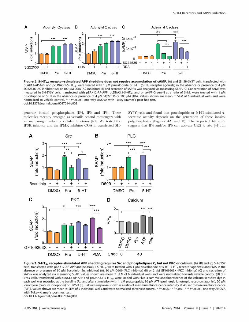
5-HT4d receptor-stimulated APP shedding does notinvolve activation of adenylyl cyclase and cAMP
Gas and cAMP mediate canonical signaling of 5-HT4 receptors
[33]. Therefore, we sought to determine whether, in SH-SY5Y
cells, accumulation of cAMP is also necessary for a-secretase
activity as previously described for CHO cells [15]. We used the
adenylyl cyclase inhibitors SQ22536 and DDA, which potently
inhibit increases in cAMP levels (Figure 2C). Interestingly, we
found that these inhibitors do not affect prucalopride or 5-HT-
stimulated sAPPa release in SH-SY5Y cells (Figures 2A and B).
These results suggest that activation of adenylyl cyclase and
accumulation of cAMP is not required for 5-HT4d receptor-
stimulated APP shedding.
5-HT4d receptor-stimulated APP shedding requires Srcand phospholipase C
Given that 5-HT4d receptor-stimulated APP shedding does not
require an elevation in cAMP levels, we sought to determine
whether generation of inositol triphosphate (IP3) is involved in 5-
HT4d receptor-stimulated sAPPa release. This second messenger is
produced by PLC and can be activated either directly down-
stream of Gaq and Gbc or through the Src non-receptor tyrosine
kinase (reviewed in [36]). To analyze the contribution of PLC and
Src, we co-treated pEAK12-AP-APP and pcDNA3.1-5-HT4d
transfected SH-SY5Y cells with the Src inhibitor Bosutinib or
5-HT4 Receptors and sAPPa Induction
PLOS ONE | www.plosone.org 4 January 2014 | Volume 9 | Issue 1 | e87014

the PLC inhibitor D609 and 5-HT4 receptor agonists. In both
cases, we observed that APP shedding was abolished compared to
control treatment (Figures 3A and B).
PLC cleaves phosphatidylinositol 4,5-bisphosphate into IP3 and
diacylglycerol, resulting in mobilization of intracellular calcium and
activation of several downstream effector proteins including PKC
[37]. In addition, several studies suggest that calcium and PKC
signaling can activate a-secretase shedding of APP [38,39]. Co-
treatment of transiently transfected SH-SY5Y cells with the PKC
inhibitor GF109203X did not induce sAPPa secretion after 5-HT4d
receptor stimulation. In contrast, direct activation of PKC with PMA
induced sAPPa, but this induction was inhibited with GF109203X
showing that the inhibitor was functional (Figure 3C). Similarly,
prucalopride did not significantly alter extracellular calcium influx in
SH-SY5Y cells, in contrast to ionomycin and ATP; two positive
controls that prove assay functionality (Figure 3D). Taken together
these data suggest that Src and PLC, but not PKC or calcium
signaling, contribute to 5-HT4d receptor-induced APP shedding.
5-HT4d receptor-stimulated APP shedding requiresinositol polyphosphates and casein kinase 2
IP3 can be further phosphorylated by inositol 1,4,5-triphosphate
3-kinase (IP3K) and inositol polyphosphate multikinase (IPMK) to
Figure 1. 5-HT4d receptor-stimulated APP shedding requires G protein signaling and is independent of b-arrestin recruitment. (A)Prucalopride induced sAPPa secretion in SH-SY5Y human neuroblastoma cells is specific for the 5-HT4 receptor. SH-SY5Y cells, transfected withpEAK12-AP-APP and pcDNA3.1-5-HT4d, were treated with 1 mM prucalopride and 5-HT (5-HT4 receptor agonists) in the absence or presence of 1 mMGR113808 (5-HT4 receptor antagonist) or PMA and secretion of sAPPa was analyzed via measuring SEAP. (B) SEAP levels were measured insupernatants of SH-SY5Y cells, transfected with pEAK12-AP-APP and pcDNA3.1-5-HT4d (WT), pcDNA3.1-5-HT4dDRY117/118AAY (DRY) or pcDNA3.1-5-HT4dD346 (D346) mutants and stimulated with 1 mM prucalopride or 5-HT. (C), (D) and (F) SEAP levels were measured in supernatants of SH-SY5Ycells, transfected with pEAK12-AP-APP and pcDNA3.1-5-HT4d and treated with 1 mM prucalopride or 5-HT in the absence or presence of 100 mM CTB(Gas inhibitor) (C), 100 mM NF449 (Gas inhibitor) (D) or 100 mM gallein (Gbc inhibitor) (F). (E) SEAP levels were measured in SH-SY5Y cells, transfectedwith pEAK12-AP-APP, pcDNA3.1-5-HT4d and pcDNAI-Amp-GasDN or pcDNA3.1 at a ratio of 2:1:4, respectively, and treated with 1 mM prucalopride or5-HT. Values shown are mean 6 SEM of 6 individual wells and were normalized to vehicle control. * P,0.05, ** P,0.01, *** P,0.001, one-way ANOVAwith Tukey-Kramer or Dunnet’s post-hoc test.doi:10.1371/journal.pone.0087014.g001
5-HT4 Receptors and sAPPa Induction
PLOS ONE | www.plosone.org 5 January 2014 | Volume 9 | Issue 1 | e87014
generate inositol polyphosphates (IP4, IP5 and IP6). These
molecules recently emerged as versatile second messengers with
an increasing number of cellular functions [40]. We tested the
IP3K inhibitor and the IPMK inhibitor CGA in transfected SH-
SY5Y cells and found that prucalopride or 5-HT-stimulated a-
secretase activity depends on the generation of these inositol
polyphosphates (Figures 4A and B). The reported literature
suggests that IP4 and/or IP6 can activate CK2 in vitro [41]. In
Figure 2. 5-HT4d receptor-stimulated APP shedding does not require accumulation of cAMP. (A) and (B) SH-SY5Y cells, transfected withpEAK12-AP-APP and pcDNA3.1-5-HT4d, were treated with 1 mM prucalopride or 5-HT (5-HT4 receptor agonists) in the absence or presence of 4 mMSQ22536 (AC inhibitor) (A) or 100 mM DDA (AC inhibitor) (B) and secretion of sAPPa was analyzed via measuring SEAP. (C) Concentration of cAMP wasmeasured in SH-SY5Y cells, transfected with pEAK12-AP-APP, pcDNA3.1-5-HT4d and pmax-FP-Green-N at a ratio of 5:4:1, were treated with 1 mMprucalopride or 5-HT in the absence or presence of 4 mM SQ22536 or 100 mM DDA. Values shown are mean 6 SEM of 6 individual wells and werenormalized to vehicle control. *** P,0.001, one-way ANOVA with Tukey-Kramer’s post-hoc test.doi:10.1371/journal.pone.0087014.g002
Figure 3. 5-HT4d receptor-stimulated APP shedding requires Src and phospholipase C, but not PKC or calcium. (A), (B) and (C) SH-SY5Ycells, transfected with pEAK12-AP-APP and pcDNA3.1-5-HT4d, were treated with 1 mM prucalopride or 5-HT (5-HT4 receptor agonists) and PMA in theabsence or presence of 50 mM Bosutinib (Src inhibitor) (A), 30 mM D609 (PLC inhibitor) (B) or 2 mM GF109203X (PKC inhibitor) (C) and secretion ofsAPPa was analyzed via measuring SEAP. Values shown are mean 6 SEM of 6 individual wells and were normalized towards vehicle control. (D) SH-SY5Y cells, transfected with pEAK12-AP-APP and pcDNA3.1-5-HT4d, were loaded with Fluo-4 NW mix and fluorescence of the calcium-sensitive dye ineach well was recorded at the baseline (F0) and after stimulation with 1 mM prucalopride, 30 mM ATP (purinergic ionotropic receptors agonist), 20 mMIonomycin (calcium ionophore) or DMSO (F). Calcium response shown is a ratio of maximum fluorescence intensity at 40 sec to baseline fluorescence(F/F0). Values shown are mean 6 SEM of 2 individual wells and were normalized to vehicle control. * P,0.05, ** P,0.01, *** P,0.001, one-way ANOVAwith Tukey-Kramer’s post-hoc test.doi:10.1371/journal.pone.0087014.g003
5-HT4 Receptors and sAPPa Induction
PLOS ONE | www.plosone.org 6 January 2014 | Volume 9 | Issue 1 | e87014

cells, Wnt3a can induce IP5 generation which then activates CK2
[42]. Inhibition of CK2 activity with TBB in pcDNA3.1-5-HT4d
receptor and pEAK12-AP-APP expressing SH-SY5Y cells stimu-
lated with prucalopride or 5-HT led to a decrease in sAPPa down
to baseline levels (Figure 4C), suggesting that CK2 is involved in 5-
HT4d receptor-stimulated APP shedding. In addition, we found
that co-transfection of CK2 siRNA completely abolished stimu-
lated APP shedding in SH-SY5Y cells treated with prucalopride or
5-HT (Figures 4D, E and F). These results demonstrate that 5-
HT4d receptor-stimulated APP shedding requires inositol polypho-
sphates and CK2.
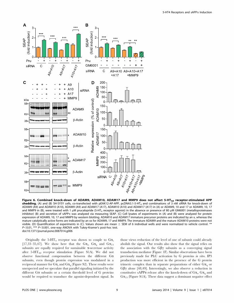
ADAM9, ADAM10, ADAM17 and MMP9 are notresponsible for 5-HT4d receptor-stimulated a-secretaseactivity
Several enzymes of the ADAM and MMP family, such as
ADAM9, 10, 17 and MMP9, are suggested candidate proteins
responsible for inducible shedding of APP (reviewed in [43]). To
determine the relative contribution of the metalloproteinases in 5-
HT4d receptor-stimulated sAPPa release, we first analyzed
expression levels of ADAM9, 10, 17 and MMP9 in SH-SY5Y
cells. Expression of ADAM9, 10, 17 and MMP9 was not changed
after prucalopride treatment of pEAK12-AP-APP transfected SH-
SY5Y cells (Figures S3A and B). To test whether a metallopro-
teinase would be responsible for induced a-secretase activity, we
treated the cells with non-toxic concentrations of the broad
spectrum metalloproteinase inhibitor GM6001 (dose response
curve shown in Figure S4). Treatment with GM6001 abolished
induction of sAPPa secretion (Figure 5A), confirming that a
metalloproteinase is indeed responsible for 5-HT4d receptor-
stimulated sAPPa release. To identify the 5-HT4d receptor-
stimulated a-secretase, we performed RNAi knock-down of the
candidate a-secretases. We found that induction of sAPPa release
was preserved after prucalopride treatment and single knock-down
of ADAM9, 10, 17 or MMP9 (Figure 5B). The efficiency of the
downregulation was between 85–95% as documented by western
blot analysis (Figures 5C and D). These data suggest that ADAM9,
10, 17 or MMP9 are not responsible for 5-HT4d receptor-
mediated inducible a-secretase activity in SH-SY5Y cells. We also
analyzed constitutive sAPPa secretion upon ADAM10 knock-
down in non-treated cells and confirmed that ADAM10 acts as the
constitutive a-secretase of APP in our experimental conditions
(data not shown).
Metalloproteinases are notorious for their functional redundan-
cy between family members. To test whether more than one
candidate metalloproteinase could be responsible for induction of
a-secretase activity, we treated transfected SH-SY5Y cells with
combinations of RNAi directed at ADAM9 and 10, ADAM9 and
17, ADAM10 and 17 (Figure 6A). We observed no change in
sAPPa secretion upon 5-HT4d receptor stimulation under any of
these conditions. Moreover, knock-down of all four candidate
Figure 4. 5-HT4d receptor-stimulated APP shedding requires inositol polyphosphates and casein kinase 2. (A), (B) and (C) SH-SY5Y cells,transfected with pEAK12-AP-APP and pcDNA3.1-5-HT4d, were treated with 1 mM prucalopride or 5-HT (5-HT4 receptor agonists) in the absence orpresence of 20 mM IP3K inhibitor (A), 80 mM CGA (IPMK inhibitor) (B) or 2.5 mM TBB (CK2 inhibitor) (C) and secretion of sAPPa was analyzed viameasuring SEAP. (D) SEAP levels were measured in supernatants of SH-SY5Y cells, co-transfected with pEAK12-AP-APP, pcDNA3.1-5-HT4d and 3 nMsiRNA for knock-down of CK2 and treated with 1 mM prucalopride. (E) Cell lysates of (D) were analyzed for CK2 expression levels by western blotting.(F) Quantification of experiments in (E). Values shown are mean 6 SEM of 6 individual wells and were normalized to vehicle control. * P,0.05, **P,0.01, *** P,0.001, one-way ANOVA with Tukey-Kramer’s post-hoc test.doi:10.1371/journal.pone.0087014.g004
5-HT4 Receptors and sAPPa Induction
PLOS ONE | www.plosone.org 7 January 2014 | Volume 9 | Issue 1 | e87014

metalloproteinases, i.e. ADAM9, 10, 17 and MMP9, still resulted
in induction of sAPPa release after 5-HT4d receptor activation
(Figure 6B). The levels of C-terminal fragments generated by the
cleavage of APP at b- and b9-sites remained unchanged after the
knock-down of ADAM9, 10, 17 and MMP9, suggesting that b-
secretase activity was not affected by reduced expression levels of
these metalloproteinases (Figures S5A and B). We used western
blotting to confirm the efficiency and specificity of RNAi mediated
downregulation (Figures 6C and D). Notice also the strong
upregulation of MMP9 expression when ADAM9, 10 and 17 are
downregulated, while single MMP9 knock-down did not affect 5-
HT4d receptor-induced sAPPa secretion. Altogether, our data
suggest that an unidentified GM6001-sensitive metalloproteinase
participates in the regulated cleavage of APP upon 5-HT4d
receptor stimulation (Figure 6B).
Discussion
In this report, we examined the signaling pathway that leads to
a-secretase induction after 5-HT4d receptor stimulation in the
human neuroblastoma SH-SY5Y cell line. We present here a
previously uncharacterized signaling pathway involved in the
mediation of 5-HT4d receptor-induced a-secretase activity
(Figure 7). The characterization of this pathway was based on a
combination of pharmacological, siRNA and site-directed muta-
genesis experiments. Our data indicate that PLC is essential for a-
secretase activation following 5-HT4d stimulation. This effect is
dependent on Ga and Gbc recruitment and signaling downstream
of the 5-HT4d receptor. Src tyrosine kinase acts as an intermediate
molecule, mediating PLC activation and inositol triphosphate
production. The latter is converted by multiple kinases to inositol
polyphosphates, which activate CK2. Downstream of CK2, a yet
unknown mechanism of a-secretase activation is triggered. The 5-
HT4d receptor-induced a-secretase activity could not be ascribed
to any known candidate a-secretase (ADAM9, 10, 17 and MMP9)
in the SH-SY5Y cells, which has hampered delineation of the final
step regulating 5-HT4d receptor-stimulated sAPPa release.
We found also that b-arrestin signaling did not contribute to a-
secretase activity upon 5-HT4d receptor stimulation as the mutant
receptor deficient in b-arrestin recruitment maintained the ability
to stimulate sAPPa secretion after agonist treatment (Figure 1B).
Interestingly, b-arrestins have recently emerged as regulators of
Ab generation downstream of the b2-adrenergic receptor and
GPR3, independently of G protein activation [44,45]. In these
studies, b-arrestins appear to bind to the Aph1 subunit of the c-
secretase complex, affecting complex localization and thereby
increasing the catalytic activity of the c-secretase complex. Our
work suggests that different signaling pathways regulate a- and c-
secretase activity as we find that G proteins are indispensable for 5-
HT4d receptor-stimulated a-secretase activity, while they are not
involved in the c-secretase regulation by GPCRs. Indeed, several
molecules that are activated downstream of G proteins are
proposed to regulate a-site APP processing, e.g. PKC, PKA,
MAPK, ERK and PI3K (reviewed in [46]).
Figure 5. Single knock-down of ADAM9, ADAM10, ADAM17 and MMP9 does not affect 5-HT4d receptor-stimulated APP shedding.(A) SH-SY5Y cells, transfected with pEAK12-AP-APP and pcDNA3.1-5-HT4d, were treated with 1 mM prucalopride or 5-HT (5-HT4 receptor agonists) inthe absence or presence of 80 mM GM6001 (metalloproteinases inhibitor) and secretion of sAPPa was analyzed via measuring SEAP. (B) SEAP levelswere measured in supernatants of SH-SY5Y cells, co-transfected with pEAK12-AP-APP, pcDNA3.1-5-HT4d and 3 nM siRNA for knock-down of ADAM9(A9), ADAM10 (A10), ADAM17 (A17) and MMP9 and treated with 1 mM prucalopride. (C) Cell lysates of (B) were analyzed for protein expression ofADAM9, 10, 17 and MMP9 by western blotting. The ADAM10 and ADAM17 immature precursor proteins are indicated by an x, whereas the maturecatalytically active forms are indicated by an xx for ADAM9, 17 and MMP9. The immature ADAM9 and the mature ADAM10 proteins were not visible.(D) Quantification of experiments in (C). Values shown are mean 6 SEM of 6 individual wells and were normalized to vehicle control. * P,0.05, **P,0.01, *** P,0.001, one-way ANOVA with Tukey-Kramer’s post-hoc test.doi:10.1371/journal.pone.0087014.g005
5-HT4 Receptors and sAPPa Induction
PLOS ONE | www.plosone.org 8 January 2014 | Volume 9 | Issue 1 | e87014
Originally the 5-HT4 receptor was shown to couple to Gas
[17,33–35,47]. We show here that the Gas, Gaq and Ga13
subunits are equally required for sustainable a-secretase activity
after 5-HT4d receptor stimulation (Figure S1A). We did not
observe functional compensation between the different Gasubunits, even though protein expression was modulated in a
reciprocal manner for Gas and Gaq (Figure S2). These results were
unexpected and we speculate that parallel signaling initiated by the
different Ga subunits or a certain threshold level of G proteins
would be required to transduce the agonist-dependent signal. In
those views reduction of the level of one of subunit could already
abolish the signal. Our results also show that the signal relies on
the association with the Gbc subunits as a converging signal
transduction mediator (Figure 1F). Similar observations have been
previously made for PLC activation by G proteins in vitro. IP3
production was more efficient in the presence of the G protein
trimeric complex than in separate preparations of either Gaq or
Gbc alone [48,49]. Interestingly, we also observe a reduction in
constitutive sAPPa release after the knock-down of Gas, Gaq and
Ga13 (Figure S1A). These data suggest a dominant negative effect
Figure 6. Combined knock-down of ADAM9, ADAM10, ADAM17 and MMP9 does not affect 5-HT4d receptor-stimulated APPshedding. (A) and (B) SH-SY5Y cells, co-transfected with pEAK12-AP-APP, pcDNA3.1-5-HT4 and combinations of 3 nM siRNA for knock-down ofADAM9 (A9) and ADAM10 (A10), ADAM9 (A9) and ADAM17 (A17), ADAM10 (A10) and ADAM17 (A17) in (A) or ADAM9, 10 and 17 or ADAM9, 10, 17and MMP9 in (B), were treated with 1 mM prucalopride (5-HT4 receptor agonist) in the absence or presence of 80 mM GM6001 (metalloproteinasesinhibitor) (B) and secretion of sAPPa was analyzed via measuring SEAP. (C) Cell lysates of experiments in (A) and (B) were analyzed for proteinexpression of ADAM9, 10, 17 and MMP9 by western blotting. ADAM10 and ADAM17 immature precursor proteins are indicated by an x, whereas themature catalytically active forms are indicated by an xx for ADAM9, 17 and MMP9. The immature ADAM9 and the mature ADAM10 proteins were notvisible. (D) Quantification of experiments in (C). Values shown are mean 6 SEM of 6 individual wells and were normalized to vehicle control. **P,0.01, *** P,0.001, one-way ANOVA with Tukey-Kramer’s post-hoc test.doi:10.1371/journal.pone.0087014.g006
5-HT4 Receptors and sAPPa Induction
PLOS ONE | www.plosone.org 9 January 2014 | Volume 9 | Issue 1 | e87014

of G proteins inhibition on constitutive a-secretase activity, which
may be mediated by additional GPCRs besides the 5-HT4d
receptor.
5-HT4 receptor coupling to different G proteins suggests several
possibilities for downstream signal transduction. Several reports
describe a PKA-independent and cAMP-dependent a-secretase
activation following 5-HT4d receptor stimulation [7,50,51]. In
CHO cells, sAPPa release is regulated by Epac1, which promotes
small GTPases Rap1 dependent Rac activation [15]. However, we
find that AC and cAMP accumulation are not required for 5-
HT4d receptor-induced APP shedding under our experimental
conditions (Figure 2). Differences in the cellular systems, a human
neuronal cell line versus a Chinese hamster ovary cell line, could
explain the discrepancy between the studies. We then found that
IP3 generation through Src and PLC activation contributes to 5-
HT4d receptor-induced a-secretase activity (Figure 3). PLC is also
an important component of the a-secretase activation pathway
through Gaq coupled GPCRs, e.g. mGluR1 and mGluR5 [52],
M1 and M3 [53], 5-HT2a and 5-HT2c [54] and thus a point of
convergence for several transduction pathways activating a-
secretase.
Investigations of the cerebral cortex and cerebellum of AD-
affected individuals reveal disturbed G protein signal transduction
compared to control patients [55]. In accordance, the phospho-
inositide hydrolysis pathway is also altered in AD because of
reduced levels of phosphatidylinositide 3-kinase and disturbed
agonist and G protein regulation of PLC [56,57]. It is proposed
that 5-HT4 receptor stimulation could counteract such detrimental
changes. We show here that the 5-HT4d receptor indeed induces
IP3K and IPMK mediated IP3 conversion to inositol polypho-
sphates and that these contribute to the non-amyloidogenic
pathway of APP processing (Figure 4). This effect is mediated
through the activation of CK2, which was recently identified to be
downstream of the cholinergic receptors in a pathway of a-
secretase induction [58]. As activation of the 5-HT4 receptor can
increase acetylcholine levels in the brain [6,13] and we need
24 hours to obtain a significant induction of the a-secretase, an
indirect mechanism through upregulation of acetylcholine could
play a role. As our cells are of the dopaminergic origin, we think
this possibility is rather unlikely. However, we cannot rule out that
other indirect mechanisms are playing a role in the 5-HT4d
receptor-mediated a-secretase induction.
To understand the molecular mechanism of a-secretase
activation downstream of the 5-HT4d receptor, we investigated
the contribution of ADAM9, 10, 17 and MMP9 in the regulation
of APP processing. Previously, regulated a-secretase activity was
partially attributed to MMP9, whose expression levels increased
after 5-HT4d receptor stimulation in APP-overexpressing H4
human neuroglioma cells [59]. However, in our cellular system,
expression levels of the investigated proteinases do not change
(Figure S3) and specific protein downregulation suggests that a
different metalloproteinase, besides the major candidate a-
secretases ADAM9, 10, 17 and MMP9, contributes to 5-HT4d
receptor-induced sAPPa release (Figures 5 and 6). Indeed,
ADAM10 was not responsible for the 5-HT4d receptor-dependent
induction of sAPPa release through the cAMP/Epac pathway
[21]. At this moment, we cannot rule out that the remaining
protein expression of these four major a-secretases contribute to
the preserved inducible a-secretase activity. Our data are
consistent with the present view of different proteases contributing
to regulated APP processing as previously reported for the M1
receptor, the insulin-like growth factor-1 receptor and the
purinergic P2Y2 and P2X7 receptors [23,60–62]. To identify
the metalloproteinase(s) responsible for induced a-secretase
activity we were reluctant to use differences in susceptibility to
GM6001 because we were working with overexpression condi-
tions. This would require a large-scale RNAi knock-down study
but is beyond the scope of the current manuscript.
In conclusion, our studies show the complexity of a-secretase
regulation upon 5-HT4d receptor stimulation. Taking into
consideration that receptor modulation of signaling pathways
depends on the cellular context and that recombinant overex-
pression and RNA interference may reveal cell type specific
results, a relevant physiological system should be used for the
confirmation of the identified signaling pathway. Clinical trials of
agonists targeting 5-HT4 serotonergic and M1 muscarinic
receptors will provide validation of a-secretase activation as a
therapeutic approach for the treatment of AD. We report here that
PLC dependent production of IP3 and CK2 activation are
important mediators of the 5-HT4d receptor signaling that
enhance the non-amyloidogenic processing of APP. These proteins
can also participate in signaling downstream of muscarinic
receptors, suggesting the possibility of a common pathway for a-
secretase activation through GPCRs. Finally, our data will also aid
with the development of 5-HT4 receptor agonists as therapeutics
for neurodegenerative or psychiatric disorders and allow for a
better understanding of potential risks associated with these drugs.
Figure 7. Schematic representation of the proposed 5-HT4d
receptor-stimulated signaling pathway leading to increasedsAPPa production. The proteins involved in 5-HT4d receptor-mediated non-amyloidogenic APP shedding are shown with greencircles while orange circles and red characters indicate proteins orsecond messengers that were tested but were ineffective in modulating5-HT4d receptor-stimulated sAPPa release. The dotted lines with thequestion marks indicate remaining areas of investigation for furtherelucidation of the molecular mechanism of a-secretase activation.cAMP-dependent pathway of a-secretase induction was previouslyreported and is depicted as a plausible way for 5-HT4d receptor-mediated sAPPa release [15].doi:10.1371/journal.pone.0087014.g007
5-HT4 Receptors and sAPPa Induction
PLOS ONE | www.plosone.org 10 January 2014 | Volume 9 | Issue 1 | e87014
Supporting Information
Figure S1 5-HT4d receptor-stimulated APP sheddingrequires the G proteins Gas, Gaq and Ga13. (A) SEAP
levels were measured in supernatants of SH-SY5Y cells, co-
transfected with pEAK12-AP-APP, pcDNA3.1-5-HT4d and 3 nM
siRNA for knock-down of Gas, Gaq and Ga13 and treated with
1 mM prucalopride (5-HT4 receptor agonist). (B) Cell lysates of (A)
were analyzed for protein expression of Gas, Gaq and Ga13 by
western blotting. (C) Quantification of experiments in (B). Values
shown are mean 6 SEM of 6 individual wells and were
normalized to vehicle control. ** P,0.01, *** P,0.001, one-way
ANOVA with Tukey-Kramer’s post-hoc test.
(TIF)
Figure S2 Knock-down of Gas and Gaq but not Ga13
alters protein expression of the G protein family. (A) SH-
SY5Y cells, transfected with 3 nM siRNA for knock-down of Gas,
Gaq and Ga13, were harvested and expression levels of G proteins
were analyzed. (B) Quantification of experiments in (A). Values
shown are mean 6 SEM of 2 individual wells and were
normalized to vehicle control. ** P,0.01, one-way ANOVA with
Tukey-Kramer’s post-hoc test.
(TIF)
Figure S3 Expression levels of major candidate a-secretases and APP do not change upon 5-HT4d receptorstimulation. (A) SH-SY5Y cells, transfected with pEAK12-AP-
APP and pcDNA3.1-5-HT4d, were treated with 1 mM prucalo-
pride (5-HT4 receptor agonist) and collected to analyze protein
expression of ADAM9, 10, 17, MMP9 and APP by western
blotting. ADAM10 and ADAM17 immature precursor proteins
are indicated by an x, whereas the mature catalytically active
forms are indicated by an xx for ADAM9, 17 and MMP9. The
immature ADAM9 and the mature ADAM10 proteins were not
visible. (B) Quantification of experiments in (A). Values shown are
mean 6 SEM of 2 individual wells and were normalized to vehicle
control.
(TIF)
Figure S4 The metalloproteinase inhibitor GM6001can inhibit secretion of sAPPa upon 5-HT4d receptor
stimulation. SH-SY5Y cells, transfected with pEAK12-AP-APP
and pcDNA3.1-5-HT4d, were treated with 1 mM prucalopride or
5-HT in the absence or presence of different concentrations of
GM6001 and secretion of sAPPa was analyzed via measuring
SEAP. Values shown are mean 6 SEM of 6 individual wells and
are normalized towards vehicle control. ** P,0.01, *** P,0.001,
one-way ANOVA with Tukey-Kramer’s post-hoc test.
(TIF)
Figure S5 Knock-down of ADAM9, 10, 17 and MMP9does not affect the pattern of CTFs generated by the 5-HT4d receptor-stimulated a-secretase activity. (A) SH-
SY5Y cells, co-transfected with pEAK12-AP-APP, pcDNA3.1-5-
HT4d and combinations of 3 nM siRNA for knock-down of
ADAM9 (A9), ADAM10 (A10) and ADAM17 (A17) or ADAM9,
10, 17 and MMP9, were treated with 1 mM prucalopride (5-HT4
receptor agonist) and secretion of sAPPa was analyzed via
measuring SEAP. Values shown are mean 6 SEM of 6 individual
wells and were normalized to vehicle control. *** P,0.001, one-
way ANOVA with Tukey-Kramer’s post-hoc test. (B) Cell lysates
of the experiment in (A) were analyzed for the levels of different
APP C-terminal fragments (CTFs) were analyzed by western
blotting using B63 antibody (16% Tricine SDS-PAGE).
(TIF)
Table S1
(DOCX)
Acknowledgments
We are grateful to Catherine Berlot for providing the pcDNAI-Amp-
GasDN plasmid. We thank Joris De Mayer and Jan Schuurkes (Shire-
Movetis, NV, Turnhout, Belgium) for providing us with prucalopride. We
thank Stefan F. Lichtenthaler (German Center for Neurodegenerative
Diseases (DZNE), Munich, Germany) for providing us with the pEAK12-
AP-APP plasmid.
Author Contributions
Conceived and designed the experiments: AAP AT BDS IT. Performed the
experiments: AAP. Analyzed the data: AAP AT BDS IT. Contributed
reagents/materials/analysis tools: AAP AT BDS IT. Wrote the paper:
AAP AT BDS IT.
References
1. De Strooper B (2010) Proteases and proteolysis in Alzheimer disease: a
multifactorial view on the disease process. Physiol Rev 90: 465–494.
2. Postina R, Schroeder A, Dewachter I, Bohl J, Schmitt U, et al. (2004) A
disintegrin-metalloproteinase prevents amyloid plaque formation and hippo-
campal defects in an Alzheimer disease mouse model. J Clin Invest 113: 1456–
1464.
3. Donmez G, Wang D, Cohen DE, Guarente L (2010) SIRT1 suppresses beta-
amyloid production by activating the alpha-secretase gene ADAM10. Cell 142:
320–332.
4. Bockaert J, Claeysen S, Compan V, Dumuis A (2008) 5-HT(4) receptors: history,
molecular pharmacology and brain functions. Neuropharmacology 55: 922–931.
5. Spencer JP, Brown JT, Richardson JC, Medhurst AD, Sehmi SS, et al. (2004)
Modulation of hippocampal excitability by 5-HT4 receptor agonists persists in a
transgenic model of Alzheimer’s disease. Neuroscience 129: 49–54.
6. Matsumoto M, Togashi H, Mori K, Ueno K, Ohashi S, et al. (2001) Evidence
for involvement of central 5-HT(4) receptors in cholinergic function associated
with cognitive processes: behavioral, electrophysiological, and neurochemical
studies. J Pharmacol Exp Ther 296: 676–682.
7. Robert SJ, Zugaza JL, Fischmeister R, Gardier AM, Lezoualc’h F (2001) The
human serotonin 5-HT4 receptor regulates secretion of non-amyloidogenic
precursor protein. J Biol Chem 276: 44881–44888.
8. Cho S, Hu Y (2007) Activation of 5-HT4 receptors inhibits secretion of beta-
amyloid peptides and increases neuronal survival. Exp Neurol 203: 274–278.
9. Tesseur I, Pimenova AA, Lo AC, Ciesielska M, Lichtenthaler SF, et al. (2013)
Chronic 5-HT4 receptor activation decreases Abeta production and deposition
in hAPP/PS1 mice. Neurobiol Aging 34: 1779–1789.
10. Cachard-Chastel M, Lezoualc’h F, Dewachter I, Delomenie C, Croes S, et al.
(2007) 5-HT4 receptor agonists increase sAPPalpha levels in the cortex and
hippocampus of male C57BL/6j mice. Br J Pharmacol 150: 883–892.
11. Shen F, Smith JA, Chang R, Bourdet DL, Tsuruda PR, et al. (2011) 5-HT(4)
receptor agonist mediated enhancement of cognitive function in vivo and
amyloid precursor protein processing in vitro: A pharmacodynamic and
pharmacokinetic assessment. Neuropharmacology 61: 69–79.
12. Micale V, Leggio GM, Mazzola C, Drago F (2006) Cognitive effects of
SL65.0155, a serotonin 5-HT4 receptor partial agonist, in animal models of
amnesia. Brain Res 1121: 207–215.
13. Consolo S, Arnaboldi S, Giorgi S, Russi G, Ladinsky H (1994) 5-HT4 receptor
stimulation facilitates acetylcholine release in rat frontal cortex. Neuroreport 5:
1230–1232.
14. Farlow MR, Cummings JL (2007) Effective pharmacologic management of
Alzheimer’s disease. Am J Med 120: 388–397.
15. Maillet M, Robert SJ, Cacquevel M, Gastineau M, Vivien D, et al. (2003)
Crosstalk between Rap1 and Rac regulates secretion of sAPPalpha. Nat Cell Biol
5: 633–639.
16. Barthet G, Framery B, Gaven F, Pellissier L, Reiter E, et al. (2007) 5-
hydroxytryptamine 4 receptor activation of the extracellular signal-regulated
kinase pathway depends on Src activation but not on G protein or beta-arrestin
signaling. Mol Biol Cell 18: 1979–1991.
17. Contesse V, Hamel C, Lefebvre H, Dumuis A, Vaudry H, et al. (1996)
Activation of 5-hydroxytryptamine4 receptors causes calcium influx in
adrenocortical cells: involvement of calcium in 5-hydroxytryptamine-induced
steroid secretion. Mol Pharmacol 49: 481–493.
5-HT4 Receptors and sAPPa Induction
PLOS ONE | www.plosone.org 11 January 2014 | Volume 9 | Issue 1 | e87014

18. Ouadid H, Seguin J, Richard S, Chaptal PA, Nargeot J (1991) Properties and
Modulation of Ca channels in adult human atrial cells. J Mol Cell Cardiol 23:
41–54.
19. Jorissen E, Prox J, Bernreuther C, Weber S, Schwanbeck R, et al. (2010) The
disintegrin/metalloproteinase ADAM10 is essential for the establishment of the
brain cortex. J Neurosci 30: 4833–4844.
20. Kuhn PH, Wang H, Dislich B, Colombo A, Zeitschel U, et al. (2010) ADAM10
is the physiologically relevant, constitutive alpha-secretase of the amyloid
precursor protein in primary neurons. EMBO J 29: 3020–3032.
21. Cochet M, Donneger R, Cassier E, Gaven F, Lichtenthaler SF, et al. (2013) 5-
HT4 receptors constitutively promote the non-amyloidogenic pathway of APP
cleavage and interact with ADAM10. ACS Chem Neurosci 4: 130–140.
22. Le Gall SM, Bobe P, Reiss K, Horiuchi K, Niu XD, et al. (2009) ADAMs 10 and
17 represent differentially regulated components of a general shedding
machinery for membrane proteins such as transforming growth factor alpha,
L-selectin, and tumor necrosis factor alpha. Mol Biol Cell 20: 1785–1794.
23. Caccamo A, Oddo S, Billings LM, Green KN, Martinez-Coria H, et al. (2006)
M1 receptors play a central role in modulating AD-like pathology in transgenic
mice. Neuron 49: 671–682.
24. Jefferson T, Causevic M, auf dem Keller U, Schilling O, Isbert S, et al. (2011)
Metalloprotease meprin beta generates nontoxic N-terminal amyloid precursor
protein fragments in vivo. J Biol Chem 286: 27741–27750.
25. Ahmad M, Takino T, Miyamori H, Yoshizaki T, Furukawa M, et al. (2006)
Cleavage of amyloid-beta precursor protein (APP) by membrane-type matrix
metalloproteinases. J Biochem 139: 517–526.
26. Agholme L, Lindstrom T, Kagedal K, Marcusson J, Hallbeck M (2010) An in
vitro model for neuroscience: differentiation of SH-SY5Y cells into cells with
morphological and biochemical characteristics of mature neurons. J Alzheimers
Dis 20: 1069–1082.
27. Hartmann D, de Strooper B, Serneels L, Craessaerts K, Herreman A, et al.
(2002) The disintegrin/metalloprotease ADAM 10 is essential for Notch
signalling but not for alpha-secretase activity in fibroblasts. Hum Mol Genet
11: 2615–2624.
28. Berlot CH (2002) A highly effective dominant negative alpha s construct
containing mutations that affect distinct functions inhibits multiple Gs-coupled
receptor signaling pathways. J Biol Chem 277: 21080–21085.
29. Lichtenthaler SF, Dominguez DI, Westmeyer GG, Reiss K, Haass C, et al.
(2003) The cell adhesion protein P-selectin glycoprotein ligand-1 is a substrate
for the aspartyl protease BACE1. J Biol Chem 278: 48713–48719.
30. Reiter E, Lefkowitz RJ (2006) GRKs and beta-arrestins: roles in receptor
silencing, trafficking and signaling. Trends Endocrinol Metab 17: 159–165.
31. Trzaskowski B, Latek D, Yuan S, Ghoshdastider U, Debinski A, et al. (2012)
Action of molecular switches in GPCRs–theoretical and experimental studies.
Curr Med Chem 19: 1090–1109.
32. Rovati GE, Capra V, Neubig RR (2007) The highly conserved DRY motif of
class A G protein-coupled receptors: beyond the ground state. Mol Pharmacol
71: 959–964.
33. Dumuis A, Bouhelal R, Sebben M, Cory R, Bockaert J (1988) A nonclassical 5-
hydroxytryptamine receptor positively coupled with adenylate cyclase in the
central nervous system. Mol Pharmacol 34: 880–887.
34. Chang WC, Ng JK, Nguyen T, Pellissier L, Claeysen S, et al. (2007) Modifying
ligand-induced and constitutive signaling of the human 5-HT4 receptor. PLoS
One 2: e1317.
35. Ponimaskin EG, Profirovic J, Vaiskunaite R, Richter DW, Voyno-Yasenetskaya
TA (2002) 5-Hydroxytryptamine 4(a) receptor is coupled to the Galpha subunit
of heterotrimeric G13 protein. J Biol Chem 277: 20812–20819.
36. Rhee SG (2001) Regulation of phosphoinositide-specific phospholipase C. Annu
Rev Biochem 70: 281–312.
37. Berridge MJ (1993) Inositol trisphosphate and calcium signalling. Nature 361:
315–325.
38. Buxbaum JD, Ruefli AA, Parker CA, Cypess AM, Greengard P (1994) Calcium
regulates processing of the Alzheimer amyloid protein precursor in a protein
kinase C-independent manner. Proc Natl Acad Sci U S A 91: 4489–4493.
39. LeBlanc AC, Koutroumanis M, Goodyer CG (1998) Protein kinase C activation
increases release of secreted amyloid precursor protein without decreasing Abeta
production in human primary neuron cultures. J Neurosci 18: 2907–2913.
40. Shears SB (1998) The versatility of inositol phosphates as cellular signals.
Biochim Biophys Acta 1436: 49–67.
41. Solyakov L, Cain K, Tracey BM, Jukes R, Riley AM, et al. (2004) Regulation of
casein kinase-2 (CK2) activity by inositol phosphates. J Biol Chem 279: 43403–
43410.
42. Gao Y, Wang HY (2007) Inositol pentakisphosphate mediates Wnt/beta-catenin
signaling. J Biol Chem 282: 26490–26502.
43. Vingtdeux V, Marambaud P (2012) Identification and biology of alpha-
secretase. J Neurochem 120 Suppl 1: 34–45.
44. Thathiah A, Horre K, Snellinx A, Vandewyer E, Huang Y, et al. (2013) beta-
arrestin 2 regulates Abeta generation and gamma-secretase activity in
Alzheimer’s disease. Nat Med 19: 43–49.
45. Liu X, Zhao X, Zeng X, Bossers K, Swaab DF, et al. (2013) beta-arrestin1
regulates gamma-secretase complex assembly and modulates amyloid-beta
pathology. Cell Res 23: 351–365.
46. Bandyopadhyay S, Goldstein LE, Lahiri DK, Rogers JT (2007) Role of the APP
non-amyloidogenic signaling pathway and targeting alpha-secretase as an
alternative drug target for treatment of Alzheimer’s disease. Curr Med Chem 14:
2848–2864.
47. Robert S, Maillet M, Morel E, Launay JM, Fischmeister R, et al. (2005)
Regulation of the amyloid precursor protein ectodomain shedding by the 5-HT4
receptor and Epac. FEBS Lett 579: 1136–1142.
48. Smrcka AV, Sternweis PC (1993) Regulation of purified subtypes of
phosphatidylinositol-specific phospholipase C beta by G protein alpha and beta
gamma subunits. J Biol Chem 268: 9667–9674.
49. Kozasa T, Hepler JR, Smrcka AV, Simon MI, Rhee SG, et al. (1993)
Purification and characterization of recombinant G16 alpha from Sf9 cells:
activation of purified phospholipase C isozymes by G-protein alpha subunits.
Proc Natl Acad Sci U S A 90: 9176–9180.
50. Lezoualc’h F, Robert SJ (2003) The serotonin 5-HT4 receptor and the amyloid
precursor protein processing. Exp Gerontol 38: 159–166.
51. Marambaud P, Ancolio K, Alves da Costa C, Checler F (1999) Effect of protein
kinase A inhibitors on the production of Abeta40 and Abeta42 by human cells
expressing normal and Alzheimer’s disease-linked mutated betaAPP and
presenilin 1. Br J Pharmacol 126: 1186–1190.
52. Lee RK, Wurtman RJ, Cox AJ, Nitsch RM (1995) Amyloid precursor protein
processing is stimulated by metabotropic glutamate receptors. Proc Natl Acad
Sci U S A 92: 8083–8087.
53. Nitsch RM, Slack BE, Wurtman RJ, Growdon JH (1992) Release of Alzheimer
amyloid precursor derivatives stimulated by activation of muscarinic acetylcho-
line receptors. Science 258: 304–307.
54. Nitsch RM, Deng M, Growdon JH, Wurtman RJ (1996) Serotonin 5-HT2a and
5-HT2c receptors stimulate amyloid precursor protein ectodomain secretion.
J Biol Chem 271: 4188–4194.
55. Lumbreras M, Baamonde C, Martinez-Cue C, Lubec G, Cairns N, et al. (2006)
Brain G protein-dependent signaling pathways in Down syndrome and
Alzheimer’s disease. Amino Acids 31: 449–456.
56. Jolles J, Bothmer J, Markerink M, Ravid R (1992) Phosphatidylinositol kinase is
reduced in Alzheimer’s disease. J Neurochem 58: 2326–2329.
57. Albasanz JL, Dalfo E, Ferrer I, Martin M (2005) Impaired metabotropic
glutamate receptor/phospholipase C signaling pathway in the cerebral cortex in
Alzheimer’s disease and dementia with Lewy bodies correlates with stage of
Alzheimer’s-disease-related changes. Neurobiol Dis 20: 685–693.
58. Lenzken SC, Stanga S, Lanni C, De Leonardis F, Govoni S, et al. (2010)
Recruitment of casein kinase 2 is involved in AbetaPP processing following
cholinergic stimulation. J Alzheimers Dis 20: 1133–1141.
59. Hashimoto G, Sakurai M, Teich AF, Saeed F, Aziz F, et al. (2012) 5-HT(4)
receptor stimulation leads to soluble AbetaPPalpha production through MMP-9
upregulation. J Alzheimers Dis 32: 437–445.
60. Jacobsen KT, Adlerz L, Multhaup G, Iverfeldt K (2010) Insulin-like growth
factor-1 (IGF-1)-induced processing of amyloid-beta precursor protein (APP) and
APP-like protein 2 is mediated by different metalloproteinases. J Biol Chem 285:
10223–10231.
61. Camden JM, Schrader AM, Camden RE, Gonzalez FA, Erb L, et al. (2005)
P2Y2 nucleotide receptors enhance alpha-secretase-dependent amyloid precur-
sor protein processing. J Biol Chem 280: 18696–18702.
62. Delarasse C, Auger R, Gonnord P, Fontaine B, Kanellopoulos JM (2010) The
purinergic receptor P2X7 triggers alpha-secretase-dependent processing of the
amyloid precursor protein. J Biol Chem 286: 2596–2606.
63. Ansanay H, Sebben M, Bockaert J, Dumuis A (1996) Pharmacological
comparison between [3H]GR 113808 binding sites and functional 5-HT4
receptors in neurons. Eur J Pharmacol 298: 165–174.
64. Van den Wyngaert I, Gommeren W, Verhasselt P, Jurzak M, Leysen J, et al.
(1997) Cloning and expression of a human serotonin 5-HT4 receptor cDNA.
J Neurochem 69: 1810–1819.
65. Milligan G, Unson CG, Wakelam MJ (1989) Cholera toxin treatment produces
down-regulation of the alpha-subunit of the stimulatory guanine-nucleotide-
binding protein (Gs). Biochem J 262: 643–649.
66. Shen KF, Crain SM (1990) Cholera toxin-B subunit blocks excitatory effects of
opioids on sensory neuron action potentials indicating that GM1 ganglioside
may regulate Gs-linked opioid receptor functions. Brain Res 531: 1–7.
67. Hohenegger M, Waldhoer M, Beindl W, Boing B, Kreimeyer A, et al. (1998)
Gsalpha-selective G protein antagonists. Proc Natl Acad Sci U S A 95: 346–351.
68. Lehmann DM, Seneviratne AM, Smrcka AV (2008) Small molecule disruption
of G protein beta gamma subunit signaling inhibits neutrophil chemotaxis and
inflammation. Mol Pharmacol 73: 410–418.
69. Harris DN, Asaad MM, Phillips MB, Goldenberg HJ, Antonaccio MJ (1979)
Inhibition of adenylate cyclase in human blood platelets by 9-substituted adenine
derivatives. J Cyclic Nucleotide Res 5: 125–134.
70. Gao Y, Usha Raj J (2002) Effects of SQ 22536, an adenylyl cyclase inhibitor, on
isoproterenol-induced cyclic AMP elevation and relaxation in newborn ovine
pulmonary veins. Eur J Pharmacol 436: 227–233.
71. Onoda JM, Braun T, Wrenn SM Jr (1987) Characterization of the purine-
reactive site of the rat testis cytosolic adenylate cyclase. Biochem Pharmacol 36:
1907–1912.
72. Legrand AB, Narayanan TK, Ryan US, Aronstam RS, Catravas JD (1990)
Effects of adenosine and analogs on adenylate cyclase activity in cultured bovine
aortic endothelial cells. Biochem Pharmacol 40: 1103–1109.
73. Coluccia AM, Benati D, Dekhil H, De Filippo A, Lan C, et al. (2006) SKI-606
decreases growth and motility of colorectal cancer cells by preventing pp60
5-HT4 Receptors and sAPPa Induction
PLOS ONE | www.plosone.org 12 January 2014 | Volume 9 | Issue 1 | e87014
(c-Src)-dependent tyrosine phosphorylation of beta-catenin and its nuclear
signaling. Cancer Res 66: 2279–2286.74. Vultur A, Buettner R, Kowolik C, Liang W, Smith D, et al. (2008) SKI-606
(bosutinib), a novel Src kinase inhibitor, suppresses migration and invasion of
human breast cancer cells. Mol Cancer Ther 7: 1185–1194.75. Amtmann E (1996) The antiviral, antitumoural xanthate D609 is a competitive
inhibitor of phosphatidylcholine-specific phospholipase C. Drugs Exp Clin Res22: 287–294.
76. Tschaikowsky K, Meisner M, Schonhuber F, Rugheimer E (1994) Induction of
nitric oxide synthase activity in phagocytic cells inhibited by tricyclodecan-9-yl-xanthogenate (D609). Br J Pharmacol 113: 664–668.
77. Martiny-Baron G, Kazanietz MG, Mischak H, Blumberg PM, Kochs G, et al.(1993) Selective inhibition of protein kinase C isozymes by the indolocarbazole
Go 6976. J Biol Chem 268: 9194–9197.78. Jacobson PB, Kuchera SL, Metz A, Schachtele C, Imre K, et al. (1995) Anti-
inflammatory properties of Go 6850: a selective inhibitor of protein kinase C.
J Pharmacol Exp Ther 275: 995–1002.79. Chang YT, Choi G, Bae YS, Burdett M, Moon HS, et al. (2002) Purine-based
inhibitors of inositol-1,4,5-trisphosphate-3-kinase. Chembiochem 3: 897–901.80. Mayr GW, Windhorst S, Hillemeier K (2005) Antiproliferative plant and
synthetic polyphenolics are specific inhibitors of vertebrate inositol-1,4,
5-trisphosphate 3-kinases and inositol polyphosphate multikinase. J Biol Chem
280: 13229–13240.
81. Sarno S, Reddy H, Meggio F, Ruzzene M, Davies SP, et al. (2001) Selectivity of
4,5,6,7-tetrabromobenzotriazole, an ATP site-directed inhibitor of protein
kinase CK2 (‘casein kinase-2’). FEBS Lett 496: 44–48.
82. Pagano MA, Andrzejewska M, Ruzzene M, Sarno S, Cesaro L, et al. (2004)
Optimization of protein kinase CK2 inhibitors derived from 4,5,6,7-tetra-
bromobenzimidazole. J Med Chem 47: 6239–6247.
83. Zien P, Duncan JS, Skierski J, Bretner M, Litchfield DW, et al. (2005)
Tetrabromobenzotriazole (TBBt) and tetrabromobenzimidazole (TBBz) as
selective inhibitors of protein kinase CK2: evaluation of their effects on cells
and different molecular forms of human CK2. Biochim Biophys Acta 1754:
271–280.
84. Galardy RE, Cassabonne ME, Giese C, Gilbert JH, Lapierre F, et al. (1994) Low
molecular weight inhibitors in corneal ulceration. Ann N Y Acad Sci 732: 315–
323.
85. Moss ML, Rasmussen FH (2007) Fluorescent substrates for the proteinases
ADAM17, ADAM10, ADAM8, and ADAM12 useful for high-throughput
inhibitor screening. Anal Biochem 366: 144–148.
5-HT4 Receptors and sAPPa Induction
PLOS ONE | www.plosone.org 13 January 2014 | Volume 9 | Issue 1 | e87014
Related Documents


