Validating science. Improving patient care. Validating science. Improving patient care. This presentation was supported by the Cooperative Agreement Number DP13-1310 from The Centers for Disease Control and Prevention. Its contents are solely the responsibility of the authors and do not necessarily represent the official views of The Centers for Disease Control and Prevention. Registrar’s Guide to Chapter 1, AJCC Seventh Edition Donna M. Gress, RHIT, CTR

Welcome message from author
This document is posted to help you gain knowledge. Please leave a comment to let me know what you think about it! Share it to your friends and learn new things together.
Transcript

Validating science. Improving patient care. Validating science. Improving patient care.
This presentation was supported by the Cooperative Agreement Number DP13-1310 from The Centers for Disease Control and Prevention. Its contents are solely the responsibility of the authors and do not necessarily represent the official views of The Centers for Disease Control and Prevention.
Registrar’s Guide to Chapter 1, AJCC Seventh Edition Donna M. Gress, RHIT, CTR
Copyright © 2013 AJCC All Rights Reserved 2
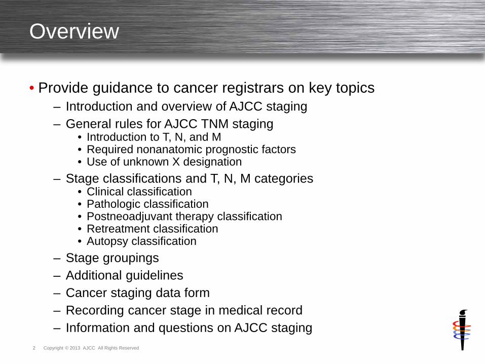
Overview
• Provide guidance to cancer registrars on key topics
– Introduction and overview of AJCC staging
– General rules for AJCC TNM staging • Introduction to T, N, and M • Required nonanatomic prognostic factors • Use of unknown X designation
– Stage classifications and T, N, M categories • Clinical classification • Pathologic classification • Postneoadjuvant therapy classification • Retreatment classification • Autopsy classification
– Stage groupings
– Additional guidelines
– Cancer staging data form
– Recording cancer stage in medical record
– Information and questions on AJCC staging
Copyright © 2013 AJCC All Rights Reserved 3
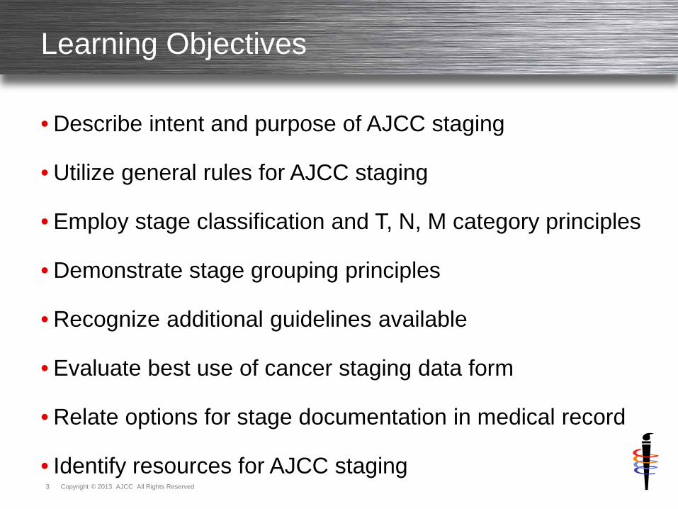
Learning Objectives
• Describe intent and purpose of AJCC staging
• Utilize general rules for AJCC staging
• Employ stage classification and T, N, M category principles
• Demonstrate stage grouping principles
• Recognize additional guidelines available
• Evaluate best use of cancer staging data form
• Relate options for stage documentation in medical record
• Identify resources for AJCC staging
Introduction and Overview of AJCC Staging

Copyright © 2013 AJCC All Rights Reserved 5
Introduction and Overview
• AJCC TNM – the common language of cancer
• International method to clearly convey without ambiguity – Clinical experience – Patient care
• Accurate staging is necessary to
– Evaluate results of treatments and clinical trials – Facilitate exchange and comparison of information among
treatment centers – Serve as basis for clinical and translational cancer research
• Stage or extent of cancer at time of diagnosis
– Defines prognosis – Determines appropriate treatment – Based on experience and outcomes of prior patients
Copyright © 2013 AJCC All Rights Reserved 6
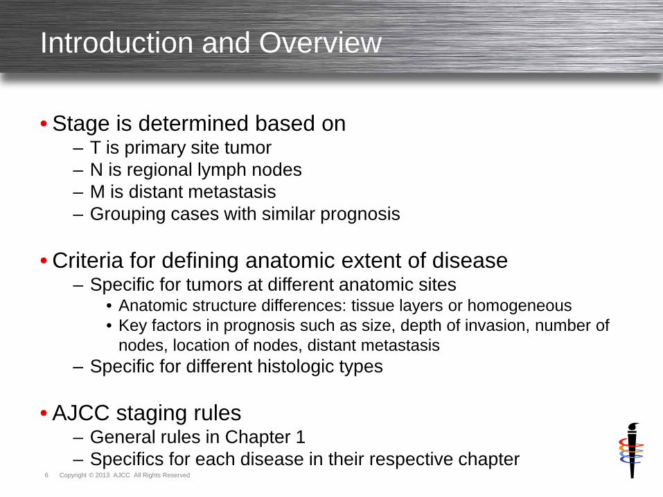
Introduction and Overview
• Stage is determined based on – T is primary site tumor – N is regional lymph nodes – M is distant metastasis – Grouping cases with similar prognosis
• Criteria for defining anatomic extent of disease
– Specific for tumors at different anatomic sites • Anatomic structure differences: tissue layers or homogeneous • Key factors in prognosis such as size, depth of invasion, number of
nodes, location of nodes, distant metastasis – Specific for different histologic types
• AJCC staging rules
– General rules in Chapter 1 – Specifics for each disease in their respective chapter
General Rules for AJCC TNM Staging

Copyright © 2013 AJCC All Rights Reserved 8
Introduction to T
• T category – Defined by size, and/or – Contiguous extension of primary tumor
• T specifically designed for each primary site
– Roles of size and contiguous spread depend on site characteristics
Primary Tumor (T) valid values
T0 No evidence of primary tumor Tis Carcinoma in situ T1, T2, T3, T4 Increasing size and/or local extension of primary tumor TX Primary tumor cannot be assessed (minimize use of TX)
Note: Subcategories are allowed, such as T1mi, T1a
Copyright © 2013 AJCC All Rights Reserved 9
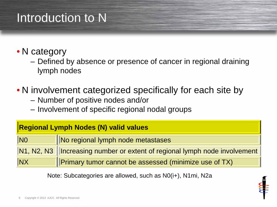
Introduction to N
• N category – Defined by absence or presence of cancer in regional draining
lymph nodes
• N involvement categorized specifically for each site by – Number of positive nodes and/or – Involvement of specific regional nodal groups
Regional Lymph Nodes (N) valid values
N0 No regional lymph node metastases N1, N2, N3 Increasing number or extent of regional lymph node involvement NX Primary tumor cannot be assessed (minimize use of TX)
Note: Subcategories are allowed, such as N0(i+), N1mi, N2a

Copyright © 2013 AJCC All Rights Reserved 10
Introduction to M
• M category – Defined by absence or presence of distant spread or metastases – Generally in locations to which cancer is spread by
• Vascular channels or • Lymphatics beyond nodes defined as regional
• M specifically designed for some sites
– Subcategories for detailed areas of involvement
Distant Metastasis (M) valid values
M0 No distant metastases M1 Distant metastases present
Note: The MX designation has been eliminated from the AJCC TNM system. Subcategories are allowed, such as cM0(i+), M1a
Copyright © 2013 AJCC All Rights Reserved 11
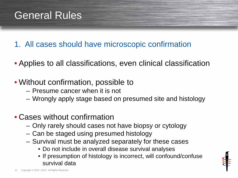
General Rules
1. All cases should have microscopic confirmation
• Applies to all classifications, even clinical classification
• Without confirmation, possible to – Presume cancer when it is not – Wrongly apply stage based on presumed site and histology
• Cases without confirmation
– Only rarely should cases not have biopsy or cytology – Can be staged using presumed histology – Survival must be analyzed separately for these cases
• Do not include in overall disease survival analyses • If presumption of histology is incorrect, will confound/confuse
survival data
Copyright © 2013 AJCC All Rights Reserved 12
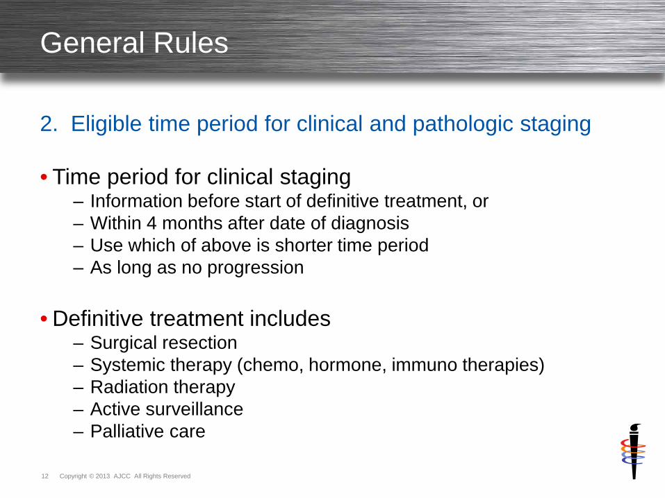
General Rules
2. Eligible time period for clinical and pathologic staging
• Time period for clinical staging – Information before start of definitive treatment, or – Within 4 months after date of diagnosis – Use which of above is shorter time period – As long as no progression
• Definitive treatment includes
– Surgical resection – Systemic therapy (chemo, hormone, immuno therapies) – Radiation therapy – Active surveillance – Palliative care

Copyright © 2013 AJCC All Rights Reserved 13
General Rules
2. Eligible time period for clinical and pathologic staging
• Time period for pathologic staging – All information including definitive surgical resection, or – Within 4 months after date of diagnosis – Use which of above is longer time period – As long as no systemic or radiation prior to surgery – As long as no progression
• Definitive surgical resection
– Must meet criteria for that specific chapter
Copyright © 2013 AJCC All Rights Reserved 14
General Rules
3. Staging with neoadjuvant or primary systemic/radiation
• Neoadjuvant therapy definition
– Systemic or radiation therapy is first treatment
– Followed by surgical resection
• Clinical stage assigned
– Only information prior to start of systemic/radiation
– Used for comparative purposes
– Used to determine response to therapy
Copyright © 2013 AJCC All Rights Reserved 15
General Rules
3. Staging with neoadjuvant or primary systemic/radiation
• Postneoadjuvant therapy stage is y – y must always be modified as yc or yp
• yc
– After systemic/radiation BUT prior to surgical resection
• yp – After systemic/radiation AND after surgical resection
Copyright © 2013 AJCC All Rights Reserved 16
General Rules
4. Progression of disease
• Evidence of disease progression – If before start of any treatment – Do not use this information for assigning stage
• Evidence of disease progression before treatment
– Use only information before progression to assign stage
Copyright © 2013 AJCC All Rights Reserved 17
General Rules
5. Uncertain information
• Assign the lower (lesser) category or stage group – If uncertain or unclear information – Not enough information to definitely choose
• Commonly called “downstaging”
• Does NOT apply to unknown information
– Unknown information does NOT use lowest category or group
Copyright © 2013 AJCC All Rights Reserved 18
General Rules
5. Uncertain information
• Examples
– Imaging unclear if one node (N1) or two nodes (N2) are involved • Use N1 which is lower category
– Colonoscopy does not provide information on T category for colon
• Use TX since information is unknown • Cannot assign T1 as this falsely skews data
– Lung clinical stage group for T2a NX M0
• Use stage group unknown since no information on nodes • Cannot assign stage group using N0 as this falsely skews data
• Physician may make clinical judgments for patient care
Copyright © 2013 AJCC All Rights Reserved 19
General Rules
6. Nonanatomic factors not available
• Not available nonanatomic factor required for stage group – Case assigned based on lowest or least advanced factor
• Use nonanatomic factor as X in stage table
• If X not available
– Use lowest level of the factor – Use least advanced category, least amount of factor
Copyright © 2013 AJCC All Rights Reserved 20
Required Nonanatomic Prognostic Factors
• Nonanatomic prognostic factors required for stage – Some AJCC chapters require these factors for assigning stage – Clearly defined and listed in stage tables, for example
• Thyroid, Chapter 8 – age and histology • Gastrointestinal Stromal Tumor, Chapter 16 – mitotic rate • Soft Tissue Sarcoma, Chapter 28 – grade
• Factors collected separately from T, N, and M
– Not part of TNM definitions – Separate additional information essential for prognosis in these
sites
• Factors needed to accurately assign stage group – Critical in some chapters, and no alternative to the information – Some chapters provide alternatives

Copyright © 2013 AJCC All Rights Reserved 21
Required Nonanatomic Prognostic Factors
• Some chapters provide alternatives to situations of – Factor is not available – Physician desires to assign group ignoring factor – Factor is not needed for that individual stage group
• Factors NOT available and needed to assign stage group
– Factor is assigned X – Allows stage group to be assigned – Allows physician to assign group ignoring factor
• Individual stage groups within table do not require factor
– Any is factor option for some individual stage groups – Any means factor is not needed to assign that stage group
• Factor can be known and documented • Factor can be unknown

Copyright © 2013 AJCC All Rights Reserved 22
Use of Unknown X Designation
• X used when – Information is unknown for specific category
• Clarification of unknown
– Unknown to physician providing patient care
– Not unknown to one physician, but known to other physicians
– Not unknown to registrar from lack of documentation in chart
• Misuse of X from registrar lacking chart information
– Can skew data analysis – Can lead to
• Inaccurate studies • Wrong conclusions about national status of patient care
Copyright © 2013 AJCC All Rights Reserved 23
Use of Unknown X Designation
• TX and/or NX cases usually cannot have stage assigned
• X category only used for T and N – When absolutely necessary
• Exception examples (not exhaustive list of every option)
– Any T and/or Any N with M1 is stage IV • Any T N2 M1, T3 Any N M1, Any T Any N M1
– TX and/or NX with M1 is stage IV • TX N2 M1, T3 NX M1, TX NX M1
– Category combinations belong in one and only one stage group • Lung TX N3 M0 is stage IIIB
– Every combination of T with N3 M0 is stage IIIB • Urethra T4 NX M0 is stage IV
– T4 M0 with every combination of N is stage IV
Copyright © 2013 AJCC All Rights Reserved 24
Use of Unknown X Designation
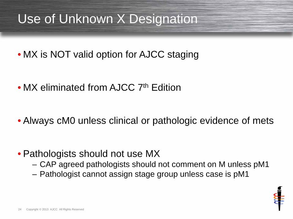
• MX is NOT valid option for AJCC staging
• MX eliminated from AJCC 7th Edition
• Always cM0 unless clinical or pathologic evidence of mets
• Pathologists should not use MX
– CAP agreed pathologists should not comment on M unless pM1 – Pathologist cannot assign stage group unless case is pM1

Stage Classifications and T, N, M Categories
Copyright © 2013 AJCC All Rights Reserved 26
Copyright © 2013 AJCC All Rights Reserved 27
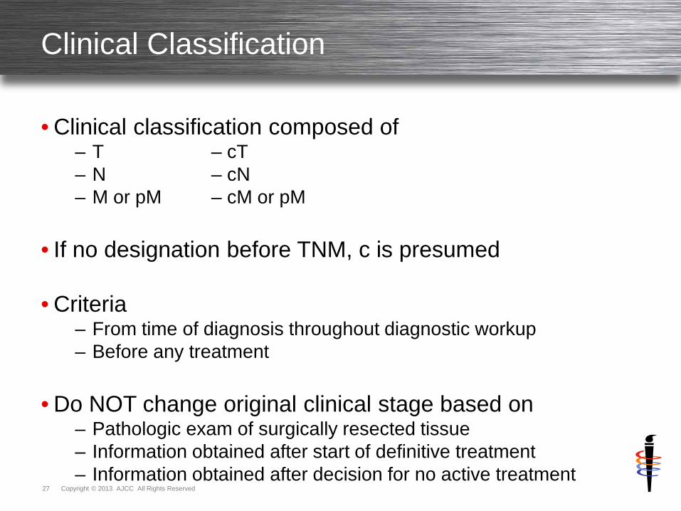
Clinical Classification
• Clinical classification composed of – T – cT – N – cN – M or pM – cM or pM
• If no designation before TNM, c is presumed
• Criteria
– From time of diagnosis throughout diagnostic workup – Before any treatment
• Do NOT change original clinical stage based on
– Pathologic exam of surgically resected tissue – Information obtained after start of definitive treatment – Information obtained after decision for no active treatment

Copyright © 2013 AJCC All Rights Reserved 28
Clinical Classification
• Information included and timing – All information during diagnostic workup – From time of diagnosis up until first treatment – Or within 4 months after diagnosis, whichever is shorter – With no systemic/radiation therapy prior to surgery – With no progression of disease
– Clinical assessment – diagnostic workup
• Clinical history • Physical examination • Imaging • Scopes and other invasive diagnostic procedures • Lab tests and biologic markers • Biopsy of primary site • Surgical exploration only • Diagnostic biopsy of lymph nodes, sentinel nodes • Diagnostic biopsy of metastatic sites • Related methods and other relevant examinations

Copyright © 2013 AJCC All Rights Reserved 29
Clinical Classification
• Clarifications of clinical assessment methods
– Surgical exploration • Can include biopsy • Cannot continue on to surgical resection in same procedure
– Biopsy for T category
• If tissue establishes highest possible T category, CAN use for pT • Also use for cT
– Biopsy of nodes is cN
• Single node or sentinel nodes as diagnostic workup, and • In absence of pathologic evaluation of primary tumor
– Imaging
• Extensive imaging NOT required to assign cT, cN or cM
– Biopsy of metastatic sites is pM • Discussion on M category follows
Copyright © 2013 AJCC All Rights Reserved 30
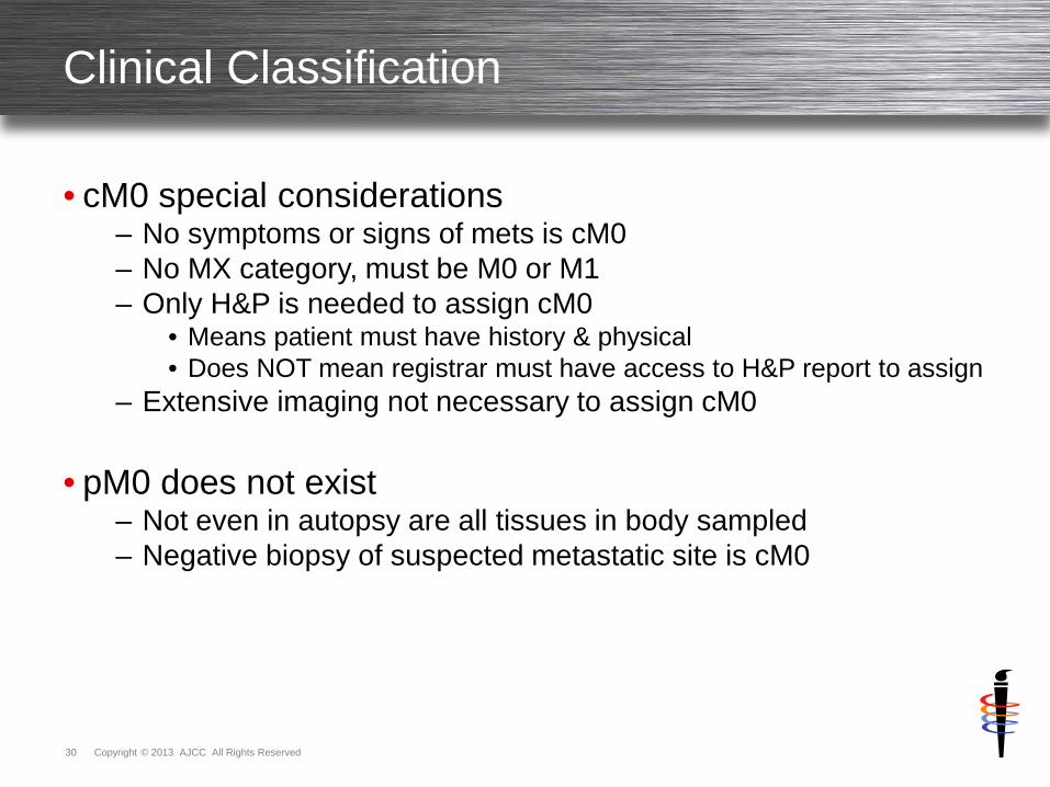
Clinical Classification
• cM0 special considerations – No symptoms or signs of mets is cM0 – No MX category, must be M0 or M1 – Only H&P is needed to assign cM0
• Means patient must have history & physical • Does NOT mean registrar must have access to H&P report to assign
– Extensive imaging not necessary to assign cM0
• pM0 does not exist – Not even in autopsy are all tissues in body sampled – Negative biopsy of suspected metastatic site is cM0
Copyright © 2013 AJCC All Rights Reserved 31
Clinical Classification
• cM1 special considerations – Evidence on physical exam of mets – Evidence on imaging of mets – Evidence seen during scopes of mets not biopsied – Operative findings during surgical resection not biopsied
• pM1 special considerations
– Positive biopsy of metastatic site – WITH cT and cN – Staged as both
• Clinical stage IV – cT cN pM1 • Pathologic stage IV – cT cN pM1
Copyright © 2013 AJCC All Rights Reserved 32
Clinical Classification
• Use of clinical classification – Select primary therapy – Treatment guidelines based on clinical classification – Critical for case comparisons
• Differences in treatment make future comparisons impossible – Only point in time where ALL cases can be compared
• Documentation
– Physician records in medical record – Recorded in cancer registry abstract clinical data fields – Essential for abstract to contain

Copyright © 2013 AJCC All Rights Reserved 33
Copyright © 2013 AJCC All Rights Reserved 34
Pathologic Classification
• Pathologic classification composed of – pT – pN – cM or pM
• Criteria
– From time of diagnosis through surgical resection findings – Use all clinical staging information AND – Add to it or change it by evidence from
• Operative findings • Pathology report on resected tissue
• Pathologic classification made up of 3 components
– Clinical classification information – Operative findings during surgical resection – Pathology report on resected specimen
Copyright © 2013 AJCC All Rights Reserved 35
Pathologic Classification
• Information included and timing – All information during diagnostic workup and surgical treatment – From time of diagnosis until end/completion of surgical treatment – Or within 4 months after diagnosis, whichever is longer – With no systemic/radiation therapy prior to surgery – With no progression of disease
– Clinical classification information – Operative findings during surgical resection – Pathology report on resected specimen
Copyright © 2013 AJCC All Rights Reserved 36
Pathologic Classification
• Clarifications of pathologic assessment methods
– Clinical classification information • Same physical exam and diagnostic studies from clinical stage
– Operative findings during surgical resection
• Surgeon’s statements of viewed/palpated involvement • Do NOT need biopsy to include in pT • Do NOT need biopsy to include in pN, unless NO nodes biopsied
– Pathology report on resected specimen
• May overrule clinically suspected involvement • Clinical or operative findings are used for stage UNLESS
– Histologic exam of resected tissue disproves those findings
– Pathology report is NOT final stage • Pathology report is only 1/3 of necessary information • Report does NOT take into consideration other 2/3 of information

Copyright © 2013 AJCC All Rights Reserved 37
Pathologic Classification
• Primary tumor (pT) assessment for pathologic classification – Resection of primary tumor, generally – Some chapters require
• More extensive resection of tumor • Partial or complete organ resection
– Generally from single specimen
• T special considerations – Physicians estimate size from several partial resections – Size recorded in whole millimeters
• Round as necessary to whole millimeter • Whole millimeter used to assign pT • Fractions of millimeters NOT used to increase pT category
– Evaluation of highest T category • Biopsy of primary tumor is adequate, then • pT can be assigned without resection
Copyright © 2013 AJCC All Rights Reserved 38
Pathologic Classification
• Regional node (pN) assessment for pathologic classification – Resection of regional nodes – Require pathologic exam of ONE node
• N special considerations
– Number of nodes resected • Minimum number to assure sufficient sampling • Expected number of nodes defined in chapters • If fewer than minimum nodes, pN is still assigned • Sentinel node procedure substitutes for expected minimum number
– Do NOT need pathologic confirmation of highest N category – pT generally necessary for pN – Microscopic evaluation of highest N category
• May use pN regardless whether T is pT or cT

Copyright © 2013 AJCC All Rights Reserved 39
Pathologic Classification
• pN0(i+) special considerations – Isolated tumor cells (ITC) in lymph nodes
• Single tumor cells or small clusters of cells • Not more than 0.2mm in greatest diameter
– Designated as pN0 – negative nodes • ITC are considered negative nodes in all sites except two • Melanoma and Merkel cell consider ITC as positive nodes • Some chapters use pN0(i+) when common in that site • Other chapters use pN0
– pN0(i+) for detected by immunohistochemistry (IHC) – pN0(i-) for IHC done and no tumor cells found – pN0(mol+) for detected by molecular techniques – pN0(mol-) for molecular technique done and no tumor cells found – Also can be detected by flow cytometry and DNA analysis – Uncertain prognostic significance of these cells – Use i+ and i- to denote status of ITC & gather data
Copyright © 2013 AJCC All Rights Reserved 40
Pathologic Classification
• Mets assessment for pathologic classification may be
– Clinical using cM0 or cM1 • pT pN cM0 or pT pN cM1
– Pathologic using pM1 • pT pN pM1
• pM1 special considerations
– Requires biopsy positive for cancer at metastatic site – Meets criteria for pathologic classification without resection of
primary site
• pM0 does NOT exist – pM0 is undefined concept – Autopsy may not satisfy since EVERY tissue must be sampled – pM0 may not be used
Copyright © 2013 AJCC All Rights Reserved 41
Pathologic Classification
• cM0 special considerations – No signs or symptoms of mets – Only H&P performed on patient is needed to assign
• cM0(i+) special considerations
– Biopsy shows isolated tumor cells (ITC) – Detected by immunohistochemistry (IHC) or molecular techniques – CTCs – circulating tumor cells in blood – DTCs – disseminated tumor cells in bone marrow or distant organs – Uncertain prognostic significance of these cells – Categorized as M0, use i+ to denote these cells & gather data
• cM1 special considerations
– Evidence from clinical assessment – Operative findings during surgical resection not biopsied
Copyright © 2013 AJCC All Rights Reserved 42
Pathologic Classification
• Use of pathologic classification – Select adjuvant therapy – Treatment guidelines for adjuvant therapy based on pathologic
classification – Significant additional prognostic information – More precise than clinical classification – Commonly used for survival studies due to precise data
• Only used for cases with surgical resection as first treatment
• Documentation – Physician records in medical record – Recorded in cancer registry abstract pathologic data fields – Essential for abstract to contain in surgically resected cases
Copyright © 2013 AJCC All Rights Reserved 43
Copyright © 2013 AJCC All Rights Reserved 44
Postneoadjuvant Therapy Classification
• Postneoadjuvant therapy classification composed of – ycT – ypT – ycN – ypN – cM or pM – cM or pM
• Neoadjuvant therapy definition
– Systemic and/or radiation therapy given prior to surgery – Systemic includes chemotherapy, hormone therapy, immunotherapy
• Criteria for yc assessment
– After systemic/radiation and before surgery – After systemic/radiation with no surgery performed
• Criteria for yp assessment
– After systemic/radiation AND after surgical resection

Copyright © 2013 AJCC All Rights Reserved 45
Postneoadjuvant Therapy Classification
• yc – information included and timing – All information at that time using clinical assessment methods – Performed after systemic/radiation and prior to surgery – Use clinical classification rules for assigning ycT and ycN – Use M as classified prior to all treatment – Clinical stage after systemic/radiation
• yp – information included and timing
– All information at that time using pathologic assessment methods – Performed after systemic/radiation/surgery – Use pathologic classification rules for assigning ypT and ypN – Use M as classified prior to all treatment – Pathologic stage after systemic/radiation/surgery
Copyright © 2013 AJCC All Rights Reserved 46
Postneoadjuvant Therapy Classification
• Provides information on response to therapy – Classification useful to physicians – Measured against clinical classification to show response – Response noted as: complete, partial, or no response – Provides important prognostic information to patients – yc
• Shows response to systemic/radiation and is prognostic • Directs type and extent of surgery to be performed
– yp • Surgical resection removes any remaining cancer • Verifies response to systemic/radiation through pathology
assessment of tissue and is prognostic • Directs subsequent systemic and/or radiation therapy
• Neoadjuvant therapy is increasingly common
– Important to assess response and document – Analyze outcomes
Copyright © 2013 AJCC All Rights Reserved 47
Postneoadjuvant Therapy Classification
• M category for yc and yp – Use M status defined PRIOR to therapy – May be either clinical (cM) or pathologic (pM)
• Positive biopsy of metastatic site
– pM1 is recorded for all classifications – Clinical stage IV – yc stage IV – yp stage IV
• Must assign clinical classification
– Estimate of disease prior to all treatment
• Clinical stage used for – Case comparisons, studies, clinical trials – Surveillance analysis
Copyright © 2013 AJCC All Rights Reserved 48
Postneoadjuvant Therapy Classification
• Use of postneoadjuvant therapy classification – Critical to assess response to therapy – Monitor success of neoadjuvant as it grows in use
• Documentation - physician
– Physician records both yc and yp in medical record
• Documentation – registrar – yc – yc NOT recorded in cancer registry abstract – No data fields available for yc classification – Cannot use clinical data fields
• Documentation – registrar – yp
– yp recorded in cancer registry abstract pathologic data fields – Must code 4 in pathologic stage descriptor data field – Identifies stage as yp and NOT p
Copyright © 2013 AJCC All Rights Reserved 49

Copyright © 2013 AJCC All Rights Reserved 50
Retreatment Classification
• Retreatment classification composed of – rT – rN – rM
• Also called recurrence classification
• Criteria
– Patient must have been disease-free prior to recurrence – Further treatment is planned
• Does NOT change original clinical and pathologic stage
• Do NOT use when patient never free of disease
Copyright © 2013 AJCC All Rights Reserved 51
Retreatment Classification
• Information included and timing – All information at time of retreatment – Biopsy confirmation is important
• May not be medically possible • Is not mandatory
• May include
– Biopsy of T, N, and/or M categories if possible – Clinical evidence
• Physical exam • Imaging • Scopes and other invasive procedures • Lab tests and biologic markers • Related methods
Copyright © 2013 AJCC All Rights Reserved 52
Retreatment Classification
• Use of retreatment classification – Extent of current disease used to guide new therapy – Prognostic information from clinical extent and therapeutic
procedures – Cannot be compared to other stage classifications
• Documentation
– Physician records in medical record – NOT recorded in cancer registry abstract – No data fields available for retreatment classification – Cannot use clinical or pathologic data fields
Copyright © 2013 AJCC All Rights Reserved 53
Copyright © 2013 AJCC All Rights Reserved 54
Autopsy Classification
• Autopsy classification composed of – aT – aN – aM
• Criteria
– NO evidence of cancer prior to death – NO possibility or suggestion of cancer prior to death – Incidental finding on autopsy
• Information included and timing
– All clinical and pathologic information obtained at time of death – Autopsy information
• Do NOT use when known cancer patient has autopsy
Copyright © 2013 AJCC All Rights Reserved 55
Autopsy Classification
• Use of autopsy classification – No opportunity for physician to intervene in course of disease – Cannot be compared to other stage classifications
• Documentation
– Physician records in medical record – NOT recorded in cancer registry abstract – No data fields available for autopsy classification – Cannot use clinical or pathologic data fields

Stage Groupings
Copyright © 2013 AJCC All Rights Reserved 57
Purpose of Stage Groupings
• Anatomic stage/prognostic groups – Comprised of T, N, and M – Nonanatomic factors sometimes required to supplement TNM – Disease specific groups – Similar prognosis for each group – Useful for guideline development – Facilitate communication regarding types of patients – Commonly referred to as stage groups
• Data tabulation and analysis
– Depends on grouping patients into a few categories – Need fewer groups of larger numbers for meaningful data
• Stage groups are summary of staging information that is
– Reproducible – Easily communicated

Copyright © 2013 AJCC All Rights Reserved 58
Principles of Stage Groupings
• Classified by Roman numerals I-IV, indicates – Increasing severity of disease – Worsening prognosis
• General definitions
– Stage I – smaller or less deeply invasive with negative nodes – Stage II and III – increasing tumor or nodal extent – Stage IV – distant metastases at diagnosis
• Additional stage group designated for
– Stage 0 – carcinoma in situ with no metastatic potential
• Expanded into subsets for – More refined prognostic information – Example stage II becomes stage IIA, stage IIB
Copyright © 2013 AJCC All Rights Reserved 59
Standard Composition of Stage Groupings
• Clinical Stage Group – cT – cN – cM or pM
• Pathologic Stage Group
– pT – pN – cM or pM
• Postneoadjuvant Therapy Stage Group
– ypT – ypN – cM or pM
Copyright © 2013 AJCC All Rights Reserved 60
Stage Grouping Principles
• Standard stage group principle defined for each case – Pure clinical stage group – Pure pathologic stage group
• Pure stage group does NOT mean
– Every category must be c • cT cN cM
– Every category must be p • pT pN pM
• Pure stage group does mean following AJCC rules
– Using c or p for categories according to established rules – Examples
• cT cN pM clinical stage group • pT pN cM pathologic stage group

Copyright © 2013 AJCC All Rights Reserved 61
Stage Grouping Principles
• Working stage
– Used by physicians in clinical setting of patient care
– Only partial information available according to staging rules
– Must combine clinical and pathologic information
– Combination allows assignment of stage group
– Used for treatment decisions and patient care
– NOT documented by cancer registry

Copyright © 2013 AJCC All Rights Reserved 62
CIS Exception to Stage Grouping
• Carcinoma in situ (CIS) definition – Does not involve any structures that allow tumor spread – Cells cannot spread to
• Other parts of primary site/organ • Regional tissues outside primary site/organ • Regional nodes • Distant sites
• CIS exception to stage grouping principles
– pTis cN0 cM0 clinical stage 0 – pTis cN0 cM0 pathologic stage 0
• Caution for pathologic stage 0
– Requires chapter specific criteria is met – Cannot assign based on small sample – Potential sampling error if less than chapter criteria
Copyright © 2013 AJCC All Rights Reserved 63
Stage Grouping Guidelines
• Assign stage group according to – Timing – Appropriate rules – Do not change due to subsequent information after time frame
• Documenting stage group in medical record
– All appropriate groups recorded in chart, not just one group
• Uncertainty general rule #5 also applies to stage group – Assign lower or less advanced group with uncertain information – Do NOT apply to unknown information such as TX and/or NX in
order to assign group

Copyright © 2013 AJCC All Rights Reserved 64
Stage Grouping Guidelines
• Exception for pCR
• pCR – Pathologic complete response to neoadjuvant therapy – After systemic/radiation followed by surgery
• No evidence of active invasive cancer cells • Based on resection pathology report • May have in situ disease
• Stage categories and group assigned for pCR is
– ypT0 ypN0 cM0 – NOT stage 0 (used for in situ disease only) – NO stage group assigned
Additional Guidelines
Copyright © 2013 AJCC All Rights Reserved 66
Multiple Tumors
• AJCC rules for multiple primary tumors – May not agree with registry MPH rules
• Multiple simultaneous tumors of same histology in 1 organ
– Tumor with highest T category is used for classification & staging – Multiplicity or number of tumors is in parentheses
• T2(m) shows multiple tumors or T2(5) shows there are five tumors
• Simultaneous bilateral cancers in paired organs – Tumors classified separately – Stage as independent tumors in different organs
• Multiple tumor criteria is part of T category for
– Thyroid, liver, and ovary

Copyright © 2013 AJCC All Rights Reserved 67
Multiple Tumors in Registry Data Field
• Registry software data field for m descriptor
• FORDS Clinical Stage (prefix/suffix) Descriptor – Code 3 M-Multiple primary tumors in a single site – NAACCR Item #980
• FORDS Pathologic Stage (prefix/suffix) Descriptor
– Code 3 M-Multiple primary tumors in a single site – Code 6 M&Y-Multiple primary tumors & initial multimodality therapy
• Meets criteria for code 3 and code 4 (y-classification for neoadjuvant) – NAACCR Item #920
Copyright © 2013 AJCC All Rights Reserved 68
Metachronous Primaries
• Metachronous – developing at a later interval
• Second or subsequent primary cancers – Occurring in same organ, or – Occurring in different organs are – Staged as NEW cancer
• Second cancers do not use y prefix
– Unless treatment of second cancer is neoadjuvant therapy
• AJCC rules for metachronous primaries – May not agree with registry MPH rules
Copyright © 2013 AJCC All Rights Reserved 69
Unknown Primary
• Staging based on clinical suspicion of primary site – No evidence of primary tumor, or – Site of primary tumor is unknown, then – T category assigned as T0
• Example 1
– Axillary node bx shows metastatic ca consistent with breast cancer – No tumor seen in breast on mammogram, US, and MRI – Stage assigned as breast cancer T0 N1 M0
• Example 2
– Cervical node bx shows metastatic squamous cell ca consistent with head and neck cancer
– History of sores in oral cavity, especially hard palate – Stage assigned as oral cavity cancer T0 N1 M0

Cancer Staging (Data) Form

Copyright © 2013 AJCC All Rights Reserved 71
Staging Form for Each Chapter
• Each chapter includes staging form for physicians
• Forms include – Clinical, pathologic, and postneoadjuvant therapy classifications – T, N, and M – Stage groups – Prognostic factors (site-specific factors) – Histologic grade – Additional descriptors
• Lymph-vascular invasion (LVI) • Residual tumor (R)
– Clinical stage used in treatment planning – National guidelines used in treatment planning – Physician signature and date – Identification of hospital and patient
Copyright © 2013 AJCC All Rights Reserved 72
Staging Form Use
• Staging form used at different points in time – Diagnosis and workup, before treatment – After surgical resection as first course of treatment – After neoadjuvant systemic/radiation therapy & before surgery – After neoadjuvant systemic/radiation therapy and surgery – Recurrence
• Best to use separate form for each point in time
• If same form used for multiple time points
– Ensure staging basis for each T, N, M category clearly identified
• Staging form is specific additional document – Not substitute for H&P, staging evaluations – Not substitute for treatment plans, follow-up
Copyright © 2013 AJCC All Rights Reserved 73
AJCC Staging Form
• Incorporation of forms into electronic record or system – Requires appropriate permission from AJCC and publisher
• Modification of forms whether paper or electronic – Requires appropriate permission from AJCC and publisher
• Paper cancer staging forms in AJCC Manual – May be duplicated for individual or institutional use
• Includes only immediate institution or work environment – Without permission from AJCC or publisher
• Permission requests submitted to – http://cancerstaging.net

Recording Cancer Stage in Medical Record
Copyright © 2013 AJCC All Rights Reserved 75
Recording Stage in Medical Record
• Physician recording stage in medical record – Critical for communication between physicians – Useful to communicate data to cancer registry – Stage in every record, all admissions and outpatient encounters
• Physician options for documenting stage
– Initial clinical evaluations: H&P, consults – Operative reports – Discharge summaries – Staging Form
• Staging Form
– Paper form included in each AJCC chapter – Electronic forms also available (e-staging tool)

Information and Questions on AJCC Staging
Copyright © 2013 AJCC All Rights Reserved 77
AJCC Web site
• https://cancerstaging.org
• Cancer Staging Education menu includes
– Articles • 18 articles on AJCC staging in various medical journals
– Resources
• Staging Moments – 15 case-based presentations in cancer conference format to promote accurate staging with answers and rationales
– Webinars
• 14 free webinars on staging rules and some disease sites
• Watch for education plans and content in the future

Copyright © 2013 AJCC All Rights Reserved 78
AJCC Cancer Staging Manual and Atlas
Order at http://cancerstaging.net
Copyright © 2013 AJCC All Rights Reserved 79
CAnswer Forum
• Submit questions to AJCC Forum
– Located within CAnswer Forum
– Provides information for all
– Allows tracking for educational purposes
• http://cancerbulletin.facs.org/forums/

Summary

Copyright © 2013 AJCC All Rights Reserved 81
Summary
• Articulate intent and purpose of AJCC staging
• Apply AJCC rules, principles, and guidelines accurately – General rules for AJCC staging – Stage classification and T, N, M category principles – Stage grouping principles – Additional guidelines available
• Recommend and operationalize
– Cancer staging data form – Stage documentation in medical record
• Identify resources for AJCC staging
– Information, guidance, and education – Obtain answers to questions

Thank you
Donna M. Gress, RHIT, CTR AJCC Technical Specialist
633 N. Saint Clair, Chicago, IL 6011-3211 cancerstaging.org
No materials in this presentation may be changed without the express written permission of the American Joint Committee on Cancer. Permission requests may be submitted at CancerStaging.net
Related Documents