Registering Retinal Vessel Images from Local to Global via Multiscale and Multicycle Features Haiyong Zheng 1 Lin Chang 1 Tengda Wei 1 Xinxin Qiu 1 Ping Lin 2 Yangfan Wang 1 1 Ocean University of China 2 The University of Dundee [email protected] {changlinok1234, tdwei123, qxx1990421}@163.com, [email protected], [email protected] Abstract We propose a comprehensive method using multiscale and multicycle features for retinal vessel image registration with a local and global strategy. The multiscale vessel maps generated by multiwavelet kernels and multiscale hierarchi- cal decomposition contain segmentation results at varying image resolutions in different levels of vessel details. Then the multicycle feature composed of various combinations of cycle structures with different numbers of vertices is ex- tracted. The cycle structure consisting of vessel bifurcation points, crossover points of arteries and veins, and the con- nected vessels can be found by our Angle-based Depth-First Search (ADFS) algorithm. Local initial registration is im- plemented by the matched Cycle-Vessel feature points and global final registration is completed by the Cycle-Vessel- Bifurcation feature points using similarity transformation. Finally, our Skeleton Alignment Error Measure (SAEM) is calculated for optimal scale and cycle feature selection, yielding the best registration result intelligently. Experi- mental results show that our method outperforms state-of- the-art methods on retinal vessel image registration using different features in terms of accuracy and robustness. 1. Introduction Retinal vessel images contain valuable local and time information as they are usually acquired from different modalities over many years, which can be aligned to one image by image registration to aid ophthalmologists for analysis and diagnosis of various diseases such as diabetic retinopathy, age-related macular degeneration, and glau- coma. In this paper, we focus on accurate and robust feature-based retinal image registration. The registration methods can be classified into intensity- based and feature-based [13]. Intensity-based methods generally optimize a similarity measure based on cross- correlation, phase correlation, and mutual information, etc. [17], which will take great computation cost to find the optimal solution, especially they need to incorporate the whole image information to finish the registration. Also, the intensity-based methods may fail to align the images if the image quality is quite low or the overlapping region be- tween the images is small. These motivate the exploitation of robust features such as retinal vessel and optic disk in- stead of intensity in retinal image registration [16]. Most of the feature-based methods use bifurcation for registra- tion since it is a prominent indicator of vasculature. Zana and Klein [23] used bifurcation points with surrounding vessel orientations for multimodal registration. Can and Stewart [2] proposed a hierarchical algorithm using branch- ing points and crossover points in the retinal vasculature to avoid unmatchable image features and mismatches between features, and then it was extended to Dual-Bootstrap Itera- tive Closest Point (ICP) algorithm that iteratively decides the optimal transformation model from simple to complex and expands the bootstrap region from local to global [20]. The blood vessel bifurcations were also identified as control points to evaluate the transformation types and the pixel- level fusion techniques [11]. Chanwimaluang et al. [4] proposed a hybrid retinal image registration approach that combines both area-based and feature-based methods us- ing crossover/bifurcation points of vascular tree as land- mark points. And the RERBEE algorithm was presented with BEES representing the vasculature structure (bifurca- tions and segments) for registration [18]. These methods largely depend on the branching angles of single bifurca- tion/crossover point, and these features have coarse preci- sion leading to matching which may not be unique and reli- able for registration purpose. Compared with the aforementioned point-matching methods, structure-matching registration is favored to over- come the possible mismatches. Chen et al. [6, 5] presented a bifurcation structure composed of a master bifurcation point and its three connected neighboring pixels or vessel segments, with the normalized branching angle and length as its characteristic vector. Shen et al. [19] then extended 50

Welcome message from author
This document is posted to help you gain knowledge. Please leave a comment to let me know what you think about it! Share it to your friends and learn new things together.
Transcript

Registering Retinal Vessel Images from Local to Global via Multiscale and
Multicycle Features
Haiyong Zheng1 Lin Chang1 Tengda Wei1 Xinxin Qiu1 Ping Lin2 Yangfan Wang1
1Ocean University of China 2The University of Dundee
{changlinok1234, tdwei123, qxx1990421}@163.com, [email protected], [email protected]
Abstract
We propose a comprehensive method using multiscale
and multicycle features for retinal vessel image registration
with a local and global strategy. The multiscale vessel maps
generated by multiwavelet kernels and multiscale hierarchi-
cal decomposition contain segmentation results at varying
image resolutions in different levels of vessel details. Then
the multicycle feature composed of various combinations
of cycle structures with different numbers of vertices is ex-
tracted. The cycle structure consisting of vessel bifurcation
points, crossover points of arteries and veins, and the con-
nected vessels can be found by our Angle-based Depth-First
Search (ADFS) algorithm. Local initial registration is im-
plemented by the matched Cycle-Vessel feature points and
global final registration is completed by the Cycle-Vessel-
Bifurcation feature points using similarity transformation.
Finally, our Skeleton Alignment Error Measure (SAEM) is
calculated for optimal scale and cycle feature selection,
yielding the best registration result intelligently. Experi-
mental results show that our method outperforms state-of-
the-art methods on retinal vessel image registration using
different features in terms of accuracy and robustness.
1. Introduction
Retinal vessel images contain valuable local and time
information as they are usually acquired from different
modalities over many years, which can be aligned to one
image by image registration to aid ophthalmologists for
analysis and diagnosis of various diseases such as diabetic
retinopathy, age-related macular degeneration, and glau-
coma. In this paper, we focus on accurate and robust
feature-based retinal image registration.
The registration methods can be classified into intensity-
based and feature-based [13]. Intensity-based methods
generally optimize a similarity measure based on cross-
correlation, phase correlation, and mutual information,
etc. [17], which will take great computation cost to find
the optimal solution, especially they need to incorporate the
whole image information to finish the registration. Also,
the intensity-based methods may fail to align the images if
the image quality is quite low or the overlapping region be-
tween the images is small. These motivate the exploitation
of robust features such as retinal vessel and optic disk in-
stead of intensity in retinal image registration [16]. Most
of the feature-based methods use bifurcation for registra-
tion since it is a prominent indicator of vasculature. Zana
and Klein [23] used bifurcation points with surrounding
vessel orientations for multimodal registration. Can and
Stewart [2] proposed a hierarchical algorithm using branch-
ing points and crossover points in the retinal vasculature to
avoid unmatchable image features and mismatches between
features, and then it was extended to Dual-Bootstrap Itera-
tive Closest Point (ICP) algorithm that iteratively decides
the optimal transformation model from simple to complex
and expands the bootstrap region from local to global [20].
The blood vessel bifurcations were also identified as control
points to evaluate the transformation types and the pixel-
level fusion techniques [11]. Chanwimaluang et al. [4]
proposed a hybrid retinal image registration approach that
combines both area-based and feature-based methods us-
ing crossover/bifurcation points of vascular tree as land-
mark points. And the RERBEE algorithm was presented
with BEES representing the vasculature structure (bifurca-
tions and segments) for registration [18]. These methods
largely depend on the branching angles of single bifurca-
tion/crossover point, and these features have coarse preci-
sion leading to matching which may not be unique and reli-
able for registration purpose.
Compared with the aforementioned point-matching
methods, structure-matching registration is favored to over-
come the possible mismatches. Chen et al. [6, 5] presented
a bifurcation structure composed of a master bifurcation
point and its three connected neighboring pixels or vessel
segments, with the normalized branching angle and length
as its characteristic vector. Shen et al. [19] then extended
50

bifurcation structure-matching by a local fine registration.
However, bifurcation structure is still not unique and reli-
able enough for registration that may make the matching
missed if the vascular tree is detailed, especially followed
by the vessel segmentation result in one scale.
In view of this, we propose the cycle structure for
feature-based retinal image registration. The cycle struc-
ture, consisting of vessel bifurcation points, crossover
points of arteries and veins, and the connected vessels, is
more unique, reliable and invariant against translation, ro-
tation, and scaling, especially for the retina from the same
person over many years, so it can also be used in many other
applications such as retina identification [1] and verifica-
tion [10]. To overcome the dependence on the vessel seg-
mentation for registration, we use multiwavelet kernels and
multiscale hierarchical decomposition [22] to generate mul-
tiscale vessel maps at varying image resolutions in differ-
ent levels of vessel details and then adopt our Angle-based
Depth-First Search (ADFS) algorithm to extract the mul-
ticycle feature composed of various combinations of cycle
structures with different numbers of vertices. For regions
away from the matching cycles, we implement the two-
stage registration from local to global to construct the robust
Cycle-Vessel-Bifurcation feature for final registration using
similarity transformation. Finally, our Skeleton Alignment
Error Measure (SAEM) is calculated for optimal scale and
cycle feature selection, yielding the best registration result.
Figure 1 shows the framework of our proposed method.
The rest of the paper is organized as follows: Section 2
gives our multiscale segmentation and skeleton method for
retinal vessel images; the detection, extraction, description
and matching of our proposed cycle structure are presented
in Section 3; and Section 4 introduces our two-stage local-
to-global strategy for accurate registration; then our SAEM
is proposed in Section 5 to obtain the best registration result
automatically; Section 6 shows the qualitative and quantita-
tive experimental results and Section 7 concludes this paper.
2. Multiscale Segmentation and Skeleton
Due to hardware limitations, retinal images usually have
characteristics of uneven illumination, large numbers of
noise points, and low contrast between vessels and back-
ground, which lead to the challenge of automatic analysis.
Retinal vessel segmentation plays a key role on the accuracy
of registration.
In this work, multiwavelet kernels and multiscale hier-
archical decomposition method [22] is used for multiscale
vessel segmentation. And 14 vessel maps are generated at
varying image resolutions representing different levels of
vessel details to avoid the possible failed feature extraction
caused by single segmentation. We then remove the invalid
scales using a threshold of pixel number for the segmenta-
tion results with very few vessel pixels because they obvi-
ously miss the important vessel information.
The multiscale segmentation results are further pro-
cessed by the following procedures for better feature ex-
traction: 1) remove the connected regions with few pixels
recognized as non-vessel by a threshold for denoising; 2)
fill the vessel holes caused by the central light reflex of reti-
nal arteries and veins based on erosion and dilation method;
3) skeletonize the vessels to obtain one pixel width vascular
tree using the contour-pruned skeletonization method 1.
The reference image and the floating image for registra-
tion are both segmented and skeletonized by the above mul-
tiscale processing, yielding up to 14 vessel skeleton results
respectively (each Nj , j = 1, 2, · · · , 14 needs to be aligned
with each Mi, i = 1, 2, · · · , 14 as shown in Figure 1). Fig-
ure 2 shows an example of the multiscale vessel segmenta-
tion and skeleton results.
3. Multicycle Matched Structure
The retinal vasculature, consisting of arteries and veins,
and their end branches, forms capillary networks (see Fig-
ure 2), thus can be formally described as an unweighted
graph G [9]. We then design and construct the cycle struc-
ture using vessel bifurcation points, crossover points of ar-
teries and veins, and the connected vessels. Multicycle
means that various combinations of cycle structures are seg-
mented by multiscale hierarchical decomposition with dif-
ferent scales. In our work, the minimum cycles, defined as
a cycle basis where the sum of the weights of the cycles is
minimum [12], are selected for retinal image registration.
The minimum cycle bases of a graph encapsulate the entire
cycle space in a concise manner so that they are very use-
ful in many contexts, e.g., structural engineering [3], cycle
analysis of electrical networks [7], and chemical ring per-
ception [8].
3.1. Cycle Structure Detection and Extraction
The extraction procedure of cycle structures can be im-
plemented by two steps: searching feature points and find-
ing cycle structures.
3.1.1 Searching Feature Points
The feature points we used for cycle structure consist of
the bifurcation and crossover points in the binary skeleton
vascular network, and they can be searched as follows:
1. Label the pixels whose sum of 8-adjacent pixels is
greater than 2 into connected components, because the
pixels cannot be bifurcation or crossover points if the
sum of its 8-adjacent pixels is less than or equal to 2.
1http://www.cs.smith.edu/∼nhowe/research/code/
51
Reference image
Floating image
35
3
4
5
34
45
345
Mi
Nj
Multicycle matched structure
L
(7 types)Lk
Local initial registration
Global final registration
Multiscale vessel skeleton
Multiscale Vessel
Segmentation and Skeleton
Space-based DFS algorithm
M
(14 scales)
N
(14 scales)
Cycle Description and Matching
Cycle-Vessel Feature Points
Cycle-Vessel-Bifurcation Feature Points
Skeleton Alignment Error Measure
Min(SAEM)
Best registration result k=1,2,…,7
i=1,2,…,14
j=1,2,…,14
reference
floating
Similarity Transformation
Similarity
Transformation
……
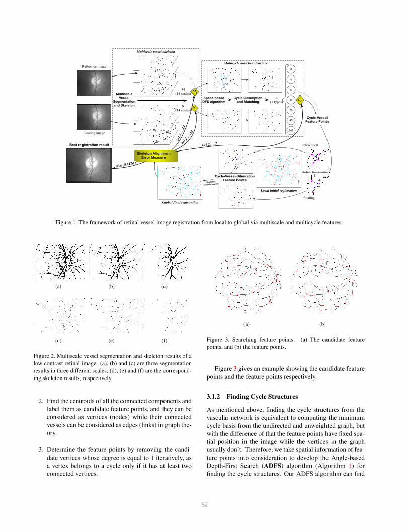
Figure 1. The framework of retinal vessel image registration from local to global via multiscale and multicycle features.
(a) (b) (c)
(d) (e) (f)
Figure 2. Multiscale vessel segmentation and skeleton results of a
low contrast retinal image. (a), (b) and (c) are three segmentation
results in three different scales, (d), (e) and (f) are the correspond-
ing skeleton results, respectively.
2. Find the centroids of all the connected components and
label them as candidate feature points, and they can be
considered as vertices (nodes) while their connected
vessels can be considered as edges (links) in graph the-
ory.
3. Determine the feature points by removing the candi-
date vertices whose degree is equal to 1 iteratively, as
a vertex belongs to a cycle only if it has at least two
connected vertices.
(a) (b)
Figure 3. Searching feature points. (a) The candidate feature
points, and (b) the feature points.
Figure 3 gives an example showing the candidate feature
points and the feature points respectively.
3.1.2 Finding Cycle Structures
As mentioned above, finding the cycle structures from the
vascular network is equivalent to computing the minimum
cycle basis from the undirected and unweighted graph, but
with the difference of that the feature points have fixed spa-
tial position in the image while the vertices in the graph
usually don’t. Therefore, we take spatial information of fea-
ture points into consideration to develop the Angle-based
Depth-First Search (ADFS) algorithm (Algorithm 1) for
finding the cycle structures. Our ADFS algorithm can find
52

the minimum cycle basis of the image-based undirected
and unweighted graph with 1 edge and n vertices effec-
tively and efficiently by reducing the time complexity from
O(n/ log n+n2) [12] to O(n2) due to the use of spatial (an-
gle) information. According to the characteristics of retinal
vasculature, only cycle structures composed of three, four
and five feature points are chosen for registration in our
work.
Algorithm 1 Angle-based Depth-First Search (ADFS)
Input: The feature points and their connectivities in the bi-
nary skeleton image.
Output: The cycle structures.
1: for all A such that A ∈ FeaturePointsSet(Pn) do
2: for all B such that B ∈ Pn and connects to A do
3: calculate the vector VBA
4: for all C such that C ∈ Pn and connects to B do
5: calculate the vector VBC
6: calculate the angle ΘABC = ∠ABC7: end for
8: determine C such that min (ΘABC)9: if C connects to A then
10: output CycleStucture(ABC)11: continue
12: else
13: repeat for all [D ∈ Pn and connects to C]
14: output CycleStucture(ABCD) and continue
15: or iterate next connected feature point
16: until no connected feature point
17: end if
18: end for
19: end for
3.2. Cycle Structure Description and Matching
All the cycle structures found in the reference image and
the floating image need to be matched to determine the Cy-
cle feature points (on matched cycle structures) for transfor-
mation and further registration. In order to make the struc-
ture more unique and reliable for registration, we describe
a cycle structure using branch lengths, branch angles, and
angles between adjacent edges, among which branch an-
gles represent the connected relationship of vessels on each
feature point while angles between adjacent edges repre-
sent the relative position of feature points. Figure 4 shows
an example description of a four-point cycle structure. The
lengths are calculated based on the pixel distance and the
angles are calculated relying on the adjacent points in the
vessels or edges. Then we normalize all the lengths and
angles by Equation 1 and 2 to preserve scaling invariant
and guarantee rotation invariant respectively, yielding the
final feature vector given in Equation 3 as an example cor-
responding to Figure 4.
θ2
θ3θ1
θ5
θ6
θ7θ8
θ9
θ10 θ11
θ12
θ13 θ14
θ15
θ17
θ18
θ19
θ20
L1
L3
L2L4
Figure 4. Cycle structure description. L1 ∼ L4, θ1 ∼ θ16 and
θ17 ∼ θ20 represent branch lengths, branch angles and angles be-
tween adjacent edges respectively in a four-point cycle structure.
LiNorm =Li∑L
(1)
θjNorm =θj
360◦(2)
v = {lengths, angles} = {L1,L2,L3,L4, θ1, θ2, θ3,0, θ5,
θ6, θ7, θ8, θ9, θ10, θ11, θ12, θ13, θ14, θ15,0, θ17, θ18, θ19, θ20}(3)
The number of feature vector elements varies with the
number of cycle feature points, and the number of branch
angles varies with the style of feature points, e.g., a bifur-
cation point usually has 3 branch angles while a crossover
point has 4 branch angles. To obtain consistent vector
length for further matching, we consider the feature points
of the cycle structure all as bifurcation or crossover points,
making maximum 4 branch angles for each point and then
the feature vector of three-, four- and five-point cycle struc-
ture becomes 18, 24, and 30 dimensions (the missing ele-
ments are set to 0 as demonstrated in Equation 3), respec-
tively.
Matching two cycle structures with the same number of
feature points can be implemented by minimizing the sim-
ilarity measure Dpq = mean(|Vp − Wq|), where Vp rep-
resents the p-th cycle structure in the reference image, Wq
represents the q-th cycle structure in the floating image and
the function mean(·) calculates the average value of vector.
To avoid the possible mismatches even with the minimum
D, a threshold is used to verify the matching again, so that
one or more of the three-, four- and five-point cycle struc-
tures may be ignored, e.g., the four-point cycle structure
is missed after the matching illustrated in Figure 1. This
method tries to find the most accurate matching cycle struc-
tures between the reference and the floating while misses
the others, so that the matched cycle feature points for reg-
istration are very accurate but not much in number. There-
53

(a) (b)
Figure 5. Local initial registration. (a) is the skeleton image regis-
tration result, and (b) is the corresponding retinal image registra-
tion result.
fore, we combine all kinds of the matched cycle structures
up to 7 types Lk, k = 1, 2, · · · , 7 as shown in Figure 1,
yielding the multicycle matched structure.
4. Registration from Local to Global
In order to get a more accurate registration, we design
a two-stage local-to-global strategy using the Cycle-Vessel
and Cycle-Vessel-Bifurcation feature points respectively.
4.1. Local Initial Registration
The multicycle matched structure only contains limited
accurate cycle feature points, which may not be enough for
further transformation and then registration. So we also
extract the closest bifurcation points or ending points con-
nected to the cycle feature points along the vessels and the
midpoints of the vessels to constitute the Cycle-Vessel fea-
ture points for local initial registration.
It’s obvious that the cycle-vessel structure is invariant
against translation, rotation, and scaling, but is variant to
shearing because of the angles between vessels, therefore,
similarity transformation is the best choice. The similarity
transformation can be defined by
X = xs cosϕ− ys sinϕ+ a (4)
Y = xs sinϕ+ ys cosϕ+ b (5)
where s denotes scaling, ϕ denotes rotational, and (a, b) de-
notes translational differences between the reference image
and the floating image. In our work, the least squares solu-
tion is adopted to obtain the transformation parameters [21].
Figure 5 shows the results of local initial registration using
the cycle-vessel feature points.
4.2. Global Final Registration
Although the cycle-vessel feature points are very accu-
rate for registration (Figure 5), it’s still not robust enough
because these points only cover vascular regions with intri-
cate capillary that may make some global regions far away
from them unaligned (Figure 6a and 6b). Therefore, we
(a) (b)
(c) (d)
Figure 6. Comparison of local initial registration and global final
registration. (a) and (b) are skeleton and retinal image results re-
spectively by local initial registration with unaligned vessels in
some local regions, while (c) and (d) are the comparative skeleton
and retinal image results respectively after global final registration
with better vessel alignment precisely.
extend the registration from local to global by finding the
unaligned bifurcation points from the local registration re-
sult to further constitute the final Cycle-Vessel-Bifurcation
feature points.
The unaligned bifurcation points are determined by bi-
furcation matching and distance thresholding: 1) find the
matching bifurcation point of each bifurcation point in the
floating local registration image among its 5 × 5 neighbor-
hood relying on the angles [23]; 2) regard the matched bi-
furcation points as unaligned if their pixel distance is greater
than 3 pixels. Then, similar to local registration, the similar-
ity transformation is adopted for final registration using the
cycle-vessel-bifurcation feature points. Figure 6c and 6d
show the global final registration results corresponding to
the local registration results Figure 6a and 6b, which can be
seen more precisely.
5. Skeleton Alignment Error Measure
One reference image and one floating image, also known
as one pair of images for registration, will produce up to
14× 14 multiscale vessel segmentation and skeleton results
with up to 7 multicycle matched structures, yielding at most
14×14×7 local-to-global registration results (Figure 1). We
then define the Skeleton Alignment Error Measure (SAEM)
and choose the optimal registration result automatically by
minimizing SAEM as follows:
1. Given a reference skeleton result Mi (i =
54

1, 2, · · · , 14) and a floating skeleton result Nj (j =1, 2, · · · , 14), Nj is aligned to Mi with one type of
multicycle matched structure Lk (k = 1, 2, · · · , 7)
under the local-to-global strategy, yielding the trans-
formed skeleton result NjMi;
2. For each vessel point in NjMi, calculate the pixel dis-
tance d of its nearest vessel pixel among its 7×7 neigh-
borhood in Mi, or mark this vessel point invalid if no
corresponding vessel pixel is found;
3. SAEM is defined by SAEMijk = (∑
d) /Numv ,
where Numv is the number of valid pixels in NjMi
that d can be calculated;
4. Constraints: SAEM is considered valid only if
Numv/NumNj≥ 50% and NumNjMi
/NumNj≥
38%, where Num(·) denotes the number of pixels in
(·).
The constraints are necessary to exclude the extremely mis-
matching situations that may make the SAEM minimized
because of very few contributing pixels.
By using SAEM, the best registration result will be se-
lected intelligently, moreover, the registration method can
be evaluated. The overall framework of our proposed
method for retinal vessel image registration can be seen in
Figure 1.
6. Experiments
There exist rare public datasets of retinal images for reg-
istration purpose, so we use VARIA database [15, 14]2 that
contains a set of retinal images for authentication purpose
to evaluate and compare the performance of our method for
retinal image registration qualitatively and quantitatively.
The database currently includes 233 images from 139 dif-
ferent individuals that have been acquired with a TopCon
non-mydriatic camera NW-100 model and are optic disc
centered with a resolution of 768× 584, among which 155pairs from 59 individuals (total 153 images from all 233retinal images) can be constructed as a new dataset for reg-
istration purpose3.
6.1. Qualitative Results
Figure 7 shows two examples of our retinal vessel image
registration from local to global via multiscale and multi-
cycle features, among which (a)(b) and (f)(g) are two pairs
of original retinal images for registration, (c) and (h) are
the corresponding best registration results selected by our
2http://www.varpa.es/varia.html3Only the two retinal images that belong to the same individual can be
considered as one pair for registration.
SAEM automatically, (d) and (i) are local initial registra-
tion results while (e) and (j) are global final registration re-
sults, respectively. Although the pairs of original retinal im-
ages are dramatically different with big deformation, it can
still be seen that the final results Figure 7(c) and (h) are
both well registered accurately by minimizing the SAEM
via multiscale and multicycle features and robustly through
the local-to-global strategy. The zoom-in regions on Fig-
ure 7(e) and (j) are shown obviously more precise of aligned
vessels than those corresponding regions on Figure 7(d) and
(i) respectively, indicating that the effectiveness and robust-
ness of our two-stage local-to-global registration strategy.
6.2. Quantitative Results
For quantitative comparison, the Success Rate (SR)
and Skeleton Alignment Error Measure (SAEM) are used
to evaluate our method and other methods on the 155pairs of retinal images. The registration is regarded as
successful evaluated by the ophthalmologists considering
the real medical applications for SR calculation (SR =(Successful Pairs)/155) and the failed registration will be
excluded to calculate SAEM .
First, the transformation models are important for dif-
ferent features of registration and have been discussed
by [20, 11], and Table 1 shows the registration results with
respect to our cycle structure using different transformation
models4: similarity, affine, and second-order polynomial.
Although the SAEM of polynomial transformation is mini-
mum with 0.231 pixel, it’s still not suitable for cycle struc-
ture due to the lowest SR (16.99%) because the failed regis-
trations will not contribute to SAEM, which may make the
SAEM very small based on very few successful registra-
tions. Therefore, because the proposed cycle-vessel struc-
ture is invariant against translation, rotation, and scaling,
but is variant to shearing due to the angles between vessels,
the similarity transformation for our cycle structure is the
best choice with the highest 96.73% SR and the acceptable
0.938 pixel SAEM for ophthalmologists.
Transformation models SR SAEM (pixel)
Similarity 96.73% 0.938Affine 50.33% 1.010Polynomial 16.99% 0.231
Table 1. Comparison of different transformation models.
Then, Table 2 shows the registration results by similar-
ity transformation using different features on the 155 pairs
of retinal images under the optimal MiNjLk with mini-
mum SAEMijk: Cycle, Cycle-Vessel, and Cycle-Vessel-
4Only Cycle features are used in this experiment, and the 155 pairs of
retinal images under the optimal MiNjLk with minimum SAEMijk are
selected for this experiment.
55

(a) (b) (c)
(d) (e)
(f) (g) (h)
(i) (j)
Figure 7. The qualitative results of our proposed retinal image reg-
istration. (a)(b) and (f)(g) are two pairs of original retinal images
for registration, (c) and (h) are the corresponding best registration
results selected by our SAEM automatically, (d) and (i) are local
initial registration results while (e) and (j) are global final regis-
tration results, respectively. The zoom-in regions on (e) and (j)
are shown obviously more precise of aligned vessels than those
corresponding regions on (d) and (i) respectively, indicating that
the effectiveness and robustness of our two-stage local-to-global
registration strategy.
Bifurcation, which obviously indicates that the Cycle-
Vessel-Bifurcation feature is the most robust and accurate
for registration with the highest SR and the lowest SAEM.
Features SR SAEM (pixel)
Cycle 96.73% 0.938Cycle-Vessel (local) 97.39% 0.903Cycle-Vessel-Bifurcation
(local-to-global)
100% 0.858
Table 2. Comparison of different features.
At last, we compare our proposed method for retinal
image registration with state-of-the-art structure-matching
registration methods: Bifurcation structure [6, 5] and Bi-
furcation structure with global fine registration [19]. This
experiment is also applied on the 155 pairs of retinal im-
ages under the optimal MiNjLk with minimum SAEMijk.
Besides, we also implement our multiscale strategy with
SAEM optimal selection on the compared two methods.
The results are shown in Table 3, and it can be seen that our
method greatly outperforms the other methods in terms of
robustness and accuracy with the highest 100% SR and the
lowest 0.858 pixel SAEM respectively. Moreover, the pro-
posed mutiscale and SAEM strategy dramatically improves
the compared two methods, which also shows the effective-
ness and robustness of our intelligent registration based on
multiscale selection automatically.
Methods SR SAEM (pixel)
Bifurcation structure 50.32% 1.009Bifurcation structure +
Global
59.35% 0.978
Bifurcation + Multiscale
+ SAEM
95.48% 0.938
Bifurcation Global +
Multiscale + SAEM
96.77% 0.877
Our method 100% 0.858
Table 3. Comparison of different methods.
7. Conclusion and Future Work
In this paper, we address the issues related to the de-
scription, matching as well as registration of the vascular
structure in retinal images. The contribution of this paper
is threefold: 1) a novel stable cycle structure is proposed
for retinal vasculature description and an effective and fast
Angle-based Depth-First Search algorithm is developed for
finding minimum cycle basis; 2) the cycle structure is ex-
tended from local to global for robust and accurate match-
ing; 3) the Skeleton Alignment Error Measure is defined to
measure and evaluate the registration intelligently. The pro-
posed feature and method can be used for other applications
such as retina identification and verification, and also other
vascular-related study.
In future work, we will test the method in larger and
more natural retinal image datasets, and tune the algorithm
for real applications helping the ophthalmologists to ana-
lyze and diagnose retina-related diseases.
Acknowledgements
This work was supported by the National Natural Sci-
ence Foundation of China under Grant Nos. 61271406,
61301240, 31302182.
56

References
[1] A. Bhuiyan, E. Lamoureux, B. Nath, K. Ramamohanarao,
and T. Y. Wong. Retinal image matching using hierarchical
vascular features. Computational Intelligence and Neuro-
science, 2011:9, 2011. 2
[2] A. Can, C. V. Stewart, B. Roysam, and H. L. Tanenbaum. A
feature-based, robust, hierarchical algorithm for registering
pairs of images of the curved human retina. IEEE Transac-
tions on Pattern Analysis and Machine Intelligence, 24:347–
364, 2002. 1
[3] A. C. Cassell, J. C. D. Henderson, and K. Ramachandran.
Cycle bases of minimal measure for the structural analysis
of skeletal structures by the flexibility method. Proceedings
of the Royal Society of London A: Mathematical, Physical
and Engineering Sciences, 350:61–70, 1976. 2
[4] T. Chanwimaluang, G. Fan, and S. R. Fransen. Hybrid reti-
nal image registration. IEEE Transactions on Information
Technology in Biomedicine, 10:129–142, 2006. 1
[5] L. Chen, X. Huang, and J. Tian. Retinal image registra-
tion using topological vascular tree segmentation and bifur-
cation structures. Biomedical Signal Processing and Control,
16:22–31, 2015. 1, 7
[6] L. Chen, Y. Xiang, Y. Chen, and X. Zhang. Retinal image
registration using bifurcation structures. In Proceedings of
the 18th IEEE International Conference on Image Process-
ing, pages 2169–2172, Brussels, September 2011. Institute
of Electrical and Electronics Engineers. 1, 7
[7] J. C. de Pina. Applications of Shortest Path Methods. PhD
thesis, University of Amsterdam, 1995. 2
[8] P. M. Gleiss. Short cycles: minimum cycle bases of graphs
from chemistry and biochemistry. PhD thesis, University of
Vienna, 2001. 2
[9] R. Gould. Graph Theory. Dover Publications, 2012. 2
[10] S. M. Lajevardi, A. Arakala, S. A. Davis, and K. J. Horadam.
Retina verification system based on biometric graph match-
ing. IEEE Transactions on Image Processing, 22:3625–
3635, 2013. 2
[11] F. Laliberte, L. Gagnon, and Y. Sheng. Registration and fu-
sion of retinal images–an evaluation study. IEEE Transac-
tions on Medical Imaging, 22:661–673, 2003. 1, 6
[12] K. Mehlhorn and D. Michail. Minimum cycle bases: Faster
and simpler. ACM Transactions on Algorithms, 6:8, 2009. 2,
4
[13] F. P. M. Oliveira and J. M. R. S. Tavares. Medical image
registration: a review. Computer Methods in Biomechanics
and Biomedical Engineering, 17:73–93, 2014. 1
[14] M. Ortega, M. G. Penedo, J. Rouco, N. Barreira, and M. J.
Carreira. Personal verification based on extraction and char-
acterisation of retinal feature points. Journal of Visual Lan-
guages & Computing, 20:80–90, 2009. 6
[15] M. Ortega, M. G. Penedo, J. Rouco, N. Barreira, and M. J.
Carreira. Retinal verification using a feature points-based
biometric pattern. EURASIP Journal on Advances in Signal
Processing, 2009:2, 2009. 6
[16] E. Peli, R. A. Augliere, and G. T. Timberlake. Feature-based
registration of retinal images. IEEE Transactions on Medical
Imaging, 6:272–278, 1987. 1
[17] G. P. Penney, J. Weese, J. A. Little, P. Desmedt, D. L. G. Hill,
and D. J. Hawkes. A comparison of similarity measures for
use in 2-d–3-d medical image registration. IEEE Transac-
tions on Medical Imaging, 17:586–595, 1998. 1
[18] A. Perez-Rovira, R. Cabido, E. Trucco, S. J. McKenna, and
J. P. Hubschman. RERBEE: robust efficient registration via
bifurcations and elongated elements applied to retinal fluo-
rescein angiogram sequences. IEEE Transactions on Medi-
cal Imaging, 31:140–150, 2012. 1
[19] B. Shen, D. Zhang, and Y. Peng. Blood bifurcation struc-
ture and global to local strategy based retinal image registra-
tion. In Proceedings of the 5th Chinese Conference on Pat-
tern Recognition, pages 394–403, Beijing, China, September
2012. Springer Berlin Heidelberg. 1, 7
[20] C. V. Stewart, C.-L. Tsai, and B. Roysam. The dual-bootstrap
iterative closest point algorithm with application to retinal
image registration. IEEE Transactions on Medical Imaging,
22:1379–1394, 2003. 1, 6
[21] S. Umeyama. Least-squares estimation of transformation
parameters between two point patterns. IEEE Transactions
on Pattern Analysis and Machine Intelligence, 13:376–380,
1991. 5
[22] Y. Wang, G. Ji, P. Lin, and E. Trucco. Retinal vessel segmen-
tation using multiwavelet kernels and multiscale hierarchical
decomposition. Pattern Recognition, 46:2117–2133, 2013. 2
[23] F. Zana and J.-C. Klein. A multimodal registration algorithm
of eye fundus images using vessels detection and hough
transform. IEEE Transactions on Medical Imaging, 18:419–
428, 1999. 1, 5
57
Related Documents


![Improved multiscale matched filter for retina vessel segmentation … · 2017. 1. 19. · Retinal Images for Vessel Extraction (DRIVE) database [9], introduced in 2004. Database contains](https://static.cupdf.com/doc/110x72/60517afc8a35ef5cec2242d8/improved-multiscale-matched-filter-for-retina-vessel-segmentation-2017-1-19.jpg)