promoting access to White Rose research papers White Rose Research Online Universities of Leeds, Sheffield and York http://eprints.whiterose.ac.uk/ This is an author produced version of a paper published in Biomaterials. White Rose Research Online URL for this paper: http://eprints.whiterose.ac.uk/5234/ Published paper Korossis, S., Bolland, F., Southgate, J., Ingham, E. and Fisher, J. (2009) Regional biomechanical and histological characterisation of the passive porcine urinary bladder: Implications for augmentation and tissue engineering strategies. Biomaterials, 30 (2). pp. 266-275. [email protected]

Welcome message from author
This document is posted to help you gain knowledge. Please leave a comment to let me know what you think about it! Share it to your friends and learn new things together.
Transcript
promoting access to White Rose research papers
White Rose Research Online
Universities of Leeds, Sheffield and York http://eprints.whiterose.ac.uk/
This is an author produced version of a paper published in Biomaterials. White Rose Research Online URL for this paper: http://eprints.whiterose.ac.uk/5234/
Published paper Korossis, S., Bolland, F., Southgate, J., Ingham, E. and Fisher, J. (2009) Regional biomechanical and histological characterisation of the passive porcine urinary bladder: Implications for augmentation and tissue engineering strategies. Biomaterials, 30 (2). pp. 266-275.
1 2 3 4 5 6 7 8 9 10 11 12 13 14 15 16 17 18 19 20 21 22 23 24 25 26 27 28 29 30 31 32 33 34 35 36 37 38 39 40 41 42 43 44 45 46 47 48 49 50 51 52 53 54 55 56 57 58 59 60 61 62 63 64 65
1
Regional Biomechanical and Histological Characterisation of the
Passive Porcine Urinary Bladder: Implications for Augmentation
and Tissue Engineering Strategies
Sotirios Korossis, PhD (Corresponding Author)
Institute of Medical and Biological Engineering
University of Leeds, Leeds, LS2 9JT, United Kingdom
E-mail: [email protected]
Tel. no: 0113 343 2197
Fax no: 0113 242 4611
Fiona Bolland, PhD
Jack Birch Unit of Molecular Carcinogenesis, Department of Biology, University of
York, Heslington, York, YO10 5YW, United Kingdom.
Jenny Southgate, PhD
Jack Birch Unit of Molecular Carcinogenesis, Department of Biology, University of
York, Heslington, York, YO10 5YW, United Kingdom.
Eileen Ingham, PhD
Institute of Medical and Biological Engineering, University of Leeds, Leeds, LS2 9JT,
United Kingdom.
John Fisher, PhD, DEng
Institute of Medical and Biological Engineering, University of Leeds, Leeds, LS2 9JT,
United Kingdom.
* Title Page
1 2 3 4 5 6 7 8 9 10 11 12 13 14 15 16 17 18 19 20 21 22 23 24 25 26 27 28 29 30 31 32 33 34 35 36 37 38 39 40 41 42 43 44 45 46 47 48 49 50 51 52 53 54 55 56 57 58 59 60 61 62 63 64 65
1
Abstract
The aim of this study was to identify and quantify potential regional and
directional variations in the quasistatic uniaxial mechanical properties of the passive
urinary bladder wall. Overall, the lower body and trigone regions demonstrated the
highest degree of directional anisotropy, whereas the ventral region demonstrated
the least directional anisotropy. Significant regional anisotropy was found only along
the apex-to-base direction. The dorsal and ventral regions demonstrated a
significantly increased distensibility along the apex-to-base direction compared to the
other bladder regions, whereas the trigone and lower body regions demonstrated the
least distensibility. The trigone, lower body and lateral regions also demonstrated the
highest tensile strength both at regional and directional level. The study detected
significant regional and directional anisotropy in the mechanical properties of the
bladder and correlated this anisotropy to the distended and non-distended tissue
histioarchitecture and whole organ mechanics. By elucidating the inhomogeneous
nature of the bladder, the results from this study will aid the regional differentiation of
bladder treatments in terms of partial bladder replacement with suitable natural or
synthetic biomaterials, as well as the development of more realistic constitutive
models of bladder wall biomechanics and improved computational simulations to
predict deformations in the natural and augmented bladder.
* Abstract
1 2 3 4 5 6 7 8 9 10 11 12 13 14 15 16 17 18 19 20 21 22 23 24 25 26 27 28 29 30 31 32 33 34 35 36 37 38 39 40 41 42 43 44 45 46 47 48 49 50 51 52 53 54 55 56 57 58 59 60 61 62 63 64 65
1
Introduction
A variety of congenital and acquired conditions result in bladder dysfunction
with consequent debilitating incontinence, which affects approximately 400 million
people worldwide. In the majority of cases, a decrease in compliance is caused by
thickening of the bladder wall due to smooth muscle cell hypertrophy and increased
connective tissue deposition [1]. This may arise due to increased distension of the
bladder wall (e.g. due to bladder outlet obstruction), which may directly or indirectly
act as a stimulus for hypertrophy and hyperplasia [2,3,4,5]. Furthermore, neuropathic
disease or trauma can induce significant alterations in the neural control of the
bladder, which in turn can cause substantial changes in bladder function. These
functional changes can produce severe alterations in the structure, thickness,
compliance and biomechanics of the bladder wall [6,7,8]. Currently, the major
surgical solution to restore lost function due to trauma, neurogenic or vascular
dysfunction, or cancer is bladder augmentation surgery. Bowel is most commonly
used in various procedures of neobladder replacement, such as augmentation
enterocystoplasty or substitution enterocystoplasty. However, its use is not without
long-term complications [9,10,11], suggesting that the materials used for the repair
may be inadequate. In fact, rupture of the repaired bladder wall is known to occur in
~5% of cases [12].The lack of an entirely satisfactory clinical procedure has led
researchers to pursue alternative bladder replacement materials involving tissue
engineering techniques [13,14].
Ideal materials for complete or partial bladder replacement should possess both
biological compatibility, to promote cellular and tissue integration, and mechanical
reliability. In order to design more appropriate long-term surgical repair procedures
and develop materials for bladder reconstruction, and indeed to gain an insight into
* Manuscript
1 2 3 4 5 6 7 8 9 10 11 12 13 14 15 16 17 18 19 20 21 22 23 24 25 26 27 28 29 30 31 32 33 34 35 36 37 38 39 40 41 42 43 44 45 46 47 48 49 50 51 52 53 54 55 56 57 58 59 60 61 62 63 64 65
2
the disease processes that lead to bladder dysfunction, it is necessary to
characterize and quantify the fundamental mechanical properties of the normal
bladder at the mesoscale-tissue level and correlate them to both whole organ
mechanics and tissue histioarchitecture. Quantitative linking of the mechanics to
bladder histioarchitecture will also help to elucidate the repercussion of cellular and
molecular level alterations on bladder function [15]. Along these lines, studies have
correlated alterations in myosin isoform and collagen type content to force
development in bladder muscle strips [16,17] or to urodynamics data [18,19]. Such
correlations are important not only for interpreting structural/functional changes in
studying patterns of bladder dysfunction, but also to predict the fate of replacement
materials when exposed to the local normal or pathological mechanical loading in the
bladder wall in vivo.
In addition to the active contraction of the detrusor smooth muscle, the bladder
demonstrates nonlinear elastic, viscous and plastic mechanical properties
[20,21,22,23,24,25,26], depending on the boundary conditions. However, during
normal physiological filling rates bladder deformation can be considered quasistatic
[27], whereas neural and contractile effects are minimal [28]. Over the years, several
mathematical models have been developed in an effort to predict the stress-strain
behaviour of the bladder wall. Most of these models assume isotropy, homogeneity,
incompressibility and a spherical shape for the bladder wall [22,29,30,31]. Although
the assumptions of a spherical shape and incompressibility can give a relatively good
description of bladder mechanics during filling [32], it is questionable how descriptive
are the assumptions of isotropy and homogeneity for the bladder wall. The bladder
demonstrates a considerable inherent inhomogeneity in its material properties [33],
and as a result, it does not stretch equally in all directions, demonstrating areas of
1 2 3 4 5 6 7 8 9 10 11 12 13 14 15 16 17 18 19 20 21 22 23 24 25 26 27 28 29 30 31 32 33 34 35 36 37 38 39 40 41 42 43 44 45 46 47 48 49 50 51 52 53 54 55 56 57 58 59 60 61 62 63 64 65
3
higher stretching and, subsequently, higher stress. In spite of this, relatively little is
known about the anisotropic mechanical properties of the bladder wall in terms of
direction or region, and only a meagre few studies have focused on this issue
[34,35]. As a first step towards the development of tissue engineered bladder repair
materials, the authors performed the first regional and directional mechanical
characterisation of the urinary bladder. In particular, the objective of this study was to
identify and quantify potential regional and directional variations in the passive
mechanical properties of the bladder wall and correlate these variations to its
histioarchitecture and whole organ mechanics. By elucidating the inhomogeneous
nature of the bladder, the aim of this work was to consider the implications for
developing suitable natural or synthetic biomaterials for bladder augmentation.
Materials & Methods
Specimen procurement & dissection
Intact bladders from 16-week-old commercial male pigs were collected from a
local abattoir and transported to the laboratory on ice in transport medium [Hanks’
balanced salt solution without Ca++ and Mg++ (HBSS, Invitrogen, Paisley, UK)
containing 10 mM HEPES, pH 7.6 (Invitrogen) and 10 KIU/ml Aprotinin (Trasylol,
Bayer, Berkshire, UK)] [36]. The absence of calcium in the solution helped ensure
that the bladders were in an inactivated state and that no spontaneous contractions
would occur during testing. Prior to testing, the bladders were sized by photographing
them in their deflated/non-distended state (Figure 1). The recorded images of the
bladders were calibrated and the maximum bladder width along the circumferential
direction was measured using an image analysis software (Image Pro PlusTM,
MediaCybernetics®). The average size of the bladders used in this study was 68 ±
11.7 mm (mean ± 95% confidence interval, n = 6).
1 2 3 4 5 6 7 8 9 10 11 12 13 14 15 16 17 18 19 20 21 22 23 24 25 26 27 28 29 30 31 32 33 34 35 36 37 38 39 40 41 42 43 44 45 46 47 48 49 50 51 52 53 54 55 56 57 58 59 60 61 62 63 64 65
4
The bladders were subsequently dissected along the apex-to-base line, as
show in Figure 2a, and samples were isolated from the dorsal, trigone, lateral, ventral
and lower body regions of the wall, as well as along the apex-to-base (longitudinal)
and transverse (circumferential) directions (Figure 2b). For the purpose of the
biomechanical characterization, specimens measuring 205 mm were isolated using
a purpose-built block cutter [37]. From each bladder, one apex-to-base and one
transverse specimen were isolated from each one of the five anatomical regions.
Samples from the five anatomical regions and along the two directions were also
harvested for histological examination. Following isolation, the specimens were
stored in transport medium and tested either biomechanically or histologically within
6 hours from slaughter.
Histological characterisation
Histological examination was performed on samples harvested along the apex-
to-base and transverse directions from the five anatomical regions of the bladder
wall, in order to analyse the general histioarchitecture, as well as the amount and
orientation of elastin, collagen and smooth muscle. The samples were retrieved
either from the procured empty bladders and fixed in 10% (v/v) neutral buffered
formalin (NBF), or from a bladder that had been distended to the mean physiological
capacity with 500 ml of 10% (v/v) NBF. Post-fixation, distended and non-distended
samples were dehydrated and embedded in paraffin wax. Histological sections were
stained with either Miller’s stain to evaluate the content and distribution of elastin,
Van Gieson’s stain to evaluate the distribution of collagen and smooth muscle, or
with haematoxylin and eosin (H&E) [38]. The stained sections were examined under
light microscopy and photographed.
Biomechanical characterisation
1 2 3 4 5 6 7 8 9 10 11 12 13 14 15 16 17 18 19 20 21 22 23 24 25 26 27 28 29 30 31 32 33 34 35 36 37 38 39 40 41 42 43 44 45 46 47 48 49 50 51 52 53 54 55 56 57 58 59 60 61 62 63 64 65
5
Bladder wall strips were subjected to low-strain rate uniaxial tensile loading to
failure in order to investigate potential regional variations in the passive stress strain-
behaviour of the bladder wall. In addition, the directional anisotropy of the bladder
wall was investigated by testing specimens along the apex-to-base and transverse
directions. In total, 10 test groups of 6 specimens each were studied. Prior to testing,
the thickness of the samples was measured at 6 points along their long axis using a
gauge with a resolution of 0.01 mm (Mitutoyo, Andover, UK), and their average
thickness (t) was recorded. Subsequently, the samples were mounted onto a
purpose-built titanium holder. The holder was supported by a removable aluminium
bracket that allowed alignment of the two holder grips, defined the gauge length of
the specimens, and ensured that no load was imposed on the specimen until the
start of the test [37]. The gauge length of the specimens was defined by a 10 mm
wide central block separating the two holder parts and screwed onto the bracket.
Once a sample was clamped onto the holder, the holder with the supporting bracket
was secured to a Howden tensile machine and the bracket was removed. Prior to
loading to failure, the specimens were preconditioned under cyclic loading using a
double-ramp wave function at a rate of 10 mm/min. A preconditioning regime of 10
cycles was sufficient to produce a steady-state load-elongation response from the
samples. Following preconditioning, the samples were sequentially stretched to
failure at a rate of 10 mm/min. All testing was conducted in physiologic saline (0.9%
w/v NaCl) and at room temperature. Total testing time was approximately 3 min per
specimen. During testing, load data from the load cell and specimen extension data
from the stroke of the cross-head of the tensile testing machine was acquired at a
rate of 20Hz.
1 2 3 4 5 6 7 8 9 10 11 12 13 14 15 16 17 18 19 20 21 22 23 24 25 26 27 28 29 30 31 32 33 34 35 36 37 38 39 40 41 42 43 44 45 46 47 48 49 50 51 52 53 54 55 56 57 58 59 60 61 62 63 64 65
6
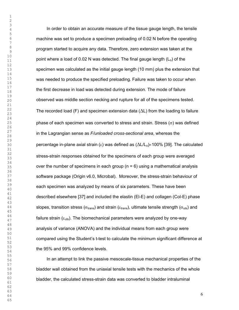
In order to obtain an accurate measure of the tissue gauge length, the tensile
machine was set to produce a specimen preloading of 0.02 N before the operating
program started to acquire any data. Therefore, zero extension was taken at the
point where a load of 0.02 N was detected. The final gauge length (Lo) of the
specimen was calculated as the initial gauge length (10 mm) plus the extension that
was needed to produce the specified preloading. Failure was taken to occur when
the first decrease in load was detected during extension. The mode of failure
observed was middle section necking and rupture for all of the specimens tested.
The recorded load (F) and specimen extension data (L) from the loading to failure
phase of each specimen was converted to stress and strain. Stress () was defined
in the Lagrangian sense as F/unloaded cross-sectional area, whereas the
percentage in-plane axial strain () was defined as (L/Lo)100% [39]. The calculated
stress-strain responses obtained for the specimens of each group were averaged
over the number of specimens in each group (n = 6) using a mathematical analysis
software package (Origin v6.0, Microbal). Moreover, the stress-strain behaviour of
each specimen was analyzed by means of six parameters. These have been
described elsewhere [37] and included the elastin (El-E) and collagen (Col-E) phase
slopes, transition stress (trans) and strain (trans), ultimate tensile strength (uts) and
failure strain (uts). The biomechanical parameters were analyzed by one-way
analysis of variance (ANOVA) and the individual means from each group were
compared using the Student’s t-test to calculate the minimum significant difference at
the 95% and 99% confidence levels.
In an attempt to link the passive mesoscale-tissue mechanical properties of the
bladder wall obtained from the uniaxial tensile tests with the mechanics of the whole
bladder, the calculated stress-strain data was converted to bladder intraluminal
1 2 3 4 5 6 7 8 9 10 11 12 13 14 15 16 17 18 19 20 21 22 23 24 25 26 27 28 29 30 31 32 33 34 35 36 37 38 39 40 41 42 43 44 45 46 47 48 49 50 51 52 53 54 55 56 57 58 59 60 61 62 63 64 65
7
pressure-bladder volume relationships using the law of Laplace for a thin-walled
sphere. While no complete survey of bladder shapes was performed, the reports of
the shapes of normal bladders tend to describe spherical bladders [40] and prolate
spheroidal bladders [33]. Although these models are only rough approximations of
the real bladder shape, it was deemed sufficient to use the spherical bladder
assumption, together with the assumptions of homogeneity and isotropy entailed by
the law of Laplace, to generate a qualitative correlation between mesoscale-tissue
and organ scale properties. The purpose of this analysis was to examine how the
whole bladder mechanics change if the regional and directional anisotropy inherent in
the bladder wall is not taken into consideration.
The law of Laplace for a segment of homogeneous thin-walled sphere relates
the internal pressure (P) applied to the segment, to its thickness (t) and radius (R),
and the membrane stress () in the segment, according to [41]:
R
2tP
σ (1)
Assuming an un-pressurised bladder ark segment of angle and radius Ro, its
original undeformed length is oo RL .When the segment is pressurised by an
internal pressure P, its radius increases to R. In addition, its length increases by L,
generating an axial membrane stress () along its length. The length of the
pressurised segment is RLL o L . Consequently, the radius R of the
pressurised segment can be estimated by:
1o
o
o L
LL
R
R (2)
Lo represents the un-stretched gauge length of the tissue specimens (final gauge
length, allowing for the preloading of 0.02 N) used in the uniaxial tensile tests,
1 2 3 4 5 6 7 8 9 10 11 12 13 14 15 16 17 18 19 20 21 22 23 24 25 26 27 28 29 30 31 32 33 34 35 36 37 38 39 40 41 42 43 44 45 46 47 48 49 50 51 52 53 54 55 56 57 58 59 60 61 62 63 64 65
8
whereas the ratio L/Lo is the in-plane axial strain () in the segment and represents
the strain calculated from the uniaxial tensile tests for the tissue strips. Therefore, the
internal bladder pressure was calculated according to:
1R
2tP
o
σ (3)
The membrane stress , produced by the stretch L in the bladder segment,
represents the corresponding axial tensile stress calculated for the tissue strips under
uniaxial tension. Moreover, the volume of the bladder, corresponding to the in-plane
axial strain in the bladder segment, was estimated from the volume of the sphere and
employing equation (2):
33 113
4 o3o VVRV (4)
The internal diameter of the bladder was assumed to be 68 mm (R = 34 mm), which
was the averaged maximum width measured along the circumferential direction of
the bladders used in the testing (Figure 1). Moreover, the bladder thickness was
assumed to be the averaged group thickness of the bladder strips tested under
uniaxial tension.
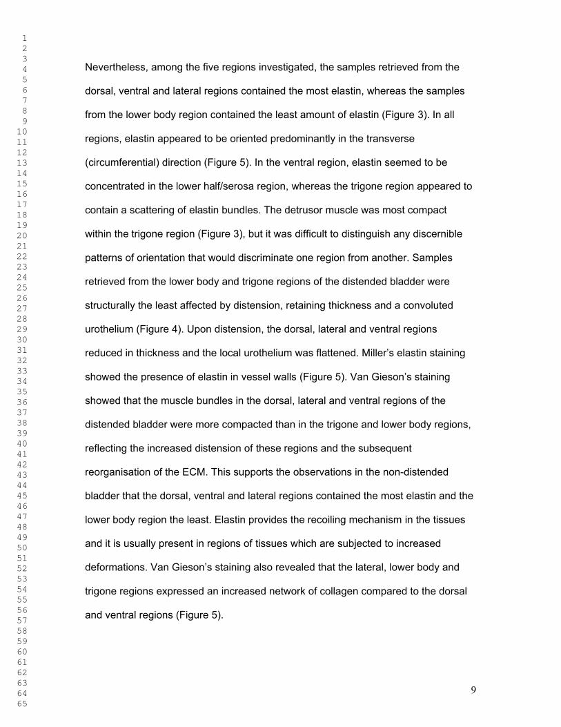
Results
Histological characterisation
The results of the structural analysis of the bladder wall, obtained from the
histological staining of samples from the dorsal, ventral, lateral lower body, and
trigone regions, as well as along the apex-to-base and transverse directions, are
illustrated in Figure 3 for the non-distended bladders, and Figure 4 and Figure 5 for
the bladder fixed while distended to 500 ml. Examination of the regional bladder
histioarchitecture revealed that elastin was generally sparse in the bladder wall.
1 2 3 4 5 6 7 8 9 10 11 12 13 14 15 16 17 18 19 20 21 22 23 24 25 26 27 28 29 30 31 32 33 34 35 36 37 38 39 40 41 42 43 44 45 46 47 48 49 50 51 52 53 54 55 56 57 58 59 60 61 62 63 64 65
9
Nevertheless, among the five regions investigated, the samples retrieved from the
dorsal, ventral and lateral regions contained the most elastin, whereas the samples
from the lower body region contained the least amount of elastin (Figure 3). In all
regions, elastin appeared to be oriented predominantly in the transverse
(circumferential) direction (Figure 5). In the ventral region, elastin seemed to be
concentrated in the lower half/serosa region, whereas the trigone region appeared to
contain a scattering of elastin bundles. The detrusor muscle was most compact
within the trigone region (Figure 3), but it was difficult to distinguish any discernible
patterns of orientation that would discriminate one region from another. Samples
retrieved from the lower body and trigone regions of the distended bladder were
structurally the least affected by distension, retaining thickness and a convoluted
urothelium (Figure 4). Upon distension, the dorsal, lateral and ventral regions
reduced in thickness and the local urothelium was flattened. Miller’s elastin staining
showed the presence of elastin in vessel walls (Figure 5). Van Gieson’s staining
showed that the muscle bundles in the dorsal, lateral and ventral regions of the
distended bladder were more compacted than in the trigone and lower body regions,
reflecting the increased distension of these regions and the subsequent
reorganisation of the ECM. This supports the observations in the non-distended
bladder that the dorsal, ventral and lateral regions contained the most elastin and the
lower body region the least. Elastin provides the recoiling mechanism in the tissues
and it is usually present in regions of tissues which are subjected to increased
deformations. Van Gieson’s staining also revealed that the lateral, lower body and
trigone regions expressed an increased network of collagen compared to the dorsal
and ventral regions (Figure 5).
1 2 3 4 5 6 7 8 9 10 11 12 13 14 15 16 17 18 19 20 21 22 23 24 25 26 27 28 29 30 31 32 33 34 35 36 37 38 39 40 41 42 43 44 45 46 47 48 49 50 51 52 53 54 55 56 57 58 59 60 61 62 63 64 65
10
Biomechanical characterisation
During uniaxial tensile loading to failure, the site of specimen failure was within
the central region of the specimens, whereas there was no evidence of specimen
slippage within the grips of the holder. The acquired force and elongation data for
each specimen tested was converted to stress and strain, respectively, and the
averaged apex-to-base and transverse stress-strain behaviours for each of the five
regional groups were plotted on the same chart in order to examine the potential
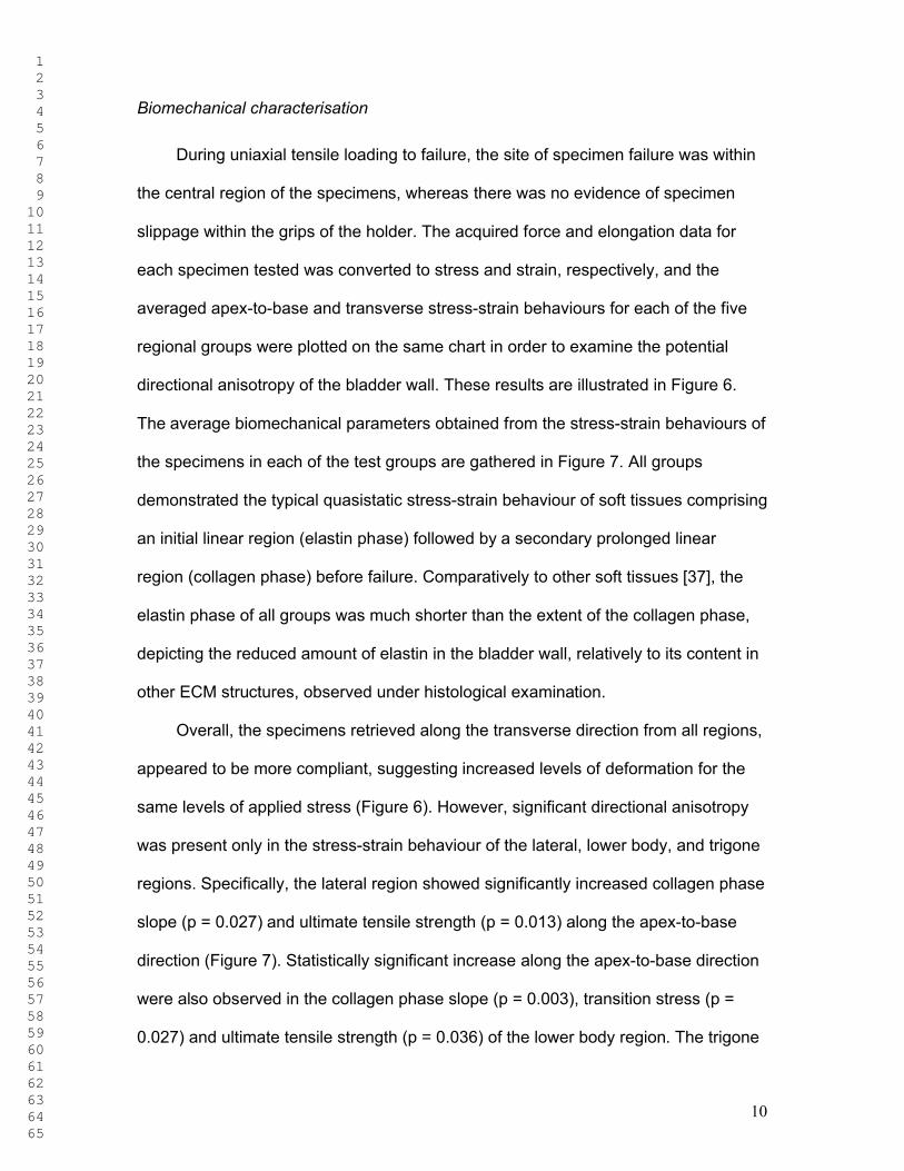
directional anisotropy of the bladder wall. These results are illustrated in Figure 6.
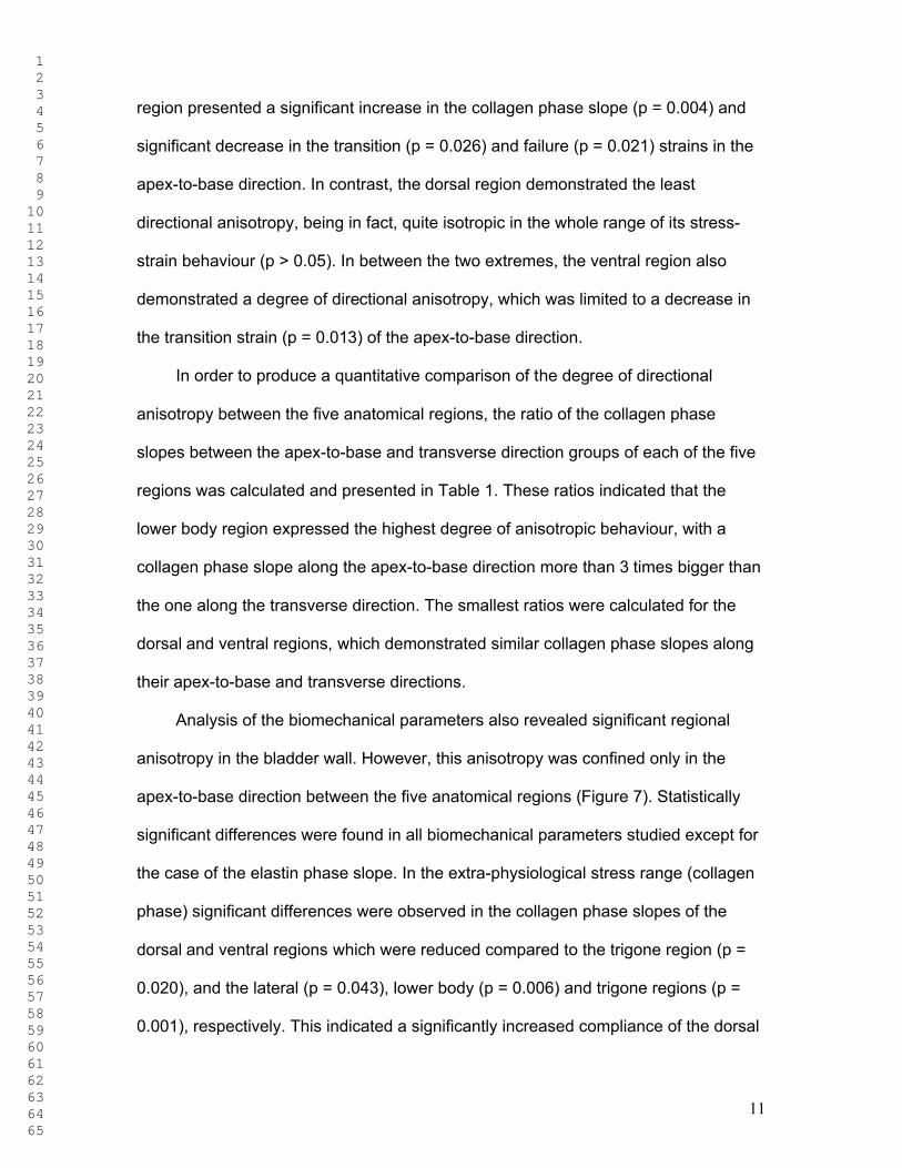
The average biomechanical parameters obtained from the stress-strain behaviours of
the specimens in each of the test groups are gathered in Figure 7. All groups
demonstrated the typical quasistatic stress-strain behaviour of soft tissues comprising
an initial linear region (elastin phase) followed by a secondary prolonged linear
region (collagen phase) before failure. Comparatively to other soft tissues [37], the
elastin phase of all groups was much shorter than the extent of the collagen phase,
depicting the reduced amount of elastin in the bladder wall, relatively to its content in
other ECM structures, observed under histological examination.
Overall, the specimens retrieved along the transverse direction from all regions,
appeared to be more compliant, suggesting increased levels of deformation for the
same levels of applied stress (Figure 6). However, significant directional anisotropy
was present only in the stress-strain behaviour of the lateral, lower body, and trigone
regions. Specifically, the lateral region showed significantly increased collagen phase
slope (p = 0.027) and ultimate tensile strength (p = 0.013) along the apex-to-base
direction (Figure 7). Statistically significant increase along the apex-to-base direction
were also observed in the collagen phase slope (p = 0.003), transition stress (p =
0.027) and ultimate tensile strength (p = 0.036) of the lower body region. The trigone
1 2 3 4 5 6 7 8 9 10 11 12 13 14 15 16 17 18 19 20 21 22 23 24 25 26 27 28 29 30 31 32 33 34 35 36 37 38 39 40 41 42 43 44 45 46 47 48 49 50 51 52 53 54 55 56 57 58 59 60 61 62 63 64 65
11
region presented a significant increase in the collagen phase slope (p = 0.004) and
significant decrease in the transition (p = 0.026) and failure (p = 0.021) strains in the
apex-to-base direction. In contrast, the dorsal region demonstrated the least
directional anisotropy, being in fact, quite isotropic in the whole range of its stress-
strain behaviour (p > 0.05). In between the two extremes, the ventral region also
demonstrated a degree of directional anisotropy, which was limited to a decrease in
the transition strain (p = 0.013) of the apex-to-base direction.
In order to produce a quantitative comparison of the degree of directional
anisotropy between the five anatomical regions, the ratio of the collagen phase
slopes between the apex-to-base and transverse direction groups of each of the five
regions was calculated and presented in Table 1. These ratios indicated that the
lower body region expressed the highest degree of anisotropic behaviour, with a
collagen phase slope along the apex-to-base direction more than 3 times bigger than
the one along the transverse direction. The smallest ratios were calculated for the
dorsal and ventral regions, which demonstrated similar collagen phase slopes along
their apex-to-base and transverse directions.
Analysis of the biomechanical parameters also revealed significant regional
anisotropy in the bladder wall. However, this anisotropy was confined only in the
apex-to-base direction between the five anatomical regions (Figure 7). Statistically
significant differences were found in all biomechanical parameters studied except for
the case of the elastin phase slope. In the extra-physiological stress range (collagen
phase) significant differences were observed in the collagen phase slopes of the
dorsal and ventral regions which were reduced compared to the trigone region (p =
0.020), and the lateral (p = 0.043), lower body (p = 0.006) and trigone regions (p =
0.001), respectively. This indicated a significantly increased compliance of the dorsal
1 2 3 4 5 6 7 8 9 10 11 12 13 14 15 16 17 18 19 20 21 22 23 24 25 26 27 28 29 30 31 32 33 34 35 36 37 38 39 40 41 42 43 44 45 46 47 48 49 50 51 52 53 54 55 56 57 58 59 60 61 62 63 64 65
12
and ventral compared to the other bladder regions. Moreover, the ultimate tensile
strength of the ventral region was significantly reduced compared to the lateral (p =
0.028) and lower body (p = 0.046) regions, whereas the transition stress of the lower
body was significantly increased compared to the dorsal region (p = 0.483). With
regards to the extensibility of the bladder wall, the trigone region was the least
distensible, demonstrating significantly reduced transition and failure strains
compared to the dorsal (p = 0.005 & 0.004), ventral (p = 0.017 & 0.012), lateral (p =
0.001 & 0.002), and lower body (p = 0.001 & 0.004) regions. The combined findings
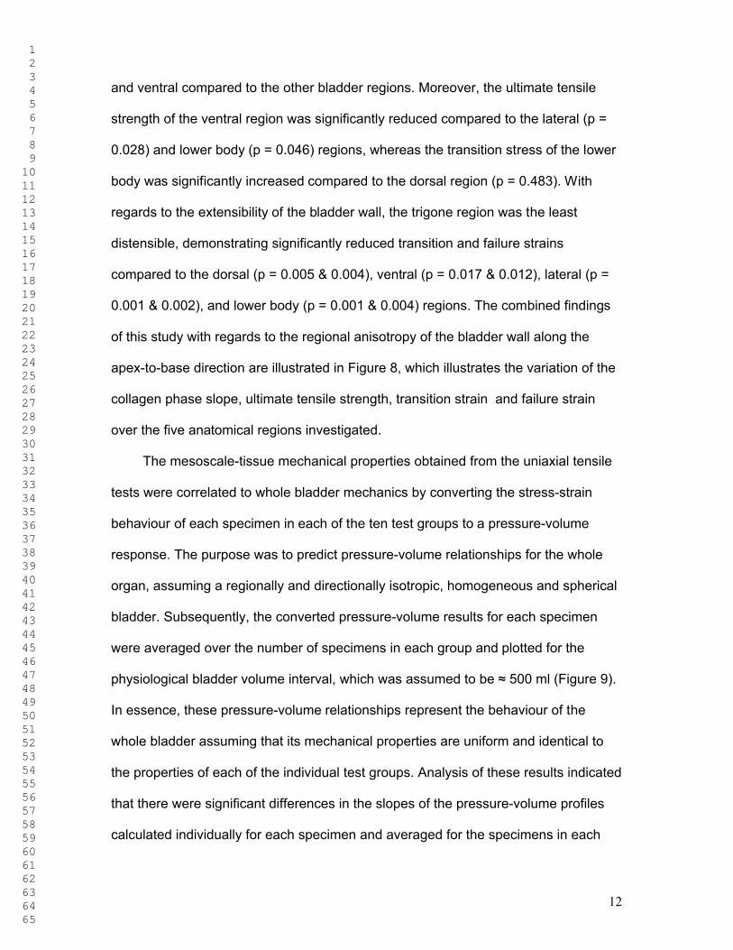
of this study with regards to the regional anisotropy of the bladder wall along the
apex-to-base direction are illustrated in Figure 8, which illustrates the variation of the
collagen phase slope, ultimate tensile strength, transition strain and failure strain
over the five anatomical regions investigated.
The mesoscale-tissue mechanical properties obtained from the uniaxial tensile
tests were correlated to whole bladder mechanics by converting the stress-strain
behaviour of each specimen in each of the ten test groups to a pressure-volume
response. The purpose was to predict pressure-volume relationships for the whole
organ, assuming a regionally and directionally isotropic, homogeneous and spherical
bladder. Subsequently, the converted pressure-volume results for each specimen
were averaged over the number of specimens in each group and plotted for the
physiological bladder volume interval, which was assumed to be ≈ 500 ml (Figure 9).
In essence, these pressure-volume relationships represent the behaviour of the
whole bladder assuming that its mechanical properties are uniform and identical to
the properties of each of the individual test groups. Analysis of these results indicated
that there were significant differences in the slopes of the pressure-volume profiles
calculated individually for each specimen and averaged for the specimens in each
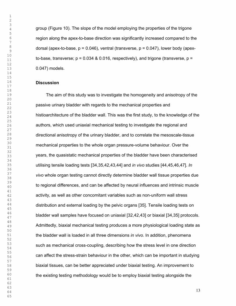
1 2 3 4 5 6 7 8 9 10 11 12 13 14 15 16 17 18 19 20 21 22 23 24 25 26 27 28 29 30 31 32 33 34 35 36 37 38 39 40 41 42 43 44 45 46 47 48 49 50 51 52 53 54 55 56 57 58 59 60 61 62 63 64 65
13
group (Figure 10). The slope of the model employing the properties of the trigone
region along the apex-to-base direction was significantly increased compared to the
dorsal (apex-to-base, p = 0.046), ventral (transverse, p = 0.047), lower body (apex-
to-base, transverse; p = 0.034 & 0.016, respectively), and trigone (transverse, p =
0.047) models.
Discussion
The aim of this study was to investigate the homogeneity and anisotropy of the
passive urinary bladder with regards to the mechanical properties and
histioarchitecture of the bladder wall. This was the first study, to the knowledge of the
authors, which used uniaxial mechanical testing to investigate the regional and
directional anisotropy of the urinary bladder, and to correlate the mesoscale-tissue
mechanical properties to the whole organ pressure-volume behaviour. Over the
years, the quasistatic mechanical properties of the bladder have been characterised
utilising tensile loading tests [34,35,42,43,44] and in vivo studies [44,45,46,47]. In
vivo whole organ testing cannot directly determine bladder wall tissue properties due
to regional differences, and can be affected by neural influences and intrinsic muscle
activity, as well as other concomitant variables such as non-uniform wall stress
distribution and external loading by the pelvic organs [35]. Tensile loading tests on
bladder wall samples have focused on uniaxial [32,42,43] or biaxial [34,35] protocols.
Admittedly, biaxial mechanical testing produces a more physiological loading state as
the bladder wall is loaded in all three dimensions in vivo. In addition, phenomena
such as mechanical cross-coupling, describing how the stress level in one direction
can affect the stress-strain behaviour in the other, which can be important in studying
biaxial tissues, can be better appreciated under biaxial testing. An improvement to
the existing testing methodology would be to employ biaxial testing alongside the
1 2 3 4 5 6 7 8 9 10 11 12 13 14 15 16 17 18 19 20 21 22 23 24 25 26 27 28 29 30 31 32 33 34 35 36 37 38 39 40 41 42 43 44 45 46 47 48 49 50 51 52 53 54 55 56 57 58 59 60 61 62 63 64 65
14
uniaxial protocol. Nevertheless, uniaxial testing is an attractive investigation tool
because it localises the investigation to a very small area of the organ from which a
tissue sample can be isolated and subjected to controlled stress states. This is a
particularly well suited approach when investigating anisotropic behaviour of tissues.
Since the purpose of this study was not to fully characterise the mechanical
properties of the bladder in terms of a constitutive three-dimensional model, in which
case a biaxial testing protocol would be more appropriate, but to investigate its
potential anisotropy and inhomogeneity, it was deemed appropriate to use uniaxial
tensile testing.
The regional and directional anisotropy of the bladder has attracted surprisingly
little attention over the years. A meagre few studies have focused on the anisotropy
of the mechanical properties of the bladder [34,35], and even these have
concentrated on the directional anisotropy. In addition to the directional anisotropy,
this study also identified a regional anisotropy inherent in the mechanical properties
of the bladder wall. Moreover, the magnitudes of the biomechanical parameters
calculated in this study were comparable to those reported by others for porcine
bladder tissue [42], considering the differences in experimental protocols, as well as
in the methods used to estimate tissue thickness which have a direct impact on the
magnitude of the estimated stress. With regards to the directional anisotropy, the
specimens retrieved along the transverse direction from all regions appeared to be
more compliant (increased transition and failure strains, reduced collagen phase
slopes) compared to the apex-to-base specimens. The increased compliance along
the transverse direction, which was more profound in the extra-physiological
mechanical properties, indicated that at the organ level the bladder distends more in
this direction than along the apex-to-base one. Within the physiological distension
1 2 3 4 5 6 7 8 9 10 11 12 13 14 15 16 17 18 19 20 21 22 23 24 25 26 27 28 29 30 31 32 33 34 35 36 37 38 39 40 41 42 43 44 45 46 47 48 49 50 51 52 53 54 55 56 57 58 59 60 61 62 63 64 65
15
limits (up to approximately the transition point of the stress-strain curve), the
increased compliance observed along the transverse direction was supported by the
histological results, which indicated that elastin was predominantly oriented in the
transverse direction (Figure 5). Elaborating, elastin provides the recoiling mechanism
in the tissues and it is most abundant in tissues, or regions of tissues, subject to
increased stretching during physiological function [48]. Directional anisotropy was
also observed in the ultimate tensile strength of the specimens, with the specimens
retrieved along the transverse direction from all regions achieving lower strengths
than the apex-to-base specimens. The difference, though, was significant only in the
lateral and lower body regions. Overall, the lower body demonstrated the highest
degree of directional anisotropy, whereas the dorsal and ventral region demonstrated
the least directional anisotropy (Figure 6 & Table1).
Significant regional anisotropy in the bladder wall was found only along the
apex-to-base direction (Figure 7 & 8). The lack of any significant regional anisotropy
along the transverse direction indicates that the organ experiences a rather uniform
circumferential expansion. Statistically significant differences were found in all
biomechanical parameters except in the slope of the elastin phase. The dorsal and
ventral regions demonstrated a significantly increased compliance along the
longitudinal direction compared to the other bladder regions, as indicated by the
reduced collagen phase slope and transition stress, and increased transition and
failure strain of these regions. The reduced transition stress of these regions
indicates that they can reach their transition point, at which the collagen and smooth
muscle fibres have uncrimped and begin to bear all the applied load, with less effort
(less pressure) than the other regions. As a complementary effect, the significantly
increased transition strain of the dorsal and ventral regions, as well as of the lateral
1 2 3 4 5 6 7 8 9 10 11 12 13 14 15 16 17 18 19 20 21 22 23 24 25 26 27 28 29 30 31 32 33 34 35 36 37 38 39 40 41 42 43 44 45 46 47 48 49 50 51 52 53 54 55 56 57 58 59 60 61 62 63 64 65
16
region, compared to the trigone, indicates that with the same effort (same pressure)
these regions are prone to deform more than the trigone in the apex-to-base
direction. In fact, the trigone region demonstrated the least distensibility, experiencing
the lowest transition and failure strains and the highest collagen phase slope in both
directions (although not significantly so in the transverse) compared to the other
regions (Figure 7 & 8). The second highest collagen phase slope and lowest failure
strain was demonstrated by the lower body region. The findings of the increased
compliance of the dorsal, ventral and lateral regions compared to the trigone and
lower body regions were supported by the increased elastin network found in these
regions, as well as by the fact that histological samples retrieved from the lower body
and trigone regions of the distended bladder were structurally the least affected by
distension. The trigone, lower body and lateral regions also demonstrated the highest
tensile strength both at regional and directional level. This can be attributed to the
increased networks of collagen, the main function of which in connective tissues is to
withstand tension, as well as to the thicker layers of muscle, observed in these
regions under histological examination.
The directional and regional anisotropy in the mesoscale-tissue mechanical
properties of the bladder was inherited in the whole organ mechanics when the
stress-strain behaviours of the different regions were used to model pressure-volume
relationships for the whole organ. The purpose was to investigate whether
mesoscale-tissue mechanical properties can be translated to meaningful whole organ
mechanics, given an appropriate model for the bladder shape and how the wall
stretch is distributed in the bladder wall. The assumptions of a spherical geometry,
homogeneity and anisotropy do not constitute a realistic bladder model.
Nevertheless, this model was sufficient to examine how the whole pressure-volume
1 2 3 4 5 6 7 8 9 10 11 12 13 14 15 16 17 18 19 20 21 22 23 24 25 26 27 28 29 30 31 32 33 34 35 36 37 38 39 40 41 42 43 44 45 46 47 48 49 50 51 52 53 54 55 56 57 58 59 60 61 62 63 64 65
17
relationship of bladder changes if the mechanical properties of a particular bladder
region are adopted as universal bladder properties. Although these results were at
best estimates based on assumptions of homogeneity, and only descriptive of whole
bladder mechanics, they were indicative of the inherent regional and directional
anisotropy present in the bladder. The modelled pressure-volume profiles were in
general agreement with similar data obtained from bladder cystometry [49]. However,
there was a considerable scatter among the results of the individual regions and
directions. The scatter ranged from a model describing a bladder that offers
considerable resistance to deformation, by employing the results of the trigone region
along the apex-to-base direction, to a bladder that is quite compliant and offers little
resistance to deformation, by employing the results of the ventral region along the
transverse direction. Moreover, the pressure-volume models verified the lack of any
significant anisotropy along the transverse direction of the anatomical regions, with
the models assuming the properties of the transverse regional groups clustering
together, towards the compliant bladder region.
Conclusions
This study detected significant regional and directional anisotropy in the
quasistatic uniaxial mechanical properties of the passive urinary bladder and
correlated this anisotropy to the distended and non-distended tissue
histioarchitecture and whole organ mechanics. The experimental protocol used to
evaluate the mesoscale mechanical properties of the bladder by employing uniaxial
tensile testing was effective in detecting bladder anisotropy. Differences between
isotropic and anisotropic behaviour can become important in regions of high stress
and in bladder augmentation surgery that changes the natural shape and boundary
conditions of the bladder. In general, the results from this study will aid the regional
1 2 3 4 5 6 7 8 9 10 11 12 13 14 15 16 17 18 19 20 21 22 23 24 25 26 27 28 29 30 31 32 33 34 35 36 37 38 39 40 41 42 43 44 45 46 47 48 49 50 51 52 53 54 55 56 57 58 59 60 61 62 63 64 65
18
differentiation of bladder treatments in terms of partial bladder replacement, as well
as the development of more realistic constitutive models of bladder wall
biomechanics and improved computational simulations to predict deformations in the
natural and augmented bladder.
Acknowledgements
This work was funded by the Biotechnology and Biological Sciences Research
Council (BBSRC Grant E20352). SK is funded by the Engineering and Physical
Sciences Research Council.
1 2 3 4 5 6 7 8 9 10 11 12 13 14 15 16 17 18 19 20 21 22 23 24 25 26 27 28 29 30 31 32 33 34 35 36 37 38 39 40 41 42 43 44 45 46 47 48 49 50 51 52 53 54 55 56 57 58 59 60 61 62 63 64 65
1
References
1. Chang SL, Chung JS, Yeung MK, Howard PS, Macarak EJ. Roles of the lamina
propria and the detrusor in tension transfer during bladder filling. Scand J Urol
Nephrol Suppl 1999; 201:38-45.
2. Elbadawi A, Meyer S, Regnier CH. Role of ischemia in structural changes in the
rabbit detrusor following partial bladder outlet obstruction: a working hypothesis and
a biochemical/structural model proposal. Neurourol Urodynam 1989; 8: 151-162.
3. Gabella G. Hypertrophy of visceral smooth muscle. Anat Embryol 1990;
182:409-424.
4. Kitada S, Wein AJ, Kato K, Levin RM. Effect of acute complete obstruction on
the rabbit urinary bladder. J Urol 1989; 141: 166-169.
5. Tammela TLJ, Levin RM, Monson FC, Wein AJ, Longhurst PA. The influence of
acute over-distension on rat bladder function and DNA synthesis. J Urol 1993;
150:15331539.
6. German K, Bedwani J, Davies J, Brading AF, Stephenson TP. An assessment
of the contribution of visco-elastic factors in the aetiology of poor compliance in the
human neuropathic bladder. Br J Urol 1994; 74(6):744-748.
7. Watanabe T, Rivas DA, Chancellor MB. Urodynamics of spinal cord injury. Urol
Clin North Am 1996; 23(3):459-473.
8. Kruse MN, Bray LA, de Groat WC. Influence of spinal cord injury on the
morphology of bladder afferent and efferent neurons. J Auton Nerv Syst 1995;
54(3):215-224.
* References
1 2 3 4 5 6 7 8 9 10 11 12 13 14 15 16 17 18 19 20 21 22 23 24 25 26 27 28 29 30 31 32 33 34 35 36 37 38 39 40 41 42 43 44 45 46 47 48 49 50 51 52 53 54 55 56 57 58 59 60 61 62 63 64 65
2
9. Bunyaratavey P, Lao S, Kongkanand A, Brasopsanti K, Vajarapongse R. Ten
years’ experience with enterocystoplasty. J Med Assoc Thai 1993; 76: 327–333.
10. Khoury JM, Timmons SL, Corbel L, Webster GD. Complications of
enterocystoplasty. Urology 1992; 40: 9–14.
11. Greenwell TJ, Venn SN, Mundy AR. Augmentation cystoplasty. BJU Int. 2001;
88(6):511-25.
12. Chancellor MB, Rivas DA, Bourgeois IM. Laplace's law and the risks and
prevention of bladder rupture after enterocystoplasty and bladder autoaugmentation.
Neurourol Urodyn 1996;15(3):223-233.
13. Bolland F, Korossis S, Wilshaw SP, Ingham E, Fisher J, Kearney JN et al.
Development and characterisation of a full-thickness acellular porcine bladder matrix
for tissue engineering. Biomaterials 2007 Feb; 28(6):1061-1070.
14. Southgate J, Cross W, Eardley I, Thomas DF, Trejdosiewicz LK. Bladder
reconstruction-from cells to materials. Proc Inst Mech Eng [H]. 2003; 217(4):311-316.
15. Korossis S, Bolland F, Ingham E, Fisher J, Kearney J, Southgate J. Review:
tissue engineering of the urinary bladder: considering structure-function relationships
and the role of mechanotransduction. Tissue Eng 2006; 12(4):635-644.
16. Roelofs M, Wein AJ, Monson FC, Passeriniglazel G, Koteliansky VE, Sartore S
et al. Contractility and phenotype transitions in serosal thickening of obstructed rabbit
bladder. J Appl Physiol 1995; 78: 1432–41.
17. Wang Z, Gopalakurup SK, Levin RM, Chacko S. Expression of smooth muscle
myosin isoforms in urinary bladder smooth muscle during hypertrophy and
regression. Lab Invest 1995; 73: 244–51.
1 2 3 4 5 6 7 8 9 10 11 12 13 14 15 16 17 18 19 20 21 22 23 24 25 26 27 28 29 30 31 32 33 34 35 36 37 38 39 40 41 42 43 44 45 46 47 48 49 50 51 52 53 54 55 56 57 58 59 60 61 62 63 64 65
3
18. Baskin LS, Constantinescu S, Duckett JW, Snyder HM, Macarak E. Type III
collagen decreases in normal foetal bovine bladder development. J Urol 1994; 152:
688–91.
19. Landau EH, Jayanthi VR, Churchill BM, Shapiro E, Gilmour RF, Khoury AE et
al. Loss of elasticity in dysfunctional bladders: urodynamic and histochemical
correlation. J Urol 1994; 152: 702–5.
20. Venegas JG. Viscoelastic properties of the contracting detrusor: I. theoretical
basis. Am J Physiol 1991; 261:C355–363.
21. Finkbeiner AE, O’Donnell PD. Responses of detrusor smooth muscle to stretch
and relaxation: in vitro study. Urology 1990; 36: 193–198.
22. Regnier CH, Kolsky H, Richardson PD, Ghoniem GM, Susset JG. The elastic
behavior of the urinary bladder for large deformations. J Biomech 1983; 16: 915–922.
23. Coolsaet BL, van Mastrigt R, Van Duyl WA, Huygen RE. Viscoelastic properties
of bladder wall strips at constant elongation. Invest Urol 1976; 13: 435–440.
24. Coolsaet BL, van Duyl WA, van Mastrigt R, van der Zwart A. Visco-elastic
properties of the bladder wall. Urol Int 1975; 30: 16–26.
25. Alexander RS. Viscoplasticity of smooth muscle of urinary bladder. Am J
Physiol 1973; 224: 618–622.
26. Kondo A, Susset JG. Physical properties of the urinary detrusor muscle. A
mechanical model based upon the analysis of stress relaxation curve. J Biomech
1973; 6: 141–151.
1 2 3 4 5 6 7 8 9 10 11 12 13 14 15 16 17 18 19 20 21 22 23 24 25 26 27 28 29 30 31 32 33 34 35 36 37 38 39 40 41 42 43 44 45 46 47 48 49 50 51 52 53 54 55 56 57 58 59 60 61 62 63 64 65
4
27. Klevmark B. Motility of the urinary bladder in cats during filling at physiological
rates. I. Intravesical pressure patterns studied by a new method of cystometry. Acta
Physiol Scand 1974; 90: 565–577.
28. Klevmark B. Motility of the urinary bladder in cats during filling at physiological
rates. II. Effects of extrinsic bladder denervation on intramural tension and on
intravesical pressure patterns. Acta Physiol Scand 1977; 101: 176–184.
29. Tözeren A. Assessment of fibber strength in a urinary bladder by using
experimental pressure volume curves: an analytical method. J Biomech Eng 1986;
108:301-305.
30. Van Mastrigt R, Griffiths DJ. An evaluation of contractility parameters
determined from isometric contractions and micturition studies. Ural Res 1986; 14:45-
52.
31. Damaser MS, Lehman SL. The effect of urinary bladder shape on its mechanics
during filling. J Biomechanics 1995; 28(6):725-732.
32. Andersson KE, Arner A. Urinary bladder contraction and relaxation: physiology
and pathophysiology. Physiol Re 2004; 84:935-986.
33. Gabella G, Uvelius B. Urinary bladder of rat: fine structure of normal and
hypertrophic musculature. Cell Tissue Res 1990; 262:67–79.
34. Gloeckner D, Sacks M, Chancellor M, de Groat W. Active and passive biaxial
mechanical properties of urinary bladder wall. In: Engineering in Medicine and
Biology. 21st Annual Conf. and the 1999 Annual Fall Meeting of the Biomedical
Engineering Society BMES/EMBS Conference. 1999; 1:17.
1 2 3 4 5 6 7 8 9 10 11 12 13 14 15 16 17 18 19 20 21 22 23 24 25 26 27 28 29 30 31 32 33 34 35 36 37 38 39 40 41 42 43 44 45 46 47 48 49 50 51 52 53 54 55 56 57 58 59 60 61 62 63 64 65
5
35. Gloeckner DC, Sacks MS, Fraser MO, Somogyi GT, de Groat WC, Chancellor
MB. Passive biaxial mechanical properties of the rat bladder wall after spinal cord
injury. J Urol. 2002; 167(5):2247-2252.
36. Southgate J, Masters JR, Trejdosiewicz LK. Culture of human urothelium. In:
Freshney RI FM, editor. Culture of epithelial cells. 2nd ed. New York: Wiley. 2002;
381–400.
37. Korossis S, Booth C, Wilcox HE, Watterson KG, Kearney JN, Ingham E et al.
Tissue engineering of cardiac valve prostheses II: Biomechanical characterisation of
decellularised porcine aortic heart valves. J Heart Valve Dis. 2002; 11(4):463-471.
38. Bancroft J, Stevens A. Theory and practice of histological techniques. London:
Churchill Livingstone. 1990.
39. Korossis S, Wilcox HE, Watterson KG, Kearney JN, Ingham E, Fisher J. In-vitro
assessment of the functional performance of the decellularised intact porcine aortic
root. J Heart Valve Dis. 2005; 14(3):408-422.
40. Griffiths CJ, Assi MS, Styles RA, Ramsden PD, Neal DE. Ambulatory
monitoring of bladder and detrusor pressure during natural filling. J Urol 1989;
142:780–784.
41. Fung YCB. Biomechanics, mechanical properties of living tissues. 2nd ed,
Springer-Verlag, New York 1993; 14-20.
42. Dahms E, Piechota HJ, Dahiya R Lue TF, Tanagho EA. Composition and
biomechanical properties of the bladder acellular matrix graft: comparative analysis in
rat, pig and human. Br J Urol 1998; 82:411-419.
43. Alexander RS. Series elasticity of urinary bladder smooth muscle. Am J Physiol
1976; 231:1337.
1 2 3 4 5 6 7 8 9 10 11 12 13 14 15 16 17 18 19 20 21 22 23 24 25 26 27 28 29 30 31 32 33 34 35 36 37 38 39 40 41 42 43 44 45 46 47 48 49 50 51 52 53 54 55 56 57 58 59 60 61 62 63 64 65
6
44. Alexander RS. Mechanical properties of urinary bladder. Am J Physiol 1971;
220:1413.
45. Rohrmann D, Zderic SA, Duckett JW, Levin RM, Damaser MS. Compliance of
the obstructed foetal rabbit bladder. Neurourol Urodyn 1997; 16(3):179-189.
46. Coplen DE, Macarak EJ, Levin RM. Developmental changes in normal foetal
bovine whole bladder physiology. J Urol 1994; 151:1391.
47. Levin RM, Horan P, Liu SP. Metabolic aspects of urinary bladder filling. Scand J
Urol Nephrol suppl. 1999; 201:59.
48. Viidik A, Danielsen CC, Oxlund H. On fundamental and phenomenological
models, structure and mechanical properties of collagen and elastin and
glycosaminoglycan complexes. Biorheology 1982; 19: 437–451.
49. Damaser MS. Whole bladder mechanics during filling. Scand J Urol Nephrol
Suppl 1999; 201:51–58
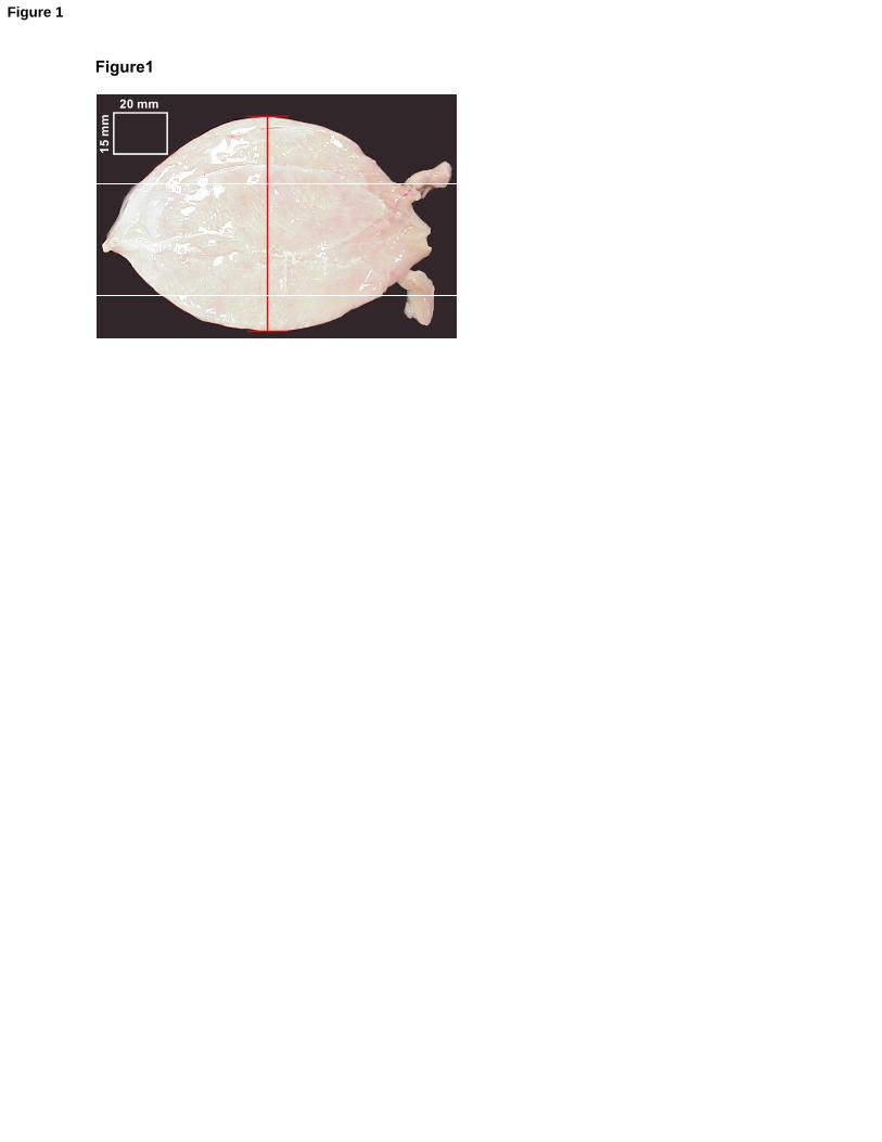
Figure1
15
mm
20 mm
Figure 1
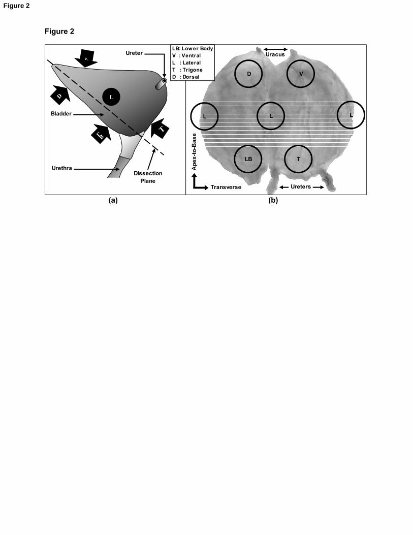
Figure 2
Uracus
UretersA
pe
x-t
o-B
as
eTransverse
Ureter
Bladder
UrethraDissection
Plane
V
D
LBT
D V
L
TLB
L L
LB: Lower BodyV : VentralL : LateralT : TrigoneD : Dorsal
L
(a) (b)
Figure 2
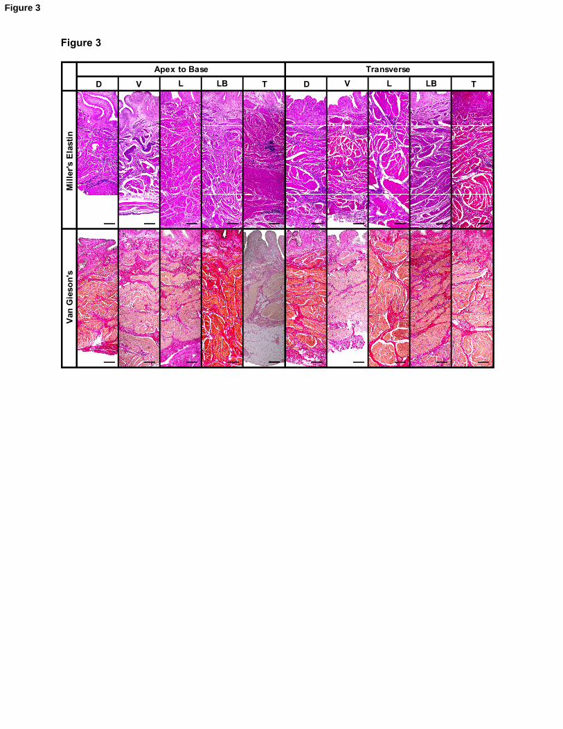
Figure 3
Apex to Base Transverse
D V L T D V L LB TLB
Mil
ler'
s E
last
inV
an G
ieso
n's
Figure 3
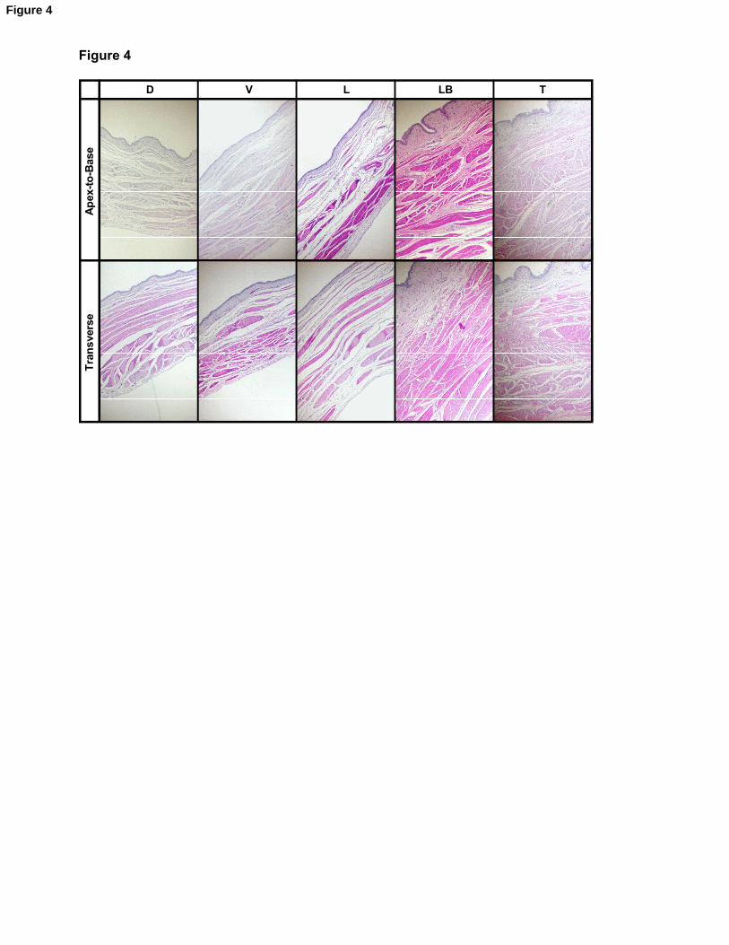
Figure 4
D V L TLBA
pex
-to
-Bas
eT
ran
sver
se
Figure 4
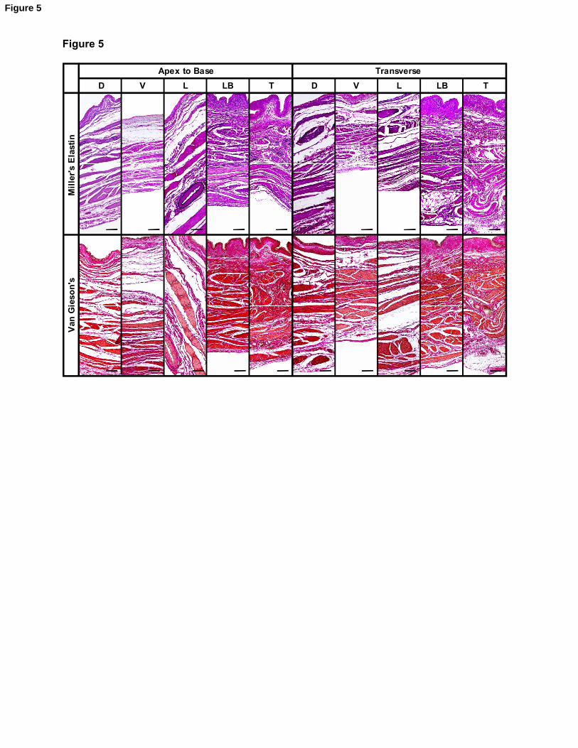
Figure 5
Apex to Base Transverse
D V L T D V L LB TLB
Mil
ler'
s E
last
inV
an G
ieso
n's
Figure 5
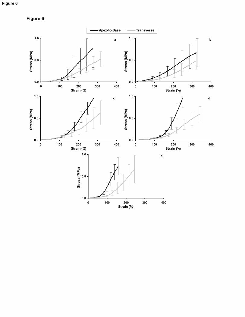
Figure 6
Apex-to-Base Transverse
0.0
0.8
1.6
0 100 200 300 400Strain (%)
Str
es
s (
MP
a)
a
0.0
0.8
1.6
0 100 200 300 400Strain (%)
Str
es
s (
MP
a)
b
0.0
0.8
1.6
0 100 200 300 400Strain (%)
Str
es
s (
MP
a)
d
0.0
0.8
1.6
0 100 200 300 400Strain (%)
Str
es
s (
MP
a)
c
0.0
0.8
1.6
0 100 200 300 400Strain (%)
Str
es
s (
MP
a)
e
Figure 6
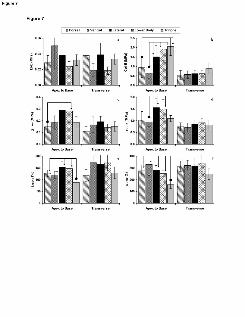
Figure 7
Dorsal Ventral Lateral Lower Body Trigone
0.0
0.5
1.0
1.5
2.0
2.5
Apex to Base Transverse
Co
l-E
(M
Pa
)
0.00
0.02
0.04
0.06
Apex to Base Transverse
El-
E (
MP
a)
a b
0.0
0.1
0.2
0.3
0.4
Apex to Base Transverse
str
ans
(M
Pa
)
0.0
0.5
1.0
1.5
2.0
Apex to Base Transverse
sU
TS
(MP
a)
dc
0
100
200
300
400
Apex to Base Transverse
eUT
S (
%)
0
50
100
150
200
Apex to Base Transverse
etra
ns
(%)
e f
Figure 7
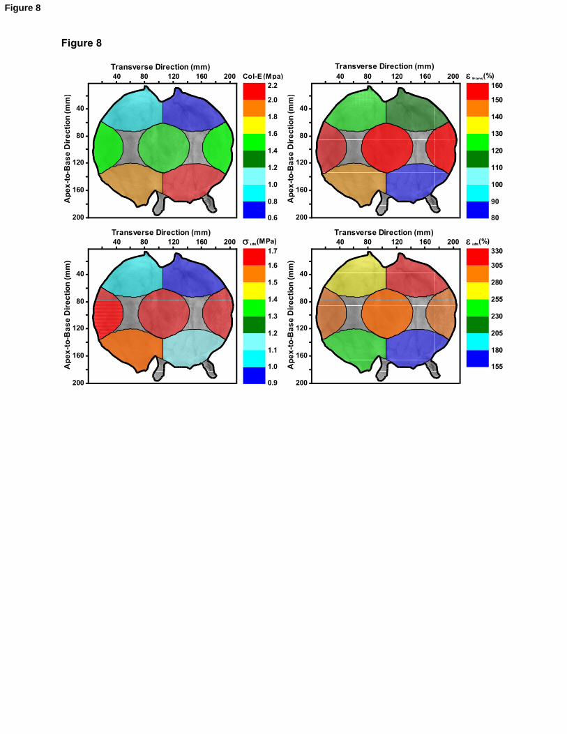
Figure 8
0.6
0.8
1.0
1.2
1.4
1.6
1.8
2.0
2.2Col-E (Mpa)
200
160
120
80
40
40 80 120 160 200Transverse Direction (mm)
Ap
ex
-to
-Ba
se
Dir
ec
tio
n (
mm
)
80
90
100
110
120
130
140
150
160e trans(%)
200
160
120
80
40
40 80 120 160 200Transverse Direction (mm)
Ap
ex
-to
-Ba
se
Dir
ec
tio
n (
mm
)
155
180
205
230
255
280
305
330e uts(%)
200
160
120
80
40
40 80 120 160 200Transverse Direction (mm)
Ap
ex
-to
-Ba
se
Dir
ec
tio
n (
mm
)
1.0
1.1
1.2
1.3
1.4
1.5
1.6
1.7s uts(MPa)
200
160
120
80
40
40 80 120 160 200Transverse Direction (mm)
Ap
ex
-to
-Ba
se
Dir
ec
tio
n (
mm
)
0.9
Figure 8
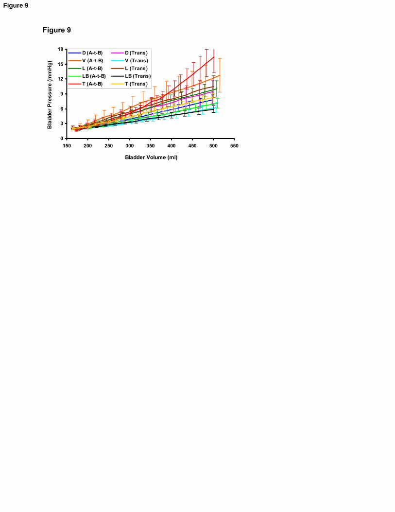
Figure 9
0
3
6
9
12
15
18
150 200 250 300 350 400 450 500 550
Bladder Volume (ml)
Bla
dd
er
Pre
ss
ure
(m
mH
g)
D (A-t-B) D (Trans)
V (A-t-B) V (Trans)
L (A-t-B) L (Trans)
LB (A-t-B) LB (Trans)
T (A-t-B) T (Trans)
Figure 9
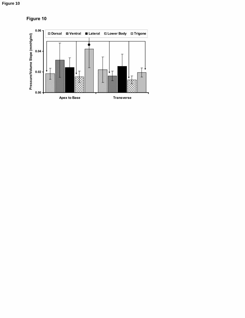
Figure 10
0.00
0.02
0.04
0.06
Apex to Base Transverse
Pre
ss
ure
/Vo
lum
e S
lop
e (
mm
Hg
/ml) Dorsal Ventral Lateral Lower Body Trigone
Figure 10
1
Figure Captions
Figure 1: Bladder sizing. Bladder width was measured along the transverse line.
Figure 2: Bladder dissection and sample localization. (a) Schematic of bladder in the anterior-posterior plane; (b)
Cut-opened porcine bladder showing the anatomical map of the five anatomical regions investigated
Figure 3: Staining of full thickness samples retrieved from the dorsal (D), ventral (V), lateral (L), lower body (LB)
and trigone (T) regions of non-distended bladder (luminal side up). Bar: 250 m.
Figure 4: H & E staining of full thickness samples retrieved from the dorsal (D), ventral (V), lateral (L), lower body
(LB) and trigone (T) regions of distended bladder (4 magnification).
Figure 5: Staining of full thickness samples retrieved from the dorsal (D), ventral (V), lateral (L), lower body (LB)
and trigone (T) regions of distended bladder. Bar: 250 m.
Figure 6: Regional mean stress-strain behaviour of the bladder wall along the apex-to-base and transverse
directions (error bars indicate the 95% confidence intervals, n = 6): a) dorsal; b) ventral; c) lateral; d) lower body;
e) trigone.
Figure 7: Regional mean biomechanical parameters of the bladder wall along the apex-to-base and transverse
directions (error bars indicate the 95% confidence intervals, n = 6): a) elastin phase slope (El-E); b) collagen
phase slope (Col-E); c) transition stress (trans); d) ultimate tensile strength (uts); e) transition strain (trans); f)
failure strain (uts). Connectors indicate significant (p<0.05) regional difference between originator column and end
arrow column.
Figure 8: Regional topographic map of the urinary bladder showing the variation of the mean collagen phase
slope (Col-E), ultimate tensile strength (uts), transition strain (trans), and failure strain (uts) over the five
anatomical regions investigated, and along the apex-to-base direction. These results correspond to the results
presented in Figure 6.
Figure 9: Mean pressure-volume profiles calculated from the stress-strain behaviour of the dorsal (D), ventral (V),
lateral (L), lower body (LB), and trigone (T) bladder regions along the apex-to-base and transverse directions
(mean ± 95% confidence interval, n = 6).
Figure 10: Average slopes of the pressure-volume profiles for the dorsal, ventral, lateral, lower body, and trigone
models (error bars indicate the 95% confidence intervals, n = 6). Connectors indicate significant difference.
Captions
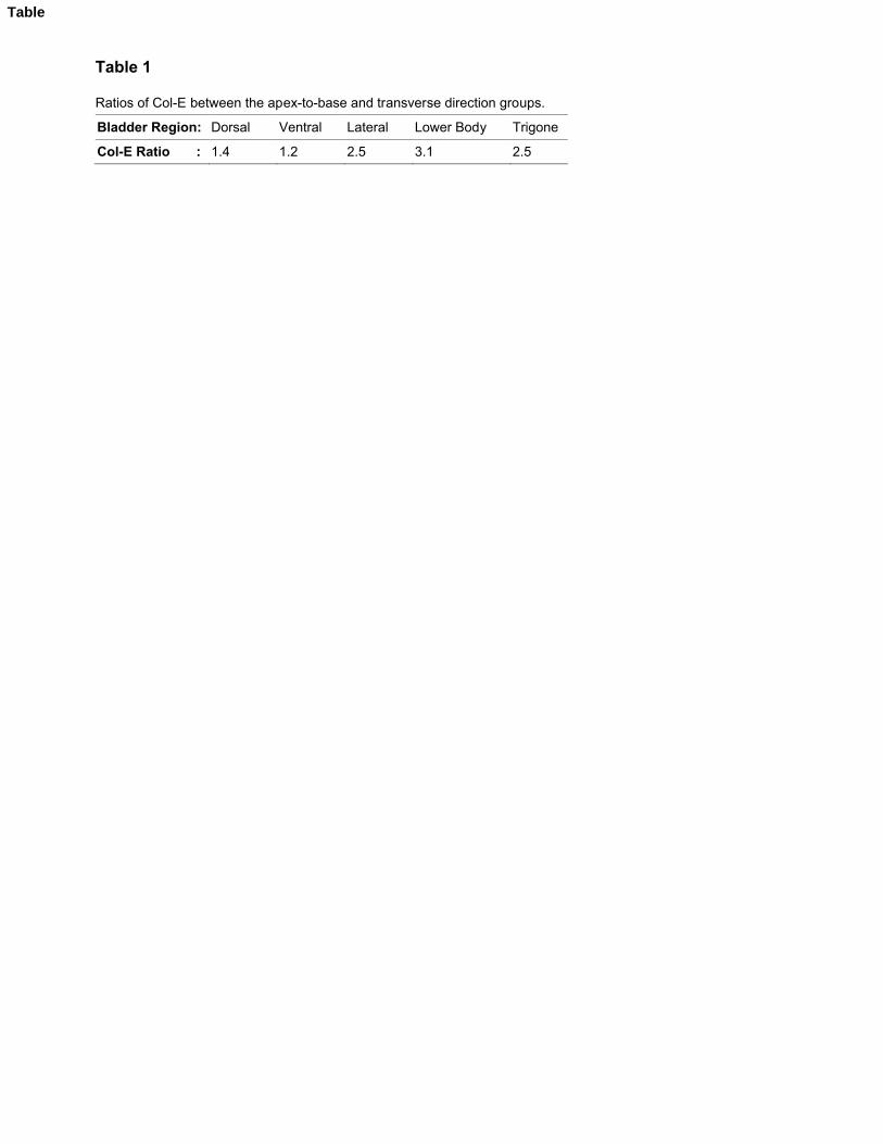
Table 1
Ratios of Col-E between the apex-to-base and transverse direction groups.
Bladder Region: Dorsal Ventral Lateral Lower Body Trigone
Col-E Ratio : 1.4 1.2 2.5 3.1 2.5
Table
Related Documents