References 1-Gray’s anatomy 2-Human nervous system Noback 3- Basic clinical neuroanatomy Young 4- Head and neck anatomy Berkovitz 5- clinical neuroanatomy.
Dec 21, 2015
Welcome message from author
This document is posted to help you gain knowledge. Please leave a comment to let me know what you think about it! Share it to your friends and learn new things together.
Transcript

References
1-Gray’s anatomy 2-Human nervous system Noback 3- Basic clinical neuroanatomy
Young 4- Head and neck anatomy
Berkovitz 5- clinical neuroanatomy Snell
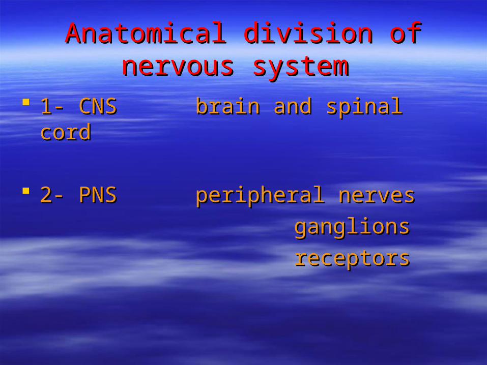
Anatomical division of nervous Anatomical division of nervous systemsystem
1- CNS brain and spinal cord1- CNS brain and spinal cord
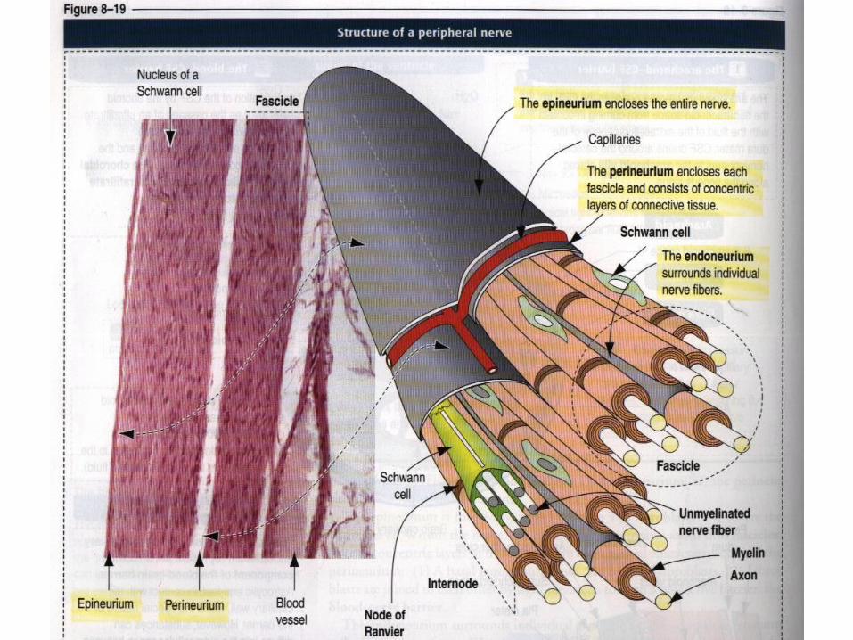
2- PNS peripheral nerves2- PNS peripheral nerves
ganglionsganglions
receptorsreceptors


Cells in this system
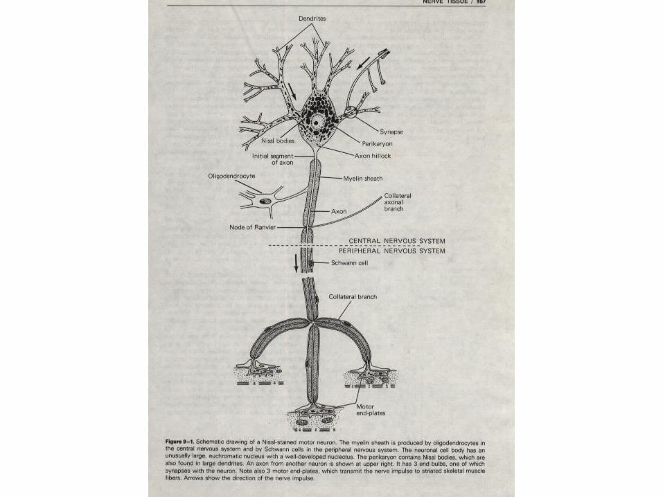
1- neuron
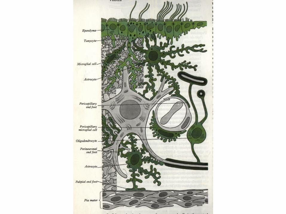
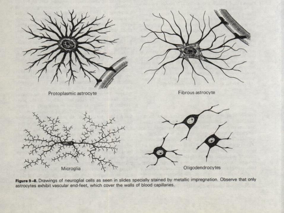
2-neuroglia A- Central neuroglia Macroglia (astrocyte,oligodendrocyte) Microglia Ependymal cells B- Peripheral neuroglia Schwann cells Satellite cells

Specialist cellSpecialist cell
1- produce action potential1- produce action potential
2- convey2- convey


central neurogliacentral neuroglia
Macroglia (astrocyte,oligodendrocyte)Macroglia (astrocyte,oligodendrocyte) MicrogliaMicroglia Ependymal cellsEpendymal cells

Peripheral neurogliaPeripheral neuroglia
1- schwann cell1- schwann cell 2- satellite cell2- satellite cell


Function of this system

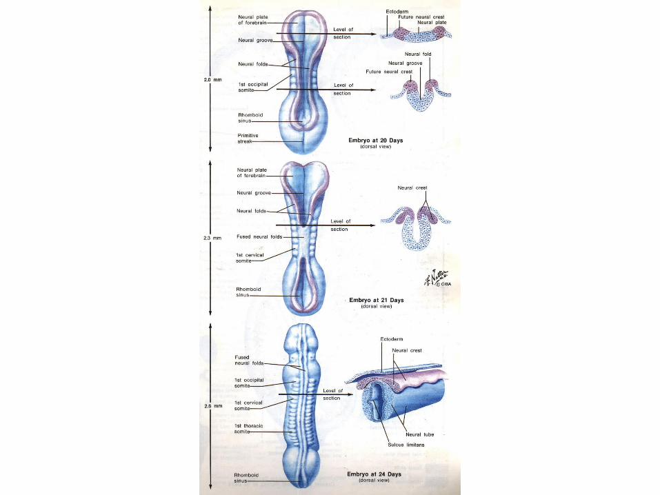
Embryology of the nervous system Embryology of the nervous system

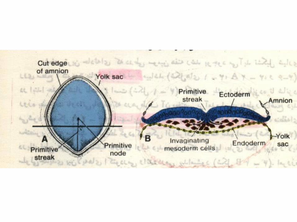

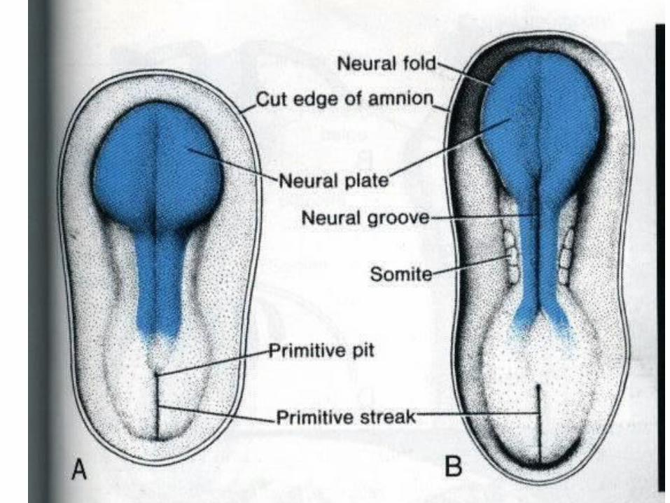
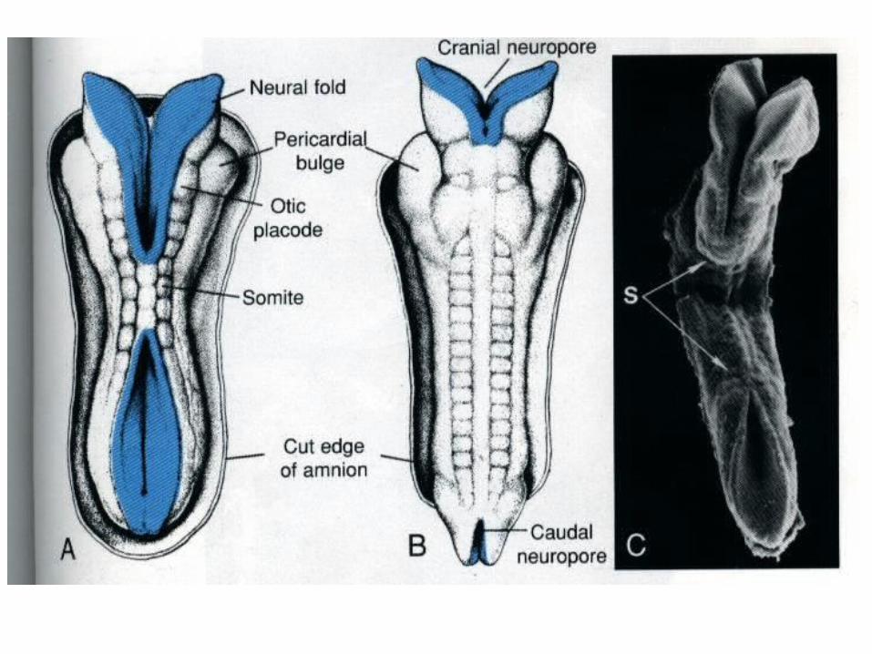
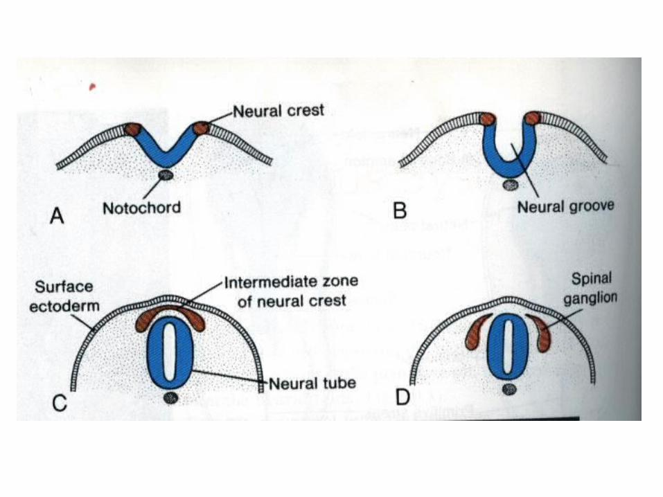


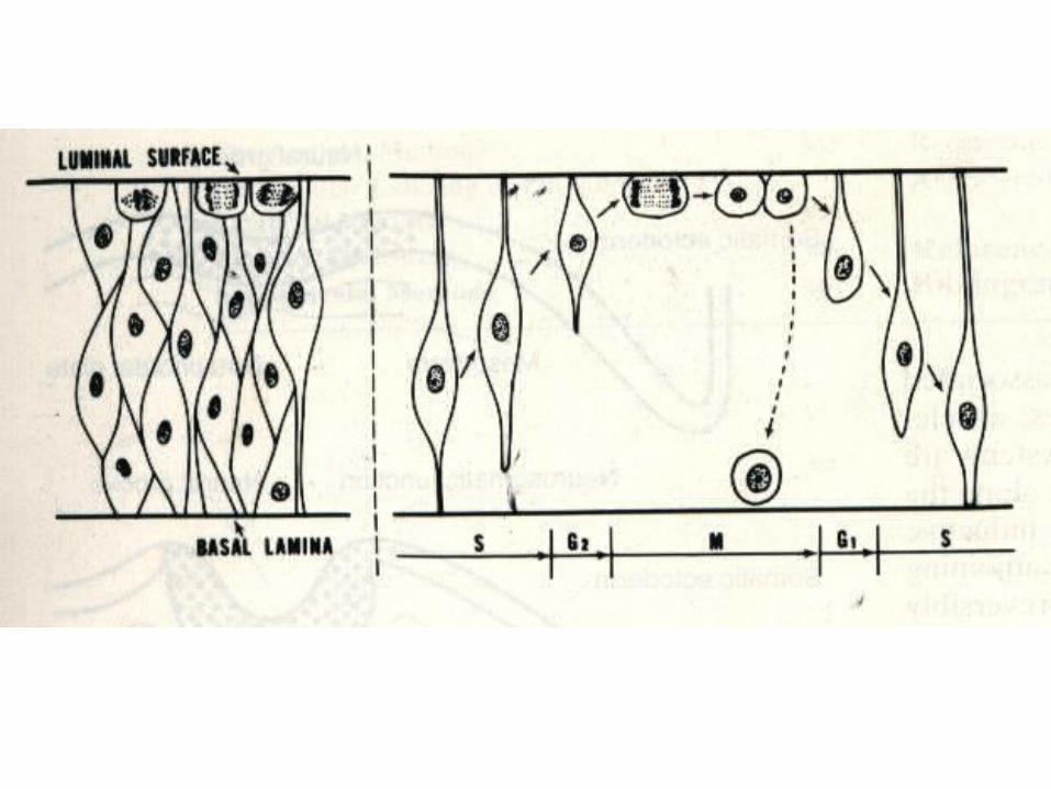
Neural Tube HistogenesisNeural Tube Histogenesis

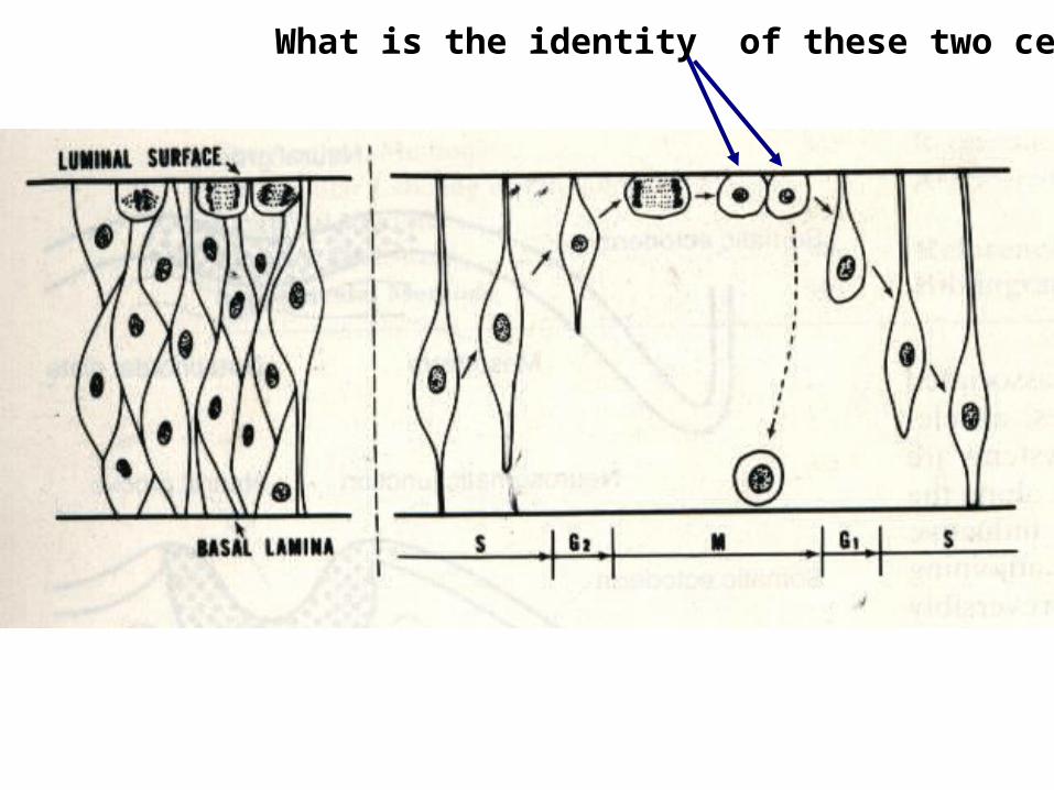
What is the identity of these two cells?
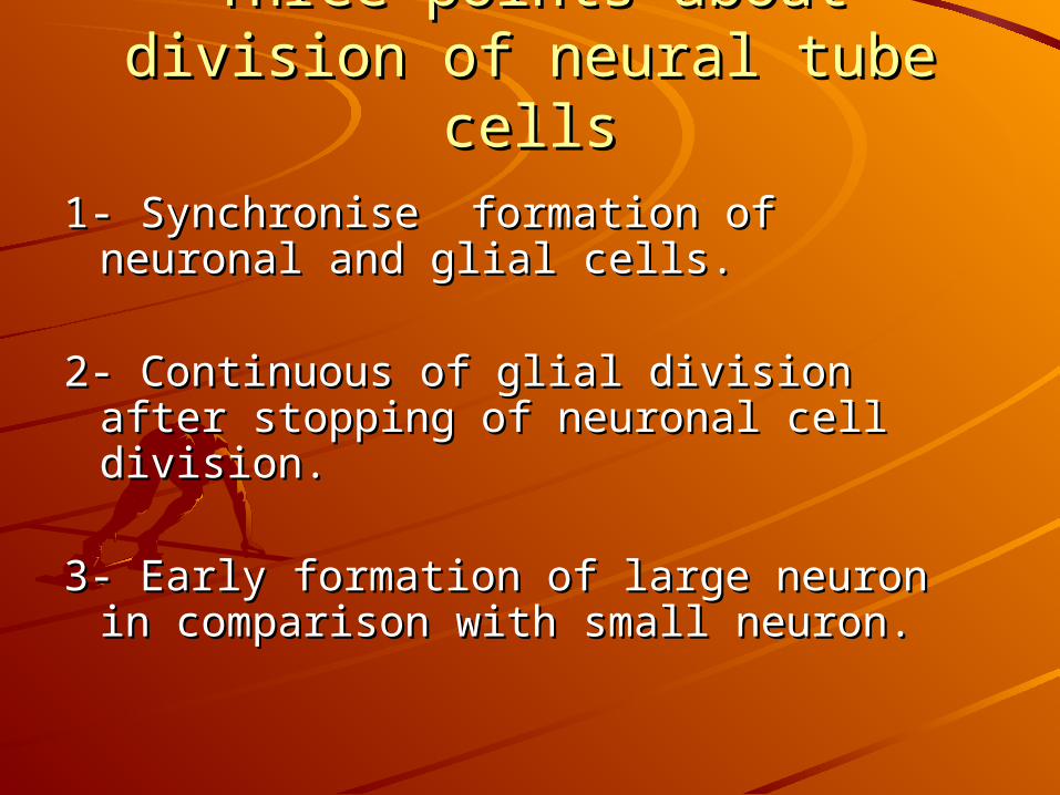
Three points about division of Three points about division of neural tube cellsneural tube cells
1- Synchronise formation of neuronal 1- Synchronise formation of neuronal and glial cells.and glial cells.
2- Continuous of glial division after 2- Continuous of glial division after stopping of neuronal cell division.stopping of neuronal cell division.
3- Early formation of large neuron in 3- Early formation of large neuron in
comparison with small neuron.comparison with small neuron.
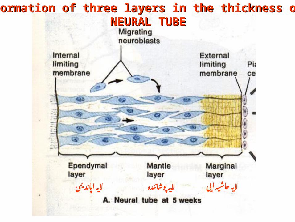
پوشاننده الیه ایی حاشیه اپاندیمی الیه الیه
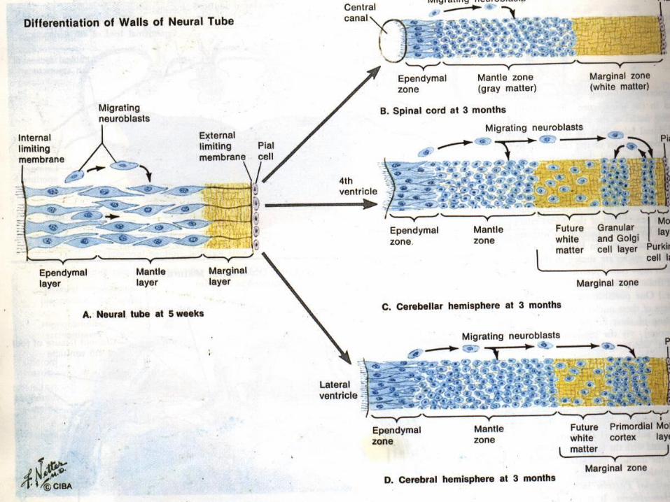
Formation of three layers in the thickness ofFormation of three layers in the thickness of NEURAL TUBE NEURAL TUBE

Neuronal cell migrationNeuronal cell migration
1- Gene activation1- Gene activation 2- Epigenetic signals 2- Epigenetic signals
(mechanical or contact guidance)(mechanical or contact guidance)

Neuronal cell aggregationNeuronal cell aggregation
Morphological appearanceMorphological appearance
NCAMs (nerve cell adhesion molecules)NCAMs (nerve cell adhesion molecules)
N cadherinN cadherin

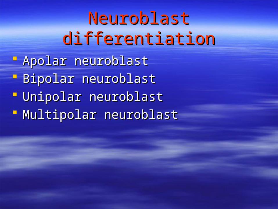
Neuroblast differentiationNeuroblast differentiation
Apolar neuroblastApolar neuroblast Bipolar neuroblastBipolar neuroblast Unipolar neuroblastUnipolar neuroblast Multipolar neuroblastMultipolar neuroblast

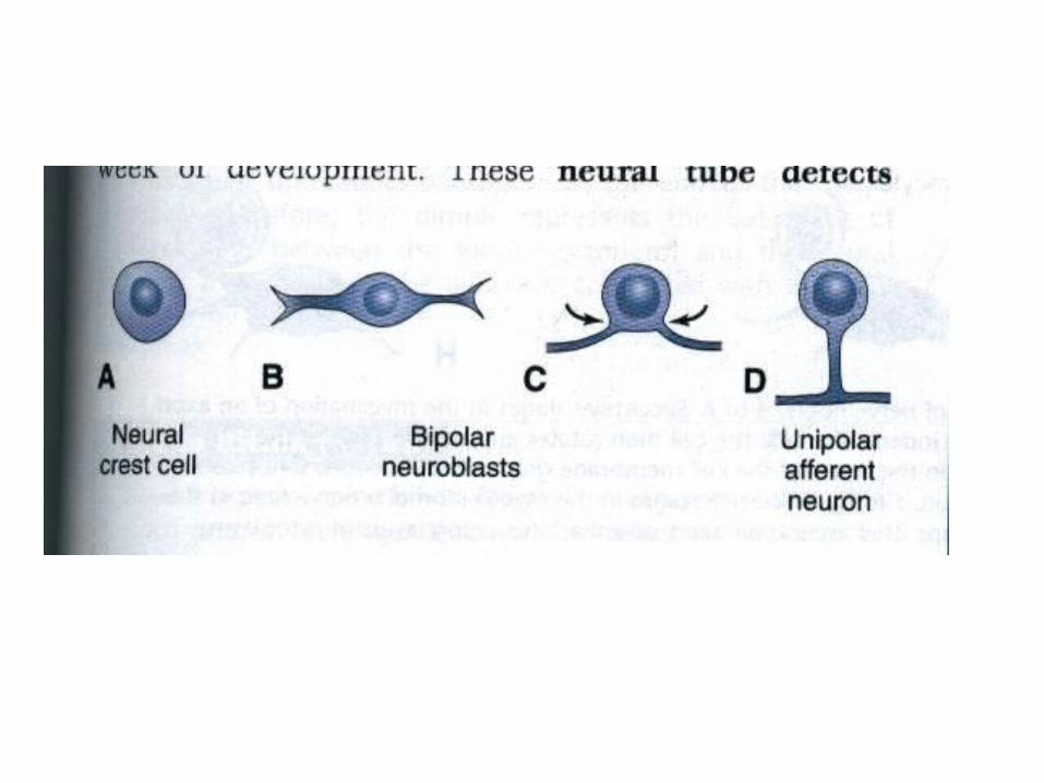

Neuroblast differentiationNeuroblast differentiation
Neurotrophic FactorsNeurotrophic Factors
1- NGFs ( Nerve Growth Factors)1- NGFs ( Nerve Growth Factors) 2- Outgrowth Promoting proteins2- Outgrowth Promoting proteins
( Laminin, Netrin, Fibronectin)( Laminin, Netrin, Fibronectin)
3-NCAMs 3-NCAMs
Cell Programmed DeathCell Programmed Death
ApoptosisApoptosis 30 to 60 percent of cells survive30 to 60 percent of cells survive
Effects of Experience and Effects of Experience and Activation on neuronsActivation on neurons
Structural PlasticityStructural Plasticity Axonal sproutingAxonal sprouting Changes in the number of Changes in the number of
Dendritic SpinesDendritic Spines
Amblyopia DiseaseAmblyopia Disease
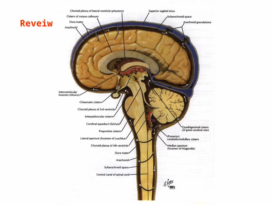
Reveiw
Formation of brain and spinal Formation of brain and spinal cordcord
1- Primary vesicles 1- Primary vesicles 2- Flexures2- Flexures
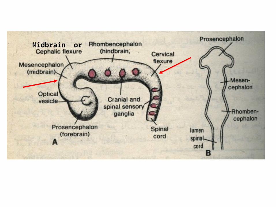
Midbrain or
Formation of brain and spinal Formation of brain and spinal cordcord
1- secondary vesicles 1- secondary vesicles


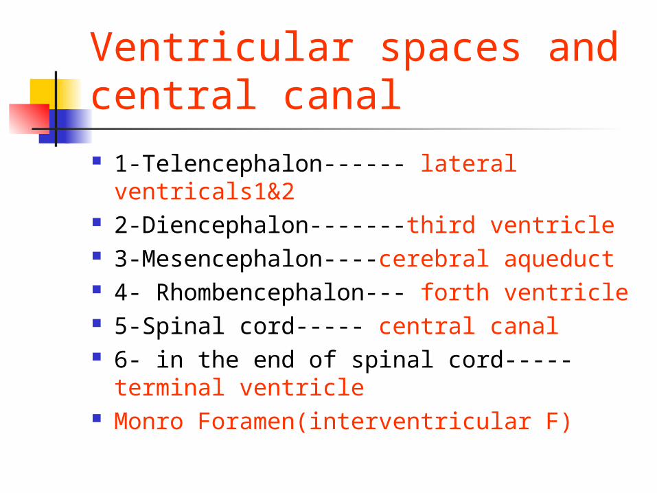
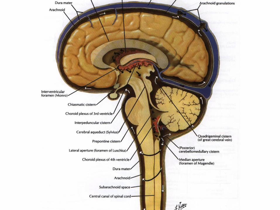
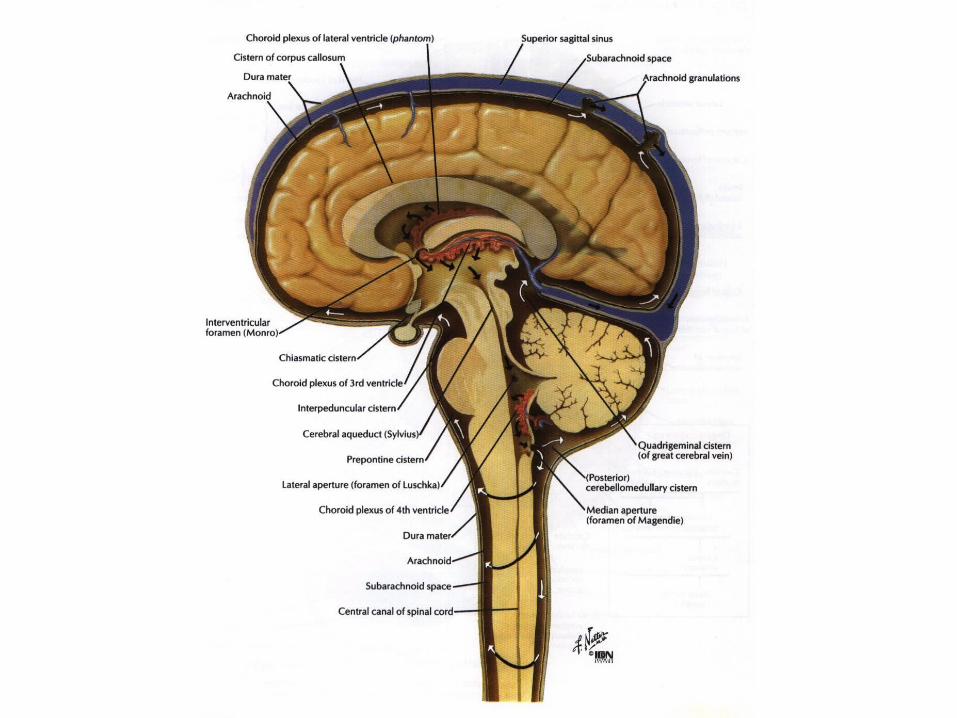
Ventricular spaces and central canal 1-Telencephalon------ lateral ventricals1&2 2-Diencephalon-------third ventricle 3-Mesencephalon----cerebral aqueduct 4- Rhombencephalon--- forth ventricle 5-Spinal cord----- central canal 6- in the end of spinal cord----- terminal
ventricle Monro Foramen(interventricular F)

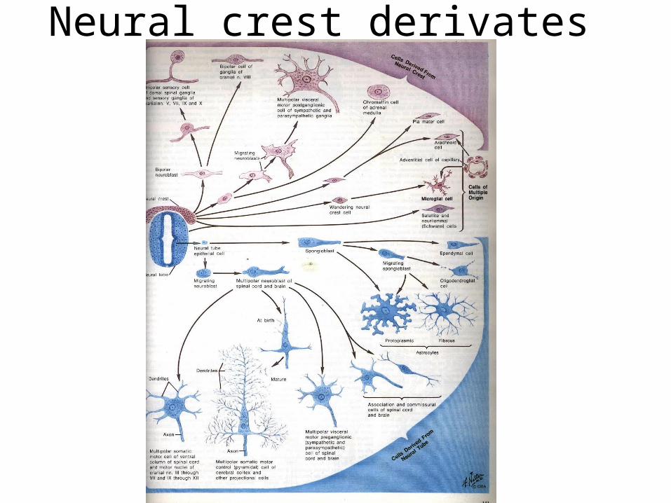
Neural crest derivates

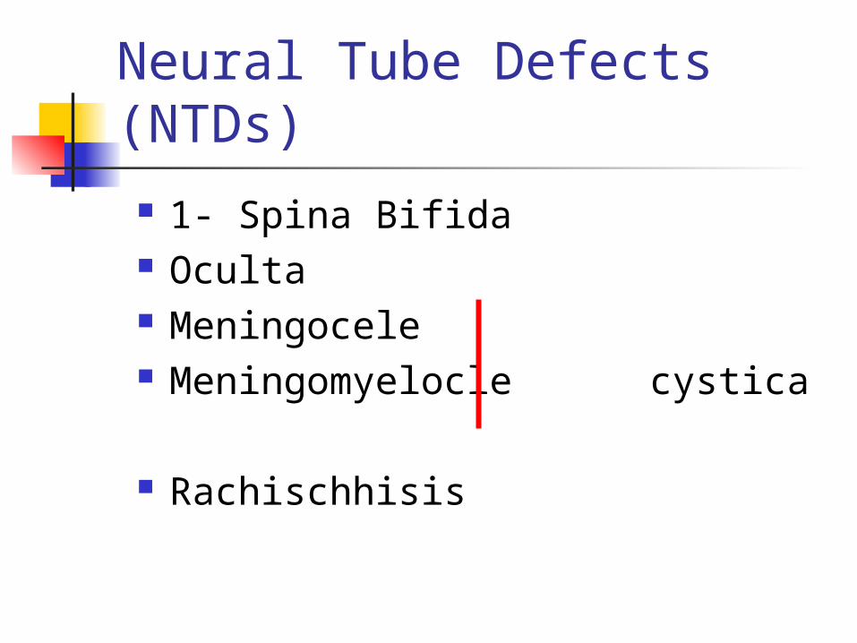
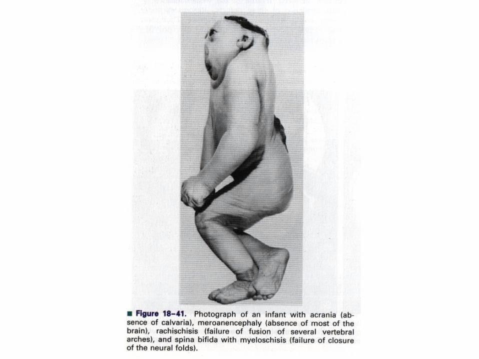
Neural Tube Defects (NTDs)
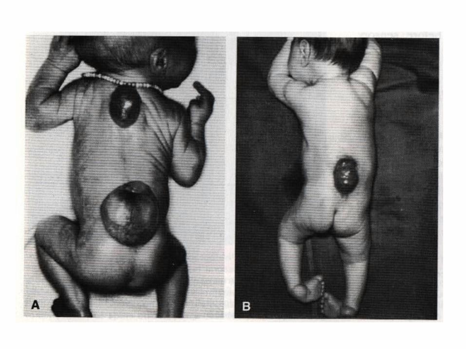
1- Spina Bifida Oculta Meningocele Meningomyelocle cystica
Rachischhisis

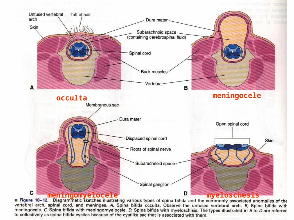
occulta meningocele
meningomyelocele myeloschesis
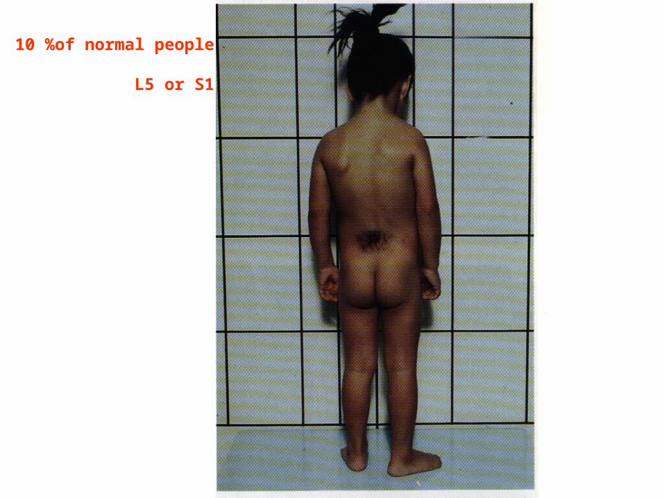
10 %of normal people
L5 or S1
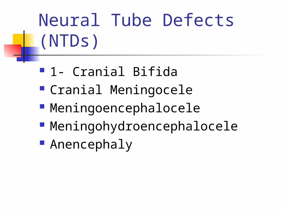
Neural Tube Defects (NTDs)
1- Cranial Bifida Cranial Meningocele Meningoencephalocele Meningohydroencephalocele Anencephaly
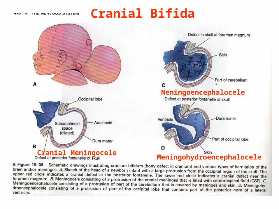
Cranial Meningocele
Meningoencephalocele
Meningohydroencephalocele
Cranial Bifida

Related Documents