by Janet Rae-Dupree and Pat DuPree Anatomy & Physiology Workbook FOR DUMmIES ‰

Welcome message from author
This document is posted to help you gain knowledge. Please leave a comment to let me know what you think about it! Share it to your friends and learn new things together.
Transcript
by Janet Rae-Dupree
and Pat DuPree
Anatomy &
Physiology
WorkbookFOR
DUMmIES‰
Anatomy & Physiology Workbook For Dummies®
Published byWiley Publishing, Inc.111 River St.Hoboken, NJ 07030-5774www.wiley.com
Copyright © 2007 by Wiley Publishing, Inc., Indianapolis, Indiana
Published simultaneously in Canada
No part of this publication may be reproduced, stored in a retrieval system, or transmitted in any form or by any means,electronic, mechanical, photocopying, recording, scanning, or otherwise, except as permitted under Sections 107 or 108 ofthe 1976 United States Copyright Act, without either the prior written permission of the Publisher, or authorization throughpayment of the appropriate per-copy fee to the Copyright Clearance Center, 222 Rosewood Drive, Danvers, MA 01923,978-750-8400, fax 978-646-8600. Requests to the Publisher for permission should be addressed to the Legal Department,Wiley Publishing, Inc., 10475 Crosspoint Blvd., Indianapolis, IN 46256, 317-572-3447, fax 317-572-4355, or online at http://www.wiley.com/go/permissions.
Trademarks: Wiley, the Wiley Publishing logo, For Dummies, the Dummies Man logo, A Reference for the Rest of Us!, TheDummies Way, Dummies Daily, The Fun and Easy Way, Dummies.com and related trade dress are trademarks or registeredtrademarks of John Wiley & Sons, Inc. and/or its affiliates in the United States and other countries, and may not be usedwithout written permission. All other trademarks are the property of their respective owners. Wiley Publishing, Inc., is notassociated with any product or vendor mentioned in this book.
LIMIT OF LIABILITY/DISCLAIMER OF WARRANTY: THE PUBLISHER AND THE AUTHOR MAKE NO REPRESENTATIONSOR WARRANTIES WITH RESPECT TO THE ACCURACY OR COMPLETENESS OF THE CONTENTS OF THIS WORK ANDSPECIFICALLY DISCLAIM ALL WARRANTIES, INCLUDING WITHOUT LIMITATION WARRANTIES OF FITNESS FOR A PAR-TICULAR PURPOSE. NO WARRANTY MAY BE CREATED OR EXTENDED BY SALES OR PROMOTIONAL MATERIALS. THEADVICE AND STRATEGIES CONTAINED HEREIN MAY NOT BE SUITABLE FOR EVERY SITUATION. THIS WORK IS SOLDWITH THE UNDERSTANDING THAT THE PUBLISHER IS NOT ENGAGED IN RENDERING LEGAL, ACCOUNTING, OROTHER PROFESSIONAL SERVICES. IF PROFESSIONAL ASSISTANCE IS REQUIRED, THE SERVICES OF A COMPETENTPROFESSIONAL PERSON SHOULD BE SOUGHT. NEITHER THE PUBLISHER NOR THE AUTHOR SHALL BE LIABLE FORDAMAGES ARISING HEREFROM. THE FACT THAT AN ORGANIZATION OR WEBSITE IS REFERRED TO IN THIS WORK ASA CITATION AND/OR A POTENTIAL SOURCE OF FURTHER INFORMATION DOES NOT MEAN THAT THE AUTHOR ORTHE PUBLISHER ENDORSES THE INFORMATION THE ORGANIZATION OR WEBSITE MAY PROVIDE OR RECOMMEN-DATIONS IT MAY MAKE. FURTHER, READERS SHOULD BE AWARE THAT INTERNET WEBSITES LISTED IN THIS WORKMAY HAVE CHANGED OR DISAPPEARED BETWEEN WHEN THIS WORK WAS WRITTEN AND WHEN IT IS READ.
For general information on our other products and services, please contact our Customer Care Department within the U.S.at 800-762-2974, outside the U.S. at 317-572-3993, or fax 317-572-4002.
For technical support, please visit www.wiley.com/techsupport.
Wiley also publishes its books in a variety of electronic formats. Some content that appears in print may not be available inelectronic books.
Library of Congress Control Number: 2007932378
ISBN: 978-0-470-16932-2
Manufactured in the United States of America
10 9 8 7 6 5 4 3 2 1
About the AuthorsJanet Rae-Dupree has been covering science and technology in Silicon Valley since 1993 fora number of publications, including U.S. News & World Report, BusinessWeek, the San JoseMercury News, and the Silicon Valley/San Jose Business Journal. She was a frequent guest on cable channel Tech TV’s “Silicon Spin” technology talk show, and she was part of thePulitzer Prize-winning team at the Los Angeles Times covering the city’s riots in 1992. Duringthe 2005–2006 academic year, Janet was a John S. Knight Journalism Fellow at StanfordUniversity, where she studied human biology and researched innovation and market transfer.She freelances for various publications, working from her home in Half Moon Bay, California,where she lives with husband, Dave Dupree, and their 9-year-old son, Matthew (although a19-year-old calico cat named Trillian actually rules the roost).
Pat DuPree taught anatomy/physiology, biology, medical terminology, and environmentalscience for 24 years at several colleges and universities in Los Angeles County. She holds twoundergraduate life science degrees and a master’s degree from Auburn University and con-ducted cancer research at Southern Research Institute in Birmingham, Alabama, before join-ing the Muscogee Health Department in Columbus, Georgia. In 1970, she moved to RedondoBeach, California, where she was a university instructor and raised her two sons, DaveDupree and Mark DuPree. Now Pat is retired and lives on lovely Pine Lake in rural Georgiawith her husband, Dr. James E. DuPree.
DedicationTo our loving family — Dave Dupree, Matthew Dupree, and Jim DuPree — for their patienceand understanding as we pulled this book together and put so many other aspects of ourlives on hold. To Dave, for being Mr. Fix-It on balky hard drives, recalcitrant computers, andfussy printers; for distracting an antsy 8-year-old with baseball, movies, and games; and forconcocting terrific — and healthy! — home-cooked meals. To Matthew, for understandingwhy Mommy couldn’t drive for every school field trip, attend every Cub Scout den meeting,or set up play dates every single day of the week.
And especially from Pat to Jim for his love, enthusiastic support, assistance, and encourage-ment without which she could not have finished this workbook.
Authors’ AcknowledgmentsWe owe gratitude to so many people, but this project has been first and foremost a DuPreefamily affair. We will never be able to thank our spouses enough for their patience and under-standing from beginning to end. Dr. James E. DuPree was a constant helpmate to Pat on theEast coast, providing research, editing, computer assistance, and graphics support as well aspriceless cheerleading and enthusiasm. Their son David E. Dupree filled the cheerleader roleon the West coast, keeping the computers and home network up and running, Janet focusedand on track, and the gaping child-rearing gaps filled while she pushed to get the book donein what surely must be record time. And we can’t forget to thank son/grandson Matthew J.Dupree for his stalwart patience while Mom and Grandmom focused their attentions on thebook instead of him.
We would also like to thank our agent, Matt Wagner, for his tireless efforts, as well as themany devoted people at Wiley Publishing, particularly Stacy Kennedy, Chrissy Guthrie (whowelcomed Sophia Rose Guthrie into the world days after receiving the final manuscript!),Stephen Clark, and Elizabeth Rea.
Publisher’s Acknowledgments
We’re proud of this book; please send us your comments through our Dummies online registration form located atwww.dummies.com/register/.
Some of the people who helped bring this book to market include the following:
Acquisitions, Editorial, and Media Development
Senior Project Editor: Christina Guthrie
Project Editor: Stephen R. Clark
Acquisitions Editor: Stacy Kennedy
Senior Copy Editor: Elizabeth Rea
Technical Editor: Michael W. Pratt, PC
Editorial Manager: Christine Meloy Beck
Editorial Assistants: Erin Calligan Mooney, Joe Niesen
Cover Photos: © Robin Lynne Gibson/Riser/Getty Images
Cartoons: Rich Tennant (www.the5thwave.com)
Composition Services
Project Coordinator: Patrick Redmond
Layout and Graphics: Stacie Brooks, Carrie A. Cesavice,Denny Hager, Stephanie D. Jumper, Julie Trippetti,Erin Zeltner
Special Art: Kathryn Born, M.A.
Anniversary Logo Design: Richard Pacifico
Proofreaders: Broccoli Information Management,Cynthia Fields, John Greenough
Indexer: Sherry Massey
Publishing and Editorial for Consumer Dummies
Diane Graves Steele, Vice President and Publisher, Consumer Dummies
Joyce Pepple, Acquisitions Director, Consumer Dummies
Kristin A. Cocks, Product Development Director, Consumer Dummies
Michael Spring, Vice President and Publisher, Travel
Kelly Regan, Editorial Director, Travel
Publishing for Technology Dummies
Andy Cummings, Vice President and Publisher, Dummies Technology/General User
Composition Services
Gerry Fahey, Vice President of Production Services
Debbie Stailey, Director of Composition Services
Contents at a GlanceIntroduction.................................................................................1
Part I: Building Blocks of the Body ...............................................5Chapter 1: The Chemistry of Life ...............................................................................................................7
Chapter 2: The Cell: Life’s Basic Building Block.....................................................................................23
Chapter 3: Divide and Conquer: Cellular Mitosis ...................................................................................37
Chapter 4: The Study of Tissues: Histology ............................................................................................47
Part II: Weaving It Together: Bones, Muscles, and Skin.................59Chapter 5: A Scaffold to Build On: The Skeleton....................................................................................61
Chapter 6: Getting in Gear: The Muscles.................................................................................................93
Chapter 7: It’s Skin Deep: The Integumentary System.........................................................................113
Part III: Feed and Fuel: Supply and Transport ............................127Chapter 8: Oxygenating the Machine: The Respiratory System ........................................................129
Chapter 9: Fueling the Functions: The Digestive System....................................................................143
Chapter 10: Spreading the Love: The Circulatory System ..................................................................163
Chapter 11: Keeping Up Your Defenses: The Lymphatic System.......................................................181
Chapter 12: Filtering Out the Junk: The Urinary System.....................................................................195
Part IV: Survival of the Species .................................................205Chapter 13: Why Ask Y?: The Male Reproductive System ..................................................................207
Chapter 14: Carrying Life Forward: The Female Reproductive System ............................................219
Part V: Mission Control: All Systems Go .....................................235Chapter 15: Feeling Jumpy: The Nervous System................................................................................237
Chapter 16: Raging Hormones: The Endocrine System.......................................................................265
Part VI: The Part of Tens...........................................................281Chapter 17: Ten Study Tips.....................................................................................................................283
Chapter 18: Ten (Plus One) Terrific Online Resources........................................................................287
Index.......................................................................................291
Table of ContentsIntroduction .................................................................................1
About This Book.........................................................................................................................1Conventions Used in This Book ...............................................................................................1Foolish Assumptions .................................................................................................................2How This Book Is Organized.....................................................................................................2
Part I: Building Blocks of the Body ................................................................................2Part II: Weaving It Together: Bones, Muscles, and Skin...............................................2Part III: Feed and Fuel: Supply and Transport ..............................................................3Part IV: Survival of the Species.......................................................................................3Part V: Mission Control: All Systems Go........................................................................3Part VI: The Part of Tens .................................................................................................3
Icons Used in This Book............................................................................................................3Where to Go from Here..............................................................................................................4
Part I: Building Blocks of the Body ................................................5
Chapter 1: The Chemistry of Life ..........................................................................................7
Building from Scratch: Atoms and Elements..........................................................................7Compounding Chemical Reactions........................................................................................10Cycling through Life: Metabolism ..........................................................................................15Answers to Questions on Life’s Chemistry...........................................................................21
Chapter 2: The Cell: Life’s Basic Building Block ............................................................23
Gaining Admission: The Cell Membrane ...............................................................................23Aiming for the Nucleus............................................................................................................26Looking Inside: Organelles and Their Functions..................................................................27Putting Together New Proteins ..............................................................................................31Cycling Along: Grow, Rest, Divide, Die ..................................................................................33Answers to Questions on the Cell..........................................................................................35
Chapter 3: Divide and Conquer: Cellular Mitosis ...........................................................37
The Mitotic Process.................................................................................................................37Waiting for action: Interphase ......................................................................................38Sorting out the parts: Prophase ...................................................................................38Dividing at the equator: Metaphase.............................................................................38Packing up to move out: Anaphase..............................................................................38Pinching off: Telophase .................................................................................................39Splitting up: Cytokinesis................................................................................................39
What Can Go Wrong.................................................................................................................42Answers to Questions on Mitosis ..........................................................................................44
Chapter 4: The Study of Tissues: Histology......................................................................47
Getting Under Your Skin..........................................................................................................47Making a Connection: Connective Tissue .............................................................................50Flexing It: Muscle Tissue .........................................................................................................53Getting the Signal Across: Nerve Tissue ...............................................................................54Answers to Questions on Histology ......................................................................................56
Part II: Weaving It Together: Bones, Muscles, and Skin .................59
Chapter 5: A Scaffold to Build On: The Skeleton ............................................................61
Understanding Dem Bones .....................................................................................................61Boning Up on Classifications, Structures, and Ossification ...............................................63Axial Skeleton: Keeping It All in Line .....................................................................................69
Making a hard head harder ...........................................................................................69Putting your backbones into it .....................................................................................70
Appendicular Skeleton: Reaching Beyond Our Girdles.......................................................79Arthrology: Articulating the Joints ........................................................................................82Answers to Questions on the Skeleton .................................................................................87
Chapter 6: Getting in Gear: The Muscles .........................................................................93
Flexing Your Muscle Knowledge ............................................................................................93Classifications: Smooth, Cardiac, and Skeletal.....................................................................95Contracting for a Contraction ................................................................................................97Pulling Together: Muscles as Organs.....................................................................................99Assuming the Right Tone ......................................................................................................100Leveraging Muscular Power .................................................................................................101What’s In a Name? Identifying Muscles ...............................................................................104Answers to Questions on Muscles.......................................................................................109
Chapter 7: It’s Skin Deep: The Integumentary System .................................................113
Dermatology Down Deep ......................................................................................................113Epidermis: Don’t judge this book by its cover .........................................................114Dermis: It’s more than skin deep................................................................................115
Touching a Nerve in the Integumentary System................................................................119Accessorizing with Hair, Nails, and Glands ........................................................................120
Wigging out about hair ................................................................................................120Nailing the fingers and toes ........................................................................................121Sweating the details .....................................................................................................121Getting an earful ...........................................................................................................122
Answers to Questions on the Skin .......................................................................................125
Part III: Feed and Fuel: Supply and Transport .............................127
Chapter 8: Oxygenating the Machine: The Respiratory System ................................129
Breathing In Oxygen, Breathing Out CO2 ............................................................................129Inhaling the Basics about the Respiratory Tract ...............................................................132
Knowing about the nose (and sinuses) .....................................................................132Dealing with throaty matters ......................................................................................134Going deep inside the lungs........................................................................................137
Damaging Air ..........................................................................................................................139Answers to Questions on the Respiratory System............................................................141
Chapter 9: Fueling the Functions: The Digestive System ............................................143
Digesting the Basics: It’s Alimentary! ..................................................................................143Nothing to Spit At: Into the Mouth and Past the Teeth.....................................................145
Entering the vestibule..................................................................................................146Moving along the oral cavity ......................................................................................147
The tongue...........................................................................................................147The salivary glands ............................................................................................148
Stomaching the Body’s Fuel..................................................................................................151
viii Anatomy & Physiology Workbook For Dummies
Breaking Down the Work of Digestive Enzymes.................................................................154Small intestine ..............................................................................................................154Liver ...............................................................................................................................155Pancreas ........................................................................................................................156Large intestine ..............................................................................................................157
Answers to Questions on the Digestive Tract ....................................................................159
Chapter 10: Spreading the Love: The Circulatory System ...........................................163
Moving to the Beat of a Pump ..............................................................................................163Finding the Key to the Heart’s Chambers ...........................................................................166
The atria ........................................................................................................................166The ventricles ...............................................................................................................167
Conducting the Heart’s Music ..............................................................................................170Riding the Network of Blood Vessels...................................................................................172Beating from the Start: Fetal Circulation.............................................................................174Answers to Questions on the Circulatory System.............................................................178
Chapter 11: Keeping Up Your Defenses: The Lymphatic System ...............................181
Duct, Duct, Lymph .................................................................................................................181Poking at the Nodes...............................................................................................................184Having a Spleen-ded Time with the Lymphatic Organs ....................................................187
The spleen.....................................................................................................................188T cell central: The thymus gland................................................................................188Opening wide and moving along: The tonsils and Peyer’s patches.......................189
Answers to Questions on the Lymphatic System ..............................................................192
Chapter 12: Filtering Out the Junk: The Urinary System..............................................195
Examining the Kidneys, the Body’s Filters .........................................................................195Going molecular ...........................................................................................................196Focusing on filtering ....................................................................................................196
Getting Rid of the Waste........................................................................................................199Surfing the ureters........................................................................................................199Ballooning the bladder ................................................................................................199The male and female urethras ....................................................................................199
Spelling Relief: Urination.......................................................................................................201Answers to Questions on the Urinary System ...................................................................203
Part IV: Survival of the Species ..................................................205
Chapter 13: Why Ask Y?: The Male Reproductive System..........................................207
Identifying the Parts of the Male Reproductive System....................................................207Packaging the Chromosomes for Delivery..........................................................................211Answers to Questions on the Male Reproductive System................................................217
Chapter 14: Carrying Life Forward: The Female Reproductive System ....................219
Identifying the Female Reproductive Parts and Their Functions ....................................219Making Eggs: A Mite More Meiosis ......................................................................................224Making Babies: An Introduction to Embryology................................................................226Growing from Fetus to Baby .................................................................................................227Growing, Changing, and Aging..............................................................................................229Answers to Questions on the Female Reproductive System............................................231
ixTable of Contents
Part V: Mission Control: All Systems Go......................................235
Chapter 15: Feeling Jumpy: The Nervous System.........................................................237
Building from Basics: Neurons, Nerves, Impulses, Synapses...........................................238Neurons .........................................................................................................................238Nerves............................................................................................................................239Impulses ........................................................................................................................240Synapses........................................................................................................................241
Minding the Central Nervous System and the Brain .........................................................243Spinal cord ....................................................................................................................244Brain...............................................................................................................................245
Medulla oblongata ..............................................................................................245Pons......................................................................................................................245Midbrain ..............................................................................................................245Cerebellum ..........................................................................................................246Diencephalon ......................................................................................................246Cerebrum.............................................................................................................246Medulla ................................................................................................................247Ventricles .............................................................................................................247
Taking Side Streets: The Peripheral Nervous System .......................................................251Keep Breathing: The Autonomic Nervous System.............................................................253Coming To Your Senses .........................................................................................................255
Eyes ................................................................................................................................256Ears ................................................................................................................................256
Answers to Questions on the Nervous System..................................................................261
Chapter 16: Raging Hormones: The Endocrine System................................................265
No Bland Glands.....................................................................................................................265Mastering the Ringmasters...................................................................................................267Supporting Cast of Glandular Characters...........................................................................270
Topping off the kidneys: The adrenal glands............................................................270Thriving with the thyroid ............................................................................................271Pairing up with the parathyroid .................................................................................271Pinging the pineal gland ..............................................................................................271Thumping the thymus .................................................................................................271Pressing the pancreas..................................................................................................272
Dealing with Stress: Homeostasis ........................................................................................274Answers to Questions on the Endocrine System...............................................................277
Part VI: The Part of Tens ...........................................................281
Chapter 17: Ten Study Tips ................................................................................................283
Chapter 18: Ten (Plus One) Terrific Online Resources.................................................287
Index .......................................................................................291
x Anatomy & Physiology Workbook For Dummies
Introduction
Whether your aim is to become a physical therapist or a pharmacist, a doctor or anacupuncturist, a nutritionist or a personal trainer, a registered nurse or a paramedic,
a parent or simply a healthy human being — your efforts have to be based on a good under-standing of anatomy and physiology. But knowing that the knee bone connects to the thighbone (or does it?) is just the tip of the iceberg. In Anatomy & Physiology Workbook ForDummies, you discover intricacies that will leave you agog with wonder. The human body isa miraculous biological machine capable of growing, interacting with the world, and evenreproducing despite any number of environmental odds stacked against it. Understandinghow the body’s interlaced systems accomplish these feats requires a close look at every-thing from chemistry to structural mechanics.
Early anatomists relied on dissections to study the human body, which is why the Greekword anatomia means “to cut up or dissect.” Anatomical references have been found inEgypt dating back to 1600 BC, but it was the Greeks — Hippocrates, in particular — who firstdissected bodies for medical study around 420 BC. That’s why more than two millennia laterwe still use words based on Greek and Latin roots to identify anatomical structures.
That’s also part of the reason so much of the study of anatomy and physiology feels likelearning a foreign language. Truth be told, you are working with a foreign language, but it’sthe language of you and the one body you’re ever going to have.
About This BookThis workbook isn’t meant to replace a textbook, and it’s certainly not meant to replace goingto an actual anatomy and physiology class. It works best as a supplement to your ongoingeducation and as a study aid in prepping for exams. That’s why we give you insight into whatyour instructor most likely will emphasize as you move from one body system or structure tothe next.
Your coursework most likely will cover things in a different order than we’ve chosen for thisbook. We encourage you to take full advantage of the table of contents and the index to findthe material addressed in your class. Whatever you do, certainly don’t feel obligated to gothrough this workbook in any particular order. However, please do answer the practice ques-tions and check the answers at the end of each chapter because, in addition to answers, weclarify why the right answer is the right answer and why the other answers are incorrect;we also provide you with memory tools and other tips whenever possible.
Conventions Used in This BookHalf the battle of studying anatomy and physiology is getting comfortable with the jargon. Tohelp you navigate through this book, we use the following typographical conventions:
� Italics are used for emphasis and to highlight new words or terms that are defined inthe text.
� Boldface is used to indicate keywords in bulleted lists or the action parts of numberedsteps.
� Monofont is used for Web site addresses.
Foolish AssumptionsIn writing Anatomy & Physiology Workbook For Dummies, we had to make someassumptions about you, the reader. If any of the following apply, this book’s for you:
� You’re an advanced high school student or college student trying to puzzle outanatomy and physiology for the first time.
� You’re a student at any level who’s returning to the topic after some time away,and you need some refreshing.
� You’re facing an anatomy and physiology exam and want a good study tool toensure that you have a firm grasp of the topic.
Because this is a workbook, we had to limit our exposition of each and every topic sothat we could include lots of practice questions to keep you guessing. (Believe us, wecould go on forever about this anatomy and physiology stuff!) In leaving out some ofthe explanation of the topics covered in this book, we assume that you’re not just look-ing to dabble in anatomy and physiology and therefore have access to at least onetextbook on the subject.
How This Book Is OrganizedAnatomy and physiology are very far-reaching topics, so it only makes sense that thisworkbook is divided into parts, each of which is divided into a number of chapters.The following sections preview the part topics to give you an idea of what you can findwhere.
Part I: Building Blocks of the BodyWe begin at the very beginning — chemistry — because it’s a very good place to start.Stop moaning and groaning — chemistry really isn’t as difficult as it’s been made outto be. It’s an integral part of understanding what the body’s cells are doing and howthey’re doing it. We cover the basics from the atom on up and introduce the processesthat keep the whole package operating smoothly. Then we take a look at the cells andthe tiny structures inside every one. Cells are living things, just like the bodies ofwhich they are a part, and they have the same cycles as all living things do: They grow,mature, reproduce, and die. By layering thousands upon thousands of similar cells ontop of one another, tissues with unique structures and functions are formed. This partcovers the primary types of tissues and where you’ll find them in the body.
Part II: Weaving It Together: Bones, Muscles, and SkinThe chapters in this part have a kind of classic leg-bone-connected-to-the-hip-bone feelto them — what people generally think of when they hear “anatomy and physiology.”We take you on a tour of the skeleton and then attach muscles to that skeleton to getthe whole package moving and grooving. Then we wrap it all together in the body’slargest single organ: the skin.
2 Anatomy & Physiology Workbook For Dummies

Part III: Feed and Fuel: Supply and TransportNo man is an island, and no one can exist without a consistent supply of life’s littlenecessities. In this part, we breathe life into the respiratory system with a close look atthe lungs and everything attached to them, we feed your hunger for knowledge abouthow nutrients fuel the anatomical package, and we get to the heart of the well-oiledhuman machine to show how the central pump is the hardest-working muscle in theentire body. None of that matters without a strong defense system, so we touch on thelymphatic system. And don’t forget: All that metabolizing is bound to lead to somewaste and by-products; we package up the trash and show you how the body takes itto the dumpster.
Part IV: Survival of the SpeciesNo, we don’t talk about who’s going to get voted off the island. But we do take a closelook at perpetuating humanity through reproductive successes. This part takes themale and female halves of the equation one at a time, delving into the parts of the maleand female reproductive systems as well as the functions of those parts. We coversperm and eggs, but we don’t even try to address which came first!
Part V: Mission Control: All Systems GoWe have lift off! We already have gotten things moving before this part, but now it’stime to study how nerves and hormones keep things hopping. In this part, we lay outthe basic building blocks of the nervous system, help you wire it all together, and thenshow you how the body sends messages flying along a solid spine of brainy material.After that, we come to our senses with an overview of the eyes and ears (we covertaste in the digestive system chapter, touch in the skin chapter, and smell in the respi-ratory chapter). Then we turn hormonal to absorb what the endocrine system does,including observing the functions of the ringmaster of this multi-ring circus, the pitu-itary gland. We also delve into the various hormones coursing through your body, whythey’re there, and how they do what they do.
Part VI: The Part of TensThis part is a classic For Dummies feature — lists of ten things that we just have toshare with our readers. First we identify ten Web sites that can help you advance yourknowledge of anatomy and physiology. Then we give you a list of ten key things tokeep in mind as you study this illustrious and fascinating topic.
Icons Used in This BookThroughout this book, you’ll find symbols in the margins that highlight critical ideasand information. Here’s what they mean:
The tip icon gives you juicy tidbits about how best to remember tricky terms or con-cepts in anatomy and physiology. It also highlights helpful strategies for fast translationand understanding.
3Introduction
The example icon marks questions for you to try your hand at. We give you the answerstraightaway to get your juices flowing and your brain warmed up for more practicequestions.
The remember icon highlights key material that you should pay extra attention to inorder to keep everything straight.
The sizzling bomb icon — otherwise known as the warning icon — points out areasand topics where common pitfalls can lead you astray.
Where to Go from HereIf you purchased this book and you’re already partway through an anatomy and physi-ology class, check the table of contents and zoom ahead to whichever segment yourinstructor is covering currently. When you have a few spare minutes, review the chap-ters that address topics your class already has covered. It’s an excellent way to prepfor a midterm or final exam. If you haven’t yet started an anatomy and physiologyclass, you have the freedom to start wherever you like (although we suggest that youbegin with Chapter 1) and proceed onward and upward through the glorious machinethat is the human body!
4 Anatomy & Physiology Workbook For Dummies
Part I
Building Blocks of the Body
In this part . . .
Before beginning to study the parts of the body, it’simportant to know about the basic building blocks
and functions that make those parts what they are. Thatmeans getting down to the true basics: chemistry, cells,cell division, and how tissues are formed. We know youreyes are glazing over, but it’s really not as bad as you maythink.
This part helps you discover that chemistry isn’t all thattough, particularly when you focus on the organic elementsinvolved in the chemistry of life. You look at how that chem-istry takes place inside the bricks-and-mortar of the body(its cells) and take things a step further with the wonders ofself-perpetuation through mitotic cell division.
Chapter 1
The Chemistry of Life
In This Chapter� Getting to the heart of all matter: Atoms
� Checking into chemical reactions and compounds
� Making sense of metabolism
We can hear your cries of alarm. You thought you were getting ready to learn aboutthe knee bone connecting to the thigh bone. How in the heck does that involve
(horrors!) chemistry? As much as you may not want to admit it, chemistry — particularlyorganic chemistry, or that branch of the field that focuses on carbon-based molecules — is acrucial starting point for understanding how the human body works. When all is said anddone, the universe boils down to two fundamental components: matter, which occupiesspace and has mass; and energy, or the ability to do work or create change. This is the chap-ter where we review the interactions between matter and energy to give you some insightinto what you need to know to ace those early-term tests.
Building from Scratch: Atoms and ElementsAll matter — be it solid, liquid, or gas — is composed of atoms. An atom is the smallest unit of matter capable of retaining the identity of an element during a chemical reaction. An element is a substance that can’t be broken down into simpler substances by normalchemical reactions. There are 92 naturally occurring atoms in nature and 17 (at last count)artificially created atoms for a total of 109 known atoms. However, additional spaces have yetto be filled in on the periodic chart of elements, which organizes all the elements by name,symbol, atomic weight, and atomic number. The key elements of interest to students ofanatomy and physiology are
� Hydrogen, symbol H
� Oxygen, symbol O
� Nitrogen, symbol N
� Carbon, symbol C
HONC your horn for the four organic elements. These four elements make up 96 percent ofall living material.
Atoms are made up of the subatomic particles protons and neutrons, which are in the atom’snucleus, and clouds of electrons orbiting the nucleus. The atomic weight, or mass, of an atomis the total number of protons and neutrons in its nucleus. The atomic number of an atom isits number of protons or electrons; conveniently, atoms always have the same number ofprotons as electrons, which means that an atom is always electrically neutral because it
always has the same number of positive charges as negative charges. Opposite chargesattract, so negatively charged electrons are attracted to positively charged protons.The attraction holds electrons in orbits outside the nucleus. The more protons thereare in the nucleus, the stronger the atom’s positive charge is and the more electrons itcan attract.
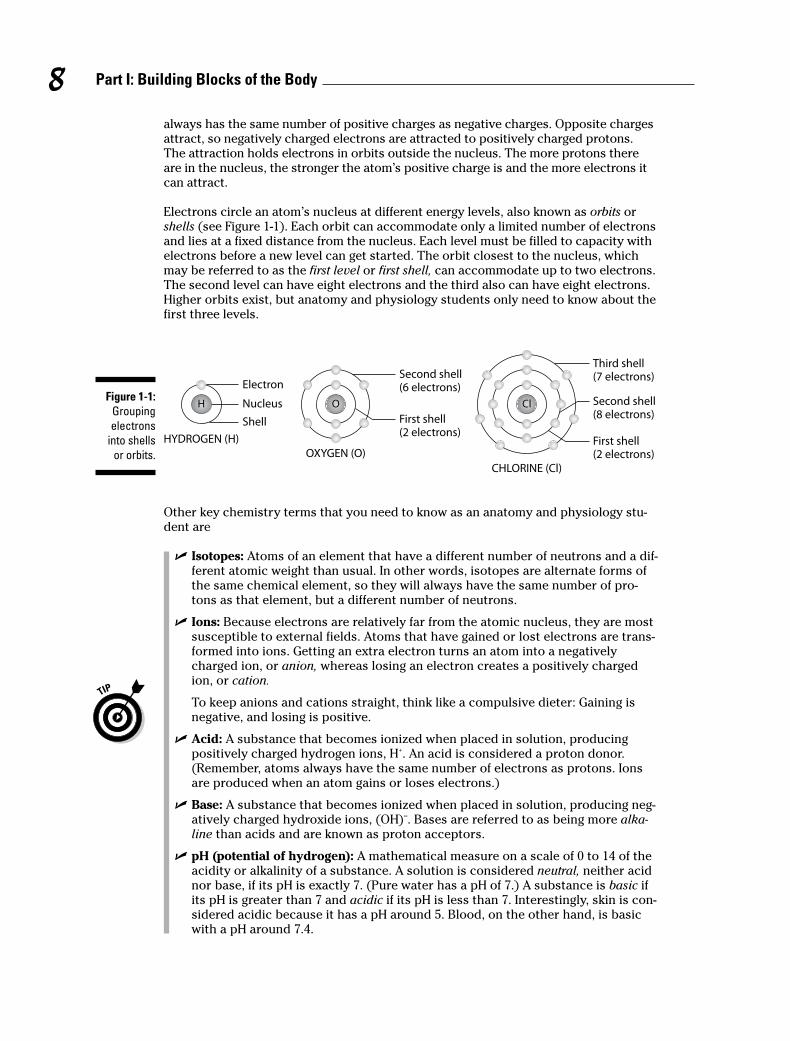
Electrons circle an atom’s nucleus at different energy levels, also known as orbits orshells (see Figure 1-1). Each orbit can accommodate only a limited number of electronsand lies at a fixed distance from the nucleus. Each level must be filled to capacity withelectrons before a new level can get started. The orbit closest to the nucleus, whichmay be referred to as the first level or first shell, can accommodate up to two electrons.The second level can have eight electrons and the third also can have eight electrons.Higher orbits exist, but anatomy and physiology students only need to know about thefirst three levels.
Other key chemistry terms that you need to know as an anatomy and physiology stu-dent are
� Isotopes: Atoms of an element that have a different number of neutrons and a dif-ferent atomic weight than usual. In other words, isotopes are alternate forms ofthe same chemical element, so they will always have the same number of pro-tons as that element, but a different number of neutrons.
� Ions: Because electrons are relatively far from the atomic nucleus, they are mostsusceptible to external fields. Atoms that have gained or lost electrons are trans-formed into ions. Getting an extra electron turns an atom into a negativelycharged ion, or anion, whereas losing an electron creates a positively chargedion, or cation.
To keep anions and cations straight, think like a compulsive dieter: Gaining isnegative, and losing is positive.
� Acid: A substance that becomes ionized when placed in solution, producing positively charged hydrogen ions, H+. An acid is considered a proton donor.(Remember, atoms always have the same number of electrons as protons. Ionsare produced when an atom gains or loses electrons.)
� Base: A substance that becomes ionized when placed in solution, producing neg-atively charged hydroxide ions, (OH)–. Bases are referred to as being more alka-line than acids and are known as proton acceptors.
� pH (potential of hydrogen): A mathematical measure on a scale of 0 to 14 of theacidity or alkalinity of a substance. A solution is considered neutral, neither acidnor base, if its pH is exactly 7. (Pure water has a pH of 7.) A substance is basic ifits pH is greater than 7 and acidic if its pH is less than 7. Interestingly, skin is con-sidered acidic because it has a pH around 5. Blood, on the other hand, is basicwith a pH around 7.4.
H Nucleus
Electron
Shell
HYDROGEN (H)
OH O
OXYGEN (O)
ClCl
CHLORINE (Cl)
First shell
(2 electrons)First shell
(2 electrons)
Second shell
(8 electrons)
Third shell
(7 electrons)Second shell
(6 electrons)Figure 1-1:
Grouping
electrons
into shells
or orbits.
8 Part I: Building Blocks of the Body
9Chapter 1: The Chemistry of Life
Answer these practice questions about atoms and elements:
1. The four key elements that make up most living matter are
a. Carbon, hydrogen, nitrogen, and phosphorus
b. Oxygen, carbon, sulfur, and nitrogen
c. Hydrogen, nitrogen, oxygen, and carbon
d. Nitrogen, potassium, carbon, and oxygen
2. Among the subatomic particles in an atom, the two that have equal weight are
a. Neutrons and electrons
b. Protons and neutrons
c. Positrons and protons
d. Neutrons and positrons
3. For an atom with an atomic number of 19 and an atomic weight of 39, the total number of neu-trons is
a. 19
b. 20
c. 39
d. 58
4. Element X has 14 electrons. How many electrons are in its outermost shell?
a. 2
b. 6
c. 14
d. 4
5. A substance that, in water, separates into a large number of hydroxide ions is
a. A weak acid
b. A weak base
c. A strong acid
d. A strong base
6. A hydroxyl, or hydroxide, ion has an oxygen atom
a. Only
b. And an extra electron
c. And a hydrogen atom and an extra electron
d. And a hydrogen atom and one less electron
7.–12. Fill in the blanks to complete the following sentences:
Different isotopes of the same element have the same number of 7. _______________ and 8. _______________ but different numbers of 9. _______________. Isotopes also have differ-ent atomic 10. _______________. An atom that gains or loses an electron is called an 11. _______________. If an atom loses an electron, it carries a 12. _______________ charge.
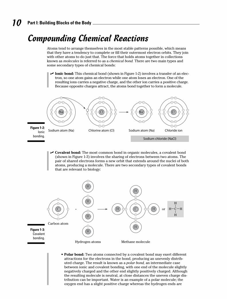
Compounding Chemical ReactionsAtoms tend to arrange themselves in the most stable patterns possible, which meansthat they have a tendency to complete or fill their outermost electron orbits. They joinwith other atoms to do just that. The force that holds atoms together in collectionsknown as molecules is referred to as a chemical bond. There are two main types andsome secondary types of chemical bonds:
� Ionic bond: This chemical bond (shown in Figure 1-2) involves a transfer of an elec-tron, so one atom gains an electron while one atom loses an electron. One of theresulting ions carries a negative charge, and the other ion carries a positive charge.Because opposite charges attract, the atoms bond together to form a molecule.
� Covalent bond: The most common bond in organic molecules, a covalent bond(shown in Figure 1-3) involves the sharing of electrons between two atoms. Thepair of shared electrons forms a new orbit that extends around the nuclei of bothatoms, producing a molecule. There are two secondary types of covalent bondsthat are relevant to biology:
• Polar bond: Two atoms connected by a covalent bond may exert differentattractions for the electrons in the bond, producing an unevenly distrib-uted charge. The result is known as a polar bond, an intermediate casebetween ionic and covalent bonding, with one end of the molecule slightlynegatively charged and the other end slightly positively charged. Althoughthe resulting molecule is neutral, at close distances the uneven charge dis-tribution can be important. Water is an example of a polar molecule; theoxygen end has a slight positive charge whereas the hydrogen ends are
CC + or
HH
HH
HH
HH
HH
HH
HH
HH
CC
Carbon atom
Hydrogen atoms Methane molecule
H
H
HH C
Figure 1-3:
Covalent
bonding.
NaNa
Sodium atom (Na)
ClCl
Chlorine atom (Cl)
Na+Na+
Sodium atom (Na)
Cl–Cl–
Chloride ion
Sodium chloride (NaCl)
Figure 1-2:
Ionic
bonding.
10 Part I: Building Blocks of the Body

slightly negative. Polarity explains why some substances dissolve readily inwater and others do not.
• Hydrogen bond: Because they’re polarized, two adjacent H2O (water) mole-cules can form a linkage known as a hydrogen bond, where a (electronega-tive) hydrogen atom of one H2O molecule is electrostatically attracted to the(electropositive) oxygen atom of an adjacent water molecule. Consequently,molecules of water join together transiently in a hydrogen-bonded lattice.Hydrogen bonds have only about 1⁄20 the strength of a covalent bond, yeteven this force is sufficient to affect the structure of water, producing manyof its unique properties, such as high surface tension, specific heat, andheat of vaporization. Hydrogen bonds are important in many life processes,such as in replication and defining the shape of DNA molecules.
A chemical reaction is the result of a process that changes the number, the types, orthe arrangement of atoms within a molecule. The substances that go through thisprocess are called the reactants. The substances produced by the reaction are calledthe products.
Chemical reactions are written in the form of an equation, with an arrow indicating thedirection of the reaction. For instance: A + B → AB. This equation translates to: Atom,ion, or molecule A plus atom, ion, or molecule B yields molecule AB.
When elements combine through chemical reactions, they form compounds. Whencompounds contain carbon, they’re called organic compounds. The four families oforganic compounds with important biological functions are
� Carbohydrates: These molecules consist of carbon, hydrogen, and oxygen in aratio of roughly 1:2:1. If a test question involves identifying a compound as a car-bohydrate, count the atoms and see if they fit that ratio. Carbohydrates areformed by the chemical reaction process of concentration, or dehydration synthe-sis, and broken apart by hydrolysis, the cleavage of a chemical by a reaction thatadds water. There are several subcategories of carbohydrates:
• Monosaccharides, also called monomers or simple sugars, are the buildingblocks of larger carbohydrate molecules and are a source of stored energy(see Figure 1-4). Key monomers include glucose (also known as bloodsugar), fructose, and galactose. These three have the same numbers ofcarbon (6), hydrogen (12), and oxygen (6) atoms in each molecule — for-mally written as C6H12O6 — but the bonding arrangements are different.Molecules with this kind of relationship are called isomers.
• Disaccharides, or dimers, are sugars formed by the bonding of two mono-saccharides, including sucrose (table sugar), lactose, and maltose.
• Polysaccharides, or polymers, are formed when many monomers bond intolong, chain-like molecules. Glycogen is the primary polymer in the body; itbreaks down to form glucose, an immediate source of energy for cells.
� Lipids: Commonly known as fats, these molecules contain carbon, hydrogen, andoxygen, and sometimes nitrogen and phosphorous. Insoluble in water becausethey contain a preponderance of nonpolar bonds, lipid molecules have six timesmore stored energy than carbohydrate molecules. Upon hydrolysis, however, fatsform glycerol and fatty acids. A fatty acid is a long, straight chain of carbon atomswith hydrogen atoms attached (see Figure 1-5). If the carbon chain has its fullnumber of hydrogen atoms, the fatty acid is saturated (examples include butterand lard). If the carbon chain has less than its full number of hydrogen atoms, thefatty acid is unsaturated (examples include margarine and vegetable oils). All fattyacids contain a carboxyl or acid group, –COOH, at the end of the carbon chain.Phospholipids, as the name suggests, contain phosphorus and often nitrogen andform a layer in the cell membrane. Steroids are fat-soluble compounds such asvitamins A or D and hormones that often serve to regulate metabolic processes.
11Chapter 1: The Chemistry of Life
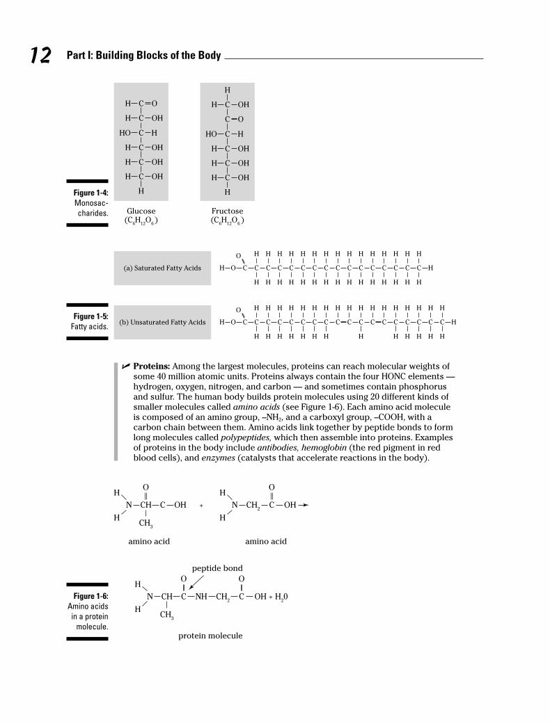
� Proteins: Among the largest molecules, proteins can reach molecular weights ofsome 40 million atomic units. Proteins always contain the four HONC elements —hydrogen, oxygen, nitrogen, and carbon — and sometimes contain phosphorusand sulfur. The human body builds protein molecules using 20 different kinds ofsmaller molecules called amino acids (see Figure 1-6). Each amino acid moleculeis composed of an amino group, –NH2, and a carboxyl group, –COOH, with acarbon chain between them. Amino acids link together by peptide bonds to formlong molecules called polypeptides, which then assemble into proteins. Examplesof proteins in the body include antibodies, hemoglobin (the red pigment in redblood cells), and enzymes (catalysts that accelerate reactions in the body).
+N CH C OH
H
HCH
3
N CH2
OHC
OOH
H
amino acidamino acid
N CH C NH
OH
HCH
3
CH2
OH + H20C
O
protein molecule
peptide bond
Figure 1-6:
Amino acids
in a protein
molecule.
COH C C C C C C C C C C C C C C C
HHHHHHHHHHHHHHH
HHHHHHHHHHHHHHH
O
H
COH C C C C C C C C C C C C C C C
HHHHHHHHHHHHHHH
HHHHHHHHHHH
O
C C
HH
HH
H
(a) Saturated Fatty Acids
(b) Unsaturated Fatty AcidsFigure 1-5:
Fatty acids.
OH C
OHH C
HHO C
OHH C
OHH C
OHH C
H
Glucose(C
6H
12O
6 )
Fructose(C
6H
12O
6 )
OHH C
H
OC
HHO C
OHH C
OHH C
OHH C
HFigure 1-4:
Monosac-
charides.
12 Part I: Building Blocks of the Body

� Nucleic acids: These long molecules, found primarily in the cell’s nucleus, act asthe body’s genetic blueprint. They’re comprised of smaller building blocks callednucleotides. Each nucleotide, in turn, is composed of a five-carbon sugar (deoxyri-bose or ribose), a phosphate group, and a nitrogenous base. The nitrogenousbases in DNA (deoxyribonucleic acid) are adenine, thymine, cytosine, and gua-nine; they always pair off A-T and C-G. In RNA (ribonucleic acid), which occurs ina single strand, thymine is replaced by uracil, so the nucleotides pair off A-U andC-G. In 1953, James Watson and Francis Crick published their discovery of thethree-dimensional structure of DNA — a polymer that looks like a ladder twistedinto a coil. They called this structure the double-stranded helix (see Figure 1-7).
A
Hydrogenbonds
G
G
C
G
AT
C
T
A
A
S A
S
S
S
S
S
P
P
P
P
G
C
A
A
G
T
T
C
A
G
T
C
= Adenine
= Guanine
Strand 1
S
P
Strand 2
Key:
S = Deoxyribose sugar
P = Phosphate sugar
= Thymine
= Cytosine
T
T
C
C
G
Figure 1-7:
The DNA
double helix.
13Chapter 1: The Chemistry of Life
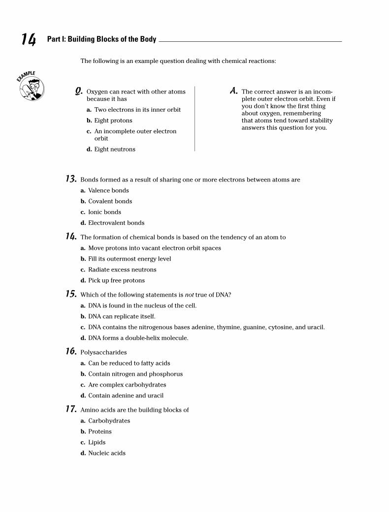
13. Bonds formed as a result of sharing one or more electrons between atoms are
a . Valence bonds
b . Covalent bonds
c . Ionic bonds
d . Electrovalent bonds
14. The formation of chemical bonds is based on the tendency of an atom to
a . Move protons into vacant electron orbit spaces
b . Fill its outermost energy level
c . Radiate excess neutrons
d . Pick up free protons
15. Which of the following statements is not true of DNA?
a . DNA is found in the nucleus of the cell.
b . DNA can replicate itself.
c . DNA contains the nitrogenous bases adenine, thymine, guanine, cytosine, and uracil.
d . DNA forms a double-helix molecule.
16. Polysaccharides
a . Can be reduced to fatty acids
b . Contain nitrogen and phosphorus
c . Are complex carbohydrates
d . Contain adenine and uracil
17. Amino acids are the building blocks of
a . Carbohydrates
b . Proteins
c . Lipids
d . Nucleic acids
14 Part I: Building Blocks of the Body
The following is an example question dealing with chemical reactions:
Q. Oxygen can react with other atomsbecause it has
a . Two electrons in its inner orbit
b . Eight protons
c . An incomplete outer electronorbit
d . Eight neutrons
A. The correct answer is an incom-plete outer electron orbit. Even ifyou don’t know the first thingabout oxygen, rememberingthat atoms tend toward stabilityanswers this question for you.
Cycling through Life: MetabolismMetabolism (from the Greek metabole, which means “change”) is the word for themyriad chemical reactions that happen in the body, particularly as they relate to gen-erating, storing, and expending energy. All metabolic reactions are either catabolic oranabolic. Catabolic reactions break food down into energy (memory tip: it can be cata-strophic when things break down). Anabolic reactions require the expenditure ofenergy to build up compounds that the body needs. The chemical alteration of mole-cules in the cell is referred to as cellular metabolism. Enzymes can be used as catalysts,accelerating chemical reactions without being changed by the reactions. The mole-cules that enzymes react with are called substrates.
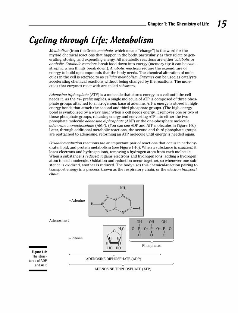
Adenosine triphosphate (ATP) is a molecule that stores energy in a cell until the cellneeds it. As the tri– prefix implies, a single molecule of ATP is composed of three phos-phate groups attached to a nitrogenous base of adenine. ATP’s energy is stored in high-energy bonds that attach the second and third phosphate groups. (The high-energybond is symbolized by a wavy line.) When a cell needs energy, it removes one or two ofthose phosphate groups, releasing energy and converting ATP into either the two-phosphate molecule adenosine diphosphate (ADP) or the one-phosphate moleculeadenosine monophosphate (AMP). (You can see ADP and ATP molecules in Figure 1-8.)Later, through additional metabolic reactions, the second and third phosphate groupsare reattached to adenosine, reforming an ATP molecule until energy is needed again.
Oxidation-reduction reactions are an important pair of reactions that occur in carbohy-drate, lipid, and protein metabolism (see Figure 1-10). When a substance is oxidized, itloses electrons and hydrogen ions, removing a hydrogen atom from each molecule.When a substance is reduced, it gains electrons and hydrogen ions, adding a hydrogenatom to each molecule. Oxidation and reduction occur together, so whenever one sub-stance is oxidized, another is reduced. The body uses this chemical-reaction pairing totransport energy in a process known as the respiratory chain, or the electron transportchain.
C
NN
N
CH
C
CC
N
HH
H H
HO HO
OO OP
O
OH
H2C
NH2
N
N
CH
C
C
C H
OP
O
OH
OP
O
OH
Phosphates
ADENOSINE DIPHOSPHATE (ADP)
Adenosine
Ribose
Adenine
ADENOSINE TRIPHOSPHATE (ATP)
Figure 1-8:
The struc-
tures of ADP
and ATP.
15Chapter 1: The Chemistry of Life
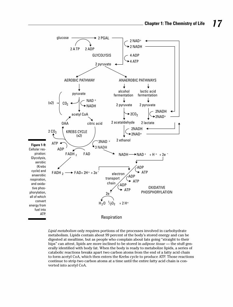
Carbohydrate metabolism involves a series of cellular respiration reactions, which areillustrated in Figure 1-9. All food carbohydrates are eventually broken down into glu-cose; therefore, carbohydrate metabolism is really glucose metabolism. Glucosemetabolism produces energy that is then stored in ATP molecules. The oxidationprocess in which energy is released from molecules, such as glucose, and transferredto other molecules is called cellular respiration. It occurs in every cell in the body andit is the cell’s source of energy. The complete oxidation of one molecule of glucose willproduce 38 molecules of ATP. It occurs in three stages: glycolysis, the Krebs cycle, andthe electron transport chain:
1. Glycolysis
From the Greek glyco (sugar) and lysis (breakdown), this is the first stage of bothaerobic (with oxygen) and anaerobic (without oxygen) respiration. Using energyfrom two molecules of ATP and two molecules of NAD+ (nicotinamide adenine di-nucleotide), glycolysis uses a process called phosphorylation to convert a mol-ecule of six-carbon glucose — the smallest molecule that the digestive systemcan produce during the breakdown of a carbohydrate — into two molecules ofthree-carbon pyruvic acid or pyruvate, as well as four ATP molecules and twomolecules of NADH (nicotinamide adenine dinucleotide). Taking place in the cell’scytoplasm (see Chapter 2), glycolysis doesn’t require oxygen to occur. The pyru-vate and NADH move into the cell’s mitochondria (detailed in Chapter 2), wherean aerobic (with oxygen) process converts them into ATP.
2. Krebs cycle
Also known as the tricarboxylic acid cycle or citric acid cycle, this series of energy-producing chemical reactions begins in the mitochondria after pyruvate arrivesfrom glycolysis. Before the Krebs cycle can begin, the pyruvate loses a carbondioxide group to form acetyl coenzyme A (acetyl CoA). Then acetyl CoA com-bines with a four-carbon molecule (oxaloacetic acid, or OAA) to form a six-carbon citric acid molecule that then enters the Krebs cycle. The CoA is releasedintact to bind with another acetyl group. During the conversion, two carbonatoms are lost as carbon dioxide and energy is released. One ATP molecule isproduced each time an acetyl CoA molecule is split. The cycle goes througheight steps, rearranging the atoms of citric acid to produce different intermedi-ate molecules called keto acids. The acetic acid is broken apart by carbon (ordecarboxylated) and oxidized, generating three molecules of NADH, one moleculeof FADH2 (flavin adenine dinucleotide), and one molecule of ATP. The energy canbe transported to the electron transport chain and used to produce more mole-cules of ATP. OAA is regenerated to get the next cycle going, and carbon dioxideproduced during this cycle is exhaled from the lungs.
3. Electron transport chain
The electron transport chain is a series of energy compounds attached to theinner mitochondrial membrane. The electron molecules in the chain are calledcytochromes. These electron-transferring proteins contain a heme, or iron, group.Hydrogen from oxidized food sources attaches to coenzymes that in turn com-bine with molecular oxygen. The energy released during these reactions is usedto attach inorganic phosphate groups to ADP and form ATP molecules.
Pairs of electrons transferred to NAD+ go through the electron transport processand produce three molecules of ATP by oxidative phosphorylation. Pairs of elec-trons transferred to FAD enter the electron transport after the first phosphoryla-tion and yield only two molecules of ATP. Oxidative phosphorylation isimportant because it makes energy available in a form the cells can use.
At the end of the chain, two positively charged hydrogen molecules combinewith two electrons and an atom of oxygen to form water. The final molecule towhich electrons are passed is oxygen. Electrons are transferred from one mole-cule to the next, producing ATP molecules.
16 Part I: Building Blocks of the Body
Lipid metabolism only requires portions of the processes involved in carbohydratemetabolism. Lipids contain about 99 percent of the body’s stored energy and can bedigested at mealtime, but as people who complain about fats going “straight to theirhips” can attest, lipids are more inclined to be stored in adipose tissue — the stuff gen-erally identified with body fat. When the body is ready to metabolize lipids, a series ofcatabolic reactions breaks apart two carbon atoms from the end of a fatty acid chainto form acetyl CoA, which then enters the Krebs cycle to produce ATP. Those reactionscontinue to strip two carbon atoms at a time until the entire fatty acid chain is con-verted into acetyl CoA.
glucose 2 PGAL
2 pyruvate
GLYCOLYSIS
2 A TP 2 ADP
4 ADP
4 ATP
2 NAD+
2 NADH
2NADH
2NAD+
2NADH
2NAD+
pyruvate
NAD +
NADHCO2
acetyl CoA
citric acid
KREBS CYCLE(x2)
OAA
2 CO2
ATP
ADP3 NADH
3NAD +
alcoholfermentation
2 pyruvate 2 pyruvate
2 acetaldehyde
2 ethanol
2CO2
lactic acidfermentation
2 lactate
ADP
ATP
ADP
ATP
ADP
ATPelectrontransport
chain
(x2)
NADH NAD + H + 2e -+ +
Respiration
F ADH F AD2
F ADH F AD+ 2H + 2e -2
+
2e -
H O 1⁄2O + 2 H2 2+
OXIDATIVE
PHOSPHORYLATION
AEROBIC PATHWAY ANAEROBIC PATHWAYS
Figure 1-9:
Cellular res-
piration:
Glycolysis,
aerobic
(Krebs
cycle) and
anaerobic
respiration,
and oxida-
tive phos-
phorylation,
all of which
convert
energy from
fuel into
ATP.
17Chapter 1: The Chemistry of Life
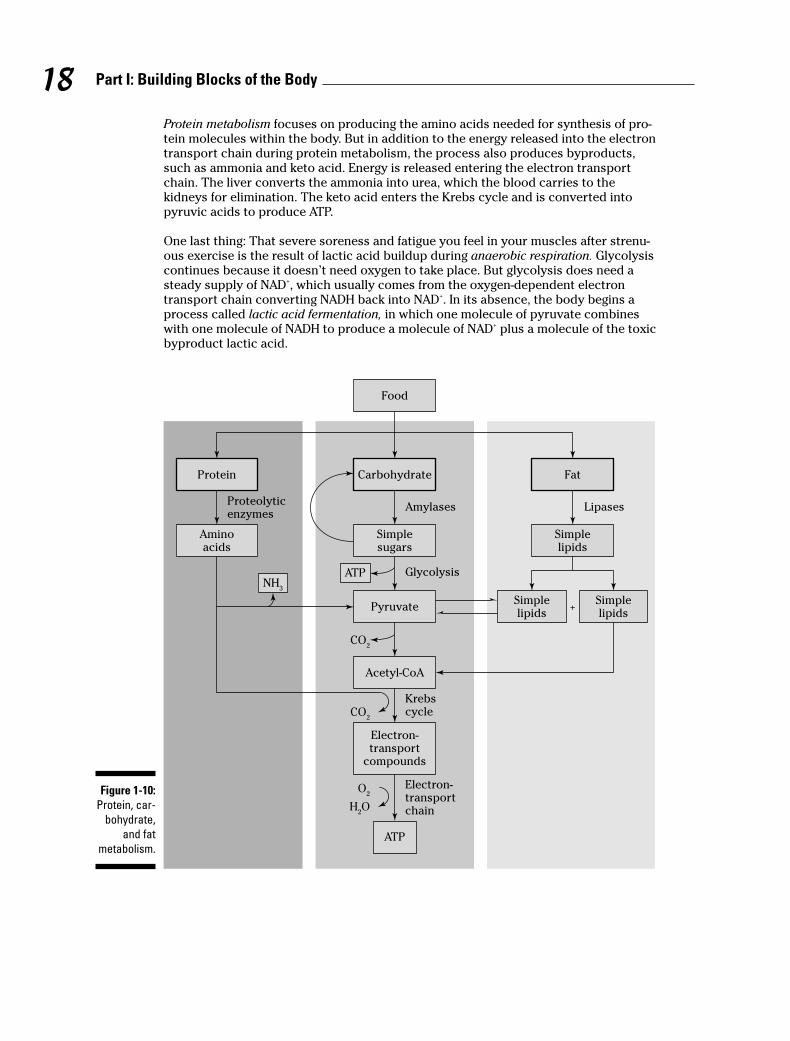
Protein metabolism focuses on producing the amino acids needed for synthesis of pro-tein molecules within the body. But in addition to the energy released into the electrontransport chain during protein metabolism, the process also produces byproducts,such as ammonia and keto acid. Energy is released entering the electron transportchain. The liver converts the ammonia into urea, which the blood carries to thekidneys for elimination. The keto acid enters the Krebs cycle and is converted intopyruvic acids to produce ATP.
One last thing: That severe soreness and fatigue you feel in your muscles after strenu-ous exercise is the result of lactic acid buildup during anaerobic respiration. Glycolysiscontinues because it doesn’t need oxygen to take place. But glycolysis does need asteady supply of NAD+, which usually comes from the oxygen-dependent electrontransport chain converting NADH back into NAD+. In its absence, the body begins aprocess called lactic acid fermentation, in which one molecule of pyruvate combineswith one molecule of NADH to produce a molecule of NAD+ plus a molecule of the toxicbyproduct lactic acid.
Food
Carbohydrate
Pyruvate +
Simplesugars
Simplelipids
Simplelipids
Simplelipids
Protein Fat
Aminoacids
Proteolyticenzymes
Amylases Lipases
Glycolysis
Krebs
cycle
Electron-transportchain
Acetyl-CoA
ATP
ATPNH
3
CO2
CO2
Electron-transport
compounds
H2O
O2Figure 1-10:
Protein, car-
bohydrate,
and fat
metabolism.
18 Part I: Building Blocks of the Body

18. A molecule of glucose is broken down to pyruvic acid by
a. Glycolysis
b. The Krebs cycle
c. The electron transport chain
d. Oxidative phosphorylation
19. Pyruvic acid enters a mitochondrion and is converted into
a. Glucose
b. Acetyl CoA
c. Water
d. Protein
20. A molecule of glucose can be converted into how many ATP molecules?
a. 2
b. 3
c. 38
d. 45
21. The part of metabolism that involves creating compounds the body needs is called
a. A catabolic reaction
b. Cellular respiration
c. An anabolic reaction
d. Oxidation
22. Metabolic processes that don’t require oxygen are called
a. Anaerobic
b. Aerobic
c. Fermentation
d. Carbon dioxination
19Chapter 1: The Chemistry of Life
Check out an example question on metabolism:
Q. Cells obtain ATP by converting theenergy in
a. Carbohydrates
b. Proteins
c. Lipids
d. All of these
A. The correct answer is all of these.While it’s true that carbohydratesprovide the most immediatelyavailable energy, proteins andlipids also contribute to the pro-duction of ATP.
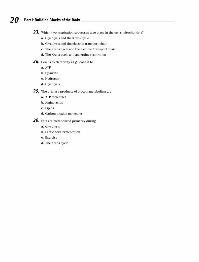
23. Which two respiration processes take place in the cell’s mitochondria?
a. Glycolysis and the Krebs cycle
b. Glycolysis and the electron transport chain
c. The Krebs cycle and the electron transport chain
d. The Krebs cycle and anaerobic respiration
24. Coal is to electricity as glucose is to
a. ATP
b. Pyruvate
c. Hydrogen
d. Glycolysis
25. The primary products of protein metabolism are
a. ATP molecules
b. Amino acids
c. Lipids
d. Carbon dioxide molecules
26. Fats are metabolized primarily during
a. Glycolysis
b. Lactic acid fermentation
c. Exercise
d. The Krebs cycle
20 Part I: Building Blocks of the Body
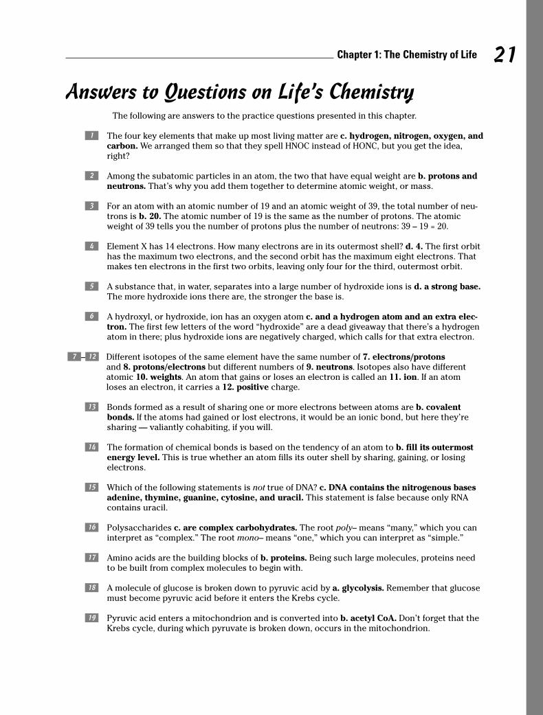
Answers to Questions on Life’s ChemistryThe following are answers to the practice questions presented in this chapter.
a The four key elements that make up most living matter are c. hydrogen, nitrogen, oxygen, andcarbon. We arranged them so that they spell HNOC instead of HONC, but you get the idea,right?
b Among the subatomic particles in an atom, the two that have equal weight are b. protons andneutrons. That’s why you add them together to determine atomic weight, or mass.
c For an atom with an atomic number of 19 and an atomic weight of 39, the total number of neu-trons is b. 20. The atomic number of 19 is the same as the number of protons. The atomicweight of 39 tells you the number of protons plus the number of neutrons: 39 – 19 = 20.
d Element X has 14 electrons. How many electrons are in its outermost shell? d. 4. The first orbithas the maximum two electrons, and the second orbit has the maximum eight electrons. Thatmakes ten electrons in the first two orbits, leaving only four for the third, outermost orbit.
e A substance that, in water, separates into a large number of hydroxide ions is d. a strong base.The more hydroxide ions there are, the stronger the base is.
f A hydroxyl, or hydroxide, ion has an oxygen atom c. and a hydrogen atom and an extra elec-tron. The first few letters of the word “hydroxide” are a dead giveaway that there’s a hydrogenatom in there; plus hydroxide ions are negatively charged, which calls for that extra electron.
g–l Different isotopes of the same element have the same number of 7. electrons/protonsand 8. protons/electrons but different numbers of 9. neutrons. Isotopes also have differentatomic 10. weights. An atom that gains or loses an electron is called an 11. ion. If an atom loses an electron, it carries a 12. positive charge.
m Bonds formed as a result of sharing one or more electrons between atoms are b. covalentbonds. If the atoms had gained or lost electrons, it would be an ionic bond, but here they’resharing — valiantly cohabiting, if you will.
n The formation of chemical bonds is based on the tendency of an atom to b. fill its outermostenergy level. This is true whether an atom fills its outer shell by sharing, gaining, or losing electrons.
o Which of the following statements is not true of DNA? c. DNA contains the nitrogenous basesadenine, thymine, guanine, cytosine, and uracil. This statement is false because only RNAcontains uracil.
p Polysaccharides c. are complex carbohydrates. The root poly– means “many,” which you caninterpret as “complex.” The root mono– means “one,” which you can interpret as “simple.”
q Amino acids are the building blocks of b. proteins. Being such large molecules, proteins needto be built from complex molecules to begin with.
r A molecule of glucose is broken down to pyruvic acid by a. glycolysis. Remember that glucosemust become pyruvic acid before it enters the Krebs cycle.
s Pyruvic acid enters a mitochondrion and is converted into b. acetyl CoA. Don’t forget that theKrebs cycle, during which pyruvate is broken down, occurs in the mitochondrion.
21Chapter 1: The Chemistry of Life
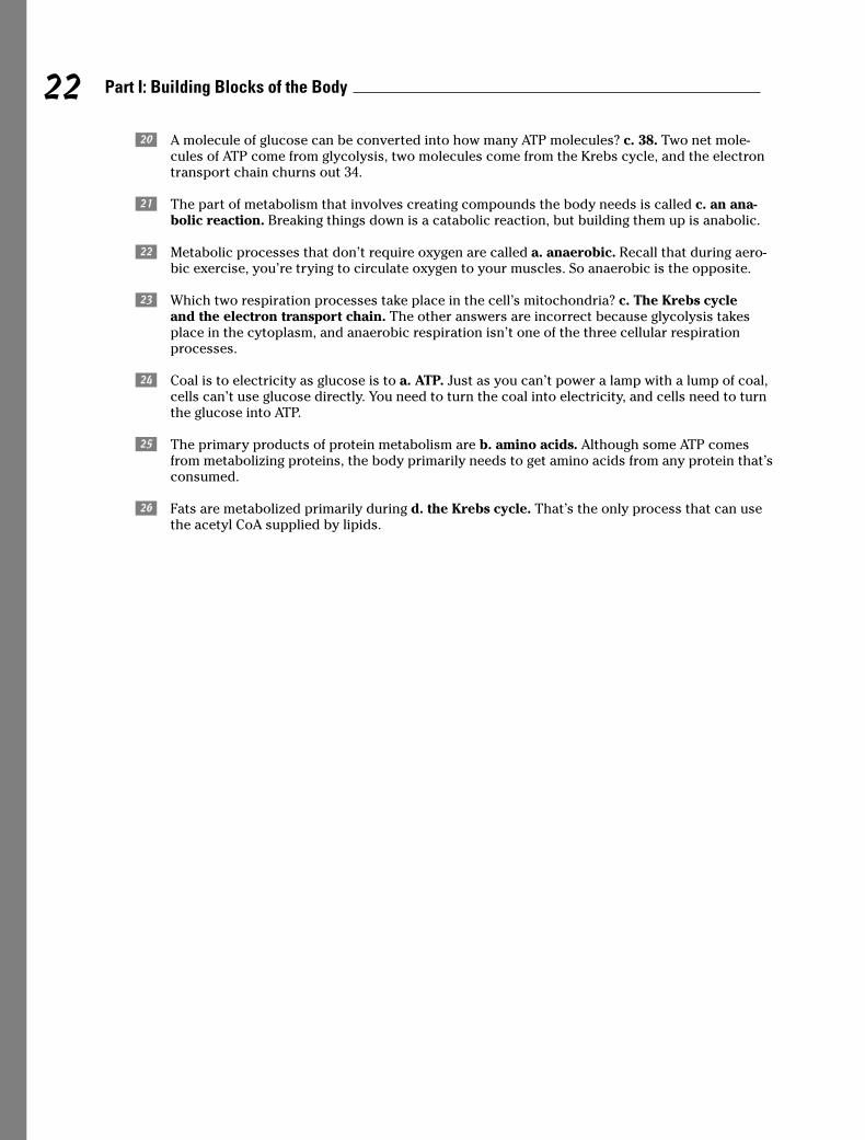
t A molecule of glucose can be converted into how many ATP molecules? c. 38. Two net mole-cules of ATP come from glycolysis, two molecules come from the Krebs cycle, and the electrontransport chain churns out 34.
u The part of metabolism that involves creating compounds the body needs is called c. an ana-bolic reaction. Breaking things down is a catabolic reaction, but building them up is anabolic.
v Metabolic processes that don’t require oxygen are called a. anaerobic. Recall that during aero-bic exercise, you’re trying to circulate oxygen to your muscles. So anaerobic is the opposite.
w Which two respiration processes take place in the cell’s mitochondria? c. The Krebs cycle and the electron transport chain. The other answers are incorrect because glycolysis takesplace in the cytoplasm, and anaerobic respiration isn’t one of the three cellular respirationprocesses.
x Coal is to electricity as glucose is to a. ATP. Just as you can’t power a lamp with a lump of coal,cells can’t use glucose directly. You need to turn the coal into electricity, and cells need to turnthe glucose into ATP.
y The primary products of protein metabolism are b. amino acids. Although some ATP comesfrom metabolizing proteins, the body primarily needs to get amino acids from any protein that’sconsumed.
A Fats are metabolized primarily during d. the Krebs cycle. That’s the only process that can usethe acetyl CoA supplied by lipids.
22 Part I: Building Blocks of the Body

Chapter 2
The Cell: Life’s Basic Building Block
In This Chapter� Breaking through the cell membrane
� Aiming for the nucleus
� Sorting through what’s inside the cell
� Putting together proteins made to order
� Following the cell cycle
Cytology, from the Greek word cyto, which means “cell,” is the study of cells. Every livingthing has cells, but not all living things have the same kinds of cells. Eukaryotes like
humans (and all other organisms besides bacteria and viruses) have eukaryotic cells, eachof which has a defined nucleus that controls and directs the cell’s activities, and cytosol,fluid material found in the gel-like cytoplasm that fills most of the cell. Plant cells havefibrous cell walls; animal cells do not, making do instead with a semipermeable cell mem-brane, which sometimes is called a plasma membrane or the plasmalemma. Because humancells don’t have cell walls, they look like gel-filled sacs with nuclei and tiny parts calledorganelles nestled inside when viewed through an electron microscope.
In this chapter, we help you sort out what makes up a cell, what all those tiny parts do, andhow cells act as protein-manufacturing plants to support life’s activities. We then take a quicklook at an individual cell’s life cycle.
Gaining Admission: The Cell MembraneThink of it as a gatekeeper, guardian, or border guard. Despite being only 6 to 10 nanometersthick and visible only through an electron microscope, the cell membrane keeps the cell’scytoplasm in place and lets only select materials enter and depart the cell as needed. Thissemipermeability, or selective permeability, is a result of a double layer (bilayer) of phospho-lipid molecules interspersed with protein molecules. The outer surface of each layer is madeup of tightly packed hydrophilic (or water-loving) polar heads. Inside, between the two layers,you find hydrophobic (or water-fearing) nonpolar tails consisting of fatty acid chains.Cholesterol molecules between the phosphate layers give the otherwise elastic membranestability and make it less permeable to water-soluble substances. Both cytoplasm and thematrix, the material in which cells lie, are primarily water. The polar heads electrostaticallyattract polarized water molecules while the nonpolar tails lie between the layers, shieldedfrom water and creating a dry middle layer. The membrane’s interior is made up of oily fattyacid molecules that are electrostatically symmetric, or nonpolarized. Lipid-soluble moleculescan pass through this layer, but water-soluble molecules such as amino acids, sugars, andproteins cannot. Because phospholipids have both polar and nonpolar regions, they’re alsocalled amphipathic molecules.
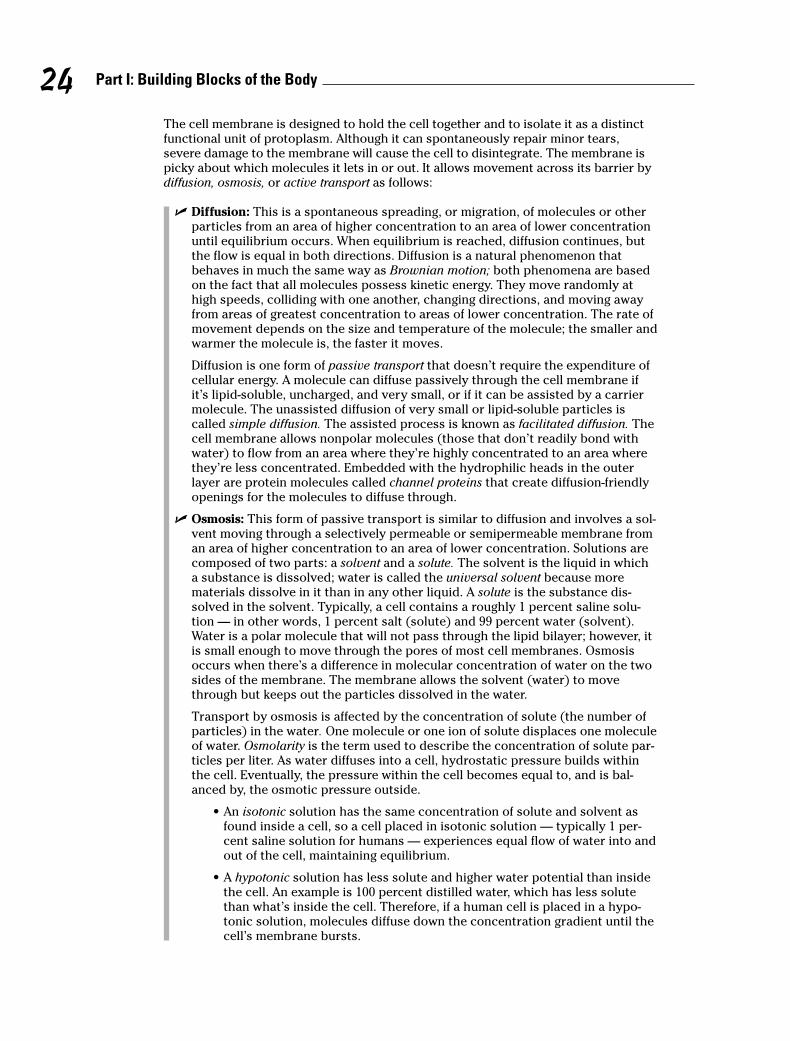
The cell membrane is designed to hold the cell together and to isolate it as a distinctfunctional unit of protoplasm. Although it can spontaneously repair minor tears,severe damage to the membrane will cause the cell to disintegrate. The membrane ispicky about which molecules it lets in or out. It allows movement across its barrier bydiffusion, osmosis, or active transport as follows:
� D if f u s io n : This is a spontaneous spreading, or migration, of molecules or otherparticles from an area of higher concentration to an area of lower concentrationuntil equilibrium occurs. When equilibrium is reached, diffusion continues, butthe flow is equal in both directions. Diffusion is a natural phenomenon thatbehaves in much the same way as Brownian motion; both phenomena are basedon the fact that all molecules possess kinetic energy. They move randomly athigh speeds, colliding with one another, changing directions, and moving awayfrom areas of greatest concentration to areas of lower concentration. The rate ofmovement depends on the size and temperature of the molecule; the smaller andwarmer the molecule is, the faster it moves.
Diffusion is one form of passive transport that doesn’t require the expenditure ofcellular energy. A molecule can diffuse passively through the cell membrane ifit’s lipid-soluble, uncharged, and very small, or if it can be assisted by a carriermolecule. The unassisted diffusion of very small or lipid-soluble particles iscalled simple diffusion. The assisted process is known as facilitated diffusion. Thecell membrane allows nonpolar molecules (those that don’t readily bond withwater) to flow from an area where they’re highly concentrated to an area wherethey’re less concentrated. Embedded with the hydrophilic heads in the outerlayer are protein molecules called channel proteins that create diffusion-friendlyopenings for the molecules to diffuse through.
� O s m o s is : This form of passive transport is similar to diffusion and involves a sol-vent moving through a selectively permeable or semipermeable membrane froman area of higher concentration to an area of lower concentration. Solutions arecomposed of two parts: a solvent and a solute. The solvent is the liquid in whicha substance is dissolved; water is called the universal solvent because morematerials dissolve in it than in any other liquid. A solute is the substance dis-solved in the solvent. Typically, a cell contains a roughly 1 percent saline solu-tion — in other words, 1 percent salt (solute) and 99 percent water (solvent).Water is a polar molecule that will not pass through the lipid bilayer; however, itis small enough to move through the pores of most cell membranes. Osmosisoccurs when there’s a difference in molecular concentration of water on the twosides of the membrane. The membrane allows the solvent (water) to movethrough but keeps out the particles dissolved in the water.
Transport by osmosis is affected by the concentration of solute (the number ofparticles) in the water. One molecule or one ion of solute displaces one moleculeof water. Osmolarity is the term used to describe the concentration of solute par-ticles per liter. As water diffuses into a cell, hydrostatic pressure builds withinthe cell. Eventually, the pressure within the cell becomes equal to, and is bal-anced by, the osmotic pressure outside.
• An isotonic solution has the same concentration of solute and solvent asfound inside a cell, so a cell placed in isotonic solution — typically 1 per-cent saline solution for humans — experiences equal flow of water into andout of the cell, maintaining equilibrium.
• A hypotonic solution has less solute and higher water potential than insidethe cell. An example is 100 percent distilled water, which has less solutethan what’s inside the cell. Therefore, if a human cell is placed in a hypo-tonic solution, molecules diffuse down the concentration gradient until thecell’s membrane bursts.
24 Part I: Building Blocks of the Body
• A hypertonic solution has more solute and lower water potential than insidethe cell. So the membrane of a human cell placed in 10 percent saline solu-tion (10 percent salt and 90 percent water) would let water flow out of thecell (from higher concentration inside to lower concentration outside),therefore shrinking it.
� Active transport: This movement occurs across a semipermeable membraneagainst the normal concentration gradient, moving from the area of lower con-centration to the area of higher concentration and requiring an expenditure ofenergy released from an ATP molecule (as discussed in Chapter 1). Embeddedwith the hydrophilic heads in the outer layer of the membrane are protein mole-cules able to detect and move compounds through the membrane. These carrieror transport proteins interact with the passenger molecules and use the ATP-sup-plied energy to move them against the gradient. The carrier molecules combinewith the transport molecules — most importantly amino acids and ions — topump them against their concentration gradients.
Active transport lets cells obtain nutrients that can’t pass through the mem-brane by other means. In addition, there are secondary active transportprocesses that are similar to diffusion but instead use imbalances in electrostaticforces to move molecules across the membrane.
1.–3. Fill in the blanks to complete the following sentences:
The lipid bilayer structure of the cell membrane is made possible because phospholipidmolecules contain two distinct regions: The 1. _______________ region is attracted to water,and the 2. _______________ region is repelled by water. Because it has both polar and non-polar regions, a phospholipid is classified as a(n) 3. _______________ molecule.
4. The movement of water molecules through a semipermeable membrane is known as
a. Diffusion
b. Filtration
c. Osmosis
d. Active transport
5. A solution having a greater concentration of water than exists in the cell is said to be
a. Hypertonic
b. Hypotonic
c. Isotonic
d. Heterotonic
6. Injecting a large quantity of distilled water into a human’s veins would cause many red bloodcells to
a. Swell and burst
b. Shrink
c. Carry more oxygen
d. Aggregate
7. The cell membrane does not function
a. In selective transport of materials into and out of the cell
b. As a barrier protecting the cell
c. In the production of energy
d. As containment for the cytoplasm
25Chapter 2: The Cell: Life’s Basic Building Block

Aiming for the NucleusThe cell nucleus is the largest cellular organelle and the first to be discovered by scien-tists. On average, it accounts for about 10 percent of the total volume of the cell, and itholds a complete set of genes.
The outermost part of this organelle is the nuclear envelope, which is composed of adouble-membrane barrier, each membrane of which is made up of a phospholipidbilayer. Between the two membranes is a fluid-filled space called the perinuclear cis-terna. The two layers fuse to form a selectively permeable barrier, but large poresallow relatively free movement of molecules and ions, including large protein mole-cules. Intermediate filaments lining the surface of the nuclear envelope make up thenuclear lamina, which functions in the disassembly and reassembly of the nuclearmembrane during mitosis and binds the membrane to the endoplasmic reticulum.The nucleus also contains nucleoplasm, a clear viscous material that forms the matrixin which the organelles of the nucleus are embedded.
DNA is packaged inside the nucleus in structures called chromatin, or chromatin networks. During cell division, the chromatin contracts, forming chromosomes.Chromosomes contain a DNA molecule encoded with the genetic information neededto direct the cell’s activities. The most prominent subnuclear body is the nucleolus, asmall spherical body that stores RNA molecules and produces ribosomes, which areexported to the cytoplasm where they translate messenger RNA (mRNA).
The following is an example question about the nucleus:
26 Part I: Building Blocks of the Body
Q. The only cellular organelle foundwithin the nucleus is called a(n)
a. Lamina
b. Envelope
c. Nucleolus
d. Chromosome
A. The correct answer is nucleolus.The other options aren’t consid-ered organelles.
8. The fluid-filled space within the nuclear envelope is called the
a. Perinuclear cisterna
b. Ribosome
c. Mitochondrion
d. Golgi appartus
9. DNA is packaged within
a. Chromatins
b. Chromosomes
c. Ribosomes
d. Genes
10. The nucleolus
a. Packages DNA
b. Enables large molecule transport
c. Forms a membrane around the nucleus
d. Assembles ribosomes
Looking Inside: Organelles and Their Functions
Molecules that pass muster with the cell membrane enter the cytoplasm, a mixture ofmacromolecules such as proteins and RNA and small organic molecules such as glu-cose, ions, and water. Because of the various materials in the cytoplasm, it’s a colloid,or mixture of phases, that alternates from a sol (a liquid colloid with solid suspendedin it) to a gel (a colloid in which the dispersed phase combines with the medium toform a semisolid material). The fluid part of the cytoplasm, called the cytosol, has a dif-fering consistency based on changes in temperature, molecular concentrations, pH,pressure, and agitation.
Within the cytoplasm lies a network of fibrous proteins collectively referred to as thecytoskeleton. It’s not rigid or permanent but changing and shifting according to theactivity of the cell. The cytoskeleton maintains the cell’s shape, enables it to move,anchors its organelles, and directs the flow of the cytoplasm. The fibrous proteins thatmake up the cytoskeleton include the following:
� Microfilaments, rodlike structures about 5 to 8 nanometers wide that consist of astacked protein called actin, the most abundant protein in eukaryotic cells. Theyprovide structural support and have a role in cell and organelle movement aswell as in cell division.
� Intermediate filaments, the strongest and most stable part of the cytoskeleton.They average about 10 nanometers wide and consist of interlocking proteins,including keratin, that chiefly are involved in maintaining cell integrity and resist-ing pulling forces on the cell.
� Hollow microtubules about 25 nanometers in diameter that are made of the pro-tein tubulin and grow with one end embedded in the centrosome near the cell’snucleus. Like microfilaments, these components of cilia, flagella, and centriolesprovide structural support and have a role in cell and organelle movement aswell as in cell division.
Organelles, literally translated as “little organs,” are nestled inside the cytoplasm(except for the two organelles that move, cilia and flagellum, which are found on thecell’s exterior). Each organelle has different responsibilities for producing materialsused elsewhere in the cell or body. Here are the key organelles and what they do:
� Centrosome: Microtubules sprout from this structure, which is located next tothe nucleus and is composed of two centrioles — arrays of microtubules — thatfunction in separating genetic material during cell division.
� Cilia: These are short, hair-like cytoplasmic projections on the external surface ofthe cell. In multicellular animals, including humans, cilia move materials over thesurface of the cell. In some single-celled organisms, they’re used for locomotion.
27Chapter 2: The Cell: Life’s Basic Building Block
� Endoplasmic reticulum (ER): This organelle makes direct contact with the cellnucleus and functions in the transport of materials such as proteins and RNAmolecules. Composed of membrane-bound canals and cavities that extend fromthe nuclear membrane to the cell membrane, the ER is the site of lipid and pro-tein synthesis. The two types of ER are rough, which is dotted with ribosomes onthe outer surface; and smooth, which has no ribosomes on the surface.
� Flagellum: This whip-like cytoplasmic projection lies on the cell’s exterior sur-face. Found in humans primarily on sperm cells, it’s used for locomotion.
� Golgi apparatus (or body): This organelle consists of a stack of flattened sacswith membranes that connect with those of the endoplasmic reticulum. Locatednear the nucleus, it functions in the storage, modification, and packaging of pro-teins for secretion to various destinations within the cell.
� Lysosome: A tiny, membranous sac containing acids and digestive enzymes, thelysosome breaks down large food molecules such as proteins, carbohydrates,and nucleic acids into materials that the cell can use. It destroys foreign particlesin the cell and helps to remove nonfunctioning structures from the cell.
� Mitochondrion: Called the powerhouse of the cell, this rod-shaped organelleconsists of two membranes — a smooth outer membrane, and an invaginated(folded inward) inner membrane that divides the organelle into compartments.The inward-folding crevices of the inner membrane are called cristae. The mito-chondrion provides critical functions in cell respiration, including oxidizing(breaking down) food molecules and releasing energy that is stored in ATP mole-cules in the mitochondrion. This energy is used to accelerate chemical reactionsin the cell, which we cover in Chapter 1.
� Ribosomes: These roughly 25-nanometer structures may be found along theendoplasmic reticulum or floating free in the cytoplasm. Composed of 60 percentRNA and 40 percent protein, they translate the genetic information on RNA mole-cules to synthesize, or produce, a protein molecule.
� Vacuoles: More commonly found in plant cells, these open spaces in the cyto-plasm sometimes carry materials to the cell membrane for discharge to the out-side of the cell. In animal cells, food vacuoles are membranous sacs formedwhen food masses are pinched-off from the cell membrane and passed into thecytoplasm of the cell. This process, called endocytosis (from the Greek wordsmeaning “within the cell”), requires energy to move large masses of material intothe cell. Vacuoles also help to remove structural debris, isolate harmful materi-als, and export unwanted substances from the cell.
Answer these practice questions about cell organelles:
11. This cigar-shaped organelle produces energy through aerobic respiration.
a. Golgi apparatus
b. Mitochondrion
c. Lysosome
d. Endoplasmic reticulum
28 Part I: Building Blocks of the Body

12. The most abundant protein in human cells is
a. Actin
b. Tubulin
c. Albumen
d. Cytoplasm
13. Which organelle gets to take out the cellular trash?
a. Golgi apparatus
b. Ribosome
c. Vacuole
d. Endoplasmic reticulum
14. The very small organelle responsible for protein synthesis (making proteins) is the
a. Ribosome
b. Lysosome
c. Centriole
d. Vesicle
15. Which organelle has ribosomes attached to it?
a. Smooth (agranular) endoplasmic reticulum
b. Golgi apparatus
c. Rough (granular) endoplasmic reticulum
d. Nucleus
16. Which organelle contains secretory materials?
a. Golgi apparatus
b. Ribosome
c. Lysosome
d. Endoplasmic reticulum
17. Which of the following can change the consistency of cytoplasm?
a. Changes in acidity or alkalinity
b. Temperature
c. Pressure
d. All of the above
18. Structures found inside the nucleus include the
a. Mitochondria
b. Lysosomes
c. Chromatin network
d. Ribosomes
29Chapter 2: The Cell: Life’s Basic Building Block
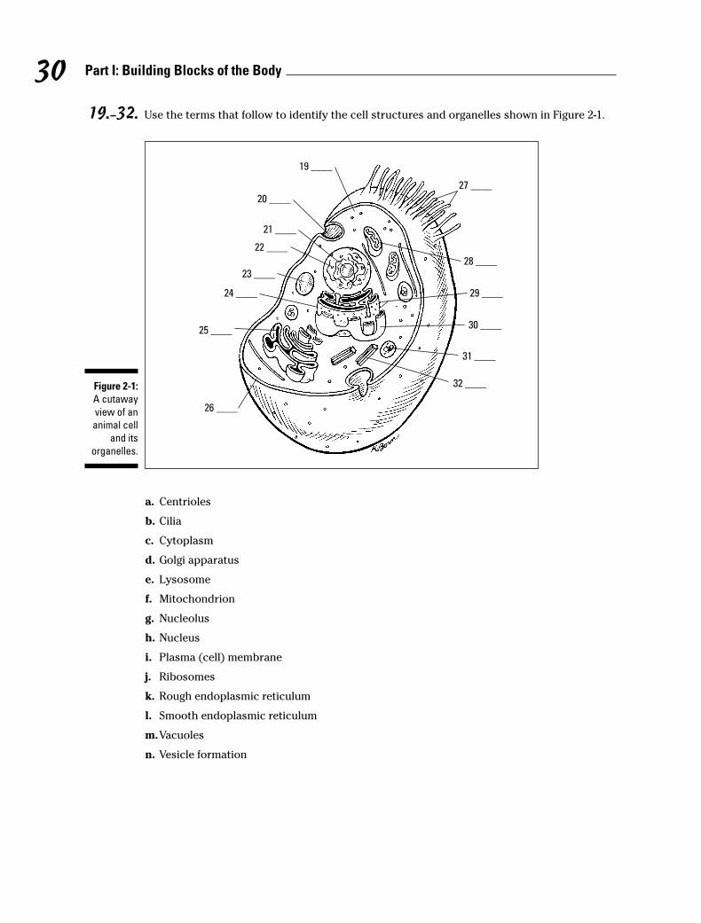
19.– 32. Use the terms that follow to identify the cell structures and organelles shown in Figure 2-1.
a. Centrioles
b. Cilia
c. Cytoplasm
d. Golgi apparatus
e. Lysosome
f. Mitochondrion
g. Nucleolus
h. Nucleus
i. Plasma (cell) membrane
j. Ribosomes
k. Rough endoplasmic reticulum
l. Smooth endoplasmic reticulum
m.Vacuoles
n. Vesicle formation
19 ____
20 ____
21 ____
23 ____
24 ____
25 ____
32 ____
26 ____
22 ____
31 ____
29 ____
28 ____
27 ____
30 ____
Figure 2-1:
A cutaway
view of an
animal cell
and its
organelles.
30 Part I: Building Blocks of the Body
33.–37. Match the organelles with their descriptions.
33. _____ Mitochondrion
34. _____ Nucleolus
35. _____ Flagellum
36. _____ Cytoplasm
37. _____ Lysosomes
Putting Together New ProteinsProteins are essential building blocks for all living systems, which helps explain whythe word is derived from the Greek term proteios, meaning “holding first place.” Cellsuse proteins to perform a variety of functions, including providing structural supportand catalyzing reactions. Cells synthesize proteins through a systematic procedurethat begins in the nucleus when the gene code for a certain protein is transcribed fromthe cell’s DNA into messenger RNA, or mRNA. The mRNA moves through nuclear poresto the rough endoplasmic reticulum (ER), where ribosomes translate the message onecodon of three nucleotides, or base pairs, at a time. The ribosome uses transfer RNA, ortRNA, to fetch each required amino acid and then link them together through peptidebonds, also known as amide bonds, to form proteins (see Figure 2-2 for details).
Don’t let the labels confuse you. Proteins are chains of amino acids (usually very longchains of at least 100 acids). Enzymes, used to catalyze reactions, also are chains ofamino acids and therefore also are categorized as proteins. Polypeptides, or simplypeptides, are shorter chains of amino acids used to bond larger protein molecules, butthey also can be regarded as proteins. Both antibodies and hormones also are pro-teins, along with almost everything else in the body — hair, muscle, cartilage, and soon. Even the four basic blood types — A, B, AB, and O — are differentiated by the pro-teins found in each.
31Chapter 2: The Cell: Life’s Basic Building Block
a. Long, whip-like organelle for locomotion
b. Fluid-like interior of the cell that may become a semisolid,or colloid
c. Membranous sacs containing digestive enzymes
d. Powerhouse of the cell
e. Stores RNA in the nucleus

G
Psite Asite
larger RNAsubunit
small rRNAsubunit
arg
C CA
glu
U UG
AA
G
G
G
G
U
U
U U
U
CC
ser
UA
pro
UGG
U A G
valA A A
G A G
G
tRNA
mRNA
ala
his
met
smallandlargeribosomesubunits
trans la
tio
ntrans lation
growingpolypeptide
5'
AA
A
C
UA
Cm
etUA
CA
AC
A
A A
cys
C
3'
5' 3'
T
U A
U
U
G
A
C
C G
5'
DNA
mRNA
3'5'
RNApolymerase
transcription
Nucleus
m
et - asn UC
G
Psite
Asite
3' 5'
Protein Synthesis
Figure 2-2:
The process
of protein
synthesis.
32 Part I: Building Blocks of the Body
38.–44. Fill in the blanks to complete the following sentences:
Protein synthesis begins in the cell’s 38. _______________ when the gene for a certain pro-tein is 39. _______________ into messenger RNA, or mRNA, which then moves on to the 40. _______________. There, ribosomes 41. _______________ three base pairs at a time, form-ing a series also referred to as a 42. _______________. Molecules called 43. _______________then collect each amino acid needed so that the ribosomes can link them together through44. _______________ bonds.
45. The word “protein” can refer to
a. Hormones
b. Enzymes
c. Antibodies
d. All of the above
46. Which of the following comes first in the protein-synthesis process?
a. Transfer RNA
b. Transcription
c. Peptide bonds
d. Translation
47. tRNA is used to gather
a. Blood cells
b. Amino acids
c. Protein molecules
d. DNA
48. A codon is a sequence of three
a. Nucleotides
b. Amino acids
c. Ribosomes
d. Base pieces
Cycling Along: Grow, Rest, Divide, DieThe cell life cycle, usually referred to simply as the cell cycle or the CDC (cell divisioncycle), extends from the beginning of one cell division to the beginning of the nextdivision. The human body produces new cells every day to replace those that aredamaged or worn out.
33Chapter 2: The Cell: Life’s Basic Building Block
The cell cycle is divided into two distinct phases:
� Interphase: Sometimes also called the resting stage, that label is a misnomerbecause the cell is actively growing and carrying out its normal metabolic func-tions as well as preparing for cell division.
� Mitosis: The period of cell division that produces new cells. (We cover this phasein detail in Chapter 3.)
New cells are produced for growth and to replace the billions of cells that stop function-ing in the adult human body every day. Some cells, like blood and skin cells, are contin-ually dividing because they have very short life cycles, sometimes only hours. Othercells, such as specialized muscle cells and certain nerve cells, may never divide at all.
49. Human cells can live
a. A few hours
b. A few days
c. Indefinitely
d. All of the above
50. The cell cycle is measured
a. By the number of times a cell divides
b. From the beginning to the end of one cell division
c. From the beginning of one cell division to the beginning of the next
d. Slowly, over time
34 Part I: Building Blocks of the Body

35Chapter 2: The Cell: Life’s Basic Building Block
Answers to Questions on the CellThe following are answers to the practice questions presented in this chapter.
a–c The lipid bilayer structure of the cell membrane is made possible because phospholipid mole-cules contain two distinct regions: The 1. hydrophilic region is attracted to water, and the 2. hydrophobic region is repelled by water. Because it has both polar and nonpolar regions, aphospholipid is classified as a(n) 3. amphipathic molecule.
d The movement of water molecules through a semipermeable membrane is known as c. osmosis.Why not diffusion? Because diffusion has to do with the passive transport of substances otherthan water.
e A solution having a greater concentration of water than exists in the cell is said to be b. hypotonic.
The prefix hypo refers to under or below normal. The prefix hyper refers to excess, or abovenormal. Someone who has been out in the cold too long suffers hypothermia — literally insuffi-cient heat. So a solution, or tonic, with very few particles would be hypotonic.
f Injecting a large quantity of distilled water into a human’s veins would cause many red bloodcells to a. swell and burst. With more water outside the cell than inside, the membrane wouldallow osmosis to continue past the breaking point.
g The cell membrane does not function c. in the production of energy. Sometimes it may useenergy in the form of ATP, but the cell membrane isn’t involved directly in the production ofenergy.
h The only organelle with direct links to the nucleus is the a. endoplasmic reticulum. Thenuclear lamina, or intermediate filaments, link the ER with the nucleus.
i DNA is packaged within b. chromosomes. None of the other answer options contains a fullpackage of DNA.
j The nucleolus d. assembles ribosomes. It’s not just a coincidence that the nucleolus sits at theheart of the genetic powerhouse.
k This cigar-shaped organelle produces energy through aerobic respiration. b. Mitochondrion.None of the other options are involved in cellular respiration or energy production
l The most abundant protein in human cells is a. actin. It makes sense that the protein makingup much of the cytoskeleton is the most abundant because the cytoskeleton accounts for up to50 percent of the cell’s volume.
m Which organelle gets to take out the cellular trash? c. Vacuole
When it’s time to clean house, you pull out the vacuum. Cells pull out the vacuoles.
n The very small organelle responsible for protein synthesis (making proteins) is the a. ribosome.

36 Part I: Building Blocks of the Body
When you think of a big protein-laden meal, you think of ribs. Ribs. Ribosome. Protein synthe-sis. Get it?
o Which organelle has ribosomes attached to it? c. Rough (granular) endoplasmic reticulum.This is a good example of using previous questions to answer later ones.
p Which organelle contains secretory materials? a. Golgi apparatus. The correct answer can’t bethe lysosome because that’s already a vesicle, and it can’t be the ribosome because you alreadyknow that handles proteins.
q Which of the following can change the consistency of cytoplasm? d. All of the above. In addi-tion, molecular concentration and agitation also can change cytoplasmic consistency.
r Structures found inside the nucleus include the c. chromatin network. Don’t confuse theorganelles in the cytoplasm with the organelles in the nucleus.
s–G Following is how Figure 2-1, the cutaway view of the cell and its organelles, should be labeled.
19. c. Cytoplasm; 20. n. Vesicle formation; 21. g. Nucleolus; 22. h. Nucleus; 23. m. Vacuoles;24. k. Rough endoplasmic reticulum; 25. d. Golgi apparatus; 26. i. Plasma (cell) membrane;27. b. Cilia; 28. f. Mitochondrion; 29. j. Ribosomes; 30. l. Smooth endoplasmic reticulum; 31.e. Lysosome; 32. a. Centriole
H Mitochondrion: d. Powerhouse of the cell
I Nucleolus: e. Stores RNA in the nucleus
J Flagellum: a. Long, whip-like organelle for locomotion
K Cytoplasm: b. Fluid-like interior of the cell that may become a semisolid, or colloid
L Lysosomes: c. Membranous sacs containing digestive enzymes
M–S Protein synthesis begins in the cell’s 38. nucleus when the gene for a certain protein is 39. tran-scribed into messenger RNA, or mRNA, which then moves on to the 40. rough endoplasmicreticulum. There, ribosomes 41. translate three base pairs at a time, forming a series alsoreferred to as a 42. codon. Molecules called 43. tRNA then collect each amino acid needed sothat the ribosomes can link them together through 44. peptide bonds.
T The word “protein” can refer to d. all of the above. Proteins come in myriad shapes, sizes, andfunctions.
U Which of the following comes first in the protein synthesis process? b. Transcription.Remember that you have to transcribe before you can translate.
V tRNA is used to gather b. amino acids. No amino acids = no protein. Simple as that.
W A codon is a sequence of three a. nucleotides. It takes three to round up a single amino acid.
X Human cells can live d. all of the above. Cell life cycles can vary widely.
Y The cell cycle is measured c. from the beginning of one cell division to the beginning of the next.

Chapter 3
Divide and Conquer: Cellular Mitosis
In This Chapter� Following the steps of cell division
� Understanding the results of errors in mitosis
Ever had so many places to be that you wished you could just divide yourself in two?Your cells already do that. Cell division is how one “mother” cell becomes two identical
twin “daughter” cells. Cell division takes place for several reasons:
� G r owth: Multicellular organisms, humans included, each start out as a single cell —the fertilized egg. That one cell divides (and divides and divides), eventually becomingan entire complex being.
� Injury repair: Uninjured cells in the areas surrounding damaged tissue divide toreplace those that have been destroyed.
� Replacement: Cells eventually wear out and cease to function. Their younger, morefunctional neighbors divide to take up the slack.
� Asexual reproduction: No, human cells don’t do this. Only single-celled organisms do.
Cell division occurs over the course of two processes: mitosis, which is when the chromo-somes within the cell’s nucleus duplicate to form two daughter nuclei; and cytokinesis, whichtakes place when the cell’s cytoplasm divides to surround the two newly formed nuclei.Although cell division breaks down into several stages, there are no pauses from one step toanother. Cell division as a whole is called mitosis because most of the changes occur duringthat process. Cytokinesis doesn’t start until later. But mitosis and cytokinesis do end together.
Keep in mind: Cells are living things, so they mature, reproduce, and die. In this chapter, wereview the cell cycle (as mitosis also is known), and you get plenty of practice figuring outwhat happens when and why.
The Mitotic ProcessIt may look like cells are living out their useful lives simply doing whatever specialized jobsthey do best, but in truth mitosis is a continuous process. When the cell isn’t actively split-ting itself in two, it’s actively preparing to do so. DNA and centrioles are being replicated, and the cell is bulking up on cytoplasm to make sure there’s enough for both daughter cells.Mitosis may look like a waiting game, but there’s plenty going on behind the scenes.
Waiting for action: InterphaseInterphase is the period when the cell isn’t dividing. It begins when the new cells are done forming and ends when the cell prepares to divide. Although it’s also called a “resting stage,” there’s constant activity in the cell during interphase.
Interphase is divided into subphases, each of which lasts anywhere from a few hoursfor those cells that divide frequently to days or years for those cells that divide lessfrequently (nerve cells, for example, can spend decades in interphase). The sub-phases are as follows:
� G1, which stands for “gap” or “growth.” During G1, the cell creates its organelles,begins metabolism, grows, and synthesizes proteins.
� S, which stands for “synthesis.” DNA synthesis or replication occurs during thissubphase. The single double-helix DNA molecule inside the cell’s nucleusbecomes two new “sister” chromatids, and the centrosome is duplicated.
� G2, which stands for “gap.” Enzymes and proteins needed for cell division areproduced during this subphase.
Sorting out the parts: ProphaseAs the first active phase of mitosis, prophase is when structures in the cell’s nucleusbegin to disappear, including the nuclear membrane (or envelope), nucleoplasm, andnucleoli. The two centrioles that have formed from the centrosome push apart toopposite ends of the nucleus. Using protein filaments, they form poles and a mitoticspindle between them as well as asters (or astral rays) which radiate from the polesinto the cytoplasm. At the same time, the chromatin threads (or chromonemata)shorten and coil, forming visible chromosomes. The chromosomes divide into chro-matids that remain attached at an area called the centromere, which produces micro-tubules called kinetochore fibers. These interact with the spindle to assure that eachdaughter cell ultimately has a full set of chromosomes. The chromatids start tomigrate toward the equatorial plane, an imaginary line between the poles.
Dividing at the equator: MetaphaseAfter the chromosomes are lined up and attached along the cell’s newly formed equa-tor, metaphase officially debuts. The nucleus itself is gone. The chromatids line upexactly along the center line of the cell (or the equatorial plane), attaching to themitotic spindle by the centromere. The centromere also is attached by microtubules toopposite poles of the cell.
Packing up to move out: AnaphaseIn anaphase, the centromeres split, separating the duplicate chromatids and formingtwo chromosomes. The spindles attached to the divided centromeres shorten, pullingthe chromosomes toward the opposite poles. The cell begins to elongate. In lateanaphase, as the chromosomes approach the poles, a slight furrow develops in thecytoplasm, showing where cytokinesis will eventually take place.
38 Part I: Building Blocks of the Body
1. Cells are dormant during interphase.
a. True
b. False
2. The G1 subphase of interphase is
a. The period of DNA synthesis
b. The most active phase
c. The phase between S and G2
d. Part of cell division
3. DNA is duplicated during which subphase?
a. S
b. G2
c. B
d. G1
39Chapter 3: Divide and Conquer: Cellular Mitosis
Pinching off: TelophaseTelophase occurs as the cell nears the end of division. The spindles and asters of earlymitosis disappear, and each newly forming cell begins to synthesize its own structure.New nuclear membranes enclose the separated chromosomes. The coiled chromo-somes unwind, becoming chromonemata once again. There’s a more pronouncedpinching, or furrowing, of the cytoplasm into two separate bodies, but there continuesto be only one cell.
Splitting up: CytokinesisCytokinesis means it’s time for the big break-up. The furrow intended to divide thenewly formed sister nuclei at last gets to finish the job. It migrates inward until itcleaves the single, altered cell into two new cells. Each new cell is smaller and containsless cytoplasm than the mother cell, but the daughter cells are genetically identical toeach other and to the original mother cell.
Try this warm-up question on cell division:
Q. Cell division takes place to
a. Repair injuries
b. Replace nonfunctioning cells
c. Grow the organism
d. All of the above
A. The correct answer is all of theabove. In addition, single-cellorganisms use cell division forasexual reproduction.
4. The nuclear membrane, or envelope, disappears during
a. Telophase
b. Metaphase
c. Prophase
d. Interphase
5. Which of the following happens in prophase?
a. The chromatids align on the equatorial plane.
b. The chromosomes divide into chromatids.
c. The nucleus reappears.
d. The chromosomes move to opposite poles.
6. Which of the following is true for metaphase?
a. The nuclear membrane appears.
b. The chromosomes move to the poles.
c. The chromatids align on the equatorial plane.
d. It’s composed of subphases G1, S, and G2.
7. During metaphase, each chromosome consists of two duplicate chromatids.
a. True
b. False
8. Identify an event that does not happen during anaphase.
a. Early cytokinesis occurs with slight furrowing.
b. The cell goes through subphase G1.
c. Spindles shorten.
d. The centromeres split.
9. Genetically identical chromosomes are pulled toward opposite poles during
a. Telophase
b. Metaphase
c. Anaphase
d. Interphase
10. Which event does not occur during telophase?
a. The chromosomes uncoil.
b. The chromosomes reach the poles.
c. The chromosomes become more distinct.
d. The nuclear membrane reforms.
11. What structures disappear during telophase?
a. Spindles and asters
b. Nuclear membranes
c. Nucleolei
d. Chromonemata
40 Part I: Building Blocks of the Body
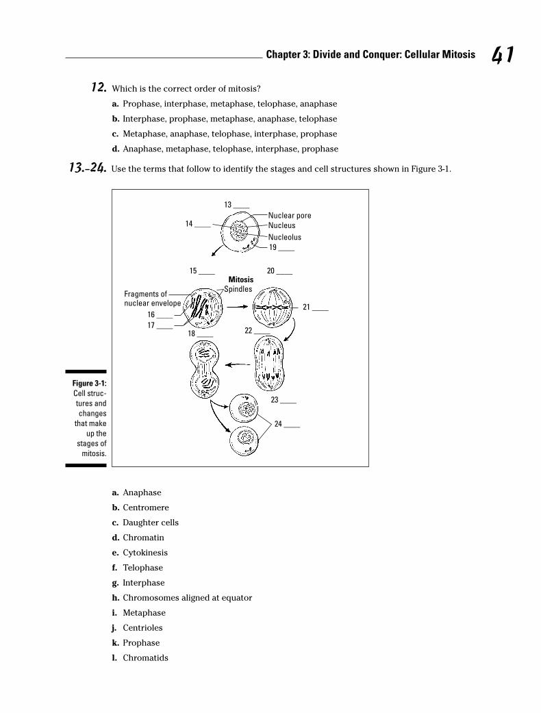
12. Which is the correct order of mitosis?
a. Prophase, interphase, metaphase, telophase, anaphase
b. Interphase, prophase, metaphase, anaphase, telophase
c. Metaphase, anaphase, telophase, interphase, prophase
d. Anaphase, metaphase, telophase, interphase, prophase
13.–24. Use the terms that follow to identify the stages and cell structures shown in Figure 3-1.
a. Anaphase
b. Centromere
c. Daughter cells
d. Chromatin
e. Cytokinesis
f. Telophase
g. Interphase
h. Chromosomes aligned at equator
i. Metaphase
j. Centrioles
k. Prophase
l. Chromatids
14 ____Nuclear pore
Nucleus
Nucleolus
19 ____
Fragments ofnuclear envelope
Spindles
16 ____
17 ____
21 ____
Mitosis
24 ____
13 ____
15 ____ 20 ____
18 ____ 22 ____
23 ____
Figure 3-1:
Cell struc-
tures and
changes
that make
up the
stages of
mitosis.
41Chapter 3: Divide and Conquer: Cellular Mitosis
25. The two newly formed daughter cells are
a. The same size as the mother cell
b. Not genetically identical to each other
c. Unequal in size
d. Genetically identical to the mother cell
26. Cytokinesis can be described as
a. The period of preparation for cell division
b. The dividing of the cytoplasm to surround the two newly formed nuclei
c. The stage of alignment of the chromatids on the equatorial plane
d. The initiation of cell division
27. Cytokinesis occurs during
a. Telophase
b. Interphase
c. Prophase
d. Metaphase
What Can Go WrongWith the millions upon millions of cell divisions that happen in the human body, it’snot surprising that sometimes things go wrong. An error during mitosis is called amutation. One kind of mutation is nondisjunction, or a failure to separate. In this muta-tion, newly formed chromosomes don’t quite divide, leaving one daughter cell withone more chromosome than normal and the other daughter cell one chromosome shyof a full complement. Down’s syndrome is an example of what happens when nondis-junction occurs. A normal human cell has 46 chromosomes, but that of a Down’s suf-ferer has 47.
Mitosis can also end up on fast-forward. Accelerated mitosis can lead to the formationof a tumor, also called a neoplasm. The rate of division usually restricts itself to replac-ing worn out or injured cells, but with accelerated mitosis, the cells don’t know whento stop dividing.
28. Any change in a cell’s genetic information is known as
a. Phagocytosis
b. Mutation
c. Pinocytosis
d. Neurotransmission
42 Part I: Building Blocks of the Body
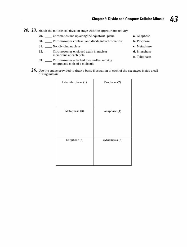
29.–33. Match the mitotic cell division stage with the appropriate activity.
29. _____ Chromatids line up along the equatorial plane a. Anaphase
30. _____ Chromosomes contract and divide into chromatids b. Prophase
31. _____ Nondividing nucleus c. Metaphase
32. _____ Chromosomes enclosed again in nuclear d. Interphasemembrane at each pole
e. Telophase33. _____ Chromosomes attached to spindles, moving
to opposite ends of a molecule
34. Use the space provided to draw a basic illustration of each of the six stages inside a cellduring mitosis.
Telophase (5) Cytokinesis (6)
Metaphase (3) Anaphase (4)
Late interphase (1) Prophase (2)
43Chapter 3: Divide and Conquer: Cellular Mitosis

Answers to Questions on MitosisThe following are answers to the practice questions presented in this chapter.
a Cells are dormant during interphase. b. False. Cells are at their most active during interphase.
b The G1 subphase of interphase is b. the most active phase. With all that organelle-growing,metabolizing, and protein-synthesizing that takes place during G1, this isn’t surprising.
c DNA is duplicated during which subphase? a. S. Remember, the cell is S-ynthesizing new DNAmolecules during this phase.
d The nuclear membrane, or envelope, disappears during c. prophase.
e Which of the following happens in prophase? b. The chromosomes divide into chromatids.But don’t forget that they remain attached at the centromere.
f Which of the following is true for metaphase? c. The chromatids align on the equatorial plane.Each of the other answer choices occurs during earlier or later phases.
g During metaphase, each chromosome consists of two duplicate chromatids. a. True. That way,each resulting daughter cell will have identical chromosomes.
h Identify an event that does not happen during anaphase. b. The cell goes through subphase G1.G1 took place back in interphase.
i Genetically identical chromosomes are pulled toward opposite poles during c. anaphase. Asthe cell nears the end of division, it makes sense that duplicate packages move to oppositeends of the cell.
j Which event does not occur during telophase? c. The chromosomes become more distinct.That change happened back in interphase.
k What structures disappear during telophase? a. Spindles and asters. These structures disap-pear because they’re no longer needed at the end of mitosis.
l Which is the correct order of mitosis? b. Interphase, prophase, metaphase, anaphase,telophase.
m–x Following is how Figure 3-1, the stages and structures of mitosis, should be labeled.
13. g. Interphase; 14. d. Chromatin; 15. k. Prophase; 16. b. Centromere; 17. l. Chromatids;18. f. Telophase; 19. j. Centrioles; 20. i. Metaphase; 21. h. Chromosomes aligned at equator; 22. a. Anaphase; 23. e. Cytokinesis; 24. c. Daughter cells
y The two newly formed daughter cells are d. genetically identical to the mother cell. None ofthe other answer options make sense.
A Cytokinesis can be described as b. the dividing of the cytoplasm to surround the two newlyformed nuclei.
B Cytokinesis occurs during a. telophase. No, it’s not a trick question. We told you early on thatmitosis and cytokinesis ended at the same time. That means, of course, that cytokinesis takesplace during the final stage of mitosis.
44 Part I: Building Blocks of the Body

C Any change in a cell’s genetic information is known as b. mutation.
D Chromatids line up along the equatorial plane: c. Metaphase
E Chromosomes contract and divide into chromatids: b. Prophase
F Nondividing nucleus: d. Interphase
G Chromosomes enclosed again in nuclear membrane at each pole: e. Telophase
H Chromosomes attached to spindles, moving to opposite ends of a molecule: a. Anaphase
I Check your answers to the fill-in-the-blanks in questions 13 through 24 (Figure 3-1), and thencompare your drawings to that figure.
45Chapter 3: Divide and Conquer: Cellular Mitosis
46 Part I: Building Blocks of the Body
Chapter 4
The Study of Tissues: Histology
In This Chapter� Checking out the skin
� Keeping things together with connective tissues
� Flexing muscle tissues
� Sending signals through nerve tissue
Oh, what tangled webs we weave! As the chapter title says, histology is the study of tis-sues, but you may be surprised to find out that the Greek histo doesn’t translate as
“tissue” but instead as “web.” It’s a logical next step after reviewing the cell and cellulardivision to take a look at what happens when groups of similar cells “web” together to formtissues. The four different types of tissue in the body are as follows:
� Epithelial, or skin, tissue (from the Greek epi– for “over” or “outer”)
� Connective tissue
� Muscle tissue
� Nerve tissue
In this chapter, you find a quick review of the basics of each of these types of tissues alongwith practice questions to test your knowledge of them.
Getting Under Your SkinPerhaps because of its unique job of both protecting the outer body and lining internalorgans, epithelial tissue comes in more varieties than any other tissue.
Epithelial tissues, which generally are arranged in sheets or tubes of tightly-packed cells,always have a free, or apical, surface that can be exposed to the air or to fluid. That free sur-face also can be covered by additional layers of epithelial tissue. But whether it’s layered ornot, each epithelial cell has polarity (a top and a bottom), and all but one side of the cell istucked snugly against neighboring cells. The apical side sometimes has cytoplasmic projec-tions such as cilia, hair-like growths that can move material over the cell’s surface, ormicrovilli, finger-like projections that increase the cell’s surface area for absorption. Oppositethe apical side is the basal side (think basement), which typically attaches to some kind ofconnective tissue.
Epithelial tissue serves several key functions, including the following:
� Protection: Skin protects vulnerable structures or tissues deeper in the body.
� Barrier: Epithelial tissues prevent foreign materials from getting inside the body.
� Sensation: Sensory nerve endings embedded in epithelial tissue connect thebody with outside stimuli.
� Secretion: Epithelial tissue in glands can be specialized to secrete enzymes, hor-mones, and fluids.
Single-layer epithelial tissue is classified as simple. Tissue with more than one layer iscalled stratified. Epithelial tissues also can be classified according to shape: Squamousis a thin, flat cell; cuboidal is, as the name implies, equal in height and width andshaped like a cube; and columnar cells are taller than they are wide.
Following are the ten primary types of epithelial tissues:
� Simple squamous epithelium: Looking a bit like rolling tundra, this flat layer ofscale-like cells is useful in diffusion, secretion, or absorption. Each cell nucleus iscentrally located and is round or oval. Simple squamous epithelium lines thelungs’ air sacs where oxygen and carbon dioxide are exchanged; forms blood fil-ters inside the kidneys; and lines the inner surface of the eardrum, known as thetympanic membrane.
� Simple cuboidal epithelium: These cube-shaped cells, found in a single layerthat looks like a microscopic mattress, have centrally located nuclei that usuallyare round. Found in the ovaries, kidneys, and some glands, this type of epithe-lium functions in secretion, absorption, and tube formation.
� Simple columnar epithelium: These densely packed cells are taller than they arewide, with nuclei located near the base of each cell. Found lining the digestivetract from the stomach to the anal canal, this type of epithelium functions insecretion and absorption.
� Simple columnar ciliated epithelium: A close cousin to simple columnar epithe-lium, this type of tissue has hair-like cilia that can move mucus and other sub-stances across the cell. It’s found lining the small respiratory tubes.
� Pseudostratified columnar epithelium: Pay attention to the prefix pseudo– here,which means “false.” It may look multilayered because the cells’ nuclei are scat-tered at different levels, but it’s not. This type of epithelium is found in the sali-vary glands and some segments of the male reproductive system, including theurethra.
� Pseudostratified columnar ciliated epithelium: Another variation on a theme,this tissue is nearly identical to pseudostratified columnar epithelium. The differ-ence is that this tissue’s free surface has cilia, making it ideal for lining air pas-sages because the cilia’s uniform waving action causes a thin layer of mucus tomove in one direction — toward the throat and mouth — and trap dust particles.
� Stratified squamous epithelium: This tissue is the stuff you see everyday —your outer skin, or epidermis. This multilayered tissue has squamous cells onthe outside plus deeper layers of cuboidal or columnar cells. Found in areaswhere the outer cell layer is constantly worn away, this type of epithelium regen-erates its surface layer with cells from lower layers.
� Stratified cuboidal epithelium: This multilayered epithelium can be found insweat glands, conjunctiva of the eye, and the male urethra. Its function is prima-rily protection.
� Stratified columnar epithelium: Also multilayered, this epithelium is foundlining parts of the male urethra, excretory ducts of glands, and some small areasof the anal mucus membrane.
� Stratified transitional epithelium: This epithelium is referred to as transitionalbecause its cells can shape-shift from cubes to squamous-like flat surfaces andback again. Found lining the bladder, the cells flatten out to make room for urine.
48 Part I: Building Blocks of the Body
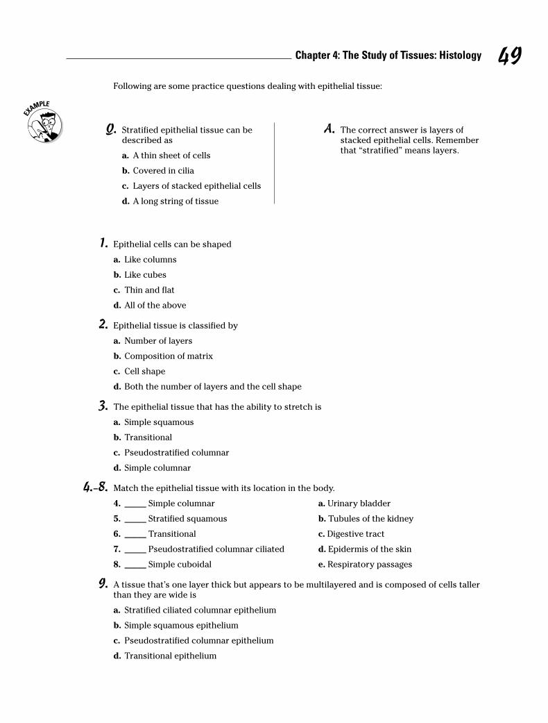
1. Epithelial cells can be shaped
a. Like columns
b. Like cubes
c. Thin and flat
d. All of the above
2. Epithelial tissue is classified by
a. Number of layers
b. Composition of matrix
c. Cell shape
d. Both the number of layers and the cell shape
3. The epithelial tissue that has the ability to stretch is
a. Simple squamous
b. Transitional
c. Pseudostratified columnar
d. Simple columnar
4.–8. Match the epithelial tissue with its location in the body.
4. _____ Simple columnar a. Urinary bladder
5. _____ Stratified squamous b. Tubules of the kidney
6. _____ Transitional c. Digestive tract
7. _____ Pseudostratified columnar ciliated d. Epidermis of the skin
8. _____ Simple cuboidal e. Respiratory passages
9. A tissue that’s one layer thick but appears to be multilayered and is composed of cells tallerthan they are wide is
a. Stratified ciliated columnar epithelium
b. Simple squamous epithelium
c. Pseudostratified columnar epithelium
d. Transitional epithelium
49Chapter 4: The Study of Tissues: Histology
Following are some practice questions dealing with epithelial tissue:
Q. Stratified epithelial tissue can bedescribed as
a. A thin sheet of cells
b. Covered in cilia
c. Layers of stacked epithelial cells
d. A long string of tissue
A. The correct answer is layers ofstacked epithelial cells. Rememberthat “stratified” means layers.
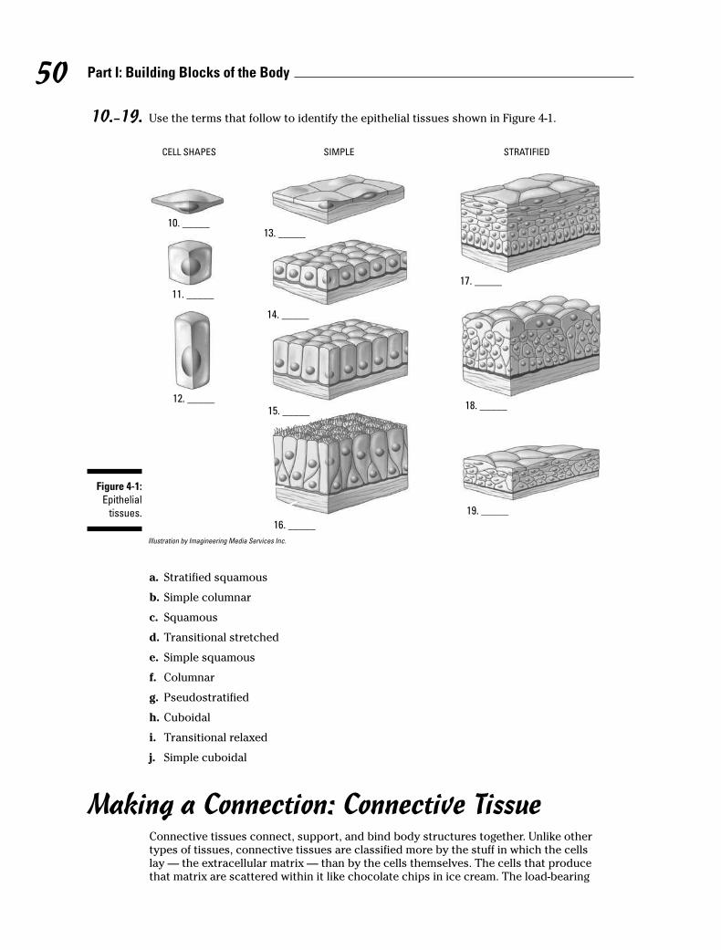
10.–19. Use the terms that follow to identify the epithelial tissues shown in Figure 4-1.
Illustration by Imagineering Media Services Inc.
a. Stratified squamous
b. Simple columnar
c. Squamous
d. Transitional stretched
e. Simple squamous
f. Columnar
g. Pseudostratified
h. Cuboidal
i. Transitional relaxed
j. Simple cuboidal
Making a Connection: Connective TissueConnective tissues connect, support, and bind body structures together. Unlike othertypes of tissues, connective tissues are classified more by the stuff in which the cellslay — the extracellular matrix — than by the cells themselves. The cells that producethat matrix are scattered within it like chocolate chips in ice cream. The load-bearing
CELL SHAPES SIMPLE STRATIFIED
10. _____
11. _____
12. _____
13. _____
14. _____
15. _____
16. _____
17. _____
18. _____
19. _____
Figure 4-1:
Epithelial
tissues.
50 Part I: Building Blocks of the Body
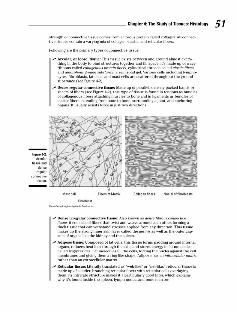
strength of connective tissue comes from a fibrous protein called collagen. All connec-tive tissues contain a varying mix of collagen, elastic, and reticular fibers.
Following are the primary types of connective tissue:
� Areolar, or loose, tissue: This tissue exists between and around almost every-thing in the body to bind structures together and fill space. It’s made up of wavyribbons called collagenous protein fibers, cylindrical threads called elastic fibers,and amorphous ground substance, a semisolid gel. Various cells including lympho-cytes, fibroblasts, fat cells, and mast cells are scattered throughout the groundsubstance (see Figure 4-2).
� Dense regular connective tissue: Made up of parallel, densely packed bands orsheets of fibers (see Figure 4-2), this type of tissue is found in tendons as bundlesof collagenous fibers attaching muscles to bone and in ligaments as bundles ofelastic fibers extending from bone to bone, surrounding a joint, and anchoringorgans. It usually resists force in just two directions.
Illustration by Imagineering Media Services Inc.
� Dense irregular connective tissue: Also known as dense fibrous connectivetissue, it consists of fibers that twist and weave around each other, forming athick tissue that can withstand stresses applied from any direction. This tissuemakes up the strong inner skin layer called the dermis as well as the outer cap-sule of organs like the kidney and the spleen.
� Adipose tissue: Composed of fat cells, this tissue forms padding around internalorgans, reduces heat loss through the skin, and stores energy in fat moleculescalled triglycerides. Fat molecules fill the cells, forcing the nuclei against the cellmembranes and giving them a ring-like shape. Adipose has an intracellular matrixrather than an extracellular matrix.
� Reticular tissue: Literally translated as “web-like” or “net-like,” reticular tissue ismade up of slender, branching reticular fibers with reticular cells overlayingthem. Its intricate structure makes it a particularly good filter, which explainswhy it’s found inside the spleen, lymph nodes, and bone marrow.
Mast cell
Fibroblast
Fibers of Matrix Collagen fibers Nuclei of fibroblasts
Figure 4-2:
Areolar
tissue and
dense
regular
connective
tissue.
51Chapter 4: The Study of Tissues: Histology
� Cartilage: These firm but flexible tissues, made up of collagen and elastic fibers,have no blood vessels or nerve cells (a state called non-vascular or avascular).Cartilage contains openings called lacunae (from the Latin word lacus for “lake”or “pit”) that enclose mature cells called chondrocytes, which are preceded bycells called chondroblasts. A membrane known as the perichondrium surroundscartilage tissue, which also contains a gelatinous protein called chondrin. Thereare three types of cartilage:
• Hyaline cartilage: The most abundant cartilage in the body, it’s elastic andmade up of a uniform matrix pocked with chondrocytes. It lays the founda-tion for the embryonic skeleton, forms the rib (or costal) cartilages, makesup nose cartilage, and covers the articulating surfaces of bones.
• Fibrocartilage: As the name implies, fibrocartilage contains thick, compactcollagen fibers. The sponge-like structure, with the lacunae and chondro-cytes lined up within the fibers, makes it a good shock absorber. It’s foundin the intervertebral discs of the vertebral column and in the symphysispubis at the front of the pelvis.
• Elastic cartilage: Similar to hyaline cartilage, elastic cartilage has moretightly packed lacunae and chondrocytes between parallel elastic fibers.This structure, which makes up the ear lobe and other structures where aspecific form is important, tends to bounce back to its original shape afterbeing bent.
� Bone, or osseous, tissue: Essentially, bone is mineralized connective tissue formedinto repeating patterns called Haversian systems. In the center of each system is alarge opening, the Haversian canal, that contains blood vessels, lymph vessels, andnerves. The central canal is surrounded by thin membranes called lamellae thatcontain the lacunae, which in turn contain osteocytes (bone cells). Smaller canali-culi connect the lacunae and circulate tissue fluids from the blood vessels to nour-ish the osteocytes. (We explore bone in more detail in Chapter 5.)
� Blood: Yes, blood is considered a type of connective tissue. Like other connec-tive tissues, it has an extracellular matrix — in this case, plasma — in which aresuspended erythrocytes (red blood cells), leukocytes (white blood cells), andthrombocytes (platelets). (Blood also is considered a vascular tissue because itcirculates inside arteries and veins, but we get into that in Chapter 10.) Roughlyhalf of blood’s volume is fluid or plasma while the other half is suspended cells.Erythrocytes are concave on both sides and contain a pigment, hemoglobin,which supplies oxygen to the body’s cells and takes carbon dioxide away. Thereare approximately 5 million erythrocytes per cubic millimeter of whole blood.Thrombocytes, which number approximately 250,000 per cubic millimeter, arefragments of cells used in blood clotting. Leukocytes are large phagocytic cells(literally “cell that eats”) that are part of the body’s immune system. There are,however, relatively few of them — less than 10,000 per cubic millimeter.
20. Adipose tissue is composed of
a. Mast cells
b. Chondrocytes
c. Osteocytes
d. Fat cells
21. Tendons are composed of
a. Elastic tissue
b. Dense regular connective tissue
c. Areolar connective tissue
d. Fibrocartilage
52 Part I: Building Blocks of the Body
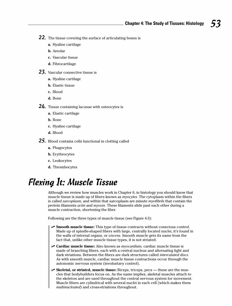
22. The tissue covering the surface of articulating bones is
a. Hyaline cartilage
b. Areolar
c. Vascular tissue
d. Fibrocartilage
23. Vascular connective tissue is
a. Hyaline cartilage
b. Elastic tissue
c. Blood
d. Bone
24. Tissue containing lacunae with osteocytes is
a. Elastic cartilage
b. Bone
c. Hyaline cartilage
d. Blood
25. Blood contains cells functional in clotting called
a. Phagocytes
b. Erythrocytes
c. Leukocytes
d. Thrombocytes
Flexing It: Muscle TissueAlthough we review how muscles work in Chapter 6, in histology you should know thatmuscle tissue is made up of fibers known as myocytes. The cytoplasm within the fibersis called sarcoplasm, and within that sarcoplasm are minute myofibrils that contain theprotein filaments actin and myosin. These filaments slide past each other during amuscle contraction, shortening the fiber.
Following are the three types of muscle tissue (see Figure 4-3):
� Smooth muscle tissue: This type of tissue contracts without conscious control.Made up of spindle-shaped fibers with large, centrally located nuclei, it’s found inthe walls of internal organs, or viscera. Smooth muscle gets its name from thefact that, unlike other muscle tissue types, it is not striated.
� Cardiac muscle tissue: Also known as myocardium, cardiac muscle tissue ismade of branching fibers, each with a central nucleus and alternating light anddark striations. Between the fibers are dark structures called intercalated discs.As with smooth muscle, cardiac muscle tissue contractions occur through theautonomic nervous system (involuntary control).
� Skeletal, or striated, muscle tissue: Biceps, triceps, pecs — these are the mus-cles that bodybuilders focus on. As the name implies, skeletal muscles attach tothe skeleton and are used throughout the central nervous system for movement.Muscle fibers are cylindrical with several nuclei in each cell (which makes themmultinucleated) and cross-striations throughout.
53Chapter 4: The Study of Tissues: Histology
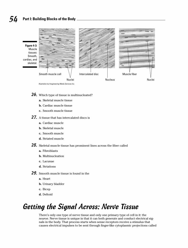
Illustration by Imagineering Media Services Inc.
26. Which type of tissue is multinucleated?
a. Skeletal muscle tissue
b. Cardiac muscle tissue
c. Smooth muscle tissue
27. A tissue that has intercalated discs is
a. Cardiac muscle
b. Skeletal muscle
c. Smooth muscle
d. Striated muscle
28. Skeletal muscle tissue has prominent lines across the fiber called
a. Fibroblasts
b. Multinucleation
c. Lacunae
d. Striations
29. Smooth muscle tissue is found in the
a. Heart
b. Urinary bladder
c. Bicep
d. Deltoid
Getting the Signal Across: Nerve TissueThere’s only one type of nerve tissue and only one primary type of cell in it: theneuron. Nerve tissue is unique in that it can both generate and conduct electrical sig-nals in the body. That process starts when sense receptors receive a stimulus thatcauses electrical impulses to be sent through finger-like cytoplasmic projections called
Nucleus
Smooth muscle cell
Nuclei
Intercalated disc
Nuclei
Muscle fiber
Figure 4-3:
Muscle
tissues:
Smooth,
cardiac, and
skeletal.
54 Part I: Building Blocks of the Body
dendrites. From there, the impulse moves through the body of the cell and into anothertype of cytoplasmic projection (or nerve process) called an axon that hands the signaloff to the next cell down the line. (We look more closely at how all that happens whenwe examine the central nervous system in Chapter 15.)
Following are some practice questions dealing with nerve tissue:
30. Cells capable of producing and transmitting electrical impulses are
a. Schwann cells
b. Neurons
c. Chondrocytes
d. Thrombocytes
31. The cytoplasmic projection of a neuron that carries impulses away from the cell body is called
a. A myofibril
b. A dendrite
c. An axon
d. A cross-striation
32. The cytoplasmic projections that receive stimuli from sense receptors are
a. Dendrites
b. Collagenous fibers
c. Axons
d. Schwann projections
33.–37. Match each description with the appropriate tissue.
33. _____ Precedes bone formation in embryonic development a. Areolar tissue
34. _____ Found in visceral walls b. Hyaline cartilage
35. _____ Found in and around most structures in the body c. Bone
36. _____ Found in the external ear d. Elastic cartilage
37. _____ Supports soft tissues of the body e. Smooth muscle
55Chapter 4: The Study of Tissues: Histology

Answers to Questions on HistologyThe following are answers to the practice questions presented in this chapter.
a Epithelial cells can be shaped d. all of the above. Cube, column, or flat and thin, it’s still anepithelial cell.
b Epithelial tissue is classified by d. both the number of layers and the cell shape. Classificationrequires looking at both simultaneously.
c The epithelial tissue that has the ability to stretch is b. transitional. This tissue lines the blad-der, so it had better be stretchy!
d Simple columnar: c. Digestive tract
e Stratified squamous: d. Epidermis of the skin
f Transitional: a. Urinary bladder
g Pseudostratified columnar ciliated: e. Respiratory passages
h Simple cuboidal: b. Tubules of the kidney
i A tissue that’s one layer thick but appears to be multilayered and is composed of cells tallerthan they are wide is c. pseudostratified columnar epithelium. To arrive at the correct answer,consider this question one piece at a time: pseudo is “false,” stratified means “layered” (so youhave “false-layered”), and columns are taller than they are wide.
j–s Following is how Figure 4-1, the types of epithelial cells and tissues, should be labeled.
10. c. Squamous; 11. h. Cuboidal; 12. f. Columnar; 13. e. Simple squamous; 14. j. Simplecuboidal; 15. b. Simple columnar; 16. g. Pseudostratified; 17. a. Stratified squamous; 18. i. Transitional relaxed; 19. d. Transitional stretched
t Adipose tissue is composed of d. fat cells. Think of the Latin adeps, which means “fat.”
u Tendons are composed of b. dense regular connective tissue. Tendons are dense and exhibit aregular pattern.
v The tissue covering the surface of articulating bones is a. hyaline cartilage.
w Vascular connective tissue is c. blood.
x Tissue containing lacunae with osteocytes is b. bone. Remember that osteo is the Latin wordfor “bone.”
y Blood contains cells functional in clotting called d. thrombocytes. Knowing that the Greekword thrombos means “clot” can help you spot the correct answer in this question.
A Which type of tissue is multinucleated? a. Skeletal muscle tissue. The other answer options aretissues that have only one nucleus per cell.
B A tissue that has intercalated discs is a. cardiac muscle. Intercalated discs, as you should orwill know from studying the circulatory system, are involved in conducting signals for the heartto pump.
56 Part I: Building Blocks of the Body
C Skeletal muscle tissue has prominent lines across the fiber called d. striations. Striations arelight and dark lines across the fiber.
D Smooth muscle tissue is found in the b. urinary bladder. The other answer choices containstriated tissue, which technically means that they aren’t smooth.
E Cells capable of producing and transmitting electrical impulses are b. neurons. These are thecells that make up nerve tissue.
F The cytoplasmic projection of a neuron that carries impulses away from the cell body is calledc. an axon. Each neuron cell usually has only one axon, although it may branch off severaltimes.
G The cytoplasmic projections that receive stimuli from sense receptors are a. dendrites. Usually,there are several dendrites per neuron cell.
H Precedes bone formation in embryonic development: b. Hyaline cartilage
I Found in visceral walls: e. Smooth muscle
J Found in and around most structures in the body: a. Areolar tissue
K Found in the external ear: d. Elastic cartilage
L Supports soft tissues of the body: c. Bone
57Chapter 4: The Study of Tissues: Histology

58 Part I: Building Blocks of the Body
Part II
Weaving It Together:Bones, Muscles, and Skin
In this part . . .
This part gets into what most people think of firstwhen it comes to anatomy and physiology: bones and
muscles. First we focus on how bones are formed beforebroadening the view to the axial skeleton (the parts thatline up from head to toe) and the appendicular skeleton(the parts that reach out from the central axis). You reviewhow muscles attach to that framework and watch thebody take shape before wrapping this newly layeredpackage in the body’s largest single organ: the skin.

Chapter 5
A Scaffold to Build On: The Skeleton
In This Chapter� Getting to know your bones
� Keeping the axial skeleton in line
� Checking out the appendicular skeleton
� Playing with joints
Human osteology, from the Greek word for “bone” (osteon) and the suffix –logy, whichmeans “to study,” focuses on the 206 bones in the adult body endoskeleton. But it’s
more than just bones; it’s also ligaments and cartilage and the joints that make the wholeassembly useful. In this chapter, you get lots of practice exploring the skeletal functions andhow the joints work together.
Understanding Dem BonesThe skeletal system as a whole serves five key functions:
� Protection: The skeleton encases and shields delicate internal organs that might other-wise be damaged during motion or crushed by the weight of the body itself. For exam-ple, the skull’s cranium houses the brain, and the ribs and sternum of the thoracic cageprotect organs in the central body cavity.
� Movement: By providing anchor sites and a scaffold against which muscles can con-tract, the skeleton makes motion possible. The bones act as levers, the joints are thefulcrums, and the muscles apply the force. For instance, when the biceps muscle con-tracts, the radius and ulna bones of the forearm are lifted toward the humerus bone ofthe upper arm.
� Support: The vertebral column’s curvatures play a key role in supporting the entirebody’s weight, as do the arches formed by the bones of the feet. Upper body supportflows from the clavicle, or collarbone, which is the only bone that attaches the upperextremities to the axial skeleton and the only horizontal long bone in the human body.
� Mineral storage: Calcium, phosphorous, and other minerals like magnesium must bemaintained in the bloodstream at a constant level, so they’re “banked” in the bones incase the dietary intake of those minerals drops. The bones’ mineral content is con-stantly renewed, refreshing entirely about every nine months. A 35 percent decrease inblood calcium will cause convulsions.
� Blood cell formation: Called hemopoiesis or hematopoiesis, most blood cell formationtakes place within the red marrow inside the ends of long bones as well as within the ver-tebrae, ribs, sternum, and cranial bones. Marrow produces three types of blood cells:erythrocytes (red cells), leukocytes (white cells), and thrombocytes (platelets). Most ofthese are formed in red bone marrow, although some types of white blood cells are pro-duced in fat-rich yellow bone marrow. At birth, all bone marrow is red. With age, it con-verts to the yellow type. In cases of severe blood loss, the body can convert yellowmarrow back to red marrow in order to increase blood cell production.
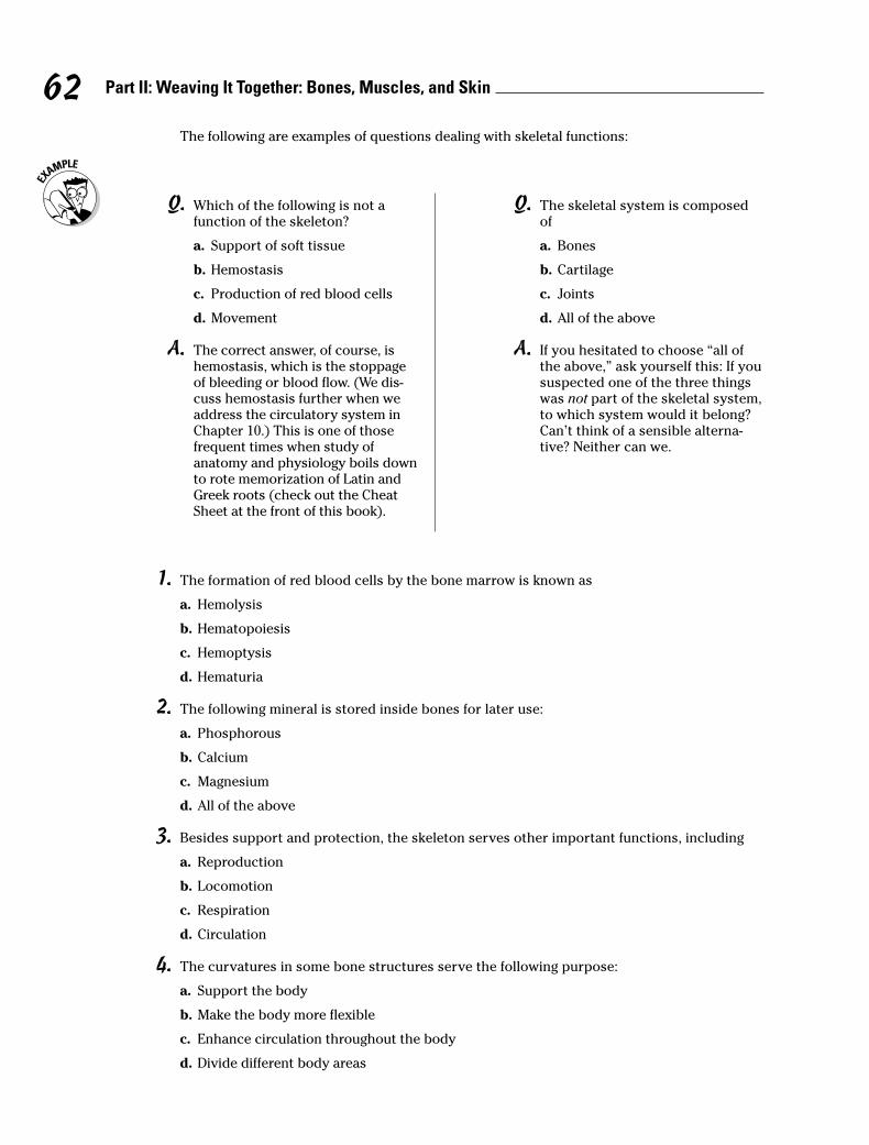
1. The formation of red blood cells by the bone marrow is known as
a. Hemolysis
b. Hematopoiesis
c. Hemoptysis
d. Hematuria
2. The following mineral is stored inside bones for later use:
a. Phosphorous
b. Calcium
c. Magnesium
d. All of the above
3. Besides support and protection, the skeleton serves other important functions, including
a. Reproduction
b. Locomotion
c. Respiration
d. Circulation
4. The curvatures in some bone structures serve the following purpose:
a. Support the body
b. Make the body more flexible
c. Enhance circulation throughout the body
d. Divide different body areas
62 Part II: Weaving It Together: Bones, Muscles, and Skin
The following are examples of questions dealing with skeletal functions:
Q. Which of the following is not afunction of the skeleton?
a. Support of soft tissue
b. Hemostasis
c. Production of red blood cells
d. Movement
A. The correct answer, of course, ishemostasis, which is the stoppageof bleeding or blood flow. (We dis-cuss hemostasis further when weaddress the circulatory system inChapter 10.) This is one of thosefrequent times when study ofanatomy and physiology boils downto rote memorization of Latin andGreek roots (check out the CheatSheet at the front of this book).
Q. The skeletal system is composedof
a. Bones
b. Cartilage
c. Joints
d. All of the above
A. If you hesitated to choose “all ofthe above,” ask yourself this: If yoususpected one of the three thingswas not part of the skeletal system,to which system would it belong?Can’t think of a sensible alterna-tive? Neither can we.
Boning Up on Classifications, Structures,and Ossification
Adult bones are composed of 30 percent protein (called ossein), 45 percent minerals(including calcium, phosphorus, and magnesium), and 25 percent water. Minerals givethe bone strength and hardness. At birth, bones are soft and pliable because of carti-lage in their structure. As the body grows, older cartilage gradually is replaced by hardbone tissue. Mineral in the bones increases with age, causing them to become morebrittle and easily fractured.
Various types of bone make up the human skeleton, but fortunately for memorizationpurposes, bone type names match what the bones look like. They are as follows:
� Long bones, like those found in the arms and legs, form the weight-bearing partof the skeleton.
� Short bones, such as those in the wrists (carpals) and ankles (tarsals), have ablocky structure and allow for a greater range of motion.
� Flat bones, such as the skull, sternum, scapulae, and pelvic bones, shield soft tissues.
� Irregular bones, such as the mandible (jawbone) and vertebrae, come in a vari-ety of shapes and sizes suited for attachment to muscles, tendons, and ligaments.Irregular bones include seed-shaped sesamoid bones found in joints such as thepatella, or kneecap.
Unfortunately for students of bone structures, there’s no easy way to memorize them.So brace yourself for a rapid summary of what your textbook probably goes into inmuch greater detail.
Compact bone is a dense layer made up of structural units, or lacunae, arranged inconcentric circles called Haversian systems (also referred to in short as osteons), eachof which has a central, microscopic Haversian canal. A perpendicular system of canals,called Volkmann’s canals, penetrate and cross between the Haversian systems. Thisnetwork ensures circulation into even the hardest bone structure. Compact bonetissue is thick in the shaft and tapers to paper thinness at the ends of the bones. Thebulbous ends of each long bone, known as the epiphyses (or singularly as an epiphy-sis), are made up of spongy bone or cancellous bone tissue covered by a thin layer ofcompact bone. The diaphysis, or shaft, contains the medullary cavity and bloodcell–producing marrow. A membrane called the periosteum covers the outer bone toprovide nutrients and oxygen, remove waste, and connect with ligaments and tendons.
Bones grow through the cellular activities of osteoblasts on the surface of the bone,which produce layers of mature bone cells called osteocytes. Osteoclasts are cells thatfunction in the developing fetus to absorb cartilage as ossification occurs and functionin adult bone to break down and remove spent bone tissue.
There are two types of ossification, which is the process by which softer tissues hardeninto bone. Both types rely on a peptide hormone produced by the thyroid gland, calci-tonin, which regulates metabolism of calcium, the body’s most abundant mineral. Thetwo types of ossification are
� Endochondral or intracartilaginous ossification: Occurs when mineral salts,particularly calcium and phosphorus, calcify along the scaffolding of cartilageformed in the developing fetus beginning about the fifth week after conception.This process, known as calcification, takes place in the presence of vitamin D and
63Chapter 5: A Scaffold to Build On: The Skeleton
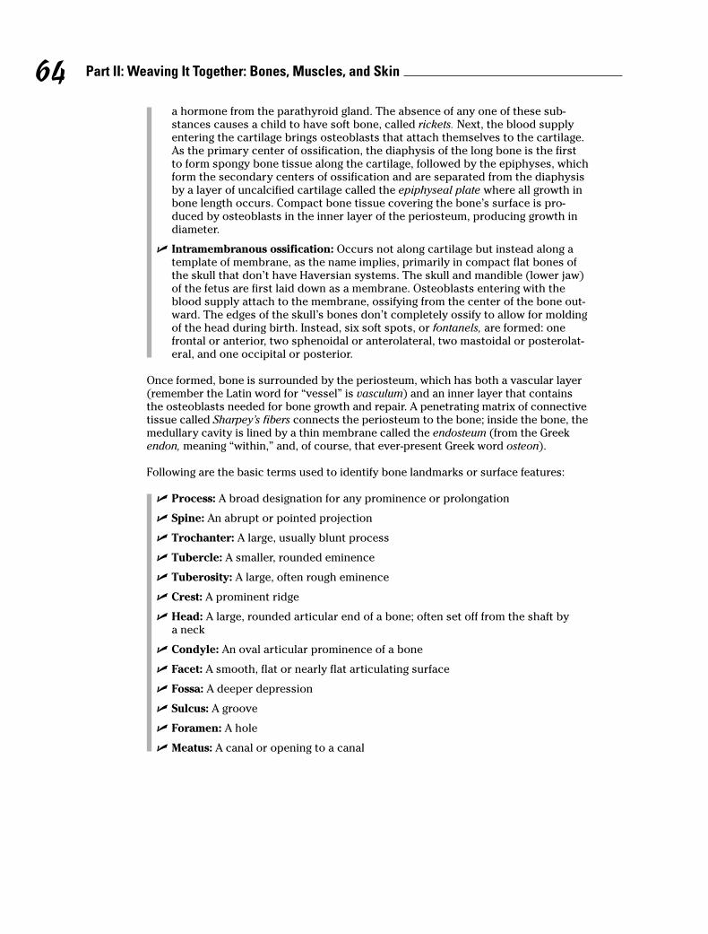
a hormone from the parathyroid gland. The absence of any one of these sub-stances causes a child to have soft bone, called rickets. Next, the blood supplyentering the cartilage brings osteoblasts that attach themselves to the cartilage.As the primary center of ossification, the diaphysis of the long bone is the firstto form spongy bone tissue along the cartilage, followed by the epiphyses, whichform the secondary centers of ossification and are separated from the diaphysisby a layer of uncalcified cartilage called the epiphyseal plate where all growth inbone length occurs. Compact bone tissue covering the bone’s surface is pro-duced by osteoblasts in the inner layer of the periosteum, producing growth indiameter.
� Intramembranous ossification: Occurs not along cartilage but instead along atemplate of membrane, as the name implies, primarily in compact flat bones ofthe skull that don’t have Haversian systems. The skull and mandible (lower jaw)of the fetus are first laid down as a membrane. Osteoblasts entering with theblood supply attach to the membrane, ossifying from the center of the bone out-ward. The edges of the skull’s bones don’t completely ossify to allow for moldingof the head during birth. Instead, six soft spots, or fontanels, are formed: onefrontal or anterior, two sphenoidal or anterolateral, two mastoidal or posterolat-eral, and one occipital or posterior.
Once formed, bone is surrounded by the periosteum, which has both a vascular layer(remember the Latin word for “vessel” is vasculum) and an inner layer that containsthe osteoblasts needed for bone growth and repair. A penetrating matrix of connectivetissue called Sharpey’s fibers connects the periosteum to the bone; inside the bone, themedullary cavity is lined by a thin membrane called the endosteum (from the Greekendon, meaning “within,” and, of course, that ever-present Greek word osteon).
Following are the basic terms used to identify bone landmarks or surface features:
� Process: A broad designation for any prominence or prolongation
� Spine: An abrupt or pointed projection
� Trochanter: A large, usually blunt process
� Tubercle: A smaller, rounded eminence
� Tuberosity: A large, often rough eminence
� Crest: A prominent ridge
� Head: A large, rounded articular end of a bone; often set off from the shaft bya neck
� Condyle: An oval articular prominence of a bone
� Facet: A smooth, flat or nearly flat articulating surface
� Fossa: A deeper depression
� Sulcus: A groove
� Foramen: A hole
� Meatus: A canal or opening to a canal
64 Part II: Weaving It Together: Bones, Muscles, and Skin
5. The most abundant mineral in the body is
a. Potassium
b. Magnesium
c. Calcium
d. Sodium
6. A decrease in calcium concentration in the body fluids by 35 percent causes
a. Unresponsive neurons
b. Death
c. Convulsions
d. Muscle spasms
7. Calcitonin is produced by the
a. Thyroid gland
b. Pituitary gland
c. Adrenal gland
d. Parathyroid
8. Bones are encased by a membrane called the
a. Vasculum
b. Periosteum
c. Endosteum
d. Fontanel
65Chapter 5: A Scaffold to Build On: The Skeleton
Q. Mature bone cells are called
a. Osteocytes
b. Osteoblasts
c. Chondrocytes
d. Osteoclasts
A. The correct answer is osteocytes.Why? Refer to the Latin root cyta,which is most commonly used torefer to a cell. Why not osteoblastsor osteoclasts? Because the Greekroot blast in biological terms refersto growth or formation, and theLatin root clast refers to breakingor fragmentation.
Q. The basic unit of structure in adultcompact bones is the
a. Osteoblast
b. Osteon
c. Osteoclast
d. Osteocytes
A. The correct answer is osteon.Always pay attention to exactlywhat a multiple-choice question isasking. Remember that descriptionof the structural part of the bone,the Haversian system? And checkout that root osteo, which comesfrom the Greek word for “bone.”

9. A bone’s encapsulating membrane is attached by
a. Haversian canals
b. Sphenoids
c. Sharpey’s fibers
d. Mastoids
10. Bone marrow can be found inside the
a. Medullary cavity
b. Sharpey’s cavity
c. Ossifying cavity
d. Diaphanous cavity
11. The six structures in the skull of an infant are called
a. Condyls
b. Fontanels
c. Facets
d. Trochanters
12. Volkmann’s canals
a. Are found in cancellous bone only
b. Contain the nutrient artery
c. Pass through the epiphysis
d. Supply articulating cartilage blood
13. Blood vessels entering through Volkmann’s canals reach the bone cells through the
a. Endosteum
b. Haversian canal
c. Lacunae
d. Medullary canal
14. The hormone released when calcium ion concentration is abnormally high is
a. Thyroxine
b. Calcitonin
c. Progesterone
d. Parathormone
15.–25. Fill in the blanks to complete the following sentences:
Bones are first laid down as 15. _______________ during the fifth week after conception.Development of the bone begins with 16._______________, the depositing of calcium andphosphorus. Next, the blood supply entering the cartilage brings 17. _______________that attach themselves to the cartilage. Ossification in long bones begins in the 18. _______________ of the long bone and moves toward the 19. _______________ of thebone. The epiphyseal and diaphyseal areas remain separated by a layer of uncalcified cartilage called the 20. _______________.
66 Part II: Weaving It Together: Bones, Muscles, and Skin
Another very large cell that enters with the blood supply is the 21. _______________, whichhelps absorb the cartilage as ossification occurs. Later it helps absorb bone tissue from thecenter of the long bone’s shaft, forming the 22. _______________ cavity. After ossification,the spaces that were formed by the osteoclasts join together to form 23. _______________systems, which contain the blood vessels, lymphatic vessels, and nerves. Unlike bones in the rest of the body, those of the skull and mandible (lower jaw) are first laid down as 24. _______________. In the skull, the edges of the bone don’t ossify in the fetus but remainmembranous and form 25. _______________.
26.–34. Classify the following bones by shape. Each classification may be used more than once.
26. _____ Vertebrae of the vertebral column a. Flat bone
27. _____ Femur in thigh b. Irregular bone
28. _____ Sternum c. Long bone
29. _____ Tarsals in ankle d. Short bone
30. _____ Humerus in upper arm
31. _____ Phalanges in fingers and toes
32. _____ Scapulae of shoulder
33. _____ Kneecap
34. _____ Carpals in wrist
35.–47. Match the description with the bone landmarks or surface features.
35. _____ An abrupt or pointed projection a. Condyle
36. _____ A large, usually blunt process b. Crest
37. _____ A designation for any prominence c. Facetor prolongation
d. Foramen38. _____ A large, often rough eminence
e. Fossa39. _____ A prominent ridge
f. Head40. _____ A large, rounded articular end of a bone; often
g. Meatusset off from the shaft by the neck
h. Process41. _____ An oval articular prominence of a bone
i. Spine42. _____ A smooth, flat or nearly flat articulating surface
j. Sulcus43. _____ A deeper depression
44. _____ A groovek. Trochanter
45. _____ A holel. Tubercle
46. _____ A canal or opening to a canalm. Tuberosity
47. _____ A smaller, rounded eminence
48.–56. Use the terms that follow to identify the regions and structures of the long bone shown inFigure 5-1.
67Chapter 5: A Scaffold to Build On: The Skeleton
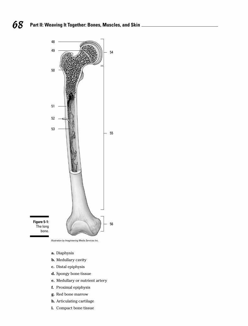
Illustration by Imagineering Media Services Inc.
a. Diaphysis
b. Medullary cavity
c. Distal epiphysis
d. Spongy bone tissue
e. Medullary or nutrient artery
f. Proximal epiphysis
g. Red bone marrow
h. Articulating cartilage
i. Compact bone tissue
48
49
50
51
52
53
54
55
56Figure 5-1:
The long
bone.
68 Part II: Weaving It Together: Bones, Muscles, and Skin
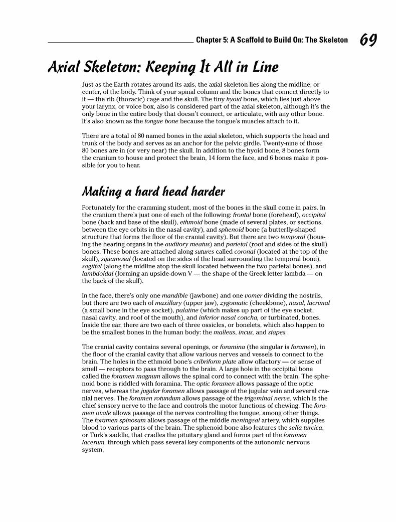
Axial Skeleton: Keeping It All in LineJust as the Earth rotates around its axis, the axial skeleton lies along the midline, orcenter, of the body. Think of your spinal column and the bones that connect directly toit — the rib (thoracic) cage and the skull. The tiny hyoid bone, which lies just aboveyour larynx, or voice box, also is considered part of the axial skeleton, although it’s theonly bone in the entire body that doesn’t connect, or articulate, with any other bone.It’s also known as the tongue bone because the tongue’s muscles attach to it.
There are a total of 80 named bones in the axial skeleton, which supports the head andtrunk of the body and serves as an anchor for the pelvic girdle. Twenty-nine of those80 bones are in (or very near) the skull. In addition to the hyoid bone, 8 bones formthe cranium to house and protect the brain, 14 form the face, and 6 bones make it pos-sible for you to hear.
Making a hard head harderFortunately for the cramming student, most of the bones in the skull come in pairs. Inthe cranium there’s just one of each of the following: frontal bone (forehead), occipitalbone (back and base of the skull), ethmoid bone (made of several plates, or sections,between the eye orbits in the nasal cavity), and sphenoid bone (a butterfly-shapedstructure that forms the floor of the cranial cavity). But there are two temporal (hous-ing the hearing organs in the auditory meatus) and parietal (roof and sides of the skull)bones. These bones are attached along sutures called coronal (located at the top of theskull), squamosal (located on the sides of the head surrounding the temporal bone),sagittal (along the midline atop the skull located between the two parietal bones), andlambdoidal (forming an upside-down V — the shape of the Greek letter lambda — onthe back of the skull).
In the face, there’s only one mandible (jawbone) and one vomer dividing the nostrils,but there are two each of maxillary (upper jaw), zygomatic (cheekbone), nasal, lacrimal(a small bone in the eye socket), palatine (which makes up part of the eye socket,nasal cavity, and roof of the mouth), and inferior nasal concha, or turbinated, bones.Inside the ear, there are two each of three ossicles, or bonelets, which also happen tobe the smallest bones in the human body: the malleus, incus, and stapes.
The cranial cavity contains several openings, or foramina (the singular is foramen), inthe floor of the cranial cavity that allow various nerves and vessels to connect to thebrain. The holes in the ethmoid bone’s cribriform plate allow olfactory — or sense ofsmell — receptors to pass through to the brain. A large hole in the occipital bonecalled the foramen magnum allows the spinal cord to connect with the brain. The sphe-noid bone is riddled with foramina. The optic foramen allows passage of the opticnerves, whereas the jugular foramen allows passage of the jugular vein and several cra-nial nerves. The foramen rotundum allows passage of the trigeminal nerve, which is thechief sensory nerve to the face and controls the motor functions of chewing. The fora-men ovale allows passage of the nerves controlling the tongue, among other things.The foramen spinosum allows passage of the middle meningeal artery, which suppliesblood to various parts of the brain. The sphenoid bone also features the sella turcica,or Turk’s saddle, that cradles the pituitary gland and forms part of the foramenlacerum, through which pass several key components of the autonomic nervoussystem.
69Chapter 5: A Scaffold to Build On: The Skeleton
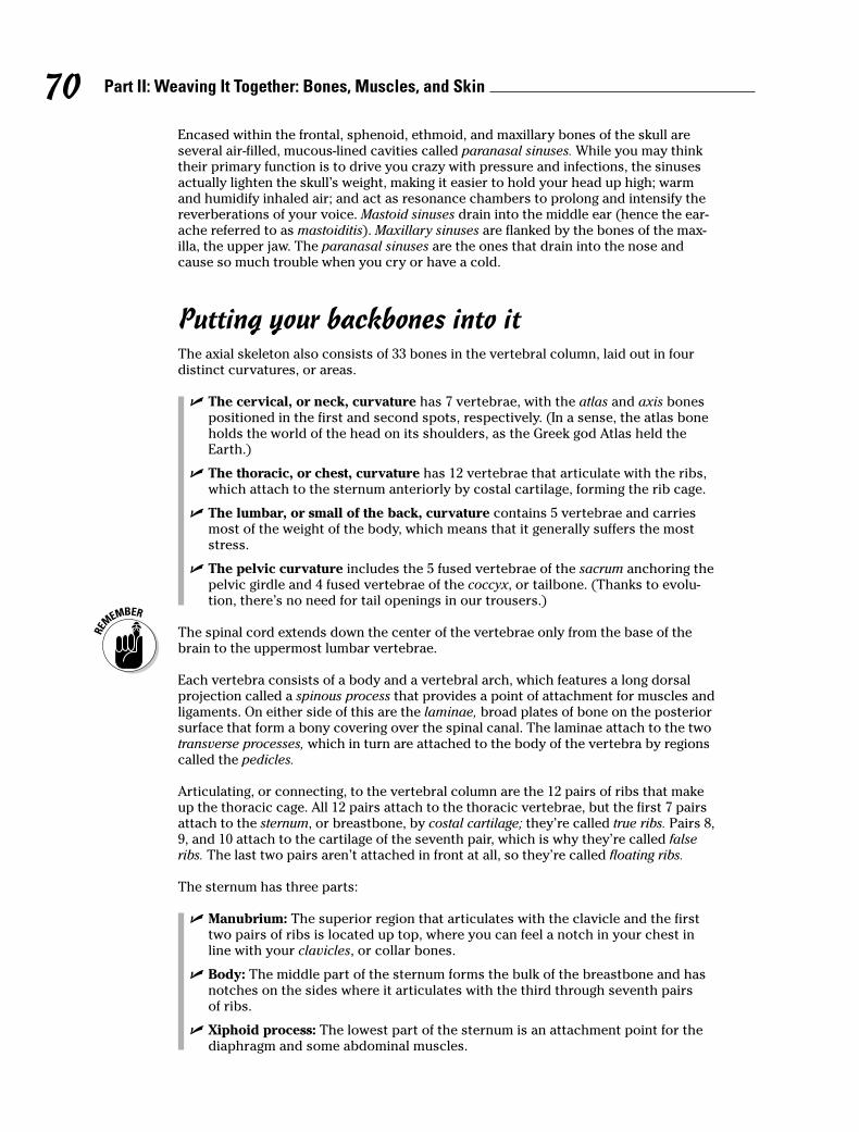
Encased within the frontal, sphenoid, ethmoid, and maxillary bones of the skull areseveral air-filled, mucous-lined cavities called paranasal sinuses. While you may thinktheir primary function is to drive you crazy with pressure and infections, the sinusesactually lighten the skull’s weight, making it easier to hold your head up high; warmand humidify inhaled air; and act as resonance chambers to prolong and intensify thereverberations of your voice. Mastoid sinuses drain into the middle ear (hence the ear-ache referred to as mastoiditis). Maxillary sinuses are flanked by the bones of the max-illa, the upper jaw. The paranasal sinuses are the ones that drain into the nose andcause so much trouble when you cry or have a cold.
Putting your backbones into itThe axial skeleton also consists of 33 bones in the vertebral column, laid out in fourdistinct curvatures, or areas.
� The cervical, or neck, curvature has 7 vertebrae, with the atlas and axis bonespositioned in the first and second spots, respectively. (In a sense, the atlas boneholds the world of the head on its shoulders, as the Greek god Atlas held theEarth.)
� The thoracic, or chest, curvature has 12 vertebrae that articulate with the ribs,which attach to the sternum anteriorly by costal cartilage, forming the rib cage.
� The lumbar, or small of the back, curvature contains 5 vertebrae and carriesmost of the weight of the body, which means that it generally suffers the moststress.
� The pelvic curvature includes the 5 fused vertebrae of the sacrum anchoring thepelvic girdle and 4 fused vertebrae of the coccyx, or tailbone. (Thanks to evolu-tion, there’s no need for tail openings in our trousers.)
The spinal cord extends down the center of the vertebrae only from the base of thebrain to the uppermost lumbar vertebrae.
Each vertebra consists of a body and a vertebral arch, which features a long dorsalprojection called a spinous process that provides a point of attachment for muscles andligaments. On either side of this are the laminae, broad plates of bone on the posteriorsurface that form a bony covering over the spinal canal. The laminae attach to the twotransverse processes, which in turn are attached to the body of the vertebra by regionscalled the pedicles.
Articulating, or connecting, to the vertebral column are the 12 pairs of ribs that makeup the thoracic cage. All 12 pairs attach to the thoracic vertebrae, but the first 7 pairsattach to the sternum, or breastbone, by costal cartilage; they’re called true ribs. Pairs 8,9, and 10 attach to the cartilage of the seventh pair, which is why they’re called falseribs. The last two pairs aren’t attached in front at all, so they’re called floating ribs.
The sternum has three parts:
� Manubrium: The superior region that articulates with the clavicle and the firsttwo pairs of ribs is located up top, where you can feel a notch in your chest inline with your clavicles, or collar bones.
� Body: The middle part of the sternum forms the bulk of the breastbone and hasnotches on the sides where it articulates with the third through seventh pairs of ribs.
� Xiphoid process: The lowest part of the sternum is an attachment point for thediaphragm and some abdominal muscles.
70 Part II: Weaving It Together: Bones, Muscles, and Skin

Emergency medical technicians learn to administer CPR at least three finger widthsabove the xiphoid.
57.–71. Use the terms that follow to identify the bones, sutures, and landmarks of the skull shown inFigure 5-2.
LifeART Image Copyright © 2007. Wolters Kluwer Health — Lippincott Willams & Wilkins
a. Temporal bone
b. Sphenoid bone
c. Nasal bone
d. Styloid process
e. Frontal bone
f. Squamosal suture
g. Maxilla
h. Parietal bone
i. Lambdoidal suture
j. Occipital bone
k. Zygomatic bone
l. Coronal suture
m.Mandibular condyle
n. Zygomatic process
o. Mandible
72.–75. Match the skull bones with their connecting sutures.
72. _____ Frontal and parietals a. Squamosal suture
73. _____ Occipital and parietals b. Lambdoidal suture
74. _____ Parietal and parietal c. Sagittal suture
75. _____ Temporal and parietal d. Coronal suture
57 _____
58 _____
59 __________ 71
_____ 65
_____ 66
_____ 67
_____ 68
_____ 69
60 _____
62 _____
64 _____
63 _____
61 _____
_____ 70
Figure 5-2:
A lateral
view of
the skull.
71Chapter 5: A Scaffold to Build On: The Skeleton
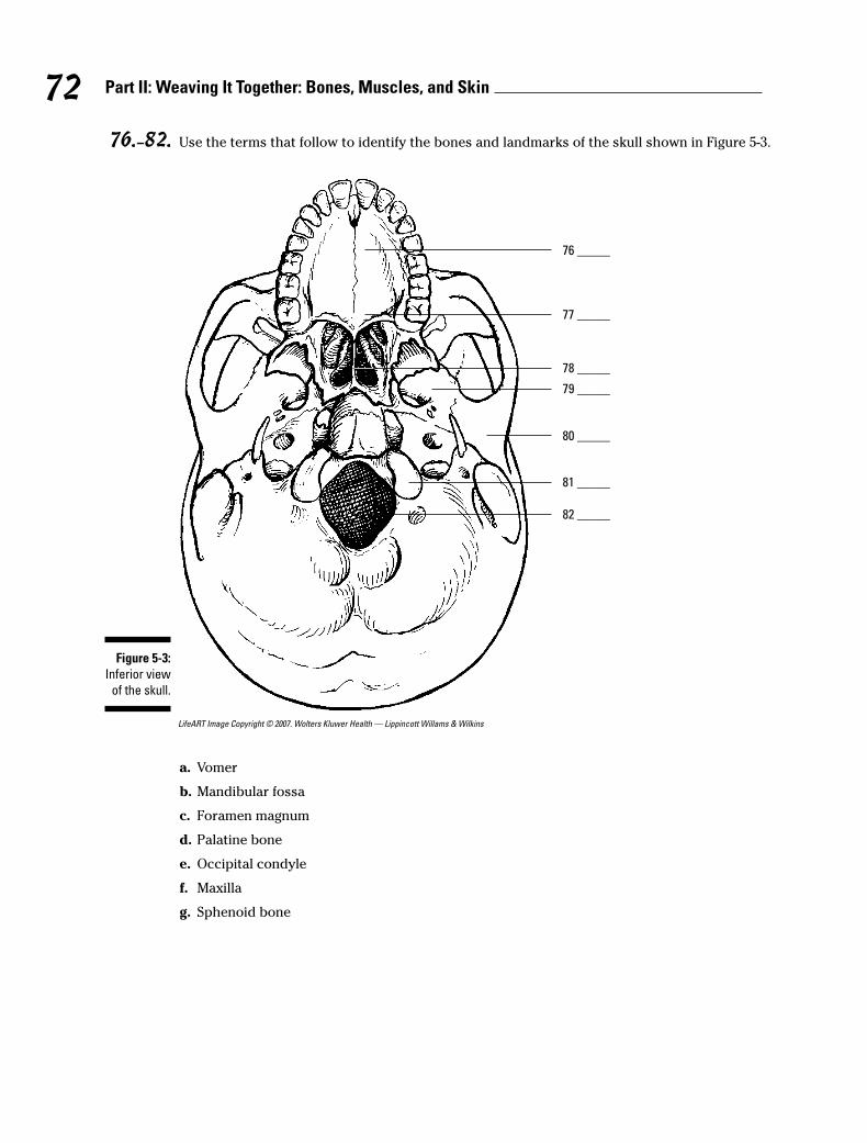
76.–82. Use the terms that follow to identify the bones and landmarks of the skull shown in Figure 5-3.
LifeART Image Copyright © 2007. Wolters Kluwer Health — Lippincott Willams & Wilkins
a. Vomer
b. Mandibular fossa
c. Foramen magnum
d. Palatine bone
e. Occipital condyle
f. Maxilla
g. Sphenoid bone
80 _____
81 _____
82 _____
76 _____
77 _____
78 _____
79 _____
Figure 5-3:
Inferior view
of the skull.
72 Part II: Weaving It Together: Bones, Muscles, and Skin
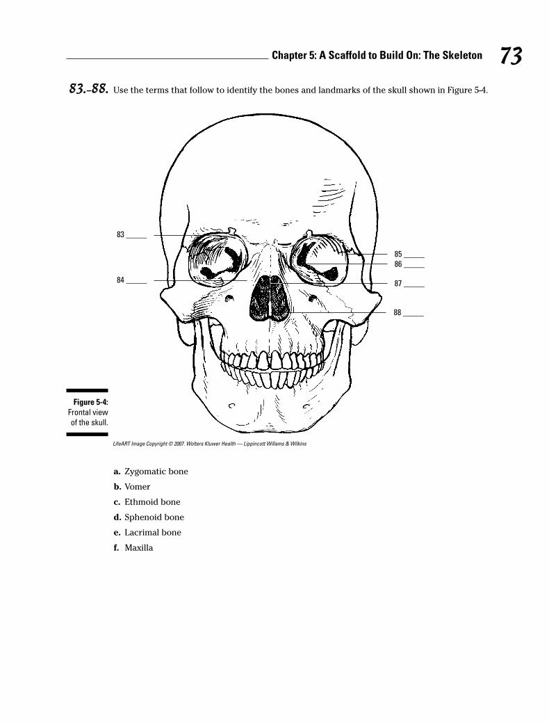
83.–88. Use the terms that follow to identify the bones and landmarks of the skull shown in Figure 5-4.
LifeART Image Copyright © 2007. Wolters Kluwer Health — Lippincott Willams & Wilkins
a. Zygomatic bone
b. Vomer
c. Ethmoid bone
d. Sphenoid bone
e. Lacrimal bone
f. Maxilla
85 _____
86 _____
87 _____
88 _____
83 _____
84 _____
Figure 5-4:
Frontal view
of the skull.
73Chapter 5: A Scaffold to Build On: The Skeleton
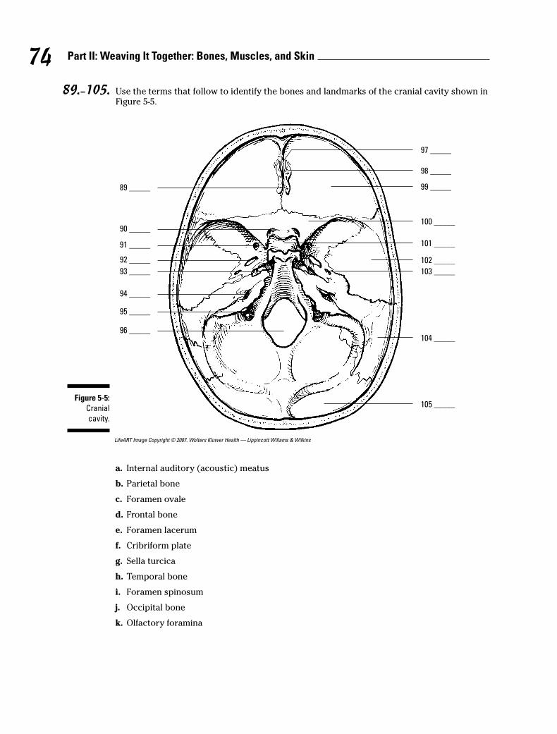
89.–105. Use the terms that follow to identify the bones and landmarks of the cranial cavity shown inFigure 5-5.
LifeART Image Copyright © 2007. Wolters Kluwer Health — Lippincott Willams & Wilkins
a. Internal auditory (acoustic) meatus
b. Parietal bone
c. Foramen ovale
d. Frontal bone
e. Foramen lacerum
f. Cribriform plate
g. Sella turcica
h. Temporal bone
i. Foramen spinosum
j. Occipital bone
k. Olfactory foramina
89 _____
90 _____
91 _____
92 _____
93 _____
94 _____
95 _____
96 _____
97 _____
105 _____
104 _____
101 _____
103 _____
102 _____
100 _____
99 _____
98 _____
Figure 5-5:
Cranial
cavity.
74 Part II: Weaving It Together: Bones, Muscles, and Skin
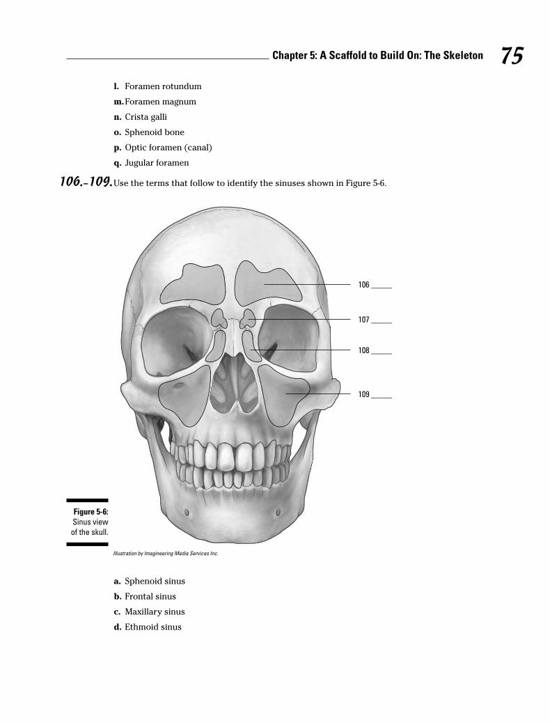
l. Foramen rotundum
m.Foramen magnum
n. Crista galli
o. Sphenoid bone
p. Optic foramen (canal)
q. Jugular foramen
106.–109.Use the terms that follow to identify the sinuses shown in Figure 5-6.
Illustration by Imagineering Media Services Inc.
a. Sphenoid sinus
b. Frontal sinus
c. Maxillary sinus
d. Ethmoid sinus
106 _____
107 _____
108 _____
109 _____
Figure 5-6:
Sinus view
of the skull.
75Chapter 5: A Scaffold to Build On: The Skeleton

110.–118.Use the terms that follow to identify the regions, structures, and landmarks of the vertebralcolumn shown in Figure 5-7.
LifeART Image Copyright © 2007. Wolters Kluwer Health — Lippincott Willams & Wilkins
a. Coccyx
b. Intervertebral foramen
c. Thoracic vertebrae or curvature
d. Sacrum
e. A vertebra
f. Cervical vertebrae or curvature
g. Lumbar vertebrae or curvature
h. A spinous process
i. Intervertebral disc
110 _____
1
12
11
10
9
8
7
6
5
4
3
2
1
76
5432
1
2
3
4
5
116 _____
117 _____
118 _____
115 _____
111 _____
112 _____
113 _____
114 _____
Figure 5-7:
Vertebral
column.
76 Part II: Weaving It Together: Bones, Muscles, and Skin
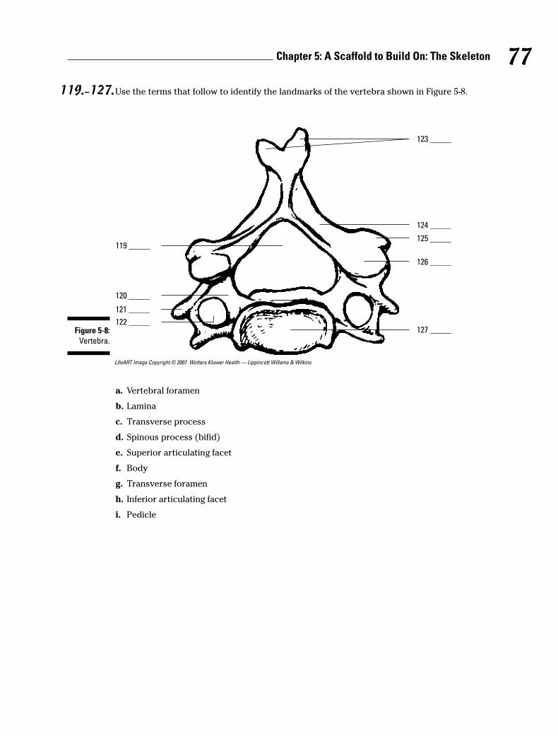
119.–127.Use the terms that follow to identify the landmarks of the vertebra shown in Figure 5-8.
LifeART Image Copyright © 2007. Wolters Kluwer Health — Lippincott Willams & Wilkins
a. Vertebral foramen
b. Lamina
c. Transverse process
d. Spinous process (bifid)
e. Superior articulating facet
f. Body
g. Transverse foramen
h. Inferior articulating facet
i. Pedicle
123 _____
124 _____
125 _____
126 _____
127 _____
119 _____
120 _____
121 _____
122 _____Figure 5-8:
Vertebra.
77Chapter 5: A Scaffold to Build On: The Skeleton
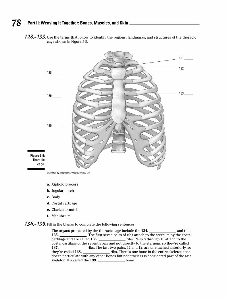
128.–133.Use the terms that follow to identify the regions, landmarks, and structures of the thoraciccage shown in Figure 5-9.
Illustration by Imagineering Media Services Inc.
a. Xiphoid process
b. Jugular notch
c. Body
d. Costal cartilage
e. Clavicular notch
f. Manubrium
134.–139.Fill in the blanks to complete the following sentences:
The organs protected by the thoracic cage include the 134. _______________ and the 135. _______________. The first seven pairs of ribs attach to the sternum by the costal cartilage and are called 136. _______________ ribs. Pairs 8 through 10 attach to the costal cartilage of the seventh pair and not directly to the sternum, so they’re called 137. _______________ ribs. The last two pairs, 11 and 12, are unattached anteriorly, sothey’re called 138. _______________ ribs. There’s one bone in the entire skeleton that doesn’t articulate with any other bones but nonetheless is considered part of the axial skeleton. It’s called the 139. _______________ bone.
128 _____
131 _____
129 _____
130 _____
132 _____
133 _____
Figure 5-9:
Thoracic
cage.
78 Part II: Weaving It Together: Bones, Muscles, and Skin
Appendicular Skeleton: Reaching Beyond Our Girdles
Whereas the axial skeleton lies along the body’s central axis, the appendicular skele-ton’s 126 bones include those in all four appendages — arms and legs — plus the twoprimary girdles to which the appendages attach: the pectoral (chest) girdle and thepelvic (hip) girdle.
The pectoral girdle is made up of a pair of clavicles, or collar bones, which attach tothe sternum medially and to the scapula laterally articulating with the acromionprocess, and a pair of scapulae, better known as shoulder blades. Each scapula has adepression in it called the glenoid fossa where the head of the humerus (upper armbone) is attached. The lower end of the humerus articulates with the forearm’s longulna bone to form the elbow joint. The process called the olecranon forms the elbowand is also referred to as the funny bone, although banging it into something usuallyfeels anything but funny. The forearm also contains a bone called the radius; togetherthe ulna and radius articulate with the eight small carpal bones that form the wrist.The carpals articulate with the five metacarpals that form the hand, which in turn con-nect with the phalanges (finger bones), which are found as a pair in the thumb and astriplets in each of the fingers. (Toe bones also are called phalanges, but we get to thatin a moment.)
The pelvic girdle consists of two hip bones, called os coxae, as well as the sacrumand coccyx, more commonly referred to as the tail bone. During early developmentalyears, the os coxa consists of three separate bones — the ilium, the ischium, and thepubis — that later fuse into one bone sometime between the ages of 16 and 20.Posteriorly, the os coxa articulates with the sacrum, forming the sacroiliac joint, thesource of much lower back pain; it’s formed by the connection of the hip bones at the sacrum. Toward the front of the pelvic girdle, the two os coxae join to form thesymphysis pubis, which is made up of fibrocartilage. A cup-like socket called the acetab-ulum articulates with the ball-shaped head of the leg’s femur (thigh bone). (The femuris the longest bone in the body.) The femur articulates with the tibia (shin bone) at theknee, which is covered by the patella (kneecap). Also inside each lower leg is the fibulabone, which joins with the tibia to connect with the seven tarsal bones that make upthe ankle. The tarsals join with the five metatarsals that form the foot, which in turnconnect to the phalanges of the toes — a pair of phalanges in the big toe and triplets ineach of the other toes.
79Chapter 5: A Scaffold to Build On: The Skeleton
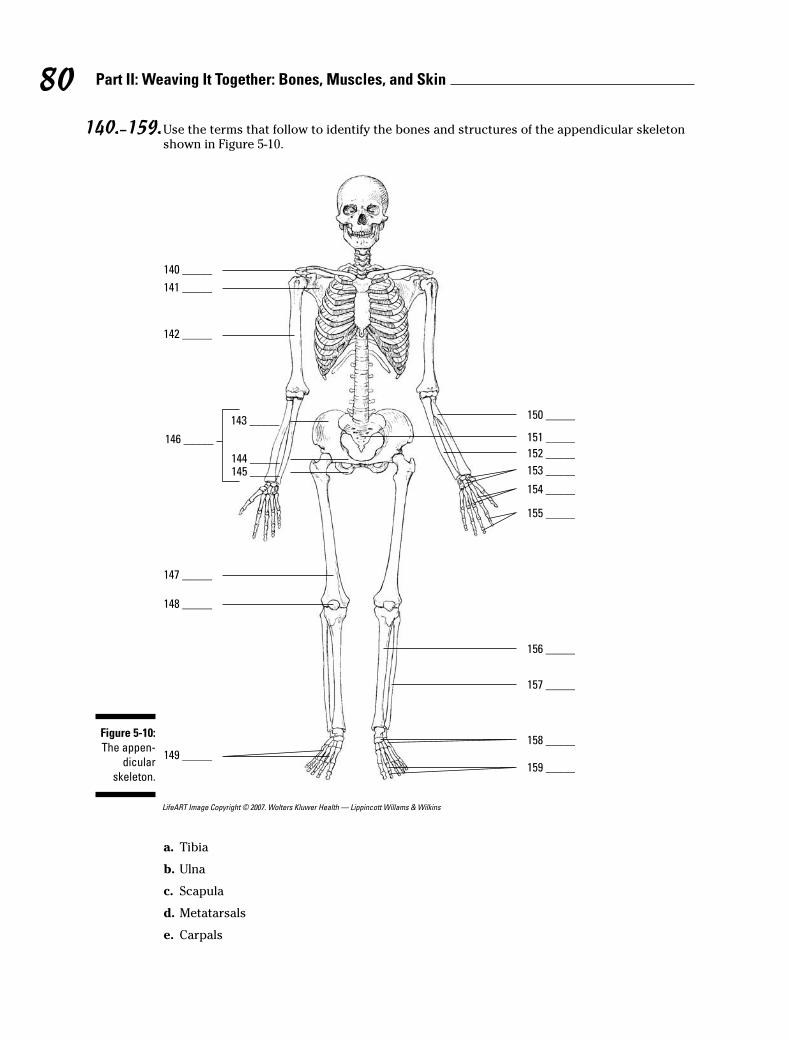
140.–159.Use the terms that follow to identify the bones and structures of the appendicular skeletonshown in Figure 5-10.
LifeART Image Copyright © 2007. Wolters Kluwer Health — Lippincott Willams & Wilkins
a. Tibia
b. Ulna
c. Scapula
d. Metatarsals
e. Carpals
140 _____
150 _____
141 _____
142 _____
147 _____
148 _____
149 _____
143 _____
144 _____145 _____
146 _____ 151 _____
152 _____
153 _____
154 _____
155 _____
156 _____
157 _____
158 _____
159 _____
Figure 5-10:
The appen-
dicular
skeleton.
80 Part II: Weaving It Together: Bones, Muscles, and Skin
f. Phalanges of the feet
g. Ilium
h. Clavicle
i. Fibula
j. Patella
k. Humerus
l. Pubis
m.Radius
n. Os coxa
o. Sacrum
p. Phalanges of the hand
q. Metacarpals
r. Tarsals
s. Femur
t. Ishium
160. The structure of the scapula that articulates with the clavicle is the
a. Acromion process
b. Glenoid fossa
c. Coracoid process
d. Spine
161. The point of attachment for the biceps muscle on the radius is the
a. Radial notch
b. Styloid process
c. Radial tuberosity
d. Ulnar notch
162. The structure of the humerus that articulates with the head of the radius is the
a. Deltoid tuberosity
b. Olecranon fossa
c. Trochlea
d. Capitulum
163. The patella is what kind of bone?
a. Wormian
b. Sesamoid
c. Pisiform
d. Hallux
81Chapter 5: A Scaffold to Build On: The Skeleton

164. Which of these bones is not part of the pelvic girdle?
a. Ilium
b. Lumbar vertebrae
c. Sacrum
d. Ischium
165. The prominence that forms the elbow is the
a. Olecranon process
b. Trochlear notch
c. Radial notch
d. Coronoid process
166. The ulna articulates with the humerus at the
a. Deltoid tuberosity
b. Greater tubercle
c. Capitulum
d. Trochlea
167. The socket for the head of the femur is the
a. Obturator foramen
b. Acetabulum
c. Ischial tuberosity
d. Greater sciatic notch
168. The largest and strongest tarsal bone is the
a. Talus
b. Cuboid
c. Navicular
d. Calcaneus
169. A person complaining of problems in their sacroiliac has pain in the
a. Lower back
b. Neck
c. Feet
d. Hands
Arthrology: Articulating the JointsArthrology, which stems from the ancient Greek word arthros (meaning “jointed”), isthe study of those structures that hold bones together, allowing them to move to vary-ing degrees — or fixing them in place — depending on the design and function of thejoint. The term articulation, or joint, applies to any union of bones, whether it movesfreely or not at all.
82 Part II: Weaving It Together: Bones, Muscles, and Skin
Inside some joints, such as knees and elbows, are fluid-filled sacs called bursae thathelp reduce friction between tendons and bones; inflammation in these sacs is calledbursitis. Some joints are stabilized by connective tissue called ligaments that rangefrom bundles of collagenous fibers that restrict movement and hold a joint in place toelastic fibers that can repeatedly stretch and return to their original shapes.
The three types of joints are as follows:
� Fibrous: Fibrous tissue rigidly joins the bones in a form of articulation calledsynarthrosis, which is characterized by no movement at all. The sutures of theskull are fibrous joints.
� Cartilaginous: This type of joint is found in two forms:
• Synchondrosis articulation involves rigid cartilage that allows no move-ment, such as the joint between the ribs, costal cartilage, and sternum.
• Symphysis joints occur where cartilage fuses bones in such a way thatpressure can cause slight movement, called amphiarthrosis. Examplesinclude the intervertebral discs and the symphysis pubis.
� Synovial: Also known as diarthrosis, or freely moving, joints, this type of articula-tion involves a synovial cavity, which contains articular fluid secreted from thesynovial membrane to lubricate the opposing surfaces of bone. The synovialmembrane is covered by a fibrous joint capsule layer that’s continuous with theperiosteum of the bone. Ligaments surrounding the joint strengthen the capsuleand hold the bones in place, preventing dislocation. In some synovial joints,such as the knee, fibrous connective tissue called meniscus develops in thecavity, dividing it into two parts. In the knee, this meniscus stabilizes the jointand acts as a shock absorber.
There are six classifications of moveable, or synovial, joints:
� Gliding: Curved or flat surfaces slide against one another, such as between thecarpal bones in the wrist or between the tarsal bones in the ankle.
� Hinge: A convex surface joints with a concave surface, allowing right-anglemotions in one plane, such as elbows, knees, and joints between the finger bones.
� Pivot (or rotary): One bone pivots or rotates around a stationary bone, such asthe atlas rotating around the odontoid process at the top of the vertebralcolumn.
� Condyloid: The oval head of one bone fits into a shallow depression in another,allowing the joint to move in two directions, such as the carpal-metacarpal jointat the wrist, or the tarsal-metatarsal joint at the ankle.
� Saddle: Each of the adjoining bones is shaped like a saddle (the technical term isreciprocally concavo-convex), allowing various movements, such as the car-pometacarpal joint of the thumb.
� Ball-and-socket: The round head of one bone fits into a cup-like cavity in theother bone, allowing movement in many directions so long as the bones are nei-ther pulled apart nor forced together, such as the shoulder joint between thehumerus and scapula and the hip joints between the femur and the os coxa.
The following are the types of joint movement:
� Flexion: Decrease the angle between two bones
� Extension: Increase the angle between two bones
� Abduction: Movement away from the midline of the body
83Chapter 5: A Scaffold to Build On: The Skeleton
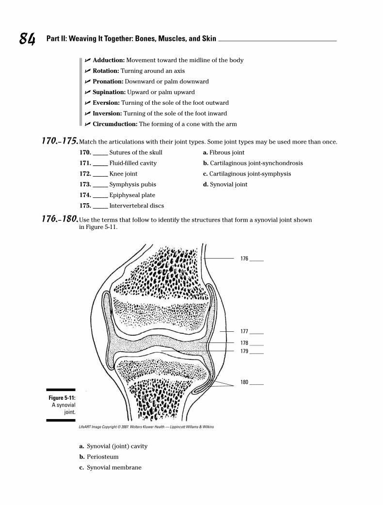
� Adduction: Movement toward the midline of the body
� Rotation: Turning around an axis
� Pronation: Downward or palm downward
� Supination: Upward or palm upward
� Eversion: Turning of the sole of the foot outward
� Inversion: Turning of the sole of the foot inward
� Circumduction: The forming of a cone with the arm
170.–175.Match the articulations with their joint types. Some joint types may be used more than once.
170. _____ Sutures of the skull a. Fibrous joint
171. _____ Fluid-filled cavity b. Cartilaginous joint-synchondrosis
172. _____ Knee joint c. Cartilaginous joint-symphysis
173. _____ Symphysis pubis d. Synovial joint
174. _____ Epiphyseal plate
175. _____ Intervertebral discs
176.–180.Use the terms that follow to identify the structures that form a synovial joint shown in Figure 5-11.
LifeART Image Copyright © 2007. Wolters Kluwer Health — Lippincott Willams & Wilkins
a. Synovial (joint) cavity
b. Periosteum
c. Synovial membrane
176 _____
177 _____
178 _____
179 _____
180 _____
Figure 5-11:
A synovial
joint.
84 Part II: Weaving It Together: Bones, Muscles, and Skin

d. Articular cartilage
e. Fibrous capsule
181. An immovable joint is
a. Amphiarthrosis
b. Synarthrosis
c. Diarthrosis
d. Synchondrosis
182. A freely moving joint is
a. Amphiarthrosis
b. Synarthrosis
c. Diarthrosis
d. Synchondrosis
183. The material or structure that allows for free movement in a joint is
a. Bursa
b. Periosteum
c. Synovial fluid
d. Bone marrow
184. An example of a ball-and-socket joint is the
a. Symphysis pubis
b. Hip
c. Ankle
d. Elbow
185. An example of a pivotal joint is
a. Between the radius and the ulna
b. The interphalanges joints
c. Between the mandible and the temporal bone
d. Between the tibia and the fibula
186. A saddle joint is located in
a. The radius and the carpals
b. The carpometacarpal joint of the thumb
c. The occipital condyles and the atlas
d. The metatarsophalanges joint
187. A shoulder joint ligament is the
a. Coracohumeral ligament
b. Popliteal ligament
c. Ischiofemoral ligament
d. Ligamentum arteriosum
85Chapter 5: A Scaffold to Build On: The Skeleton
188. A knee joint ligament is the
a. Iliofemoral ligament
b. Coracohumeral ligament
c. Oblique popliteal ligament
d. Annular ligament
189. A hip joint ligament is the
a. Subscapularis ligament
b. Pubofemoral ligament
c. Glenohumeral ligament
d. Coracohumeral ligament
190. The structure in the knee that divides the synovial joint into two separate compartments is the
a. Bursa
b. Joint fat
c. Tendon sheath
d. Meniscus or articular disc
191.–200.Match the type of joint movement with its description.
191. _____ Flexion a. Upward or palm upward
192. _____ Extension b. Decrease the angle between two bones
193. _____ Abduction c. Turning of the sole of the foot inward
194. _____ Adduction d. Downward or palm downward
195. _____ Rotation e. Increase the angle between two bones
196. _____ Pronation f. Turning of the sole of the foot outward
197. _____ Supination g. Movement away from the midline of the body
198. _____ Eversion h. The forming of a cone with the arm
199. _____ Inversion i. Turning around an axis
200. _____ Circumduction j. Movement toward the midline of the body
86 Part II: Weaving It Together: Bones, Muscles, and Skin
Answers to Questions on the SkeletonThe following are answers to the practice questions presented in this chapter.
a The formation of red blood cells by the bone marrow is known as b. hematopoiesis. The termhemopoiesis also would be correct here, but it’s not one of the answer options.
b The following mineral is stored inside bones for later use: d. All of the above (phosphorous,calcium, and magnesium). The bones act as a mineral bank for the entire body.
c Besides support and protection, the skeleton serves other important functions, including b. locomotion. You’d be a motionless blob without a skeleton to support coordinated movement.
d The curvatures in some bone structures serve the following purpose: a. Support the body. Andnow you know why your back aches most where it curves.
e The most abundant mineral in the body is c. calcium.
f A decrease in calcium concentration in the body fluids by 35 percent causes c. convulsions. Gomuch beyond that and the situation becomes fatal.
g Calcitonin is produced by the a. thyroid gland. There’s that busy little thyroid gland, control-ling metabolism.
h Bones are encased by a membrane called the b. periosteum. Back to Greek again: peri means“around” and osteon means “bone,” so the periosteum is “around the bone.”
i A bone’s encapsulating membrane is attached by c. Sharpey’s fibers. Described by anatomistWilliam Sharpey in 1846, these are also called perforating fibers.
j Bone marrow can be found inside the a. medullary cavity. This is where you’ll find yellowmarrow, although in infants red marrow also is present.
k The six structures in the skull of an infant are called b. fontanels. These separate floating platesare why you can see a bald baby’s pulse throbbing on the top of its head.
l Volkmann’s canals a. are found in cancellous bone only. Ironically, anatomist Alfred WilhelmVolkmann was most noted for his observations of the physiology of the nervous system, notbones.
m Blood vessels entering through Volkmann’s canals reach the bone cells through the b. Haversian canal. The name comes from the first physician to describe them, CloptonHavers.
n The hormone released when calcium ion concentration is abnormally high is b. calcitonin. Thispeptide hormone lowers plasma calcium.
o–y Bones are first laid down as 15. cartilage during the fifth week after conception. Developmentof the bone begins with 16. calcification, the depositing of calcium and phosphorus. Next, theblood supply entering the cartilage brings 17. osteoblasts that attach themselves to the carti-lage. Ossification in long bones begins in the 18. diaphysis of the long bone and moves towardthe 19. epiphysis of the bone. The epiphyseal and diaphyseal areas remain separated by a layerof uncalcified cartilage called the 20. epiphyseal plate.
87Chapter 5: A Scaffold to Build On: The Skeleton
Another very large cell that enters with the blood supply is the 21. osteoclast, which helpsabsorb the cartilage as ossification occurs. Later it helps absorb bone tissue from the center ofthe long bone’s shaft, forming the 22. medullary or marrow cavity. After ossification, thespaces that were formed by the osteoclasts join together to form 23. Haversian canal systems,which contain the blood vessels, lymphatic vessels, and nerves. Unlike bones in the rest of thebody, those of the skull and mandible (lower jaw) are first laid down as 24. membrane. In theskull, the edges of the bone don’t ossify in the fetus but remain membranous and form 25.fontanels.
A Vertebrae of the vertebral column: b. Irregular bone
B Femur in thigh: c. Long bone
C Sternum: a. Flat bone
D Tarsals in ankle: d. Short bone
E Humerus in upper arm: c. Long bone
F Phalanges in fingers and toes: d. Short bone
G Scapulae of shoulder: a. Flat bone
H Kneecap: b. Irregular bone
I Carpals in wrist: d. Short bone
J An abrupt or pointed projection: i. Spine
K A large, usually blunt process: k. Trochanter
L A designation for any prominence or prolongation: h. Process
M A large, often rough eminence: m. Tuberosity
N A prominent ridge: b. Crest
O A large, rounded articular end of a bone; often set off from the shaft by the neck: f. Head
P An oval articular prominence of a bone: a. Condyle
Q A smooth, flat or nearly flat articulating surface: c. Facet
R A deeper depression: e. Fossa
S A groove: j. Sulcus
T A hole: d. Foramen
U A canal or opening to a canal: g. Meatus
V A smaller, rounded eminence: l. Tubercle
W–4 Following is how Figure 5-1, the long bone, should be labeled.
48. h. Articulating cartilage; 49. d. Spongy bone tissue; 50. g. Red bone marrow; 51. i.Compact bone tissue; 52. e. Medullary or nutrient artery; 53. b. Medullary cavity; 54. f.Proximal epiphysis; 55. a. Diaphysis; 56. c. Distal epiphysis
88 Part II: Weaving It Together: Bones, Muscles, and Skin
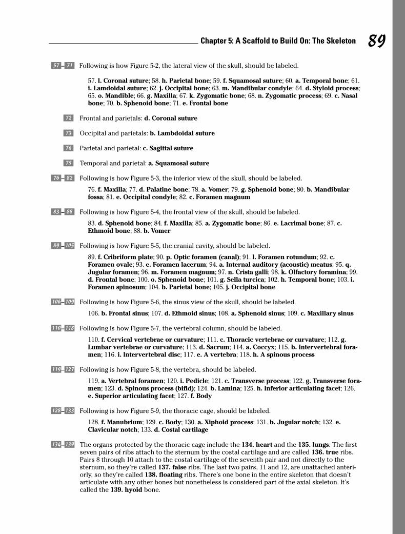
5–( Following is how Figure 5-2, the lateral view of the skull, should be labeled.
57. l. Coronal suture; 58. h. Parietal bone; 59. f. Squamosal suture; 60. a. Temporal bone; 61.i. Lamdoidal suture; 62. j. Occipital bone; 63. m. Mandibular condyle; 64. d. Styloid process;65. o. Mandible; 66. g. Maxilla; 67. k. Zygomatic bone; 68. n. Zygomatic process; 69. c. Nasalbone; 70. b. Sphenoid bone; 71. e. Frontal bone
) Frontal and parietals: d. Coronal suture
- Occipital and parietals: b. Lambdoidal suture
_ Parietal and parietal: c. Sagittal suture
= Temporal and parietal: a. Squamosal suture
+–| Following is how Figure 5-3, the inferior view of the skull, should be labeled.
76. f. Maxilla; 77. d. Palatine bone; 78. a. Vomer; 79. g. Sphenoid bone; 80. b. Mandibularfossa; 81. e. Occipital condyle; 82. c. Foramen magnum
;–> Following is how Figure 5-4, the frontal view of the skull, should be labeled.
83. d. Sphenoid bone; 84. f. Maxilla; 85. a. Zygomatic bone; 86. e. Lacrimal bone; 87. c.Ethmoid bone; 88. b. Vomer
/–œ Following is how Figure 5-5, the cranial cavity, should be labeled.
89. f. Cribriform plate; 90. p. Optic foramen (canal); 91. l. Foramen rotundum; 92. c.Foramen ovale; 93. e. Foramen lacerum; 94. a. Internal auditory (acoustic) meatus; 95. q.Jugular foramen; 96. m. Foramen magnum; 97. n. Crista galli; 98. k. Olfactory foramina; 99.d. Frontal bone; 100. o. Sphenoid bone; 101. g. Sella turcica; 102. h. Temporal bone; 103. i.Foramen spinosum; 104. b. Parietal bone; 105. j. Occipital bone
®–ö Following is how Figure 5-6, the sinus view of the skull, should be labeled.
106. b. Frontal sinus; 107. d. Ethmoid sinus; 108. a. Sphenoid sinus; 109. c. Maxillary sinus
õ–§ Following is how Figure 5-7, the vertebral column, should be labeled.
110. f. Cervical vertebrae or curvature; 111. c. Thoracic vertebrae or curvature; 112. g.Lumbar vertebrae or curvature; 113. d. Sacrum; 114. a. Coccyx; 115. b. Intervertebral fora-men; 116. i. Intervertebral disc; 117. e. A vertebra; 118. h. A spinous process
¶–« Following is how Figure 5-8, the vertebra, should be labeled.
119. a. Vertebral foramen; 120. i. Pedicle; 121. c. Transverse process; 122. g. Transverse fora-men; 123. d. Spinous process (bifid); 124. b. Lamina; 125. h. Inferior articulating facet; 126.e. Superior articulating facet; 127. f. Body
…–ı Following is how Figure 5-9, the thoracic cage, should be labeled.
128. f. Manubrium; 129. c. Body; 130. a. Xiphoid process; 131. b. Jugular notch; 132. e.Clavicular notch; 133. d. Costal cartilage
í–ˆ The organs protected by the thoracic cage include the 134. heart and the 135. lungs. The firstseven pairs of ribs attach to the sternum by the costal cartilage and are called 136. true ribs.Pairs 8 through 10 attach to the costal cartilage of the seventh pair and not directly to the sternum, so they’re called 137. false ribs. The last two pairs, 11 and 12, are unattached anteri-orly, so they’re called 138. floating ribs. There’s one bone in the entire skeleton that doesn’tarticulate with any other bones but nonetheless is considered part of the axial skeleton. It’scalled the 139. hyoid bone.
89Chapter 5: A Scaffold to Build On: The Skeleton
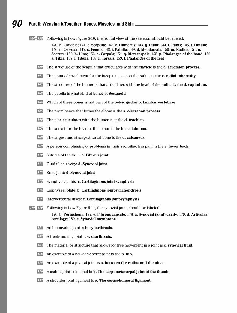
Ô–å Following is how Figure 5-10, the frontal view of the skeleton, should be labeled.
140. h. Clavicle; 141. c. Scapula; 142. k. Humerus; 143. g. Ilium; 144. l. Pubis; 145. t. Ishium;146. n. Os coxa; 147. s. Femur; 148. j. Patella; 149. d. Metatarsals; 150. m. Radius; 151. o.Sacrum; 152. b. Ulna; 153. e. Carpals; 154. q. Metacarpals; 155. p. Phalanges of the hand; 156.a. Tibia; 157. i. Fibula; 158. r. Tarsals; 159. f. Phalanges of the feet
ç The structure of the scapula that articulates with the clavicle is the a. acromion process.
ë The point of attachment for the biceps muscle on the radius is the c. radial tuberosity.
î The structure of the humerus that articulates with the head of the radius is the d. capitulum.
ï The patella is what kind of bone? b. Sesamoid
ñ Which of these bones is not part of the pelvic girdle? b. Lumbar vertebrae
† The prominence that forms the elbow is the a. olecranon process.
‚ The ulna articulates with the humerus at the d. trochlea.
„ The socket for the head of the femur is the b. acetabulum.
• The largest and strongest tarsal bone is the d. calcaneus.
´ A person complaining of problems in their sacroiliac has pain in the a. lower back.
¨ Sutures of the skull: a. Fibrous joint
≠ Fluid-filled cavity: d. Synovial joint
∞ Knee joint: d. Synovial joint
± Symphysis pubis: c. Cartilaginous joint-symphysis
≤ Epiphyseal plate: b. Cartilaginous joint-synchondrosis
≥ Intervertebral discs: c. Cartilaginous joint-symphysis
∂–∫ Following is how Figure 5-11, the synovial joint, should be labeled.
176. b. Periosteum; 177. e. Fibrous capsule; 178. a. Synovial (joint) cavity; 179. d. Articularcartilage; 180. c. Synovial membrane
Ê An immovable joint is b. synarthrosis.
ø A freely moving joint is c. diarthrosis.
¿ The material or structure that allows for free movement in a joint is c. synovial fluid.
√ An example of a ball-and-socket joint is the b. hip.
≈ An example of a pivotal joint is a. between the radius and the ulna.
∆ A saddle joint is located in b. The carpometacarpal joint of the thumb.
» A shoulder joint ligament is a. The coracohumeral ligament.
90 Part II: Weaving It Together: Bones, Muscles, and Skin

à A knee joint ligament is the c. oblique popliteal ligament.
Õ A hip joint ligament is the b. pubofermoral ligament.
’ The structure in the knee that divides the synovial joint into two separate compartments is thed. meniscus or articular disc.
◊ Flexion: b. Decrease the angle between two bones
ÿ Extension: e. Increase the angle between two bones
Ÿ Abduction: g. Movement away from the midline of the body
⁄ Adduction: j. Movement toward the midline of the body
€ Rotation: i. Turning around an axis
‹ Pronation: d. Downward or palm downward
› Supination: a. Upward or palm upward
fi Eversion: f. Turning of the sole of the foot outward
fl Inversion: c. Turning of the sole of the foot inward
‡ Circumduction: h. The forming of a cone with the arm
91Chapter 5: A Scaffold to Build On: The Skeleton
92 Part II: Weaving It Together: Bones, Muscles, and Skin
Chapter 6
Getting in Gear: The Muscles
In This Chapter� Understanding the functions and structure of muscles
� Classifying types of muscle
� Pulling together: Muscles as organs
� Breaking down muscle contractions, tone, and power
� Deciphering muscle names
Much of what we think of as “the body” centers around our muscles and what they cando, what we want them to do, and how tired we get trying to make them do it. With
all that muscles do and are, it’s hard to believe the word “muscle” is rooted in the Latinword musculus, which is a diminutive of the word for “mouse.” Well, the muscle is a mousethat roars. Muscles make up most of the fleshy parts of the body and average 43 percent ofthe body’s weight. Layered over the skeleton, they largely determine the body’s form. Thereare over 500 muscles large enough to be seen by the unaided eye, and thousands more arevisible only through a microscope. Although there are three distinct types of muscle tissue,every muscle in the human body shares one important characteristic: contractility, the abil-ity to shorten, or contract.
Flexing Your Muscle KnowledgeThe study of muscles is called myology after the Greek word mys, which means “mouse.”Muscles perform a number of functions vital to maintaining life, including
� Movement: Skeletal muscles (those attached to bones) convert chemical energy intomechanical work, producing movement ranging from finger tapping to a swift kick of aball by contracting, or shortening. Reflex muscle reactions protect your fingers whenyou put them too close to a fire and startle you into watchfulness when an unexpectednoise sounds. Many purposeful movements require several sets, or groups, of musclesto work in unison.
� Vital functions: Without muscle activity, you die. Muscles are doing their job whenyour heart beats, when your blood vessels constrict, and when your intestines squeezefood along your digestive tract in peristalsis.
� Antigravity: Perhaps that’s overstating it, but muscles do make it possible for you tostand and move about in spite of gravity’s ceaseless pull. Did your mother tell you toimprove your posture? Just think how bad it would be without any muscles!
� Heat generation: You shiver when you’re cold and stamp your feet and jog in placewhen you need to warm up. That’s because chemical reactions in muscles result inheat, helping to maintain the body’s temperature.
� Keep the body together: Muscles are the warp and woof of your body’s structure,binding one part to another.
As you may remember from studying tissues, muscle cells — called fibers — are someof the longest in the body. Fibers are held together by connective tissue and enclosedin a fibrous sheath called fascia. Some muscle fibers contract rapidly, whereas othersmove at a leisurely pace. Generally speaking, however, the smaller the structure to bemoved, the faster the muscle action. Exercise can increase the thickness of musclefibers, but it doesn’t make new fibers. Skeletal muscles have a rich vascular supplythat dilates during exercise to give the working muscle the extra oxygen it needs tokeep going.
Two processes are central to muscle development in the developing embryo: myogene-sis, during which muscle tissue is formed; and morphogenesis, when the muscles forminto internal organs. By the eighth week of gestation, a fetus is capable of coordinatedmovement.
Following are some important muscle terms to know:
� Fascia: Loose, or areolar, connective tissue that holds muscle fibers together toform a muscle organ
� Fiber: An individual muscle cell
� Insertion: The more movable attachment of a muscle
� Ligament: Elastic connective tissue that supports joints and anchors organs
� Motor nerve: Nerve that stimulates contraction of a muscle
� Myofibril: Fibrils within a muscle cell that contain protein filaments such asactin and myosin that slide during contraction, shortening the fiber (or cell)
� Origin: The immovable attachment of a muscle, or the point at which a muscle isanchored by a tendon to the bone
� Sarcoplasm: The cellular cytoplasm in a muscle fiber
� Tendon: Connective tissue made up of collagen, a fibrous protein that attachesmuscles to bone; lets muscles apply their force at some distance from where acontraction actually takes place
� Tone, or tonus: State of tension present to a degree at all times, even when themuscle is at rest
Complete the following practice questions to see how well you understand the basicsof myology:
1. Which of the following is not a true statement?
a. Muscles represent 90 percent of the total body weight.
b. The ancient Greek word mys means “mouse.”
c. The muscles covering the bones largely determine the form of the body.
d. Posture is an expression of muscle action.
2. Muscle functions include
a. Support of the bony tissues of the body
b. Blood formation
c. Converting chemical energy into mechanical work
d. Only a and c
94 Part II: Weaving It Together: Bones, Muscles, and Skin
3. A necessary property for a muscle to perform work is
a. Extensibility
b. Contractility
c. Elasticity
d. All of the above
4. The cellular unit in muscle tissue is the
a. Filament
b. Myofibril
c. Fiber
d. Fasciculus
5. A partial state of contraction, in part, defines
a. Rigor
b. Tonus
c. Clovus
d. Paralysis
6. It’s possible to completely relax every muscle in the body.
a. True
b. False
7. During embryonic development, tissue development is called
a. Myogelosis
b. Morphogenesis
c. Myogenesis
d. Morpholysis
8. Exercise forms new muscle fibers.
a. True
b. False
Classifications: Smooth, Cardiac, and Skeletal
Muscle tissue is classified in three ways based on the tissue’s function, shape, andstructure:
� Smooth muscle tissue: So-called because it doesn’t have the cross-striations typ-ical of other kinds of muscle, the spindle-shaped fibers of smooth muscle tissuedo have faint longitudinal striping. This muscle tissue forms into sheets andmakes up the walls of hollow organs such as the stomach, intestines, and blad-der. The tissue’s involuntary movements are relatively slow, so contractions last
95Chapter 6: Getting in Gear: The Muscles
longer than those of other muscle tissue, and fatigue is rare. Each fiber is about 6 microns in diameter and can vary from 15 microns to 500 microns long. Ifarranged in a circle inside an organ, contraction constricts the cavity inside theorgan. If arranged lengthwise, contraction of smooth muscle tissue shortensthe organ.
� Cardiac muscle tissue: Found only in the heart, cardiac muscle fibers arebranched, cross-striated, feature one central nucleus, and move through involun-tary control. An electron microscope view of the tissue shows separate fiberstightly pressed against each other, forming cellular junctions called intercalateddiscs that look like tiny, dark-colored plates. Some experts believe intercalateddiscs are not cellular junctions but rather special structures that help move anelectrical impulse throughout the heart.
� Skeletal muscle tissue: This is the tissue that most people think of as muscle. It’s the only muscle subject to voluntary control through the central nervoussystem. Its long, striated cylindrical fibers contract quickly but tire just as fast.Skeletal muscle, which is also what’s considered meat in animals, is 20 percentprotein, 75 percent water, and 5 percent organic and inorganic materials. Eachmultinucleated fiber is encased in a thin, transparent membrane called a sar-colemma that receives and conducts stimuli. The fibers, which vary from 10microns to 100 microns in diameter and up to 4 centimeters in length, are subdi-vided lengthwise into tiny myofibrils roughly 1 micron in diameter that are sus-pended in the cell’s sarcoplasm.
The following practice questions test your knowledge of muscle classifications:
9. This type of muscle tissue lacks cross-striations.
a. Cardiac
b. Smooth
c. Skeletal
d. Contracting
10. Skeletal muscle fibers are encased in
a. A sarcolemma
b. Sarcoplasm
c. Sarcomeres
d. A sarcophagus
11. Which muscle type appears only in a single organ?
a. Contractile
b. Smooth
c. Cardiac
d. Skeletal
12. Intercalated discs
a. Anchor cardiac muscle fibers to one another
b. May play a role in moving electrical impulses through the heart
c. Are found only in the muscles of the back
d. Contribute to tactile perception
96 Part II: Weaving It Together: Bones, Muscles, and Skin
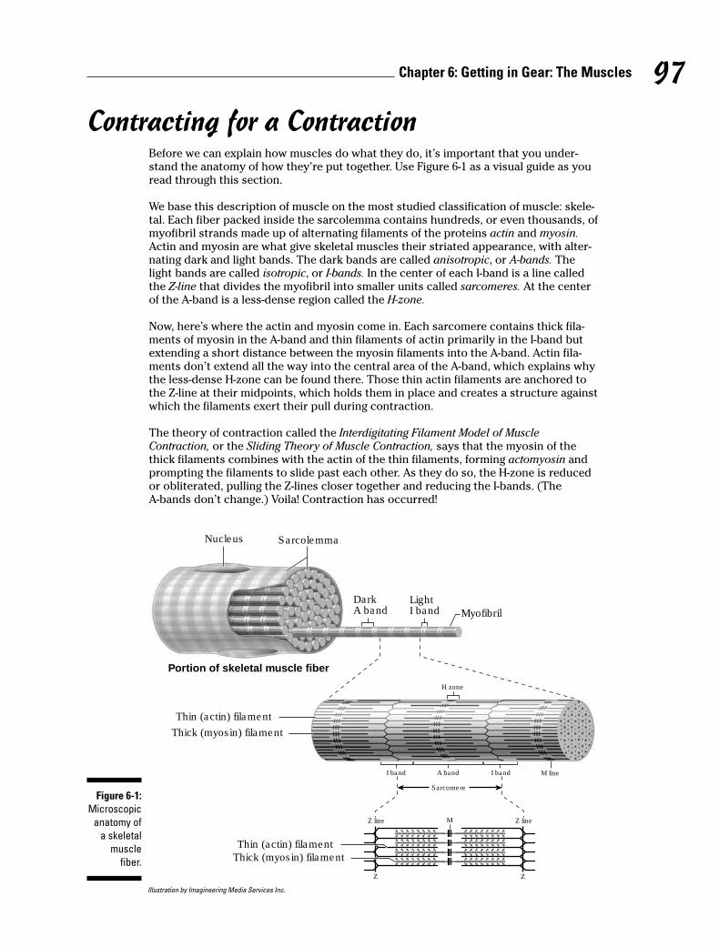
Contracting for a ContractionBefore we can explain how muscles do what they do, it’s important that you under-stand the anatomy of how they’re put together. Use Figure 6-1 as a visual guide as youread through this section.
We base this description of muscle on the most studied classification of muscle: skele-tal. Each fiber packed inside the sarcolemma contains hundreds, or even thousands, ofmyofibril strands made up of alternating filaments of the proteins actin and myosin.Actin and myosin are what give skeletal muscles their striated appearance, with alter-nating dark and light bands. The dark bands are called anisotropic, or A-bands. Thelight bands are called isotropic, or I-bands. In the center of each I-band is a line calledthe Z-line that divides the myofibril into smaller units called sarcomeres. At the centerof the A-band is a less-dense region called the H-zone.
Now, here’s where the actin and myosin come in. Each sarcomere contains thick fila-ments of myosin in the A-band and thin filaments of actin primarily in the I-band butextending a short distance between the myosin filaments into the A-band. Actin fila-ments don’t extend all the way into the central area of the A-band, which explains whythe less-dense H-zone can be found there. Those thin actin filaments are anchored tothe Z-line at their midpoints, which holds them in place and creates a structure againstwhich the filaments exert their pull during contraction.
The theory of contraction called the Interdigitating Filament Model of MuscleContraction, or the Sliding Theory of Muscle Contraction, says that the myosin of thethick filaments combines with the actin of the thin filaments, forming actomyosin andprompting the filaments to slide past each other. As they do so, the H-zone is reducedor obliterated, pulling the Z-lines closer together and reducing the I-bands. (The A-bands don’t change.) Voila! Contraction has occurred!
Illustration by Imagineering Media Services Inc.
Nucleus
Portion of skeletal muscle fiber
Thin (actin) filament
Thick (myosin) filament
Thin (actin) filament
Thick (myosin) filament
Sarcolemma
LightI band Myofibril
I band
Z line
I band M line
M
A band
Sarcomere
DarkA band
H zone
Z line
Z Z
Figure 6-1:
Microscopic
anatomy of
a skeletal
muscle
fiber.
97Chapter 6: Getting in Gear: The Muscles
So you know how muscles contract. Now you need to figure out what stimulates themto do so. We cover the details of the nervous system in Chapter 15, but here you canfind out what’s happening as an impulse stimulates a skeletal muscle.
The impulse, or stimulus, from the central nervous system is brought to the musclethrough a nerve called the motor, or efferent, nerve. On entering the muscle, the motornerve fibers separate to distribute themselves among the thousands of muscle fibers.Because the muscle has more fibers than the motor nerve, individual nerve fibersbranch repeatedly so that a single nerve fiber innervates from 5 to as many as 200muscle fibers. These small terminal branches penetrate the sarcolemma and form aspecial structure known as the motor end plate, or synapse. This neuromuscular unitconsisting of one motor neuron and all the muscle fibers that it innervates is called themotor unit.
Interference — either chemical or physical — with the nerve pathway can affect theaction of the muscle or stop the action altogether, resulting in muscle paralysis. Therealso are afferent, or sensory, nerves that carry information about muscle condition tothe brain.
When an impulse moves through the synapse and the motor unit, it must arrive virtu-ally simultaneously at each of the individual sarcomeres to create an efficient contrac-tion. Enter the transverse system, or T-system, of tubules. The fiber’s membrane formsdeep invaginations, or inward-folding sheaths, at the Z-line of the myofibrils. Theresulting inward-reaching tubules ensure that the sarcomeres are stimulated at nearlythe same time.
Does it matter whether the signal received is strong or weak? Nope. That’s the all-or-none law of muscle contraction. The fiber either contracts completely or not at all. Inother words, if a single muscle fiber is going to contract, it’s going to do so to its fullestextent.
Following are some practice questions that deal with muscle anatomy and contraction:
13.–17. Match each muscle component with the appropriate region.
13. _____ Myosin a. H-zone
14. _____ Segment of fibril from Z-line to Z-line b. Z-line
15. _____ Less-dense region of the A-band c. I-band
16. _____ Structure to which filaments are attached d. A-band
17. _____ Actin e. Sarcomere
18. Which of these terms doesn’t belong in the following list?
a. Anisotropic
b. Actin
c. Myosin
d. Isotropic
e. Sarcolemma
19. This part of a muscle doesn’t change during contraction:
a. The H-zone
b. The A-bands
c. The I-bands
d. The Z-lines
98 Part II: Weaving It Together: Bones, Muscles, and Skin
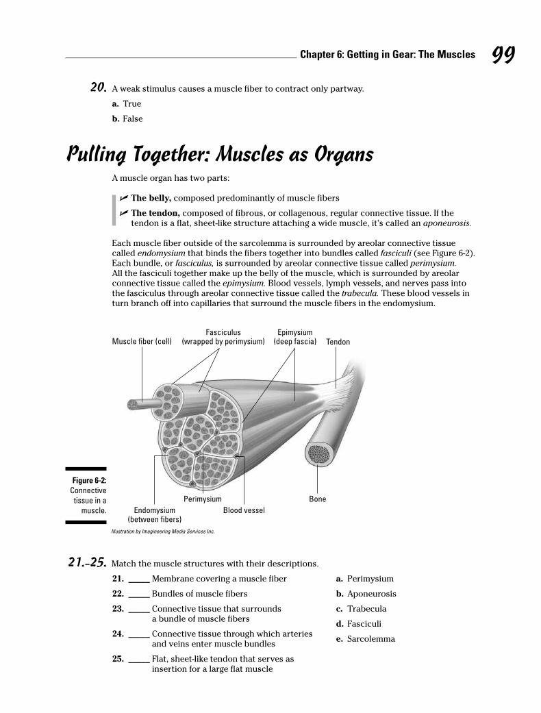
20. A weak stimulus causes a muscle fiber to contract only partway.
a. True
b. False
Pulling Together: Muscles as OrgansA muscle organ has two parts:
� The belly, composed predominantly of muscle fibers
� The tendon, composed of fibrous, or collagenous, regular connective tissue. If thetendon is a flat, sheet-like structure attaching a wide muscle, it’s called an aponeurosis.
Each muscle fiber outside of the sarcolemma is surrounded by areolar connective tissue called endomysium that binds the fibers together into bundles called fasciculi (see Figure 6-2).Each bundle, or fasciculus, is surrounded by areolar connective tissue called perimysium.All the fasciculi together make up the belly of the muscle, which is surrounded by areolar connective tissue called the epimysium. Blood vessels, lymph vessels, and nerves pass into the fasciculus through areolar connective tissue called the trabecula. These blood vessels inturn branch off into capillaries that surround the muscle fibers in the endomysium.
Illustration by Imagineering Media Services Inc.
21.–25. Match the muscle structures with their descriptions.
21. _____ Membrane covering a muscle fiber a. Perimysium
22. _____ Bundles of muscle fibers b. Aponeurosis
23. _____ Connective tissue that surrounds c. Trabeculaa bundle of muscle fibers
24. _____ Connective tissue through which arteries d. Fasciculi
and veins enter muscle bundlese. Sarcolemma
25. _____ Flat, sheet-like tendon that serves as insertion for a large flat muscle
Muscle fiber (cell)
Endomysium(between fibers)
Fasciculus(wrapped by perimysium)
Epimysium(deep fascia) Tendon
Perimysium
Blood vessel
Bone
Figure 6-2:
Connective
tissue in a
muscle.
99Chapter 6: Getting in Gear: The Muscles
Assuming the Right ToneAs we note earlier in this chapter, when it comes to contraction of a muscle fiber,it’s an all-or-nothing affair. Nonetheless, it has been demonstrated that fewer actionpotentials — a weaker stimulus, as it were — causes fewer motor units to becomeinvolved in a contraction. Maximum stimulus, on the other hand, brings all motor unitsto bear together. So it’s true that a muscle organ can have varying degrees of contrac-tion depending on the level of stimulation. As for how this can be so, one theory pro-poses that individual fibers have specific thresholds of excitation; thus, those withhigher thresholds only respond to stronger stimuli. The other theory holds that thedeeper a fiber is buried in the muscle, the less accessible it is to incoming stimuli.
In physiology, a muscle contraction is referred to as a muscle twitch. A twitch is the fun-damental unit of recordable muscular activity. Complete fatigue occurs when no moretwitches can be elicited, even with increasing intensity of stimulation.
The short lapse of time between the application of a stimulus and the beginning ofmuscular response is called the latent period. In mammalian muscle, latency is about.001 second, or one one-thousandth of a second.
Two types of muscle contraction relate to tone:
� Isometric: Occurs when a contracting muscle is unable to move a load (or heft apiece of luggage or push a building to one side). It retains its original length butdevelops tension. No mechanical work is accomplished, and all energy involvedis expended as heat.
� Isotonic: Occurs when the resistance offered by the load (or the gardening hoeor the cold can of soda) is less than the tension developed, thus shortening themuscle and resulting in mechanical work.
But muscles aren’t independent sole proprietors. Each muscle depends upon compan-ions in a muscle group to assist in executing a particular movement. That’s why mus-cles are categorized by their actions. The brain coordinates the following groupsthrough the cerebellum.
� Prime movers: Just as it sounds, these muscles are the workhorses that producemovement.
� Antagonists: These muscles exist in opposition to prime movers.
� Fixators or fixation muscles: These muscles serve to steady a part while othermuscles execute movement. They don’t actually take part in the movement itself.
� Synergists: These muscles control movement of the proximal joints so that theprime movers can bring about movements of distal joints.
Flex your knowledge of muscle tone and function with these practice questions:
100 Part II: Weaving It Together: Bones, Muscles, and Skin
Q. Muscle movement that lifts anobject involves an action known as
a. Isometric
b. Eccentric
c. Isotonic
A. The correct answer is isotonic.When the tension leads to move-ment (actual work), it’s isotonic.
26. Muscles that tend to counteract or slow an action are called
a. Antagonists
b. Fixators
c. Primary movers
d. Synergists
27. Which of the following statements finishes this sentence and makes it not true: A contractingmuscle unable to move a load
a. Involves an action known as isometric
b. Expends energy as heat
c. Is exemplified in the effect of the force of gravity on muscle contraction
d. Does no mechanical work and therefore doesn’t develop any tension
e. Retains its original length
28. A muscle contraction is referred to as
a. Latency
b. Synergy
c. A twitch
d. Isotonic motion
Leveraging Muscular PowerSkeletal muscle power is nothing without lever action. The bone acts as a rigid bar, thejoint is the fulcrum, and the muscle applies the force. Levers are divided into theweight arm, the area between the fulcrum and the weight; and the power arm, the areabetween the fulcrum and the force. When the power arm is longer than the weightarm, less force is required to lift the weight, but range, or distance, and speed are sac-rificed. When the weight arm is longer, the range of action and speed increase, butpower is sacrificed. Therefore, 90 degrees is the optimum angle for a muscle to attachto a bone and apply the greatest force.
Three classes of levers are at work in the body:
� Class I, or seesaw: The fulcrum is located between the weight and the forcebeing applied. An example is a nod of the head: The head-neck joint is the ful-crum, the head is the weight, and the muscles in the back of the neck apply theforce.
� Class II, or wheelbarrow: The weight is located between the fulcrum and thepoint at which the force is applied. An example is standing on your tiptoes: Thefulcrum is the joint between the toes and the foot, the weight is the body, andthe muscles in the back of the leg at the heel bone apply the force.
� Class III: The force is located between the weight and the fulcrum. An example isflexing your arm and showing off your biceps: The elbow joint is the fulcrum, theweight is the lower arm and hand, and the biceps insertion on the lower armapplies the force.
101Chapter 6: Getting in Gear: The Muscles
The direction in which the muscle fibers run also plays a critical role in leverage. Hereare the possible directions:
� Longitudinal: Fibers run parallel to each other, or longitudinally, the length ofthe muscle. Example: sartorius.
� Pennate: Fibers attach to the sides of the tendon, which extends the length ofthe muscle. These come in subcategories:
• Unipennate, where fibers attach to one side of the tendon; example: tibialisposterior
• Bipennate, where fibers attach to two sides of the tendon; example: rectusfemoris
• Multipennate, where fibers attach to many sides of the tendon; example:deltoideus
� Radiate: Fibers converge from a broad area into a common point. Example: pec-toralis major.
� Sphincter: Fibers are arranged in a circle around an opening. Example: orbicularisoculi.
The three types of fasciae, which Gray’s Anatomy describes as “dissectable, fibrousconnective tissues of the body,” are as follows:
� Superficial fasciae: Found under the skin and consisting of two layers: an outerlayer called the panniculus adiposus containing fat; and an inner layer made up ofa thin, membranous, and highly elastic layer. Between the two layers are thesuperficial arteries, veins, nerves, and mammary glands.
� Deep fasciae: Holds muscles or structures together or separates them intogroups that function in unison. It’s a system of splitting, rejoining, and fusingmembranes involving
• An outer investing layer that’s found under the superficial fasciae coveringa large part of the body
• An internal investing layer that lines the inside of the body wall in thetorso, or trunk, region
• An intermediate investing layer that connects the outer investing layer andthe internal investing layer
� Subserous fasciae: Located between the internal investing layer of the deep fas-ciae and the peritoneum. It’s the serous membrane that lines the abdominopelviccavity, also known as the peritoneal cavity.
Got all that? Then try your hand at the following questions:
102 Part II: Weaving It Together: Bones, Muscles, and Skin
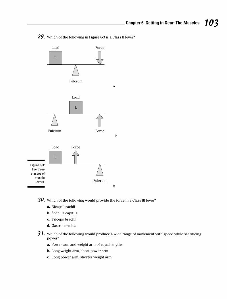
29. Which of the following in Figure 6-3 is a Class II lever?
30. Which of the following would provide the force in a Class III lever?
a. Biceps brachii
b. Spenius capitus
c. Triceps brachii
d. Gastrocnemius
31. Which of the following would produce a wide range of movement with speed while sacrificingpower?
a. Power arm and weight arm of equal lengths
b. Long weight arm, short power arm
c. Long power arm, shorter weight arm
Load
L
L
L
Fulcrum Force
Load Force
Fulcrum
Load Force
Fulcrum
b
c
a
Figure 6-3:
The three
classes of
muscle
levers.
103Chapter 6: Getting in Gear: The Muscles
32. Identify the bipennate bundle arrangement.
a. Sartorius
b. Rectus femoris
c. Pectoralis major
d. Tibialis posterior
e. Deltoideus
33. Which of the following is considered a dissectable connective tissue?
a. Aponeurosis
b. Bursae
c. Fasciae
d. Tendons
e. Ligaments
34. The most extensive fascia in the body is
a. Superficial
b. Deep
c. Subserous
d. None is more extensive than the other
What’s In a Name? Identifying MusclesIt may seem like a jumble of meaningless Latin at first, but muscle names follow a strictconvention that lets them be named for one or more of four things:
� Function: These muscle names usually have a verb root and end in a suffix (–oror –eus), followed by the name of the affected structure. Example: levator scapu-lae (elevates the scapulae).
� Compounding points of attachment: These muscle names blend the origin andinsertion attachment with an adjective suffix (–eus or –is). Examples: sternoclei-domastoideus (sternum, clavicle, and mastoid process) and sternohyoideus(sternum and hyoid).
� Shape or position: These muscle names usually have descriptive adjectives thatmay be followed by the names of the locations of the muscles. Examples: rectus(straight) femoris, rectus abdominus, and serratus (sawtooth) anterior.
� Figurative names: These muscle names are based on the muscles’ resemblanceto some objects. Examples: gastrocnemius (resembles the stomach) and trapez-ius (resembles a tablet).
Check out Table 6-1 for a rundown of prominent muscles in the body and key points toremember about each one.
104 Part II: Weaving It Together: Bones, Muscles, and Skin
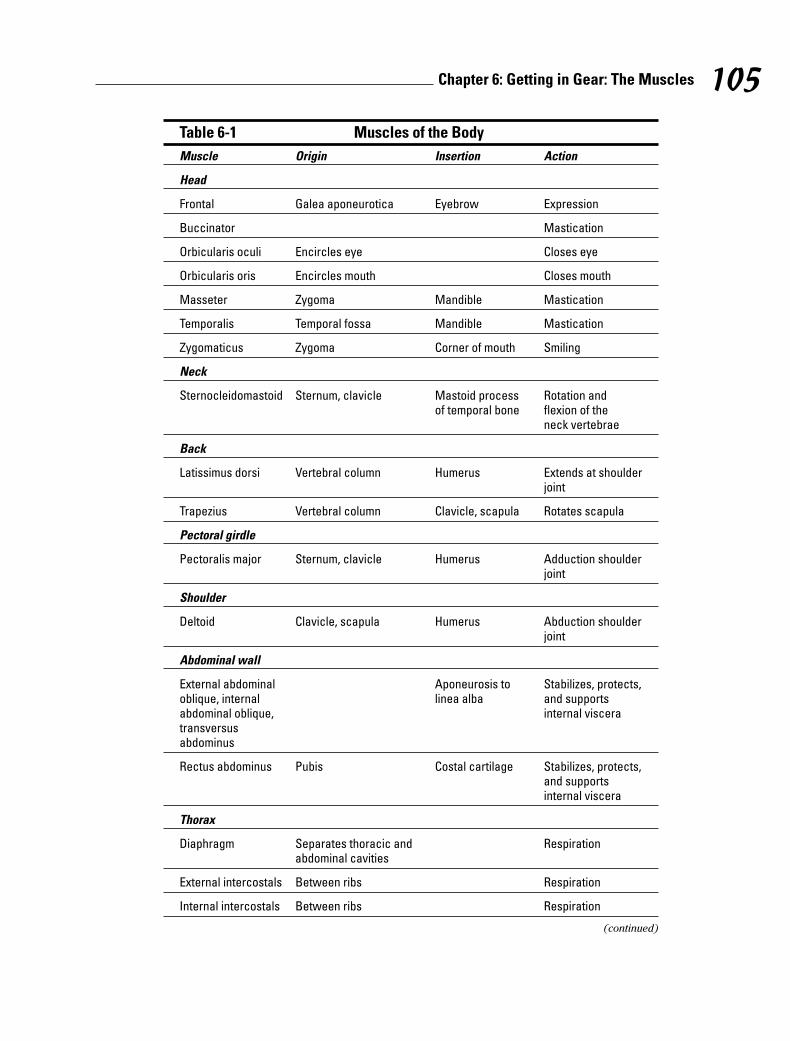
Table 6-1 Muscles of the Body
Muscle Origin Insertion Action
Head
Frontal Galea aponeurotica Eyebrow Expression
Buccinator Mastication
Orbicularis oculi Encircles eye Closes eye
Orbicularis oris Encircles mouth Closes mouth
Masseter Zygoma Mandible Mastication
Temporalis Temporal fossa Mandible Mastication
Zygomaticus Zygoma Corner of mouth Smiling
Neck
Sternocleidomastoid Sternum, clavicle Mastoid process Rotation and of temporal bone flexion of the
neck vertebrae
Back
Latissimus dorsi Vertebral column Humerus Extends at shoulder joint
Trapezius Vertebral column Clavicle, scapula Rotates scapula
Pectoral girdle
Pectoralis major Sternum, clavicle Humerus Adduction shoulderjoint
Shoulder
Deltoid Clavicle, scapula Humerus Abduction shoulderjoint
Abdominal wall
External abdominal Aponeurosis to Stabilizes, protects, oblique, internal linea alba and supports abdominal oblique, internal visceratransversus abdominus
Rectus abdominus Pubis Costal cartilage Stabilizes, protects,and supports internal viscera
Thorax
Diaphragm Separates thoracic and Respirationabdominal cavities
External intercostals Between ribs Respiration
Internal intercostals Between ribs Respiration
(continued)
105Chapter 6: Getting in Gear: The Muscles
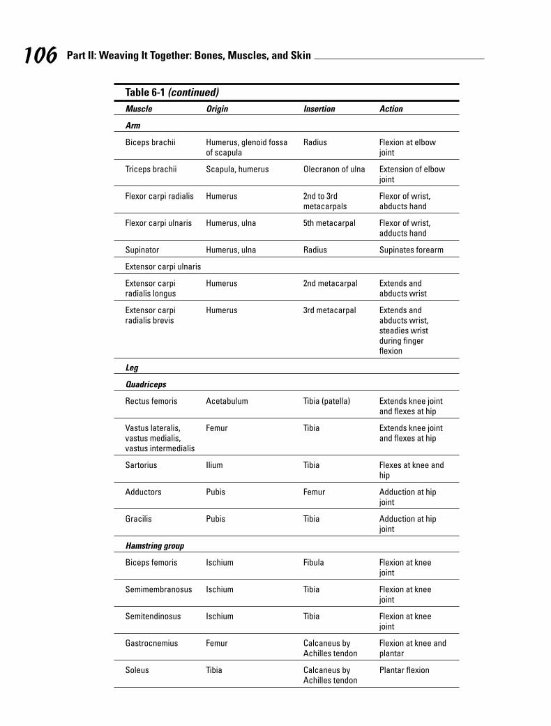
Table 6-1 (continued)
Muscle Origin Insertion Action
Arm
Biceps brachii Humerus, glenoid fossa Radius Flexion at elbow of scapula joint
Triceps brachii Scapula, humerus Olecranon of ulna Extension of elbowjoint
Flexor carpi radialis Humerus 2nd to 3rd Flexor of wrist, metacarpals abducts hand
Flexor carpi ulnaris Humerus, ulna 5th metacarpal Flexor of wrist,adducts hand
Supinator Humerus, ulna Radius Supinates forearm
Extensor carpi ulnaris
Extensor carpi Humerus 2nd metacarpal Extends and radialis longus abducts wrist
Extensor carpi Humerus 3rd metacarpal Extends and radialis brevis abducts wrist,
steadies wristduring finger flexion
Leg
Quadriceps
Rectus femoris Acetabulum Tibia (patella) Extends knee jointand flexes at hip
Vastus lateralis, Femur Tibia Extends knee joint vastus medialis, and flexes at hipvastus intermedialis
Sartorius Ilium Tibia Flexes at knee andhip
Adductors Pubis Femur Adduction at hipjoint
Gracilis Pubis Tibia Adduction at hipjoint
Hamstring group
Biceps femoris Ischium Fibula Flexion at kneejoint
Semimembranosus Ischium Tibia Flexion at kneejoint
Semitendinosus Ischium Tibia Flexion at kneejoint
Gastrocnemius Femur Calcaneus by Flexion at knee and Achilles tendon plantar
Soleus Tibia Calcaneus by Plantar flexionAchilles tendon
106 Part II: Weaving It Together: Bones, Muscles, and Skin
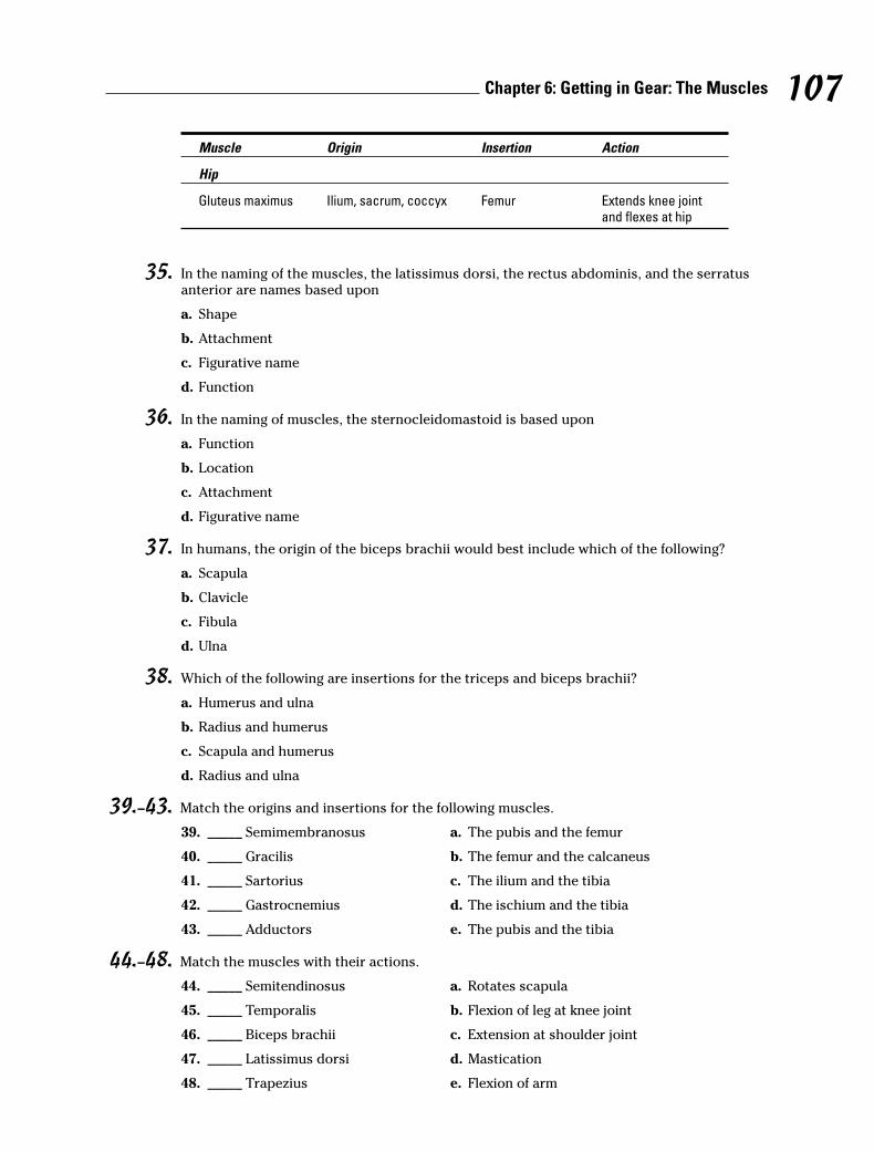
Muscle Origin Insertion Action
Hip
Gluteus maximus Ilium, sacrum, coccyx Femur Extends knee jointand flexes at hip
35. In the naming of the muscles, the latissimus dorsi, the rectus abdominis, and the serratusanterior are names based upon
a. Shape
b. Attachment
c. Figurative name
d. Function
36. In the naming of muscles, the sternocleidomastoid is based upon
a. Function
b. Location
c. Attachment
d. Figurative name
37. In humans, the origin of the biceps brachii would best include which of the following?
a. Scapula
b. Clavicle
c. Fibula
d. Ulna
38. Which of the following are insertions for the triceps and biceps brachii?
a. Humerus and ulna
b. Radius and humerus
c. Scapula and humerus
d. Radius and ulna
39.–43. Match the origins and insertions for the following muscles.
39. _____ Semimembranosus a. The pubis and the femur
40. _____ Gracilis b. The femur and the calcaneus
41. _____ Sartorius c. The ilium and the tibia
42. _____ Gastrocnemius d. The ischium and the tibia
43. _____ Adductors e. The pubis and the tibia
44.–48. Match the muscles with their actions.
44. _____ Semitendinosus a. Rotates scapula
45. _____ Temporalis b. Flexion of leg at knee joint
46. _____ Biceps brachii c. Extension at shoulder joint
47. _____ Latissimus dorsi d. Mastication
48. _____ Trapezius e. Flexion of arm
107Chapter 6: Getting in Gear: The Muscles
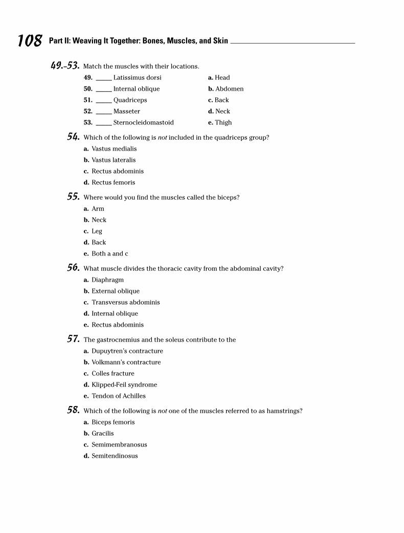
49.–53. Match the muscles with their locations.
49. _____ Latissimus dorsi a. Head
50. _____ Internal oblique b. Abdomen
51. _____ Quadriceps c. Back
52. _____ Masseter d. Neck
53. _____ Sternocleidomastoid e. Thigh
54. Which of the following is not included in the quadriceps group?
a. Vastus medialis
b. Vastus lateralis
c. Rectus abdominis
d. Rectus femoris
55. Where would you find the muscles called the biceps?
a. Arm
b. Neck
c. Leg
d. Back
e. Both a and c
56. What muscle divides the thoracic cavity from the abdominal cavity?
a. Diaphragm
b. External oblique
c. Transversus abdominis
d. Internal oblique
e. Rectus abdominis
57. The gastrocnemius and the soleus contribute to the
a. Dupuytren’s contracture
b. Volkmann’s contracture
c. Colles fracture
d. Klipped-Feil syndrome
e. Tendon of Achilles
58. Which of the following is not one of the muscles referred to as hamstrings?
a. Biceps femoris
b. Gracilis
c. Semimembranosus
d. Semitendinosus
108 Part II: Weaving It Together: Bones, Muscles, and Skin
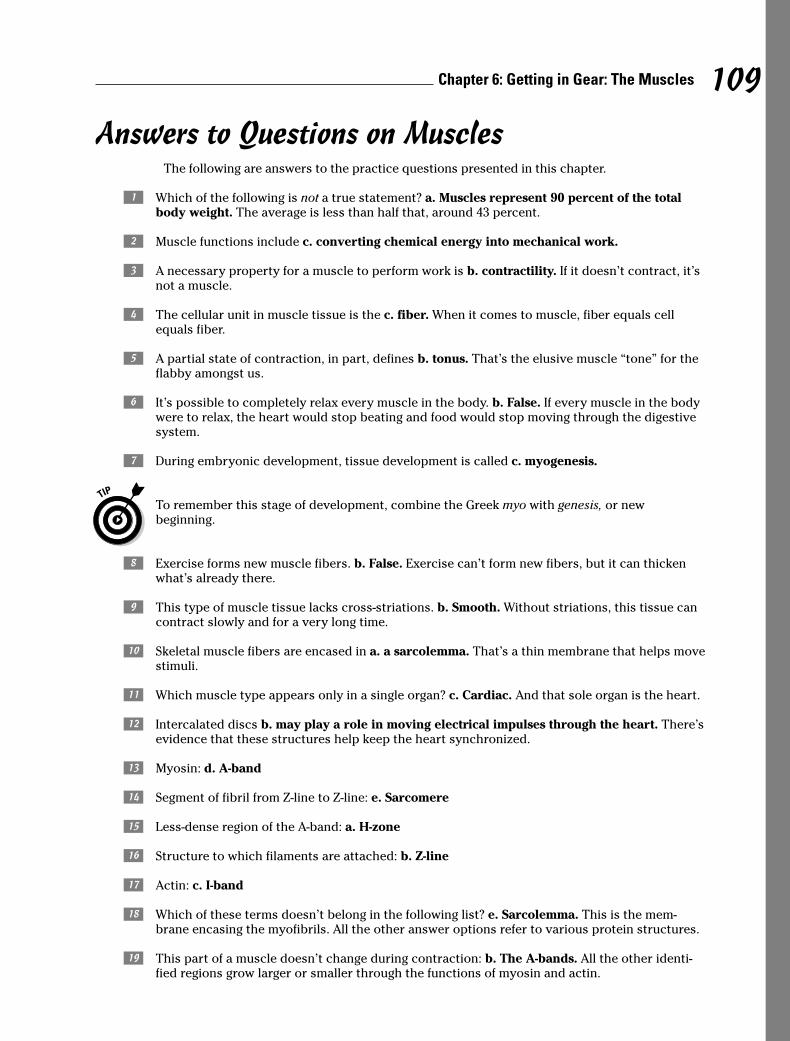
109Chapter 6: Getting in Gear: The Muscles
Answers to Questions on MusclesThe following are answers to the practice questions presented in this chapter.
a Which of the following is not a true statement? a. Muscles represent 90 percent of the totalbody weight. The average is less than half that, around 43 percent.
b Muscle functions include c. converting chemical energy into mechanical work.
c A necessary property for a muscle to perform work is b. contractility. If it doesn’t contract, it’snot a muscle.
d The cellular unit in muscle tissue is the c. fiber. When it comes to muscle, fiber equals cellequals fiber.
e A partial state of contraction, in part, defines b. tonus. That’s the elusive muscle “tone” for theflabby amongst us.
f It’s possible to completely relax every muscle in the body. b. False. If every muscle in the bodywere to relax, the heart would stop beating and food would stop moving through the digestivesystem.
g During embryonic development, tissue development is called c. myogenesis.
To remember this stage of development, combine the Greek myo with genesis, or new beginning.
h Exercise forms new muscle fibers. b. False. Exercise can’t form new fibers, but it can thickenwhat’s already there.
i This type of muscle tissue lacks cross-striations. b. Smooth. Without striations, this tissue cancontract slowly and for a very long time.
j Skeletal muscle fibers are encased in a. a sarcolemma. That’s a thin membrane that helps movestimuli.
k Which muscle type appears only in a single organ? c. Cardiac. And that sole organ is the heart.
l Intercalated discs b. may play a role in moving electrical impulses through the heart. There’sevidence that these structures help keep the heart synchronized.
m Myosin: d. A-band
n Segment of fibril from Z-line to Z-line: e. Sarcomere
o Less-dense region of the A-band: a. H-zone
p Structure to which filaments are attached: b. Z-line
q Actin: c. I-band
r Which of these terms doesn’t belong in the following list? e. Sarcolemma. This is the mem-brane encasing the myofibrils. All the other answer options refer to various protein structures.
s This part of a muscle doesn’t change during contraction: b. The A-bands. All the other identi-fied regions grow larger or smaller through the functions of myosin and actin.
t A weak stimulus causes a muscle fiber to contract only partway. b. False. Muscle contractionsare all-or-nothing; there’s no such thing as a partial contraction.
u Membrane covering a muscle fiber: e. Sarcolemma
v Bundles of muscle fibers: d. Fasciculi
w Connective tissue that surrounds a bundle of muscle fibers: a. Perimysium
x Connective tissue through which arteries and veins enter muscle bundles: c. Trabecula
y Flat, sheet-like tendon that serves as insertion for a large flat muscle: b. Aponeurosis
A Muscles that tend to counteract or slow an action are called a. antagonists.
The muscles are against the action, so think of them as antagonistic.
B Which of the following statements finishes this sentence and makes it not true: A contractingmuscle unable to move a load d. does no mechanical work and therefore doesn’t develop anytension. This statement is false because the contraction of the muscle causes tension in allcases.
C A muscle contraction is referred to as c. a twitch. Keep in mind the twitch is the fundamentalmeasure of muscle activity.
D Which of the following in Figure 6-3 is a Class II lever? b.
E Which of the following would provide the force in a Class III lever? a. Biceps brachii. It’s thePopeye weight-lifting class, after all.
F Which of the following would produce a wide range of movement with speed while sacrificingpower? b. Long weight arm, short power arm. The longer the weight arm, the greater therange of action and speed but the less power there is.
G Identify the bipennate bundle arrangement. b. Rectus femoris. Keep in mind the muscle namingconventions.
H Which of the following is considered a dissectable connective tissue? c. Fasciae. This connec-tive tissue can be found in most of the body.
I The most extensive fascia in the body is b. deep.
J In the naming of the muscles, the latissimus dorsi, the rectus abdominis, and the serratus ante-rior are names based upon a. shape. Latissimus stems from the Latin for “wide,” rectus from theLatin word for “straight,” and serratus from the Latin word for “notched” or “scalloped.”
K In the naming of muscles, the sternocleidomastoid is based upon c. attachment. You can figureout this answer by recalling that cleido stems from the Latin word for “collarbone.”
L In humans, the origin of the biceps brachii would best include which of the following? a. Scapula
M Which of the following are insertions for the triceps and biceps brachii? d. Radius and ulna
110 Part II: Weaving It Together: Bones, Muscles, and Skin
N Semimembranosus: d. The ischium and the tibia
O Gracilis: e. The pubis and the tibia
P Sartorius: c. The ilium and the tibia
Q Gastrocnemius: b. The femur and the calcaneus
R Adductors: a. The pubis and the femur
S Semitendinosus: b. Flexion of leg at knee joint
T Temporalis: d. Mastication
U Biceps brachii: e. Flexion of arm
V Latissimus dorsi: c. Extension at shoulder joint
W Trapezius: a. Rotates scapula
X Latissimus dorsi: c. Back
Y Internal oblique: b. Abdomen
z Quadriceps: e. Thigh
Z Masseter: a. Head
1 Sternocleidomastoid: d. Neck
2 Which of the following is not included in the quadriceps group? c. Rectus abdominis. The termabdominis is the giveaway here because it should make you think of the abdomen. The rest ofthe quadriceps group is found in the upper leg, where they belong.
3 Where would you find the muscles called the biceps? e. Both a and c (arm and leg). Biceps is amuscle with two heads or points of origin. Although people usually think of the biceps brachiiin the arm, you can’t forget about the biceps femoris at the back of the thigh.
4 What muscle divides the thoracic cavity from the abdominal cavity? a. Diaphragm. And with-out it, you couldn’t breathe.
5 The gastrocnemius and the soleus contribute to the e. tendon of Achilles. The soleus liesunder the gastrocnemius in the calf of each leg.
6 Which of the following is not one of the muscles referred to as hamstrings? b. Gracilis. Theother three answer options all are listed as hamstring muscles.
111Chapter 6: Getting in Gear: The Muscles

112 Part II: Weaving It Together: Bones, Muscles, and Skin

Chapter 7
It’s Skin Deep: The IntegumentarySystem
In This Chapter� Going below the surface of the skin
� Getting on your nerves
� Checking out hair, nails, and glands
Did you know that the skin is the body’s largest organ? In an average person, its 17 to 20 square feet of surface area represents 15 percent of the body’s weight. Self-repairing
and surprisingly durable, the skin is the first line of defense against the harmful effects ofthe outside world. It helps retain moisture; regulates body temperature; hosts the sensereceptors for touch, pain, and heat; excretes excess salts and small amounts of waste; andeven stores blood to be moved quickly to other parts of the body when needed.
Skin is jam-packed with components; it has been estimated that every square inch of skincontains 15 feet of blood vessels, 4 yards of nerves, 650 sweat glands, 100 oil glands, 1,500sensory receptors, and over 3 million cells with an average lifespan of 26 days that are con-stantly being replaced.
In this chapter, we peel back the surface of this most-visible organ system. We also give youplenty of opportunities to test your knowledge.
Dermatology Down DeepSkin — together with hair, nails, and glands — composes the integumentary system (shown inFigure 7-1). The name stems from the Latin verb integere, which means “to cover.” The rele-vant Greek and Latin roots include dermato and cutis, both of which mean “skin.”
The skin consists of two primary parts: the epidermis and the dermis. (Recall the Greek rootepi–, which means “upon” or “above.”) Underlying the epidermis and dermis is the hypoder-mis or superficial fascia (also sometimes called subcutaneous tissue), which acts as a founda-tion but is not part of the skin. Composed of areolar (porous) and adipose (fat) tissue, itanchors the skin through fibers that extend from the dermis. Underneath, the hypodermisattaches loosely to tissues and organs so that muscles can move freely. Around elbow andknee joints, the hypodermis contains fluid-filled sacs called bursae. The fat in the hypodermisbuffers deeper tissues and acts as insulation, preventing heat loss from within the body’score. The hypodermis also is home to pressure-sensitive nerve endings called lamellated orPacinian corpuscles that respond to a deeper poke in the skin.
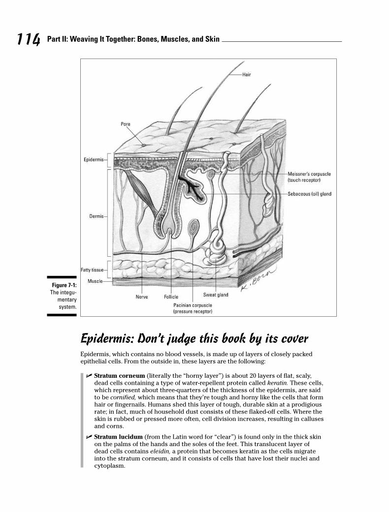
Epidermis: Don’t judge this book by its coverEpidermis, which contains no blood vessels, is made up of layers of closely packedepithelial cells. From the outside in, these layers are the following:
� Stratum corneum (literally the “horny layer”) is about 20 layers of flat, scaly,dead cells containing a type of water-repellent protein called keratin. These cells,which represent about three-quarters of the thickness of the epidermis, are saidto be cornified, which means that they’re tough and horny like the cells that formhair or fingernails. Humans shed this layer of tough, durable skin at a prodigiousrate; in fact, much of household dust consists of these flaked-off cells. Where theskin is rubbed or pressed more often, cell division increases, resulting in callusesand corns.
� Stratum lucidum (from the Latin word for “clear”) is found only in the thick skinon the palms of the hands and the soles of the feet. This translucent layer ofdead cells contains eleidin, a protein that becomes keratin as the cells migrateinto the stratum corneum, and it consists of cells that have lost their nuclei andcytoplasm.
Figure 7-1:
The integu-
mentary
system.
114 Part II: Weaving It Together: Bones, Muscles, and Skin

� Stratum granulosum is three to five layers of flattened cells containing kerato-hyalin, a substance that marks the beginning of keratin formation. No nourish-ment from blood vessels reaches this far into the epidermis, so cells are eitherdead or dying by the time they reach the stratum granulosum. The nuclei of cellsfound in this layer are degenerating; when the nuclei break down entirely, thecell can’t metabolize nutrients and dies.
� Stratum spinosum (also sometimes called the spinous layer) has ten layers con-taining prickle cells, named for the spine-like projections that connect them withother cells in the layer. Langerhans cells, believed to be involved in the body’simmune response, are prevalent in the upper portions of this layer and some-times the lower part of the stratum granulosum; they migrate from the skin tothe lymph nodes in response to infection. Some mitosis (cell division) takesplace in the stratum spinosum, but the cells lose the ability to divide as theymature.
� Stratum basale (or stratum germinativum) is also referred to as the germinal layerbecause this single layer of mostly columnar stem cells generates all the cellsfound in the other epidermal layers. It rests on the papillary (rough or bumpy)surface of the dermis, close to the blood supply needed for nourishment andoxygen. The mitosis that constantly occurs here replenishes the skin; it takesabout two weeks for the cells that originate here to migrate up to the stratumcorneum, and it’s another two weeks before they’re shed. About a quarter of thislayer’s cells are melanocytes, cells that synthesize a pale yellow to black pigmentcalled melanin that contributes to skin color and provides protection againstultraviolet radiation (the kind of radiation found in sunlight). The remaining cells in this layer become keratinocytes, the primary epithelial cell of the skin.Melanocytes secrete melanin directly into the keratinocytes in a process calledcytocrine secretion. Merkel’s cells, a large oval cell believed to be involved in thesense of touch, occasionally appear amid the keratinocytes.
In addition to melanin, the epidermis contains a yellowish pigment called carotene (thesame one found in carrots and sweet potatoes). Found in the stratum corneum and thefatty layers beneath the skin, it produces the yellowish hue associated with Asianancestry or increased carrot consumption. The pink to red color of Caucasian skin iscaused by hemoglobin, the red pigment of the blood cells. Because Caucasian skin con-tains relatively less melanin, hemoglobin can be seen more easily through the epider-mis. Sometimes the limited melanin in Caucasian skin pools in small patches. Can youguess the name of those patches of color? Yep, they’re freckles. Albinos, on the otherhand, have no melanin in their skin at all, making them particularly sensitive to ultravi-olet radiation.
Ridges and grooves form on the outer surface of the epidermis to increase the frictionneeded to grasp objects or move across slick surfaces. On hands and feet, these ridgesform patterns of loops and whorls — fingerprints, palm prints, and footprints — thatare unique to each person. You leave these imprints on smooth surfaces because ofthe oily secretions of the sweat glands on the skin’s surface. In addition to these finerpatterns, the areas around joints develop patterns called flexion lines. Deeper andmore permanent lines are called flexion creases.
Dermis: It’s more than skin deepBeneath the epidermis is a thicker, fibrous structure called the dermis, or corium. Itconsists of the following two layers, which blend together:
� The outer, soft papillary layer contains elastic and reticular (net-like) fibers thatproject into the epidermis to bring blood and nerve endings closer. Papillae(finger-like projections) containing loops of capillaries increase the surface areaof the dermis and anchor the epidermis. Some of these papillae contain
115Chapter 7: It’s Skin Deep: The Integumentary System

1. The layer of epidermis in which mitosis takes place is the stratum
a. Corneum
b. Lucidum
c. Granulosum
d. Papilla
e. Basale
2. The papillary layer of the dermis
a. Is composed of numerous projections
b. Extends into the epidermis
c. Carries the blood and nerve endings close to the epidermis
d. Aids in holding the epidermis and dermis together
e. All of the above
116 Part II: Weaving It Together: Bones, Muscles, and Skin
Meissner’s corpuscles, nerve endings that are sensitive to soft touch. It’s thedermal papillae that form the epidermal ridges referred to as fingerprints.
� The inner, thicker reticular layer (from the Latin word rete for “net”) is made up ofdense, irregular connective tissue containing interlacing bundles of collagenousand elastic fibers that form the strong, resistant layer used to make leather andsuede from animal hides. This layer is what gives skin its strength, extensibility,and elasticity. Within the reticular layer are sebaceous glands (oil glands), sweatglands, fat cells, and larger blood vessels.
Cells in the dermis include fibroblasts (from which connective tissue develops),macrophages (which engulf waste and foreign microorganisms), and adipose tissue.Thickest on the palms of the hands and soles of the feet, the dermis is thinnest overthe eyelids, penis, and scrotum. It’s thicker on the back (posterior) than on the stom-ach (anterior) and thicker on the sides (lateral) than toward the middle of the body(medial). The various skin “accessories” — blood vessels, nerves, glands, and hair follicles — are embedded here.
See if you’ve got the skinny on skin so far:
Q. The layer of epidermis that’s com-posed of a horny, cornified tissuethat sloughs off is called thestratum
a. Corneum
b. Lucidum
c. Granulosum
d. Spinosum
e. Basale
A. The correct answer is corneum.Cornified, corns — think of howhard a kernel of popcorn can be.
3. The function of the epidermal ridges on the fingers is to
a. Provide a means of identification
b. Increase the friction of the epidermal surface
c. Decrease water loss by the tissues
d. Aid in regulating body temperature
e. Prevent bacterial infection
4. The color of Caucasian skin is due to
a. Carotene pigment in the dermis
b. The high level of melanin in the epidermis
c. Less melanin in the skin, allowing the blood pigment to be seen
d. The absence of all pigment
e. Melanin and carotene pigments
5. The sequence of layers in the epidermis from the dermis outward is
a. Corneum, lucidum, granulosum, spinosum, basale
b. Corneum, granulosum, lucidum, basale, spinosum
c. Spinosum, basale, granulosum, corneum, lucidum
d. Basale, spinosum, granulosum, lucidum, corneum
e. Basale, lucidum, corneum, spinosum, granulosum
6. The subcutaneous layer of tissue can be called the
a. Epidermis
b. Superficial fascia
c. Papillary layer
d. Inner reticular layer
e. Dermis
7. The lines in fingerprints are determined by
a. The contours of the dermal papillae
b. The thickness of the dermis
c. The bundles of collagenous and elastic fibers in the epidermis
d. The fibroblasts, macrophages, and adipose tissue surrounding the nerves
e. The surface layer of cells that are constantly being shed
8. A function not pertaining to the skin is
a. Aids in retaining water
b. Regulates body temperature
c. Contains sense receptors
d. Excretes some waste materials
e. Provides movement
117Chapter 7: It’s Skin Deep: The Integumentary System
9. A layer of dense, irregular connective tissue containing interlacing bundles of collagenous andelastic fibers is the
a. Basale layer of the epidermis
b. Reticular layer of the dermis
c. Outer layer of the hypodermis
d. Papillary layer of the dermis
e. Inner layer of the hypodermis
10. The stratum corneum cells contain a tough, water-repellant protein called
a. Keratohyalin
b. Eleidin
c. Keratin
d. Cerumen
e. Sweat
11. The epidermal layer containing keratohyalin is the
a. Stratum germinativum
b. Stratum spinosum
c. Stratum lucidum
d. Stratum granulosum
e. Stratum corneum
12. The layer of skin attached to the superficial fascia is the
a. Papillary layer of the dermis
b. Stratum granulosum
c. Stratum germinativum
d. Holorine layer of the epidermis
e. Reticular layer of the dermis
13. Flattened and irregular cells with small, spine-like projections that connect them with othercells in the layer are referred to as
a. Prickle cells
b. Langerhans cells
c. Melanocytes
d. Merkel’s cells
e. Keratohyalin cells
14. If melanin forms into patches, it’s referred to as
a. Flexion creases
b. Freckles
c. Dermal papillae
d. Lamellated corpuscles
e. Matrix
118 Part II: Weaving It Together: Bones, Muscles, and Skin
Touching a Nerve in the Integumentary System
At least four kinds of receptors are involved in creating the sensation of touch.
� Free nerve endings: These afferent nerve endings are dendrites (branched exten-sions) of sensory neurons that act primarily as pain receptors, although somesense temperature, touch, and muscles (including the sensation of “stretch”).Found all over the body, free nerve endings are especially prevalent in epithelialand connective tissue. These small-diameter fibers have a swelling at the endthat responds to touch and sometimes heat, cold, or pain. Some of the endingsare disc-shaped structures called Merkel discs that function as light-touch recep-tors within the deep layers of the epidermis.
� Meissner’s corpuscles: These light-touch mechanoreceptors lie within thedermal papillae. They’re small, egg-shaped capsules of connective tissue sur-rounding a spiraled end of a dendrite. Most abundant in sensitive skin areas suchas the lips and fingertips, these corpuscles and free nerve endings can sense aquick touch but not a sustained one. That’s why your skin is able to ignore thetouch sensation of your own clothing.
� Pacinian corpuscles: These deep-pressure mechanoreceptors are dendrites surrounded by concentric layers of connective tissue. Found deep within thedermis, they respond to deep or firm pressure and vibrations. Each is over 2 mil-limeters long and therefore visible to the naked eye.
� Hair nerve endings: These mechanoreceptors respond to a change in positionof a hair. They consist of bare dendrites.
There are two primary temperature receptors, one for heat and one for cold:
� End-bulbs of Krause: Also known as Krause’s corpuscles, these cold receptorsusually activate below 68 degrees F (20 degrees C). They consist of a bulbouscapsule surrounding the dendrite and are commonly found throughout the bodyin the dermis as well as in the lips, the tongue, and the conjunctiva of the eyes.
� Brushes of Ruffini: Also known as Ruffini cylinders or Ruffini’s corpuscles, thesewarmth receptors respond to temperatures between 77 degrees and 113 degrees F(25 degrees to 45 degrees C). Found primarily in the dermis and subcutaneoustissue, they’re dendrite endings enclosed in a flattened capsule. Because thereare fewer of them than Krause’s end-bulbs and because they lie in deeper tissue,the body is less sensitive to heat than to cold.
In addition to the receptors for touch and temperature, the dermis has neuromuscularspindles (also called proprioceptors) that transmit information to the spinal cord andbrain about the lengths and tensions of muscles. This information helps provideawareness of the body’s position and the relative position of body parts. The spindlesalso assist with muscle coordination and muscle action efficiency.
119Chapter 7: It’s Skin Deep: The Integumentary System
Test whether you’re staying in touch with this section:
15. The sensation of a soft touch is received by
a. Ruffini’s corpuscles
b. Pacinian corpuscles
c. The Crypt of Lieberkuhn
d. The end-bulbs of Krause
e. Meissner’s corpuscles
16. End-bulbs of Krause are receptors for cold that usually are activated at temperatures below
a. 98.6 degrees F
b. 20 degrees F
c. 68 degrees F
d. 45 degrees F
e. 77 degrees F
Accessorizing with Hair, Nails, and GlandsMother Nature has accessorized your fashionable over-wrap with a variety of special-ized structures that grow from the epidermis: hair, fingernails, toenails, sebaceousglands, and sweat glands.
Wigging out about hairLike most mammals, hair covers the entire human body except for the lips, eyelids,palms of the hands, soles of the feet, nipples, and portions of external reproductiveorgans. But human body hair generally is sparser and much lighter in color than thatsported by most other mammals. Animals have hair for protection and temperaturecontrol. For humans, however, body hair is largely a secondary sex characteristic.A thick head of hair protects the scalp from exposure to the sun’s harmful rays andlimits heat loss. Eyelashes block sunlight and deflect debris from the eyes. Hair in thenose and ears prevents airborne particles and insects from entering. Touch receptorsconnected to hair follicles respond to the lightest brush. The average adult has about5 million hairs, with about 100,000 of those growing from the scalp. Normal hair lossfrom an adult scalp is about 70 to 100 hairs each day, although baldness can resultfrom genetic factors, hormonal imbalances, scalp injuries, disease, dietary deficien-cies, radiation, or chemotherapy.
Each hair grows at an angle from a follicle embedded in the epidermis and extendinginto the dermis; scalp hairs sometimes reach as far as the hypodermis. Nerves reachthe hair at the follicle’s expanded base, called the bulb, where a nipple-shaped papillaof connective tissue and capillaries provide nutrients to the growing hair. Epithelialcells in the bulb divide to produce the hair’s shaft (the part that extends out of the folli-cle). (The part of the hair within the follicle is called the root.) The shape of a hair’scross section can vary from round to oval or even flat; oval hairs grow out appearingwavy or curly, flat hairs appear kinky, and round hairs grow out straight. Each scalphair grows for two to three years at a rate of about 1⁄3 to 1⁄2 millimeter per day, or 10 to18 centimeters per year. When mature, the hair rests for three or four months beforeslowly losing its attachment. Eventually, it falls out and is replaced by a new hair.
120 Part II: Weaving It Together: Bones, Muscles, and Skin
Hair pigment (which is melanin, just as in the skin) is produced by melanocytes in thefollicle and transferred to the hair’s cortex and medulla cells. Three types of melanin —black, brown, and yellow — combine in different quantities for each individual to pro-duce different hair colors ranging from light blonde to black. Gray and white hairs growin when melanin levels decrease and air pockets form where the pigment used to be.
Wondering why you have to shampoo so often? Hair becomes oily over time thanks tosebum, a mixture of cholesterol, fats, and other substances secreted from a sebaceous(or holocrine) gland found next to each follicle. Sebum keeps both hair and skin soft, pli-able, and waterproof. Attached to each follicle is a smooth muscle called an arrector pili(literally “raised hair”) that both applies pressure to the sebaceous gland and straight-ens the hair shaft, depressing the skin in a pattern called goose bumps or goose pimples.
Each hair is made up of three concentric layers of keratinized cells:
� A central core, called the medulla, consists of large cells containing eleidin that areseparated by air spaces; in fine hair, the medulla may be small or entirely absent.
� A cortex surrounding the medulla forms the major part of the hair shaft with sev-eral layers of flattened cells. The cortex also has elongated pigment-bearing cellsin dark hair and air pockets in white hair.
� The outermost cuticle is a single layer of overlapping cells with the free endpointing upward. The cuticle strengthens and compacts the inner layers, butabrasion tends to wear away the end of the shaft, exposing the medulla andcortex in a pattern known as split ends.
Nailing the fingers and toesHuman nails (which actually are vestigial claws) have three parts: a root bed at the nailbase, a body that’s attached to the fingertip, and a free edge that grows beyond the endof the finger or toe. Heavily cornified tissue forms the nails from modified stratacorneum and lucidum. A narrow fold of the stratum corneum turns back to form theeponychium, or cuticle. Under the nail, the nail bed is formed by the strata basale andspinosum. At the base of the nail, partially tucked under the cuticle, the strata thickento form a whitish area called the lunula (literally “little moon”) that can be seenthrough the nail. Beneath the lunula is the nail matrix, a region of thickened stratawhere mitosis pushes previously formed cornified cells forward, making the nail grow.Under the free edge of the nail, the stratum corneum thickens to form the hypony-chium. Nails are pinkish in color because of hemoglobin in the underlying capillaries,which are visible through the translucent cells of the nail.
On average, fingernails grow about 1 millimeter each week. Toenails tend to grow evenmore slowly. Nails function as an aid to grasping, as a tool for manipulating smallobjects, and as protection against trauma to the ends of fingers and toes.
Sweating the detailsHumans perspire over nearly every inch of skin, but anyone with sweaty palms orsmelly feet can attest to the fact that sweat glands are most numerous in the palmsand soles, with the forehead running a close third. There are two types of sweat, orsudoriferous, glands: eccrine and apocrine. Both are coiled tubules embedded in thedermis or subcutaneous layer composed of simple columnar cells.
Eccrine glands are distributed widely over the body — an average adult has roughly3 million of them — and produce the watery, salty secretion you know as sweat. Eachgland’s duct passes through the epidermis to the skin’s surface, where it opens as a
121Chapter 7: It’s Skin Deep: The Integumentary System
sweat pore. The sympathetic division of the autonomic nervous system controls whenand how much perspiration is secreted depending on how hot the body becomes.Sweat helps cool the skin’s surface by evaporating as fast as it forms. About 99 percentof eccrine-type sweat is water, but the remaining 1 percent is a mixture of sodium chlo-ride and other salts, uric acid, urea, amino acids, ammonia, sugar, lactic acid, andascorbic acid.
Apocrine sweat glands are located primarily in armpits (known as the axillary region)and the groin area. Usually associated with hair follicles, they produce a white, cloudysecretion that contains organic matter. Although apocrine-type sweat contains thesame basic components as eccrine sweat and also is odorless when first secreted, bac-teria quickly begin to break down its additional fatty acids and proteins — explainingthe post-exercise underarm stench. In addition to exercise, sexual and other emotionalstimuli can cause contraction of cells around these glands, releasing sweat.
Getting an earfulThe occasionally troublesome yellowish substance known as earwax is secreted in theouter part of the ear canal from modified sudoriferous glands called ceruminous glands(the Latin word cera means “wax”). Lying within the subcutaneous layer of the earcanal, these glands have ducts that either open directly into the ear canal or emptyinto the ducts of nearby sebaceous glands. Technically called cerumen, earwax is thecombined secretion of these two glands. Working with ear hairs, cerumen traps anyforeign particles before they reach the eardrum. As the cerumen dries, it flakes andfalls from the ear, carrying particles out of the ear canal.
Think you’ve got a grip on everything to do with hair, nails, and glands? Find out byanswering the following practice questions:
17. The cuticle is also called the
a. Lunula
b. Hyponychium
c. Eponychium
d. Nail matrix
e. Perinychium
18. Perspiration is formed by the
a. Sebaceous glands
b. Ceruminous glands
c. Endocrine glands
d. Merkel’s glands
e. Sudoriferous glands
19. The cause of graying hair is
a. Production of melanin in the shaft of the hair
b. Production of carotene in the shaft of the hair
c. Decrease in blood supply to the hair
d. Lack of melanin in the shaft of the hair
e. Parenthood
122 Part II: Weaving It Together: Bones, Muscles, and Skin
20. Hair develops from a(n)
a. Arrector pili
b. Shaft
c. Follicle
d. Sebaceous gland
e. Lanugo
21. The nails are modifications of the epidermal layers
a. Corneum and lucidum
b. Lucidum and granulosum
c. Granulosum and spinosum
d. Spinosum and basale
22. The muscle that straightens a hair and puts pressure on a gland causing it to secrete is the
a. Terminalis muscle
b. Arrector pili muscle
c. Internal oblique muscle
d. External transversus muscle
e. Internal rectus muscle
23. A factor not associated with baldness is
a. Genetics
b. Hormonal imbalances
c. Scalp injuries
d. Lack of carotene
e. Disease
24. Sebaceous glands
a. Produce a watery solution called sweat
b. Produce an oily mixture of cholesterol, fats, and other substances
c. Produce a waxy secretion called cerumen
d. Accelerate aging
e. Are associated with endocrine glands
25. The bulb of the follicle of a hair contains epithelial cells (germinating cells) that are continu-ous with the
a. Papillary layer of the dermis
b. Stratum corneum
c. Stratum germinativum
d. Reticular layer of the dermis
e. Statum lucidum
123Chapter 7: It’s Skin Deep: The Integumentary System
26. Cooling of the skin’s surface is aided by the
a. Endocrine glands
b. Sebaceous glands
c. Ceruminous glands
d. Prickle glands
e. Sweat glands
27. This gland contains true sweat, fatty acids, and proteins, and acquires an unpleasant odorwhen bacteria breaks down the organic molecules it secretes.
a. Apocrine sweat gland
b. Sebaceous gland
c. Ceruminous gland
d. Eccrine sweat gland
e. Mammary gland
28. Which of the following is true about fingernails?
a. They’re derived from the hypodermis.
b. They contain carotene.
c. They grow more slowly than toenails.
d. They grow about 1 millimeter per week.
e. They’re not a skin accessory.
29. The gland that secretes an oily mixture of cholesterol, fats, and other substances into hair fol-licles to keep hair and skin soft, pliable, and waterproof is the
a. Sweat gland
b. Sebaceous gland
c. Ceruminous gland
d. Apocrine gland
e. Eccrine gland
124 Part II: Weaving It Together: Bones, Muscles, and Skin
Answers to Questions on the SkinThe following are answers to the practice questions presented in this chapter.
a The layer of epidermis in which mitosis takes place is the stratum e. basale.
This layer also is called the stratum germinativum, but a simpler memory tool is simply toassociate it with the “base” of the epidermis.
b The papillary layer of the dermis e. all of the above. Busy little finger-like projections, thosepapillae.
c The function of the epidermal ridges on the fingers is to b. increase the friction of the epider-mal surface. It’s Mother Nature’s way of helping you cling to tree branches or grab food.
d The color of Caucasian skin is due to c. less melanin in the skin, allowing the blood pigment tobe seen. Here’s a fun experiment: Turn off the lights, press your fingers together, and hold aflashlight under them. See the red glow? That’s hemoglobin, too.
e The sequence of layers in the epidermis from the dermis outward is d. basale, spinosum,granulosum, lucidum, corneum.
Memory tool time: Base, spine, grain, Lucy, corny. Or try the first letters of Be Super Greedy,Less Caring. Insensitive, yes, but effective.
f The subcutaneous layer of tissue can be called the b. superficial fascia. Subcutaneous is thesame as hypodermis (from the Greek hypo– for “beneath”).
g The lines in fingerprints are determined by a. the contours of the dermal papillae.
h A function not pertaining to the skin is e. provides movement. That would be in the realm ofmuscles.
i A layer of dense, irregular connective tissue containing interlacing bundles of collagenous andelastic fibers is the b. reticular layer of the dermis. The description in this question soundslike a tough structure, so it may help you to remember that the reticular layer is what’s used tomake leather from animal hides.
j The stratum corneum cells contain a tough, water-repellant protein called c. keratin. Associatethe words “corneum” and “keratin,” and you’re in great shape.
k The epidermal layer containing keratohyalin is the d. stratum granulosum. Keratohyalin even-tually becomes keratin, so think of the layer where the cells are starting to die off.
l The layer of skin attached to the superficial fascia is the e. reticular layer of the dermis.Reticular means net-like; it makes sense that this netting lies between the dermis and the hypodermis.
m Flattened and irregular cells with small, spine-like projections that connect them with othercells in the layer are referred to as a. prickle cells. The spines make them look prickly, hencethe name.
n If melanin forms into patches, it’s referred to as b. freckles. Ever noticed how kids have morefreckles at the end of a long summer spent outdoors? That’s ultraviolet radiation working onthose melanin patches.
125Chapter 7: It’s Skin Deep: The Integumentary System
126 Part II: Weaving It Together: Bones, Muscles, and Skin
o The sensation of a soft touch is received by e. Meissner’s corpuscles. While it’s true that sev-eral different nerves are involved in the overall sense of touch, the Meissner’s are the mostresponsive to touch.
p End-bulbs of Krause are receptors for cold that usually are activated at temperatures below c. 68 degrees F.
Specific temperatures may seem tough to remember, but look at it this way: When it’s 45degrees F, you definitely need a jacket. When it’s 77 degrees F, you don’t. But when it’s 68degrees F, you’ll want to carry a light jacket in case it gets colder. Voila! A cold receptor activa-tion temperature!
q The cuticle is also called the c. eponychium. Recall that the prefix ep– refers to “upon” or“around,” whereas the prefix hypo– refers to “below” or “under.” The cuticle is around the baseof the nail, so it’s the eponychium, not the hyponychium.
r Perspiration is formed by the e. sudoriferous glands. The Latin word sudor means “sweat.”
s The cause of graying hair is d. lack of melanin in the shaft of the hair. Despite the medicalcause, people often suspect that answer “e. parenthood” has a lot to do with graying hair.
t Hair develops from a(n) c. follicle. The Latin translation of this word is “small cavity” or “sac,”so it makes sense that this would be an origination place.
u The nails are modifications of the epidermal layers a. corneum and lucidum. These are the twoupper layers.
v The muscle that straightens a hair and puts pressure on a gland causing it to secrete is the b. arrector pili muscle. “Arrector” is similar to “erector” (in fact, the muscle is sometimescalled that), which implies straightening, and the Latin word for “hair” is pili.
w A factor not associated with baldness is d. lack of carotene. This answer just means that yourhair won’t turn orange, not necessarily that it will fall out of your scalp.
x Sebaceous glands b. produce an oily mixture of cholesterol, fats, and other substances. Thatsecretion’s called sebum — hence “sebaceous glands.”
y The bulb of the follicle of a hair contains epithelial cells (germinating cells) that are continuouswith the c. stratum germinativum. Germinating cells from the germinativum. Don’t forget,though, that this layer also is called the stratum basale, or base stratum.
A Cooling of the skin’s surface is aided by the e. sweat glands. The evaporation of perspirationcools the skin naturally.
B This gland contains true sweat, fatty acids, and proteins, and acquires an unpleasant odorwhen bacteria breaks down the organic molecules it secretes. a. Apocrine sweat gland. Theseare the truly stinky sweat glands.
Here’s a memory tool for the difference between apocrine and eccrine sweat glands: You mayhave to APOlogize for your APOcrine glands but not your eccrine glands.
C Which of the following is true about fingernails? d. They grow about 1 millimeter per week.The other answer options are either just plain wrong or nonsensical.
D The gland that secretes an oily mixture of cholesterol, fats, and other substances into hairfollicles to keep hair and skin soft, pliable, and waterproof is the b. sebaceous gland. Sebum,sebaceous, oily — just don’t call it sweat.
Part III
Feed and Fuel: Supply and Transport
In this part . . .
There are a few things humans just can’t live without.This part gives you a rundown of how your body gets
what it needs and what it does to take advantage of theseresources.
Each of the chapters in this part delves into a differentmajor body system, starting with the respiratory systemand what a few deep breaths can do for the humanmachine. Next up is the digestive system, fueling thesystem with food; you follow a mouthful of food from itsentry in the mouth to expulsion of waste after every possi-ble nutrient has been wrung from it. We check in on the cir-culatory system and its blood-filled internal transit routesthat carry both nutrients and oxygen to every nook andcranny of the body. Then it’s on to the lymphatic system’sdistribution of crucial immune system functions. Of course,all this supply and transport is bound to lead to a wasteissue; we close out this part with a look at how the urinarysystem collects the body’s trash and dispenses with it.

Chapter 8
Oxygenating the Machine: The Respiratory System
In This Chapter� Tracking respiration: In with oxygen, out with carbon dioxide
� Identifying the organs and muscles of the respiratory tract
� Taking note of common pulmonary diseases
People need lots of things to survive, but the most urgent need from moment to momentis oxygen. Without a continual supply of this vital element, we don’t last long. But if we
have reserves of the other things we need — carbohydrates, fats, and proteins — why don’twe have some kind of storehouse of oxygen, too? Simple. It’s readily available in the airaround us, so we’ve never needed to evolve a means for storing it. Nonetheless, our storedfood supplies would be useless without oxygen; our bodies can’t metabolize the energy theyneed from these substances without a constant stream of oxygen to keep things percolatingalong. All that metabolizing creates another equally important need, however. We must havea means for getting rid of our bodies’ key gaseous waste — carbon dioxide, or CO2. If itbuilds up in our systems, we die. It must be removed from our bodies almost as fast as it’sformed. Conveniently, breathing in fulfills our need for oxygen and breathing out fulfills ourneed to expel carbon dioxide.
In this chapter, you get a quick review of Mother Nature’s dual-purpose system and plenty ofopportunities to test your knowledge about the lungs and other parts of the respiratorysystem.
Breathing In Oxygen, Breathing Out CO2Respiration, or the exchange of gases between an organism and its environment, occurs inthree distinct processes:
� Breathing: The technical term is pulmonary ventilation, or the movement of air intoand out of the lungs. (Breathing is also called inspiration and expiration.)
� Exchanging gases: This takes place between the alveolar cells in the lungs, the blood,and the body’s cells in two ways:
• Pulmonary, or external, respiration: The exchange in the lungs when bloodgains oxygen and loses carbon dioxide, transforming it from venous blood intoarterial blood
• Systemic, or internal, respiration: The exchange within systemic capillarieswhen the blood releases some of its oxygen and collects carbon dioxide from thetissues
� Cellular respiration: Oxygen is used in the catabolism of substances like glucose forthe production of energy (see Chapter 1).
Here are some key respiratory terms to keep in mind:
� Adult breathing rate: About 12 to 20 times per minute.
� Anoxia: Oxygen deficiency in which the cells either don’t have or can’t utilizesufficient oxygen to perform normal functions.
� Asphyxia: Lack of oxygen with an increase in carbon dioxide in the blood andtissues; accompanied by a feeling of suffocation leading to coma.
� Expiration or exhalation: The diaphragm returns to its domed shape as themuscle fibers relax, via elastic recoil of the lungs and tissues lining the thoraciccavity, the external intercostal muscles relax, and the internal intercostal musclescontract. This movement pulls the ribs back into place, decreasing the volume ofthe thoracic cavity and increasing pressure, forcing air out of the lungs.
� Hypoxia: Low oxygen content in the inspired air.
� Inspiration or inhalation: When the muscles of the diaphragm contract, itsdome shape flattens; simultaneously, the contraction of the external intercostalmuscles pulls the ribs upward and increases the volume of the thoracic cavity,decreasing the intra-alveolar pressure. The pressure difference between theatmosphere and the lungs diffuses air into the respiratory tract.
� Lung capacity: The vital capacity plus the residual air.
� Mediastinum: The region between the lungs extending from the sternum ven-trally (at the front) to the thoracic vertebrae dorsally (at the back), and superi-orly (top) from the entrance of the thoracic cavity to the diaphragm inferiorly(at the bottom).
� Minimal air: The volume of air in the lungs when they’re completely collapsed(150 cubic centimeters in an adult).
� Phrenic nerve: The nerve that innervates (stimulates) the diaphragm.
� Residual air: The volume of air remaining in the lungs after the most forcefulexpiration (1,200 cubic centimeters in an adult).
� Respiratory centers: Nerve centers for regulating breathing located in themedulla oblongata, or brain stem. The centers are influenced by the amount ofcarbon dioxide in the blood.
� Tidal air: The volume of air inspired and expired in the resting state (500 cubiccentimeters in an adult).
� Vital capacity: The volume of air moved by the most forceful expiration after amaximum inspiration. It represents the total moveable air in the lungs (4,600cubic centimeters in an adult).
Here’s what happens as you breathe in and out (see Figure 8-1): Red blood cells use apigment called hemoglobin to carry oxygen and carbon dioxide throughout the bodythrough the circulatory system (for more on that system, turn to Chapter 10).Hemoglobin bonds loosely with oxygen, or O2, to carry it throughout the body; thebonded hemoglobin is called oxyhemoglobin.
After hemoglobin releases its oxygen molecules, it picks up carbon dioxide, or CO2, to deliver to the lungs for exhalation. The freshly bonded hemoglobin becomes carbohemoglobin (carbhemoglobin or carbaminohemoglobin).
130 Part III: Feed and Fuel: Supply and Transport
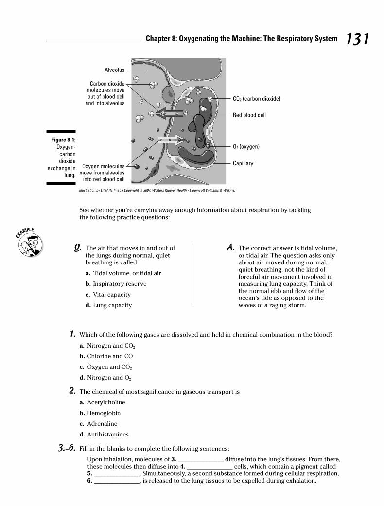
1. Which of the following gases are dissolved and held in chemical combination in the blood?
a. Nitrogen and CO2
b. Chlorine and CO
c. Oxygen and CO2
d. Nitrogen and O2
2. The chemical of most significance in gaseous transport is
a. Acetylcholine
b. Hemoglobin
c. Adrenaline
d. Antihistamines
3.–6. Fill in the blanks to complete the following sentences:
Upon inhalation, molecules of 3. _______________ diffuse into the lung’s tissues. From there,these molecules then diffuse into 4. _______________ cells, which contain a pigment called 5. _______________. Simultaneously, a second substance formed during cellular respiration,6. _______________, is released to the lung tissues to be expelled during exhalation.
131Chapter 8: Oxygenating the Machine: The Respiratory System
Illustration by LifeART Image Copyright 2007. Wolters Kluwer Health - Lippincott Williams & Wilkins.
See whether you’re carrying away enough information about respiration by tacklingthe following practice questions:
Alveolus
Carbon dioxidemolecules moveout of blood cell
and into alveolus
Oxygen moleculesmove from alveolus
into red blood cell
CO2 (carbon dioxide)
Red blood cell
O2 (oxygen)
Capillary
Figure 8-1:
Oxygen-
carbon
dioxide
exchange in
lung.
Q. The air that moves in and out ofthe lungs during normal, quietbreathing is called
a. Tidal volume, or tidal air
b. Inspiratory reserve
c. Vital capacity
d. Lung capacity
A. The correct answer is tidal volume,or tidal air. The question asks onlyabout air moved during normal,quiet breathing, not the kind offorceful air movement involved inmeasuring lung capacity. Think ofthe normal ebb and flow of theocean’s tide as opposed to thewaves of a raging storm.
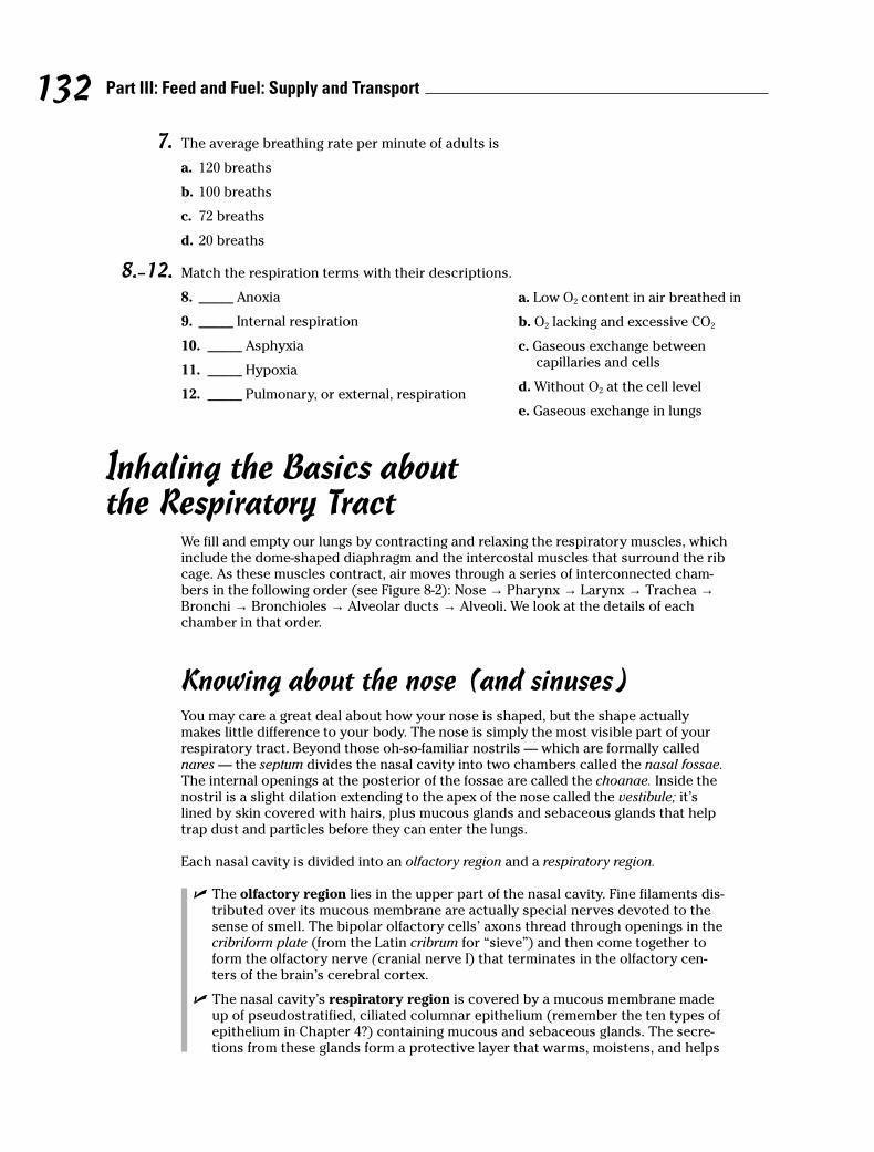
7. The average breathing rate per minute of adults is
a. 120 breaths
b. 100 breaths
c. 72 breaths
d. 20 breaths
8.–12. Match the respiration terms with their descriptions.
8. _____ Anoxia
9. _____ Internal respiration
10. _____ Asphyxia
11. _____ Hypoxia
12. _____ Pulmonary, or external, respiration
Inhaling the Basics about the Respiratory Tract
We fill and empty our lungs by contracting and relaxing the respiratory muscles, whichinclude the dome-shaped diaphragm and the intercostal muscles that surround the ribcage. As these muscles contract, air moves through a series of interconnected cham-bers in the following order (see Figure 8-2): Nose → Pharynx → Larynx → Trachea →Bronchi → Bronchioles → Alveolar ducts → Alveoli. We look at the details of eachchamber in that order.
Knowing about the nose (and sinuses)You may care a great deal about how your nose is shaped, but the shape actuallymakes little difference to your body. The nose is simply the most visible part of yourrespiratory tract. Beyond those oh-so-familiar nostrils — which are formally callednares — the septum divides the nasal cavity into two chambers called the nasal fossae.The internal openings at the posterior of the fossae are called the choanae. Inside thenostril is a slight dilation extending to the apex of the nose called the vestibule; it’slined by skin covered with hairs, plus mucous glands and sebaceous glands that helptrap dust and particles before they can enter the lungs.
Each nasal cavity is divided into an olfactory region and a respiratory region.
� The olfactory region lies in the upper part of the nasal cavity. Fine filaments dis-tributed over its mucous membrane are actually special nerves devoted to thesense of smell. The bipolar olfactory cells’ axons thread through openings in thecribriform plate (from the Latin cribrum for “sieve”) and then come together toform the olfactory nerve (cranial nerve I) that terminates in the olfactory cen-ters of the brain’s cerebral cortex.
� The nasal cavity’s respiratory region is covered by a mucous membrane madeup of pseudostratified, ciliated columnar epithelium (remember the ten types ofepithelium in Chapter 4?) containing mucous and sebaceous glands. The secre-tions from these glands form a protective layer that warms, moistens, and helps
132 Part III: Feed and Fuel: Supply and Transport
a. Low O2 content in air breathed in
b. O2 lacking and excessive CO2
c. Gaseous exchange between capillaries and cells
d. Without O2 at the cell level
e. Gaseous exchange in lungs
to filter air as it’s inhaled. Beneath the protective layer, areolar connective tissuecontaining lymphocytes (which form a thin lymphoid tissue) removes foreignmaterials. A layer of blood vessels next to the periosteum (the membrane cover-ing the surface of bones) forms a rich plexus (network) that tends to swell whenirritated or inflamed, closing the ostia (openings) of the nasal sinuses. Don’t youjust hate it when that happens?
Ah, the sinuses. They can be such headaches. Lined with a ciliated columnar epithe-lium (refer to Chapter 4’s tissue discussion), sinuses are cavities in the bone thatreduce the skull’s weight and act as resonators for the voice. Each of the sinuses isnamed for the bone containing it, as follows:
� Frontal sinuses are located in the front bone behind the eyebrows. (If you’veever flown in an airplane with a sinus infection, these are the suckers that hit youright behind the eyes.)
� Maxillary sinuses are located in the maxillae, or upper lip.
� Ethmoid and sphenoid sinuses are located in the ethmoid and sphenoid bonesin the cranial cavity’s floor.
Beyond the sinuses and connected to them are nasal ducts that extend from the medialangle of the eyes to the nasal cavity. These ducts let serous fluid — a biology termreferring to any fluid resembling serum — from the eyes’ lacrimal glands (tear ducts)flow into the nasal cavity.
The nasal cavity performs several important functions:
� It drains mucous secretions from the sinuses.
� It drains lacrimal secretions from the eyes.
� It prepares inhaled air for the lungs by warming, moistening, and filtering it. Dustand bacteria are caught in the mucous and passed outward from the nasal cavityby the motion of the cilia. Some of that gunk is taken up by lymphatic tissue in thenasal cavity and respiratory tubes for delivery to the lymph nodes, whichdestroy invading germs.
Beyond the nasal cavity is the nasopharynx, which connects — you guessed it — thenasal cavity to the pharynx.
With a bit of a refresher on the nasal and sinus passages, do you think you can hit thefollowing practice questions on the nose?
13. Which of the following statements about the mucous membranes of the nasal cavity is not true?
a. They contain an abundant blood supply.
b. They moisten the air that flows over them.
c. They’re composed of stratified squamous epithelium.
d. They become inflamed, causing the membrane to swell and close the nasal sinuses.
14. A nasal sinus would not be found in the
a. Frontal bone
b. Ethmoid
c. Maxilla
d. Vomer
e. Sphenoid
133Chapter 8: Oxygenating the Machine: The Respiratory System
15.–30. Use the terms that follow to identify the structures of the respiratory tract shown in Figure 8-2.
Illustration by LifeART Image Copyright 2007. Wolters Kluwer Health - Lippincott Williams & Wilkins.
a. Esophagus
b. Larynx
c. Nasal cavities
d. Oropharynx
e. Nose
f. Right lung
g. Epiglottis
h. Mouth
i. Alveoli
j. Nasopharynx
k. Thyroid cartilage
l. Left lung
m.Trachea
n. Bronchioles
o. Laryngopharynx
p. Left bronchus
Dealing with throaty mattersIn laymen’s terms, it’s the throat. But you know better, right? The top of the throat con-sists of these key parts:
21. _____15. _____
16. _____
17. _____
20. _____
18. _____
19. _____
22. _____
23. _____
25. _____
26. _____
27. _____
28. _____
29. _____
30. _____
24. _____
Figure 8-2:
The respira-
tory tract.
134 Part III: Feed and Fuel: Supply and Transport

� Pharynx: The pharynx is an oval, fibromuscular sac about 5 inches long andtapering to 1⁄2 inch in diameter at its anteroposterior end, which is a fancy biologyterm meaning “front to back.” In fact, the point where the pharynx connects tothe esophagus is the narrowest part of the entire digestive tract. Eustachiantubes connected to the middle ears enter the pharynx on each side. On the backwall of the pharynx is a mass of lymphoid tissue called the pharyngeal tonsil, oradenoids.
� Larynx: Connecting the pharynx with the trachea, this collection of nine carti-lages is what makes a man’s prominent Adam’s apple. Also called the voice box,the larynx looks like a triangular box flattened dorsally and at the sides thatbecomes narrow and cylindrical toward the base (see Figure 8-3). Ligaments con-nect the cartilages controlled by several muscles; the inside of the larynx is linedwith a mucous membrane that continues into the trachea.
Three of the larynx’s nine cartilages go solo — the thyroid, the cricoid, and the epiglottis — while three more come in pairs — the arytenoids, the corniculates, and thecuneiforms. The thyroid cartilage (thyroid in Greek means “shield-shaped) is largestand consists of two plates called laminae that are fused just beneath the skin to form ashield-shaped process, the Adam’s apple. Immediately above the Adam’s apple, thelaminae are separated by a V-shaped notch called the superior thyroid notch. The ring-shaped cricoid cartilage is smaller but thicker and stronger, with shallow notches atthe top of its broad back that connect, or articulate, with the base of the arytenoid car-tilages. The arytenoid cartilages both are shaped like pyramids, with the vocal foldsattached at the back and the controlling muscles that move the arytenoids attached atthe sides, moving the vocal cords. On top of the arytenoids are the corniculate carti-lages, small conical structures for attachment of muscles regulating tension on thevocal cords. Nestled in front of these and inside the aryepiglottic fold, the cuneiformcartilages stiffen the soft tissues in the vicinity. The epiglottis, sometimes called the lidon the voice box, is a leaf-shaped cartilage that projects upward behind the root of thetongue. Attached at its stem end, the epiglottis opens during respiration and reflex-ively closes during swallowing to keep food and liquids from getting into the respira-tory tract.
Two types of folds play different roles inside the larynx. The true vocal folds, or cords,are V-shaped when relaxed. When talking, the folds stretch for high sounds or slackenfor low sounds, causing the opening into the glottis — the opening in the larynx — toform an oval. Sounds are produced when air is forced over the folds, causing them tovibrate. Just above these folds are the ventricular vocal folds, also known as vestibularor false folds, that don’t produce sounds. Muscle fibers within these folds help closethe glottis during swallowing.
Following are some practice questions dealing with the throat:
31. The vocal folds change position by the movement of the cartilage known as
a. Cuneiforms
b. Thyroid
c. Arytenoids
d. Cricoid
e. Epiglottis
135Chapter 8: Oxygenating the Machine: The Respiratory System

32.–36. Match the anatomical structure with its function.
32. _____ Epiglottis a. Voice box lid
33. _____ Nasal fossae b. Respiratory center
34. _____ Medulla oblongata c. Prevents collapse of trachea
35. _____ C-shaped cartilaginous rings d. Nasal cavity
36. _____ Thyroid cartilage e. Adam’s apple
37. The opening between the two vocal folds is called the
a. Epiglottis
b. Bronchi
c. Alveoli
d. Glottis
e. Larynx
38. The loud voice of a person speaking is due to
a. Vibrating vocal folds
b. Vibrating chest muscles
c. Increased air from lungs
d. Vibrating pharynx
e. Vibrating trachea
39.–52. Use the terms that follow to identify the structures of the larynx shown in Figure 8-3. Someterms may be used more than once.
Illustration by Imageering Media Services Inc.
a. Thyroid cartilage
b. Cricoid cartilage
c. Hyoid bone
d. Epiglottis
49. _____
50. _____
51. _____
52. _____
42. _____
43. _____
44. _____
46. _____
45. _____
47. _____
48. _____
39. _____
40. _____
41. _____
a b
Figure 8-3:
Front (a) and
lateral (b)
views of the
larynx.
136 Part III: Feed and Fuel: Supply and Transport

e. Arytenoid cartilage
f. Ventricular fold (false vocal cord)
g. Laryngenal prominence (Adam’s apple)
h. Cuneiform cartilage
i. Arytenoid muscle
j. True vocal cord
k. Tracheal cartilages
l. Corniculate cartilage
Going deep inside the lungsAfter the pharynx and larynx comes the trachea, more popularly known as the wind-pipe. Roughly 6 inches long in adults, it’s a tube connected to the larynx in front of theesophagus that’s made up of C-shaped rings of hyaline cartilage and fibrous connec-tive tissue that strengthen it and keep it open. Like the larynx, the trachea’s lined withmucous membrane covered in cilia. Just above the heart, the trachea splits into twobronchi divided by a sharp ridge called the carina, with each leading to a lung. Butthey’re not identical: The right primary bronchus is shorter and wider than the left pri-mary bronchus. Each primary bronchus divides into secondary bronchi with a branchgoing to each lobe of the lung; the right side gets three secondary bronchi while theleft gets only two. Once inside a designated lobe, the bronchus divides again into terti-ary bronchi. The right lung has ten such branches: three in the superior (or upper)lobe, two in the middle lobe, and five in the inferior (or lower) lobe. The left lung hasonly four tertiary bronchi: two in the upper lobe and two in the lower lobe.
Each tertiary bronchi subdivides one more time into smaller tubes called bronchioles(see Figure 8-4), which lack the supporting cartilage of the larger structures. Eachbronchiole ends in an elongated sac called the atrium (also known as an alveolar ductor alveolar sac). Alveoli (or air cells) surround the atria, as do small capillaries thatpick up oxygen for delivery elsewhere in the body and dump off carbon dioxidefetched from elsewhere. Overall, there are 23 branches in the respiratory system, witha combined surface area (counting the alveoli) the size of a tennis court!
Knowing that the bronchi aren’t evenly distributed, you may have guessed that thelungs aren’t identical either. You’re right. They’re both spongy and porous because ofthe air in the sacs, but the right lung is larger, wider, and shorter than the left lung andhas three lobes. The left lung divides into only two lobes and is both narrower andlonger to make room for the heart because two-thirds of that organ lies to the left ofthe body’s midline. Each lobe is made up of many lobules, each with a bronchioleending in an atrium inside.
Covering each lung is a thin serous membrane called the visceral pleura that folds backon itself to form a second outer layer, the parietal pleura, with a pleural cavity betweenthe two layers. These two layers secrete a watery fluid into the cavity to lubricate thesurfaces that rub against each other as you breathe. When the pleural membranebecomes inflamed in a condition called pleurisy, a sticky discharge roughens thepleura, causing painful irritation. An accompanying bacterial infection means that pusaccumulates in the pleural cavity in a condition known as empyema.
137Chapter 8: Oxygenating the Machine: The Respiratory System
Blood comes to the lungs through two sources: the pulmonary arteries and thebronchial arteries. The pulmonary trunk comes from the right ventricle of the heartand then branches into the two pulmonary arteries carrying venous blood (the onlyarteries that contain blood loaded with carbon dioxide from various parts of the body)to the lungs. That blood goes through capillaries in the lungs where the carbon dioxideleaves the blood and enters the alveoli to be expelled during exhalation; oxygen leavesthe alveoli through the capillaries to enter the bloodstream. After that, oxygenatedarterial blood returns to the left atrium through the pulmonary veins (the only veinsthat contain oxygenated blood), completing the cycle. Bronchial arteries branch offthe thoracic aorta of the heart, supplying the lung tissue with nutrients and oxygen.
Following are some practice questions dealing with the lungs:
53. Cartilage is not found in the
a. Primary bronchi
b. Secondary bronchi
c. Bronchioles
d. Trachea
e. Larynx
54. Gaseous exchange occurs between the capillaries and the
a. Trachea
b. Alveolar sacs
c. Primary bronchi
d. Terminal bronchioles
e. Secondary bronchi
55.–59. Fill in the blanks to complete the following sentences:
The trachea divides into two 55. _______________, which then divide into 56. _______________with a branch going to each lobe of the lung. Upon entering the lobe, each divides into 57. _______________, subdividing into smaller tubes called 58. _______________. They terminate in an elongated sac called the atrium surrounded by 59. _______________ or air cells.
60. If a pin were to pierce the body from the outside in the thoracic region, the third structure itwould reach would be the
a. Pleural cavity
b. Visceral pleura
c. Parietal pleura
d. Lung
138 Part III: Feed and Fuel: Supply and Transport
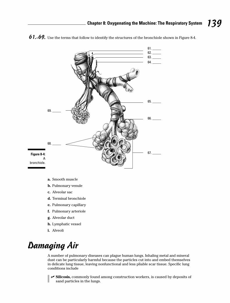
61.–69. Use the terms that follow to identify the structures of the bronchiole shown in Figure 8-4.
a. Smooth muscle
b. Pulmonary venule
c. Alveolar sac
d. Terminal bronchiole
e. Pulmonary capillary
f. Pulmonary arteriole
g. Alveolar duct
h. Lymphatic vessel
i. Alveoli
Damaging AirA number of pulmonary diseases can plague human lungs. Inhaling metal and mineraldust can be particularly harmful because the particles cut into and embed themselvesin delicate lung tissue, leaving nonfunctional and less pliable scar tissue. Specific lungconditions include
� Silicosis, commonly found among construction workers, is caused by deposits ofsand particles in the lungs.
69. _____
68. _____
61. _____
62. _____
63. _____
64. _____
65. _____
66. _____
67. _____Figure 8-4:
A
bronchiole.
139Chapter 8: Oxygenating the Machine: The Respiratory System
� Anthracosis, or black lung, occurs among coal miners because of coal dust accu-mulating in the lungs.
� Rhinitis, or the common cold, can be caused by several different kinds of viralinfections. Undue exposure may activate the virus or cause the body to becomemore susceptible to the virus.
70. The disease referred to as anthracosis is caused by
a. A bacterial infection
b. Inhaling coal dust
c. Inhaling sand particles
d. Undue exposure
e. Inflammation of the pleura membrane
140 Part III: Feed and Fuel: Supply and Transport
Answers to Questions on the Respiratory System
The following are answers to the practice questions presented in this chapter.
a Which of the following gases are dissolved and held in chemical combination in the blood? c. Oxygen and CO2. Yes, the human body’s two greatest needs are to inhale oxygen and to exhale carbon dioxide.
b The chemical of most significance in gaseous transport is b. hemoglobin.
Remember that the Latin root for blood is hemo; none of the other answer options incorporatethat root.
c–f Upon inhalation, molecules of 3. oxygen diffuse into the lung’s tissues. From there, these mole-cules then diffuse into 4. red blood cells, which contain a pigment called 5. hemoglobin.Simultaneously, a second substance formed during cellular respiration, 6. carbon dioxide, isreleased to the lung tissues to be expelled during exhalation.
g The average breathing rate per minute of adults is d. 20 breaths. The rates in the other answeroptions are more akin to pulse rates or blood pressures than to average respirations perminute.
h Anoxia: d. Without O2 at the cell level
i Internal respiration: c. Gaseous exchange between capillaries and cells
j Asphyxia: b. O2 lacking and excessive CO2
k Hypoxia: a. Low O2 content in air breathed in
l Pulmonary, or external, respiration: e. Gaseous exchange in lungs
m Which of the following statements about the mucous membranes of the nasal cavity is not true?c. They’re composed of stratified squamous epithelium.
n A nasal sinus would not be found in the d. vomer.
o–E Following is how Figure 8-2, the respiratory tract, should be labeled.
15. c. Nasal cavities; 16. e. Nose; 17. h. Mouth; 18. k. Thyroid cartilage; 19. b. Larynx; 20 f.Right lung; 21. j. Nasopharynx; 22. d. Oropharynx; 23. g. Epiglottis; 24. o. Laryngopharynx;25. a. Esophagus; 26. m. Trachea; 27. p. Left bronchus; 28. n. Bronchioles; 29. i. Alveoli; 30. l. Left lung
F The vocal folds change position by the movement of the cartilage known as c. arytenoids. Thiscartilage is tough to spell and pronounce but easy to move.
G Epiglottis: a. Voice box lid
H Nasal fossae: d. Nasal cavity
I Medulla oblongata: b. Respiratory center
141Chapter 8: Oxygenating the Machine: The Respiratory System
J C-shaped cartilaginous rings: c. Prevents collapse of trachea
K Thyroid cartilage: e. Adam’s apple
L The opening between the two vocal folds is called the d. glottis.
M The loud voice of a person speaking is due to c. increased air from lungs. That’s why peopletend to take a deep breath before they start yelling.
N–Z Following is how Figure 8-3, the larynx, should be labeled.
39. c. Hyoid bone; 40. a. Thyroid cartilage; 41. g. Laryngenal prominence (Adam’s apple);42. d. Epiglottis; 43. h. Cuneiform cartilage; 44. l. Corniculate cartilage; 45. e. Arytenoid cartilage; 46. i. Arytenoid muscle; 47. b. Cricoid cartilage; 48. k. Tracheal cartilages; 49. c. Hyoid bone; 50. f. Ventricular fold (false vocal cord); 51. a. Thyroid cartilage; 52. j. True vocal cord
1 Cartilage is not found in the c. bronchioles. They’re so small that they need to be more elasticand less cartilaginous.
2 Gaseous exchange occurs between the capillaries and the b. alveolar sacs. These sacs are thesmallest parts of the lungs, so it makes sense that molecular exchange would take place here.
3–7 The trachea divides into two 55. primary bronchi, which then divide into 56. secondarybronchi with a branch going to each lobe of the lung. Upon entering the lobe, each divides into57. tertiary bronchi, subdividing into smaller tubes called 58. bronchioles. They terminate inan elongated sac called the atrium surrounded by 59. alveoli or air cells.
8 If a pin were to pierce the body from the outside in the thoracic region, the third structure itwould reach would be the b. visceral pleura. Note that the question asks you to choose fromthe list provided, not from the entire structure of the body.
9–& Following is how Figure 8-4, the bronchiole, should be labeled.
61. d. Terminal bronchiole; 62. f. Pulmonary arteriole; 63. b. Pulmonary venule; 64. h.Lymphatic vessel; 65. a. Smooth muscle; 66. g. Alveolar duct; 67. c. Alveolar sac; 68. i.Alveoli; 69. e. Pulmonary capillary
* The disease referred to as anthracosis is caused by b. inhaling coal dust. Anthracosis equalsblack lung equals coal dust.
142 Part III: Feed and Fuel: Supply and Transport
Chapter 9
Fueling the Functions: The Digestive System
In This Chapter� Getting down and dirty with digestion basics
� Examining the mouth
� Spending time in the stomach
� Passing through the intestines and other organs for enzyme digestion
It’s time to feed your hunger for knowledge about how nutrients fuel the whole packagethat is the human body. In this chapter, we help you swallow the basics about getting
food into the system and digest the details about how nutrients move into the rest of thebody. You also get plenty of practice following the nutritional trail from first bite to finalelimination.
Digesting the Basics: It’s Alimentary!Before jumping into a discussion on the alimentary tract, we need to review some basicterms.
� Ingestion: Taking in food
� Digestion: Changing the composition of food — splitting large molecules into smallerones — to make it usable by the cells
� Deglutition: Swallowing, or moving food from the mouth to the stomach
� Absorption: Occurs when digested food moves through the intestinal wall and intothe blood
� Egestion: Eliminating waste materials or undigested foods at the lower end of thedigestive tract; also known as defecation
The alimentary tract develops early on in a growing embryo. The primitive gut, or archen-teron, develops from the endoderm (inner germinal layer) during the third week after concep-tion, a stage during which the embryo is known as a gastrula. At the anterior end (head end),the oral cavity, nasal passages, and salivary glands develop from a small depression called a stomodaeum in the ectoderm (outer germinal layer). The anal and urogenital structuresdevelop at the opposite, or posterior, end from a depression in the ectoderm called the proctodaeum. In other words, the digestive tract develops from an endodermal tube withectoderm at each end.
Whereas the respiratory tract is a two-way street — oxygen flows in and carbon dioxide flowsout — the digestive tract is designed to have a one-way flow (although when you’re sick oryour body detects something bad in the food you’ve eaten, what goes down sometimes comes
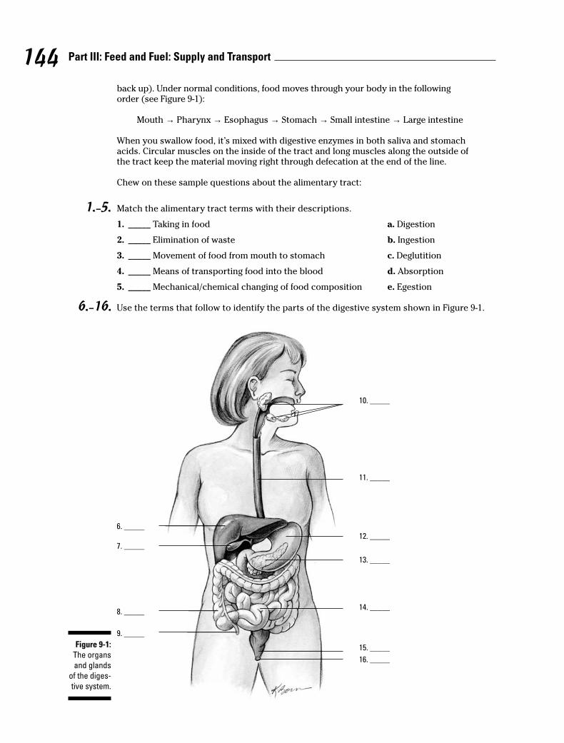
back up). Under normal conditions, food moves through your body in the followingorder (see Figure 9-1):
Mouth → Pharynx → Esophagus → Stomach → Small intestine → Large intestine
When you swallow food, it’s mixed with digestive enzymes in both saliva and stomachacids. Circular muscles on the inside of the tract and long muscles along the outside ofthe tract keep the material moving right through defecation at the end of the line.
Chew on these sample questions about the alimentary tract:
1.–5. Match the alimentary tract terms with their descriptions.
1. _____ Taking in food a. Digestion
2. _____ Elimination of waste b. Ingestion
3. _____ Movement of food from mouth to stomach c. Deglutition
4. _____ Means of transporting food into the blood d. Absorption
5. _____ Mechanical/chemical changing of food composition e. Egestion
6.–16. Use the terms that follow to identify the parts of the digestive system shown in Figure 9-1.
10. _____
11. _____
12. _____
13. _____
14. _____
15. _____
16. _____
6. _____
7. _____
8. _____
9. _____
Figure 9-1:
The organs
and glands
of the diges-
tive system.
144 Part III: Feed and Fuel: Supply and Transport
a. Pancreas
b. Colon
c. Liver
d. Small intestine
e. Salivary glands
f. Gallbladder
g. Appendix
h. Anus
i. Esophagus
j. Rectum
k. Stomach
17. The alimentary tract forms from the following layer(s) of the developing embryo:
a. Endoderm
b. Ectoderm
c. Both the endoderm and the ectoderm
d. Neither the endoderm nor the ectoderm
18. Identify the correct sequence of the movement of food through the body:
a. Mouth → Pharynx → Esophagus → Stomach → Small intestine → Large intestine
b. Mouth → Esophagus → Pharynx → Stomach → Small intestine → Large intestine
c. Mouth → Pharynx → Esophagus → Stomach → Large intestine → Small intestine
d. Mouth → Pharynx → Stomach → Esophagus → Small intestine → Large intestine
Nothing to Spit At: Into the Mouth and Past the Teeth
In addition to being very useful for communicating, the mouth serves a number ofimportant roles in the digestive process:
� Chewing, formally known as mastication, breaks down food mechanically intosmaller particles.
� The act of chewing increases blood flow to all the mouth’s structures and thelower part of the head.
� Saliva from salivary glands in the mouth helps prepare food to be swallowed andbegins the chemical breakdown of carbohydrates.
� Taste buds on the tongue stimulate saliva production. Interestingly, studies haveshown that taste preferences can change in reaction to the body’s specific needs.In addition, the smell of food can get gastric juices flowing in preparation fordigestion.
The mouth’s anatomy begins, of course, with the lips, which are covered by a thin,modified mucous membrane. That membrane is so thin that you can see the red blood
145Chapter 9: Fueling the Functions: The Digestive System
in the underlying capillaries. (That’s the unromantic reason for the lips’ natural rosyglow.) The mouth itself is divided into two regions defined by the arches of the upperand lower jaws. The vestibule is the region between these dental arches, cheeks, andlips, whereas the oral cavity is the region inside the dental arches.
Entering the vestibuleThe inner surface of the lips is covered by a mucous membrane. Sickle-shaped pieces oftissue called labial frenula attach the lips to the gums. Within the mucous membraneare labial glands, which produce mucus to prevent friction between the lips and theteeth. The cheeks are made up of buccinator muscles and a buccal pad, a subcutaneouslayer of fat. The buccinator muscles keep the food between the teeth during the act ofchewing. Elastic tissue in the mucous membrane keeps the lining of the cheeks fromforming folds that would be bitten during chewing (usually — most people have bittenthe insides of their cheeks at one time or another). Also stashed away in the cheek, justin front of and below each ear, is a parotid gland, which is the largest salivary gland; itreleases saliva through a duct opposite the second upper molar tooth. Two other pairsof salivary glands also secrete into the mouth: the submaxillary glands along the side ofthe lower jaw and the sublingual glands in the floor of the mouth near the chin.
The dental arches are formed by the maxillae (upper jaw) and the mandible (lowerjaw) along with the gingivae (gums) and teeth of both jaws. The gingivae are dense,fibrous tissues attached to the teeth and the underlying jaw bones; they’re covered bya mucous membrane extending from the lips and cheeks to form a collar around theneck of each tooth. The gums are very vascular (meaning that lots of blood vessels runthrough them) but poorly innervated (meaning that, fortunately, they’re not generallyvery sensitive to pain).
Teeth rise from openings in the jawbone called sockets, or alveoli. You have a numberof different kinds of teeth, and each has a specific contribution to the process of bitingand chewing. Humans get two sets of teeth in a lifetime. The first temporary, or decidu-ous, set is known as milk teeth. Babies between 6 months and 2 years old “cut,” orerupt, four incisors, two canines, and two molars in each jaw. These teeth are slowlyreplaced by permanent teeth from about 5 or 6 years of age until the final molars —referred to as wisdom teeth — erupt between 17 and 25 years of age.
An adult human has the following 16 teeth in each jaw (for a total set of 32 teeth):
� Four incisors, which are chisel-shaped teeth at the front of the jaw for biting intoand cutting food
� Two canines, or cuspids, which are pointed teeth on either side of the incisors forgrasping and tearing
� Four premolars, or bicuspids, which are flatter, shallower teeth that come in pairsjust behind the canines
� Six molars, which are triplets of broad, flat teeth on either side of the jawbone forgrinding and mixing food prior to swallowing
Regardless of type, each tooth has three primary parts, which you can see in Figure 9-2:
� Crown: The part that projects above the gum
� Neck: The region where the gum attaches to the tooth
� Root: The internal structure that firmly fixes the tooth in the alveolus (socket)
146 Part III: Feed and Fuel: Supply and Transport
Teeth primarily consist of yellowish dentin with a layer of enamel over the crown and a layer of cementum over the root and neck, which are connected to the bone by theperiodontal membrane. Cementum and dentin are nearly identical in composition tobone; enamel consists of 94 percent calcium phosphate and calcium carbonate and isthickest over the chewing surface of the tooth.
Depending on the structure of the tooth, the root can be a single-, double-, or eventriple-pointed structure. In addition, each tooth has a pulp cavity at the center that’sfilled with connective and lymphatic tissue, nerves, and blood vessels that enter thetooth through the root canal via an opening at the bottom called the apical foramen.Now you know why it hurts so much when dentists have to drill down and take outthat part of an infected tooth!
Moving along the oral cavityThe roof of the oral cavity is formed by both the hard palate, a bony structure coveredby fibrous tissue and the ever-present mucous membrane, and the soft palate, a mov-able partition of fibromuscular tissue that prevents food and liquid from getting intothe nasal cavity. (It’s also the tissue that sometimes vibrates in sleep, causing asonorous grating sound referred to as snoring.) The soft palate hangs at the back of theoral cavity in two curved folds that form the palatine arches. The uvula, a soft conicalprocess (or piece of tissue), hangs in the center between those folds.
Beyond the soft palate, the palatopharyngeal (or pharyngopalatine) arch curves sharplytoward the midline and blends with the wall of the pharynx, ending at the dorsum(back) of the tongue. Another structure, the anterior palatoglossal (or glossopalatine)arch, starts on the surface of the palate at the base of the uvula and continues in a widecurve forward and downward, ending next to the posterior (back) one-third of thetongue. At the base of these arches and between the folds lie the palatine tonsils — if asurgeon hasn’t removed them because of frequent childhood infections. The faucial isthmus or oropharynx is the junction between the oral cavity and the pharynx(described in detail in Chapter 8). It opens during swallowing and closes when youmove the dorsum of the tongue against the soft palate when breathing.
The tongueThe tongue, which is a tight bundle of interlaced muscles, and its associated mucousmembrane form the floor of the oral cavity. Two distinct groups of muscles — extrinsicand intrinsic — are used in tandem for mastication (chewing), deglutition (swallowing),and to articulate speech.
� The extrinsic muscles, which are used to move the tongue in different directions,originate outside the tongue and are attached to the mandible, styloid processesof the temporal bone and the hyoid and, along with a fold of mucous membranecalled the lingual frenulum, anchor the tongue.
� The intrinsic muscles are a complex muscle network allowing the tongue tochange shape for talking, chewing and swallowing.
Three primary types of papillae (nipple-shaped protrusions) cover the tongue’s for-ward upper surface:
� Filiform papillae are fine, brush-like papillae that cover the dorsum, the tip, andthe lateral margins of the tongue. They’re the most numerous papillae and don’thold any taste buds.
147Chapter 9: Fueling the Functions: The Digestive System

19.–30. Use the terms that follow to identify the parts of a tooth shown in Figure 9-2.
148 Part III: Feed and Fuel: Supply and Transport
� Fungiform papillae are large, red, mushroom-shaped papillae scattered amongthe filiform papillae. They have taste buds, which are special receptors that com-municate taste signals to the brain.
� Vallate papillae, also called circumvallate papillae, are flattened structures, eachwith a moat-like trough ringing it. There are 12 of these on the tongue, and theysurround a V-shaped furrow toward the back of the tongue called the sulcus terminalis.
There are no papillae on the back (posterior) one-third of the tongue; that part hasonly a mucous membrane covering lymphatic tissue, which forms the lingual tonsils.
The salivary glandsAs we explain in the earlier section “Entering the vestibule,” the oral cavity has threepairs of salivary glands producing saliva. The submandibular (or submaxillary) salivaryglands are about the size of a walnut and release fluid onto the floor of the mouth,under the tongue. The smallest pair of the trio, the sublingual salivary glands, lies nearthe tongue under the oral cavity’s mucous membrane floor to release secretionsdirectly onto the mucous membrane.
And those secretions are nothing to spit at. Saliva does the following:
� Dissolves and lubricates food to make it easier to swallow
� Contains ptyalin, or salivary amylase, an enzyme that initiates chemical digestionof certain carbohydrates
� Moistens and lubricates the mouth and lips, keeping them pliable and resilientfor speech and chewing
� Frees the mouth and teeth of food, foreign particles, and epithelial cells
� Produces the sensation of thirst to prevent you from becoming dehydrated
Following are some practice questions regarding the vestibule and oral cavity:
Q. The function of the mouth is
a. Mixing of solid foods with saliva
b. Breaking down of the milk pro-tein by the enzyme rennin
c. Mastication or the breakingdown of food into small particles
d. A and c
e. A, b, and c
A. The correct answer is mixing ofsolid foods with saliva and masti-cation. The mouth does lots ofthings, including mixing saliva into the food to add the enzymeptyalin, but that’s not rennin. Withanswer options like these, it’s bestto stick to the basics.
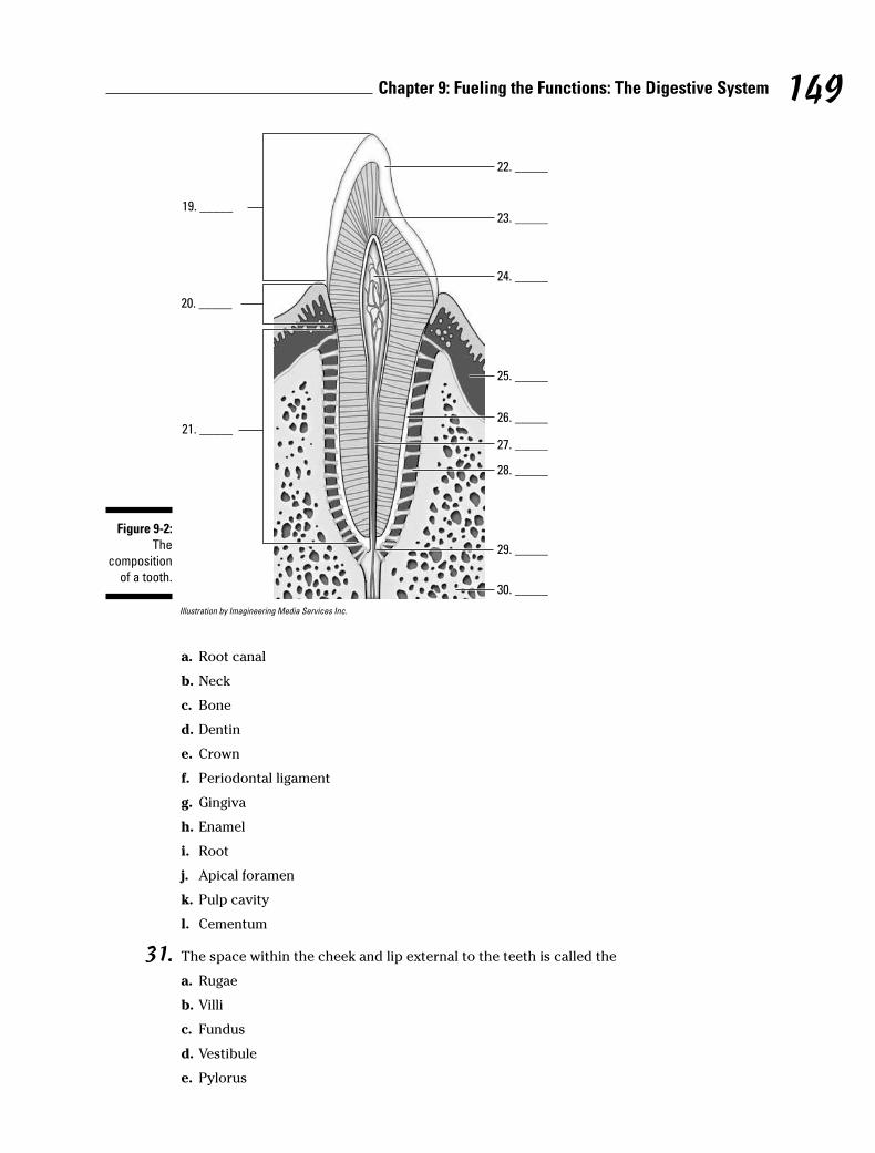
Illustration by Imagineering Media Services Inc.
a. Root canal
b. Neck
c. Bone
d. Dentin
e. Crown
f. Periodontal ligament
g. Gingiva
h. Enamel
i. Root
j. Apical foramen
k. Pulp cavity
l. Cementum
31. The space within the cheek and lip external to the teeth is called the
a. Rugae
b. Villi
c. Fundus
d. Vestibule
e. Pylorus
19. _____
20. _____
21. _____
22. _____
23. _____
24. _____
25. _____
26. _____
27. _____
28. _____
29. _____
30. _____
Figure 9-2:
The
composition
of a tooth.
149Chapter 9: Fueling the Functions: The Digestive System
32. The roof of the oral cavity is formed by
a. The hard and soft palates
b. The sulcus terminalis
c. A rigid bony structure covered by fibrous tissue and a mobile partition composed of fibro-muscular tissue in a fold of mucous membrane
d. A and c
e. A, b, and c
33. Which of the following statements is not true of the teeth?
a. The permanent teeth in each human jaw are four incisors, two canines, four premolars, andsix molars.
b. Each tooth has a single cuspid anchoring it.
c. The tooth cavity contains the tooth pulp.
d. The enamel consists of 94 percent calcium phosphate and calcium carbonate.
e. Each tooth is composed of a crown, a neck, and a root.
34.–38. Match each description with the proper anatomical structure.
150 Part III: Feed and Fuel: Supply and Transport
a. Pharyngopalatine arch
b. Faucial isthmus
c. Gingivae
d. Glossopalatine arch
e. Uvula
34. _____ Soft conical process projecting from the soft palate
35. _____ The junction between the mouth and pharyn
36. _____ Forms a collar around the teeth and is poorly innervated
37. _____ Sharply curved arch that bends lateral1y with the walls of the pharynx
38. _____ Arch that starts at the buccal surface of the palate at the base of the uvula and ends alongside the back third of the tongue
39. The palatine tonsil is located
a. In the posterior wall of the pharynx
b. In the smooth posterior one-third of the tongue
c. In the region between the rigid hard palate and the soft palate
d. Under the mucous membrane of the tongue
e. In the region between the palatopharyngeal and palatoglossal arches
40. The function of saliva is
a. To facilitate swallowing
b. To initiate the digestion of certain carbohydrates
c. To moisten and lubricate the mouth and lips
d. A and c
e. A, b, and c
41. The largest salivary gland is the
a. Submandibular gland
b. Brunner’s gland
c. Sublingual gland
d. Submaxillary gland
e. Parotid gland
42.–44. Match the descriptions with the anatomical structures.
151Chapter 9: Fueling the Functions: The Digestive System
a. Vallate papillae
b. Filiform papillae
c. Fungiform papillae
42. _____ Fine brush-like structures found covering the dorsum of the tongue
43. _____ Large mushroom-shaped structures
44. _____ Large structures, each surrounded by a moatthat form a V-shaped furrow in the tongue
Stomaching the Body’s FuelDeglutition (swallowing) occurs in three phases:
1. The tip of the tongue elevates slightly, pushing against the hard palate, slidingfood onto the back of the tongue, and ultimately propelling it toward the pharynx.
2. Tensor muscles tighten the palate while levator muscles raise it until thepalate meets the pharyngeal wall, sealing off the nasopharynx from theoropharynx.
This action momentarily stops breathing and ensures that food and fluid won’tregurgitate through the nose — unless someone makes you laugh, of course.
3. The bolus (food mass) heads “down the hatch.”
The pharynx is an oval fibrous muscular sac, about 5 inches long. It opens into thenasal cavity, the oral cavity, the larynx, and the esophagus. On the lateral walls arelocated the openings to the Eustachian tubes, which connect with the middle ear. Inthe posterior wall is a mass of lymphatic tissue, the pharyngeal tonsil or adenoid.
This “hatch,” borrowed nautical slang for the esophagus, is approximately 10 incheslong and 1⁄2 inch in diameter and carries food through three body regions: the neck, thethorax, and the abdomen. It’s not a straight tube, but rather curves slightly to the left asit passes through the diaphragm 1 inch to the left of the midline. The very thick walls of the esophagus are lined with non-keratinized stratified squamous epithelium andinclude a fibrous outer layer made up of elastic fibers that permit distention during swallowing (think of a snake swallowing a whole egg — there’s some major stretchinggoing on there). A muscular layer contains both longitudinal and circular layers ofsmooth muscle. The circular layers contract in sequence, like a series of shrinking andexpanding rings, in a movement called peristalsis that forces the bolus downward. Thelongitudinal layers act in concert with the circular muscles, pulling the esophagus overthe bolus as it moves downward.
All this pushing and pulling ultimately releases the bolus into the stomach, a pear-shaped bag of an organ that lies just beneath the ribs and diaphragm. About 1 footlong and 1⁄2 foot wide, a human stomach’s normal capacity is about 1 quart. Whenempty, the stomach’s mucous lining lies in folds called rugae; rugae allow expansion ofthe tummy when you gorge and then shrink the stomach when it’s empty to decreasethe surface area exposed to acid. Food enters the upper end of the stomach, called thecardiac region, through a ring of muscles called the cardiac sphincter, which generallyremains closed to prevent gastric acids from moving up into the esophagus. The

dome-shaped area below the cardiac region is called the fundus region; it expandssuperiorly with really big meals. The lower part of the stomach, shaped like the letter J,is the pylorus. The middle part of the body of the stomach forms a large curve calledthe greater curvature. The right, much shorter, border of the stomach’s body iscalled the lesser curvature. The far end of the stomach remains closed off by the pyloricsphincter until its contents have been digested sufficiently to pass into the duodenumof the small intestine.
The wall of the stomach consists of three layers of smooth muscle lined by mucousmembrane and covered by the peritoneum (see Figure 9-3). The fibers of the outerlayer of muscle run longitudinally, the middle layer of muscle consists of circular fibersthat encircle the stomach, and the inner layer of muscle fibers runs obliquely onlyalong the fundus region. The stomach’s mucous membrane is covered with nonciliatedcolumnar epithelium containing mucous glands.
The three types of gastric glands in the stomach’s epithelium (lining) are
� Cardiac glands: Found in the cardiac region (of course)
� Pyloric glands: Secrete mucous in the pyloric region
� Fundic glands: Lined with chief cells and parietal cells and are located through-out the stomach’s body and fundus
The three types of cells in the mucosa (lining) of the stomach are
� Mucous cells: Secrete mucin (mucous) to protect the mucosa from the high acid-ity of the gastric juices
� Chief cells: Secrete pepsinogen, a precursor to the enzyme pepsin that helpsbreak down certain proteins into peptides. (Chief cells in children also producean enzyme called rennin, not found in adults, which acts upon milk proteins.)
� Parietal cells: Lie alongside chief cells and secrete the hydrochloric acid thatcombines with pepsinogen to form pepsin to catalyze protein digestion
The peristaltic contractions that get the bolus into the stomach aren’t limited to theesophagus. Instead, peristalsis continues into the musculature of the stomach andstimulates the release of a hormone called gastrin. Within minutes, gastrin triggerssecretion of gastric juices that reduce the bolus of food to a thick semiliquid masscalled chyme, which passes through the pyloric sphincter into the small intestinewithin one to four hours of the food’s consumption.
Gastric juices are thin, colorless fluids with an extremely acid pH that ranges from 1 to 4.The quantity of acid released depends on the amount and type of food being digested.
One more part attached to the stomach that we should mention is the greater omen-tum. This is a peritoneal fold that hangs like an apron from the greater curvature of thestomach all the way down to the transverse colon, covering all the small intestine andmost of the large intestine. Also called a caul or velum, this lining can be laden with fat,particularly in obese people.
152 Part III: Feed and Fuel: Supply and Transport
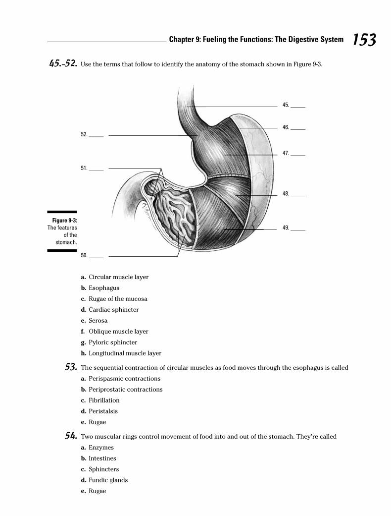
45.–52. Use the terms that follow to identify the anatomy of the stomach shown in Figure 9-3.
a. Circular muscle layer
b. Esophagus
c. Rugae of the mucosa
d. Cardiac sphincter
e. Serosa
f. Oblique muscle layer
g. Pyloric sphincter
h. Longitudinal muscle layer
53. The sequential contraction of circular muscles as food moves through the esophagus is called
a. Perispasmic contractions
b. Periprostatic contractions
c. Fibrillation
d. Peristalsis
e. Rugae
54. Two muscular rings control movement of food into and out of the stomach. They’re called
a. Enzymes
b. Intestines
c. Sphincters
d. Fundic glands
e. Rugae
45. _____
52. _____
46. _____
47. _____
48. _____
49. _____
51. _____
50. _____
Figure 9-3:
The features
of the
stomach.
153Chapter 9: Fueling the Functions: The Digestive System

55. The lower part of the stomach that’s shaped like a J is called the
a. Esophagus
b. Pylorus
c. Peritoneal fold
d. Cardiac region
e. Fundus region
56. Food that’s ready to leave the stomach has been reduced to a thick, semiliquid mass called
a. Omentum
b. Gastric juices
c. Peritoneum
d. Chyme
e. Enzymes
Breaking Down the Work of Digestive Enzymes
So what exactly does all the work of digesting and breaking down food? That questionbrings you back into the realm of proteins. Proteins called enzymes act as catalysts,meaning that they initiate and accelerate chemical reactions without themselves beingpermanently changed in the reaction. Enzymes are very picky proteins indeed; they’reeffective only in their own pH range, they catalyze only a single chemical reaction, theyact on a specific substance called a substrate, and they function best at 98.6 degreesFahrenheit, which just happens to be normal body temperature!
The following sections take you on a tour of the organs in which digestive enzymes dotheir job.
Small intestineMost enzyme reactions — in fact most digestion and practically all absorption of nutrients — takes place in the small intestine. Stretching 7 meters (which is nearly 23 feet!), this long snake of an organ extends from the stomach’s pylorus to the ileoce-cal junction (the point where the small intestine meets the large intestine), graduallydiminishing in diameter along the way.
Three regions of the small intestine play unique roles as chyme moves through it:
� Duodenum: The first section of the small intestine is also the shortest andwidest section. As partially digested food enters the duodenum, its acidity stimu-lates the intestine to secrete the intestinal hormone enterocrinin, which controlsthe secretion of intestinal juices, stimulates the pancreas to secrete its juices,and stimulates the liver to secrete bile. Both the liver and pancreas share acommon opening into the duodenum. Lined with large and numerous villi, theduodenum also has Brunner’s glands that secrete a clear alkaline mucous. Theglands are most numerous near the entry to the stomach and decrease innumber toward the opposite, or jejunum, end.
154 Part III: Feed and Fuel: Supply and Transport

� Jejunum: This region of the small intestine also contains villi, but unlike the duo-denum, it has numerous large circular folds at the beginning that decrease innumber toward the ileum end.
� Ileum: Peyer’s patches, which are aggregates of lymph nodes, line this region ofthe small intestine, becoming largest and most numerous at the distal end. Theileum opens into the cecum of the large intestine through the ileocaecal valve.
A microscopic look at the small intestine reveals circular folds called plicae circularis,which project 3 to 10 millimeters into the intestinal lumen and extend anywhere fromhalf to entirely around the tube. These are permanent folds that don’t smooth out evenwhen the intestine is distended. Also present are finger-like projections called villi thatgreatly increase the surface area through which the small intestine can absorb nutri-ents. Each villus contains a network of capillaries and a central lymph vessel, orlacteal, which contains a milk-white substance called chyle. Simple sugars, aminoacids, vitamins, minerals, and water are absorbed by the lacteal and combine to formthe triglycerides found in the blood. The surface of the villus is simple columnarepithelium (if you can’t recall what that means, flip to Chapter 4). Electron microscopy,which can magnify tissues far more than an optical microscope can, reveals that thesurface of each villus is further increased by microvilli. Peristalsis continues into thesmall intestine, shortening and lengthening the villi to mix intestinal juices with foodand increase absorption. Intestinal glands lie in the depressions between villi, andpacked inside these glands are antimicrobial Paneth cells within glands called thecrypts of Lieberkühn, which secrete enzymes that assist pancreatic enzymes.
Intestinal juices contain three types of enzymes:
� Enterokinase has no enzyme action by itself, but when added to pancreaticjuices, it combines with trypsinogen to form trypsin, which can break down proteins.
� Erepsins, or proteolytic enzymes, don’t directly digest proteins but insteadcomplete protein digestion started elsewhere. They split polypeptide bonds,separating amino acids.
� Inverting enzymes split disaccharides into monosaccharides as follows:
Enzyme Disaccharide Monosaccharides
Maltase Maltose Glucose + Glucose
Lactase Lactose Glucose + Galactose
Sucrase Sucrose Glucose + Fructose
LiverThe largest gland in the body, the liver is divided into a large right lobe and a small leftlobe by the falciform ligament, another peritoneal fold. Two smaller lobes — thequadrate and caudate lobes — are found on the lower (inferior) and back (posterior)sides of the right lobe. The quadrate lobe surrounds and cushions the gallbladder, apear-shaped structure that stores and concentrates bile, which it empties periodicallythrough the cystic duct to the common bile duct and on into the duodenum duringdigestion. Bile aids in the digestion and absorption of fats; it consists of bile pigments,bile salts, and cholesterol.
The liver secretes diluted bile through the hepatic ducts into the cystic duct and oninto the gallbladder. Liver tissue is made up of rows of cuboidal cells separated by
155Chapter 9: Fueling the Functions: The Digestive System
microscopic blood spaces called sinusoids. Blood from the interlobular veins and arter-ies circulates through the sinusoids with food and oxygen for the liver cells, picking upmaterials along the way. The blood then enters the intralobular veins, which carry it tothe sublobular veins, which empty into the hepatic vein, which leads to the inferiorvena cava. Bile secreted from the liver cells is carried by biliary canaliculi (bile capil-laries) to the bile ducts and then to the hepatic ducts.
Considering the number of vital roles the liver plays, the complexity of that processisn’t too surprising. Among the liver’s various functions are
� Production of blood plasma proteins including albumin, antibodies to fend offdisease, a blood anticoagulant called heparin that prevents clotting, and bile pig-ments from red blood cells, the yellow pigment bilirubin, and the green bile pig-ment biliverdin
� Storage of vitamins and minerals as well as glucose in the form of glycogen
� Conversion and utilization through enzyme activity of fats, carbohydrates, andproteins
� Filtering and removal of nonfunctioning red blood cells, toxins (isolated byKupffer cells in the liver) and waste products from amino acid breakdown, suchas urea and ammonia
Unfortunately, a number of serious diseases can damage the liver. The hepatitis virusinflames the gland, and cirrhosis caused by repeated toxic injury (often through alco-hol or other substance abuse) destroys Kupffer cells and replaces them with scartissue. Also, painful gallstones can develop when cholesterol clumps together to forma center around which the gallstone can form.
PancreasEqually important. though not as large as the liver, the pancreas looks like a roughly 7-inch long, irregularly shaped prism. It has a broad head lodged in the curve of theduodenum. The head is attached to the body of the gland by a slight constrictioncalled the neck, and the opposite end gradually tapers to form a tail. The pancreaticduct extends from the head to the tail, receiving the ducts of various lobules thatmake up the gland. It generally joins the common bile duct, but some 40 percent ofhumans have a pancreatic duct and a common bile duct that open separately into the duodenum.
Uniquely, the pancreas is both an exocrine gland, meaning that it releases its secretionexternally either directly or through a duct, and an endocrine gland, meaning that itproduces hormonal secretions that pass directly into the bloodstream without using aduct. However, most of the pancreas is devoted to being an exocrine gland secretingpancreatic juices into the duodenum. The endocrine portion of the gland secretesinsulin vital to the control of sugar metabolism in the body through small, scatteredclumps of cells known as islets of Langerhans. Because it contains sodium bicarbonate,pancreatic juice is alkaline, or base, with a pH of 8. Enzymes released by the pancreasact upon all types of foods, making its secretions the most important to digestion. Itsenzymes include pancreatic amylase, or carbohydrate enzymes; pancreatic lipase, orfat enzymes; trypsin, or protein enzymes; and nuclease, or nucleic acid enzymes.
The most commonly known pancreatic disease is called diabetes mellitus, or sugardiabetes, which occurs when the islets of Langerhans cease producing insulin. Withoutinsulin, the body can’t use sugar, which builds up in the blood and is excreted by thekidneys.
156 Part III: Feed and Fuel: Supply and Transport

Large intestineAfter chyme works its way through the small intestine, it then must move through5 feet or so of large intestine. The byproduct of the small intestine’s work enters at theileocaecal valve and then moves through the following regions of the large intestine:
Cecum → Vermiform appendix → Ascending colon→ Transverse colon →Descending colon → Sigmoid colon → Rectum → Anus
The large intestine is about 3 inches wide at the start and decreases in width all theway to the anus. As the unabsorbed material moves through the large intestine, excesswater is reabsorbed, drying out the material. In fact, most of the body’s water absorp-tion takes place in the large intestine. Peristaltic movement continues, albeit ratherfeebly, in the cecum and ascending colon. The large intestine has a longitudinal musclelayer in the form of three bands running from the cecum to the rectum called thetaenia coli. The large intestine serves no digestive function and secretes only mucus.It has no villi, nor does it have any intestinal glands. Truly, it is the end of the line.
That’s a lot of material to digest. See how much you remember:
57. Which of the following terms doesn’t belong?
a. Enterokinase
b. Maltose
c. Amylase
d. Sucrase
e. Erepsin
58. The parietal cells of the gastric glands secrete
a. HCl
b. Pepsinogen
c. Trypsinogen
d. Pepsin
e. Mucous
59. The liver is least likely to be involved in
a. Production of insulin
b. Production of bile pigments
c. Storage of vitamins and minerals
d. Removal of old blood cells
e. Formation of glycogen
60. The muscle that contracts to prevent gastric juices of the stomach from entering the esopha-gus is the
a. Pyloric sphincter
b. Cardiac sphincter
c. Ileocecal sphincter
d. Fundic sphincter
e. Gastric sphincter
157Chapter 9: Fueling the Functions: The Digestive System

61. The organ in which most digestion occurs is the
a. Mouth
b. Stomach
c. Esophagus
d. Large intestine
e. Small intestine
62. The enzyme found in the intestinal juices that activates the pancreatic enzyme into an activeenzyme that can break down protein is called
a. Maltase
b. Proteolytic enzyme
c. Erepsin
d. Inverting enzyme
e. Enterokinase
63. What structure of the small intestine is composed of a network of capillaries with a centrallymph vessel or lacteal, which contains a milky-white substance?
a. Rugae
b. Villi
c. Paneth cells
d. Islets of Langerhans
e. Plicae circularis
64. Microscopical1y, the liver is composed of rows of cuboidal cells with small blood spacesrunning between the cells called
a. Sinusoids
b. Cubisoids
c. Freakasoids
d. Rugae
e. Biliary canaliculi
158 Part III: Feed and Fuel: Supply and Transport

Answers to Questions on the Digestive TractThe following are answers to the practice questions presented in this chapter.
a Taking in food: b. Ingestion
b Elimination of waste: e. Egestion
c Movement of food from mouth to stomach: c. Deglutition
d Means of transporting food into the blood: d. Absorption
e Mechanical/chemical changing of food composition: a. Digestion
f–p Following is how Figure 9-1, the digestive system, should be labeled.
6. c. Liver; 7. f. Gallbladder; 8. b. Colon; 9. g. Appendix; 10. e. Salivary glands; 11. i.Esophagus; 12. k. Stomach; 13. a. Pancreas; 14. d. Small intestine; 15. j. Rectum; 16. h. Anus
q The alimentary tract forms from the following layer(s) of the developing embryo: c. Both theendoderm and the ectoderm. Keep in mind that the tube that becomes the digestive tractdevelops from endoderm with ectoderm at each end.
r Identify the correct sequence of the movement of food through the body: a. Mouth →Pharynx → Esophagus → Stomach → Small intestine → Large intestine
Although remembering the sequence M-P-E-S-small-large can be helpful, you can also try thisphrase to jog your memory: Most Phones Enable Speeches, from Small to Large.
s–E Following is how Figure 9-2, the tooth, should be labeled.
19. e. Crown; 20. b. Neck; 21. i. Root; 22. h. Enamel; 23. d. Dentin; 24. k. Pulp cavity; 25. g.Gingiva; 26. l. Cementum; 27. a. Root canal; 28. f. Periodontal ligament; 29. j. Apical fora-men; 30. c. Bone
F The space within the cheek and lip external to the teeth is called the d. vestibule. You enter abuilding through its vestibule, right? That makes it easy to remember the name of the entranceto the mouth, too.
G The roof of the oral cavity is formed by d. a and c (the hard and soft palates and a rigid bonystructure covered by fibrous tissue and a mobile partition composed of fibromuscular tissuein a fold of mucous membrane). Admittedly, the latter answer is just a fancy description of thehard and soft palate, but you need to recognize the fancy descriptions along with the commonterms.
H Which of the following statements is not true of the teeth? b. Each tooth has a single cuspidanchoring it. You can rule out this answer option as false because a cuspid is a type of tooth,so it makes no sense that each tooth would have another type of tooth anchoring it.
I Soft conical process projecting from the soft palate: e. Uvula
J The junction between the mouth and pharynx: b. Faucial isthmus
K Forms a collar around the teeth and is poorly innervated: c. Gingivae
159Chapter 9: Fueling the Functions: The Digestive System
L Sharply curved arch that bends lateral1y with the walls of the pharynx: a. Pharyngopalatinearch
M Arch that starts at the buccal surface of the palate at the base of the uvula and ends alongsidethe back third of the tongue: d. Glossopalatine arch
N The palatine tonsil is located e. in the region between the palatopharyngeal and palatoglos-sal arches.
O The function of saliva is e. a, b, and c. Multifunctional stuff, that saliva. It facilitates swallowing,initiates the digestion of certain carbohydrates, and moistens and lubricates the mouth andlips.
P The largest salivary gland is the e. parotid gland. It lies below and in front of the ear, hence theGreek roots para–, meaning “beside,” and ot–, meaning “ear.”
Q Fine brush-like structures found covering the dorsum of the tongue: b. Filiform papillae
R Large mushroom-shaped structures: c. Fungiform papillae
S Large structures, each surrounded by a moat, that form a V-shaped furrow in the tongue: a. Vallate papillae
T–Z Following is how Figure 9-3, the stomach, should be labeled.
45. b. Esophagus; 46. e. Serosa; 47. h. Longitudinal muscle layer; 48. a. Circular musclelayer; 49. f. Oblique muscle layer; 50. c. Rugae of the mucosa; 51. g. Pyloric sphincter; 52. d. Cardiac sphincter
1 The sequential contraction of circular muscles as food moves through the esophagus is calledd. peristalsis.
A bit of Greek may help you remember this term, which comes from the word peristaltikos,which means “to wrap around.”
2 Two muscular rings control movement of food into and out of the stomach. They’re called c.sphincters.
3 The lower part of the stomach that’s shaped like a J is called the b. pylorus. This question callsupon your knowledge of Greek prefixes and suffixes: pyl– means “gate,” and –orus means“guard.”
4 Food that’s ready to leave the stomach has been reduced to a thick, semiliquid mass called d. chyme.
A silly but effective memory tool for this term is this: When food is ready to leave the stomach,it rings a chime.
5 Which of the following terms doesn’t belong? b. Maltose. This is a sugar, whereas the otheranswer options are all enzymes.
6 The parietal cells of the gastric glands secrete a. HCl. That’s chemical shorthand for hydrochloric acid.
7 The liver is least likely to be involved in a. production of insulin. Insulin production is the jobof the pancreas.
160 Part III: Feed and Fuel: Supply and Transport

8 The muscle that contracts to prevent gastric juices of the stomach from entering the esophagusis the b. cardiac sphincter.
To remember this one, keep in mind that the sphincter that serves this purpose is the closestdigestive sphincter to the heart.
9 The organ in which most digestion occurs is the e. small intestine. It’s certainly the longestpath for the food to follow! Seeing as the food spends the most time there, it makes sense thatit’s the site of a lot of digestion.
0 The enzyme found in the intestinal juices that activates the pancreatic enzyme into an activeenzyme that can break down protein is called e. enterokinase.
It’s tricky to remember which of these enzymes is inactive until it combines with somethingelse. You can either try to memorize the function of each enzyme, or you can pick apart theterms. The prefix entero– comes from the Greek word for “intestine.” The suffix –kinase stemsfrom the Greek word for “moving.” “Moving through the intestine” sounds like a good guess,don’t you think?
! What structure of the small intestine is composed of a network of capillaries with a centrallymph vessel or lacteal, which contains a milky-white substance? b. Villi. A network of capillar-ies must be pretty small, and villi are definitely small. Besides, all but one of the other answeroptions — rugae, islets of Langerhans, and plicae circularis — aren’t even in the small intestine.
@ Microscopical1y, the liver is composed of rows of cuboidal cells with small blood spaces run-ning between the cells called a. sinusoids. To help you answer this question, it may help tohark back to Chapter 8’s discussion of the nasal sinuses, which we defined as empty spaces.
161Chapter 9: Fueling the Functions: The Digestive System

162 Part III: Feed and Fuel: Supply and Transport

Chapter 10
Spreading the Love: The Circulatory System
In This Chapter� Understanding the heart’s rhythm and structure
� Identifying the heart’s chambers and valves
� Tracing arteries, veins, and capillaries
� Touching on fetal circulation
This chapter gets to the heart of the well-oiled human machine to see how its centralpump is the hardest-working muscle in the entire body. From a month after you’re con-
ceived to the moment of your death, this phenomenal powerhouse pushes a liquid connec-tive tissue — blood — and its precious cargo of oxygen and nutrients to every nook andcranny of the body, and then it keeps things moving to bring carbon dioxide and wasteproducts back out again. In the first seven decades of human life, the heart beats roughly2.5 billion times. Do the math: How many pulses has your ticker clocked if the average heartkeeps up a pace of 72 beats per minute, 100,000 per day, or roughly 36 million per year?
Moving to the Beat of a PumpAlso called the cardiovascular system, the circulatory system includes the heart, all bloodvessels, and the blood that moves endlessly through it all (see Figure 10-1). It’s what’sreferred to as a closed double system; the term “closed” is used for three reasons: because the blood is contained in the heart and its vessels; because the vessels specifically target theblood to the tissues; and because the heart critically regulates blood flow to the tissues. The system is called “double” because there are two distinct circuits and cavities within theheart separated by a wall of muscle called the septum. (Each cavity in turn has two chamberscalled atria on top and ventricles below). The double circuits are the following:
� The pulmonary circuit carries blood to and from the lungs for gaseous exchange.Centered in the right side of the heart, this circuit receives blood saturated withcarbon dioxide from the veins and pumps it through the pulmonary artery (or trunk)to capillaries in the lungs, where the carbon dioxide departs the system. That sameblood, freshly loaded with oxygen, then returns to the left side of the heart throughthe pulmonary veins where it enters the second circuit.
� The systemic circuit uses the oxygen-rich blood to maintain a constant internal environ-ment around the body’s tissues. From the left side of the heart, the blood movesthrough the aorta to a variety of systemic arteries for distribution throughout the body.
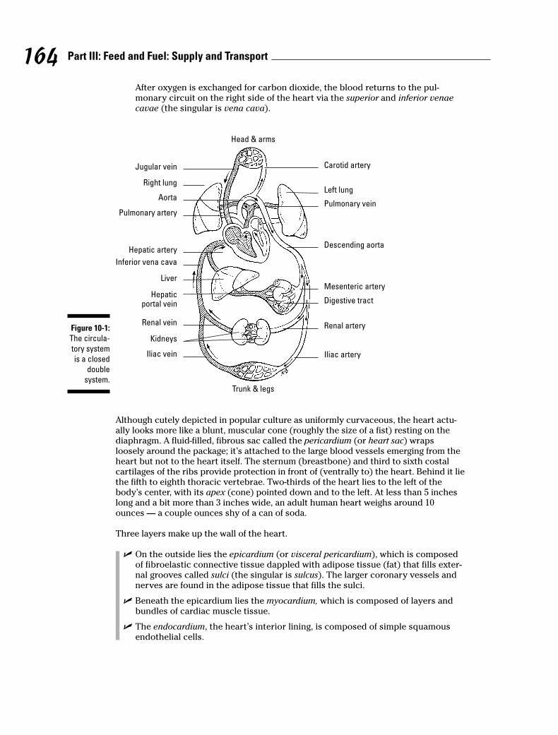
After oxygen is exchanged for carbon dioxide, the blood returns to the pul-monary circuit on the right side of the heart via the superior and inferior venaecavae (the singular is vena cava).
Although cutely depicted in popular culture as uniformly curvaceous, the heart actu-ally looks more like a blunt, muscular cone (roughly the size of a fist) resting on thediaphragm. A fluid-filled, fibrous sac called the pericardium (or heart sac) wrapsloosely around the package; it’s attached to the large blood vessels emerging from theheart but not to the heart itself. The sternum (breastbone) and third to sixth costalcartilages of the ribs provide protection in front of (ventrally to) the heart. Behind it liethe fifth to eighth thoracic vertebrae. Two-thirds of the heart lies to the left of thebody’s center, with its apex (cone) pointed down and to the left. At less than 5 incheslong and a bit more than 3 inches wide, an adult human heart weighs around 10ounces — a couple ounces shy of a can of soda.
Three layers make up the wall of the heart.
� On the outside lies the epicardium (or visceral pericardium), which is composedof fibroelastic connective tissue dappled with adipose tissue (fat) that fills exter-nal grooves called sulci (the singular is sulcus). The larger coronary vessels andnerves are found in the adipose tissue that fills the sulci.
� Beneath the epicardium lies the myocardium, which is composed of layers andbundles of cardiac muscle tissue.
� The endocardium, the heart’s interior lining, is composed of simple squamousendothelial cells.
Head & arms
Jugular vein
Right lungLeft lung
Inferior vena cava
Pulmonary artery
Hepatic artery
Aorta
Carotid artery
Liver
Kidneys
Mesenteric artery
Renal vein
Iliac vein Iliac artery
Trunk & legs
Digestive tract
Descending aorta
Renal artery
Hepaticportal vein
Pulmonary vein
Figure 10-1:
The circula-
tory system
is a closed
double
system.
164 Part III: Feed and Fuel: Supply and Transport
Too much to remember? To keep the layers straight, turn to the Greek roots. Epi– isthe Greek term for “upon” or “on” whereas endo– comes from the Greek endon mean-ing “within.” The medical definition of myo– is “muscle.” And peri– comes from theGreek term for “around” or “surround.” Hence the epicardium is on the heart, theendocardium is inside the heart, the myocardium is the muscle between the two,and the pericardium surrounds the whole package. By the way, the root cardi– comesfrom the Greek word for heart, kardia.
The pericardium is made up of two parts — a tough inelastic sac called the fibrous peri-cardium on the outside and a serous (or lubricated) membrane nearer the heart calledthe parietal pericardium. Between the serous layers of the epicardium and the parietalpericardium is the small pericardial space and its tiny amount of lubricating pericardialfluid. This watery substance prevents irritation during systole (contraction of theheart) and diastole (relaxation of the heart).
Give these practice questions a try to see if you have the rhythm of all this:
1.–5. Match the description to its anatomical term.
1. _____ The system for gaseous exchange in the lungs
2. _____ The system for maintaining a constant internal environment in other tissues
3. _____ The membranous sac that surrounds the heart
4. _____ The wall that divides the heart into two cavities
5. _____ Uppermost two chambers of the heart
6. The heart contracts at an average rate of
a. 110 times/minute
b. 72 times/minute
c. 12 times/minute
d. 42 times/minute
e. 24 times/minute
7. A closed system of circulation involves
a. Non-confinement of blood, general dispersal, minimal regulation
b. Confinement of blood, general dispersal, critical regulation
c. Non-confinement of blood, specific targeting, critical regulation
d. Confinement of blood, specific targeting, critical regulation
e. Confinement of blood, specific targeting, minimal regulation
8. The inner layer of the heart’s wall is called the
a. Pericardium
b. Epicardium
c. Endocardium
d. Endothelium
165Chapter 10: Spreading the Love: The Circulatory System
a. Pericardium
b. Pulmonary circuit
c. Systemic circuit
d. Atria
e. Septum
9.–13. Match the description to its anatomical term.
9. _____ A membranous, serous layer attached to a fibrous sac
10. _____ A tissue composed of layers and bundles of cardiac muscles
11. _____ Outside layer of the heart wall that’s interspersed with adipose
12. _____ The interior lining of the heart
13. _____ External grooves that indicate the regions of the heart
Finding the Key to the Heart’s ChambersThe heart’s lower two chambers, the ventricles, are quite a bit larger than the two atriaup top. Yet the proper anatomical terms for their positions refer to the atria as being“superior” (above) and the ventricles “inferior” (below). In this case, size isn’t theissue at all.
The atriaSometimes referred to as “receiving chambers” because they receive blood returningto the heart through the veins, each atrium has two parts: a principal cavity with asmooth interior surface and an auricle, a smaller, dog-ear-shaped pouch with muscularridges inside called pectinate muscles, or musculi pectinati, that resemble the teeth of acomb.
The right atrium appears slightly larger than the left and has somewhat thinner wallsthan the left. Its principal cavity, the sinus venarum cavarum, is between the two venacavae and the atrioventricular (between an atrium and a ventricle) openings. The pointwhere the right atrium’s auricle joins with its principal cavity is marked externally bythe sulcus terminalis and internally by the crista terminalis. Openings into the rightatrium include the following:
� The superior vena cava, which has no valve and returns blood from the head,thorax, and upper extremities and directs it toward the atrioventricular opening
� The inferior vena cava, which returns blood from the trunk and lower extremitiesand directs it toward the fossa ovalis in the interatrial septum, which also has novalve
� The coronary sinus, which opens between the inferior vena cava and the atrio-ventricular opening, returns blood from the heart, and is covered by the ineffec-tive Thebesian valve
� An atrioventricular opening covered by the tricuspid valve
166 Part III: Feed and Fuel: Supply and Transport
a. Visceral pericardium
b. Sulci
c. Endocardium
d. Myocardium
e. Parietal pericardium
� The fossa ovalis is an oval depression in the interatrial septum that correspondsto the foramen ovale of the fetal heart. If the foramen ovale does not close atbirth, it causes a condition known as “blue baby.”
The left atrium’s principal cavity contains openings for the four pulmonary veins, twofrom each lung, which have no valves. Frequently, the two left veins share a commonopening. The left auricle, a dog-ear-shaped blind pouch, is longer, narrower, and morecurved than the right, marked interiorly by the pectinate muscles, and the left atrium’satrioventricular (or AV) opening is smaller than on the right and is protected by themitral, or bicuspid, valve.
The ventriclesThe heart’s ventricles are sometimes called the pumping chambers because it’s theirjob to receive blood from the atria and pump it back to the lungs and out into thebody’s network of arteries. More force is needed to move the blood great distances,so the myocardium of the ventricles is thicker than that of either atrium, and themyocardium of the left ventricle is thicker than that of the right.
The right ventricle only has to move blood to the lungs, so its myocardium is only one-third as thick as that of its neighbor to the left. Roughly triangular in shape, the rightventricle occupies much of the sternocostal (front) surface of the heart and forms theconus arteriosus where it joins the pulmonary artery, or trunk. The right ventricleextends downward toward where the heart rests against the diaphragm. A circularopening into the pulmonary trunk is covered by the pulmonary semilunar valve, so-called because of its three crescent-shaped cusps. When the ventricle relaxes, theblood from the pulmonary artery tends to flow back toward the ventricle, filling thepockets of the cusps and causing the valve to close. The oval AV opening is sur-rounded by a strong fibrous ring and covered by the tricuspid valve, named after itsthree unequally sized cusps. The atrial surface of the tricuspid valve is smooth, butthe side toward the ventricle is irregular, forming a ragged edge where the chordaetendineae attach. These fibrous cords, which are attached to nipple-shaped projec-tions called papillary muscles in the ventricle’s wall, prevent blood from flowing backinto the atrium. Cardiac muscle in the ventricle’s wall is in an irregular pattern of bun-dles and bands called the trabeculae carneae.
Longer and more conical in shape, the left ventricle’s tip forms the apex of the heart.Its walls are three times thicker than those of the right ventricle. Its AV opening issmaller than that in the right ventricle and is covered by the bicuspid, or mitral, valvethat’s comprised of two unequal cusps. This ventricle’s chordae tendineae are fewer,thicker, and stronger, and they’re attached by only two larger papillary muscles, oneon the front (anterior) wall and one on the back (posterior). More ridges are packedmore densely in the muscular trabeculae carneae. Its opening to the aorta is protectedby the aortic semilunar valve, composed of three half-moon cusps that are larger,thicker, and stronger than the pulmonary valve’s cusps. Between these cusps and theaortic wall are dilated pockets called aortic sinuses, which are openings for the coro-nary arteries.
Pump up your practice time with these questions related to the chambers of the heart:
167Chapter 10: Spreading the Love: The Circulatory System
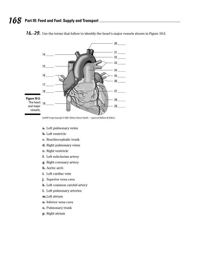
14.–29. Use the terms that follow to identify the heart’s major vessels shown in Figure 10-2.
LifeART Image Copyright © 2007. Wolters Kluwer Health — Lippincott Williams & Wilkins
a. Left pulmonary veins
b. Left ventricle
c. Brachiocephalic trunk
d. Right pulmonary veins
e. Right ventricle
f. Left subclavian artery
g. Right coronary artery
h. Aortic arch
i. Left cardiac vein
j. Superior vena cava
k. Left common carotid artery
l. Left pulmonary arteries
m.Left atrium
n. Inferior vena cava
o. Pulmonary trunk
p. Right atrium
20 _____
21 _____
22 _____
23 _____
24 _____
26 _____
25 _____
27 _____
28 _____
29 _____
14 _____
15 _____
16 _____
17 _____
18 _____
19 _____
Figure 10-2:
The heart
and major
vessels.
168 Part III: Feed and Fuel: Supply and Transport
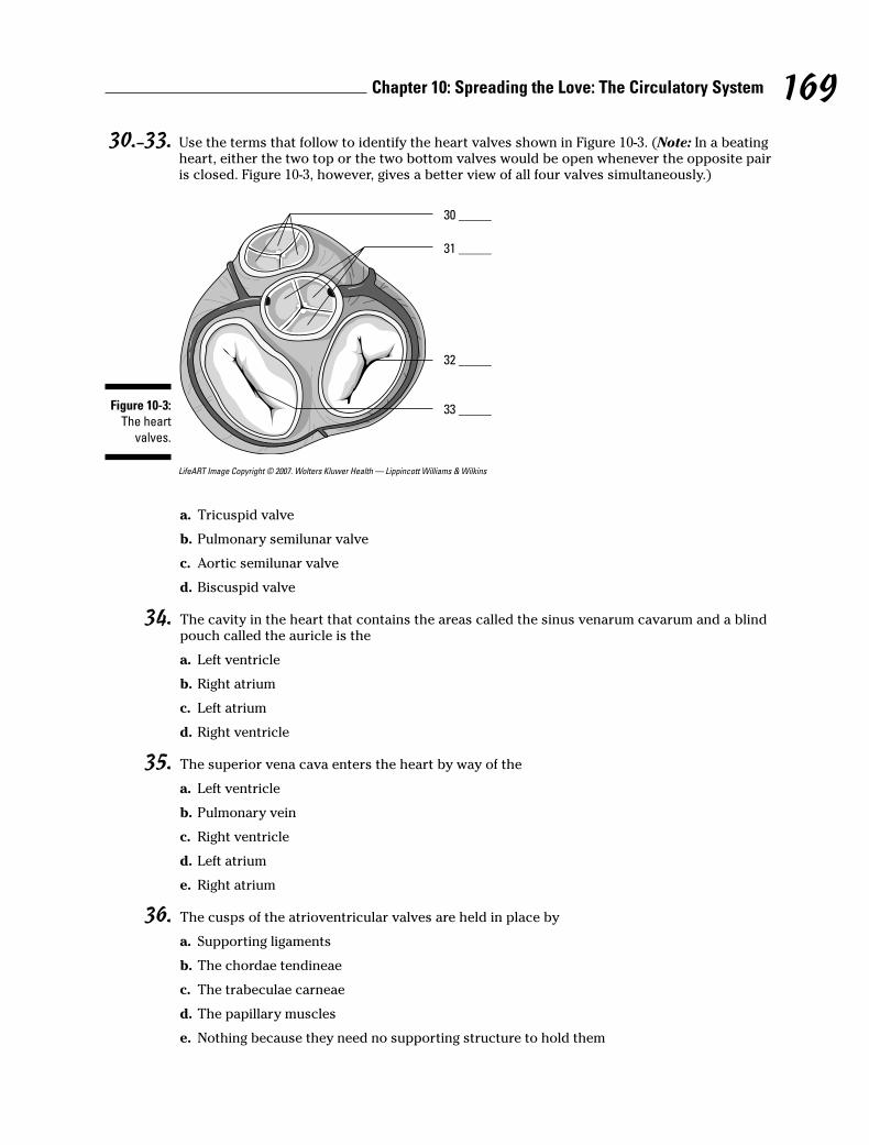
30.–33. Use the terms that follow to identify the heart valves shown in Figure 10-3. (Note: In a beatingheart, either the two top or the two bottom valves would be open whenever the opposite pairis closed. Figure 10-3, however, gives a better view of all four valves simultaneously.)
LifeART Image Copyright © 2007. Wolters Kluwer Health — Lippincott Williams & Wilkins
a. Tricuspid valve
b. Pulmonary semilunar valve
c. Aortic semilunar valve
d. Biscuspid valve
34. The cavity in the heart that contains the areas called the sinus venarum cavarum and a blindpouch called the auricle is the
a. Left ventricle
b. Right atrium
c. Left atrium
d. Right ventricle
35. The superior vena cava enters the heart by way of the
a. Left ventricle
b. Pulmonary vein
c. Right ventricle
d. Left atrium
e. Right atrium
36. The cusps of the atrioventricular valves are held in place by
a. Supporting ligaments
b. The chordae tendineae
c. The trabeculae carneae
d. The papillary muscles
e. Nothing because they need no supporting structure to hold them
30 _____
31 _____
32 _____
33 _____Figure 10-3:
The heart
valves.
169Chapter 10: Spreading the Love: The Circulatory System
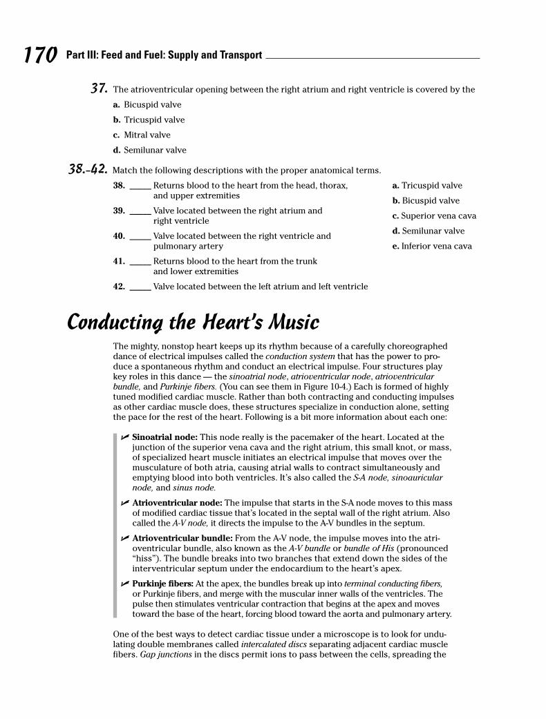
37. The atrioventricular opening between the right atrium and right ventricle is covered by the
a. Bicuspid valve
b. Tricuspid valve
c. Mitral valve
d. Semilunar valve
38.–42. Match the following descriptions with the proper anatomical terms.
38. _____ Returns blood to the heart from the head, thorax, and upper extremities
39. _____ Valve located between the right atrium and right ventricle
40. _____ Valve located between the right ventricle and pulmonary artery
41. _____ Returns blood to the heart from the trunk and lower extremities
42. _____ Valve located between the left atrium and left ventricle
Conducting the Heart’s MusicThe mighty, nonstop heart keeps up its rhythm because of a carefully choreographeddance of electrical impulses called the conduction system that has the power to pro-duce a spontaneous rhythm and conduct an electrical impulse. Four structures playkey roles in this dance — the sinoatrial node, atrioventricular node, atrioventricularbundle, and Purkinje fibers. (You can see them in Figure 10-4.) Each is formed of highlytuned modified cardiac muscle. Rather than both contracting and conducting impulsesas other cardiac muscle does, these structures specialize in conduction alone, settingthe pace for the rest of the heart. Following is a bit more information about each one:
� Sinoatrial node: This node really is the pacemaker of the heart. Located at thejunction of the superior vena cava and the right atrium, this small knot, or mass,of specialized heart muscle initiates an electrical impulse that moves over themusculature of both atria, causing atrial walls to contract simultaneously andemptying blood into both ventricles. It’s also called the S-A node, sinoauricularnode, and sinus node.
� Atrioventricular node: The impulse that starts in the S-A node moves to this massof modified cardiac tissue that’s located in the septal wall of the right atrium. Alsocalled the A-V node, it directs the impulse to the A-V bundles in the septum.
� Atrioventricular bundle: From the A-V node, the impulse moves into the atri-oventricular bundle, also known as the A-V bundle or bundle of His (pronounced“hiss”). The bundle breaks into two branches that extend down the sides of theinterventricular septum under the endocardium to the heart’s apex.
� Purkinje fibers: At the apex, the bundles break up into terminal conducting fibers,or Purkinje fibers, and merge with the muscular inner walls of the ventricles. Thepulse then stimulates ventricular contraction that begins at the apex and movestoward the base of the heart, forcing blood toward the aorta and pulmonary artery.
One of the best ways to detect cardiac tissue under a microscope is to look for undu-lating double membranes called intercalated discs separating adjacent cardiac musclefibers. Gap junctions in the discs permit ions to pass between the cells, spreading the
170 Part III: Feed and Fuel: Supply and Transport
a. Tricuspid valve
b. Bicuspid valve
c. Superior vena cava
d. Semilunar valve
e. Inferior vena cava
171Chapter 10: Spreading the Love: The Circulatory System
action potential of the electrical impulse and synchronizing cardiac muscle contrac-tions. Potential problems include fibrillation, a breakdown in rhythm or propagation ofthe impulses that causes individual fibers to act independently, and heart block, aninterruption that causes the atria and ventricles to take on their own rates of contrac-tion. Usually the atria contract faster than the ventricles.
LifeART Image Copyright © 2007. Wolters Kluwer Health — Lippincott Williams & Wilkins
A healthy heart makes a “lub-dub” sound as it beats. The first sound (the “lub”) isheard most clearly near the apex of the heart and comes at the beginning of ventricu-lar systole (the closing of the atrioventricular valves and opening of the semilunarvalves). It’s lower in pitch and longer in duration than the second sound (the “dub”),heard most clearly over the second rib, which results from the semilunar valves clos-ing during ventricular diastole. Defects in the valves can cause turbulence or regurgita-tion of blood that can be heard through a stethoscope. Called murmurs, these soundsindicate imperfect closure of one or more valves.
Have you got the beat? Try the following practice questions that deal with the heart’srhythm:
Sinoatrial node(pacemaker)
Atrioventricularnode
Right atrium
Left atrium
Purkinje fibers
Purkinjefibers
Atrioventricularbundle
Right and leftbundle branches
Interventricularseptum
Figure 10-4:
The
conductive
system of
the heart.
Q. Cardiac tissue is distinctive microscopically because of thepresence of
a. Hemoglobin
b. Intercalated discs
c. Fibrin
d. Ganglia
e. Nuclei
A. The correct answer is intercalateddiscs.

43. The pacemaker of the heart is also known as the
a. Sinoatrial node (S-A node)
b. Extrinsic nerve control center
c. Bundle of His
d. Atrioventricular node (A-V node)
e. Purkinje control fibers
44. Stimulation of myocardial contractions in the ventricles radiates from the
a. Bundle of His
b. Atrioventricular bundles
c. Sinoatrial node (S-A node)
d. Purkinje fibers
e. Atrioventricular node (A-V node)
45. Choose the correct conductive pattern.
a. S-A node → Bundle of His → A-V node → Purkinje fibers
b. A-V node → S-A node → Purkinje fibers → Bundle of His
c. S-A node → A-V node → Bundle of His → Purkinje fibers
d. S-A node → Purkinje fibers → Bundle of His → A-V node
Riding the Network of Blood VesselsBlood vessels come in three varieties, which you can see illustrated in Figure 10-5:
� Arteries carry blood away from the heart. The largest artery is the aorta. Smallones are called arterioles, and microscopically small ones are called metarterioles.
� Veins carry blood toward the heart; all veins except the pulmonary veins containdeoxygenated blood. Small ones are called venules, and large venous spaces arecalled sinuses.
� Microscopically small capillaries carry blood from arterioles to venules, butsometimes tiny spaces in the liver and elsewhere called sinusoids replace capillaries.
The walls of arteries and veins have three layers: the outermost tunica externa (some-times called tunica adventitia) composed of white fibrous connective tissue, a central“active” layer called the tunica media composed of smooth muscle fibers and yellowelastic fibers, and an inner layer called the tunica intima made up of endothelium thataids in preventing blood coagulation by reducing the resistance of blood flow. Arterialwalls are very strong, thick, and very elastic to withstand the great pressure to whichthe arteries are subjected. Arteries have no valves.
There are two types of arteries: elastic and muscular. In elastic arteries, found prima-rily near the heart, the tunica media is composed of yellow elastic fibers that stretch
172 Part III: Feed and Fuel: Supply and Transport
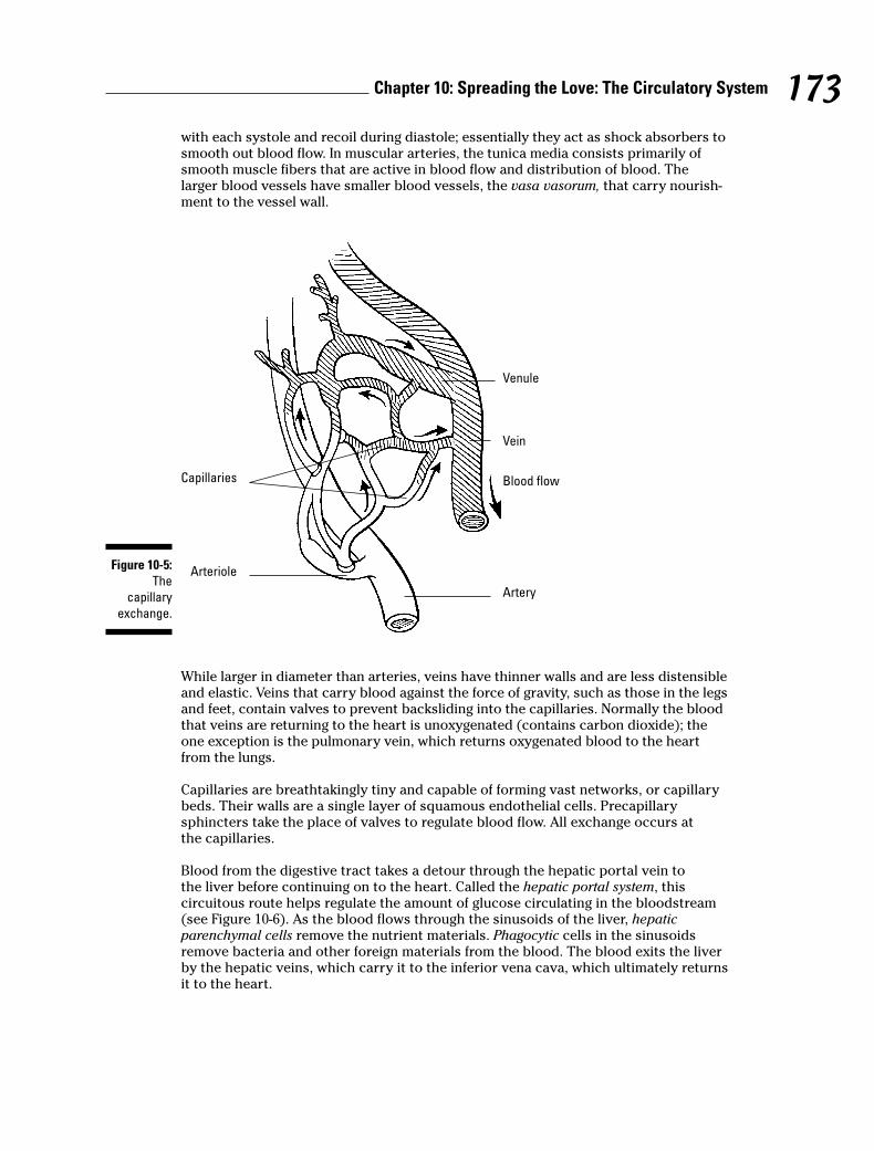
with each systole and recoil during diastole; essentially they act as shock absorbers tosmooth out blood flow. In muscular arteries, the tunica media consists primarily ofsmooth muscle fibers that are active in blood flow and distribution of blood. Thelarger blood vessels have smaller blood vessels, the vasa vasorum, that carry nourish-ment to the vessel wall.
While larger in diameter than arteries, veins have thinner walls and are less distensibleand elastic. Veins that carry blood against the force of gravity, such as those in the legsand feet, contain valves to prevent backsliding into the capillaries. Normally the bloodthat veins are returning to the heart is unoxygenated (contains carbon dioxide); theone exception is the pulmonary vein, which returns oxygenated blood to the heartfrom the lungs.
Capillaries are breathtakingly tiny and capable of forming vast networks, or capillarybeds. Their walls are a single layer of squamous endothelial cells. Precapillary sphincters take the place of valves to regulate blood flow. All exchange occurs at the capillaries.
Blood from the digestive tract takes a detour through the hepatic portal vein tothe liver before continuing on to the heart. Called the hepatic portal system, this circuitous route helps regulate the amount of glucose circulating in the bloodstream(see Figure 10-6). As the blood flows through the sinusoids of the liver, hepaticparenchymal cells remove the nutrient materials. Phagocytic cells in the sinusoidsremove bacteria and other foreign materials from the blood. The blood exits the liverby the hepatic veins, which carry it to the inferior vena cava, which ultimately returnsit to the heart.
Venule
Capillaries
Artery
Vein
Blood flow
ArterioleFigure 10-5:
The
capillary
exchange.
173Chapter 10: Spreading the Love: The Circulatory System

LifeART Image Copyright © 2007. Wolters Kluwer Health — Lippincott Williams & Wilkins
Beating from the Start: Fetal CirculationBecause nutrients and oxygen come from the mother’s bloodstream, fetal circulationrequires extra vessels to get the job done. Two umbilical arteries — the umbilical veinand the ductus venosus — fill the bill. Fetal blood leaves the placenta through theumbilical vein, which branches at the liver to become the ductus venosus before enter-ing the inferior vena cava that carries blood to the right atrium and then through ahole in the septum called the foramen ovale into the left atrium. From there it flowsinto the left ventricle and is pumped through the aorta to the head, neck, and upperextremities. It returns to the heart through the superior vena cava, to the right atrium,to the right ventricle, to the pulmonary trunk (lungs inactive), goes through the ductusarteriosus into the aorta, to the abdominal and pelvic viscera and lower extremities,and to the placenta through the umbilical artery. After birth, these circulation path-ways quickly shut down, eventually leaving a depression in the septum, the fossaovale, where the hole of the foramen ovale once was.
Now is your chance to practice circulating through the circulatory system:
46. In fetal hepatic portal circulation, blood flows directly into the systemic circulation through the
a. Conus arteriosus
b. Hepatic vein
c. Ductus venosus
d. Ductus arteriosus
e. Truncus arteriosus
Inferior vena cava
Stomach
Gastric vein
Left gastroepiploic vein
Spleen
Tail of pancreas
Right gastroepiploic vein
Descending colon
Inferior mesentric vein
Small intestine
Hepatic veins
Liver
Cystic vein
Hepatic portal vein
Duodenum
Head of pancreas
Ascending colon
Superior mesentric vein
Appendix
Figure 10-6:
The veins of
the hepatic
portal
system.
174 Part III: Feed and Fuel: Supply and Transport
47. Follow a drop of blood through the heart, starting in the superior vena cava. Number the fol-lowing structures in sequential order.
_____ Pulmonary vein
_____ Right ventricle
_____ Lung
_____ Right atrium
_____ Pulmonary artery
48. Number the structures in the correct sequence of blood flow from the heart to the radialartery for pulse. Start at the heart with the aortic semilunar valve.
_____ Axillary artery
_____ Subclavian artery
_____ Ascending aorta
_____ Brachial artery
_____ Aortic arch
49. Number the structures in the correct sequence of blood flow from the forearm to the heart.
_____ Basilic vein
_____ Subclavian vein
_____ Superior vena cava
_____ Brachial vein
_____ Axillary vein
50. Number the structures in the correct sequence of blood flow from the great saphenous veinback to the heart.
_____ External iliac vein
_____ Right atrium
_____ Common iliac vein
_____ Femoral vein
_____ Inferior vena cava
51. Follow a drop of blood from the right atrium to the radial artery (for pulse). Number thestructures in sequential order.
__1__ Right atrium
_____ Pulmonary artery
_____ Left atrium
_____ Bicuspid valve
_____ Ascending aorta
_____ Axillary artery
_____ Radial artery
_____ Right ventricle
175Chapter 10: Spreading the Love: The Circulatory System
_____ Pulmonary vein
_____ Aortic semilunar valve
_____ Brachial artery
_____ Tricuspid valve
_____ Lung capillary
_____ Aortic arch
_____ Pulmonary semilunar valve
_____ Left ventricle
_____ Subclavian artery
52. Follow a drop of blood from the stomach to the inferior vena cava. (Remember the portalsystem?) Number the structures in sequential order.
__1__ Superior mesenteric vein
_____ Sinusoids of liver
_____ Hepatic vein
_____ Hepatic portal vein
_____ Inferior vena cava
53. Follow a drop of blood from the aortic semilunar valve of the heart to the forearm and back tothe heart. Number the structures in sequential order.
__1__ Ascending aorta
_____ Basilic vein
_____ Axillary artery
_____ Subclavian vein
_____ Brachial vein
_____ Radial artery
_____ Right atrium
_____ Brachial artery
_____ Axillary vein
_____ Capillaries in the hand
_____ Superior vena cava
_____ Subclavian artery
_____ Aortic arch
54. Follow a drop of blood from the saphenous vein back to the heart. Number the structuresin sequential order.
__1__ Saphenous vein
_____ External iliac vein
_____ Inferior vena cava
_____ Right atrium
_____ Femoral vein
_____ Common iliac vein
_____ Right ventricle
176 Part III: Feed and Fuel: Supply and Transport
55. Follow a drop of blood from the anterior tibial vein to the lungs. Number the structuresin sequential order.
__1__ Anterior tibial vein
_____ External iliac vein
_____ Inferior vena cava
_____ Right ventricle
_____ Popliteal vein
_____ Common iliac vein
_____ Pulmonary artery
_____ Femoral vein
_____ Right atrium
_____ Lung capillaries
177Chapter 10: Spreading the Love: The Circulatory System
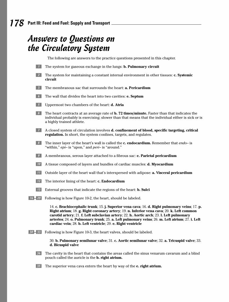
Answers to Questions on the Circulatory System
The following are answers to the practice questions presented in this chapter.
a The system for gaseous exchange in the lungs: b. Pulmonary circuit
b The system for maintaining a constant internal environment in other tissues: c. Systemic circuit
c The membranous sac that surrounds the heart: a. Pericardium
d The wall that divides the heart into two cavities: e. Septum
e Uppermost two chambers of the heart: d. Atria
f The heart contracts at an average rate of b. 72 times/minute. Faster than that indicates theindividual probably is exercising; slower than that means that the individual either is sick or isa highly trained athlete.
g A closed system of circulation involves d. confinement of blood, specific targeting, criticalregulation. In short, the system confines, targets, and regulates.
h The inner layer of the heart’s wall is called the c. endocardium. Remember that endo– is“within,” epi– is “upon,” and peri– is “around.”
i A membranous, serous layer attached to a fibrous sac: e. Parietal pericardium
j A tissue composed of layers and bundles of cardiac muscles: d. Myocardium
k Outside layer of the heart wall that’s interspersed with adipose: a. Visceral pericardium
l The interior lining of the heart: c. Endocardium
m External grooves that indicate the regions of the heart: b. Sulci
n–D Following is how Figure 10-2, the heart, should be labeled.
14. c. Brachiocephalic trunk; 15. j. Superior vena cava; 16. d. Right pulmonary veins; 17. p.Right atrium; 18. g. Right coronary artery; 19. n. Inferior vena cava; 20. k. Left commoncarotid artery; 21. f. Left subclavian artery; 22. h. Aortic arch; 23. l. Left pulmonaryarteries; 24. o. Pulmonary trunk; 25. a. Left pulmonary veins; 26. m. Left atrium; 27. i. Leftcardiac vein; 28. b. Left ventricle; 29. e. Right ventricle
E–H Following is how Figure 10-3, the heart valves, should be labeled.
30. b. Pulmonary semilunar valve; 31. c. Aortic semilunar valve; 32. a. Tricuspid valve; 33.d. Bicuspid valve
I The cavity in the heart that contains the areas called the sinus venarum cavarum and a blindpouch called the auricle is the b. right atrium.
J The superior vena cava enters the heart by way of the e. right atrium.
178 Part III: Feed and Fuel: Supply and Transport
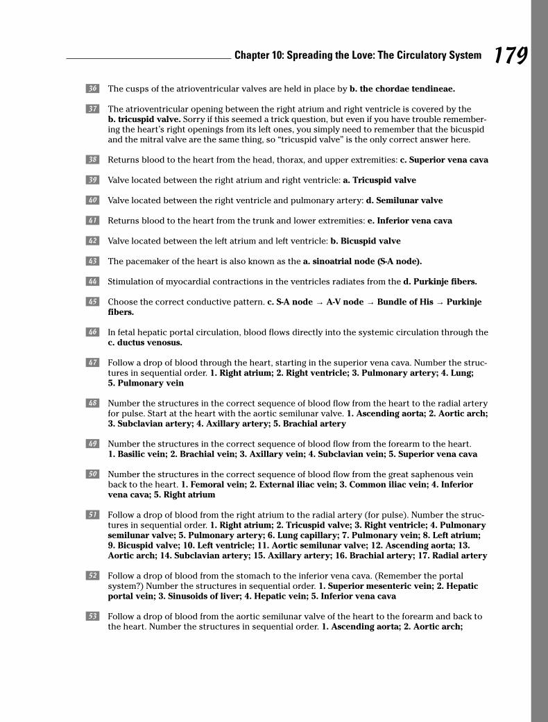
K The cusps of the atrioventricular valves are held in place by b. the chordae tendineae.
L The atrioventricular opening between the right atrium and right ventricle is covered by theb. tricuspid valve. Sorry if this seemed a trick question, but even if you have trouble remember-ing the heart’s right openings from its left ones, you simply need to remember that the bicuspidand the mitral valve are the same thing, so “tricuspid valve” is the only correct answer here.
M Returns blood to the heart from the head, thorax, and upper extremities: c. Superior vena cava
N Valve located between the right atrium and right ventricle: a. Tricuspid valve
O Valve located between the right ventricle and pulmonary artery: d. Semilunar valve
P Returns blood to the heart from the trunk and lower extremities: e. Inferior vena cava
Q Valve located between the left atrium and left ventricle: b. Bicuspid valve
R The pacemaker of the heart is also known as the a. sinoatrial node (S-A node).
S Stimulation of myocardial contractions in the ventricles radiates from the d. Purkinje fibers.
T Choose the correct conductive pattern. c. S-A node → A-V node → Bundle of His → Purkinjefibers.
U In fetal hepatic portal circulation, blood flows directly into the systemic circulation through thec. ductus venosus.
V Follow a drop of blood through the heart, starting in the superior vena cava. Number the struc-tures in sequential order. 1. Right atrium; 2. Right ventricle; 3. Pulmonary artery; 4. Lung;5. Pulmonary vein
W Number the structures in the correct sequence of blood flow from the heart to the radial arteryfor pulse. Start at the heart with the aortic semilunar valve. 1. Ascending aorta; 2. Aortic arch;3. Subclavian artery; 4. Axillary artery; 5. Brachial artery
X Number the structures in the correct sequence of blood flow from the forearm to the heart.1. Basilic vein; 2. Brachial vein; 3. Axillary vein; 4. Subclavian vein; 5. Superior vena cava
Y Number the structures in the correct sequence of blood flow from the great saphenous veinback to the heart. 1. Femoral vein; 2. External iliac vein; 3. Common iliac vein; 4. Inferiorvena cava; 5. Right atrium
z Follow a drop of blood from the right atrium to the radial artery (for pulse). Number the struc-tures in sequential order. 1. Right atrium; 2. Tricuspid valve; 3. Right ventricle; 4. Pulmonarysemilunar valve; 5. Pulmonary artery; 6. Lung capillary; 7. Pulmonary vein; 8. Left atrium;9. Bicuspid valve; 10. Left ventricle; 11. Aortic semilunar valve; 12. Ascending aorta; 13.Aortic arch; 14. Subclavian artery; 15. Axillary artery; 16. Brachial artery; 17. Radial artery
Z Follow a drop of blood from the stomach to the inferior vena cava. (Remember the portalsystem?) Number the structures in sequential order. 1. Superior mesenteric vein; 2. Hepaticportal vein; 3. Sinusoids of liver; 4. Hepatic vein; 5. Inferior vena cava
1 Follow a drop of blood from the aortic semilunar valve of the heart to the forearm and back tothe heart. Number the structures in sequential order. 1. Ascending aorta; 2. Aortic arch;
179Chapter 10: Spreading the Love: The Circulatory System
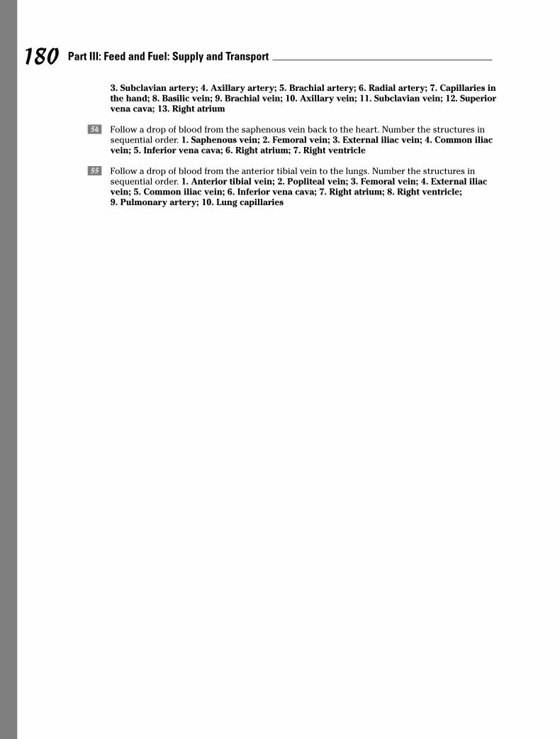
3. Subclavian artery; 4. Axillary artery; 5. Brachial artery; 6. Radial artery; 7. Capillaries inthe hand; 8. Basilic vein; 9. Brachial vein; 10. Axillary vein; 11. Subclavian vein; 12. Superiorvena cava; 13. Right atrium
2 Follow a drop of blood from the saphenous vein back to the heart. Number the structures insequential order. 1. Saphenous vein; 2. Femoral vein; 3. External iliac vein; 4. Common iliacvein; 5. Inferior vena cava; 6. Right atrium; 7. Right ventricle
3 Follow a drop of blood from the anterior tibial vein to the lungs. Number the structures insequential order. 1. Anterior tibial vein; 2. Popliteal vein; 3. Femoral vein; 4. External iliacvein; 5. Common iliac vein; 6. Inferior vena cava; 7. Right atrium; 8. Right ventricle; 9. Pulmonary artery; 10. Lung capillaries
180 Part III: Feed and Fuel: Supply and Transport
Chapter 11
Keeping Up Your Defenses: The Lymphatic System
In This Chapter� Delving into lymphatic ducts
� Noodling around with nodes
� Exploring the lymphatic organs
You see it every rainy day — water, water everywhere, rushing along gutters and downstorm drains into a complex underground system that most would rather not give a
second thought. Well, it’s time to give hidden drainage systems a second thought: Yourbody has one. You already know that the body is made up mostly of fluid. Interstitial orextracellular fluid moves in and around the body’s tissues and cells constantly. It leaks outof blood capillaries at the rate of nearly 51 pints a day, carrying various substances to andaway from the smallest nooks and crannies. Most of that fluid gets reabsorbed into bloodcapillaries. But the one or two liters of extra fluid that remain around the tissues become asubstance called lymph that needs to be managed to maintain fluid balance in the internalenvironment. That’s where the lymphatic system steps in, forming an alternative route forthe return of tissue fluid to the bloodstream.
But the lymphatic system is more than a drainage network. It’s a body-wide filter that trapsand destroys invading microorganisms as part of the body’s immune response network. Itcan remove impurities from the body, help absorb and digest excess fats, and maintain astable blood volume despite varying environmental stresses. Without it, the cardiovascularsystem would grind to a halt.
We bet that you won’t take your little lymph nodes for granted anymore after you’re donewith this chapter.
Duct, Duct, LymphThe story of the lymphatic system (shown in Figure 11-1) begins deep within the body’s tis-sues at the farthest reaches of blood capillaries, where nutrients, plasma, and plasma pro-teins move out into cells, while waste products like carbon dioxide and the fluid carryingthose molecules move back in through a process known as diffusion. Roughly 10 percent ofthe fluid that leaves the capillaries remains deep within the tissues as part of the interstitial(meaning “between the tissues”) fluid. But in order for the body to maintain sufficientvolume of water within in the circulatory system, eventually this plasma and its protein mustget back into the blood. So the lymphatic vessels act as a recycling system to gather, trans-port, cleanse, and return this fluid to the bloodstream.

To collect the fluid, minute vessels called lymph capillaries are woven throughout the body, with afew caveats and exceptions. There are no lymph capillaries in the central nervous system, teeth,outermost layer of the skin, certain types of cartilage, any other avascular tissue, and bones. Andbecause bone marrow makes lymphocytes, which we explain in the next section, it’s consideredpart of the lymphatic system. Plus, lacteals (lymphatic capillaries found in the villi of the intes-tines) absorb fats to mix with lymph, forming a milky fluid called chyle. (See Chapter 9 for detailson lacteals.) Unlike blood capillaries, lymph capillaries dead-end (terminate) within tissue. Madeup of loosely overlapping endothelial cells anchored by fine filaments, lymph capillaries behaveas if their walls are made of cellular one-way valves. When the pressure outside the capillary is
Figure 11-1:
The
lymphatic
system.
182 Part III: Feed and Fuel: Supply and Transport

greater than it is inside, the filaments anchoring the cells allow them to open, permit-ting interstitial fluid to seep in. Rising differential pressure across the capillary wallseventually forces the cell junctions to close. Once in the capillaries, the trapped fluid isknown as lymph, and it moves into larger, vein-like lymphatic vessels. The lymph movesslowly and without any kind of central pump through a combination of peristalsis, theaction of semilunar valves, and the squeezing influence of surrounding skeletal mus-cles, much like occurs in veins.
In the skin, lymph vessels form networks around veins, but in the trunk of the body andaround internal organs, they form networks around arteries. Lymph vessels have thin-ner walls than veins, are wider, have more valves, and — most important — regularlybulge with bean-shaped sacs called lymph nodes (more on those in the later section“Poking at the Nodes”). Just as small tree branches merge into larger ones and then intothe trunk, lymphatics eventually merge into the nine largest lymphatic vessels calledlymphatic trunks. The biggest of these at nearly 11⁄2 feet in length is the thoracic duct;nearly all the body’s lymph vessels empty into it. Only those vessels in the right half ofthe head, neck, and thorax empty into its smaller mate, the right lymphatic duct. Lymphreturns to the bloodstream when both ducts connect with the subclavian (under thecollarbone) veins.
The thoracic duct, which also sometimes is called the left lymphatic duct, arises froma triangular sac called the chyle cistern (or cisterna chyli) into which one intestinaltrunk and two lumbar lymphatic trunks (which drain the lower limbs) flow. Both thethoracic duct and the much smaller right lymphatic duct drain into the subclavian(behind the collarbone) veins. The remaining four trunks are a pair serving the jugularregion (sides of the throat) and a pair serving the bronchomediastinal region (the cen-tral part of the chest).
To see how much of this information is seeping in, answer the following questions:
1. The lymphatic system plays an important role in regulating
a. Intracellular energy function
b. Interstitial fluid protein
c. Metabolizing unused fats from the small intestine
d. Intercellular transportation of oxygen
e. None of these
2. Terminated vessels that return plasma proteins to the blood are
a. Blood capillaries
b. Venules
c. Lymph capillaries
d. Arterioles
e. Cardiac ducts
3. The thoracic duct does not drain lymph from the
a. Right lower extremity
b. Right side of the head
c. Digestive tract
d. Left axilla
e. Posterior abdominal wall
183Chapter 11: Keeping Up Your Defenses: The Lymphatic System
4. The largest lymphatic vessel in the body is the
a. Right lymphatic duct
b. Spleen
c. Thoracic duct
d. Chyle cistern
5. The lymphatic system does not function to
a. Return interstitial fluid to the blood
b. Destroy bacteria
c. Remove old erythrocytes
d. Produce erythrocytes
e. Produce lymphocytes
Poking at the NodesLymph nodes (see Figure 11-2) are the site of filtration of the lymphatic system. Also sometimes incorrectly referred to as lymph glands — they don’t secrete anything,so technically they’re not glands — these kidney-shaped sacs are surrounded by connective tissue (and therefore are tough to spot). Lymph nodes containmacrophages, which destroy bacteria, cancer cells, and other matter in the lymphfluid. Lymphocytes, which produce an immune response to microorganisms, also arefound in lymph nodes. Like the kidneys, the indented part of each node is referred toas the hilus. The stroma (body) of each node is surrounded by a fibrous capsule thatdips into the node to form trabeculae, or septa (thin dividing walls) that divide thenode into compartments. Reticular (net-like) fibers are attached to the trabeculae andform a framework for the lymphoid tissue and lymphocytes (white blood cells) in clus-ters called lymphatic nodules.
Inside the node is a cortex where most of the lymphocytes gather, and at the center is amedulla, which is less dense than the cortex but also contains lymphocytes. The outercortex consists of lymphocytes arranged in masses called lymphatic nodules, whichhave central areas called germinal centers that produce the lymphocytes. Lymph fluidenters the node on its convex side through afferent (inbound) vessels that have valvesopening only toward the node. Lymph circulates through the node, where it’s filteredand then allowed to depart through efferent (outbound) vessels in the hilus withvalves pointing exclusively away from the node. (If you have trouble rememberingyour afferent from your efferent, think of the “a” as standing for “access” and the “e” as standing for “exit”.)
Although some lymph nodes are isolated from others, most nodes occur in groups, orclusters, particularly in the inguinal (groin), axillary (armpit), and mammary glandareas. The following are the primary lymph node regions:
� Head
� Neck
� Upper extremities
� Lower extremities
184 Part III: Feed and Fuel: Supply and Transport
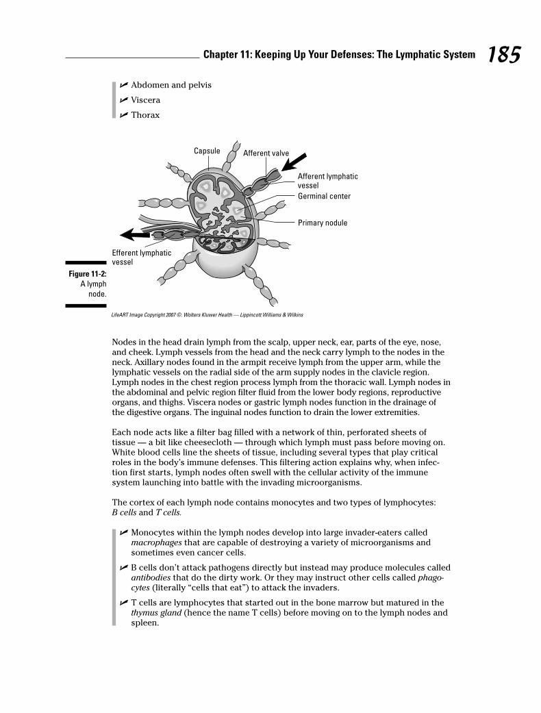
� Abdomen and pelvis
� Viscera
� Thorax
LifeART Image Copyright 2007 ©. Wolters Kluwer Health — Lippincott Williams & Wilkins
Nodes in the head drain lymph from the scalp, upper neck, ear, parts of the eye, nose,and cheek. Lymph vessels from the head and the neck carry lymph to the nodes in theneck. Axillary nodes found in the armpit receive lymph from the upper arm, while thelymphatic vessels on the radial side of the arm supply nodes in the clavicle region.Lymph nodes in the chest region process lymph from the thoracic wall. Lymph nodes inthe abdominal and pelvic region filter fluid from the lower body regions, reproductiveorgans, and thighs. Viscera nodes or gastric lymph nodes function in the drainage ofthe digestive organs. The inguinal nodes function to drain the lower extremities.
Each node acts like a filter bag filled with a network of thin, perforated sheets oftissue — a bit like cheesecloth — through which lymph must pass before moving on.White blood cells line the sheets of tissue, including several types that play criticalroles in the body’s immune defenses. This filtering action explains why, when infec-tion first starts, lymph nodes often swell with the cellular activity of the immunesystem launching into battle with the invading microorganisms.
The cortex of each lymph node contains monocytes and two types of lymphocytes:B cells and T cells.
� Monocytes within the lymph nodes develop into large invader-eaters calledmacrophages that are capable of destroying a variety of microorganisms andsometimes even cancer cells.
� B cells don’t attack pathogens directly but instead may produce molecules calledantibodies that do the dirty work. Or they may instruct other cells called phago-cytes (literally “cells that eat”) to attack the invaders.
� T cells are lymphocytes that started out in the bone marrow but matured in thethymus gland (hence the name T cells) before moving on to the lymph nodes andspleen.
Capsule Afferent valve
Germinal center
Primary nodule
Afferent lymphaticvessel
Efferent lymphaticvessel
Figure 11-2:
A lymph
node.
185Chapter 11: Keeping Up Your Defenses: The Lymphatic System
Think you have a node-tion (sorry!) about what’s happening here? Test your knowledge:
186 Part III: Feed and Fuel: Supply and Transport
6. An encapsulated mass of lymph tissue connected to lymph vessels is a
a. Tonsil
b. Spleen
c. Peyer’s patch
d. Thymus
e. Lymph node
7. The lymph organ that has afferent and efferent lymph vessels is the
a. Lymph node
b. Tonsil
c. Peyer’s patches
d. Spleen
e. Thymus
8. Cells attached to the reticular fibers in a lymph node are
a. Macrophages
b. Monocytes
c. Neutrophils
d. Lymphocytes
e. Basophils
9. A function of the lymph nodes is to
a. Remove dead erythrocytes
b. Produce bilirubin
c. Produce lymphocytes
d. Conserve iron
e. Remove erythrocytes
Q. An area of the body where nolymph nodes are found is the
a. Integument
b. Liver
c. Stomach
d. Central nervous system (brainand spinal cord)
e. Urinary system
A. The correct answer is the centralnervous system (brain and spinalcord).
10. The stroma of the lymph node does not consist of
a. Capsule
b. Hilus
c. Cortex
d. Trabeculae
e. Villi
11. Lymphocytes are produced in the lymph nodules in the region called the
a. Medulla
b. Germinal center
c. Lymphatic reservoir
d. Trabeculae
e. Hilus
12. The connective tissue fiber that forms the framework of the lymphoid tissue is
a. Cartilaginous
b. Collagenous
c. Bone
d. Elastic
e. Reticular
13. When infection first starts, the lymph nodes tend to
a. Recede
b. Swell
c. Multiply
d. Divide
e. Fade
14. T cells get their name because they start out in the bone marrow and mature in the
a. Thigh
b. Thyroid
c. Thymus
d. Tongue
e. Tailbone
Having a Spleen-ded Time with the Lymphatic Organs
While the lymph nodes are the most numerous lymphatic organs, several other vitalorgans exist in the lymphatic system, including the spleen, thymus gland, and tonsils.
187Chapter 11: Keeping Up Your Defenses: The Lymphatic System
The spleenThe spleen, the largest lymphatic organ in the body, is a 5-inch, roughly egg-shapedstructure to the left of and slightly behind the stomach. Like lymph nodes, it has ahilus through which the splenic artery, splenic vein, and efferent (remember “e” for“exit”) vessels pass. Also like lymph nodes, the spleen’s surrounded by a fibrous cap-sule that folds inward to section it off. Arterioles leading into each section are sur-rounded by masses of developing lymphocytes that give those areas of so-called whitepulp their appearance. On the outer edges of each compartment, tissue called red pulpconsists of blood-filled cavities. Unlike lymph nodes, the spleen doesn’t have any affer-ent (access) lymph vessels, which means that it doesn’t filter lymph, only blood.
Blood flows slowly through the spleen to allow it to remove microorganisms, exhaustederythrocytes (red blood cells), and any foreign material that may be in the stream.Among its various functions, the spleen can be a blood reservoir. When blood circula-tion drops while the body is at rest, the spleen’s vessels can dilate to store any excessvolume. Later, during exercise or if oxygen concentrations in the blood begin to drop,the spleen’s blood vessels constrict and push any stored blood back into circulation.
But the spleen’s primary role is as a biological recycling unit, capturing and breakingdown defective and aged blood cells to reuse their components later. Iron stored bythe spleen’s macrophages goes to the bone marrow where it’s turned into hemoglobinin new blood cells. By the same token, bilirubin for the liver is generated during break-down of hemoglobin. The spleen produces red blood cells during embryonic develop-ment but shuts down that process after birth; in cases of severe anemia, the spleensometimes starts up production of red blood cells again.
Fortunately, the spleen isn’t considered a vital organ; if it’s damaged or has to be surgi-cally removed, the liver and bone marrow can pick up where the spleen leaves off.
T cell central: The thymus glandTucked just behind the breastbone and between the lungs in the upper chest, thethymus gland was a medical mystery until recent decades. Its two oblong lobes arelargest at puberty when they weigh around 40 grams (somewhat less than an adultmouse). Through a process called involution, however, the gland atrophies and shrinksto roughly 6 grams by the time an adult is 65. (You can remember that term as theinverse of evolution.)
The thymus gland serves its most critical role — as a nursery for immature T lympho-cytes, or T cells — during fetal development and the first few years of a human’s life.Prior to birth, fetal bone marrow produces lymphoblasts (early stage lymphocytes)that migrate to the thymus. Shortly after birth and continuing until adolescence, thethymus secretes several hormones, collectively called thymosin, that prompt the earlycells to mature into full-grown T cells that are immunocompetent, ready to go forthand conquer invading microorganisms. (These hormones are the reason the thymus isconsidered part of the endocrine system, too.)
As with other lymphatic structures, the thymus is surrounded by a fibrous capsulethat dips inside to create chambers called lobules. Within each lobule is a cortex madeof T cells held in place by reticular fibers and a central medulla of unusually onion-likelayered epithelial cells called thymic corpuscles, or Hassall’s corpuscles, as well as scat-tered lymphocytes.
188 Part III: Feed and Fuel: Supply and Transport
Opening wide and moving along: The tonsils and Peyer’s patchesLike the thymus gland, the tonsils, which are misunderstood masses of lymphoidtissue, are largest around puberty and tend to atrophy as an adult ages. Unlike thethymus, however, the tonsils don’t secrete hormones but do produce lymphocytes andantibodies to protect against microorganisms that are inhaled or eaten. Although onlytwo are visible on either side of the pharynx, there are actually six tonsils: the two youcan identify, which are called palatine tonsils; two more called adenoids or pharyngealtonsils in the wall of the pharynx; and two in the posterior one-third of the tonguecalled lingual tonsils. Invaginations (ridges) in the tonsils form pockets called crypts,which trap bacteria and other foreign matter.
Peyer’s patches, also called aggregate glands or agminate glands, are masses of lymphtissue just below the surface of the ileum, the lowest section of the small intestine.When harmful microorganisms get into the intestine, Peyer’s patches can mobilize anarmy of B cells and macrophages to fight off infection.
You’ve absorbed a lot in this section. See how much of it is getting caught in your filters:
15. Cells that remove foreign matter from the lymph in the lymph nodes are the
a. Endothelial cells
b. Erythrocytes
c. Neutrophils
d. Lymphocytes
e. Macrophages
16. Which of the following is not a lymphatic organ?
a. Tonsil
b. Thymus
c. Liver
d. Spleen
17. The lymphatic organ that stops growth during adolescence and atrophies with aging is the
a. Thymus
b. Lymph nodes
c. Tonsil
d. Adenoids
e. Spleen
18. Lymphoid tissue located in the pharynx that protects against inhaled or ingested pathogensand foreign substances is called the
a. Thymus
b. Tonsils
c. Peyer’s patches
d. Spleen
e. Lymph nodes
189Chapter 11: Keeping Up Your Defenses: The Lymphatic System
19. Lymphatic nodules found in the ileum of the small intestines are
a. Tonsils
b. Lymph nodes
c. Thymus
d. Macrophages
e. Peyer’s patches
20. The spleen is in close relation with the
a. Stomach
b. Liver
c. Colon
d. Kidney
e. Duodenum
21. The lymphatic organ found in the superior mediastinum is the
a. Tonsil
b. Spleen
c. Thymus
d. Reticular formation
e. Germinal center
22. Lymphocytes are the predominant cells in the
a. Bone tissue
b. Cartilage
c. White pulp of the spleen
d. Red pulp of the spleen
e. Elastic connective tissue
23. The lymphatic organ responsible for removal of aged and defective blood cells is the
a. Tonsil
b. Spleen
c. Peyer’s patches
d. Lymph nodes
e. Thymus
24. The lymphatic organ that secretes hormones to make T lymphocytes immunocompetent is the
a. Lymph node
b. Tonsil
c. Peyer’s patch
d. Spleen
e. Thymus
190 Part III: Feed and Fuel: Supply and Transport
25. Which of the following is not a true tonsil?
a. Pharyngeal tonsil
b. Palatine tonsils
c. Adenoids
d. Sublingual tonsil
e. Lingual tonsil
26.–35. Mark the statement with a T if it’s true or an F if it’s false:
26. _____ The lymph system offers an alternative route for the return of the tissue fluid to thebloodstream.
27. _____ The fluid surrounding the cells that will enter the lymph capillaries is called intersti-tial fluid.
28. _____ Lymph from the lymph vessels flows into the right thoracic duct and the left thoracicduct.
29. _____ The lymph capillaries are composed of loosely overlapping reticular fiber.
30. _____ The spleen filters both lymph and blood.
31. _____ The thymus gland is functional in the early years of life and is most active in old age.
32. _____ Tonsils function to protect against pathogens and foreign substances that areinhaled or ingested.
33. _____ The spleen functions in the removal of aged and defective blood cells and plateletsfrom the blood.
34. _____ The gastric lymph nodes drain the lymphatic vessels on the radial side of the arm.
35. _____ The thoracic duct originates from a triangular sac called the chyle cistern (or cis-terna chyli).
36.–41. Fill in the blanks:
36. The bilobular thymus gland is located in the ______________________________.
37. Once in the lymphatic system, interstitial fluid becomes known as _______________.
38. ______________________________ are masses of lymphatic nodules found in the distal por-tion of the small intestines.
39. The largest lymphatic organ in the body is the _______________.
40. The lymph nodes of the lower extremities drain the _______________, _______________, and_______________.
41. In the center of the nodules of the lymph node are areas called______________________________.
191Chapter 11: Keeping Up Your Defenses: The Lymphatic System

Answers to Questions on the Lymphatic System
The following are answers to the practice questions presented in this chapter.
a The lymphatic system plays an important role in regulating b. interstitial fluid protein. Bykeeping the interstitial fluid volume between tissue cells in balance, the lymphatic system alsokeeps the body in homeostasis.
b Terminated vessels that return plasma proteins to the blood are c. lymph capillaries.“Terminated vessels” is a technical way of saying they’re a one-way path straight back to thesource.
c The thoracic duct does not drain lymph from the b. right side of the head. In fact, that’s one ofthe very few areas the thoracic duct doesn’t drain.
d The largest lymphatic vessel in the body is the c. thoracic duct. Yes, this duct is the largestlymphatic vessel. “Spleen” isn’t the correct answer because that’s the largest lymphatic organ.
e The lymphatic system does not function to d. produce erythrocytes. Those are red blood cells,which develop in the bone marrow.
f An encapsulated mass of lymph tissue connected to lymph vessels is a e. lymph node. Don’t leta $5 word like “encapsulated” fool you. It just means that the mass is wrapped in connectivetissue.
g The lymph organ that has afferent and efferent lymph vessels is the a. lymph node. Thethymus and spleen have no inbound (afferent) vessels, and Peyer’s patches and tonsils don’thave much to do with lymph circulation.
h Cells attached to the reticular fibers in a lymph node are d. lymphocytes. The reticular fiberscreate a net on which these cells can cluster.
i A function of the lymph nodes is to c. produce lymphocytes. That’s one of their two primaryfunctions.
j The stroma of the lymph node does not consist of e. villi. Nope, no finger-like projections here.
k Lymphocytes are produced in the lymph nodules in the region called the b. germinal center.That’s the heart of lymphocyte production in a nodule.
l The connective tissue fiber that forms the framework of the lymphoid tissue is e. reticular. Itprovides both a tissue framework and a type of netting to hold clusters of lymphocytes.
m When infection first starts, the lymph nodes tend to b. swell. This reaction occurs as the battlebegins in your immune system at the cellular level.
n T cells get their name because they start out in the bone marrow and mature in the c. thymus.
o Cells that remove foreign matter from the lymph in the lymph nodes are the e. macrophages.These are the mature monocytes that can engulf a microorganism.
p Which of the following is not a lymphatic organ? c. Liver. No lymph fluid here.
192 Part III: Feed and Fuel: Supply and Transport

q The lymphatic organ that stops growth during adolescence and atrophies with aging is the a. thymus.
Here’s a memory tool that only word-play students will love: “The thymus runs out of thyme.”
r Lymphoid tissue located in the pharynx that protects against inhaled or ingested pathogensand foreign substances is called the b. tonsils. When you remember where the pharynx is —the back of the throat — this question becomes more obvious.
s Lymphatic nodules found in the ileum of the small intestines are e. Peyer’s patches. It’s almostlike they’re “patched” onto the ileum.
t The spleen is in close relation with the a. stomach. It’s certainly nearest to the stomach.
u The lymphatic organ found in the superior mediastinum is the c. thymus.
Break this question into parts and it becomes easier to locate which gland is being referenced:Superior means “upper,” media– means “middle” (or “midline”), and –stinum refers to the ster-num, or breastbone.
v Lymphocytes are the predominant cells in the c. white pulp of the spleen. They’re what give itits whitish color.
w The lymphatic organ responsible for removal of aged and defective blood cells is the b. spleen.It recycles critical components from the spent blood cells and sends them to the bone marrowto be turned into fresh cells.
x The lymphatic organ that secretes hormones to make T lymphocytes immunocompetent is thee. thymus. It’s where these cells get the “T” in their name.
y Which of the following is not a true tonsil? d. Sublingual tonsil. If it’s not pharyngeal, palantine,or lingual, it’s not a real tonsil.
A The lymph system offers an alternative route for the return of the tissue fluid to the blood-stream. True
B The fluid surrounding the cells that will enter the lymph capillaries is called interstitial fluid.True
C Lymph from the lymph vessels flows into the right thoracic duct and the left thoracic duct.False. There is no right thoracic duct, only a right lymphatic duct.
D The lymph capillaries are composed of loosely overlapping reticular fiber. False. The lymphcapillaries actually are composed of overlapping endothelial cells.
E The spleen filters both lymph and blood. False
F The thymus gland is functional in the early years of life and is most active in old age. False. Theopposite is true; the thymus is practically nonexistent in old age.
G Tonsils function to protect against pathogens and foreign substances that are inhaled oringested. True
H The spleen functions in the removal of aged and defective blood cells and platelets from theblood. True
193Chapter 11: Keeping Up Your Defenses: The Lymphatic System

I The gastric lymph nodes drain the lymphatic vessels on the radial side of the arm. False. Thisstatement doesn’t make much sense because “gastric” refers to the digestive system.
J The thoracic duct originates from a triangular sac called the chyle cistern (or cisterna chyli).True
K The bilobular thymus gland is located in the superior mediastinum.
L Once in the lymphatic system, interstitial fluid becomes known as lymph. What else could it becalled?
M Peyer’s patches are masses of lymphatic nodules found in the distal portion of the small intes-tines. Don’t let that “distal” fool you; just think of it as “distant.”
N The largest lymphatic organ in the body is the spleen. You may be tempted to write “thoracicduct” here, but that’s incorrect because the duct is the largest vessel, not the largest organ.
O The lymph nodes of the lower extremities drain the knee, leg, and foot. It’s a dead giveawayseeing as how those are all part of the lower extremities.
P In the center of the nodules of the lymph node are areas called germinal centers. When youread “germinal,” think of the word “germinate,” and then think of a place where lymphocytescan sprout and mature.
194 Part III: Feed and Fuel: Supply and Transport
Chapter 12
Filtering Out the Junk: The Urinary System
In This Chapter� Putting the kidneys on clean-up duty
� Tracking urinary waste out of the body
If you read Chapter 9 on the digestive system, you may be chewing on the idea that undi-gested food is the body’s primary waste product. But it’s not — that title belongs to
urine. We make more of it than we do feces — in fact, our bodies are making small amountsof urine all the time — and we release it more often throughout the day. Most important,urine captures all the leftovers from our cells’ metabolic activities and jettisons them beforethey can build up and become toxic. In addition, urine helps maintain homeostasis, or theproper balance of body fluids.
In short, the urinary system
� Excretes useless and harmful material that it filters from blood plasma, including urea,uric acid, creatinine, and various salts
� Removes excess materials, particularly anything normally present in the blood thatbuilds up to excessive levels
� Maintains proper osmotic pressure, or fluid balance, by eliminating excess water whenconcentration rises too high at the tissue level
In this chapter, we look at how the urinary system collects, manages, and excretes the wastethat the body’s cells produce as they go about busily metabolizing all day. You practice iden-tifying parts of the kidneys, ureter, urinary bladder, and urethra.
Examining the Kidneys, the Body’s FiltersThe kidneys are nonstop filters that sift through 1.2 liters of blood per minute. Humans have apair of kidneys just above the waist (lumbar region) toward the back of the abdominal cavity.While sometimes the same size, the left kidney tends to be a bit larger than the right. The lasttwo pairs of ribs surround and protect each kidney, and a layer of fat, called perirenal fat, pro-vides additional cushioning. Kidneys are retroperitoneal, which means that they’re posteriorto the peritoneum. The renal capsule, or outer lining of the kidney, is a layer of collagen fibers;these fibers extend outward to anchor the organ to surrounding structures.
Each kidney is dark red, about 41⁄2 inches long, and shaped like a bean (hence the type oflegumes called kidney beans). The portion of the bean that folds in on itself, referred to asthe medial border, is concave with a deep depression in it called the hilus, or hilum. Thehilus opens into a fat-filled space called the renal sinus, which in turn contains the renalpelvis, renal calices, blood vessels, nerves, and fat. The renal artery and renal vein, which
provide the kidney’s blood supply, as well as the ureter that carries urine to the bladderleave the kidney through the hilus.
Immediately below the renal capsule is a granular layer called the renal cortex, and justbelow that is an inner layer called the medulla that folds into anywhere from 8 to 18conical projections called the renal pyramids. Between the pyramids are renal columnsthat extend from the cortex inward to the renal sinus. The tips of these pyramids, therenal papillae, empty their contents into a collecting area called the minor calyx. It’sone of several sac-like structures referred to as the minor and major calyces whichform the start of the urinary tract’s “plumbing” system and collect urine transmittedthrough the papillae from the cortex and medulla. Although the number variesbetween individuals, generally each of two or three major calyces branches into fouror five minor calyces, with a single minor calyx surrounding the papilla of one pyra-mid. Urine passes through the minor calyx into its major calyx and then on into theureter for the trip to the bladder.
Going molecularAt the microscopic level, each kidney contains hundreds of thousands of tiny tubesknown as uriniferous tubules, or nephrons. These are the primary functional units of theurinary system. At one end, each nephron is closed off and folded into a small double-cupped structure called a Bowman’s capsule, or the glomerular capsule, where theactual process of filtration occurs. Leading away from the capsule, the nephron formsinto the first or proximal convoluted tubule (PCT), which is lined with cuboidal epithe-lial cells having microvilli brush borders that increase the area of absorption. Thistube straightens to form a structure called the descending loop of Henle and thenbends back in a hairpin turn into another structure called the ascending loop of Henle.After that, the tube becomes convoluted again, forming the second or distal convolutedtubule (DCT), which is made of the same types of cells as the first tubule but withoutany microvilli. This tubule connects to a collecting tubule that it shares with theoutput ends of many other nephrons. The collecting tubules open into the minorcalyces of the renal pelvis, which in turn open into the major calyces.
Because of their role as the body’s key filters, the kidneys receive about 20 percent of allthe blood pumped by the heart each minute. A large branch of the abdominal aorta,called the renal artery, carries that blood to them. After branching into smaller andsmaller vessels, the blood eventually enters afferent arterioles, each of which branchesinto tufts of five to eight capillaries called a glomerulus (the plural is glomeruli) insidethe Bowman’s capsule. After picking up waste products from the filters inside the wall ofthe capsule, the capillaries come back together to form efferent arterioles, which thenbranch to form the peritubular, or second, capillary bed surrounding the convolutedtubules, the loop of Henle, and the collecting tubule. The capillaries come together onceagain to form a small vein that empties blood into the renal vein to depart the kidneys.
Each glomerulus and its surrounding Bowman’s capsule make up a single renal corpus-cle where basic filtration takes place. Like all capillaries, glomeruli have thin, membra-nous walls, but unlike their capillary cousins elsewhere, these vessels have unusuallylarge pores called fenestrations or fenestrae (from the Latin word fenestra for “window”).
Focusing on filteringTo understand how the renal corpuscles work, think of an espresso machine: Water isforced under pressure through a sieve containing ground coffee beans, and a filtratecalled brewed coffee trickles out the other end. Something similar takes place in therenal corpuscles. Hydrostatic pressure forces fluids across the glomerular membranes,which capture about 125 milliliters of material per minute in the Bowman’s capsules.
196 Part III: Feed and Fuel: Supply and Transport
Selective reabsorption occurs (mostly in the PCT) as these filtered materials thenmove through the nephrons’ network of tubules, returning the bulk of the water andmuch of the needed materials back to the bloodstream through the peritubular capil-lary bed surrounding the nephrons’ structures.
So despite 125 milliliters of material coming out of the blood every minute, only 1 milliliter of urine is generated each minute. This is a matter of simple subtraction:Reabsorption of about 100 milliliters per minute takes place in the proximal convolutedtubules. The loop of Henle returns 7 milliliters per minute more. The distal convolutedtubules return 12 milliliters, and the collecting tubules return about 5 milliliters. Voila!That totals 124 milliliters of reabsorption per minute and explains the 1 milliliter ofurine that comes out when all is said and done.
While all this filtering and absorption is going on, the kidneys also sometimes secretean enzyme called renin (also known by its more complicated chemical name ofangiotensinogenase) that converts a peptide generated in the liver, called angiotensino-gen, into angiotensin I. Angiotensin I moves into the lungs where a converting enzymeturns it into angiotensin II, a potent vasoconstrictor. Say what? Try this explanation,instead: The kidneys work to ensure that systemic blood pressure remains highenough for them to do their filtering job properly. That’s what a vasoconstrictor is: asubstance that causes blood vessels to narrow, increasing the pressure of the fluidsmoving through them. Rising blood pressure also triggers the adrenal glands perchedatop each kidney to release aldosterone, causing the renal tubules to absorb moresodium and pumping up blood volume. The pituitary gland also plays a role in urineproduction by releasing an antidiuretic hormone (ADH) that causes water retention atthe kidneys and elevated blood pressure.
You’ve absorbed a lot in the last few paragraphs. See how much of it is getting caughtin your filters:
1.–5. Match the anatomical terms with their descriptions.
1. _____ Cortex
2. _____ Medulla
3. _____ Renal pelvis
4. _____ Calyx
5. _____ Collecting tubule
6. The functional kidney is responsible for
a. Removal of carbon dioxide
b. Excretion of waste
c. Removal of excessive materials in the blood
d. All of the above
e. Only b and c
7. The ureter leaves the kidney through the depression known as the
a. Hilus
b. Pyramids
c. Cortex
d. Medulla
e. Calyx
197Chapter 12: Filtering Out the Junk: The Urinary System
a. Composed of folds forming the renal pyramids
b. Granular outer layer
c. Irregular sac-like structures for collecting urine in the renal pelvis
d. Transports urine from the cortex
e. Found in the renal sinus
8. The human kidney is not included in the abdominal cavity, which makes it
a. Retroperitoneal
b. Parietal
c. Endocoelomic
d. Exterocoelomic
e. None of these
9. The functional unit of the kidney is the
a. Glomerulus
b. Henle’s loop
c. Collecting tubule
d. Nephron
10. The correct sequence for removal of material from the blood through the nephron is
a. Afferent arteriole → Glomerulus → Proximal convoluted tubule → Loop of Henle → Distalconvoluted tubule → Collecting tubule
b. Afferent arteriole → Glomerulus → Distal convoluted tubule → Loop of Henle → Proximalconvoluted tubule → Collecting tubule
c. Afferent arteriole → Collecting tubule → Glomerulus → Proximal convoluted tubule → Loopof Henle → Distal convoluted tubule
d. Efferent arteriole → Proximal convoluted tubule → Glomerulus → Loop of Henle → Distalconvoluted tubule → Collecting tubule
11. Brush borders of villi are found primarily in the
a. Ascending loop of Henle
b. Proximal convoluted tubule
c. Bowman’s capsule
d. Descending loop of Henle
e. Collecting tubules
12. Filtration occurs primarily in the
a. Distal convoluted tubules
b. Loop of Henle
c. Collecting tubule
d. Glomerulus
e. Proximal convoluted tubules
13. Reabsorption occurs primarily in the
a. Collecting tubules
b. Ureter
c. Glomerulus
d. Proximal convoluted tubules
e. Distal convoluted tubules
198 Part III: Feed and Fuel: Supply and Transport
Getting Rid of the WasteAfter your kidneys filter out the junk, it’s time to deliver it to the bladder. Here is howthat’s done.
Surfing the uretersUreters are narrow, muscular tubes through which the collected waste travels. About10 inches long, each ureter descends from a kidney to the posterior lower third of thebladder. Like the kidneys themselves, the ureters are behind the peritoneum outsidethe abdominal cavity, so the term retroperitoneal applies to them, too. The inner wallof the ureter is a simple mucous membrane. It also has a middle layer of smoothmuscle tissue that propels the urine by peristalsis — the same process that moves foodthrough the digestive system. So rather than trickling into the bladder, urine arrives insmall spurts as the muscular contractions force it down. The tube is surrounded by anouter fibrous layer of connective tissue that supports it during peristalsis.
Ballooning the bladderThe urinary bladder is a large muscular bag that lies in the pelvis behind the pubisbones. In female humans, it’s beneath the uterus and in front of the vagina. In malehumans, the bladder lies between the rectum and the symphysis pubis. There arethree openings in the bladder: two on the back side where the ureters enter and oneon the front for the urethra, the tube that carries urine outside the body. The bladder’strigone is the triangular area between these three openings. The neck of the bladdersurrounds the urethral attachment, and the internal sphincter (smooth muscle that pro-vides involuntary control) encircles the junction between the urethra and the bladder.
Inside, the bladder is lined with highly elastic transitional epithelium tissue. When full,the bladder’s lining is smooth and stretched; when empty, the lining lies in a series offolds called rugae (just as the stomach does). When the bladder fills, the increasedpressure stimulates the organ’s stretch receptors, prompting the individual to urinate.
The male and female urethrasBoth males and females have a urethra, the tube that carries urine from the bladder toa body opening, or orifice. Both males and females have an internal sphincter con-trolled by the autonomic nervous system and composed of smooth muscle to guardthe exit from the bladder. Both males and females also have an external sphincter com-posed of circular striated muscle that’s under voluntary control. But as we all wellknow, the exterior plumbing is rather different.
The female urethra is about one and a half inches long and lies close to the vagina’santerior (front) wall. It opens just in front of the vaginal opening. The external sphinc-ter for the female urethra lies just inside the urethra’s exit point.
The male urethra is about 8 inches long and carries a different name as it passesthrough each of three regions:
199Chapter 12: Filtering Out the Junk: The Urinary System

14. The interior of the bladder is lined with elastic
a. Ciliated columnar epithelium
b. Transitional epithelium
c. Cuboidal epithelium
d. White fibrous connective tissue
e. Squamous epithelium
15. The urine formed in the kidney is passed down the ureter by
a. Fibrillation
b. Flexure
c. Gravity
d. Gestation
e. Peristaltic contractions
16. The circular striated muscle known as the external sphincter is found in the
a. Kidney
b. Ureter
c. Uterus
d. Urethra
e. Bladder
200 Part III: Feed and Fuel: Supply and Transport
� The prostatic urethra leaving the bladder contains the internal sphincter andpasses through the prostate gland. Several openings appear in this region of theurethra, including a small opening where sperm from the vas deferens and ejacu-latory duct enters, and prostatic ducts where fluid from the prostate enters.
� The membranous urethra is a small 1- or 2-centimeter portion that contains theexternal sphincter and penetrates the pelvic floor. The tiny bulbourethral (orCowper’s) glands lie on either side of this region.
� The cavernous urethra, also known as the spongy urethra, runs the length of thepenis on its ventral surface through the corpus spongiosum, ending at a verticalslit at the end of the penis. Ducts from the Cowper’s glands enter at this region.
Now test your knowledge of how the human body gets rid of its waste:
Q. The separation of the reproductiveand urinary systems is complete inthe human
a. Male
b. Female
c. Both
A. The correct answer is female. Themale urethra runs through thesame “plumbing” as the malereproductive system.
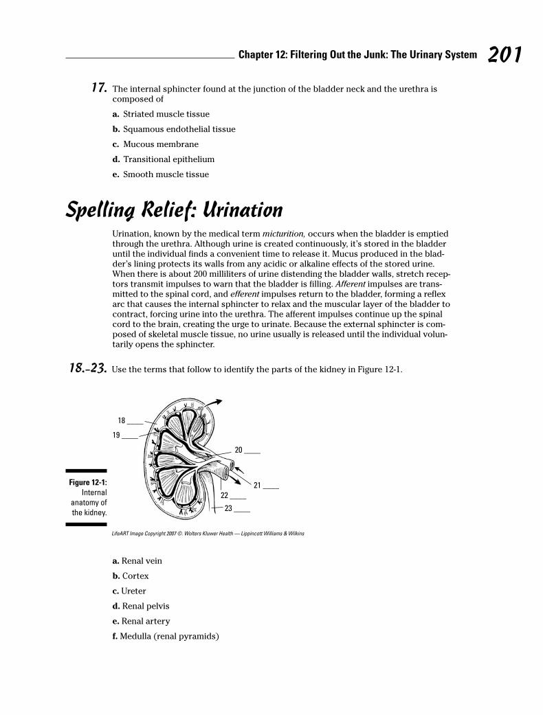
17. The internal sphincter found at the junction of the bladder neck and the urethra is composed of
a. Striated muscle tissue
b. Squamous endothelial tissue
c. Mucous membrane
d. Transitional epithelium
e. Smooth muscle tissue
Spelling Relief: UrinationUrination, known by the medical term micturition, occurs when the bladder is emptiedthrough the urethra. Although urine is created continuously, it’s stored in the bladderuntil the individual finds a convenient time to release it. Mucus produced in the blad-der’s lining protects its walls from any acidic or alkaline effects of the stored urine.When there is about 200 milliliters of urine distending the bladder walls, stretch recep-tors transmit impulses to warn that the bladder is filling. Afferent impulses are trans-mitted to the spinal cord, and efferent impulses return to the bladder, forming a reflexarc that causes the internal sphincter to relax and the muscular layer of the bladder tocontract, forcing urine into the urethra. The afferent impulses continue up the spinalcord to the brain, creating the urge to urinate. Because the external sphincter is com-posed of skeletal muscle tissue, no urine usually is released until the individual volun-tarily opens the sphincter.
18.–23. Use the terms that follow to identify the parts of the kidney in Figure 12-1.
LifeART Image Copyright 2007 ©. Wolters Kluwer Health — Lippincott Williams & Wilkins
a. Renal vein
b. Cortex
c. Ureter
d. Renal pelvis
e. Renal artery
f. Medulla (renal pyramids)
18 ____
19 ____
23 ____
22 ____
21 ____
20 ____
Figure 12-1:
Internal
anatomy of
the kidney.
201Chapter 12: Filtering Out the Junk: The Urinary System
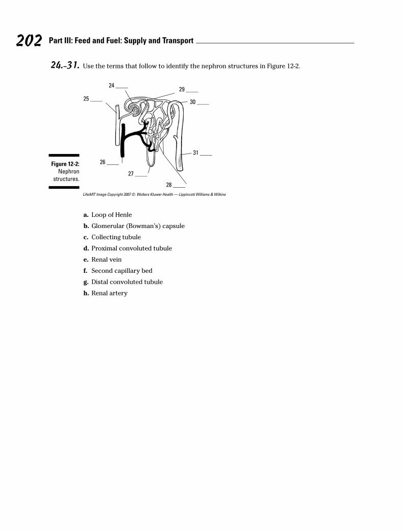
24.–31. Use the terms that follow to identify the nephron structures in Figure 12-2.
LifeART Image Copyright 2007 ©. Wolters Kluwer Health — Lippincott Williams & Wilkins
a. Loop of Henle
b. Glomerular (Bowman’s) capsule
c. Collecting tubule
d. Proximal convoluted tubule
e. Renal vein
f. Second capillary bed
g. Distal convoluted tubule
h. Renal artery
26 ____
27 ____
31 ____
28 ____
25 ____30 ____
29 ____24 ____
Figure 12-2:
Nephron
structures.
202 Part III: Feed and Fuel: Supply and Transport
Answers to Questions on the Urinary SystemThe following are answers to the practice questions presented in this chapter.
a Cortex: b. Granular outer layer
b Medulla: a. Composed of folds forming the renal pyramids
c Renal pelvis: e. Found in the renal sinus
d Calyx: c. Irregular sac-like structures for collecting urine in the renal pelvis
e Collecting tubule: d. Transports urine from the cortex
f The functional kidney is responsible for e. only b and c (excretion of waste and removal ofexcessive materials in the blood). The other answer option can’t be correct because carbondioxide exits the body through the lungs.
g The ureter leaves the kidney through the depression known as the a. hilus. That’s the partwhere the “bean” folds in on itself.
h The human kidney is not included in the abdominal cavity, which makes it a. retroperitoneal.Peritoneal refers to the peritoneum, the membrane lining the abdominal cavity; and retro canbe defined as “situated behind.”
i The functional unit of the kidney is the d. nephron. Each nephron contains a series of the partsneeded to do the kidney’s filtering job.
j The correct sequence for removal of material from the blood through the nephron is a.Afferent arteriole → Glomerulus → Proximal convoluted tubule → Loop of Henle → Distalconvoluted tubule → Collecting tubule. In short, blood comes through the artery (arteriole)and material gloms onto the nephron before twisting through the near (proximal) tubes, loop-ing the loop, twisting through the distant (distal) tubes, and collecting itself at the other end.Try remembering artery-glom-proxy-loop-distant-collect.
k Brush borders of villi are found primarily in the b. proximal convoluted tubule. Those brushborders provide extra surface area for reabsorption, so it makes sense that they congregate inthe first area after filtration.
l Filtration occurs primarily in the d. glomerulus. The glomerulus is a collection of capillarieswith big pores, so think of it as the initial filtering sieve.
m Reabsorption occurs primarily in the d. proximal convoluted tubules. These tubules have themost surface area with all those villi brush borders, so they reabsorb the most.
n The interior of the bladder is lined with elastic b. transitional epithelium. It’s transitionalbecause it needs to be able to stretch and collapse as needed.
o The urine formed in the kidney is passed down the ureter by e. peristaltic contractions. It’s thesame action that moves food through the digestive system.
p The circular striated muscle known as the external sphincter is found in the d. urethra. If it’sexternal, it has to be in the tube that heads outside.
203Chapter 12: Filtering Out the Junk: The Urinary System

q The internal sphincter found at the junction of the bladder neck and the urethra is composed ofe. smooth muscle tissue. The internal sphincter is smooth muscle tissue that prevents urineleakage from the bladder.
r–w Following is how Figure 12-1, the internal anatomy of the kidney, should be labeled.
18. b. Cortex; 19. f. Medulla (renal pyramids); 20. d. Renal pelvis; 21. e. Renal artery; 22. a. Renal vein; 23. c. Ureter
x–F Following is how Figure 12-2, the nephron, should be labeled.
24. b. Glomerular (Bowman’s) capsule; 25. h. Renal artery; 26. e. Renal vein; 27. a. Loop ofHenle; 28. f. Secondary capillary bed; 29. d. Proximal convoluted tubule; 30. g. Distalconvoluted tubule; 31. c. Collecting tubule
204 Part III: Feed and Fuel: Supply and Transport
Part IV
Survival of the Species
In this part . . .
In Part I, we review how cells perpetuate themselves.So in this part, we look at how the entire human species
ensures its future through that process of cell division.We go with the guys first because — let’s face it — theirbiological role in reproduction isn’t as involved as women’s.Then we explain reproduction on the female side of theequation, including a review of the human life cycle frombirth to death.
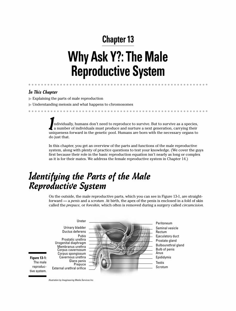
Chapter 13
Why Ask Y?: The MaleReproductive System
In This Chapter� Explaining the parts of male reproduction
� Understanding meiosis and what happens to chromosomes
Individually, humans don’t need to reproduce to survive. But to survive as a species,a number of individuals must produce and nurture a next generation, carrying their
uniqueness forward in the genetic pool. Humans are born with the necessary organs todo just that.
In this chapter, you get an overview of the parts and functions of the male reproductivesystem, along with plenty of practice questions to test your knowledge. (We cover the guysfirst because their role in the basic reproduction equation isn’t nearly as long or complexas it is for their mates. We address the female reproductive system in Chapter 14.)
Identifying the Parts of the MaleReproductive System
On the outside, the male reproductive parts, which you can see in Figure 13-1, are straight-forward — a penis and a scrotum. At birth, the apex of the penis is enclosed in a fold of skincalled the prepuce, or foreskin, which often is removed during a surgery called circumcision.
Illustration by Imagineering Media Services Inc.
Ureter
Urinary bladderDuctus deferens
PubisProstatic urethra
Urogenital diaphragmMembranus urethraCorpus cavernosumCorpus spongiosum
Cavernous urethraGlans penis
PrepuceExternal urethral orifice
Peritoneum
Seminal vesicleRectum
Ejaculatory duct
Prostate gland
Bulbourethral glandBulb of penisAnus
Epididymis
Testis
Scrotum
Figure 13-1:
The male
reproduc-
tive system.
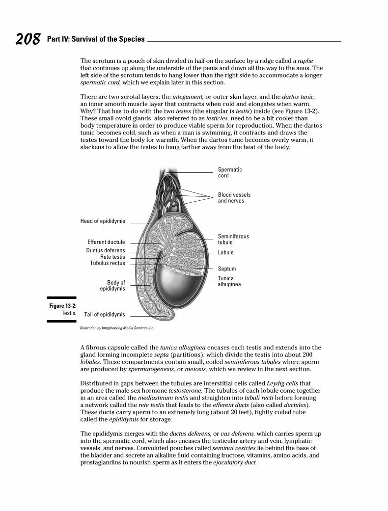
The scrotum is a pouch of skin divided in half on the surface by a ridge called a raphethat continues up along the underside of the penis and down all the way to the anus. Theleft side of the scrotum tends to hang lower than the right side to accommodate a longerspermatic cord, which we explain later in this section.
There are two scrotal layers: the integument, or outer skin layer, and the dartos tunic,an inner smooth muscle layer that contracts when cold and elongates when warm.Why? That has to do with the two testes (the singular is testis) inside (see Figure 13-2).These small ovoid glands, also referred to as testicles, need to be a bit cooler thanbody temperature in order to produce viable sperm for reproduction. When the dartostunic becomes cold, such as when a man is swimming, it contracts and draws thetestes toward the body for warmth. When the dartos tunic becomes overly warm, itslackens to allow the testes to hang farther away from the heat of the body.
Illustration by Imagineering Media Services Inc.
A fibrous capsule called the tunica albuginea encases each testis and extends into thegland forming incomplete septa (partitions), which divide the testis into about 200lobules. These compartments contain small, coiled seminiferous tubules where spermare produced by spermatogenesis, or meiosis, which we review in the next section.
Distributed in gaps between the tubules are interstitial cells called Leydig cells thatproduce the male sex hormone testosterone. The tubules of each lobule come togetherin an area called the mediastinum testis and straighten into tubuli recti before forminga network called the rete testis that leads to the efferent ducts (also called ductules).These ducts carry sperm to an extremely long (about 20 feet), tightly coiled tubecalled the epididymis for storage.
The epididymis merges with the ductus deferens, or vas deferens, which carries sperm upinto the spermatic cord, which also encases the testicular artery and vein, lymphaticvessels, and nerves. Convoluted pouches called seminal vesicles lie behind the base ofthe bladder and secrete an alkaline fluid containing fructose, vitamins, amino acids, andprostaglandins to nourish sperm as it enters the ejaculatory duct.
Head of epididymis
Efferent ductule
Ductus deferensRete testis
Tubulus rectus
Body ofepididymis
Tail of epididymis
Tunicaalbuginea
Septum
Lobule
Seminiferoustubule
Blood vesselsand nerves
Spermaticcord
Figure 13-2:
Testis.
208 Part IV: Survival of the Species
From there the mixture containing sperm enters the prostatic urethra that’s surroundedby the prostate gland. This gland secretes a thin, opalescent substance that precedesthe sperm in an ejaculation. The alkaline nature of this substance reduces the naturalacidity of the female’s vagina to prepare it to receive the sperm.
Two yellowish pea-sized bodies called Cowper’s glands, or bulbourethral glands, lie oneither side of the urethra and secrete a clear alkaline lubricant prior to ejaculation; itneutralizes the acidity of the urethra and acts as a lubricant for the penis. Once all theglands have added protective and nourishing fluids to the 400 to 500 million departingsperm, the mixture is known as seminal fluid or semen.
During sexual arousal, two columns of spongy erectile tissue in the penis — the corpusspongiosum penis and the corpus cavernosum penis — swell with blood to make it rigidand capable of entering the female’s vagina. At the time of ejaculation, smooth musclesin the wall of the epididymis force sperm through the ductus deferens, located in theinguinal canal, toward the urinary bladder. After mixing with the secretions from theseminal vesicles and the prostate gland, the semen travels along the urethra and out avertical slit in the glans penis, or head of the penis.
See how familiar you are with the male anatomy by tackling these practice questions:
1. The cutaneous pouch containing the testes and part of the spermatic cord is the
a. Scrotum
b. Ejaculatory pouch
c. Ductus deferens
d. Seminal vesicle
e. Epididymis
2. The scrotum adjusts to surrounding temperatures through the action of the
a. Testes
b. Bulbourethral glands
c. Dartos tunic
d. Prostatic urethra
3. Spermatogenesis occurs in the
a. Inguinal cells
b. Interstitial cells
c. Seminiferous tubules
d. Rete testis
e. Epididymis
4. Testosterone is produced in the
a. Seminiferous tubules
b. Epididymis
c. Adenohypophysis
d. Subtentacular cells
e. Leydig cells
209Chapter 13: Why Ask Y?: The Male Reproductive System
5. Select the correct sequence for the movement of sperm:
a. Seminiferous tubules → Tubuli recti → Rete testis → Epididymis → Ductus deferens →Ejaculatory duct → Urethra
b. Seminiferous tubules → Tubuli recti → Rete testis → Ejaculatory duct → Epididymis →Duct deferens → Urethra
c. Epididymis → Ejaculatory duct → Tubuli recti → Rete testis → Seminiferous tubules →Ductus deferens → Urethra
d. Seminiferous tubules → Ejaculatory duct → Tubuli recti → Rete testis → Epididymis →Ductus deferens → Urethra
6. Which of the following does not add a secretion to the sperm as it moves through thereproductive ducts?
a. Interstitial cells
b. Epididymis
c. Seminal vesicle
d. Prostate
7. The convoluted tube that stores sperm is called the
a. Seminiferous tubule
b. Rete testis
c. Spermatic cord
d. Epididymis
e. Ductus deferens
8. The fluid accompanying the sperm is called the
a. Stroma
b. Semen
c. Prepuce
d. Inguinal
e. Perineum
9. An average ejaculation will contain sperm numbering approximately
a. 40 to 50 million
b. 400 to 500 million
c. 400 to 500
d. 4 to 5 million
10. A thin, milky liquid imparting alkaline characteristics to the seminal fluid is produced by the
a. Seminal vesicle
b. Interstitial cells
c. Sertoli cells
d. Bulbourethral gland
e. Prostate gland
210 Part IV: Survival of the Species
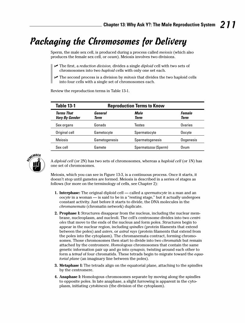
Packaging the Chromosomes for DeliverySperm, the male sex cell, is produced during a process called meiosis (which alsoproduces the female sex cell, or ovum). Meiosis involves two divisions.
� The first, a reduction division, divides a single diploid cell with two sets ofchromosomes into two haploid cells with only one set each.
� The second process is a division by mitosis that divides the two haploid cellsinto four cells with a single set of chromosomes each.
Review the reproduction terms in Table 13-1.
Table 13-1 Reproduction Terms to Know
Terms That General Male Female Vary By Gender Term Term Term
Sex organs Gonads Testes Ovaries
Original cell Gametocyte Spermatocyte Oocyte
Meiosis Gametogenesis Spermatogenesis Oogenesis
Sex cell Gamete Spermatozoa (Sperm) Ovum
A diploid cell (or 2N) has two sets of chromosomes, whereas a haploid cell (or 1N) hasone set of chromosomes.
Meiosis, which you can see in Figure 13-3, is a continuous process. Once it starts, itdoesn’t stop until gametes are formed. Meiosis is described in a series of stages asfollows (for more on the terminology of cells, see Chapter 2):
1 . Interphase: The original diploid cell — called a spermatocyte in a man and anoocyte in a woman — is said to be in a “resting stage,” but it actually undergoesconstant activity. Just before it starts to divide, the DNA molecules in thechromanemata (chromatin network) duplicate.
2. Prophase I: Structures disappear from the nucleus, including the nuclear mem-brane, nucleoplasm, and nucleoli. The cell’s centrosome divides into two centri-oles that move to the ends of the nucleus and form poles. Structures begin toappear in the nuclear region, including spindles (protein filaments that extendbetween the poles) and asters, or astral rays (protein filaments that extend fromthe poles into the cytoplasm). The chromanemata contract, forming chromo-somes. Those chromosomes then start to divide into two chromatids but remainattached by the centromere. Homologous chromosomes that contain the samegenetic information pair up and go into synapsis, twisting around each other toform a tetrad of four chromatids. These tetrads begin to migrate toward the equa-torial plane (an imaginary line between the poles).
3. Metaphase I: The tetrads align on the equatorial plane, attaching to the spindlesby the centromere.
4. Anaphase I: Homologous chromosomes separate by moving along the spindlesto opposite poles. In late anaphase, a slight furrowing is apparent in the cyto-plasm, initiating cytokinesis (the division of the cytoplasm).
211Chapter 13: Why Ask Y?: The Male Reproductive System
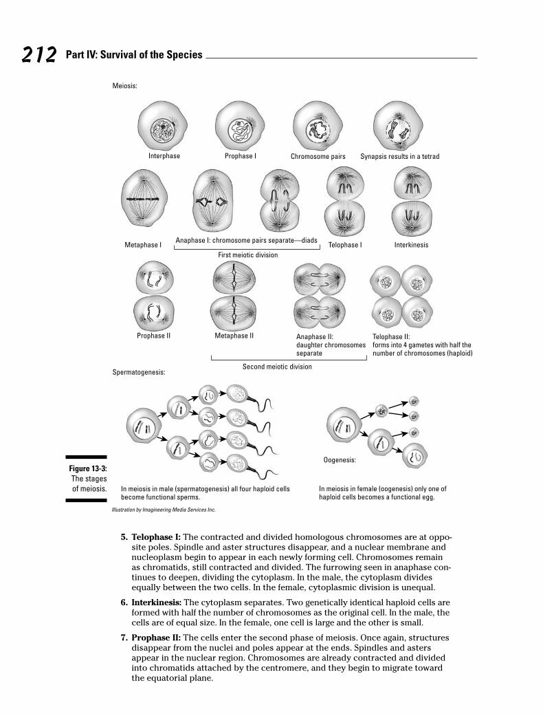
Illustration by Imagineering Media Services Inc.
5 . Telophase I: The contracted and divided homologous chromosomes are at oppo-site poles. Spindle and aster structures disappear, and a nuclear membrane andnucleoplasm begin to appear in each newly forming cell. Chromosomes remainas chromatids, still contracted and divided. The furrowing seen in anaphase con-tinues to deepen, dividing the cytoplasm. In the male, the cytoplasm dividesequally between the two cells. In the female, cytoplasmic division is unequal.
6. Interkinesis: The cytoplasm separates. Two genetically identical haploid cells areformed with half the number of chromosomes as the original cell. In the male, thecells are of equal size. In the female, one cell is large and the other is small.
7. Prophase II: The cells enter the second phase of meiosis. Once again, structuresdisappear from the nuclei and poles appear at the ends. Spindles and astersappear in the nuclear region. Chromosomes are already contracted and dividedinto chromatids attached by the centromere, and they begin to migrate towardthe equatorial plane.
Interphase Prophase I Chromosome pairs Synapsis results in a tetrad
Metaphase IAnaphase I: chromosome pairs separate—diads
Telophase I Interkinesis
Prophase II Metaphase II Telophase II: forms into 4 gametes with half the number of chromosomes (haploid)
First meiotic division
Second meiotic division
Anaphase II:daughter chromosomes separate
Spermatogenesis:
Meiosis:
Oogenesis:
In meiosis in male (spermatogenesis) all four haploid cellsbecome functional sperms.
In meiosis in female (oogenesis) only one of haploid cells becomes a functional egg.
Figure 13-3:
The stages
of meiosis.
212 Part IV: Survival of the Species

8 . Metaphase II: The chromatids align on the equatorial plane and attach to thespindles by the centromere.
9. Anaphase II: The chromatids separate, becoming chromosomes that move alongthe spindles to the poles. A slight furrowing appears in the cytoplasm.
10. Telophase II: With the chromosomes at the poles, spindles and asters disappearwhile new nuclear structures appear. The chromosomes uncoil, returning tochromanemata and their chromatin network. Cytoplasmic division continues todeepen and each haploid cell divides, forming four cells.
At the end of this process, the male has four haploid sperm of equal size. As the spermmatures further, a flagellum (tail) develops. The female, on the other hand, has producedone large cell, the ovum, and three small cells called polar bodies; all four structures con-tain just one set of chromosomes. The polar bodies eventually disintegrate and theovum becomes the functional cell. When fertilized by the sperm, the resulting zygote(fertilized egg) is diploid, containing two sets of chromosomes.
Think you’ve conquered this process? Find out by tackling these practice questions:
213Chapter 13: Why Ask Y?: The Male Reproductive System
Q. The metaphase II stage in meiosisinvolves
a. The slipping of the centromerealong the chromosome
b. The alignment of the chromo-somes on the equatorial plane
c. The contraction of thechromosomes
d. The disappearance of thenuclear membrane
e. The polar migration of thechromosomes
A. The correct answer is the align-ment of the chromosomes on theequatorial plane. Think “divide andconquer.”
11. The process in sexual reproduction involving the union of gametes is called
a. Fission
b. Conjugation
c. Invagination
d. Fertilization
e. Pollination
12. Gametes are formed by
a. Interkinesis
b. Cytokinesis
c. Meiosis
d. Mitosis
e. Photosynthesis
13. A man has 46 chromosomes in a spermatocyte. How many chromosomes are in each sperm?
a. 23 pairs
b. 23
c. 184
d. 46
e. 92
14. Synapsis, or side-by-side pairing, of homologous chromosomes
a. Occurs in mitosis
b. Completes fertilization
c. Is followed immediately by the splitting of each centromere
d. Signifies the end of prophase of the second meiotic division
e. Occurs in meiosis
15. Anaphase I of meiosis is characterized by which of the following?
a. Synapsed homologous chromosomes move toward the poles.
b. DNA duplicates itself.
c. Synapsis of homologous chromosomes occurs.
d. Homologous chromosomes separate and move poleward with centromeres intact.
e. Centromeres split, and chromosomes migrate toward the poles.
16. During ovum production, the three nonfunctional cells produced are called
a. Diploid cells
b. Male sex cells
c. Polar bodies
d. Somatic cells
e. Cross-over gametes
17. The stage (or period) in meiosis between the first and second division is called
a. Anaphase I
b. Interphase
c. Metaphase I
d. Interkinesis
e. Prophase II
18. After meiosis, each diploid spermatocyte has become
a. Four haploid sperm of varying sizes
b. One functional diploid sperm and three cells that eventually will disintegrate
c. Four haploid sperm of equal size
d. Two haploid sperm of equal size
e. Three haploid flagellum and a diploid sperm
214 Part IV: Survival of the Species
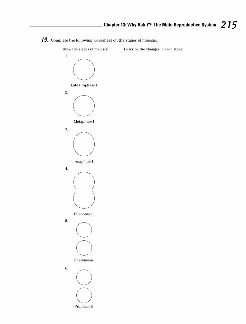
19. Complete the following worksheet on the stages of meiosis.
Draw the stages of meiosis:
1.
Late Prophase I
5.
Interkinesis
6.
Prophase II
2.
Metaphase I
3.
Anaphase I
4.
Teleophase I
Describe the changes in each stage:
215Chapter 13: Why Ask Y?: The Male Reproductive System

7.
Metaphase II
8.
Anaphase II
9.
Teleophase II
10.
Sperm
11.
Ovum and Polar Bodies
216 Part IV: Survival of the Species
Answers to Questions on the MaleReproductive System
The following are answers to the practice questions presented in this chapter.
a The cutaneous pouch containing the testes and part of the spermatic cord is the a. scrotum.“Cutaneous” simply means “skin.”
b The scrotum adjusts to surrounding temperatures through the action of the c. dartos tunic.This is the inner smooth muscle layer of the scrotum.
c Spermatogenesis occurs in the c. seminiferous tubules. Immature sperm cells line the walls ofthe tubules.
d Testosterone is produced in the e. Leydig cells. “Interstitial cells” would also be correctbecause the word “interstitial” can be translated as “placed between.” But alas, that’s notone of the answer options.
e Select the correct sequence for the movement of sperm: a. Seminiferous tubules → Tubulirecti → Rete testis → Epididymis → Ductus deferens → Ejaculatory duct → Urethra. Thesperm develop in the coiled tubules, move through the straighter tubes (tubuli recti), continueacross the network of the testis (rete testis) and into the epididymis (remember the really longtube), and travel past the ductus (or vas) deferens and the ejaculatory duct into the urethra.
f Which of the following does not add a secretion to the sperm as it moves through the reproduc-tive ducts? a. Interstitial cells. Interstitial cells secrete testosterone, which goes into the blood.
g The convoluted tube that stores sperm is called the d. epididymis. The other answer optionsdon’t come into play until it’s time to release semen.
h The fluid accompanying the sperm is called the b. semen.
i An average ejaculation will contain sperm numbering approximately b. 400 to 500 million.Keep in mind that sperm are microscopically small, so quite a few can fit in a tiny amountof semen.
j A thin, milky liquid imparting alkaline characteristics to the seminal fluid is produced by thee. prostate gland.
k The process in sexual reproduction involving the union of gametes is called d. fertilization.Oh, come now — fission? Pollination? Remember that we’re talking human anatomy here!
l Gametes are formed by c. meiosis. Gametes — sperm and ova — are the end goal of thisprocess.
m A man has 46 chromosomes in a spermatocyte. How many chromosomes are in each sperm?b. 23. Divide the number 46 in half.
n Synapsis, or side-by-side pairing, of homologous chromosomes e. occurs in meiosis.
o Anaphase I of meiosis is characterized by which of the following? d. Homologous chromo-somes separate and move poleward with centromeres intact.
217Chapter 13: Why Ask Y?: The Male Reproductive System
218 Part IV: Survival of the Species
p During ovum production, the three nonfunctional cells produced are called c. polar bodies.They eventually disintegrate.
q The stage (or period) in meiosis between the first and second division is called d. interkinesis.Inter– means “between,” and –kinesis means “motion,” so it’s clear that this phase is “betweenmotions.”
r After meiosis, each diploid spermatocyte has become c. four haploid sperm of equal size.There’s no such thing as a diploid sperm because as a sex cell, sperm carries only half the regu-lar complement of 46 chromosomes. And because another division takes place after the initialdivision in meiosis, the final product of the process is four cells, not two.
s Following is a summary of what should appear in your drawings and descriptions of the stagesof meiosis. For further reference, check out Figure 13-3.
In the drawing for late prophase I, at least two pairs of homologous chromosomes shouldbe shown grouped into tetrads (in truth, there are 23 pairs, but simplified illustrations tendto show just two). The description for prophase I should include reference to the tetrad for-mation. The drawing for metaphase I should show the equatorial plane (a center horizontalline) with the tetrads aligned along it. The illustration also should show spindles radiatingfrom each pole, with the tetrads attached to them by their centromeres. The descriptionshould include reference to the equatorial plane, the poles, and the spindles.
The drawing for anaphase I should show the tetrads moving to the top and bottom of the cellalong the spindles and the cytoplasm slowly beginning to divide. In telophase I, the divisionbecomes more pronounced and two new nuclei form. As the process enters interkinesis, thecytoplasm pinches off into two cells.
During prophase II, which also is the start of the second meiotic division, the contracted anddivided chromatids migrate toward a new equatorial plane. The drawing of metaphase IIshould show all chromatids aligned on the equatorial plane. For anaphase II, you should showthe chromatids pulling apart into chromosomes and moving toward the poles. In the finalstage, telophase II, you should draw new nuclei forming around the chromosomes.

Chapter 14
Carrying Life Forward: The FemaleReproductive System
In This Chapter� Mapping out the female reproductive parts and what they do
� Understanding meiosis as the process that makes eggs
� Explaining embryology
� Nursing a fetus into a baby
� Following the process of growth and aging in women
Men may have quite a few hard-working parts in their reproductive systems, but womenare the ones truly responsible for survival of the species (biologically speaking,
anyway). The female body prepares for reproduction every month for most of a woman’sadult life, producing an ovum and then measuring out delicate levels of hormones to preparefor nurturing a developing embryo. When a fertilized ovum fails to show up, the body hits thebiological reset button and sloughs off the uterine lining before building it up all over again fornext month’s reproductive roulette. But that’s nothing compared to what the female bodydoes when a fertilized egg actually settles in for a nine-month stay. Strap yourselves in for atour of the incredible female baby-making machinery — practice questions included.
Identifying the Female Reproductive Parts and Their Functions
First and foremost in the female reproductive repertoire are the two ovaries, which usuallytake a turn every other month to produce a single ovum. Roughly the size and shape of largeunshelled almonds, the female gonads lie on either side of the uterus, below and slightlybehind the Fallopian tubes (also called the uterine tubes). Each ovary has a stroma (body) ofconnective tissue surrounded by a dense fibrous connective tissue called the tunica albuginea(literally “white covering”); yes, that’s the same name as the tissue surrounding the testes. Infact, the ovaries in a female and the testes in a male are homologous, meaning that they sharesimilar origins. External to the tunica albuginea is a layer of cuboidal cells known as thegerminal epithelium. During growth of the ovary in a female fetus prior to birth, the germinalepithelium dips into the body of the ovary in various places. Over time, a mass of epithelialcells called primordial follicles, or primary follicles, becomes separated from the main body ofthe ovary. The ovaries of a young girl contain from 100,000 to 400,000 of these follicles, mostof them present at birth.

At puberty and approximately once each month until menopause (which we cover inthe later section “Growing, Changing, and Aging”), the following happens:
1. The pars distal (anterior lobe) of the hypophysis (pituitary gland) secretesfollicle-stimulating hormones, or FSH, which prompt about 1,000 of the pri-mordial follicles to resume cellular division by meiosis.
Usually, only one follicle matures to become a Graafian follicle (twins, triplets, oreven more fetuses result if more than one follicle matures to the point of releas-ing an ovum).
2. One cell of this mass, the oocyte (produced by oogenesis, or meiosis), becomesthe ovum while the remaining cells surround the ovum as part of the cumulusoophorus and others line the fluid-filled follicular cavity as the membranagranulosa.
3. As the ovum matures, its follicle moves toward the ovary’s surface and beginssecreting the hormone estrogen, which signals the endometrium (uterinelining) to build up in preparation for pregnancy.
4. As blood levels of estrogen begin to rise, the pituitary stops releasing FSH andbegins releasing luteinizing hormone, or LH, which prompts the Graafian fol-licle now at the surface of the ovary to rupture, triggering ovulation — therelease of the ovum.
5. Ringed by follicular cells in what’s called the corona radiata, the ovumenters the coelom (body cavity) and is swept into the Fallopian tube by afringe of tissue called fimbriae.
6. It takes approximately three days for the ovum to travel from the Fallopiantube to the uterus.
Meanwhile, back at the ovary, a clot has formed inside the ruptured follicle and themembrana granulosa cells are being replaced by yellow luteal cells, forming a corpusluteum (literally “yellow body”) on the surface of the ovary. This new endocrine glandsecretes progesterone, a hormone that signals the uterine lining to prepare for possibleimplantation of a fertilized egg, inhibits the maturing of Graafian follicles, ovulation,and the production of estrogen to prevent menstruation; and stimulates furthergrowth in the mammary glands (which is why some women get sore breasts a few daysbefore their periods begin). If pregnancy occurs, the placenta also will release proges-terone to prevent menstruation throughout the pregnancy.
If the ovum isn’t fertilized, the corpus luteum dissolves after 10 to 14 days to bereplaced by scar tissue called the corpus albicans. If pregnancy does occur, the corpusluteum remains and grows for about six months before disintegrating. Only about 400of a woman’s primordial follicles ever get a chance to make the trip to the uterus. Therest ripen to various stages before degenerating into what are known as atretic follicles(or corpora atretica) over the course of her lifetime. Figure 14-1 shows what happensinside an ovary.
Fallopian tubes, oviducts, uterine tubes — call them what you will, but they’re wherethe real business of fertilization takes place. Why? Because an egg must be fertilizedwithin 24 hours of its release from the ovary to remain viable. These small, musculartubes lined with cilia are nearly 5 inches long and, somewhat surprisingly, aren’tdirectly connected to the ovaries. Instead, the funnel-shaped end, the infundibulum,of a tube is fragmented into finger-like projections called fimbriae that help to move
220 Part IV: Survival of the Species
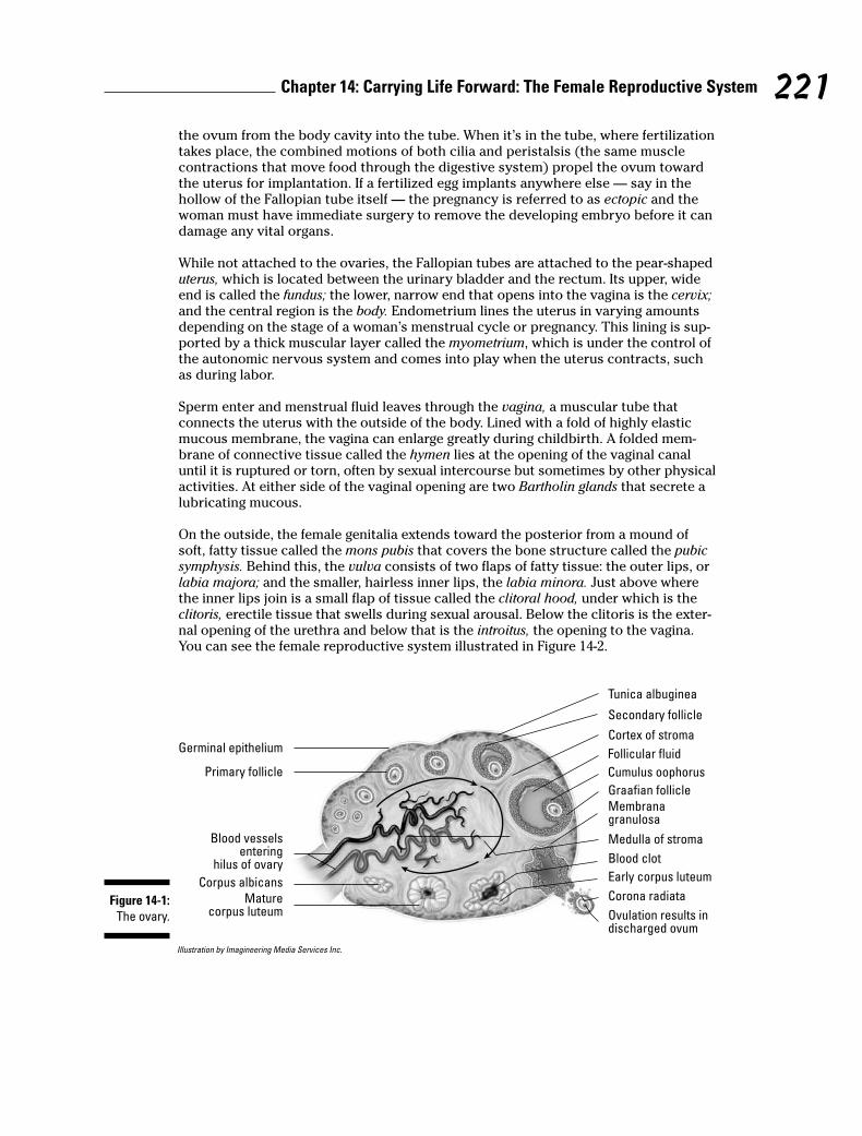
the ovum from the body cavity into the tube. When it’s in the tube, where fertilizationtakes place, the combined motions of both cilia and peristalsis (the same musclecontractions that move food through the digestive system) propel the ovum towardthe uterus for implantation. If a fertilized egg implants anywhere else — say in thehollow of the Fallopian tube itself — the pregnancy is referred to as ectopic and thewoman must have immediate surgery to remove the developing embryo before it candamage any vital organs.
While not attached to the ovaries, the Fallopian tubes are attached to the pear-shapeduterus, which is located between the urinary bladder and the rectum. Its upper, wideend is called the fundus; the lower, narrow end that opens into the vagina is the cervix;and the central region is the body. Endometrium lines the uterus in varying amountsdepending on the stage of a woman’s menstrual cycle or pregnancy. This lining is sup-ported by a thick muscular layer called the myometrium, which is under the control ofthe autonomic nervous system and comes into play when the uterus contracts, suchas during labor.
Sperm enter and menstrual fluid leaves through the vagina, a muscular tube thatconnects the uterus with the outside of the body. Lined with a fold of highly elasticmucous membrane, the vagina can enlarge greatly during childbirth. A folded mem-brane of connective tissue called the hymen lies at the opening of the vaginal canaluntil it is ruptured or torn, often by sexual intercourse but sometimes by other physicalactivities. At either side of the vaginal opening are two Bartholin glands that secrete alubricating mucous.
On the outside, the female genitalia extends toward the posterior from a mound ofsoft, fatty tissue called the mons pubis that covers the bone structure called the pubicsymphysis. Behind this, the vulva consists of two flaps of fatty tissue: the outer lips, orlabia majora; and the smaller, hairless inner lips, the labia minora. Just above wherethe inner lips join is a small flap of tissue called the clitoral hood, under which is theclitoris, erectile tissue that swells during sexual arousal. Below the clitoris is the exter-nal opening of the urethra and below that is the introitus, the opening to the vagina.You can see the female reproductive system illustrated in Figure 14-2.
Illustration by Imagineering Media Services Inc.
Blood vesselsentering
hilus of ovary
Corpus albicans
Maturecorpus luteum
Blood clot
Early corpus luteum
Medulla of stroma
Ovulation results indischarged ovum
Corona radiata
Membranagranulosa
Cortex of stroma
Graafian follicle
Follicular fluid
Cumulus oophorus
Tunica albuginea
Germinal epithelium
Secondary follicle
Primary follicle
Figure 14-1:
The ovary.
221Chapter 14: Carrying Life Forward: The Female Reproductive System

Find out how familiar you are with the female anatomy:
1. Follicular growth (or change) in the ovary is started by
a. Progesterone
b. Estrogen
c. LH
d. FSH
e. Testosterone
2. Which one of the following is not part of the maturing Graafian follicle?
a. Follicular fluid
b. Ovum
c. Membrana granulosum
d. Corpora atretica
3. Which one of the following is not a function of estrogen?
a. Preparing the endometrium
b. Supporting development of the ovum
c. Supporting development of the corpus luteum
d. Preventing secretion of FSH from the pituitary
4. Which of the following does not produce a hormone?
a. Corpus luteum
b. Pars distalis
c. Graafian follicle
d. Corpus albicans
e. Placenta
5.–9. Match the term to its description.
5. _____ Ectopic a. Period of intrauterine development
6. _____ Gestation b. Cessation of menses
7. _____ Ovulation c. Development of out-of-place embryo
8. _____ Menopause d. Release of ovum into coelom
9. _____ Luteinization e. Glandular development by membrana granulosa
10. Progesterone is produced by the
a. Endometrium
b. Pars distalis
c. Follicle
d. Corpus luteum
e. Pituitary
222 Part IV: Survival of the Species
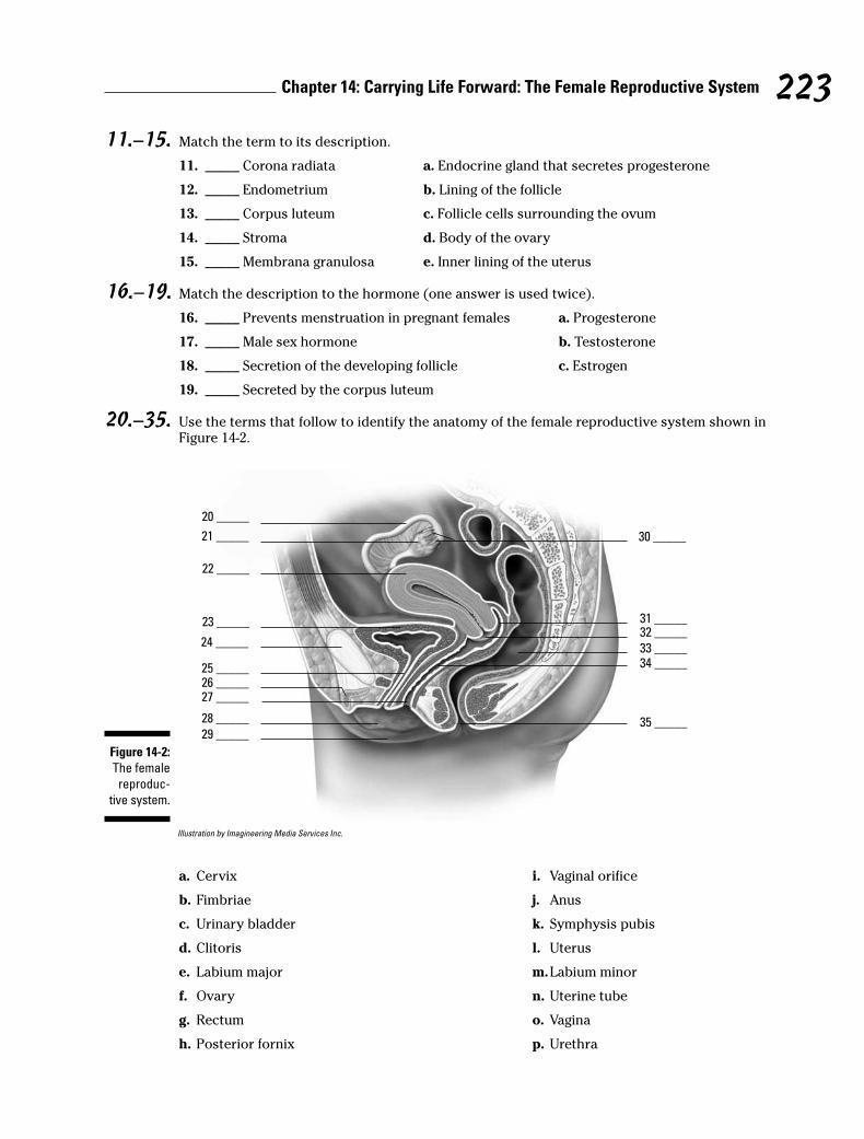
11.–15. Match the term to its description.
11. _____ Corona radiata a. Endocrine gland that secretes progesterone
12. _____ Endometrium b. Lining of the follicle
13. _____ Corpus luteum c. Follicle cells surrounding the ovum
14. _____ Stroma d. Body of the ovary
15. _____ Membrana granulosa e. Inner lining of the uterus
16.–19. Match the description to the hormone (one answer is used twice).
16. _____ Prevents menstruation in pregnant females a. Progesterone
17. _____ Male sex hormone b. Testosterone
18. _____ Secretion of the developing follicle c. Estrogen
19. _____ Secreted by the corpus luteum
20.–35. Use the terms that follow to identify the anatomy of the female reproductive system shown inFigure 14-2.
Illustration by Imagineering Media Services Inc.
20 _____
21 _____
22 _____
23 _____
29 _____
28 _____
27 _____26 _____25 _____
24 _____
30 _____
31 _____32 _____
33 _____34 _____
35 _____
Figure 14-2:
The female
reproduc-
tive system.
223Chapter 14: Carrying Life Forward: The Female Reproductive System
a. Cervix
b. Fimbriae
c. Urinary bladder
d. Clitoris
e. Labium major
f. Ovary
g. Rectum
h. Posterior fornix
i. Vaginal orifice
j. Anus
k. Symphysis pubis
l. Uterus
m.Labium minor
n. Uterine tube
o. Vagina
p. Urethra
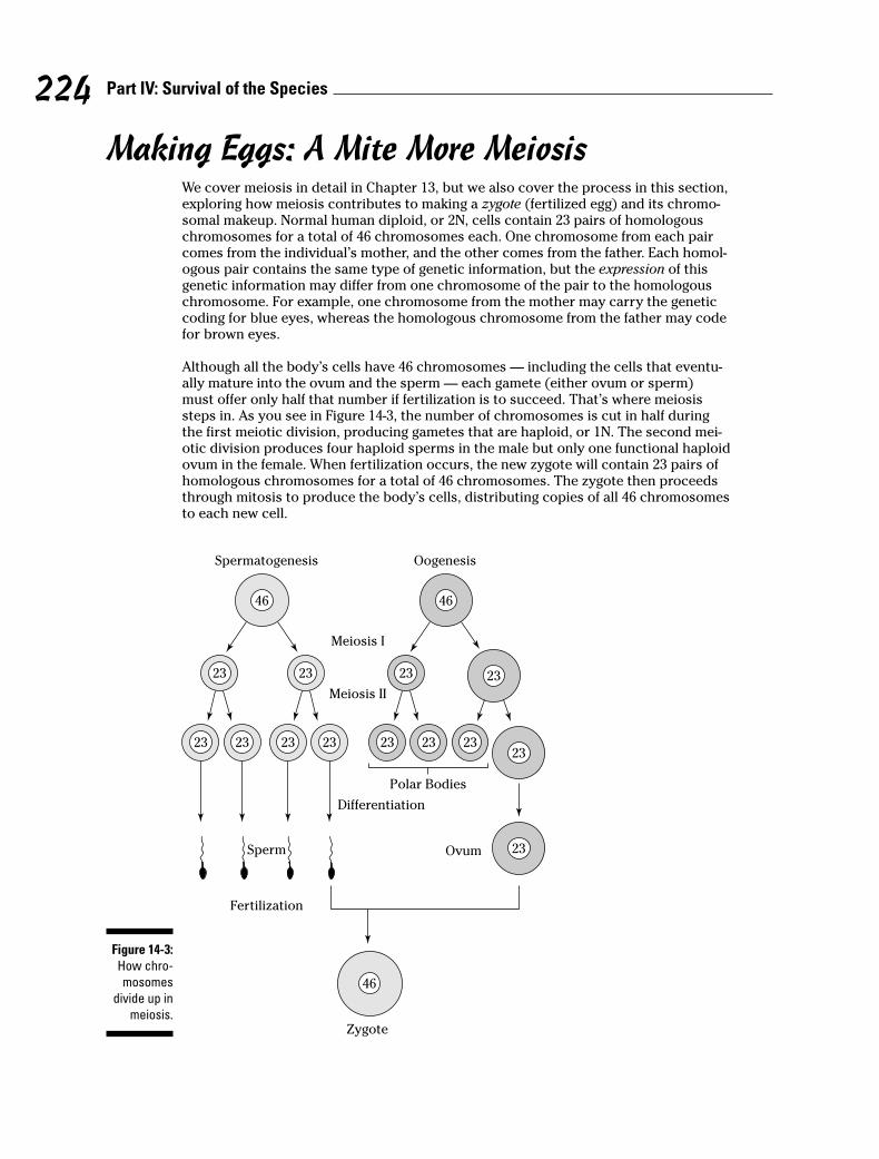
Making Eggs: A Mite More MeiosisWe cover meiosis in detail in Chapter 13, but we also cover the process in this section,exploring how meiosis contributes to making a zygote (fertilized egg) and its chromo-somal makeup. Normal human diploid, or 2N, cells contain 23 pairs of homologouschromosomes for a total of 46 chromosomes each. One chromosome from each paircomes from the individual’s mother, and the other comes from the father. Each homol-ogous pair contains the same type of genetic information, but the expression of thisgenetic information may differ from one chromosome of the pair to the homologouschromosome. For example, one chromosome from the mother may carry the geneticcoding for blue eyes, whereas the homologous chromosome from the father may codefor brown eyes.
Although all the body’s cells have 46 chromosomes — including the cells that eventu-ally mature into the ovum and the sperm — each gamete (either ovum or sperm)must offer only half that number if fertilization is to succeed. That’s where meiosissteps in. As you see in Figure 14-3, the number of chromosomes is cut in half duringthe first meiotic division, producing gametes that are haploid, or 1N. The second mei-otic division produces four haploid sperms in the male but only one functional haploidovum in the female. When fertilization occurs, the new zygote will contain 23 pairs ofhomologous chromosomes for a total of 46 chromosomes. The zygote then proceedsthrough mitosis to produce the body’s cells, distributing copies of all 46 chromosomesto each new cell.
Spermatogenesis
Sperm
Fertilization
Zygote
46
23
23
23
23 23 23
Oogenesis
Meiosis I
Meiosis II
Differentiation
Polar Bodies
Ovum
46
46
23
23
23
23
23
23 23
Figure 14-3:
How chro-
mosomes
divide up in
meiosis.
224 Part IV: Survival of the Species
Think you’ve conquered this process? Find out by answering the following practicequestions:
225Chapter 14: Carrying Life Forward: The Female Reproductive System
Q. How many chromosomes are inmost of the cells in your body?
a. 46
b. 23
c. 92
A. The correct answer is 46. That’sthe usual human complement.
36. How many chromosomes are in each sperm produced by a male?
a. 46
b. 23
c. 92
37. How many chromosomes are in each ovum produced by a female?
a. 46
b. 23
c. 92
38. How many chromosomes are in a newly fertilized egg, or zygote?
a. 46
b. 23
c. 92
39. A haploid cell contains how many chromosomes?
a. 46
b. 23
c. 92
40. The process of chromosomal reduction is called
a. Mitosis
b. Menses
c. Meiosis
d. Gene expression
41.–48. Fill in the blanks to complete the following sentences:
Meiosis produces sperm and ovum, which contributes to making a 41. _________________(fertilized egg) and its chromosomal makeup. Normal humans have 42. ___________________,or 2N, cells containing 23 pairs of homologous chromosomes for a total of 46 chromosomeseach. A pair of chromosomes containing the same type of genetic information are 43. ______________________ chromosomes. An ovum and a sperm are called sex cells or 44. _____________. The number of chromosomes is cut in half during the first meiotic division,producing gametes that are 45. ____________, or 1N. The second meiotic division produces 46. _____________________ sperms in the male but only 47. __________________ovum in thefemale. The zygote then proceeds through 48. _______________________ to produce the body’scells.

Making Babies: An Introductionto Embryology
Fertilization (the joining of ovum and sperm) occurs in the Fallopian tube as the ovumtravels toward the uterus. Fertilization must occur within 24 hours of ovulation or theovum degenerates. But that doesn’t mean that sexual intercourse outside that timeframe can’t lead to pregnancy. Research indicates that spermatozoa can survive up toseven days inside the uterus and Fallopian tubes. If a sperm is still motile — that is, ifit’s still whipping its flagellum tail — when an ovum comes down the tube, it will dowhat it was made to do and penetrate the ovum’s membrane.
When the sperm penetrates the ovum, it releases enzymes that allow it to digest its wayinto the ovum, leaving its flagellum behind. After that first sperm penetrates, the mem-brane instantly solidifies around the ovum, preventing any other sperm from gettinginside. Presto! You’ve got a zygote. Over the next three to five days, the zygote movesthrough the Fallopian tube to the uterus, undergoing cleavage (mitotic cell division)along the way: Two cells become four smaller cells, four cells become eight smaller cells,and then those eight cells become a solid 16-cell ball called a morula. After five days ofcleavage, the cells form a hollow ball of approximately 32 cells called a blastula, or blasto-cyst. The inner hollow region is called the blastocoele, and the outer-layer cells are calledthe trophoblast. Figure 14-4 illustrates this process of development.
Within three days of its arrival in the uterine cavity (generally within a week of fertiliza-tion), the blastocyst implants in the endometrium, and some of the blastocyst’s cells —called totipotent embryonic stem cells — organize into an inner cell mass called theembryonic disk, or embryoblast. Over time, the embryonic disk differentiates into the tis-sues of the developing embryo (see Figure 14-4). Cells above the disk form the amnioticcavity, and those below form the gut cavity and two primitive germ layers. The layer near-est the amniotic cavity forms the ectoderm while that nearest the gut cavity forms theendoderm. Between the two layers, additional ectodermal cells develop to form a thirdlayer, the mesoderm. The ectoderm forms skin and nerve tissue; the mesoderm formsbones, cartilage, connective tissue, muscles, and organs; and the endoderm forms thelinings of the organs and glands.
To keep these terms straight, remember that endo– means “inside or within,” ecto–means “outer or external,” and meso– means “middle.”
After three weeks of development, the heart begins beating. In the fourth week ofdevelopment, the embryonic disk forms an elongated structure that attaches to thedeveloping placenta by a connecting stalk. A head and jaws form while primitive budssprout; the buds will develop into arms and legs. During the fifth through seventhweeks, the head grows rapidly and a face begins to form (eyes, nose, and a mouth).Fingers and toes grow at the ends of the elongating limb buds. All internal organs havestarted to form. After eight weeks of development, the embryo begins to have a morehuman appearance and is referred to as a fetus.
The outer cells of the embryo, together with the endometrium of the uterus, form theplacenta, a new internal organ that exists only during pregnancy. The placenta attachesthe fetus to the uterine wall, exchanges gases and waste between the maternal andfetal bloodstreams, and secretes hormones to sustain the pregnancy.
226 Part IV: Survival of the Species
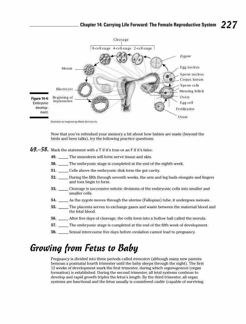
Illustration by Imagineering Media Services Inc.
Now that you’ve refreshed your memory a bit about how babies are made (beyond thebirds and bees talks), try the following practice questions:
49.–58. Mark the statement with a T if it’s true or an F if it’s false:
49. _____ The mesoderm will form nerve tissue and skin.
50. _____ The embryonic stage is completed at the end of the eighth week.
51. _____ Cells above the embryonic disk form the gut cavity.
52. _____ During the fifth through seventh weeks, the arm and leg buds elongate and fingersand toes begin to form.
53. _____ Cleavage is successive mitotic divisions of the embryonic cells into smaller andsmaller cells.
54. _____ As the zygote moves through the uterine (Fallopian) tube, it undergoes meiosis.
55. _____ The placenta serves to exchange gases and waste between the maternal blood andthe fetal blood.
56. _____ After five days of cleavage, the cells form into a hollow ball called the morula.
57. _____ The embryonic stage is completed at the end of the fifth week of development.
58. _____ Sexual intercourse five days before ovulation cannot lead to pregnancy.
Growing from Fetus to BabyPregnancy is divided into three periods called trimesters (although many new parentsbemoan a postnatal fourth trimester until the baby sleeps through the night). The first12 weeks of development mark the first trimester, during which organogenesis (organformation) is established. During the second trimester, all fetal systems continue todevelop and rapid growth triples the fetus’s length. By the third trimester, all organsystems are functional and the fetus usually is considered viable (capable of surviving
Ovum
Egg cell
Sperm cells
Fertilization
Sperm nucleus
Egg nucleus
Zygote
Morula
Blastocyst
Beginning ofimplantation
Ovary
Corpus luteum
Maturing follicle
2-cell stage4-cell stage8-cell stage
Cleavage
Figure 14-4:
Embryonic
develop-
ment.
227Chapter 14: Carrying Life Forward: The Female Reproductive System
outside the womb) even if it’s born prematurely. The overall growth rate slows in thethird trimester, but the fetus gains weight rapidly.
Milestones in fetal development can be marked monthly.
� At the end of the second month (when the terminology changes from “embryo”to “fetus”), the head remains overly large compared to the developing body, andthe limbs are still short. All major regions of the brain have formed.
� During the third month, body growth accelerates and head growth slows. Thearms reach the length they will maintain during fetal development. The bonesbegin to ossify, and all body systems have begun to form. The circulatory(cardiovascular) system supplies blood to all the developing extremities, andeven the lungs begin to practice “breathing” amniotic fluid. By the end of thethird month, the external genitalia are visible in the male (ultrasound technicianscall this a “turtle sign”). The fetus is a bit less than 4 inches long and weighsabout 1 ounce.
� The body grows rapidly during the fourth month as legs lengthen, and the skele-ton continues to ossify as joints begin to form. The face looks more human. Thefetus is about 7 inches long and weighs 4 ounces.
� Growth slows during the fifth month, and the legs reach their final fetal propor-tions. Skeletal muscles become so active that the mother can feel fetal movement.Hair grows on the head, and lanugo, a profusion of fine soft hair, covers the skin.The fetus is about 12 inches long and weighs 1⁄2 to 1 pound.
� The fetus gains weight during the sixth month, and eyebrows and eyelashesform. The skin is wrinkled, translucent, and reddish because of dermal bloodvessels. The fetus is between 11 and 14 inches long and weighs a bit less than11⁄2 pounds.
� During the seventh month, skin becomes smoother as the fetus gains subcuta-neous fat tissue. The eyelids, which are fused during the sixth month, open.Usually, the fetus turns to an upside-down position. It’s between 13 and 17 incheslong and weighs from 21⁄2 to 3 pounds.
� During the eighth month, subcutaneous fat increases, and the fetus shows morebaby-like proportions. The testes of a male fetus descend into the scrotum. Thefetus is now 16 to 18 inches long and has grown to just under 5 pounds.
� During the ninth month, the fetus plumps up considerably with additional sub-cutaneous fat. Much of the lanugo is shed, and fingernails extend all the way tothe tips of the fingers. The average newborn at the end of the ninth month is20 inches long and weighs about 71⁄2 pounds.
The following practice questions deal with the development of the fetus during its40 weeks in the womb:
59. A fetus usually is considered viable
a. By the second trimester
b. By the third trimester
c. By the fifth month of gestation
d. By the 19th week of gestation
228 Part IV: Survival of the Species

60. Describe one new fetal development for each month:
3rd month: ________________________________________________________________________
4th month: ________________________________________________________________________
5th month: ________________________________________________________________________
6th month: ________________________________________________________________________
7th month: ________________________________________________________________________
8th month: ________________________________________________________________________
9th month: ________________________________________________________________________
Growing, Changing, and AgingAfter a baby arrives, the female reproductive system goes into a different form ofoverdrive. Throughout the pregnancy, the placenta has been producing estrogen andprogesterone to sustain the fetus. But after the baby is born, the sudden drop in hor-monal blood levels triggers the pituitary gland to release prolactin, a hormone thatstimulates the woman’s mammary glands to secrete milk in a process called lactation.First, however, the glands produce colostrum, a thin, yellowish fluid rich in antibodiesand minerals to sustain a newborn. Both colostrum and milk flow from a number oflobes inside the breast through lactiferous ducts that converge on the nipple.
From birth to 4 weeks of age, the newborn is called a neonate. Faced with survival afterits physical separation from the mother, a neonate must abruptly begin to processfood, excrete waste, obtain oxygen, and make circulatory adjustments.
From 4 weeks to 2 years of age, the baby is called an infant. Growth during this period isexplosive under the stimulation of circulatory growth hormones from the pituitary gland,adrenal steroids, and thyroid hormones. The infant’s deciduous teeth, also called baby ormilk teeth, begin to form and erupt through the gums. The nervous system advances,making coordinated activities possible. The baby begins to develop language skills.
From 2 years to puberty, you’re looking at a child. Influenced by growth hormones,growth continues its rapid pace as deciduous teeth are replaced by permanent teeth.Muscle coordination, language skills, and intellectual skills also develop rapidly.
From puberty, which starts between the ages of 11 and 14, to adulthood, the child iscalled an adolescent. Growth occurs in spurts. Girls achieve their maximum growthrate between the ages of 10 and 13, whereas boys experience their fastest growthbetween the ages of 12 and 15. Primary and secondary sex characteristics begin toappear. Growth terminates when the epiphyseal plates of the long bones ossify some-time between the ages of 18 and 21. Motor skills and intellectual abilities continue todevelop, and psychological changes occur as adulthood approaches.
The young adult stage covers roughly 20 years, from the age of 20 to about 40. Physicaldevelopment reaches its peak and adult responsibilities are assumed, often including acareer, marriage, and a family. After about age 30, physical changes that indicate theonset of aging begin to occur.
From age 40 to about 65, physiological aging continues. Gray hair, diminished physicalabilities, and skin wrinkles are outward signs of aging. Women go through menopause,the cessation of monthly cycles, which is also known as climacteric or the change oflife. While menarche (the onset of menstruation) may begin any time between 10 and15 years of age, the female body’s monthly reproductive cycle slows and stops entirely
229Chapter 14: Carrying Life Forward: The Female Reproductive System
between the ages of 40 and 55. With the cessation of menses comes a decrease in sizeof the uterus, shortening of the vagina, shrinkage of the mammary glands, disappear-ance of Graafian follicles, and shrinkage of the ovaries. For about six years prior tomenopause, many women experience a stage called perimenopause during whichincreasingly irregular hormone secretions can cause fluctuations in menstruation anda sensation called hot flashes.
From age 65 until death is the period of senescence, the process of aging. Individualadults can show widely varying patterns of aging in part because of differences ingenetic background and physical activities. Signs of senescence include loss of skinelasticity and accompanying sagging or wrinkling; weakened bones and decreasinglymobile joints; weakened muscles; impaired coordination, memory, or intellectual func-tion; cardiovascular problems; reduced immune responses; decreased respiratoryfunction caused by reduced lung elasticity; and decreased peristalsis and muscle tonein the digestive and urinary tracts.
Following are some practice questions on the aging cycle:
61. The gland involved in lactation is called the
a. Pituitary gland
b. Mammary gland
c. Pars distalis
d. Adrenal gland
e. Parathyroid gland
62. An individual’s height stops increasing
a. During adolescence
b. Upon onset of menopause
c. When the epiphyseal plates of the long bones ossify
d. During middle age
63. A 20-month-old child is formally called a(n)
a. Baby
b. Infant
c. Toddler
d. Neonate
64.–70. Match the description to its life stage.
64. _____ Assume adult responsibilities, a. Neonatepossibly including marriage and a family b. Infant
65. _____ Faced with survival, it must process food, c. Childexcrete waste, and obtain oxygen d. Adolescent
66. _____ Primary and secondary sex characteristics begin e. Young adultto appear f. Middle-aged adult
67. _____ Experiences the period of senescence g. Old adult
68. _____ Deciduous teeth begin to form
69. _____ Women go through menopause
70. _____ From 2 years of age to puberty
230 Part IV: Survival of the Species
Answers to Questions on the FemaleReproductive System
The following are answers to the practice questions presented in this chapter.
a Follicular growth (or change) in the ovary is started by d. FSH. That’s short for follicle stimulat-ing hormone, which is released by the pituitary gland.
b Which one of the following is not part of the maturing Graafian follicle? d. Corpora atretica.Follicles that never matured all the way become corpora atretica over time.
c Which one of the following is not a function of estrogen? c. Supporting development of thecorpus luteum. By the time the corpus luteum starts to develop, estrogen already has begun tobow out of the reproductive equation.
d Which of the following does not produce a hormone? d. Corpus albicans. Corpus albicans is thescar tissue that follows disintegration of the corpus luteum, so it makes sense that it doesn’tproduce any hormones.
e Ectopic: c. Development of out-of-place embryo. Ektopos in Greek literally means “out of place.”
f Gestation: a. Period of intrauterine development. “Gestation” is just a fancy term for pregnancy,and intra– means “within.”
g Ovulation: d. Release of ovum into coelom. Ignore the reference to the body cavity. “Release ofovum” is the giveaway here.
h Menopause: b. Cessation of menses. Menses is the same as menstruation.
i Luteinization: e. Glandular development by membrana granulosa. The granulosa cellsbecome luteal cells to form the corpus luteum, which is actually a new gland each month.
j Progesterone is produced by the d. corpus luteum.
k Corona radiata: c. Follicle cells surrounding the ovum
l Endometrium: e. Inner lining of the uterus
m Corpus luteum: a. Endocrine gland that secretes progesterone
n Stroma: d. Body of the ovary
o Membrana granulosa: b. Lining of the follicle
p Prevents menstruation in pregnant females: a. Progesterone
q Male sex hormone: b. Testosterone
r Secretion of the developing follicle: c. Estrogen
s Secreted by the corpus luteum: a. Progesterone
231Chapter 14: Carrying Life Forward: The Female Reproductive System
t–J Following is how Figure 14-2, the female reproductive system, should be labeled.
20. n. Uterine tube; 21. f. Ovary; 22. l. Uterus; 23. c. Urinary bladder; 24. k. Symphysis pubis;25. p. Urethra; 26. d. Clitoris; 27. i. Vaginal orifice; 28. m. Labium minor; 29. e. Labium major;30. b. Fimbrae; 31. h. Posterior fornix; 32. a. Cervix; 33. g. Rectum; 34. o. Vagina; 35. j. Anus
K How many chromosomes are in each sperm produced by a male? b. 23. Cut the usual comple-ment in half to form a gamete.
L How many chromosomes are in each ovum produced by a female? b. 23. Same answer for thesame reason as in the preceding question.
M How many chromosomes are in a newly fertilized egg, or zygote? a. 46. The 23 in the ovum plusthe 23 in the sperm equals 46.
N A haploid cell contains how many chromosomes? b. 23.
The number of chromosomes in a haploid cell may be easier to remember this way: Haploid ishalf, but diploid is double that.
O The process of chromosomal reduction is called c. meiosis. Don’t be fooled by this question;mitosis is when a cell replicates itself — with a full complement of chromosomes. Meiosis is alsocell division, but it occurs only when the body is working to make gametes for reproduction.
P–W Meiosis produces sperm and ovum, which contributes to making a 41. zygote (fertilized egg)and its chromosomal makeup. Normal humans have 42. diploid, or 2N, cells containing 23 pairsof homologous chromosomes for a total of 46 chromosomes each. A pair of chromosomes con-taining the same type of genetic information are 43. homologous chromosomes. An ovum and asperm are called sex cells or 44. gametes. The number of chromosomes is cut in half during thefirst meiotic division, producing gametes that are 45. haploid, or 1N. The second meiotic divi-sion produces 46. four haploid sperms in the male but only 47. one functional haploid ovumin the female. The zygote then proceeds through 48. mitosis to produce the body’s cells.
X The mesoderm will form nerve tissue and skin. False
Y The embryonic stage is completed at the end of the eighth week. True
z Cells above the embryonic disk form the gut cavity. False
Z During the fifth through seventh weeks, the arm and leg buds elongate and fingers and toesbegin to form. True
1 Cleavage is successive mitotic divisions of the embryonic cells into smaller and smaller cells.True
2 As the zygote moves through the uterine (Fallopian) tube, it undergoes meiosis. False
3 The placenta serves to exchange gases and waste between the maternal blood and the fetalblood. True
4 After five days of cleavage, the cells form into a hollow ball called the morula. False
5 The embryonic stage is completed at the end of the fifth week of development. False
232 Part IV: Survival of the Species

6 Sexual intercourse five days before ovulation cannot lead to pregnancy. False. It certainly canhappen — and does.
7 A fetus usually is considered viable b. by the third trimester. Even then, a premature birth canhave serious health consequences for the newborn.
8 Describe one new fetal development for each month:
3rd month: Bones begin to ossify, body growth accelerates while head growth slows, lungsbegin to practice breathing amniotic fluid, external genitalia visible in male, 4 inches long andabout 1 ounce
4th month: Body grows rapidly, legs lengthen, joints begin to form, face looks more human,roughly 7 inches long and 4 ounces
5th month: Mother can feel fetal movement, hair grows on head, lanugo covers skin, about12 inches long and 8 to 16 ounces
6th month: Eyebrows and eyelashes form, weight gain accelerates, roughly 11 to 14 incheslong and just under 1 1⁄2 pounds
7th month: Subcutaneous fat begins to form, eyelids open, usually turns to upside-down posi-tion, between 13 and 17 inches long and 2 1⁄2 to 3 pounds
8th month: Subcutaneous fat increases, fetus appears more “baby-like,” testes of maledescend into scrotum, 16 to 18 inches long and roughly 5 pounds
9th month: Substantial “plumping” with subcutaneous fat, lanugo shed, fingernails extend tofingertips, average newborn is 20 inches long and weighs 7 1⁄2 pounds
9 The gland involved in lactation is called the b. mammary gland. Everybody say “moo”!
0 An individual’s height stops increasing c. when the epiphyseal plates of the long bones ossify.And that occurs sometime between the ages of 18 and 21.
! A 20-month-old child is formally called a(n) b. infant. The terms “baby” and “toddler” aren’tpart of the formal medical lexicon.
@ Assume adult responsibilities, possibly including marriage and a family: e. Young adult
# Faced with survival, it must process food, excrete waste, and obtain oxygen: a. Neonate
$ Primary and secondary sex characteristics begin to appear: d. Adolescent
% Experiences the period of senescence: g. Old adult
^ Deciduous teeth begin to form: b. Infant
& Women go through menopause: f. Middle-aged adult
* From 2 years of age to puberty: c. Child
233Chapter 14: Carrying Life Forward: The Female Reproductive System

234 Part IV: Survival of the Species

Part V
Mission Control: All Systems Go

In this part . . .
This part reviews how the body you know so wellmoves, thinks, feels, and thrives on a day-to-day basis.
You get to know the intricacies of the nerves and brain,arguably the most complex of all the anatomical systemsas well as the fine-tuning capacity of the endocrinesystem and its hormones.
Chapter 15
Feeling Jumpy: The Nervous System
In This Chapter� Breaking down the structure of nerves
� Centering with the central nervous system
� Branching out with the peripheral nervous system
� Taking a hands-off approach to the autonomic nervous system
� Examining the senses
Throughout this book, you look at the human body from head to toe, exploring how itcollects and distributes the molecules it needs to grow and thrive, how it reproduces
itself, and even how it gets rid of life’s nastier byproducts. In this chapter, however, you lookat the living computer that choreographs the whole show, the one system that contributesthe most to making us who we are as humans.
The nervous system is the communications network that goes into nearly every part of thebody, enervating your muscles, pricking your pain sensors, and letting you reach beyondyourself into the larger world. More than 80 major nerves make up this intricate network,and each nerve contains somewhere around 1 million neurons (individual nerve cells).It’s through this complex network that you respond both to external and internal stimuli,demonstrating a characteristic called irritability (the capacity to respond to stimuli, not thetendency to yell at annoying people).
There are three functional types of cells in the nervous system: receptor cells that receive astimulus (sensing); conductor cells that transmit impulses (integrating); and effector cells, ormotor neurons, which bring about a response such as contracting a muscle. Put another way,there are three functions of the human nervous system as a whole: orientation, or the abilityto generate nerve impulses in response to changes in the external and internal environments(this also can be referred to as perception); coordination, or the ability to receive, sort, anddirect those signals to channels for response (this also can be referred to as integration); andconceptual thought, or the capacity to record, store, and relate information received and toform plans for future reactions to environmental change (which includes specific action).
In this chapter, you get a feel for how the nervous system is put together. You practice identi-fying the parts and functions of nerves and the brain itself as well as the structure and activi-ties of the Big Three parts of the whole nervous system: the central, the peripheral, and theautonomic systems. In addition, we touch on the sensory organs that bring information intothe human body.
Building from Basics: Neurons, Nerves,Impulses, Synapses
Before trying to study the system as a whole, it’s best to break it down into buildingblocks first.
NeuronsThe basic unit that makes up nerve tissue is the neuron (also called a nerve cell). Itsproperties include that marvelous irritability that we speak of in the chapter introduc-tion as well as conductivity, otherwise known as the ability to transmit a nerve impulse.
The central part of a neuron is the cell body, or soma, that contains a large nucleuswith one or more nucleoli, mitochondria, Golgi apparatus, numerous ribosomes, andNissl bodies that are associated with conduction of a nerve impulse. (See Chapter 2 foran overview of a cell’s primary parts.) Extending from the cell body are threads ofcytoplasm, or cytoplasmic projections, containing specialized fibrils, or neurofibrillae.Two types of cytoplasmic projections play a role in neurons: Dendrites conductimpulses to the cell body while axons (nerve fibers) usually conduct impulses awayfrom the cell body (see Figure 15-1). Each neuron has only one axon; however, eachaxon can have many branches called axon collaterals, enabling communication withmany target cells. The point of attachment on the soma is called the axon hillock. Inaddition, each neuron may have one dendrite, several dendrites, or none at all.
There are three types of neurons, as follows:
� Motor neurons, or efferent neurons, transmit messages from the brain and spinalcord to effector organs, including muscles and glands, triggering them torespond. Motor neurons are classified structurally as multipolar because they’restar-shaped cells with a single large axon and numerous dendrites.
� Sensory neurons, or afferent neurons, are triggered by physical stimuli, such aslight, and pass the impulses on to the brain and spinal cord. Sensory fibers havespecial structures called receptors, or end organs, where the stimulus is propa-gated. Sensory neurons can be classified structurally as either monopolar or bipo-lar. Monopolar neurons have a single process (a projection or outgrowth of tissue)that divides shortly after leaving the cell body; one branch conveys impulses fromsense organs while the other branch carries impulses to the central nervoussystem. Bipolar neurons have two processes — one dendrite and one axon.
� Association neurons (also called internuncial neurons, interneurons, or interca-lated neurons) are triggered by sensory neurons and relay messages betweenneurons within the brain and spinal cord. Interneurons, like motor neurons, areclassified structurally as multipolar.
Here are a couple of handy memory devices: Afferent connections arrive, and efferentconnections exit. Dendrites deliver impulses while axons send them away.
238 Part V: Mission Control: All Systems Go
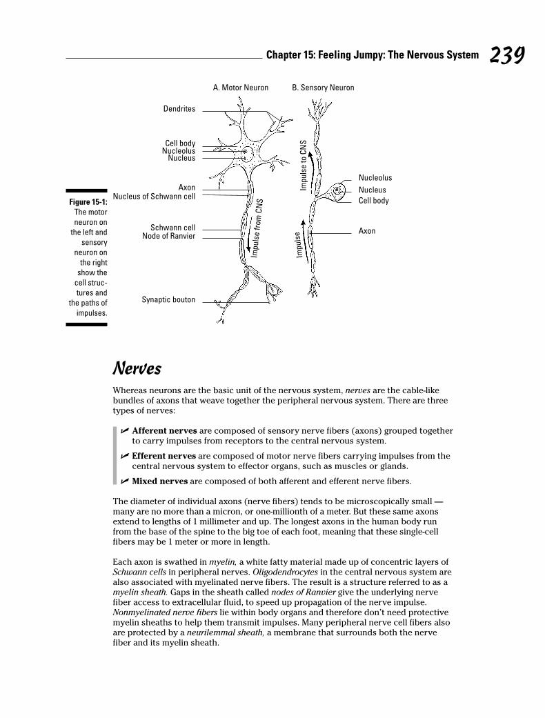
NervesWhereas neurons are the basic unit of the nervous system, nerves are the cable-likebundles of axons that weave together the peripheral nervous system. There are threetypes of nerves:
� Afferent nerves are composed of sensory nerve fibers (axons) grouped togetherto carry impulses from receptors to the central nervous system.
� Efferent nerves are composed of motor nerve fibers carrying impulses from thecentral nervous system to effector organs, such as muscles or glands.
� Mixed nerves are composed of both afferent and efferent nerve fibers.
The diameter of individual axons (nerve fibers) tends to be microscopically small —many are no more than a micron, or one-millionth of a meter. But these same axonsextend to lengths of 1 millimeter and up. The longest axons in the human body runfrom the base of the spine to the big toe of each foot, meaning that these single-cellfibers may be 1 meter or more in length.
Each axon is swathed in myelin, a white fatty material made up of concentric layers ofSchwann cells in peripheral nerves. Oligodendrocytes in the central nervous system arealso associated with myelinated nerve fibers. The result is a structure referred to as amyelin sheath. Gaps in the sheath called nodes of Ranvier give the underlying nervefiber access to extracellular fluid, to speed up propagation of the nerve impulse.Nonmyelinated nerve fibers lie within body organs and therefore don’t need protectivemyelin sheaths to help them transmit impulses. Many peripheral nerve cell fibers alsoare protected by a neurilemmal sheath, a membrane that surrounds both the nervefiber and its myelin sheath.
Dendrites
Cell body
A. Motor Neuron B. Sensory Neuron
Cell body
NucleolusNucleus
Nucleolus
Imp
uls
e t
o C
NS
Imp
uls
e
NucleusNucleus of Schwann cell
Axon
Schwann cellNode of Ranvier
Synaptic bouton
Imp
uls
e f
rom
CN
S
Axon
Figure 15-1:
The motor
neuron on
the left and
sensory
neuron on
the right
show the
cell struc-
tures and
the paths of
impulses.
239Chapter 15: Feeling Jumpy: The Nervous System

From the inside out, nerves are composed of the following:
� Axon: The impulse-conducting process of a neuron
� Myelin sheath: An insulating envelope that protects the nerve fiber and facilitatestransmission of nerve impulses
� Neurolemma (or neurilemma): A thin membrane present in many peripheralnerves that surrounds the nerve fiber and the myelin sheath
� Endoneurium: Loose, or areolar, connective tissue surrounding individual fibers
� Fasciculi: Bundles of fibers within a nerve
� Perineurium: The same kind of connective tissue as endoneurium; surrounds abundle of fibers
� Epineurium: The same kind of connective tissue as endoneurium and perineurium;surrounds several bundles of fibers
There also is a class of cells called neuroglia, or simply glia, that act as the supportivecells of the nervous system, providing neurons with nutrients and otherwise protectingthem. Glia include oligodendrocytes that support the myelin sheath within the centralnervous system; star-shaped cells called astrocytes that both support nerve tissue andcontribute to repairs when needed; and microgliacytes, cells that remove dead or dyingparts of tissue (this type of cell is called a phagocyte, which literally translates from theGreek words for “cell that eats”).
ImpulsesNeuron membranes are semi-permeable (meaning that certain small molecules likeions can move in and out but larger molecules can’t), and they’re electrically polarized(meaning that positively charged ions called cations rest around the outside mem-brane surface while negatively charged ions called anions line the inner surface; youcan find more about ions in Chapter 1).
A neuron that isn’t busy transmitting an impulse is said to be at its resting potential. Butthe nerve impulse theory, or membrane theory, says that things switch around when astimulus — a nerve impulse, or action potential — moves along the neuron. A stimuluschanges the specific permeability of the fiber membrane and causes a depolarizationdue to a reshuffling of the cations and anions. This change spreads along the nervefiber and constitutes the nerve impulse. It’s called an all-or-none response becauseeach neuron has a specific threshold of excitation. Once that threshold is exceeded, thenerve fiber responds with a fixed impulse. After depolarization, repolarization occursfollowed by a refractory period, during which no further impulses occur, even if thestimuli’s intensity increases.
Intensity of sensation, however, depends on the frequency with which one nerveimpulse follows another and the rate at which the impulse travels. That rate is deter-mined by the diameter of the impacted fiber and tends to be more rapid in large nervefibers. It’s also more rapid in myelinated fibers than nonmyelinated fibers. The cyto-plasm of the axon or nerve fiber is electrically conductive and the myelin decreasesthe capacitance to prevent charge leakage through the membrane. Depolarization atone node of Ranvier is sufficient to trigger regeneration of the voltage at the next node.Therefore, in myelinated nerve fibers the action potential does not move as a wave butrecurs at successive nodes, traveling faster than in nonmyelinated fibers. This isreferred to as saltatory conduction (from the Latin word saltare, which means “to hopor leap”).
240 Part V: Mission Control: All Systems Go
SynapsesNeurons don’t touch, which means that when a nerve impulse reaches the end of aneuron, it needs to cross a gap to the next neuron or to the gland or muscle cell forwhich the message is intended. That gap is called a synapse, or synaptic cleft. An electricsynapse — generally found in organs and glial cells — uses channels known as gap junc-tions to permit direct transmission of signals between neurons. But in other parts of thebody, chemical changes occur to let the impulse make the leap. The end branches of anaxon each form a terminal knob or bulb called a bouton terminal (that first word’s pro-nounced boo-taw), beyond which there is a space between it and the next nerve path-way. When an impulse reaches the bouton terminal, the following happens:
1. Synaptic vesicles in the knob release a transmitter called acetylcholine thatflows across the gap and increases the permeability of the next cell mem-brane in the chain.
2. An enzyme called cholinesterase breaks the transmitter down into acetyl andcholine, which then diffuse back across the gap.
3. An enzyme called choline acetylase in the synaptic vesicles reunites theacetyl and choline, prepping the bouton terminal to do its job again whenthe next impulse rolls through.
Nervous about getting all this right? Try some practice questions:
1.–5. Match the term to its description.
1. _____ Irritability
2. _____ Conductivity
3. _____ Orientation
4. _____ Coordination
5. _____ Conceptual thought
6. The brain and spinal cord are called the
a. Central nervous system
b. Visceral afferent system
c. Autonomic nervous system
d. Peripheral nervous system
7. The functional unit of the nervous system is the
a. Axon
b. Nephron
c. Dendron
d. Neuron
241Chapter 15: Feeling Jumpy: The Nervous System
a. Tissue’s ability to respond to stimulation
b. Ability to receive impulses and direct them to channelsfor favorable response
c. Sense organs’ capacity to generate nerve impulse tostimulation
d. Spreading of the nerve impulse
e. Capacity to record, store, and relate information to be used to determine future action
8. The terminal structure of the cytoplasmic projection of the neuron cannot be a(n)
a. Node of Ranvier
b. End organ
c. Effector
d. End bulb
e. Receptor
9. The afferent fiber that carries impulses to a neuron’s cell body is called a
a. Nissl body
b. Neuron
c. Dendrite
d. Axon
e. Mitochondria
10. The membrane surrounding the axon (nerve fiber) is the
a. Sarcolemma
b. Neurilemma
c. Perineurium
d. Epineurium
11.–15. Match the term to its description.
11. _____ Astrocytes
12. _____ Microgliacytes
13. _____ Oligodendrocytes
14. _____ Axons
15. _____ Dendrites
16. The neuroglia cells are important as
a. Sensory tissue
b. Supporting tissue
c. Irritable tissue
d. Conducting tissue
17. Axons tend to consist of
a. Single processes
b. Several synapses
c. Multiple processes
d. None of the above
e. Both a and c
242 Part V: Mission Control: All Systems Go
a. Cytoplasmic projections carrying impulses to the cell body
b. Cells that form and preserve myelin sheaths
c. Cytoplasmic projections carrying impulses from the cell body
d. Cells that are phagocytic
e. Cells that contribute to the repair process of the centralnervous system
18. A synapse between neurons is best described as the
a. Transmission of a continuous impulse
b. Transmission of an electrical impulse
c. Transmission of an impulse through a chemical and physical change
d. Transmission of an impulse through a physical change
e. Transmission of an impulse through a chemical change
19.–23. Match the term to its description.
19. _____ Endoneurium a. Fatty layer around an axon fiber
20. _____ Neurilemma b. Outer thin membrane around an axon fiber
21. _____ Schwann cell c. Cell in the sheath of an axon
22. _____ Node of Ranvier d. Depression in the sheath around a fiber
23. _____ Myelin sheath e. Connective tissue surrounding individual fibers ina nerve
24.–28. Match the term to its description.
24. _____ All-or-none response
25. _____ Cation
26. _____ Anion
27. _____ Polarization
28. _____ Depolarization
29.–33. Match the term to its description.
29. _____ Cholinesterase
30. _____ Choline acetylase
31. _____ Terminal bulb
32. _____ Acetylcholine
33. _____ Synapse
Minding the Central Nervous System and the Brain
Together, the brain and spinal cord make up the central nervous system. The spinalcord, which forms very early in the embryonic spinal canal, extends down into the tailportion of the vertebral column. But because bone grows much faster than nervetissue, the end of the cord soon is too short to extend into the lowest reaches of thespinal canal. In an adult, the 18-inch spinal cord ends between the first and secondlumbar vertebrae, roughly where the last ribs attach. Its tapered end is called the conusmedullaris. The cord continues as separate strands below that point and is referred toas the cauda equina (horse tail). A thread of fibrous tissue called the filum terminaleextends to the base of the coccyx (tailbone) and is attached by the coccygeal ligament.
243Chapter 15: Feeling Jumpy: The Nervous System
a. Impermeability of cell membrane
b. Negatively charged ion on the inner surface of the cellmembrane
c. Threshold of excitation determines ability to respond
d. Positively charged ion on the outer surface of the cellmembrane
e. Reshuffling of cell membrane ions; permeability ofcell membrane
a. Excitatory chemical necessary for continualnerve pathway
b. Enzyme for breakdown of excitatory chemical
c. Enzyme for reformation of excitatory chemical
d. Space between neurons
e. Contains storage vesicles for excitatory chemical
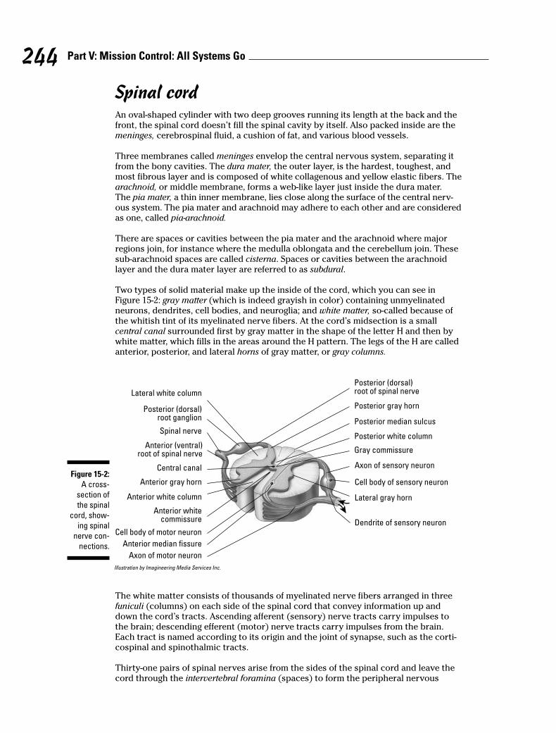
Spinal cordAn oval-shaped cylinder with two deep grooves running its length at the back and thefront, the spinal cord doesn’t fill the spinal cavity by itself. Also packed inside are themeninges, cerebrospinal fluid, a cushion of fat, and various blood vessels.
Three membranes called meninges envelop the central nervous system, separating itfrom the bony cavities. The dura mater, the outer layer, is the hardest, toughest, andmost fibrous layer and is composed of white collagenous and yellow elastic fibers. Thearachnoid, or middle membrane, forms a web-like layer just inside the dura mater.The pia mater, a thin inner membrane, lies close along the surface of the central nerv-ous system. The pia mater and arachnoid may adhere to each other and are consideredas one, called pia-arachnoid.
There are spaces or cavities between the pia mater and the arachnoid where majorregions join, for instance where the medulla oblongata and the cerebellum join. Thesesub-arachnoid spaces are called cisterna. Spaces or cavities between the arachnoidlayer and the dura mater layer are referred to as subdural.
Two types of solid material make up the inside of the cord, which you can see inFigure 15-2: gray matter (which is indeed grayish in color) containing unmyelinatedneurons, dendrites, cell bodies, and neuroglia; and white matter, so-called because ofthe whitish tint of its myelinated nerve fibers. At the cord’s midsection is a smallcentral canal surrounded first by gray matter in the shape of the letter H and then bywhite matter, which fills in the areas around the H pattern. The legs of the H are calledanterior, posterior, and lateral horns of gray matter, or gray columns.
Illustration by Imagineering Media Services Inc.
The white matter consists of thousands of myelinated nerve fibers arranged in threefuniculi (columns) on each side of the spinal cord that convey information up anddown the cord’s tracts. Ascending afferent (sensory) nerve tracts carry impulses tothe brain; descending efferent (motor) nerve tracts carry impulses from the brain.Each tract is named according to its origin and the joint of synapse, such as the corti-cospinal and spinothalmic tracts.
Thirty-one pairs of spinal nerves arise from the sides of the spinal cord and leave thecord through the intervertebral foramina (spaces) to form the peripheral nervous
Spinal nerve
Lateral white column
Anterior (ventral)root of spinal nerve
Anterior gray horn
Central canal
Anterior white column
Anterior whitecommissure
Anterior median fissure
Cell body of motor neuron
Axon of motor neuron
Posterior (dorsal)root of spinal nerve
Posterior gray horn
Posterior median sulcus
Posterior white column
Gray commissure
Axon of sensory neuron
Lateral gray horn
Cell body of sensory neuron
Dendrite of sensory neuron
Posterior (dorsal)root ganglion
Figure 15-2:
A cross-
section of
the spinal
cord, show-
ing spinal
nerve con-
nections.
244 Part V: Mission Control: All Systems Go
system, which we discuss in the later section “Taking Side Streets: The PeripheralNervous System.”
BrainOne of the largest organs in the adult human body, the brain tips the scales at 3 poundsand packs roughly 100 billion neurons (yes, that’s billion with a “b”) and 900 billionsupporting neuroglia cells. In this section, we review six major divisions of the brainfrom the bottom up (see Figure 15-3): medulla oblongata, pons, midbrain, cerebellum,diencephalon, and cerebrum.
Medulla oblongataThe spinal cord meets the brain at the medulla oblongata, or brainstem, just below theright and left cerebellar hemispheres of the brain. In fact, the medulla oblongata is con-tinuous with the spinal cord at its base (inferiorly) and back (dorsally) and locatedanteriorly and superiorly to the pons. All the afferent and efferent tracts of the cordcan be found in the brainstem as part of two bulges of white matter forming an areareferred to as the pyramids. Many of the tracts cross from one side to the other at thepyramids, which explains why the right side of the brain controls the left side of thebody and vice versa.
Along with the pons, the medulla oblongata also forms a network of gray and whitematter called the reticular formation, the upper part of the so-called extrapyramidalpathway. With its capacity to arouse the brain to wakefulness, it keeps the brain alert,directs messages in the form of impulses, monitors stimuli entering the sense recep-tors (accepting some and rejecting others it deems to be irrelevant), refines bodymovements, and effects higher mental processes such as attention, introspection, andreasoning. Although the cortex of the cerebrum is the actual powerhouse of thought, itmust be stimulated into action by signals from the reticular formation.
Nerve cells in the brainstem are grouped together to form nerve centers (nuclei) thatcontrol bodily functions, including cardiac activities, and respiration as well as reflexactivities such as sneezing, coughing, vomiting, and alimentary tract movements. Themedulla oblongata affects these reactions through the vagus, also referred to as cra-nial nerve X or the 10th cranial nerve. Three other cranial nerves also originate fromthis area: the 9th (IX) or glossopharyngeal, 11th (XI) or accessory, and 12th (XII) orhypoglossal.
PonsThe pons (literally “bridge”) does exactly as its name implies: It connects the cerebel-lum through a structure called the middle peduncle, the cerebrum by the superiorpeduncle, and the medulla oblongata by the inferior peduncle. It also unites the cere-bellar hemispheres, coordinates muscles on both sides of the body, controls facialmuscles (including those used to chew), and regulates the first stage of respiration.Oh, and it contains the nuclei for the following cranial nerves: the 5th (V) or trigemi-nal, the 6th (VI) or abducens, 7th (VII) or facial, and 8th (VIII) or vestibulocochlear.
MidbrainBetween the pons and the diencephalon lies the mesencephalon, or midbrain. It con-tains the corpora quadrigemina, which correlates optical and tactile impulses as wellas regulates muscle tone, body posture, and equilibrium through reflex centers in thesuperior colliculus. The inferior colliculus contains auditory reflex centers and isbelieved to be responsible for the detection of musical pitch. The midbrain contains
245Chapter 15: Feeling Jumpy: The Nervous System
the cerebral aqueduct, which connects the third ventricle of the thalamus with thefourth ventricle of the medulla oblongata (see the section “Ventricles” later in thischapter for more). The mesencephalon contains nuclei for the 3rd (III) or oculomotorcranial nerve and the 4th (IV) or trochlear cranial nerve. The red nucleus that containsfibers of the rubrospinal tract, a motor tract that acts as a relay station for impulsesfrom the cerebellum and higher brain centers, also lies within the midbrain, constitut-ing the superior cerebellar peduncle.
CerebellumThe cerebellum also is known as the little brain or small brain. The second-largest divi-sion of the brain, it’s just above and overhangs the medulla oblongata and lies justbeneath the rear portion of the cerebrum. Inside, the cerebellum resembles a tree calledthe arbor vitae, or “tree of life.” A central body called the vermis connects the two lateralmasses called the cerebellar hemispheres and assists in motor coordination and refine-ment of muscular movement, aiding equilibrium and muscle tone. The cerebellar cortexor gray matter contains Purkinje neurons with pear-shaped cell bodies, a multitude ofdendrites, and a single axon. It sends impulses to the white matter of the cerebellum andto other deeper nuclei in the cerebellum, and then to the brainstem. The cerebellarcortex has parallel ridges called the folia cerebelli, which are separated by deep sulci.
DiencephalonThe diencephalon, a region between the mesencephalon and the cerebrum, containsseparate brain structures called the thalamus, epithalamus, subthalamus, and hypothal-amus. The region where the two sides of the thalami come in contact and join forces iscalled the intermediate mass. The thalamus is a primitive receptive center throughwhich the sensory impulses travel on their way to the cerebral cortex. Here, nervefibers from the spinal cord and lower parts of the brain synapse with neurons leadingto the sensory areas of the cortex of the cerebrum. The thalamus is the great integrat-ing center of the brain with the ability to correlate the impulses from tactile, pain,olfactory, and gustatory (taste) senses with motor reactions.
The epithalamus contains the choroid plexus, a vascular structure that produces spinalfluid. The pineal body and olfactory centers also lie within the epithalamus, whichforms the roof of the third ventricle. The subthalamus is located below the thalamusand regulates the muscles of emotional expression.
The hypothalamus contains the centers for sexual reflexes; body temperature; water,carbohydrate, and fat metabolism; and emotions that affect the heartbeat and bloodpressure. It also has the optic chiasm (connecting the optic nerves to the optic tract),the posterior lobe of the pituitary gland, and a funnel-shaped region called theinfundibulum that forms the stalk of the pituitary gland.
CerebrumThe cerebrum, or forebrain, is often called the true brain. It has two cerebral hemi-spheres — the right and the left. A thin outer layer of gray matter called the cerebralcortex features folds or convolutions called gyri; furrows and grooves are referred to assulci, and deeper grooves are called fissures. A longitudinal fissure separates the cere-brum. The transverse fissure separates the cerebrum and the cerebellum. Each hemi-sphere has a set of controls for sensory and motor activities of the body. Interestingly,it’s not just right-side/left-side controls that are reversed in the cerebrum; the upperareas of the cerebral cortex control the lower body activities while the lower areas ofthe cortex control upper-body activities in a reversal called “little man upside down.”
Commissural fibers, a tract of nerves running from one side of the brain to the other,coordinate activities between the right and left hemispheres. The corpus callosum
246 Part V: Mission Control: All Systems Go
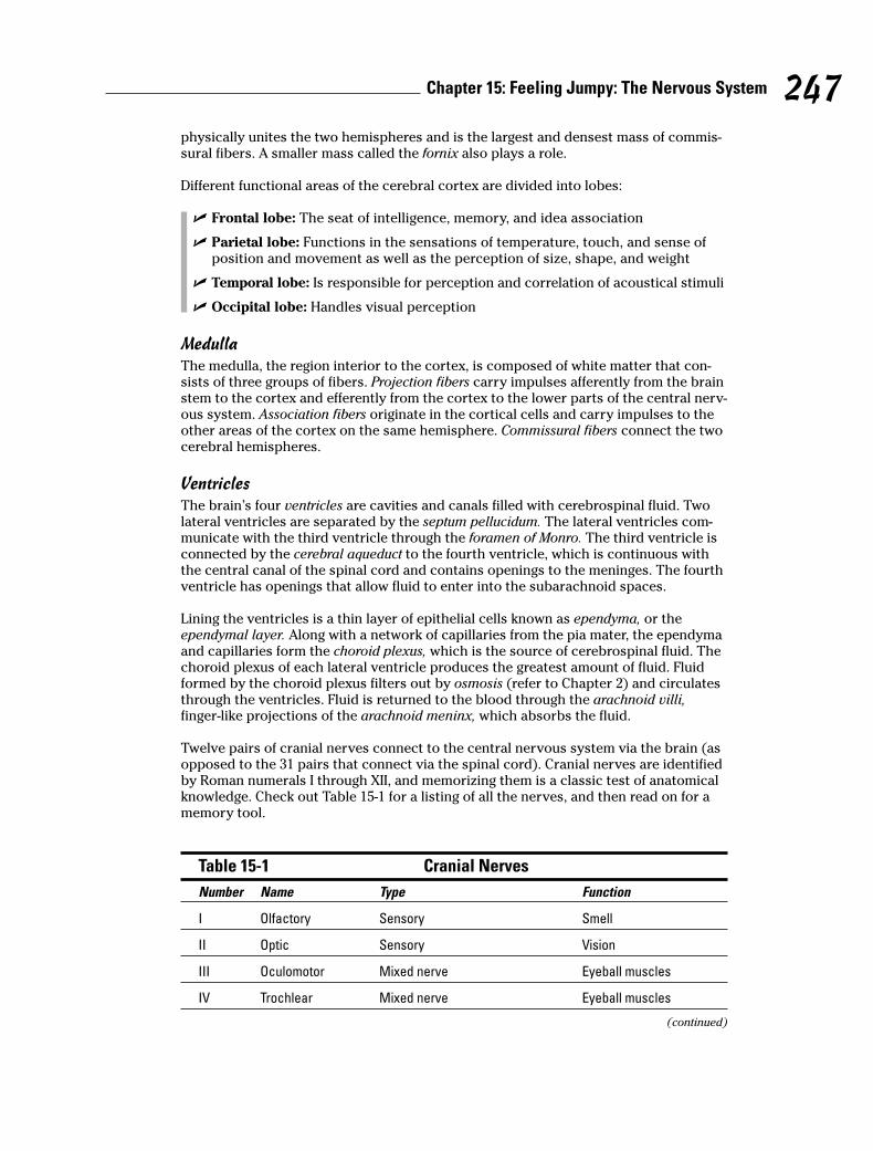
physically unites the two hemispheres and is the largest and densest mass of commis-sural fibers. A smaller mass called the fornix also plays a role.
Different functional areas of the cerebral cortex are divided into lobes:
� Frontal lobe: The seat of intelligence, memory, and idea association
� Parietal lobe: Functions in the sensations of temperature, touch, and sense ofposition and movement as well as the perception of size, shape, and weight
� Temporal lobe: Is responsible for perception and correlation of acoustical stimuli
� Occipital lobe: Handles visual perception
MedullaThe medulla, the region interior to the cortex, is composed of white matter that con-sists of three groups of fibers. Projection fibers carry impulses afferently from the brainstem to the cortex and efferently from the cortex to the lower parts of the central nerv-ous system. Association fibers originate in the cortical cells and carry impulses to theother areas of the cortex on the same hemisphere. Commissural fibers connect the twocerebral hemispheres.
VentriclesThe brain’s four ventricles are cavities and canals filled with cerebrospinal fluid. Twolateral ventricles are separated by the septum pellucidum. The lateral ventricles com-municate with the third ventricle through the foramen of Monro. The third ventricle isconnected by the cerebral aqueduct to the fourth ventricle, which is continuous withthe central canal of the spinal cord and contains openings to the meninges. The fourthventricle has openings that allow fluid to enter into the subarachnoid spaces.
Lining the ventricles is a thin layer of epithelial cells known as ependyma, or theependymal layer. Along with a network of capillaries from the pia mater, the ependymaand capillaries form the choroid plexus, which is the source of cerebrospinal fluid. Thechoroid plexus of each lateral ventricle produces the greatest amount of fluid. Fluidformed by the choroid plexus filters out by osmosis (refer to Chapter 2) and circulatesthrough the ventricles. Fluid is returned to the blood through the arachnoid villi,finger-like projections of the arachnoid meninx, which absorbs the fluid.
Twelve pairs of cranial nerves connect to the central nervous system via the brain (asopposed to the 31 pairs that connect via the spinal cord). Cranial nerves are identifiedby Roman numerals I through XII, and memorizing them is a classic test of anatomicalknowledge. Check out Table 15-1 for a listing of all the nerves, and then read on for amemory tool.
Table 15-1 Cranial Nerves
Number Name Type Function
I Olfactory Sensory Smell
II Optic Sensory Vision
III Oculomotor Mixed nerve Eyeball muscles
IV Trochlear Mixed nerve Eyeball muscles
(continued)
247Chapter 15: Feeling Jumpy: The Nervous System
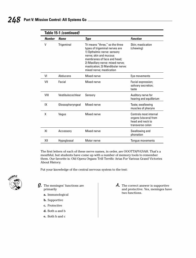
Table 15-1 (continued)
Number Name Type Function
V Trigeminal Tri means “three,” so the three Skin; mastication types of trigeminal nerves are (chewing)1) Opthalmic nerve: sensory nerve; skin and mucous membranes of face and head; 2) Maxillary nerve: mixed nerve; mastication; 3) Mandibular nerve: mixed nerve; mastication
VI Abducens Mixed nerve Eye movements
VII Facial Mixed nerve Facial expression; salivary secretion;taste
VIII Vestibulocochlear Sensory Auditory nerve forhearing and equilibrium
IX Glossopharyngeal Mixed nerve Taste; swallowingmuscles of pharynx
X Vagus Mixed nerve Controls most internalorgans (viscera) fromhead and neck totransverse colon
XI Accessory Mixed nerve Swallowing andphonation
XII Hypoglossal Motor nerve Tongue movements
The first letters of each of these nerve names, in order, are OOOTTAFVGVAH. That’s amouthful, but students have come up with a number of memory tools to rememberthem. Our favorite is: Old Opera Organs Trill Terrific Arias For Various Grand VictoriesAbout History.
Put your knowledge of the central nervous system to the test:
248 Part V: Mission Control: All Systems Go
Q. The meninges’ functions areprimarily
a. Immunological
b. Supportive
c. Protective
d. Both a and b
e. Both b and c
A. The correct answer is supportiveand protective. Yes, meninges havetwo functions.
34. The cerebrum consists of two major halves called
a. Cerebellar hemispheres
b. Cerebral spheres
c. Cerebellar spheres
d. Cerebral hemispheres
35. The cerebrum is divided into two major halves by the
a. Lateral fissure
b. Transverse fissure
c. Longitudinal fissure
d. Fissure of Sylvius
e. Central sulcus
36. In the cerebrum, the
a. Right side tends to control the left side of the body and vice versa
b. Upper area controls lower-body activity
c. Lower area controls upper-body activity
d. None of the above are correct
e. A, b, and c are correct
37. The functions of the occipital lobe of the cerebrum pertain principally to
a. Visual activity
b. Autonomic control
c. Associative reasoning
d. Motor coordination
e. Auditory control
38. The cerebellum functions primarily as a center of
a. Visual activity
b. Associative reasoning
c. Auditory activity
d. Autonomic coordination
e. Motor control
39.–43. Match the term to its description.
39. _____ White matter
40. _____ Reticular formation
41. _____ Funiculus
42. _____ Dorsal root ganglion
43. _____ Gray matter
249Chapter 15: Feeling Jumpy: The Nervous System
a. Has the capacity to arouse the brain to wakefulness
b. Myelinated fibers
c. Bundles of nerve fibers arranged in tracts
d. Collection of cell bodies outside of the central nervoussystem
e. Unmyelinated fibers, cell bodies, and neuroglia
44.-48.Match the term to its description.
44. _____ Pons
45. _____ Cerebellum
46. _____ Medulla oblongata
47. _____ Cerebrum
48. _____ Mesencephalon
49. The largest quantity of cerebrospinal fluid originates from the
a. Foramen of Monro
b. Arachnoid villi
c. Lateral ventricle
d. Optic chiasm
e. Foramen of Luschka
50. The part of the brain that contains the thalamus, pituitary gland, and the optic chiasm is the
a. Diencephalon
b. Mesencephalon
c. Myelencephalon
d. Telencephalon
e. Metencephalon
51.–62. Use the terms that follow to identify the parts of the brain shown in Figure 15-3.
a. Pons
b. Thalamus
c. Cerebellum
d. Corpus callosum
e. Third ventricle
f. Hypothalamus
g. Cerebrum
h. Cerebral aqueduct
i. Midbrain
j. Pituitary gland
k. Medulla oblongata
l. Fourth ventricle
250 Part V: Mission Control: All Systems Go
a. Bridge connecting the medulla oblongata andcerebellum
b. Contains the centers that control cardiac, respiratory,and vasomotor functions
c. Contains the corpora quadrigemina and nuclei for theoculomotor and trochlear nerves
d. Controls motor coordination and refinement ofmuscular movement
e. Controls sensory and motor activity of the body

LifeART Image Copyright © 2007. Wolters Kluwer Health — Lippincott Williams & Wilkins
Taking Side Streets: The PeripheralNervous System
The peripheral nervous system is the network that carries information to and from thespinal cord. Among its key structures are 31 pairs of spinal nerves (see Figure 15-4),each originating in a segment of the spinal cord called a neuromere. Eight of the spinalnerve pairs are cervical (having to do with the neck), 12 are thoracic (relating to thechest, or thorax), five are lumbar (between the lowest ribs and the pelvis), five aresacral (the posterior section of the pelvis), and one is coccygeal (relating to the tail-bone). Spinal nerves connect with the spinal cord by two bundles of nerve fibers, orroots. The dorsal root contains afferent fibers that carry sensory information fromreceptors to the central nervous system. The cell bodies of these sensory neurons lieoutside the spinal cord in a bulging area called the dorsal root ganglion (refer to thecross-section of the spinal cord in Figure 15-2). A second bundle, the ventral root, con-tains efferent motor fibers with cell bodies that lie inside the spinal cord. In eachspinal nerve, the two roots join outside the spinal cord to form what’s called amixed spinal nerve.
Spinal reflexes, or reflex arcs, occur when a sensory neuron transmits a “danger”signal — like a sensation of burning heat — through the dorsal root ganglion. An inter-nuncial neuron (or association neuron) in the spinal cord passes along the signal to amotor neuron (or efferent fiber) that stimulates a muscle, which immediately pulls theburning body part away from heat (see Figure 15-5).
To spinal cord
51 _____
52 _____
53 _____
54 _____55 _____
56 _____
57 _____
58 _____59 _____
60 _____
61 _____
62 _____
Figure 15-3:
Sagittal
view of the
brain.
251Chapter 15: Feeling Jumpy: The Nervous System
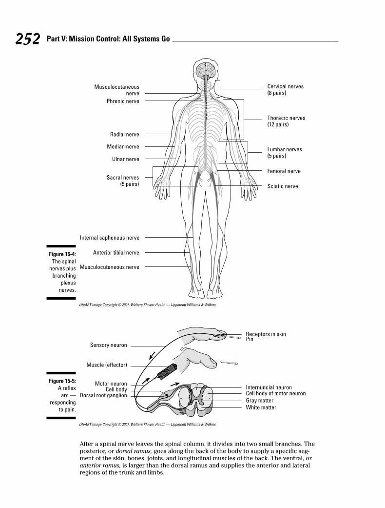
LifeART Image Copyright © 2007. Wolters Kluwer Health — Lippincott Williams & Wilkins
LifeART Image Copyright © 2007. Wolters Kluwer Health — Lippincott Williams & Wilkins
After a spinal nerve leaves the spinal column, it divides into two small branches. Theposterior, or dorsal ramus, goes along the back of the body to supply a specific seg-ment of the skin, bones, joints, and longitudinal muscles of the back. The ventral, oranterior ramus, is larger than the dorsal ramus and supplies the anterior and lateralregions of the trunk and limbs.
Receptors in skin
Sensory neuron
Muscle (effector)
Motor neuron
Pin
Dorsal root ganglion
Internuncial neuronCell bodyCell body of motor neuron
Gray matter
White matter
Figure 15-5:
A reflex
arc —
responding
to pain.
Cervical nerves(8 pairs)
Thoracic nerves(12 pairs)
Lumbar nerves(5 pairs)
Femoral nerve
Sciatic nerve
Musculocutaneousnerve
Phrenic nerve
Radial nerve
Sacral nerves(5 pairs)
Internal saphenous nerve
Anterior tibial nerve
Musculocutaneous nerve
Median nerve
Ulnar nerve
Figure 15-4:
The spinal
nerves plus
branching
plexus
nerves.
252 Part V: Mission Control: All Systems Go
Groups of spinal nerves interconnect to form an extensive network called a plexus(Latin for “braid”), each of which connects through the anterior ramus, including thecervical plexus of the neck, brachial plexus of the arms, and lumbosacral plexus ofthe lower back (including the body’s largest nerve, the sciatic nerve). However, there’sno plexus in the thoracic area. Instead, the anterior ramus directly supplies the inter-costal muscles (literally “between the ribs”) and the skin of the region.
63. The network of nerves formed by the ventral branch of the spinal nerve is a
a. Pedicle
b. Papilla
c. Plexus
d. Plica
e. Phallus
64. How many spinal nerves are there?
a. 61
b. 31 pairs
c. 13 pairs
d. 1
e. 12
65. Which spinal nerve area does not form a plexus?
a. Cervical
b. Lumbar
c. Sacral
d. Thoracic
Keep Breathing: The AutonomicNervous System
Just as the name implies, the autonomic nervous system functions automatically.Divided into the sympathetic and parasympathetic systems, it activates the involuntarysmooth and cardiac muscles and glands to serve such vital systems that function auto-matically as the digestive tract, circulatory system, respiratory, urinary, and endocrinesystems. Autonomic functions are under the control of the hypothalamus, cerebralcortex, and medulla oblongata. The sympathetic system, which is responsible for thebody’s involuntary fight-or-flight response to stress, is defined by the autonomic fibersthat exit the thoracic and lumbar segments of the spinal cord. The parasympatheticsystem is defined by the autonomic fibers that either exit the brainstem via the cranialnerves or exit the sacral segments of the spinal cord.
The sympathetic and parasympathetic systems oppose each other in function, helpingto maintain homeostasis, or balanced activity in the body systems. Yet, often the sympa-thetic and parasympathetic systems work in concert. The sympathetic system dilatesthe eye’s pupil, but the parasympathetic system contracts it again. The sympatheticsystem quickens and strengthens the heart while the parasympathetic slows the heart’saction. The sympathetic system contracts blood vessels in the skin so more blood goesto muscles for a fight-or-flight reaction to stress, and the parasympathetic systemdilates the blood vessels when the stress concludes.
253Chapter 15: Feeling Jumpy: The Nervous System
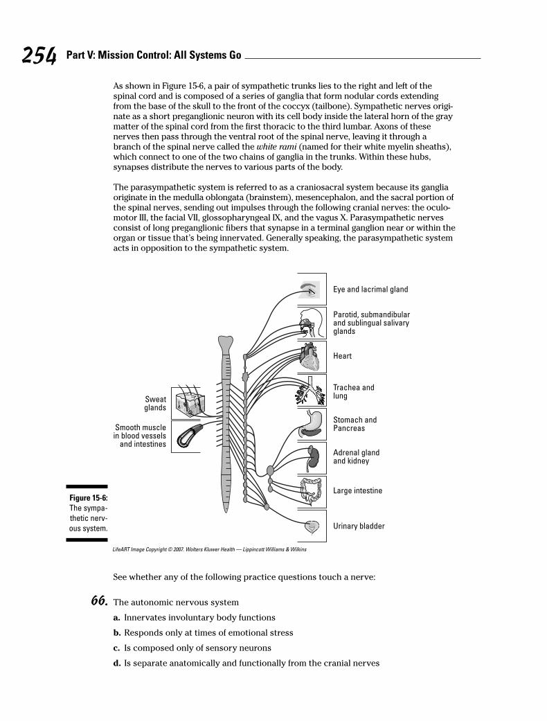
As shown in Figure 15-6, a pair of sympathetic trunks lies to the right and left of thespinal cord and is composed of a series of ganglia that form nodular cords extendingfrom the base of the skull to the front of the coccyx (tailbone). Sympathetic nerves origi-nate as a short preganglionic neuron with its cell body inside the lateral horn of the graymatter of the spinal cord from the first thoracic to the third lumbar. Axons of thesenerves then pass through the ventral root of the spinal nerve, leaving it through abranch of the spinal nerve called the white rami (named for their white myelin sheaths),which connect to one of the two chains of ganglia in the trunks. Within these hubs,synapses distribute the nerves to various parts of the body.
The parasympathetic system is referred to as a craniosacral system because its gangliaoriginate in the medulla oblongata (brainstem), mesencephalon, and the sacral portion ofthe spinal nerves, sending out impulses through the following cranial nerves: the oculo-motor III, the facial VII, glossopharyngeal IX, and the vagus X. Parasympathetic nervesconsist of long preganglionic fibers that synapse in a terminal ganglion near or within theorgan or tissue that’s being innervated. Generally speaking, the parasympathetic systemacts in opposition to the sympathetic system.
LifeART Image Copyright © 2007. Wolters Kluwer Health — Lippincott Williams & Wilkins
See whether any of the following practice questions touch a nerve:
66. The autonomic nervous system
a. Innervates involuntary body functions
b. Responds only at times of emotional stress
c. Is composed only of sensory neurons
d. Is separate anatomically and functionally from the cranial nerves
Eye and lacrimal gland
Sweatglands
Smooth musclein blood vessels
and intestines
Heart
Trachea andlung
Stomach andPancreas
Adrenal glandand kidney
Large intestine
Urinary bladder
Parotid, submandibularand sublingual salivaryglands
Figure 15-6:
The sympa-
thetic nerv-
ous system.
254 Part V: Mission Control: All Systems Go
67. Which of the following statements is true about the autonomic nervous system?
a. It has two parts: the parasympathetic that controls all normal functions, and the sympatheticthat carries out the same functions.
b. It’s the nervous system that controls all reflexes.
c. It doesn’t function when the body’s under stress.
d. It has two divisions that are antagonistic to each other, meaning that one counteracts theeffects of the other one.
e. It controls the contractions of the skeletal, smooth, and cardiac muscle tissue.
68. The divisions of the autonomic nervous system are
a. Sympathetic and peripheral
b. Somatic and peripheral
c. Parasympathetic and peripheral
d. Sympathetic and parasympathetic
69. Which part of the autonomic nervous system can be called a craniosacral system?
a. Ganglia
b. Sympathetic trunks
c. Parasympathetic system
d. Medulla oblongata
Coming To Your SensesThe nervous system must have some way to perceive its environment in order to gen-erate appropriate responses. That’s where the senses come in. Sense receptors arethose numerous organs that respond to stimuli — like increased temperature, bittertastes, and sharp points — by generating a nerve impulse. While there are millions ofgeneral sense receptors found throughout the body that can convey touch, pain, andphysical contact, there are far fewer of the special sense receptors — those located inthe head — that really bring meaning to your world.
Sense receptors are classified by the stimuli they receive, as follows:
� Exteroceptors: Receive stimuli from the external environment. These are sen-sory nerve terminals, such as those in the skin and mucous membranes, that arestimulated by the immediate external environment.
� Interoceptors: Receive stimuli from the internal environment. These can be anyof the sensory nerve terminals located in and transmitting impulses from theviscera.
� Proprioceptors: Part of the “true” internal environment. They’re sensory nerveterminals chiefly found in muscles, joints, and tendons that give informationconcerning movements and position of the body.
� Teleceptors: Sensory nerve terminals stimulated by emanations from distantobjects. They exist in the eyes, ears, and nose.
255Chapter 15: Feeling Jumpy: The Nervous System
EyesAlthough there are many romantic notions about eyes, the truth is that an eyeball issimply a hollow sphere bounded by a trilayer wall and filled with a gelatinous fluidcalled, oddly enough, vitreous humor (see Figure 15-7). The outer fibrous coat is madeup of the sclera in back and the cornea in front. The sclera provides mechanicalsupport, protection, and a base for attachment of eye muscles, and it assists in thefocusing process. The cornea covers the anterior with a clear window.
An intermediate, or vascular, coat called the uvea provides blood and lymphatic fluids tothe eye, regulates light, and also secretes and reabsorbs aqueous humor, a thin wateryliquid that fills the anterior chamber of the eyeball in front of the iris. A pigmented coathas three layers: the iris, containing blood vessels, pigment cells, and smooth musclefibers to control the pupil’s diameter; the ciliary body, which is attached to the peripheryof the iris; and the choroid, a thin, dark brown, vascular layer lining most of the sclera onthe back and sides of the eye. The choroid contains arteries, veins, and capillaries thatsupply the retina with nutrients, and it also contains pigment cells to absorb light andprevent reflection and blurring. An optic nerve enters at the back (posterior) of each eye.
The retina is part of an internal nervous layer that connects with the optic nerve. Thenervous tissue layers along the inner back of the eye contain rods and cones (types ofneurons that analyze visual input). The rods are dim light receptors whereas the conesdetect bright light and construct form, structure, and color. The retina has an opticdisc, which is essentially a blind spot incapable of producing an image.
The crystalline lens consists of concentric layers of protein. It’s biconcave in shape,bulging outward. Located behind the pupil and iris, the lens is held in place by liga-ments attached to the ciliary muscles. When the ciliary muscles contract, the shape ofthe lens changes, altering the visual focus. This process of accommodation allows theeye to see objects both at a distance and close up.
The palpebrae (eyelids) extend from the edges of the eye orbit, into which roughlyfive-sixths of the eyeball is recessed. Eyelids come together at medial and lateralangles of the eye that are called the canthi. In the medial angle of the eye is a pinkregion called the caruncula, or caruncle. The caruncula contains sebaceous glands andsudoriferous (sweat) glands. A mucous membrane called the conjunctiva covers theinner surface of each eyelid and the anterior surface of the eye. Up top and to the sideof the orbital cavities are lacrimal glands that secrete tears that are carried through aseries of lacrimal ducts to the conjunctiva of the upper eyelid. Ultimately, secretionsdrain from the eyes through the nasolacrimal ducts.
EarsHuman ears — otherwise called vestibulocochlear organs — are more than just organsof hearing. They also serve as organs of equilibrium, or balance. Here are the threedivisions of the ear:
� The external ear includes the auricle, or pinna, which is the folded, roundedappendage made of cartilage and skin. Extending into the skull is the ear canal,or external auditory meatus, a short passage through the temporal bone endingat the tympanic membrane, or eardrum. Sebaceous glands near the external open-ing and ceruminous glands in the upper wall produce the brownish substanceknown as earwax, or cerumen.
256 Part V: Mission Control: All Systems Go
� The middle ear is a small, usually air-filled cavity in the skull that’s lined withmucous membrane. It communicates through the Eustachian tube with the phar-ynx. The Eustachian tube keeps air pressure equal on both sides of the eardrum(tympanic membrane), equalizing pressure in the middle ear with atmosphericpressure from outside. Three small bones called auditory ossicles occupy themiddle ear, deriving their names from their shapes: the malleus (hammer), theincus (anvil), and the stapes (stirrup).
� The internal ear is the most complex structure of the entire organ because it’swhere vibrations are translated. It’s composed of a group of interconnectedcanals or channels called the cochlea. Within the cochlea are three canals sepa-rated from each other by thin membranes; two of the canals — the vestibular andthe tympanic — are bony chambers filled with a perilymph fluid, and the thirdcanal — the cochlear canal — is a membranous chamber filled with endolymph.The cochlear canal lies between the vestibular and tympanic canals and containsthe organ of Corti, a spiral-shaped organ made up of cells with projecting hairsthat transmit auditory impulses.
The process of hearing a sound follows these basic steps:
1. Sound waves travel through the auditory canal, striking the eardrum andmaking it vibrate and setting the three ossicle bones into motion.
2. The stapes at the end of the chain strikes against the oval window of thevestibular canal, translating the motion into the perilymph fluid in the vestibu-lar and tympanic canals of the cochlea.
3. The vibrating fluid begins moving the basilar membrane that separates thetwo canals, stimulating the endolymph fluid in the membranous area ofthe cochlea.
4. The stimulated endolymph fluid in turn stimulates the hair cells of the organof Corti, which transmit the impulses to the brain over the auditory nerve.
That’s the hearing part of your ears. Equilibrium requires that some additional partscome into play. Three semicircular canals, each with an ampulla (or small, dilated por-tion) at each end, lie at right angles to each other. The ampullae connect to a fluid-filledsac called a utricle, which in turn connects to another fluid-filled sac called a saccule.Both sacs contain regions called maculae that are lined with sensitive hairs and containconcretions (solid masses) of calcium carbonate called otoliths (or otoconia). Whenlinear acceleration pulls at them, the otoliths press on the hair cells and initiate animpulse to the brain through basal sensory nerve fibers. When the head changes posi-tion, it causes a change in the direction of force on the hairs. Movement of the hairsstimulates dendrites of the vestibulocochlear nerve (the eighth cranial nerve) to carryimpulses to the brain.
257Chapter 15: Feeling Jumpy: The Nervous System
Q. The most sensitive region of theretina producing the greatest visualacuity is the
a. Blind spot
b. Cornea
c. Fovea centralis
d. Macula lutea
e. Lens
A. The correct answer is foveacentralis. It’s loaded with light-sensitive cones.
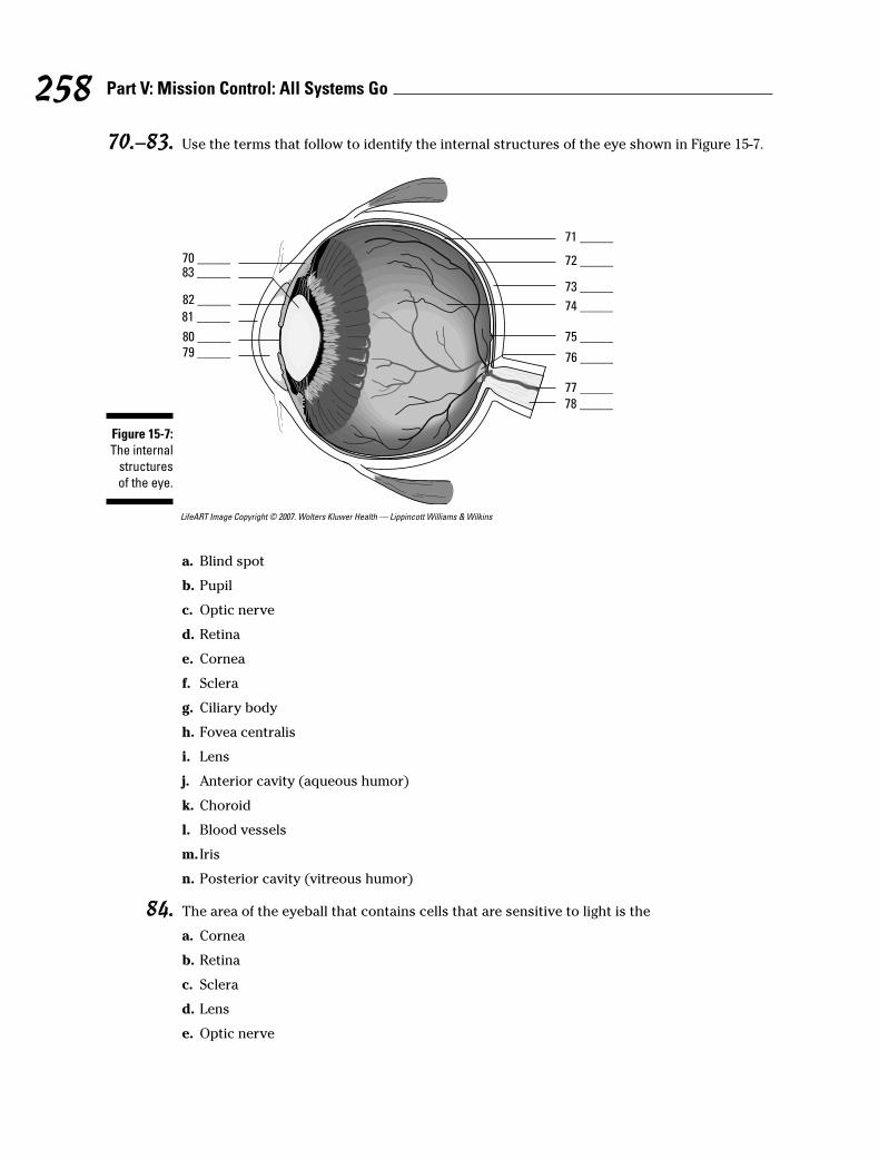
70.–83. Use the terms that follow to identify the internal structures of the eye shown in Figure 15-7.
LifeART Image Copyright © 2007. Wolters Kluwer Health — Lippincott Williams & Wilkins
a. Blind spot
b. Pupil
c. Optic nerve
d. Retina
e. Cornea
f. Sclera
g. Ciliary body
h. Fovea centralis
i. Lens
j. Anterior cavity (aqueous humor)
k. Choroid
l. Blood vessels
m. Iris
n. Posterior cavity (vitreous humor)
84. The area of the eyeball that contains cells that are sensitive to light is the
a. Cornea
b. Retina
c. Sclera
d. Lens
e. Optic nerve
70 _____83 _____
82 _____
81 _____
80 _____79 _____
78 _____
77 _____
76 _____
75 _____
74 _____
73 _____
72 _____
71 _____
Figure 15-7:
The internal
structures
of the eye.
258 Part V: Mission Control: All Systems Go
85. Which of the following structures is not part of the eyeball?
a. Optic nerve
b. Iris
c. Cornea
d. Pupil
e. Ciliary body
86. The accommodation (focusing) of the eye is accomplished by the
a. Sphincter of the pupil
b. Contraction of the iris
c. Action of the ciliary muscles
d. Dilator of the pupil
e. Contraction of the pupil
87. The structure in the eye that responds to the ciliary muscles during focusing is the
a. Pupil
b. Lens
c. Retina
d. Iris
e. Choroid
88. The middle ear is separated from the external ear by the
a. Tympanic membrane
b. Round window
c. The organ of Corti
d. Oval window
e. Cochlea
89. The structure that contains the receptor cells for the perception of sound is the
a. Tympanic membrane
b. Semicircular canals
c. Mastoid air cells
d. Organ of Corti
e. Middle ear cavity
90. The fluid in the membranous canal of the cochlea is called
a. Aqueous humor
b. Plasma
c. Endolymph
d. Perilymph
e. Vitreous humor
259Chapter 15: Feeling Jumpy: The Nervous System
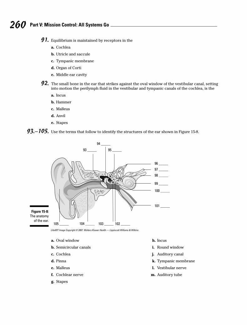
91. Equilibrium is maintained by receptors in the
a. Cochlea
b. Utricle and saccule
c. Tympanic membrane
d. Organ of Corti
e. Middle ear cavity
92. The small bone in the ear that strikes against the oval window of the vestibular canal, settinginto motion the perilymph fluid in the vestibular and tympanic canals of the cochlea, is the
a. Incus
b. Hammer
c. Malleus
d. Anvil
e. Stapes
93.–105. Use the terms that follow to identify the structures of the ear shown in Figure 15-8.
LifeART Image Copyright © 2007. Wolters Kluwer Health — Lippincott Williams & Wilkins
93 _____
94 _____
95 _____
96 _____
97 _____
98 _____
99 _____
100 _____
101 _____
102 _____103 _____104 _____105 _____
Figure 15-8:
The anatomy
of the ear.
260 Part V: Mission Control: All Systems Go
a. Oval window
b. Semicircular canals
c. Cochlea
d. Pinna
e. Malleus
f. Cochlear nerve
g. Stapes
h. Incus
i. Round window
j. Auditory canal
k. Tympanic membrane
l. Vestibular nerve
m. Auditory tube

Answers to Questions on the Nervous SystemThe following are answer to the practice questions presented in this chapter.
a Irritability: a. Tissue’s ability to respond to stimulation
b Conductivity: d. Spreading of the nerve impulse
c Orientation: c. Sense organs’ capacity to generate nerve impulse to stimulation
d Coordination: b. Ability to receive impulses and direct them to channels for favorableresponse
e Conceptual thought: e. Capacity to record, store, and relate information to be used to deter-mine future action
f The brain and spinal cord are called the a. central nervous system. They’re at the center ofeverything.
g The functional unit of the nervous system is the d. neuron. Some of the other answer optionsare parts of a neuron, but the neuron is the central unit.
h The terminal structure of the cytoplasmic projection of the neuron cannot be a(n) a. node ofRanvier. The nodes of Ranvier are gaps along the myelin sheath, so one of them can’t be foundat the end of the line.
i The afferent fiber that carries impulses to a neuron’s cell body is called a c. dendrite. Dendritescarry impulses to the neurons; axons carry them away.
j The membrane surrounding the axon (nerve fiber) is the b. neurilemma. When present, thisfiber actually wraps around the myelin sheath, so it’s always on the outside.
k Astrocytes: e. Cells that contribute to the repair process of the central nervous system
l Microgliacytes: d. Cells that are phagocytic
m Oligodendrocytes: b. Cells that form and preserve myelin sheaths
n Axons: c. Cytoplasmic projections carrying impulses from the cell body
o Dendrites: a. Cytoplasmic projections carrying impulses to the cell body
p The neuroglia cells are important as b. supporting tissue. They nourish and protect neurons.
q Axons tend to consist of a. single processes. A neuron may have one, many, or no dendrites,but it always has a single axon.
r A synapse between neurons is best described as the e. transmission of an impulse through achemical change. With all that acetylcholine and cholinesterase floating around, it must be achemical transmission.
s Endoneurium: e. Connective tissue surrounding individual fibers in a nerve
t Neurilemma: b. Outer thin membrane around an axon fiber
261Chapter 15: Feeling Jumpy: The Nervous System
u Schwann cell: c. Cell in the sheath of an axon
v Node of Ranvier: d. Depression in the sheath around a fiber
w Myelin sheath: a. Fatty layer around an axon fiber
x All-or-none response: c. Threshold of excitation determines ability to respond
y Cation: d. Positively charged ion on the outer surface of the cell membrane
A Anion: b. Negatively charged ion on the inner surface of the cell membrane
B Polarization: a. Impermeability of cell membrane
C Depolarization: e. Reshuffling of cell membrane ions; permeability of cell membrane
D Cholinesterase: b. Enzyme for breakdown of excitatory chemical
E Choline acetylase: c. Enzyme for reformation of excitatory chemical
F Terminal bulb: e. Contains storage vesicles for excitatory chemical
G Acetylcholine: a. Excitatory chemical necessary for continual nerve pathway
H Synapse: d. Space between neurons
I The cerebrum consists of two major halves called d. cerebral hemispheres. Cerebrum = cere-bral, and two halves = hemispheres.
J The cerebrum is divided into two major halves by the c. longitudinal fissure. Longitudinal isthe most likely position for an equal division.
K In the cerebrum, the e. a, b, and c are correct (right side tends to control the left side of thebody and vice versa, upper area controls lower-body activity, and lower area controlsupper-body activity). Right = left, and up = down. Clear as mud?
L The functions of the occipital lobe of the cerebrum pertain principally to a. visual activity. Toremember, use the word “occipital” to bring to mind the word “optic,” which of course is relatedto visual activity.
M The cerebellum functions primarily as a center of e. motor control.
N White matter: b. Myelinated fibers
O Reticular formation: a. Has the capacity to arouse the brain to wakefulness
P Funiculus: c. Bundles of nerve fibers arranged in tracts
Q Dorsal root ganglion: d. Collection of cell bodies outside of the central nervous system
R Gray matter: e. Unmyelinated fibers, cell bodies, and neuroglia
S Pons: a. Bridge connecting the medulla oblongata and cerebellum
T Cerebellum: d. Controls motor coordination and refinement of muscular movement
262 Part V: Mission Control: All Systems Go
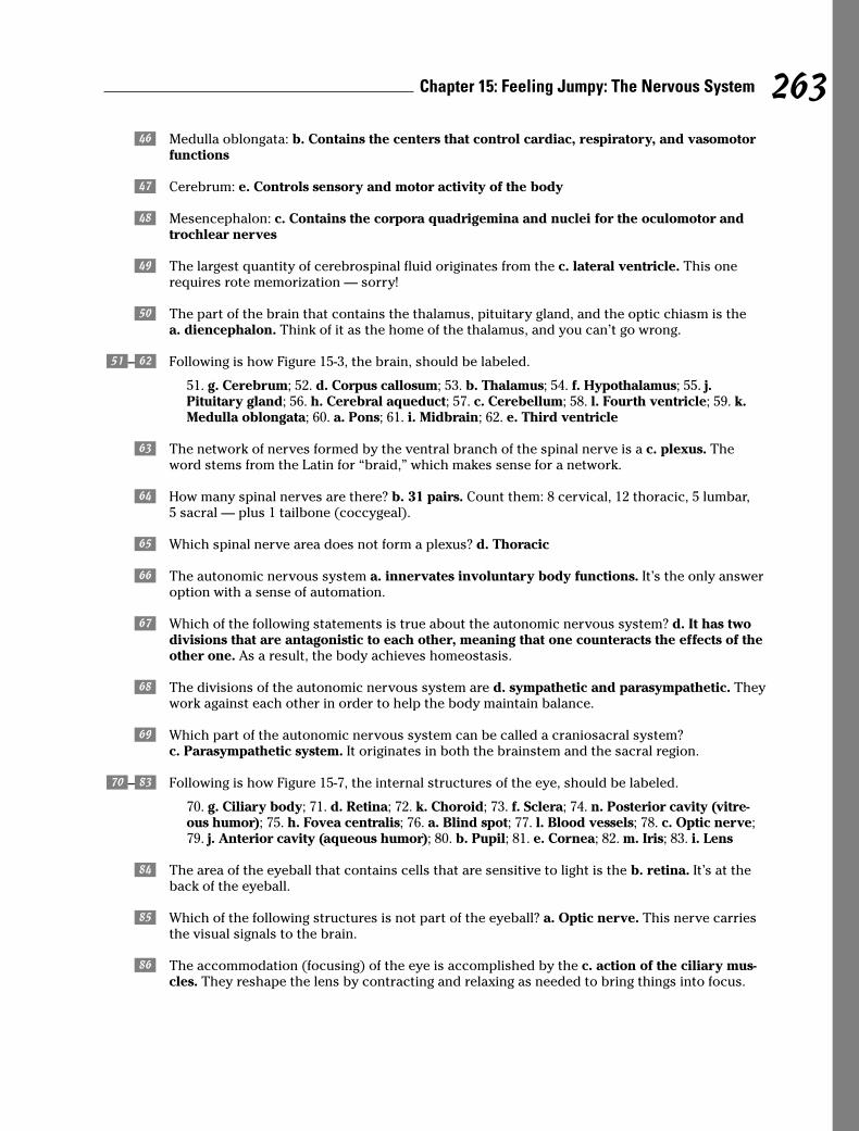
U Medulla oblongata: b. Contains the centers that control cardiac, respiratory, and vasomotorfunctions
V Cerebrum: e. Controls sensory and motor activity of the body
W Mesencephalon: c. Contains the corpora quadrigemina and nuclei for the oculomotor andtrochlear nerves
X The largest quantity of cerebrospinal fluid originates from the c. lateral ventricle. This onerequires rote memorization — sorry!
Y The part of the brain that contains the thalamus, pituitary gland, and the optic chiasm is thea. diencephalon. Think of it as the home of the thalamus, and you can’t go wrong.
z–0 Following is how Figure 15-3, the brain, should be labeled.
51. g. Cerebrum; 52. d. Corpus callosum; 53. b. Thalamus; 54. f. Hypothalamus; 55. j.Pituitary gland; 56. h. Cerebral aqueduct; 57. c. Cerebellum; 58. l. Fourth ventricle; 59. k.Medulla oblongata; 60. a. Pons; 61. i. Midbrain; 62. e. Third ventricle
! The network of nerves formed by the ventral branch of the spinal nerve is a c. plexus. Theword stems from the Latin for “braid,” which makes sense for a network.
@ How many spinal nerves are there? b. 31 pairs. Count them: 8 cervical, 12 thoracic, 5 lumbar,5 sacral — plus 1 tailbone (coccygeal).
# Which spinal nerve area does not form a plexus? d. Thoracic
$ The autonomic nervous system a. innervates involuntary body functions. It’s the only answeroption with a sense of automation.
% Which of the following statements is true about the autonomic nervous system? d. It has twodivisions that are antagonistic to each other, meaning that one counteracts the effects of theother one. As a result, the body achieves homeostasis.
^ The divisions of the autonomic nervous system are d. sympathetic and parasympathetic. Theywork against each other in order to help the body maintain balance.
& Which part of the autonomic nervous system can be called a craniosacral system?c. Parasympathetic system. It originates in both the brainstem and the sacral region.
*–; Following is how Figure 15-7, the internal structures of the eye, should be labeled.
70. g. Ciliary body; 71. d. Retina; 72. k. Choroid; 73. f. Sclera; 74. n. Posterior cavity (vitre-ous humor); 75. h. Fovea centralis; 76. a. Blind spot; 77. l. Blood vessels; 78. c. Optic nerve;79. j. Anterior cavity (aqueous humor); 80. b. Pupil; 81. e. Cornea; 82. m. Iris; 83. i. Lens
: The area of the eyeball that contains cells that are sensitive to light is the b. retina. It’s at theback of the eyeball.
, Which of the following structures is not part of the eyeball? a. Optic nerve. This nerve carriesthe visual signals to the brain.
< The accommodation (focusing) of the eye is accomplished by the c. action of the ciliary mus-cles. They reshape the lens by contracting and relaxing as needed to bring things into focus.
263Chapter 15: Feeling Jumpy: The Nervous System
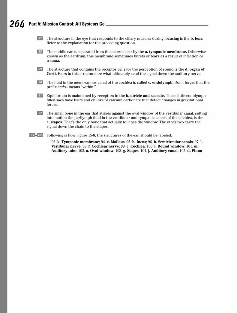
. The structure in the eye that responds to the ciliary muscles during focusing is the b. lens.Refer to the explanation for the preceding question.
> The middle ear is separated from the external ear by the a. tympanic membrane. Otherwiseknown as the eardrum, this membrane sometimes bursts or tears as a result of infection ortrauma.
/ The structure that contains the receptor cells for the perception of sound is the d. organ ofCorti. Hairs in this structure are what ultimately send the signal down the auditory nerve.
? The fluid in the membranous canal of the cochlea is called c. endolymph. Don’t forget that theprefix endo– means “within.”
` Equilibrium is maintained by receptors in the b. utricle and saccule. These little endolymph-filled sacs have hairs and chunks of calcium carbonate that detect changes in gravitationalforces.
~ The small bone in the ear that strikes against the oval window of the vestibular canal, settinginto motion the perilymph fluid in the vestibular and tympanic canals of the cochlea, is thee. stapes. That’s the only bone that actually touches the window. The other two carry thesignal down the chain to the stapes.
ú–œ Following is how Figure 15-8, the structures of the ear, should be labeled.
93. k. Tympanic membrane; 94. e. Malleus; 95. h. Incus; 96. b. Semicircular canals; 97. l.Vestibular nerve; 98. f. Cochlear nerve; 99. c. Cochlea; 100. i. Round window; 101. m.Auditory tube; 102. a. Oval window; 103. g. Stapes; 104. j. Auditory canal; 105. d. Pinna
264 Part V: Mission Control: All Systems Go
Chapter 16
Raging Hormones: The Endocrine System
In This Chapter� Absorbing what endocrine glands do
� Checking out the ringmasters: Pituitary and hypothalamus glands
� Surveying the supporting glands
� Understanding how the body balances under stress
The human body has two separate command and control systems that work in harmonymost of the time but also work in very different ways. Designed for instant response, the
nervous system cracks its cellular whip using electrical signals that make entire systemshop to their tasks with no delay (refer to Chapter 15). By contrast, the endocrine system’sglands use chemical signals called hormones that behave like the steering mechanism on alarge, fully loaded ocean tanker; small changes can have big impacts, but it takes quite a bitof time for any evidence of the change to make itself known. At times, parts of the nervoussystem stimulate or inhibit the secretion of hormones, and some hormones are capable ofstimulating or inhibiting the flow of nerve impulses.
The word “hormone” originates from the Greek word hormao, which literally translates as“I excite.” And that’s exactly what hormones do. Each chemical signal stimulates some specificpart of the body, known as target tissues or target cells. The body needs a constant supply ofhormonal signals to grow, maintain homeostasis, reproduce, and conduct myriad processes.
In this chapter, we go over which glands do what and where, as well as review the types ofchemical signals that play various roles in the body. You also get to practice discerning whatthe endocrine system does, how it does it, and why the body responds like it does.
No Bland GlandsTechnically, there are ten or so primary endocrine glands with various other hormone-secreting tissues scattered throughout the body. Unlike exocrine glands (such as mammaryglands and sweat glands), endocrine glands have no ducts to convey their secretions. Instead,hormones move directly into extracellular spaces surrounding the gland and from there moveinto capillaries and the greater bloodstream. Although they spread throughout the body in thebloodstream, hormones are uniquely tagged by their chemical composition. Thus they haveseparate identities and stimulate specific receptors on target cells so that usually only theintended cells or tissues respond to their signals.
All of the many hormones can be classified either as steroid (derived from cholesterol) ornonsteroid (derived from amino acids and other proteins). The steroid hormones — whichinclude testosterone, estrogen, progesterone, and cortisol — are the ones most closely
associated with emotional outbursts and mood swings. Steroidal hormones, which arenonpolar (see Chapter 2 for details on cell diffusion), penetrate cell membranes easilyand initiate protein production at the nucleus.
Nonsteroid hormones are divided among four classifications:
� Some are derived from modified amino acids, including such things as epineph-rine and norepinephrine, as well as melatonin.
� Others are peptide-based, including an antidiuretic hormone called ADH, oxytocin,and a melanocytes-stimulating hormone called MSH.
� Glycoprotein-based hormones include follicle-stimulating hormone (FSH),luteinizing hormone (LH), and chorionic gonadotropin — all closely associatedwith the female reproductive system.
� Protein-based nonsteroid hormones include such crucial substances as insulinand growth hormone as well as prolactin and parathyroid hormone.
Hormone functions include controlling the body’s internal environment by regulatingits chemical composition and volume, activating responses to changes in environmen-tal conditions to help the body cope, influencing growth and development, enablingseveral key steps in reproduction, regulating components of the immune system, andregulating organic metabolism.
See if all this hormone-speak is sinking in:
1.–5. Mark the statement with a T if it’s true or an F if it’s false:
1. _____ The endocrine system brings about changes in the metabolic activities of the bodytissue.
2. _____ The amount of hormone released is determined by the body’s need for that hormoneat the time.
3. _____ The glands of the endocrine system are composed of cartilage cells.
4. _____ Endocrine glands aren’t functional in reproductive processes.
5. _____ Some hormones can be derivatives of amino acids, whereas others are synthesizedfrom cholesterol.
6. Glands that secrete their product into the interstitial fluid, which flows into the blood, are
a. Exocrine glands
b. Endocrine glands
c. Heterocrine glands
d. Pericrintal glands
e. Interocrine glands
7. Cells that respond to a hormone are
a. Affectors
b. Effectors
c. Target cells
d. Chromosomal cells
e. Rickets cells
266 Part V: Mission Control: All Systems Go
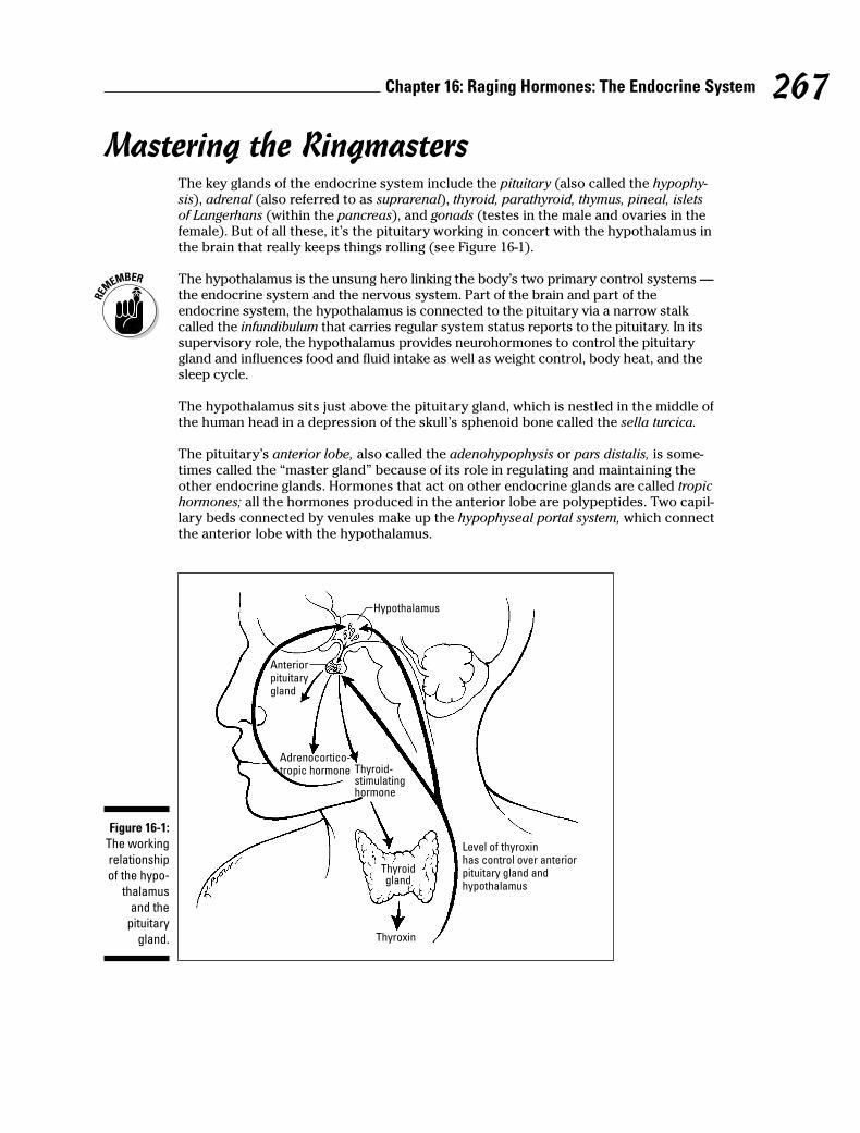
Mastering the RingmastersThe key glands of the endocrine system include the pituitary (also called the hypophy-sis), adrenal (also referred to as suprarenal), thyroid, parathyroid, thymus, pineal, isletsof Langerhans (within the pancreas), and gonads (testes in the male and ovaries in thefemale). But of all these, it’s the pituitary working in concert with the hypothalamus inthe brain that really keeps things rolling (see Figure 16-1).
The hypothalamus is the unsung hero linking the body’s two primary control systems —the endocrine system and the nervous system. Part of the brain and part of theendocrine system, the hypothalamus is connected to the pituitary via a narrow stalkcalled the infundibulum that carries regular system status reports to the pituitary. In itssupervisory role, the hypothalamus provides neurohormones to control the pituitarygland and influences food and fluid intake as well as weight control, body heat, and thesleep cycle.
The hypothalamus sits just above the pituitary gland, which is nestled in the middle ofthe human head in a depression of the skull’s sphenoid bone called the sella turcica.
The pituitary’s anterior lobe, also called the adenohypophysis or pars distalis, is some-times called the “master gland” because of its role in regulating and maintaining theother endocrine glands. Hormones that act on other endocrine glands are called tropichormones; all the hormones produced in the anterior lobe are polypeptides. Two capil-lary beds connected by venules make up the hypophyseal portal system, which connectthe anterior lobe with the hypothalamus.
Hypothalamus
Anteriorpituitarygland
Adrenocortico-tropic hormone Thyroid-
stimulatinghormone
Thyroidgland
Thyroxin
Level of thyroxinhas control over anteriorpituitary gland andhypothalamus
Figure 16-1:
The working
relationship
of the hypo-
thalamus
and the
pituitary
gland.
267Chapter 16: Raging Hormones: The Endocrine System
Among the hormones produced in the anterior lobe of the pituitary gland are thefollowing:
� Follicle-stimulating hormone (FSH): Signals an immature Graafian follicle in anovary to mature into an ovum, which then produces the hormone estrogen.Negative feedback from the estrogen blocks further secretion of FSH. Guys, don’tthink you needn’t worry about FSH: It’s present in you, too, encouraging develop-ment and maturation of sperm.
� Luteinizing hormone (LH): Stimulates formation of the yellow body, or corpusluteum, on the surface of the ovary after an ovum has been released. In men,LH stimulates the development of interstitial cells and fresh production oftestosterone.
� Lactogenic hormone, or prolactin (PRL): Promotes milk production in mammaryglands, which are considered nonendocrine targets.
� Interstitial-cell stimulating hormone (ICSH): Stimulates formation and secretionof testosterone.
� Thyrotropic hormone, or thyroid-stimulating hormone (TSH): Controls thedevelopment and release of thyroid gland hormones thyroxin and triiodothyronine.The hypothalamus regulates TSH secretion by secreting thyrotropin-releasinghormone (TRH).
� Adrenocorticotropic hormone (ACTH), or corticotropin: Is a polypeptidecomposed of 39 amino acids that regulates the development, maintenance,and secretion of the cortex of the adrenal gland.
� Somatotropic hormone, or growth hormone (GSH): Stimulates body weightgrowth and regulates skeletal growth. This is the only hormone secreted by theanterior lobe that has a general effect on nearly every cell in the body (alsoregarded as nonendocrine targets).
For a review of the male and female reproductive systems, flip to Chapters 13 and 14.
The posterior lobe, or neurohypophysis, of the pituitary gland stores and releases secre-tions produced by the hypothalamus. This lobe is connected to the hypothalamusby the hypophyseal tract, nerve axons with cell bodies lying in the hypothalamus.Whereas the adenohypophysis is made up of epithelial cells, the neurohypophysis islargely composed of modified nerve fibers and neuroglial cells called pituicytes.
Among the hormones produced in the posterior lobe of the pituitary gland are thefollowing:
� Oxytocin: Stimulates contraction of the uterine smooth muscle during childbirthand release of breast milk in nursing women
� Vasopressin, or antidiuretic hormone (ADH): Constricts smooth muscle tissue inthe blood vessels, elevating blood pressure and increasing the amount of waterreabsorbed by the kidneys, which reduces the production of urine. The hypothal-amus has special neurons called osmoreceptors that monitor the amount of solutein the blood.
See how much of this information you’re absorbing:
268 Part V: Mission Control: All Systems Go
8.–12. Mark the statement with a T if it’s true or an F if it’s false:
8. _____ The pituitary gland consists of two parts: an endocrine gland and modified nervetissue.
9. _____ The pituitary gland is found in the sella turcica of the temporal bone.
10. _____ The adenohypophysis is called the master gland because of its influence on all thebody’s tissues.
11. _____ ADH causes constriction of smooth muscle tissue in the blood vessels, whichelevates the blood pressure.
12. _____ The neurohypophysis stores and releases secretions produced by the hypothalamus.
13. The gland that does the most to regulate and maintain the function of other glands is the
a. Pineal
b. Pituitary
c. Thyroid
d. Thymus
e. Parathyroid
14. Which of the following is not a pituitary hormone?
a. Progesterone
b. Follicle-stimulating hormone (FSH)
c. Growth hormone (GSH)
d. Prolactin
e. Luteinizing hormone (LH)
269Chapter 16: Raging Hormones: The Endocrine System
Q. The hormone that stimulatesovulation is the
a. Follicle-stimulating hormone(FSH)
b. Antidiuretic hormone (ADH)
c. Oxytocin
d. Thyroid-stimulating hormone(TSH)
e. Luteinizing hormone (LH)
A. The correct answer is luteinizinghormone (LH). Don’t be fooled intothinking it’s FSH; that hormonedoes its job earlier, when it encour-ages an ovum to mature.
Supporting Cast of Glandular CharactersWhile the pituitary orchestrates the show at center stage, the endocrine system enjoysthe support of a number of other important glands. Lying in various locations through-out the body, these glands secrete check-and-balance hormones that keep the bodyin tune.
Topping off the kidneys: The adrenal glandsAlso called suprarenals, the adrenal glands lie atop each kidney. The central area ofeach is called the adrenal medulla, and the outer layers are called the adrenal cortex.Each glandular area secretes different hormones. The cells of the cortex produceover 30 steroids, including the hormones aldosterone, cortisone, and some sex hor-mones. The medullar cells secrete epinephrine (you may know it as adrenaline) andnorepinephrine (also known as noradrenaline).
Made up of closely packed epithelial cells, the adrenal cortex is loaded with blood ves-sels. Layers form an outer, middle, and inner zone of the cortex. Each zone is com-posed of a different cellular arrangement and secretes different steroid hormones.
� The zona glomerulosa (outer zone) produces aldosterone.
� The zona fasciculata (middle zone) secretes cortisone (also called cortisol).
� The zona reticularis (inner zone) secretes small amounts of gonadocorticoids orsex hormones.
The following are among the hormones produced by the cortex:
� Aldosterone, or mineralocorticoid, regulates electrolytes (sodium and potassiummineral salts) retained in the body. It promotes the conservation of water andreduces urine output.
� Cortisone, or cortisol, acts as an antagonist to insulin, causing more glucose toform and increasing blood sugar to maintain normal levels. Elevated levels ofcortisone speed up protein breakdown and inhibit amino acid absorption.
� Androgens and estrogen are cortical sex hormones. Androgens generally conveyantifeminine effects, thus accelerating maleness, although in women adrenalandrogens maintain the sexual drive. Too much androgen in females can causevirilism (male secondary sexual characteristics). Estrogen has the oppositeeffect, accelerating femaleness. Too much estrogen in a male produces femininecharacteristics.
The adrenal medulla is made of irregularly shaped chromaffin cells arranged in groupsaround blood vessels. The sympathetic division of the autonomic nervous system con-trols these cells as they secrete adrenaline and noradrenaline. Both hormones havesimilar molecular structure and physiological functions. The adrenal cortex producesapproximately 80 percent adrenaline and 20 percent noradrenaline. Adrenaline accel-erates the heartbeat, stimulates respiration, slows digestion, increases muscle effi-ciency, and helps muscles resist fatigue. Noreadrenaline does similar things but alsoraises blood pressure by stimulating contraction of muscular arteries.
270 Part V: Mission Control: All Systems Go
The terms “adrenaline” and “noradrenaline” are interchangeable with the terms“epinephrine” and “norepinephrine.” You’re likely to encounter both in textbooksand exams.
Thriving with the thyroidThe largest of the endocrine glands, the thyroid is like a large butterfly with two lobesconnected by a fleshy isthmus positioned in the front of the neck, just below the larynxand on either side of the trachea. A transport mechanism called the iodide pumpmoves the iodides from the bloodstream for use in creating its two primary hormones,thyroxin and triiodothyronine, which regulate the body’s metabolic rate. Extrafollicularcells (also called parafollicular or C cells) secrete calcitonin, a polypeptide hormonethat helps regulate the concentration of calcium and phosphate ions by inhibiting therate at which they leave the bones. High blood calcium levels stimulate the secretionof more calcitonin.
Thyroxin (T4) and triiodothyronine (T3) regulate cellular metabolism throughout thebody, but the thyroid needs iodine to manufacture those hormones. Iodine insufficiencycauses the thyroid to swell in a condition called a goiter.
Pairing up with the parathyroidThe parathyroid consists of four pea-sized glands that lie posterior to the thyroid glandsecreting parathormone, or parathyroid hormone (PTH). This large polypeptide regu-lates the balance of calcium levels in the blood and bones as well as controls the rate atwhich calcium is excreted into urine. When blood calcium levels dip, the parathyroidsecretes PTH, which increases calcium absorption from the intestine, decreases cal-cium excretion, increases phosphate excretion, removes calcium from the bones, andstimulates secretion of calcitonin by the thyroid C cells. Blood calcium ion homeostasisis critical to the conduction of nerve impulses, muscle contraction, and blood clotting.
Pinging the pineal glandThe pineal gland, also called the epiphysis, is a small, oval gland thought to play a rolein regulating the body’s biological clock. It lies between the cerebral hemispheres andis attached to the thalamus near the roof of its third ventricle.
Because it both secretes a hormone and receives visual nerve stimuli, the pineal glandis considered part of both the nervous system and the endocrine system. Its hormonemelatonin is believed to play a role in circadian rhythms, the pattern of repeated behav-ior associated with the cycles of night and day. The pineal gland is affected by changesin light, producing its highest levels of secretion at night and its lowest levels duringdaylight hours.
Thumping the thymusAs discussed in Chapter 11, the thymus is thought to secrete a group of peptidescalled thymosin that affect the production of lymphocytes (white blood cells).Thymosin promotes the production and maturation of T lymphocyte cells as part ofthe body’s immune system. The gland is large in children and atrophies with age.
271Chapter 16: Raging Hormones: The Endocrine System
Pressing the pancreasThe pancreas is both an exocrine and an endocrine gland, which means that it secretessome substances through ducts while others go directly into the bloodstream. (Wecover its exocrine functions in Chapter 9.) The pancreatic endocrine glands are clustersof cells called the islets of Langerhans. Within the islets are a variety of cells, including
� A cells (alpha cells) that secrete the hormone glucagon, a polypeptide of29 amino acids that increases blood sugar
� B cells (beta cells) that secrete insulin, a two-linked polypeptide chain of21 amino acids that decreases blood sugar levels, increases lipid synthesis,and stimulates protein synthesis
� D cells (delta cells) that secrete somatostatin, a growth hormone–inhibitingfactor that inhibits the secretion of insulin and glucagons
� F cells (PP cells) that secrete a pancreatic polypeptide that regulates the releaseof pancreatic digestive enzymes
See if all this information has your hormones raging:
15.–19. Mark the statement with a T if it’s true or an F if it’s false:
15. _____ The adrenal glands are located in the cortex of the kidneys.
16. _____ Adrenaline is functional in the absorption of stored carbohydrates and fat.
17. _____ Aldosterone is functional in regulating the amount of insulin in the body.
18. _____ The sympathetic division of the autonomic nervous system controls the cells of theadrenal medulla.
19. _____ The layers of the adrenal medulla form outer, middle, and inner zones.
20. The endocrine gland that initiates antibody development by producing thymosin is the
a. Pineal body
b. Pituitary gland
c. Thymus
d. Hypothalamus
e. Adrenal gland
21. The hormone that regulates the amount of electrolytes retained in the body is
a. Aldosterone
b. Cortisone
c. Epinephrine
d. Androgens
e. Norepinephrine
272 Part V: Mission Control: All Systems Go
22.–26. Mark the statement with a T if it’s true or an F if it’s false:
22. _____ Iodine is a necessary component of thyroxin (T4) and triiodothyronine (T3).
23. _____ Follicular cells of the thyroid produce hormones that affect the metabolic rate ofthe body.
24. _____ A transport mechanism called the sodium pump moves the iodides into the folliclecells.
25. _____ Thyroxin (T4) is normally secreted in lower quantity than triiodothyronine (T3).
26. _____ The hormone calcitonin helps regulate the concentration of sodium and potassium.
27. Which statement is not true of the pineal gland?
a. It secretes melatonin.
b. Nerve fibers stimulate the pineal cells.
c. As light decreases, secretion increases.
d. It’s a small, oval gland.
e. It promotes immunity.
28. Insufficiency of iodine causes the thyroid gland to enlarge, causing
a. Dwarfism
b. Diabetes
c. Giantism
d. Acromegaly
e. Simple or endemic goiter
29.–33. Mark the statement with a T if it’s true or an F if it’s false:
29. _____ The parathyroid gland contains cells that secrete parathormone or parathyroidhormone (PTH).
30. _____ Melatonin is a polypeptide that regulates the balance of calcium in the blood andbones.
31. _____ The pineal gland responds to light, producing higher levels of secretions at nightthan during the day.
32. _____ Thymosin promotes the production and maturation of erythrocyte cells.
33. _____ The parathyroid hormone can prompt calcium to move from bone.
34. The endocrine gland that produces 80 percent epinephrine is the
a. Hypothalamus
b. Pituitary
c. Medulla of the adrenal
d. Thyroid
e. Thymus
273Chapter 16: Raging Hormones: The Endocrine System
35. The endocrine gland associated with metabolic rate is the
a. Parathyroid
b. Thyroid
c. Pineal
d. Posterior lobe of the pituitary
e. Thymus
Dealing with Stress: HomeostasisNothing upsets your delicate cells more than a change in their internal environment.A stimulus such as fear or pain provokes a response that upsets your body’s carefullymaintained equilibrium. Such a change initiates a nerve impulse to the hypothalamusthat activates the sympathetic division of the autonomic nervous system and increasessecretions from the adrenal glands. This change — called a stressor — produces a con-dition many know oh so well: stress. The body’s immediate response is to push forhomeostasis — keeping everything the same inside.
The body’s effort to maintain homeostasis invokes a series of reactions called the gen-eral stress syndrome that’s controlled by the hypothalamus. When the hypothalamusreceives stress information, it responds by preparing the body for fight or flight; in otherwords some kind of decisive, immediate, physical action. This reaction increases bloodlevels of glucose, glycerol, and fatty acids; increases the heart rate and breathing rate;redirects blood from skin and internal organs to the skeletal muscles; and increasesthe secretion of adrenaline from the adrenal medulla. The hypothalamus releases corti-cotropin-releasing hormone (CRH) that stimulates the anterior lobe of the pituitary tosecrete adrenocorticotropic hormone (ACTH), which tells the adrenal cortex to secretemore cortisone. That cortisone supplies the body with amino acids and an extraenergy source needed to repair any injured tissues that may result from the impendingcrisis.
As part of the general stress syndrome, the pancreas produces glucagon, and the ante-rior pituitary secretes growth hormones, both of which prepare energy sources andstimulate the absorption of amino acids to repair damaged tissue. The posterior pitu-itary secretes antidiuretic hormone, making the body hang on to sodium ions andspare water. The subsequent decrease in urine output is important to increase bloodvolume, especially if there’s bleeding or excessive sweating.
Wow. With the body gearing up like that every time, it’s no wonder that people subjectedto repeated stress are often sickly.
We try not to stress you out with these practice questions:
36.–40. Mark the statement with a T if it’s true or an F if it’s false:
36. _____ The hypothalamus controls reactions to combat general stress syndrome.
37. _____ The pancreas is an endocrine gland only.
38. _____ During stress, the pancreas produces thyroxin (T4).
39. _____ Alpha cells in the pancreas secrete the hormone insulin.
40. _____ Changes in the body’s environment called stressors produce a condition calledstress.
274 Part V: Mission Control: All Systems Go
41. When changes occur in the body’s internal environment, a reaction is initiated by
a. Neurohormones
b. Glucocorticoids
c. The hypothalamus
d. The adrenal cortex
e. The pituitary gland
42. Stress activates a set of body responses called
a. The survival response
b. The general stress syndrome
c. The repair response
d. The resistance response
e. The stress reflex
43. The body’s initial reaction to a stressor is
a. Fight or flight response
b. Repair response
c. To promote rapid wound healing
d. Stress reflex
e. To promote normal metabolism
44. Which of the following is a response to stress?
a. Decrease the heart rate
b. Increase the urine output
c. Redirect blood from the skeletal muscles
d. Increase the respiratory rate
e. Decrease the glucose in the blood
45. The pancreas, testes, and ovaries all have this in common:
a. All are influenced by hormones from the parathyroid.
b. All are considered to be both exocrine and endocrine.
c. None were formed from embryonic tissues.
d. They influence secondary sex characteristics.
e. They have no blood supply.
275Chapter 16: Raging Hormones: The Endocrine System
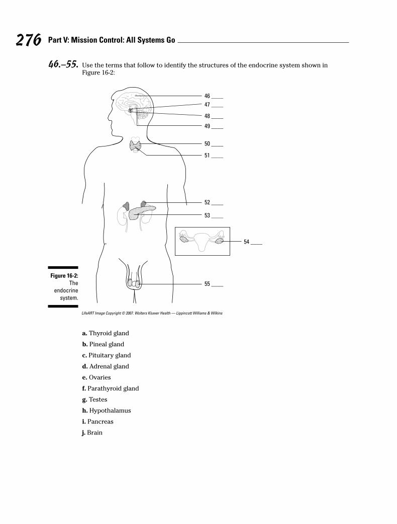
46.–55. Use the terms that follow to identify the structures of the endocrine system shown inFigure 16-2:
LifeART Image Copyright © 2007. Wolters Kluwer Health — Lippincott Williams & Wilkins
a. Thyroid gland
b. Pineal gland
c. Pituitary gland
d. Adrenal gland
e. Ovaries
f. Parathyroid gland
g. Testes
h. Hypothalamus
i. Pancreas
j. Brain
46 ____
47 ____
48 ____
49 ____
50 ____
51 ____
52 ____
53 ____
54 ____
55 ____
Figure 16-2:
The
endocrine
system.
276 Part V: Mission Control: All Systems Go

Answers to Questions on the Endocrine SystemThe following are answers to the practice questions presented in this chapter.
a The endocrine system brings about changes in the metabolic activities of the body tissue.True. Metabolism is one of the areas influenced by hormones.
b The amount of hormone released is determined by the body’s need for that hormone at thetime. True. In many ways, it’s a self-regulating system: Just enough hormones are distributedto balance everything else out.
c The glands of the endocrine system are composed of cartilage cells. False. That connectionmakes no sense whatsoever.
d Endocrine glands aren’t functional in reproductive processes. False. The endocrine system is akey component in reproduction.
e Some hormones can be derivatives of amino acids, whereas others are synthesized from choles-terol. True. Amino acids for the nonsteroids and cholesterol for the steroid-based hormones.
f Glands that secrete their product into the interstitial fluid, which flows into the blood, areb. endocrine glands.
g Cells that respond to a hormone are c. target cells. Hormones actually go looking for thesespecific targets.
h The pituitary gland consists of two parts: an endocrine gland and modified nerve tissue. True.Remember that the anterior lobe is mostly epithelial cells, whereas the posterior lobe containsprimarily nerve cells.
i The pituitary gland is found in the sella turcica of the temporal bone. False. The pituitary glandis in the sphenoid bone, not the temporal bone.
j The adenohypophysis is called the master gland because of its influence on all the body’s tis-sues. False. It earned the title “master gland” because of its influence over the other endocrineglands.
k ADH causes constriction of smooth muscle tissue in the blood vessels, which elevates theblood pressure. True.
l The neurohypophysis stores and releases secretions produced by the hypothalamus. True.Seems a strange thing for a structure made of nerve cells to do, but it does its job well.
m The gland that does the most to regulate and maintain the function of other glands is theb. pituitary. That’s why it’s the master gland.
n Which of the following is not a pituitary hormone? a. Progesterone. That’s made by the corpusluteum on the ovary following ovulation.
o The adrenal glands are located in the cortex of the kidneys. False. They’re atop the kidneys.
p Adrenaline is functional in the absorption of stored carbohydrates and fat. False. Adrenalinedoes lots of things, but not that.
277Chapter 16: Raging Hormones: The Endocrine System
q Aldosterone is functional in regulating the amount of insulin in the body. False. This hormoneregulates mineral salts.
r The sympathetic division of the autonomic nervous system controls the cells of the adrenalmedulla. True.
s The layers of the adrenal medulla form outer, middle, and inner zones. False. The layers of theadrenal cortex form those three zones.
t The endocrine gland that initiates antibody development by producing thymosin is thec. thymus.
The name of the hormone, thymosin, should be your first clue that it’s produced by thethymus. See the resemblance?
u The hormone that regulates the amount of electrolytes retained in the body is a. aldosterone.
v Iodine is a necessary component of thyroxin (T4) and triiodothyronine (T3). True. The bodycan’t make those hormones without iodine.
w Follicular cells of the thyroid produce hormones that affect the metabolic rate of the body. True.
x A transport mechanism called the sodium pump moves the iodides into the follicle cells. False.Don’t let the thyroid’s iodide pump make you think it also has a sodium pump. It doesn’t.
y Thyroxin (T4) is normally secreted in lower quantity than triiodothyronine (T3). False. In fact,it’s just the opposite — more T4 is secreted than T3.
A The hormone calcitonin helps regulate the concentration of sodium and potassium. False.Actually, calcitonin lowers plasma calcium and phosphate levels.
B Which statement is not true of the pineal gland? e. It promotes immunity. It’s more of abiological-clock kind of gland.
C Insufficiency of iodine causes the thyroid gland to enlarge, causing e. simple or endemicgoiter. Sometimes this swelling becomes visible at the base of the neck.
D The parathyroid gland contains cells that secrete parathormone or parathyroid hormone(PTH). True.
E Melatonin is a polypeptide that regulates the balance of calcium in the blood and bones. False.Melatonin is thought to regulate circadian rhythms.
F The pineal gland responds to light, producing higher levels of secretions at night than duringthe day. True. That’s why it’s also considered part of the nervous system.
G Thymosin promotes the production and maturation of erythrocyte cells. False. Thymosinworks on lymphocytes.
H The parathyroid hormone can prompt calcium to move from bone. True. After all, the bonesare mineral reservoirs.
I The endocrine gland that produces 80 percent epinephrine is the c. medulla of the adrenal.That’s about all the adrenal medulla does.
278 Part V: Mission Control: All Systems Go
J The endocrine gland associated with metabolic rate is the b. thyroid. Controlling the body’smetabolic rate is its primary role.
K The hypothalamus controls reactions to combat general stress syndrome. True.
L The pancreas is an endocrine gland only. False. It’s also an exocrine gland.
M During stress, the pancreas produces thyroxin (T4). False. That’s the thyroid’s job, and T4 hasnothing to do with stress, anyway.
N Alpha cells in the pancreas secrete the hormone insulin. False. A cells secrete glucagon.
O Changes in the body’s environment called stressors produce a condition called stress. True.
P When changes occur in the body’s internal environment, a reaction is initiated by c. the hypo-thalamus. It stimulates the sympathetic division of the autonomic nervous system and adrenalmedulla.
Q Stress activates a set of body responses called b. the general stress syndrome.
R The body’s initial reaction to a stressor is a. fight or flight response. You’re either going to putup your dukes or run like the wind.
S Which of the following is a response to stress? d. Increase the respiratory rate. That brings inmore oxygen in case you need to move fast.
T The pancreas, testes, and ovaries all have this in common: b. All are considered to be bothexocrine and endocrine. Secretions go directly into the bloodstream and through ducts.
U–3 Following is how Figure 16-2, the endocrine system, should be labeled.
46 j. Brain; 47. h. Hypothalamus; 48. c. Pituitary gland; 49. b. Pineal gland; 50. f. Parathyroidgland; 51. a. Thyroid gland; 52. d. Adrenal gland; 53. i. Pancreas; 54. e. Ovaries; 55. g. Testes
279Chapter 16: Raging Hormones: The Endocrine System

280 Part V: Mission Control: All Systems Go
Part VI
The Part of Tens

In this part . . .
This classic For Dummies feature contains two chaptersidentifying ten useful tidbits for students of anatomy
and physiology. We identify ten Web sites that can helpyou advance your knowledge and ten top study tipsto help you get A’s (or at least get closer to them!) inanatomy and physiology courses.
Chapter 17
Ten Study Tips
In This Chapter� Playing with words and outlines
� Getting the most out of study groups
� Tackling tests and moving forward
What’s the best way to tackle anatomy and physiology and come out successful on theother side? Of course, a good memory helps, but it’s not as critical to anatomy and
physiology success as it’s cracked up to be. With a little advance planning and tricks of thestudy trade, even students who complain that they can’t remember their own names onexam day can summon the right terminology and information from their scrambled synapticpathways. In this chapter, we cover ten key things you can start doing today to ensure suc-cess not only in anatomy and physiology but in any number of other classes.
Write It Down in Your Own WordsThis is a simple idea that far too few students practice regularly. Don’t stop at underliningand highlighting important material in your textbooks and study guides: Write it down. Ortype it up. Whatever you do, don’t just regurgitate it exactly as presented in the materialyou’re studying. Find your own words. Create your own analogies. Tell your own tale ofwhat happens to the bolus as it ventures into the digestive tract. Detail the course followedby a molecule of oxygen as it enters through the nose. Draw pictures of the differencesbetween meiosis and mitosis. Completely relax into the process with the knowledge that noone else ever has to see what you write, type, or sketch. All they’ll ever see is your successfulcompletion of the course!
Better Knowledge through MnemonicsStudying anatomy and physiology involves remembering lists of terms, functions, andprocesses. Sprinkled throughout this book are suggestions to take just the first letter or two ofeach word from a list to create an acronym. Occasionally, we help you go one step beyond theacronym to a clever little thing called a mnemonic device. Simply put, the mnemonic is thething you commit to memory as a means for remembering the more technical thing for whichit stands. For example, a question in Chapter 7 asks you to list in order the epidermal layersfrom the dermis outward; we suggest that you commit the following phrase to memory: BeSuper Greedy, Less Caring. Just like that, a complicated list like basale, spinosum, granulosum,lucidum, corneum gets a little closer to a permanent home in your brain.
Not feeling terribly clever at the moment you need a useful mnemonic? Surf on over towww.medicalmnemonics.com, which touts itself as the world’s database of theseuseful tools. Here’s a sampling of the site’s offerings:
� To remember the three types of tonsils: “PPL (people) have tonsils: P haryngeal,P alatine, and L ingual.”
� To remember the spleen’s location and dimensions: “Count 1, 3, 5, 7, 9, 11: Thespleen is 1 inch by 3 inches by 5 inches, weighs 7 ounces, and underlies ribs 9through 11.”
� To remember the cranial bones; “Think PEST OF 6: P arietal, E thmoid, Sphenoid,Temporal, Occipital, Frontal.”
Stylish LearningEvery person has his or her own sense of style, and woe betide anyone who tries toshoehorn the masses into a single style. The same, of course, is true of students. To getthe most out of your study time, you need to figure out what your learning style is andalter your study habits to accommodate it. No idea what we’re talking about? Answer thequestionnaire posted at www.vark-learn.com/english/page.asp?p=questionnaire,and the VARK guide to learning styles will tell you more about yourself than your lastpsychotherapist.
VARK, as you may have suspected, is an acronym that stands for Visual (learning byseeing), Aural (learning by hearing), Reading/Writing (learning by reading and writing),and Kinesthetic (learning by touching, holding, or feeling). If you’re a visual learner, youmay get more out of anatomy and physiology by seeing the real thing in the flesh. Ifyou’re an aural learner, you may learn best in the classroom as the teacher lectures.If you’re a reading and writing kind of learner, you’ll get the most out of our first tip towrite stuff down. And if you’re a kinesthetic learner, there’s nothing like touching orholding to commit something to memory.
Grecian FormulaIf you keep thinking “It’s all Greek to me,” congratulations on your insight! The truth ofthe matter is that most of it actually is Greek. So dust off your foreign language learningskills and begin with the basic vocabulary of medical terminology. (Get started with theGreek and Latin roots, prefixes, and suffixes that appear on this book’s Cheat Sheet —the tear-out page in the front of the book.) You’ll soon discover that for every “little”word you learn, there’s a whole mountain of additional terms and phrases just waitingto be discovered.
Connecting with ConceptsIt happens time and again in anatomy and physiology that one concept or connectionmirrors another yet to be learned. But because you’re focusing so hard on this week’slesson, you lose sight of the value in the previous month’s lessons. For example, a
284 Part VI: The Part of Tens

concept like metabolism comes up in a variety of ways throughout your study ofanatomy and physiology. When you encounter a repeat concept like that, create aspecial page or two for it at the back of your notebook, or link the concept to a sepa-rate computer file. Then, every time the term comes up in class or in your textbook,add to the running list of notes on that concept. You’ll have references to metabolismat each point it comes up and you’ll be able to analyze its influences across differentbody systems.
Grouping StudiesIf you’re really lucky, someone in your class (or maybe it’s even you) has already sug-gested forming that time-honored tradition — a study group. The power of groupmembers to fill gaps in your knowledge is priceless. But don’t restrict it to late-nightcramming just before each test. Meet with your group at least once a week to go overlecture notes and textbook readings. If it’s true that people only retain about 10 per-cent of what they hear or read, then it makes sense that your fellow group memberswill recall things that slipped immediately from your mind.
Outlining What’s to ComeAs you read through a chapter of your textbook to prepare for the next lecture, pre-pare an outline of what you’re reading, leaving plenty of space between subheadings.Then, during the lecture, take your notes within the outline you’ve already created.Piecing together an incomplete puzzle shows you where the key gaps in your knowl-edge may be.
Practice, Practice, PracticeFlash cards, mnemonic drills, practice tests — be creative and practice, practice,practice! The more you know about the format of any upcoming exam, the better.Sometimes instructors share tidbits about what they plan to emphasize, but some-times they don’t. In the end, if you’ve done the work and put in the time to study andpractice with information outside of class, the exact structure and content of an examshouldn’t make much difference.
Sleuthing Out CluesOkay, it’s test time! Take advantage of the test itself. You may find that the answer toan exam question that stumps you is revealed — at least partially — in the phrasing ofa subsequent question. Stay alert to these blessed little gifts even when you think thatyou already understand the whole anatomical process. You wouldn’t be the first stu-dent to change an answer after working your way through an exam.
285Chapter 17: Ten Study Tips
Learning from MistakesThe test is done and the grades are in. So there was a really tough question or two onthe test and you blew it big-time? It’s hardly a missed opportunity — this is whererolling with the punches really pays off. Go back over the entire test and pay extraattention to what you got wrong. Start your next practice sessions with those ques-tions, and stay alert for upcoming material that may trip you up in a similar way.
286 Part VI: The Part of Tens

Chapter 18
Ten (Plus One) Terrific Online Resources
In This Chapter� Scanning online reference books
� Exploring databases and virtual tours
No matter how much you study or how many Latin and Greek roots you memorize,it’s inevitable that some aspects of anatomy and physiology will leave you dazed and
confused. But if you study within reach of an Internet connection, you don’t have to staythat way for long. Simply surf over to one of the 11 sites covered in this chapter and startentering search terms. As with anything Internet-related, however, you have to be cautiousabout the accuracy of what you find. Just keep our mantra in mind: “When in doubt, trustthe textbook.”
Answers and More Answerswww.answers.com
Admittedly, this free Web site doesn’t focus on anatomy and physiology, but you won’t reallycare about that technicality after you’ve dived into it a few times. The site offers about 4 mil-lion “answers” based more on keyword searches than on any actual questions. Material isdrawn from scores of brand-name content publishers as well as Answers.com’s own editorialteam. Can’t quite figure out where an “anteroinferior” something is supposed to be? Forgotwhere Peyer’s patches are hiding? Not entirely sure what a gallbladder does? Answers.com’sfriendly little “Tell me about . . .” box at the top of the home page takes you straight to a Webpage that aggregates what various sources say on your chosen topic.
Into the Lion’s Denwww.lionden.com/ap.htm
We told you to trust the textbook, didn’t we? That means that you can probably trustthe textbook’s author, too. This delightful site is maintained by a Missouri communitycollege professor who also happens to be coauthor of an anatomy and physiology textbook.Dr. Kevin T. Patton has been teaching the subject for more than two decades and has devel-oped a refreshingly gentle sense of humor along the way. His Web site is packed with studytips, downloadable PowerPoint slides, and guided tours of various anatomical systems.
The Venerable Gray’swww.bartleby.com/107
When we refer to “Gray’s Anatomy,” we’re not talking about the popular TV series of asimilar name. We’re talking about the venerable reference book that dates back to 1858and is now online for quick and easy access. Included in the “virtual” Gray’s Anatomy ofthe Human Body are more than 1,200 color illustrations and a subject index with 13,000entries. If your textbook is missing critical illustrations, don’t worry; a quick keywordsearch on this Web site can reveal a number of relevant graphics for you to study.
Alluring Anatomistswww.anatomy.org
The American Association of Anatomists believes that anatomy is “truly the backboneof biomedical science.” With that in mind, the organization has put together an Ask theExpert feature that lets anyone — students and educators alike — query the group’sworking professionals. To access this feature, click the Education and Teaching Toolslink on the organization’s home page; then select Ask the Expert. (To view the answerto a particular posted question, click on the question.)
From the Education and Teaching Tools page, you also can find a list of popular linksto various subspecialties, including cell biology, genetics, imaging, molecular develop-ment, endocrinology, forensics, and physical anthropology.
Getting Body Smartwww.getbodysmart.com
This Web site is the brainchild and passion of an anatomy and physiology instructor,Scott Sheffield, who says in his site’s mission statement that he’s attempting to distilltwo decades of teaching into a single, fully animated and interactive e-book about thehuman body. In addition to “flash” windows that drill down into various systems,GetBodySmart offers free tutorials and quizzes to explain complex physiologicalinteractions. Sheffield readily acknowledges that his work will last “many years,”so perhaps the best is yet to come.
Pop Quiz Centralmsjensen.education.umn.edu/webanatomy
Murray Jensen, an associate professor at the University of Minnesota, conductsresearch on the use of technology in science education. His Web site is both an out-growth of his research and a source of ideas for it. As of this writing, the site consistsmostly of dozens of quizzes of varying lengths and difficulty. Treat this site like yourown personal flash card system and you’ll be head and shoulders above your fellowstudents.
288 Part VI: The Part of Tens
MEDTropolis – Virtual Bodywww.medtropolis.com/VBody.asp
When you visit this site, click on English or Spanish, and then sit back and enjoy theshow. This site only covers the brain, skeleton, heart, and digestive tract, but its clear,concise three-dimensional representations of these organs and systems make it wortha look.
Drilling, Drilling, Drilling Some Morematcmadison.edu/faculty/cshuster/wiley.html
This Web site links to an extensive set of anatomy drill and practice exercises main-tained by John Wiley and Sons (who also happens to be the publisher of this book).To work through the multiple-choice practice questions, you need to download theShockwave plug-in (if you don’t already have it), but that’s a small price to pay forsuch a useful site. Full-color images with blank labels give you the opportunity tofigure out which part is what and why; then you can clear your labels and begin againas often as you like.
Human Biodyssey: Exploring Anatomy and Physiology
www.gwc.maricopa.edu/home_pages/crimando/jcHumanBiodyssey.htm
This page is the impressive work of Dr. James Crimando at Gateway CommunityCollege in Phoenix, Arizona. Dr. Crimando lists every scrap of information a studentneeds to succeed in his classes (or any anatomy and physiology class), includingextensive practice questions, lecture outlines, and quick summaries of class sessions.Regardless of whether you’re among Dr. Crimando’s students, his site is an incrediblyuseful receptacle for information about how the body is organized.
List of Listswww.mhhe.com/biosci/ap/saladin/www.mhtml
Textbook author Kenneth Saladin uses a small portion of this site to provide descrip-tions of the last three versions of his anatomy and physiology textbooks. But thatdoesn’t begin to compare with what he has done pulling together resources from allover the Web in the Student Resources page we guide you to here. So much to see, solittle time!
289Chapter 18: Ten (Plus One) Terrific Online Resources
Virtual Anatomylibrary.thinkquest.org/16421/noframes/index.htm
Believe it or not, this site was created by a trio of high school kids in San Antonio, Texas.The site’s capabilities are somewhat limited, but it contains some good interactiveanatomical practice areas and a couple of educational videos, too.
290 Part VI: The Part of Tens
• A •abdomen, lymph node region, 185abdominopelvic cavity, subserous fasciae, 102abducens (cranial nerve VI), pons, 245abduction, joint movement type, 83absorption, digestive system, 143accelerated mitosis, 42acetabulum, appendicular skeleton, 79acetylocholine transmitters, synaptic vesicles, 241acidic, pH level, 8acids, 11–13, 16, 18actin, 27, 53, 97action potential, 170–171, 240active transport, molecule transport method, 25actomyosin, skeletal muscles, 97adduction, joint movement type, 84adenine, DNA nitrogenous base, 13adenoids (pharyngeal tonsil), 135, 189adenosine diphosphate (ADP), 15adenosine monophosphate (AMP), 15adenosine triphosphate (ATP), 15adipose tissue, 17, 51adolescent, human growth stage, 229adrenal (suparenal) glands, 267, 270–271adrenocorticotropic hormone (ACTH), 268, 274afferent (inbound) vessels, lymph nodes, 184afferent (sensory) nerves, 98afferent (sensory) neurons, stimuli triggers, 238afferent arterioles, kidneys, 196afferent impulses, urination process, 201afferent, nerve type, 239aggregate glands, 189agminate glands, 189albinos, melanin deprivation, 115albumin, liver function, 156aldosterone, kidneys, 197alveoli, teeth openings, 146amino acids, 12, 18, 31amorphous ground substance, 51amphiarthrosis, symphysis joints, 83amphipathic molecules, cell membrane, 23ampulla, hearing sense, 257anabolic reactions, metabolism, 15anaerobic respiration, 16, 18anaphase, cellular mitosis, 38anaphase, meiosis stage, 211–212angiotensinogenase (renin), kidney secretion, 197anion, 8, 240anisotropic (A-bands), skeletal muscles, 97anoxia, oxygen deficiency, 130antagonists, muscle groups, 100anterior lobe, pituitary gland, 267anterior ramus, spinal nerves, 252anteroposterior end, pharynx, 135anthracosis (black lung), lung condition, 140
antibodies, 12, 31, 185antidiuretic hormone (ADH), 197, 266, 268anvil (incus), middle ear, 257aorta, systemic circuit, 163–164aortic semilunar valve, left ventricle, 167apical foramen, root canal opening, 147apical surface, skin tissue, 47apocrine glands, integumentary system, 121–122appendicular skeleton, appendages, 79–82arachnoid meninx, brain ventricles, 247arachnoid, spinal cord, 244arachnoid villi, brain ventricles, 247arbor vitae (tree of life), cerebellum, 246archenteron, developing embryo, 143areolar (loose tissue), connective tissue type, 51arrector pili, hair muscle, 121arterioles, blood vessels, 172–173artificially created atoms, number of, 7aryepiglottic fold, larynx, 135arytenoids, larynx cartilage, 135ascending colon, large intestines, 157ascending loop of Henle, kidneys, 196association fibers, medulla, 247association (internuncial) neurons, 238asters (astral rays), 38, 211atlas bones, cervical (neck) curvature, 70atretic follicles, female reproductive system, 220atria, heart chamber, 163, 166–167atrioventricular , 170–171atrioventricular opening, 166–167atrium, respiratory system, 137auditory meatus bone, axial skeleton, 69auditory ossicles, middle ear, 257auricle, external ear, 256autonomic nervous system, 69, 253–255avascular (non-vascular), nerve state, 52axillary region, apocrine sweat glands, 122axis bones, cervical (neck) curvature, 70axon collaterals, neurons, 238axon hillock, neurons, 238axons (nerve fibers), 55, 238–241
• B •B cells, lymph nodes, 185ball-and-socket, synovial joint classification, 83barrier, epithelial tissue function, 47basal side, epithelial tissues, 47base, defined, 8basic, pH level, 8beta cells (B cells), islets of Langerhans, 272biconcave, eyes, 256bicuspid (mitral) valve, 167bicuspids, teeth, 146bile, gallbladder storage, 155biliary canaliculi, liver, 156bilirubin, liver production, 156
Index

biliverdin, liver production, 156bipennate fibers, muscle direction, 102bipolar, sensory neuron classification, 238bladder, urinary system, 199blastocoele, female reproductive system, 226–227blastula (blastocyst), 226–227blood cell formation, skeletal system function, 61blood sugar (glucose), monosaccharide, 11–12body fat, adipose tissue, 17bolus (food mass), swallowing process, 151bone (osseous) tissue, connective tissue, 52bouton terminal, axons, 241brachial plexus, spinal nerves, 253brain, 243–251breastbone (sternum), axial skeleton, 70breathing (pulmonary ventilation), 129bronchi, trachea, 137bronchial arteries, lungs, 138bronchioles, respiratory system, 137bronchomediastinal region, lymphatic trunks, 183Brownian motion, molecule transport method, 24Brunner’s glands, duodenum, 154buccal pad, cheeks, 146buccinator muscles, cheeks, 146bulb, hair base, 120bulbourethral (Cowper’s) glands, males, 200, 209bursae, 83, 113bursitis, joints, 83
• C •calcification, developing fetus, 63–64calcitonin, thyroid gland, 271calcium, bone composition, 63canaliculi, connective tissues, 52canals, internal ear, 257canthi, eyes, 256capillaries, 137, 172–173carbohemoglobin, exhalation process, 130–131carbohydrate metabolism, 16–17cardiac (myocardium), muscle tissue type, 53–54cardiac glands, stomach, 152cardiac muscle tissue, muscle classification, 96cardiac region, stomach, 151carina, trachea, 137carotene, epidermis pigment, 115carpal bones, appendicular skeleton, 79carpals (wrists), short bones, 63carrier proteins, active transport, 25cartilage, 52, 64, 70cartilaginous joints, forms, 83caruncula, eyes, 246catabolic reactions, metabolism, 15catalysts, enzymes, 15cation, 8, 240caudate lobe, liver, 155cavernous urethra, males, 200CDC (cell division cycle), cells, 33–34cecum, 155, 157cell body, neurons, 238cell membrane, 23–25cell nucleus, largest cellular organelle, 26
cell-cell stimulating hormone (ICHS), 268cellular metabolism, 15cellular mitosis, 38–39cellular respiration, gas exchange process, 129cellular respiration reactions, 16–17cementum, tooth element, 147central canal, spinal cord, 244central nervous system. See nervous systemcentral nervous system, motor (efferent) nerve, 98centrioles, 27, 211centromere, prophase, 38centrosome, 27cerebellar hemispheres, cerebellum, 246cerebral aqueduct, 245–247cerebral cortex, cerebrum, 246cerumen (earwax), 122, 256ceruminous glands, 122, 256cervical (neck) curvature, bones, 70cervical nerves, 251–252cervical plexus, spinal nerves, 253channel proteins, molecule transport method, 24cheekbone (zygomatic), axial skeleton, 69cheeks, buccinator muscles, 146chest (pectoral) girdle, 79–82chest (thoracic) curvature, bones, 70chief cells, stomach lining, 152choanae, nasal cavity, 132cholesterol molecules, cell membrane, 23choline acetylase enzyme, synaptic vesicles, 241cholinesterase enzyme, neurons, 241chondrin, cartilage tissues, 52chondroblasts, cartilage, 52chondrocytes, cartilage, 52chordae tendineae, right ventricle, 167chorionic gonadotropin, nonsteroid hormone, 266choroid, eyes, 256choroid plexus, 246–247chromanemata (chromatin network), 211chromatids, 211, 238chromatin networks, DNA packaging, 26chromatin, prophase, 38chromonemata. See chromatinchromosomes, 26, 37–39, 42, 211–213, 224chyle cistern, thoracic duct, 183cilia, 27, 47, 220ciliary body, eyes, 256circulatory system, 163–174circumdaction, joint movement type, 84cisterna, sub-arachnoid spaces, 244classes, muscle levers, 101clavicle (collarbone), skeletal system, 61clitoris, female reproductive system, 221coccygeal nerves, 251–252coccyx (tailbone), 70, 79, 243cochlea, internal ear, 257cochlear canal, internal ear, 257coelom, female reproductive system, 220collagen, connective tissues, 51colloid, cytoplasm mixtures, 27colostrum, female reproductive system, 229columnar, epithelial tissue shape, 48commissural fibers, 246–247
292 Anatomy & Physiology Workbook For Dummies
common bile duct, liver, 155conduction system, heart, 170–172conductivity, neuron property, 238conductor cells, nervous system, 237condyle, bone landmark, 64condyloid, synovial joint classification, 83cones, eyes, 256contractility, muscle characteristic, 93contraction, muscle theory, 97–99conus arteriosus, right ventricle, 167conus medullaris, spinal cord end, 243coordination, nervous system function, 237cornea, eyes, 256corniculates, larynx cartilage, 135corona radiata, female reproductive system, 220coronal, axial skeleton, 69coronary arteries, left ventricle, 167coronary sinus, right atrium opening, 166corpora atretica, female reproductive system, 220corpora quadrigemina, midbrain, 245corpus albicans, female reproductive system, 220corpus callosum, cerebrum, 246–247corpus cavernosum penis, males, 209corpus luteum, female reproductive system, 220corpus spongiosum penis, males, 209cortex, 121, 184corticotropin, pituitary gland, 268corticotropin-releasing hormone (CRH), 274cortisol, steroid hormone, 265costal cartilage, sternum, 70covalent bond, organic molecules, 10Cowper’s (bulbourethral) glands, males, 200Cowper’s glands, male reproductive system, 209cranial bones, mnemonic, 284cranial nerve X (10th cranial nerve), 245cranial nerves, 245–246craniosacral system. See parasympathetic systemscrest, bone landmark, 64CRH (corticotropin-releasing hormone), 274cribriform plate, 69, 132cricoid, larynx cartilage, 135crista terminalis, atria chamber, 166crown, tooth element, 146, 149cuboidal, epithelial tissue shape, 48cumulus oophorus, females, 220cuneiforms, larynx cartilage, 135curvatures, vertebral column, 70cuticle (eponychium), 121cystic duct, liver, 155cytochromes, electron transport chain, 16cytocrine secretion, epidermis, 115cytokinesis, 39, 211cytoplasm, 23, 27–28, 37, 39cytosine, DNA nitrogenous base, 13cytoskeleton, fibrous proteins, 27cytosol, 23, 27
• D •dartos tunic (inner skin layer), scrotum, 208deep fasciae, muscles, 102dehydration synthesis, 11
delta cells (D cells), islets of Langerhans, 272dendrites, 54–55, 119, 238–239dental arches, mouth region, 146dentin, tooth element, 147deoxyribonucleic acid (DNA), 13, 26deoxyribose, nucleotides, 13dermis, 51, 113–116diabetes mellitus, pancreatic disease, 156diaphragm, phrenic nerve, 130diaphysis, bones, 63diastole, heart relaxation, 165diencephalon, brain division, 246diffusion process, 24, 181digestive system, 143–148, 151–157diploid cells (2N), meiosis, 211disaccharides, carbohydrate subcategory, 11diseases, respiratory system, 139–140distal convoluted tubule (DCT), kidneys, 196distension, swallowing process, 151DNA molecules, hydrogen bonds, 11dorsal ramus, spinal nerves, 252dorsal root ganglion, spinal nerves, 251–252dorsal root, spinal nerves, 251–252dorsum, oral cavity, 147ducts, lymphatic system, 181–184ductules (efferent ducts), males, 208ductus venosus, fetal circulation, 174dura matter, spinal cord, 244
• E •ears, 69–70, 122, 256–257eccrine glands, integumentary system, 121–122ectoderm (outer germinal layer), 143, 2226ectopic pregnancy, females, 221effector cells, nervous system, 237efferent (motor) nerve, muscle impulse, 98efferent (motor) neurons, 238efferent (outbound) vessels, lymph nodes, 184efferent arterioles, kidneys, 196efferent ducts (ductules), males, 208efferent impulses, urination process, 201efferent, nerve type, 239electrically polarized, neuron membranes, 240electron transport chain, 15–17electrons, 7–11eleidin, stratum lucidum, 114embryology, females, 226–227embryonic disk (embryoblast), females, 226empyema, lung condition, 137endocardium, heart layer, 164–165endocrine gland, pancreas, 156endocrine system, 265–276endoderm (inner germinal layer), 143, 226endolymph, cochlear canal, 257endometrium, female reproductive system, 220endomysium, muscle fibers, 99endoneurim, areolar connective tissue, 240endoplasmic reticulum (ER), cell organelle, 28endosteum, bone membrane, 64enterocrinin hormone, small intestines, 154enterokinase, small intestines, 155
293Index

enzymes, 12, 15, 21, 154–157, 197ependyma, ventricle lining, 247epicardium, heart layer, 164–165epidermal layers, mnemonic, 283epidermis, integumentary system, 113–115epididymis, sperm storage, 208epiglottis, larynx cartilage, 135epimysium, muscle fibers, 99epinephrine, nonsteroid hormone, 266epineurium, nerve fiber connective tissue, 240epiphyseal plate, uncalcified cartilage, 64epiphyses, long bones, 63epithalamus, diencephalon, 246epithelial tissues, 47–48.equations, chemical reactions, 11erepsins (proteolytic enzymes), 155erythrocytes (red blood cells), 52, 61esophagus, 144, 151estrogen, female reproductive system, 220estrogen, steroid hormone, 265ethmoid bone, axial skeleton, 69eukaryotes, eukaryotic cells, 23Eustachian tube, middle ear, 257eversion, joint movement type, 84exhalation, respiration process, 130exocrine glands, 156, 265extension, joint movement type, 83external auditory meatus, 256external ear, ear division, 256external (pulmonary) respiration, 129exteroceptors, sense receptor classification, 255extrafollicular cells, thyroid gland, 271extrapyramidal pathway, medulla oblongata, 245extrinsic muscles, tongue, 147eye sockets, axial skeleton, 69eyelids (palpebrae), eyes, 256
• F •F cells (PP cells), islets of Langerhans, 272facet, bone landmark, 64facial (cranial nerve VII), pons, 245facilitated diffusion, molecule transport, 24falciform ligament, liver, 155Fallopian tubes (uterine tubes), females, 219–220false ribs, axial skeleton, 70fascia, muscle sheath, 94fasciae, muscles, 102fasciculus, muscle fibers, 99fat-soluble compounds, steroids, 11fatty acids, lipids, 11–12faucial isthmus, oral cavity, 147female reproductive system, 219–233, 266–267femur (thigh bone), appendicular skeleton, 79fenestrations, kidneys, 196fertilization, females, 226–227fetus (developing), 63–64, 174, 226–228fibers, 51, 53, 94, 102, 115–116fibrillation, heart conduction system, 171fibrocartilage, connective tissues, 52fibrous pericardium, heart sac, 165fibrous tissue, joints, 83
fibula bone, appendicular skeleton, 79filiform papillae, tongue surface, 147filum terminale, spinal cord, 243fimbriae, female reproductive system, 220–221fingernails, integumentary system, 121fissures, cerebrum, 246fixators, muscle groups, 100flagellum, 28, 214flat bones, skeletal system, 63flexion creases, epidermis, 115flexion, joint movement type, 83flexion lines, epidermis, 115floating ribs, axial skeleton, 70fontanels, soft spots, 64foramen (foramina), 64, 69foramen, axial skeleton, 69foramen of Monro, brain ventricles, 247foramen ovale, 69, 174foramina (foramen), 64, 69fornix, cerebrum, 247fossa, bone landmark, 64fossa ovalis, blue baby condition, 167frontal (forehead) bone, axial skeleton, 69frontal lobe, cerebrum, 247fructose, monosaccharide, 11–12function, muscle naming convention, 104fundic glands, stomach, 152fundus, female reproductive system, 221fungiform papillae, tongue surface, 148funiculi, spinal cord columns, 244
• G •galactose, monosaccharide, 11–12gallbladder, bile storage, 155gap junctions, 170–171, 241gastric glands, stomach epithelium, 152gastrin hormone, stomach, 152gastrula, embryo stage, 143germinal centers, lymph nodes, 184germinal epithelium, females, 219germinal layer, epidermis, 115glands, 113–116, 121–122, 133, 145–146, 148, 152, 154,
156, 209, 256, 265–276glenoid fossa, shoulder blades, 79glial cells, gap junctions, 241gliding, synovial joint classification, 83glomerular capsule, kidneys, 196glomerulus, kidneys, 196glossopharyngeal (cranial nerve IX), 245glucagon hormone, islets of Langerhans, 272glucose (blood sugar), monosaccharide, 11–12glucose metabolism, energy production, 16–17glycogen, polysaccharide, 11glycoprotein-based, nonsteroid hormone, 266glycosis, cellular respiration reaction, 16–17golgi apparatus, cell organelle, 28gonads, endocrine system, 271gray matter, spinal cord, 244greater curvature, stomach, 152greater omentum, stomach, 152
294 Anatomy & Physiology Workbook For Dummies
growth hormone (GSH), pituitary gland, 268growth, cell division reason, 37guanine, DNA nitrogenous base, 13gyri, cerebrum, 246
• H •hair nerve endings, sense of touch, 119hammer (malleus), middle ear, 257haploid cells (1N), meiosis, 211heart block, heart conduction system, 171heart sac (pericardium), circulatory system, 164hematopoiesis (hemopoiesis), 61hemoglobin, 12, 52, 115, 130–131heparin, liver production, 156hepatic ducts, liver, 155hepatic parenchymal cells, liver, 173hepatic portal system, 173–174hilus, 184, 195–196hinge, synovial joint classification, 83holocrine (sebaceous) gland, hair secretions, 121homeostasis, 195, 253, 274homologous chromosomes, males, 211hormones, 11, 31, 63–64, 152, 154, 188, 220, 265–276hyaline cartilage, connective tissues, 52hydrochloric acid, parietal cell secretion, 152hydrogen bond, organic molecules, 11hydrolysis, 11hyoid bone, axial skeleton, 69hypertonic solutions, osmosis, 25hypodermis (superficial fascia), 113hypoglossal (cranial nerve XII), 245hypophyseal portal system, hypothalamus, 267hypophyseal tract, pituitary gland, 268hypophysis (pituitary) gland, 267–269hypothalamus, 246, 274hypotonic solutions, osmosis, 24hypoxia, low oxygen content, 130
• I •ileocaecal valve, 155, 157incus (anvil), 69, 257inferior colliculus, midbrain, 245inferior nasal concha, axial skeleton, 69inferior venae cavae, 164, 166infundibulum, 220, 245, 267inner germinal layer (endoderm), 143insulin, 266, 272integration, nervous system function, 237integument (outer skin layer), scrotum, 208integumentary system, 113–116, 119–121intercalated discs, 53–54, 96, 170–171interkinesis, meiosis stage, 212interlobular veins, liver, 156intermediate filaments, cytoskeleton protein, 27intermediate mass, diencephalon, 246internal (systemic), respiration, 129internal sphincter, bladder, 199internuncial (association) neurons, 238interoceptors, sense receptor classification, 255interphase, 34, 38, 211–212
intervertebral foramina spaces, 244–245intestinal trunk, lymphatic system, 183intracartilaginous (endochondral) ossification, 63–64intracellular matrix, adipose tissue, 51intramembranous ossification, skeletal system, 64intrinsic muscles, tongue, 147introitus, female reproductive system, 221invaginations, tonsil ridges, 189inversion, joint movement type, 84inverting enzymes, small intestines, 155involution, thymus gland process, 188iodine pump, thyroid gland, 271ionic bond, organic molecules, 10ischium, appendicular skeleton, 79isomers, molecule relationship, 11isometric contraction, muscles, 100isotonic contraction, muscles, 100isotopes, defined, 8isotropic (I-bands), skeletal muscles, 97isthmus, thyroid gland, 271
• K •keratin, stratum corneum, 114keratinocytes, epithelial cell, 115keratohyalin, stratum granulosum, 115keto acids, Krebs cycle, 16kidneys, 195–198kinetochore fibers, prophase, 38
• L •labia , 221lacrimal bone, axial skeleton, 69lacrimal glands (tear ducts), eyes, 133lactation, female reproductive system, 229lacteal, 155, 182lactic acid fermentation, body process, 18lactiferous ducts, female reproductive system, 229lactogenic hormone, pituitary gland, 268lactose, disaccharide, 11lacunae, 52, 63lambdoidal, axial skeleton, 69lamellated (Pacinian) corpuscles, 113laminae, 70, 135langerhans cells, stratum spinosum, 115larynx, throat component, 135left lymphatic duct, 63, 79–82lesser curvature, stomach, 152leukocytes (white blood cells), 52, 61levator muscles, swallowing process, 151lingual frenulum, tongue anchor, 147lingual tonsils, lymphatic system, 189lipid metabolism, adipose tissue, 17lipids, 11–12, 15liver, 155–156, 173–174lobes, lungs, 137lobules, 37, 188, 208longitudinal fibers, muscle direction, 102loose tissue (areolar), connective tissue type, 51lower extremities, lymph node region, 184–185lumbar lymphatic trunks, lymphatic system, 183
295Index
lumbar nerves, 251–252lumbar (small of the back) curvature, bones, 70lumbosacral plexus, spinal nerves, 253lung capacity, vital capacity plus residual air, 130luteinizing hormone (LH), 220, 266, 268lymph capillaries, fluid collection, 182lymph glands, 184–187.lymph nodes, 184lymphatic system, 183–189lymphatic trunks, 183lymphatic vessels, 183lymphoblasts, fetal bone marrow production, 188lymphocytes (white blood cells), 133, 184–185lymphoid tissue, nasal cavity, 133lysosome, cell organelle, 28
• M •macrophages, dermis, 116, 185maculae, hearing sense, 257magnesium, bone composition, 63major calyces, kidneys, 196malleus (hammer), 69, 257maltose, disaccharide, 11manubrium, sternum component, 70marrow, blood cell formation, 61mastoid sinuses, axial skeleton, 70mastoiditis, earache, 70matrix, cell material, 23maxillary sinuses, 70, 133meatus, bone landmark, 64medial border, kidneys, 195mediastinum, respiratory system, 130mediastinum testis, males, 208medical terminology, study method, 284medulla, 121, 196, 247medullary cavity, bones, 63meiosis (spermatogenesis), 208, 211–213, 224melanin, skin color contributor, 115melanocytes-stimulating (MSH), 266melanocytes, stratum basale, 115membrana granulosa, females, 220membranous urethra, males, 200menarche, female growth stage, 229–230meningeal artery, foramen spinosum, 69meninges, spinal cord, 244menopause, female growth stage, 229mesoderm, female reproductive system, 226messenger RNA (mRNA), 26–31metabolism, 15–18metaphase I, meiosis stage, 211–212metaphase II, meiosis stage, 212–213metaphase, cellular mitosis, 38metarterioles, blood vessels, 172–173metatarsals, appendicular skeleton, 79microfilaments, cytoskeleton protein, 27microtubules, cytoskeleton protein, 27microvilli, 47, 155micturition, urination process, 201midbrain, brain division, 245–246middle ear, mastoid sinuses, 70mineral storage, skeletal system function, 61
minimal air, adult capacity, 130minor calyx, kidneys, 196miotic spindle, prophase, 38mitochondria, cells, 16mitochondrion, cell organelle, 28mitosis, 34, 37–39, 42mitosis division, meiosis, 211mitral (bicuspid) valve, 167mixed, nerve type, 239mixed spinal nerve, spinal cord, 251–252mnemonic device, study method, 283–284modified amino acids, 266monocytes, lymph nodes, 185monopolar, sensory neuron classification, 238monosaccharides, 11–12mons pubis, female reproductive system, 221morphogenesis, developing embryo, 94morula, female reproductive system, 226–227motor end plate (synapse), muscle stimulus, 98motor nerves, muscles, 94motor unit, muscle stimulus, 98movement, 61, 93mucin, mucous cell secretion, 152mucosa, stomach lining, 152mucous cells, stomach lining, 152multinucleated, skeletal muscle tissue, 53multipennate fibers, muscle direction, 102multipolar, motor neuron classification, 238murmurs, defective heart sounds, 171muscle tissues, 53, 54muscle twitch, muscle contraction, 100muscles, 93–102, 121, 146–147, 151, 163–165musculi pectinati, atria chamber, 166mutations, cell division error, 42myelin, axon layer, 239myelin sheath, nerves, 239, 240myocardium (cardiac), 53–54, 164–165myocytes, muscle tissues, 53myofibrils, muscle tissues, 53, 94myogenesis, developing embryo, 94myometrium, female reproductive system, 221myosin, 53, 97
• N •nares, nostrils, 132nasal bone, axial skeleton, 69nasal cavity, 132–133nasal ducts, serous fluid, 133nasal fossae, nasal cavity, 132nasopharynx, nasal cavity, 133neck (cervical) curvature, bones, 70neonate, newborn development stage, 229neoplasm, cell division error, 42nephrons, kidneys, 196nerve fibers (axons), neurons, 238–240nerve tissues, 54–55nerves, 98, 130, 132, 239, 247–248, 251–252, 254nervous system, 237–264neurilemmal sheath, nerve cell fibers, 239neurofibrillae, neurons, 238neurohypophysis, pituitary gland, 268
296 Anatomy & Physiology Workbook For Dummies

neurolemma, peripheral nerve membrane, 240neuromere, spinal cord, 251neuron cells, nerve tissue, 54–55neurons, 237–240neutrons, subatomic particle, 7–8nicotinamide adenine di-nucleotide (NAD), 16nondisjunction, cell mutation type, 42nonmyelinated nerve fibers, body organs, 239nonpolarized fatty acid molecules, 23nonsteroids, hormone classification, 265–266non-vascular (avascular), nerve state, 52norepinephrine, nonsteroid hormone, 266nuclear envelope, cell nucleus, 26nuclear lamina, cell nucleus, 26nucleic acids, organic compound, 13nucleolus, RNA molecule storage, 26nucleoplasm, cell nucleus, 26nucleotides, nucleic acids, 13nucleus, 7–8, 23
• O •occipital bone, axial skeleton, 69occipital lobe, cerebrum, 247oculomotor cranial nerve III), midbrain, 246oil glands (sebaceous glands), dermis, 116olecranon (funny bone), appendicular skeleton, 79olfactory nerve, nasal cavity, 132olfactory receptors, cribriform plate, 69olfactory region, nasal cavity, 132oligodendrocytes, myelinated nerve fibers, 239oocyte, 211, 220optic chiasm, hypothalamus, 246optic disc, eyes, 256optic foramen, axial skeleton, 69optic nerve, 69, 256oral cavity, 8, 146–148orbits, electrons, 8organelles, 23, 27organic compounds, 11–13organic elements, 7organogenesis, fetal development, 227organs, 99, 188–189, 241oropharynx. See faucial isthmusos coxae, appendicular skeleton, 79osmolarity 24osmoreceptors, hypothalamus, 268osmosis, molecule transport method, 24–25ossein, adult bone protein, 63osseous (bone) tissue, connective tissue, 52ossification, skeletal system, 63–64osteoblasts, 63osteocytes, 52, 63ostia, nasal sinuses, 133otoliths, hearing sense, 257outer germinal layer (ectoderm), 143ovaries, females, 219, 221oxidation-reduction reactions, metabolism, 15oxidized, chemical reaction, 15oxyhemoglobin, oxygen transport, 130–131
• P •pacinian corpuscles, sense of touch, 119palatine bone, axial skeleton, 69palatoglossal arch, oral cavity, 147palatopharyngeal arch, oral cavity, 147palpebrae (eyelids), eyes, 256pancreas, 156, 267, 272Paneth cells, small intestines, 155papillae, 115–116, 147–148papillary muscles, right ventricle, 167paranasal sinuses, axial skeleton, 70parasympathetic systems, 253–255parathormone, parathyroid gland, 271parathyroid gland, 64, 267, 271parathyroid hormone (PTH), 266, 271parietal bone, axial skeleton, 69parietal cells, stomach lining, 152parietal lobe, cerebrum, 247parietal pleura, lungs, 137parotid gland, cheeks, 146pars distal (anterior lobe), females, 220patella (kneecap), appendicular skeleton, 79pectinate muscles, atria chamber, 166pectoral (chest) girdle, 79–82pedicles, axial skeleton, 70pelvic (hip), girdle, appendicular skeleton, 79–82pelvis, lymph node region, 185pennate fibers, muscle direction, 102pepsinogen, chief cell secretion, 152peptide-based, nonsteroid hormone, 266perception, nervous system function, 237pericardial space, pericardial fluid, 165pericardium (heart sac), circulatory system, 164perichondrium, cartilage tissues, 52perilymph fluid, internal ear, 257perimenopause, female growth stage, 230perineurium, nerve fiber connective tissue, 240perinuclear cisterna, cell nucleus, 26periodontal membrane, 147periosteum, 63, 133perirenal fat, kidneys, 195peristalsis, 93, 151peritoneal cavity, subserous fasciae, 102peritubular capillary bed, kidneys, 196phagocytes, lymph nodes, 185phagocytic cells, 52, 173phalanges (finger bones), 79pharyngeal tonsil (adenoids), 135, 189pharyngopalatine. See palatopharyngealpharynx, 133, 135, 144phospholipid molecules, cell membrane, 23phospholipids, lipids, 11phosphorylation, glocolysis process, 16phrenic nerve, diaphragm, 130pia-arachnoid, spinal cord, 244pia mater, spinal cord, 244pineal gland, endocrine system, 267, 271pituicytes, pituitary gland, 268pituitary (hypophysis) gland, 69, 197, 267pivot (rotary), synovial joint classification, 83platelets (thrombocytes), 52, 61
297Index
pleural cavity, lungs, 37plexus, spinal nerves, 253plicae circularis, small intestines, 155polar bodies, female reproductive system, 213polar bond, organic molecules, 10–11polarity, epithelial tissues, 47polypeptides, amino acids, 12, 31polysaccharides, carbohydrate subcategory, 11pons, brain division, 245pores, cell nucleus, 26posterior lobe, pituitary gland, 268potential of hydrogen (pH), defined, 8prickle cells, stratum spinosum, 115primary bronchus, respiratory system, 137primary follicles, female reproductive system, 219prime movers, muscle groups, 100primordial follicles, females, 219principal cavity, heart atria, 166proctodaeum, developing embryo, 143progesterone, female reproductive system, 220progesterone, steroid hormone, 265projection fibers, medulla, 247prolactin (PRL), 229, 266, 268pronation, joint movement type, 84prophase, cellular mitosis, 38prophase, meiosis stage, 211–212proprioceptors, 119, 255prostate gland, males, 209prostatic urethra, males, 200, 209protein-based, nonsteroid hormone, 266protein metabolism, amino acid production, 18proteins, 12, 15, 24–25, 27, 31, 61, 63, 97, 114, 154–157proteolytic enzymes (erepsins), 155proximal convoluted tubule (PCT), kidneys, 196pseudostratified columnar ciliated epithelium, 48pubis, appendicular skeleton, 79pulmonary arteries, lungs, 138pulmonary circuit, circulatory system, 163–164pulmonary veins, 163–164, 167pulmonary ventilation, 129pyloric glands, stomach, 152pyloric sphincter, stomach, 152pylorus, stomach, 152pyramids, medulla oblongata, 245pyruvic acid, glocolysis process, 16
• R •radiate fibers, muscle direction, 102radius bone, appendicular skeleton, 79raphe, scrotal ridge, 208reactants, defined, 11receptor cells, nervous system, 237reciprocally concavo-convex, synovial joints, 83rectum, large intestines, 157red blood cells (erythrocytes), 52, 61red pulp, spleen, 188reduction division, meiosis, 211reflex arcs, peripheral nervous system, 251renal artery, kidneys, 195–196renin (angiotensinogenase), kidney secretion, 197replication, hydrogen bonds, 11reproductive systems, 207–217, 219–233
respiratory centers, medulla oblongata, 130respiratory region, nasal cavity, 132–133respiratory system, 129–140reticular (net-like) fibers, lymph nodes, 184reticular formation, medulla oblongata, 245reticular layer, dermis, 116reticular tissues, connective tissue type, 51retroperitoneal, kidneys, 195rhinitis (common cold), lung condition, 140rib cage, thoracic (chest) curvature, 70ribosomes, 26, 28rickets, soft bone, 64right lymphatic duct, lymphatic trunk, 183roots, spinal nerves, 251–252rotary (pivot), synovial joint classification, 83rotation, joint movement type, 84rough, ER (endoplasmic reticulum) type, 28rubrospinal tract, midbrain, 246rugae, 151, 199
• S •S (synthesis), interphase subphase, 38saccule, hearing sense, 257sacral nerves, 251–252sacroiliac joint, appendicular skeleton, 79sacrum, 70, 79saddle, synovial joint classification, 83sagittal, axial skeleton, 69salatory conduction, myelinated nerve fibers, 240salivary glands, 145, 148sarcomeres, skeletal muscles, 97sarcoplasm, 53, 94scapulae, flat bones, 63sciatic nerve, spinal cord, 253sclera, eyes, 256sebaceous (holocrine) gland, hair secretions, 121sebaceous glands (oil glands), dermis, 116secondary bronchi, respiratory system, 137secretion, epithelial tissue function, 48seesaw (Class I), muscle levers, 101selective permeability, cell membrane, 23sella turcica (Turk’s saddle), 69, 267seminal vesicles, male reproductive system, 208seminiferous tubules, sperm production, 208semi-permeable, neuron membranes, 240senescence, human growth stage, 230sensation, epithelial tissue function, 48sense receptors, nervous system, 255sensory (afferent) nerves, 98sensory (afferent) neurons, stimuli triggers, 238septa (traveculae), 184, 208septum, 132, 163septum pellucidum, brain ventricles, 247serous fluid, nasal ducts, 133shin bone (tibia), appendicular skeleton, 79sigmoid colon, large intestines, 157silcosis, lung condition, 139simple columnar ciliated epithelium, 48simple cuboidal epithelium, 48simple (single-layer), epithelial tissue, 48simple diffusion, molecule transport method, 24
298 Anatomy & Physiology Workbook For Dummies
simple squamous epithelium, 48sinoatrial node, heart conduction system, 170–171sinus vernarum cavarum, atria chamber, 166sinuses, 70, 132–134, 172–173skeletal (striated), muscle tissue type, 53–54skeletal muscle tissue, muscle classification, 96skeletal system, 61–82skin, 8, 113–126small intestine, 133, 154–155smooth, ER (endoplasmic reticulum) type, 28smooth muscle tissue, 53–54, 95–96sockets, teeth openings, 146soft palate, oral cavity, 147soft spots, fontanels, 64sol, colloid mixture, 27solutes, osmosis, 24solvents, osmosis, 24soma, neurons, 238somatotropic hormone, pituitary gland, 268sounds, processing steps, 257spermatic cord, male reproductive system, 208spermatocyte, diploid cell, 211spermatogenesis (meiosis), males, 208, 211–213sphenoid bone, axial skeleton, 69sphenoid sinuses, respiratory system, 133sphincter fibers, muscle direction, 102spinal cord, 69, 243–245, 351–253spinal reflexes, peripheral nervous system, 251spindles, male reproductive system, 211spine, bone landmark, 64spinous layer, epidermis, 115spinous process, axial skeleton, 70spleen, 188, 284spongy urethra, males, 200squamosal, axial skeleton, 69squamous, epithelial tissue shape, 48stapes (stirrup), 69, 237sternocostal (front) surface, right ventricle, 167sternum (breastbone), 63, 70steroids, 11, 265–266stimulus, muscles, 98stirrup (stapes), 69, 257stomach, 144, 151–154stomodaeum, developing embryo, 143stratified, epithelial tissue, 48stratum basale (stratum germinativum), 115stratum corneum, epidermis layer, 114stratum granulosum, epidermis layer, 115stratum lucidum, epidermis layer, 114stratum spinosum, epidermis layer, 115striated (skeletal), muscle tissue type, 53–54stroma, 184, 219subcutaneous tissue, integumentary system, 113subdural spaces, spinal cord, 244sublingual glands, mouth, 146sublingular salivary glands, oral cavity, 148sublobular veins, liver, 156submandibular salivary glands, oral cavity, 148submaxillary glands, mouth, 146submaxillary salivary glands, oral cavity, 148subphases, interphase, 38subserous fasciae, muscles, 102
substrates, enzymes, 15, 154subthalamus, diencephalon, 246sulci, 164, 246sulcus, bone landmark, 64sulcus terminalis, 148, 166superficial fascia (hypodermis), 113superficial fasciae, muscles, 102superior colliculus, midbrain, 245superior vena cava, right atrium opening, 166superior venae cavae, systemic circuit, 164supination, joint movement type, 84support, skeletal system function, 61sutures, axial skeleton, 69sweat glands, integumentary system, 121–122sympathetic systems, 253–255symphysis joints, cartilaginous joint, 83symphysis pubis, appendicular skeleton, 79synapse (motor end plate), muscle stimulus, 98synapses, nervous system element, 241synaptic vesicles, acetylcholine transmitters, 241synchondrosis articulation, joints, 83synergists, muscle groups, 100synovial joints, 83systemic circuit, circulatory system, 163–164systemic (internal) respiration, 129systole, heart contraction, 165
• T •T cells, lymph nodes, 185taenia coli, large intestines, 157tail bone (coccyx), 70, 79, 243target cells, endocrine system 265target tissues, endocrine system, 265tarsals (ankles), 63, 79teleceptors, sense receptor classification, 255telophase, cellular mitosis, 39telophase, meiosis stage, 212–213temperature receptors, integumentary system, 119temporal bones, axial skeleton, 69temporal lobe, cerebrum, 247tensor muscles, swallowing process, 151terminal conducting fibers, Purkinje fibers, 170tertiary bronchi, respiratory system, 137testes, male reproductive system, 208tetrad, male reproductive system, 211thalamus, 246thigh bone (femur), appendicular skeleton, 79thoracic (chest) curvature, bones, 70thoracic duct, lymphatic trunk, 183thoracic nerves, 251–252thorax, lymph node region, 185throat, respiratory system, 134–137thrombocytes (platelets), 52, 61thymic corpuscles, thymus gland, 188thymine, DNA nitrogenous base, 13thymosin, thymus gland, 188, 271thymus gland, 185, 188, 267, 271thyroid gland, 135, 267, 271thyroid-stimulating hormone (TSH), 268thyrotropic hormone, pituitary gland, 268thyroxin, thyroid gland, 268, 271tibia (shin bone), appendicular skeleton, 79
299Index
tissues, 47–55tone (tonus), muscle tension, 94, 100–101tonus (tone), muscle tension, 94, 100–101totipotent embryonic stem cells, females, 226trabecula, muscle fibers, 99trabeculae carneae, right ventricle, 167trachea (windpipe), respiratory system, 137transfer RNA (tRNA), proteins, 31transverse colon, large intestines, 157transverse processes, axial skeleton, 70transverse system (T-system), 98traveculae (septa), lymph nodes, 184tree of life (arbor vitae), cerebellum, 246tricuspid valve, 166–167trigeminal (cranial nerve V), 69, 245trigone, bladder, 199triiodothyronine, thyroid gland, 268, 271trochanter, bone landmark, 64trochlear (cranial nerve IV), midbrain, 246trophoblast, female reproductive system, 226–227tropic hormones, endocrine system, 267true brain, 246true ribs, axial skeleton, 70trunk (pulmonary artery), 163–164, 167TSH (thyroid-stimulating hormone), 268T-system (transverse system), 98tubercle, bone landmark, 64tuberosity, bone landmark, 64tubuli recti, male reproductive system, 208tubulin proteins, microtubules, 27tumors, neoplasm, 42tunica adventitia, 219tunica albuginea, 208, 219tunica, blood vessel wall, 172tympanic canal, internal ear, 257tympanic membrane (eardrum), external ear, 256
• U •ulna bone, appendicular skeleton, 79umbilical vein, fetal circulation, 174uncalcified cartilage, epiphyseal plate, 64unipennate fibers, muscle direction, 102upper extremities, lymph node region, 184–185uracil, RNA (ribonucleic acid), 13urethra, 199–200urinary system, 195–201uriniferous tubules, kidneys, 196uterus, female reproductive system, 221utricle, hearing sense, 257uvea, eyes, 256uvula, soft conical process, 147
• V •vacuoles, cell organelle, 28vagus, medulla oblongata, 245vallate papillae, tongue structure, 148vas deferens, male reproductive system, 208vasa vasorum, blood vessels, 173vasoconstrictors, kidneys, 197vasopressin, pituitary gland, 268
velum. See greater omentumvena cava, systemic circuit, 164vena cavae, atria chamber, 166venous blood, pulmonary arteries, 138ventral root, spinal nerves, 251–252ventricles, 163, 167, 247venules, blood vessels, 172–173vermiform appendix, large intestines, 157vermis, cerebellum, 246vertebrae, irregular bones, 63vestibule, 132, 146–147vestibulocochlear (cranial nerve VIII), 245, 257villi, small intestines, 155viscearal pleura, lungs, 137viscera, 53, 185visceral pericardium, heart layer, 164vital capacity, adults, 130vital organs, muscle functions, 93vitamin A, fat-soluble compound, 11vitamins, liver storage, 156vitreous humor, eyes, 256vomer, axial skeleton, 69vulva, female reproductive system, 221
• W •walls, blood vessels, 172–174water, 10–11, 23, 63wheelbarrow (Class II), muscle levers, 101white blood cells (leukocytes), 52, 61white blood cells (lymphocytes), 184white matter, spinal cord, 244white pulp, spleen, 188white rami, sympathetic nerves, 254windpipe (trachea), respiratory system, 137wrists (carpals), short bones, 63
• Z •Z-line, skeletal muscles, 97zygomatic (cheekbone), axial skeleton, 69zygote (fertilized egg), 213, 224
300 Anatomy & Physiology Workbook For Dummies
Related Documents

![Anatomyphysiology for yoga students201516 [compatibility mode]](https://static.cupdf.com/doc/110x72/58ecb10a1a28abed5f8b45d5/anatomyphysiology-for-yoga-students201516-compatibility-mode.jpg)