Reducing ppGpp Level Rescues an Extreme Growth Defect Caused by Mutant EF-Tu Jessica M. Bergman . , Disa L. Hammarlo ¨f .¤ , Diarmaid Hughes* Department of Medical Biochemistry and Microbiology, Uppsala University, Uppsala, Sweden Abstract Transcription and translation of mRNA’s are coordinated processes in bacteria. We have previously shown that a mutant form of EF-Tu (Gln125Arg) in Salmonella Typhimurium with a reduced affinity for aa-tRNA, causes ribosome pausing, resulting in an increased rate of RNase E-mediated mRNA cleavage, causing extremely slow growth, even on rich medium. The slow growth phenotype is reversed by mutations that reduce RNase E activity. Here we asked whether the slow growth phenotype could be reversed by overexpression of a wild-type gene. We identified spoT (encoding ppGpp synthetase/ hydrolase) as a gene that partially reversed the slow growth rate when overexpressed. We found that the slow-growing mutant had an abnormally high basal level of ppGpp that was reduced when spoT was overexpressed. Inactivating relA (encoding the ribosome-associated ppGpp synthetase) also reduced ppGpp levels and significantly increased growth rate. Because RelA responds specifically to deacylated tRNA in the ribosomal A-site this suggested that the tuf mutant had an increased level of deacylated tRNA relative to the wild-type. To test this hypothesis we measured the relative acylation levels of 4 families of tRNAs and found that proline isoacceptors were acylated at a lower level in the mutant strain relative to the wild-type. In addition, the level of the proS tRNA synthetase mRNA was significantly lower in the mutant strain. We suggest that an increased level of deacylated tRNA in the mutant strain stimulates RelA-mediated ppGpp production, causing changes in transcription pattern that are inappropriate for rich media conditions, and contributing to slow growth rate. Reducing ppGpp levels, by altering the activity of either SpoT or RelA, removes one cause of the slow growth and reveals the interconnectedness of intracellular regulatory mechanisms. Citation: Bergman JM, Hammarlo ¨ f DL, Hughes D (2014) Reducing ppGpp Level Rescues an Extreme Growth Defect Caused by Mutant EF-Tu. PLoS ONE 9(2): e90486. doi:10.1371/journal.pone.0090486 Editor: Eric Jan, University of British Columbia, Canada Received July 21, 2013; Accepted February 1, 2014; Published February 28, 2014 Copyright: ß 2014 Bergman et al. This is an open-access article distributed under the terms of the Creative Commons Attribution License, which permits unrestricted use, distribution, and reproduction in any medium, provided the original author and source are credited. Funding: This work was supported by grants from The Swedish Science Research Council (Vetenskapsra ˚det), and the Knut and Alice Wallenberg Foundation (RiboCore project), to DH. The funders had no role in study design, data collection and analysis, decision to publish, or preparation of the manuscript. Competing Interests: The authors have declared that no competing interests exist. * E-mail: [email protected] . These authors contributed equally to this work. ¤ Current address: Institute of Integrative Biology, University of Liverpool, Liverpool, United Kingdom Introduction Translation Elongation Factor Tu (EF-Tu) plays a crucial role in protein synthesis [1], forming a complex with each aminoacy- lated tRNA and carrying it to the decoding site on translating ribosomes. The degree of saturation of elongating ribosomes by ternary complex (EF-Tu?GTP?aa-tRNA) is a major determinant of the maximum growth rate of bacteria [2]. In Salmonella enterica subsp. enterica serovar Typhimurium strain LT2 (hereafter referred to as S. Typhimurium) EF-Tu is encoded by two widely separated genes, tufA and tufB, that encode identical proteins [3,4]. Each gene can be individually inactivated without lethal effect [5]. Strains in which one tuf gene is inactivated produce approximately 66% of the wild-type amount of EF-Tu and have a maximum growth rate in rich medium (Luria broth, LB) that is reduced to a similar degree [2,3,6]. Strains in which only one tuf gene is present (or active) facilitate the study of the phenotypes associated with mutant variants of EF-Tu. We have previously shown that strains depending on a single copy of the tufA499 allele, encoding a mutant form of EF-Tu, Gln125Arg [7], have an extremely slow growth rate even in rich medium [8]. This mutant EF-Tu has a reduced affinity for aa- tRNA but is otherwise proficient in translation in vitro [9]. In an effort to understand the basis of the extreme slow growth phenotype we have previously selected chromosomal mutants which almost completely rescue the growth defect and determined that in the majority of cases they had acquired amino acid substitution mutations in rne, the gene for RNase E [8]. Analysis of translation and RNA processing in single and double mutants (tuf, rne) led us to suggest an explanation for the slow growth rate associated with tufA499, and its reversal by mutations in rne [8]. Thus, mutant EF-Tu, defective in aa-tRNA binding, reduces the saturation of the ribosome by ternary complex, causing the ribosome following the RNA polymerase to pause, probably in a codon-specific manner, exposing the nascent mRNA to RNase E cleavage. Normal growth rate could be restored to the mutant strain either by increasing the total activity of EF-Tu or by reducing the specific activity of RNase E [8]. The tufA499 mutation apparently initiated a vicious cycle in which a reduced specific activity of the EF-Tu protein was coupled with reduced production of EF-Tu because of increased RNase E-mediated cleavage of tuf mRNA. PLOS ONE | www.plosone.org 1 February 2014 | Volume 9 | Issue 2 | e90486

Welcome message from author
This document is posted to help you gain knowledge. Please leave a comment to let me know what you think about it! Share it to your friends and learn new things together.
Transcript

Reducing ppGpp Level Rescues an Extreme GrowthDefect Caused by Mutant EF-TuJessica M. Bergman., Disa L. Hammarlof.¤, Diarmaid Hughes*
Department of Medical Biochemistry and Microbiology, Uppsala University, Uppsala, Sweden
Abstract
Transcription and translation of mRNA’s are coordinated processes in bacteria. We have previously shown that a mutantform of EF-Tu (Gln125Arg) in Salmonella Typhimurium with a reduced affinity for aa-tRNA, causes ribosome pausing,resulting in an increased rate of RNase E-mediated mRNA cleavage, causing extremely slow growth, even on rich medium.The slow growth phenotype is reversed by mutations that reduce RNase E activity. Here we asked whether the slow growthphenotype could be reversed by overexpression of a wild-type gene. We identified spoT (encoding ppGpp synthetase/hydrolase) as a gene that partially reversed the slow growth rate when overexpressed. We found that the slow-growingmutant had an abnormally high basal level of ppGpp that was reduced when spoT was overexpressed. Inactivating relA(encoding the ribosome-associated ppGpp synthetase) also reduced ppGpp levels and significantly increased growth rate.Because RelA responds specifically to deacylated tRNA in the ribosomal A-site this suggested that the tuf mutant had anincreased level of deacylated tRNA relative to the wild-type. To test this hypothesis we measured the relative acylation levelsof 4 families of tRNAs and found that proline isoacceptors were acylated at a lower level in the mutant strain relative to thewild-type. In addition, the level of the proS tRNA synthetase mRNA was significantly lower in the mutant strain. We suggestthat an increased level of deacylated tRNA in the mutant strain stimulates RelA-mediated ppGpp production, causingchanges in transcription pattern that are inappropriate for rich media conditions, and contributing to slow growth rate.Reducing ppGpp levels, by altering the activity of either SpoT or RelA, removes one cause of the slow growth and revealsthe interconnectedness of intracellular regulatory mechanisms.
Citation: Bergman JM, Hammarlof DL, Hughes D (2014) Reducing ppGpp Level Rescues an Extreme Growth Defect Caused by Mutant EF-Tu. PLoS ONE 9(2):e90486. doi:10.1371/journal.pone.0090486
Editor: Eric Jan, University of British Columbia, Canada
Received July 21, 2013; Accepted February 1, 2014; Published February 28, 2014
Copyright: � 2014 Bergman et al. This is an open-access article distributed under the terms of the Creative Commons Attribution License, which permitsunrestricted use, distribution, and reproduction in any medium, provided the original author and source are credited.
Funding: This work was supported by grants from The Swedish Science Research Council (Vetenskapsradet), and the Knut and Alice Wallenberg Foundation(RiboCore project), to DH. The funders had no role in study design, data collection and analysis, decision to publish, or preparation of the manuscript.
Competing Interests: The authors have declared that no competing interests exist.
* E-mail: [email protected]
. These authors contributed equally to this work.
¤ Current address: Institute of Integrative Biology, University of Liverpool, Liverpool, United Kingdom
Introduction
Translation Elongation Factor Tu (EF-Tu) plays a crucial role
in protein synthesis [1], forming a complex with each aminoacy-
lated tRNA and carrying it to the decoding site on translating
ribosomes. The degree of saturation of elongating ribosomes by
ternary complex (EF-Tu?GTP?aa-tRNA) is a major determinant
of the maximum growth rate of bacteria [2]. In Salmonella enterica
subsp. enterica serovar Typhimurium strain LT2 (hereafter referred
to as S. Typhimurium) EF-Tu is encoded by two widely separated
genes, tufA and tufB, that encode identical proteins [3,4]. Each
gene can be individually inactivated without lethal effect [5].
Strains in which one tuf gene is inactivated produce approximately
66% of the wild-type amount of EF-Tu and have a maximum
growth rate in rich medium (Luria broth, LB) that is reduced to a
similar degree [2,3,6]. Strains in which only one tuf gene is present
(or active) facilitate the study of the phenotypes associated with
mutant variants of EF-Tu.
We have previously shown that strains depending on a single
copy of the tufA499 allele, encoding a mutant form of EF-Tu,
Gln125Arg [7], have an extremely slow growth rate even in rich
medium [8]. This mutant EF-Tu has a reduced affinity for aa-
tRNA but is otherwise proficient in translation in vitro [9].
In an effort to understand the basis of the extreme slow growth
phenotype we have previously selected chromosomal mutants
which almost completely rescue the growth defect and determined
that in the majority of cases they had acquired amino acid
substitution mutations in rne, the gene for RNase E [8]. Analysis of
translation and RNA processing in single and double mutants (tuf,
rne) led us to suggest an explanation for the slow growth rate
associated with tufA499, and its reversal by mutations in rne [8].
Thus, mutant EF-Tu, defective in aa-tRNA binding, reduces the
saturation of the ribosome by ternary complex, causing the
ribosome following the RNA polymerase to pause, probably in a
codon-specific manner, exposing the nascent mRNA to RNase E
cleavage. Normal growth rate could be restored to the mutant
strain either by increasing the total activity of EF-Tu or by
reducing the specific activity of RNase E [8]. The tufA499
mutation apparently initiated a vicious cycle in which a reduced
specific activity of the EF-Tu protein was coupled with reduced
production of EF-Tu because of increased RNase E-mediated
cleavage of tuf mRNA.
PLOS ONE | www.plosone.org 1 February 2014 | Volume 9 | Issue 2 | e90486

Here we asked whether the extreme growth defect associated
with tufA499 could be rescued by overexpression of a wild-type
gene other than tuf. The aim was to gain further insights into the
nature of the mutant growth defect and its possible connections
with normal growth rate regulation.
Results
Identification of plasmid clones that improve the growthrate of TH7509
A P22 phage lysate grown on a S. Typhimurium LT2 genomic
library made in pBR328 (Experimental Procedures) was used to
transduce TH7509 (Table 1) carrying the mutation tufA499 [8].
Selection plates were screened visually for faster growing
transductants. Seven colonies were chosen for further analysis
based on their apparent faster growth rate. DNA sequencing of the
inserts in plasmids purified from each of the faster-growing clones
revealed that five of the seven plasmids carried different
overlapping inserts corresponding to nts 3927882–3942536 in
the LT2 genome [10]. We decided to focus on these clones. The
five clones differed from each other but carried in common the
complete sequences of three genes: spoT, spoU, recG. This
coincidence suggested that one or more of these genes were
responsible for the improved growth rate of the tufA499 mutant
strain. These genes are part of the same operon (gmk – rpoZ – spoT
– spoU - recG), and encode a ppGpp synthetase/hydrolase: spoT
[11]; a tRNA methyltransferase: spoU [12], and an RNA helicase:
recG [13], respectively.
Controlled overexpression of spoT, spoU and recGTo determine whether any of the three genes was individually
responsible for the observed growth compensation, each of them
was amplified by PCR and sub-cloned into a pBAD TOPO vector
under the control of an arabinose-inducible promoter (Experi-
mental Procedures). The different plasmid constructs were then
introduced into the slow-growing tufA499 strain TH7509. As
negative controls both the empty vector and the vector carrying a
non-coding 70 nt sequence (from upstream of the gene csgD) were
also introduced.
The growth of each of the strains was assayed in liquid medium
in the absence and presence of the inducer L-arabinose. Only one
strain showed an increase in exponential growth rate and a higher
growth yield in the presence of arabinose: TH7964 carrying the
mutant allele tufA499 and the plasmid pBAD-spoT (Figure 1A).
The improvement in growth yield as a function of the presence of
arabinose was also visualized as faster colony growth rate on rich
agar medium (Figure 1B). We concluded that overexpression of
wild-type spoT partially reversed the extreme growth defect
associated with tufA499. The longer lag-phase associated with
the TH7964 strain, even in the presence of arabinose, is not
compensated by overexpression of spoT.
Overexpression of spoT reduces ppGpp levels in themutant strain
The enzyme encoded by spoT is associated with two different
activities: ppGpp synthetase and ppGpp hydrolase activity [11,14].
We asked whether the induction of spoT was associated with either
an increased or a decreased level of ppGpp in the mutant strain.
Cultures of the mutant carrying the plasmid pBAD-spoT were
grown in rich medium with different levels of arabinose. After
overnight growth cultures were harvested and assayed for ppGpp.
The level of ppGpp was highest in the uninduced culture and
decreased by approximately 80% in fully induced cultures
(Figure 2). Thus, overexpression of spoT significantly reduces the
Table 1. Bacterial strains.
Strain Genotype
TH673 metA22 metE551 galE496 rpsL120 xyl-404 (Fels2-) Hlnb nml- H2 enx hsdL6 hsdA29 ilv proB1657::Tn10 srl-203::Tn10d-Cam recA1
TH4527 Salmonella enterica subsp. enterica serovar Typhimurium strain LT2 wild-type
TH7480 tufB::FRTa trpE91/F9128 pro+ lac+ zzf-1831::Tn10dspc
TH7483 tufA499 tufB::FRTa trpE91/F9128 pro+ lac+ zzf-1831::Tn10dspc
TH7507 tufB::FRTa trpE91
TH7509 tufA499 tufB::FRTa trpE91
TH7960 tufB::FRTa trpE91/pBAD TOPO (spoT)
TH7963 tufA499 tufB::FRTa trpE91/pBAD TOPO (empty vector)
TH7964 tufA499 tufB::FRTa trpE91/pBAD TOPO (spoT)
TH7965 tufA499 tufB::FRTa trpE91/pBAD TOPO (spoU)
TH7966 tufA499 tufB::FRTa trpE91/pBAD TOPO (recG)
TH7975 tufA499 tufB::FRTa trpE91 relA21::Tn10
TH7976 tufB::FRTa trpE91 relA21::Tn10
TH7991 tufA499 tufB::FRTa trpE91/pBAD TOPO (csgD 70 nt fragment)
TH8132 tufB::FRTa trpE91 dksA::KanR
TH8133 tufA499 tufB::FRTa trpE91 dksA::KanR
TH8385 tufA499 tufB::FRTa trpE91 relA21::Tn10/pBAD TOPO (spoT)
TH8386 tufB::FRTa trpE91 relA21::Tn10/pBAD TOPO (spoT)
TH8634 tufA499 tufB::FRTa trpE91 relA21::Tn10/F9128 pro+ lac+ zzf-1831::Tn10dspc
TH8635 tufB::FRTa trpE91 relA21::Tn10/F9128 pro+ lac+ zzf-1831::Tn10dspc
aThe coding sequence of tufB was precisely replaced with an FRT sequence by l-Red-mediated chromosome recombineering [43,44].doi:10.1371/journal.pone.0090486.t001
ppGpp Level Influences tuf Mutant Growth
PLOS ONE | www.plosone.org 2 February 2014 | Volume 9 | Issue 2 | e90486

level of ppGpp in the mutant cell and this reduction is associated
with increased growth rate (Figure 1).
Inactivation of relA decreases ppGpp level and increasesgrowth rate
Based on the effect of spoT overexpression in reducing ppGpp
level, we hypothesized that inactivation of relA, which encodes the
RelA ppGpp synthetase, would also improve the growth rate of a
strain dependent on tufA499 for protein synthesis.
We constructed a strain (TH7975) carrying tufA499 as the only
active tuf gene and with relA inactivated by a transposon insertion
(relA21::Tn10). As expected, the basal level of ppGpp in TH7975
was reduced significantly relative to the level found in TH7509,
carrying tufA499 and wild-type relA (Figure 3A). TH7975 also had
a significantly higher growth yield and faster growth rate than the
isogenic TH7509 (Figure 3B). The positive effect of relA
inactivation on growth rate was greater than that associated with
the overexpression of spoT and greatly increased colony growth
rate (Figure 3C). We concluded from this experiment that,
irrespective of how it was achieved, a reduction in the level of
ppGpp in a strain that depends on tufA499 for production of EF-
Tu to drive protein synthesis, resulted in a significant improvement
in growth rate. Combining the overexpression of spoT with
inactivation of relA by growing a strain with relA21::Tn10 carrying
the pBAD-spoT plasmid (TH8385) on 0.2% L-arabinose, did not
result in any further increase in colony size, compared to the size
of the tufA499 relA21::Tn10 strain TH7975 (data not shown).
Basal levels of ppGpp differ in mutant and wild-typeBecause of the inverse correlation between growth rate of the
tufA499 mutant and level of ppGpp noted above, we decided to
compare the basal levels of ppGpp during exponential growth in
the wild-type and the mutant. This was done by quantifying the
incorporation of radioactive orthophosphate into ppGpp in vivo,
taking samples throughout the logarithmic growth phase. The
incorporation of radioactive orthophosphate into ppGpp was
consistently higher, by a factor of 2, in the mutant strain relative to
the wild-type: 4.106104 for the tufA499 mutant, compared to
1.966104 for the wild-type (the values are means of three time
points taken at different culture ODs during early exponential
growth, with three independent experiments for each strain). We
concluded that the tufA499 mutation is associated with unusually
high basal levels of ppGpp during exponential growth.
Inactivation of dksA does not improve mutant growthrate
In E. coli the protein DksA binds to RNA polymerase and it has
been suggested that it acts as a co-factor, sensitizing the
polymerase to changes in the cellular levels of ppGpp [15,16]
possibly by stabilizing the ppGpp-RNA polymerase complex [17].
However, the exact relationship between DksA and ppGpp may
be more complex because in E. coli the absence of ppGpp or DksA
exerts opposite phenotypes on cell adhesion [18]. In addition, a
transcriptomic analysis of gene expression in E. coli deficient in
ppGpp or DksA found that many genes were oppositely affected,
showing that the regulation of gene expression by ppGpp can in
some cases be independent of DksA [19]. To test whether the slow
growth phenotype of the EF-Tu mutant would be reversed in the
absence of DksA we inactivated the gene by insertion of a
kanamycin resistance cassette. Apart from causing a slight
reduction in growth rate (also observed in an isogenic strain with
a wild-type tufA gene) loss of DksA activity did not increase the
growth rate of the tufA499 mutant strain (Table 2). Thus, the
Figure 1. Overexpression of spoT increases mutant growth rateand growth yield. (A) Growth as a function of overexpression of spoT.TH7507 (tufA+), TH7509 (tufA499), and TH7964 (tufA499/pBAD-spoT)grown with or without added arabinose (0.2%) in LB. Growth curves arefrom a single, representative experiment. (B) TH7964 (tufA499/pBAD-spoT) grown on LA plates for 16 h at 37uC. Left panel: no arabinoseadded; Right panel: 0.2% arabinose added to induce expression.doi:10.1371/journal.pone.0090486.g001
Figure 2. Reduction in ppGpp levels associated with inductionof spoT overexpression. Thin layer chromatography of guaninenucleotides isolated from TH7964 (tufA499/pBAD-spoT) grown withdifferent concentrations of arabinose as indicated. The positions ofppGpp, pppGpp and GTP are indicated.doi:10.1371/journal.pone.0090486.g002
ppGpp Level Influences tuf Mutant Growth
PLOS ONE | www.plosone.org 3 February 2014 | Volume 9 | Issue 2 | e90486

effects of altered ppGpp levels on mutant growth rate are not
dependent on DksA activity.
Inactivation of relA does not decrease protein synthesisstep-time in the mutant
Although the major regulatory effect of ppGpp is through its
interaction with RNA polymerase, ppGpp can also interact with
the guanine-nucleotide-binding translation factors IF-2, EF-G and
EF-Tu [20] and it was shown, using an in vitro translation system,
that competition by ppGpp for binding to EF-Tu and EF-G could
reduce translation elongation rate [21]. The step time for b-
galactosidase synthesis is significantly increased by tufA499 [8],
raising the question of whether the ppGpp effects on mutant
growth rate and step-time are mediated through effects on
transcription and/or translation. To test this we measured step-
times for b-galactosidase synthesis in four isogenic strains
(TH7480, TH7483, TH8634 and TH8635) carrying wild-type
or mutant tuf, and with different basal levels of ppGpp due to the
presence of a wild-type or inactivated copy of relA. The step-times
after induction of lacZ were as expected significantly dependent on
whether the tuf gene was mutant or wild-type, but they did not
differ significantly as a function of relA activity (Table 3). This
result is consistent with the major effects of ppGpp on mutant
growth rate and protein synthesis step-time being primarily
mediated through transcription rather than translation.
Increased ppGpp level in the tufA499 mutant is reflectedin decreased expression of 16S rRNA
Because ppGpp negatively regulates transcription of ribosomal
RNA genes [22] we asked whether the increased basal level of
ppGpp in the tufA499 mutant strain was sufficient to reduce
transcription of 16S rRNA. RNA was prepared from exponentially
growing cultures of the wild-type strain (TH7507), an isogenic
strain with inactivated relA (TH7976), the slow-growing tufA499
mutant (TH7509), and an isogenic strain carrying tufA499 and
inactivated relA (TH7975). Relative transcription levels of 16S
RNA and tmRNA (used as a standard) were measured by
Figure 3. Inactivation of relA reduces ppGpp level and increases mutant growth rate. (A) Reduction in ppGpp levels associated withinactivation of relA. Thin layer chromatography of guanine nucleotides isolated from strains with the tufA499 allele. Lane 1: TH7509 (tufA499). Lane 2:TH7975 (tufA499, relA21::Tn10). The positions of ppGpp, pppGpp and GTP are indicated. (B) Growth curves of TH7507 (tufA+), TH7975 (tufA499relA::Tn10), TH7964 (tufA499/pBAD-spoT) grown with 0.2% arabinose to cause overexpression of spoT or 0% arabinose as a control, and TH7509(tufA499), all grown in LB (Bioscreen). Growth curves are from a single, representative, experiment. (C) Strains grown on an LA plate for 18 h at 37uC.Left panel: TH7509 (tufA499); Right panel: TH7975 (tufA499 relA21::Tn10).doi:10.1371/journal.pone.0090486.g003
Table 2. Inactivating dksA does not compensate for tufA499.
Strain Genotype Dt±sda N
TH7507 tufA tufB::FRT 23.462.4 27
TH8132 tufA tufB::FRT dksA::KanR 24.461.1 9
TH7509 tufA499 tufB::FRT 69.8611.6 30
TH8133 tufA499 tufB::FRT dksA::KanR 71.666.2 12
aDt is doubling time of the bacterial cultures, 6 standard deviation.doi:10.1371/journal.pone.0090486.t002
ppGpp Level Influences tuf Mutant Growth
PLOS ONE | www.plosone.org 4 February 2014 | Volume 9 | Issue 2 | e90486

quantitative real-time PCR. The 16S rRNA level in the slow-
growing tufA499 strain was significantly reduced (to 44% of the
wild-type level) but was restored back to the wild-type level in the
strain carrying both tufA499 and inactivated relA (Table 4). These
data are in agreement with the direct measurements of different
ppGpp levels in these strains and show that the increased basal
level of ppGpp associated with tufA499 is sufficiently high to
negatively regulate transcription of 16S rRNA.
The tufA499 mutant shows decreased expression of fourtRNA aminoacyl synthetases
The tufA499 mutant has previously been shown to cause
increased RNase E cleavage of mRNA [8]. Since RNase E is well-
known to regulate the expression of several genes, including
aminoacyl-tRNA synthetases, via mRNA cleavage [23], we asked
if the strain TH7509, carrying tufA499 as its only tuf gene, had an
altered expression of tRNA synthetase genes compared to the
wild-type. RNA from exponentially growing cultures of the wild-
type strain (TH7507) and the slow-growing tufA499 mutant
(TH7509) was prepared. Transcript levels of six different tRNA
aminoacyl synthetase genes, thrS, cysS, asnC, valS, proS and tyrS,
relative to tmRNA, were measured by quantitative real-time PCR
(Materials and Methods). The expression levels of four of these
synthetase genes, thrS, cysS, valS and proS were significantly reduced
in the tufA499 strain compared to the wild-type strain: down to 50–
75% of the wild-type levels (Figure 4).
Proline tRNAs are aminoacylated to a lower level in theslow-growing tufA499 mutant
Next, we asked if this reduction in tRNA synthetase mRNA
expression was associated with any reduction in the aminoacyla-
tion levels of the tRNAs charged by the synthetases with reduced
mRNA levels. To examine this, RNA from mid-exponential
cultures of wild-type (TH7507) and the tufA499 mutant (TH7509)
was prepared under acidic conditions and analyzed by Northern
blotting (Materials and Methods). The relative levels of aminoa-
cylation for the each of the tRNA isoacceptor species were
compared in the wild-type strain and the tufA499 strain. The
charging levels of the Thr, Cys, and Val tRNAs showed no
difference between mutant and wild-type (data not shown), but all
three of the proline tRNAs had a significantly decreased level of
aminoacylation in the tufA499 mutant: 60–80% of the wild-type
levels (Figure 5). This confirms that the slow-growing tufA499
strain is associated with a lower tRNA acylation level for at least
one amino acid.
Discussion
Free-living bacteria constantly adjust their rates and patterns of
macromolecular synthesis in response to the nutritional status of
their environment [24,25]. This ability to make appropriate
adjustments is key to their survival in natural environments and
understanding the details of these processes may also be key to
manipulating or controlling bacterial growth and persistence in
clinical settings. Under conditions of exponential growth in rich
media, S. Typhimurium, like its close relative E. coli, contains tens
of thousands of ribosomes per cell [24]. Under such nutritionally
rich conditions the major activity of RNA polymerase is
transcription of the 7 rRNA operons and the parts of the genome
closely associated with the translation apparatus [24]. If nutritional
conditions deteriorate the bacteria can rapidly adjust their
transcriptional pattern, directing RNA polymerase away from
ribosomal RNA transcription, and favouring transcription of genes
and operons required for the biosynthesis of cellular building
blocks such as amino acids and nucleotides. A key player in
directing and modulating these changes in the pattern of
transcription is the guanine nucleotide ppGpp [16], an alarmone
and global regulator of transcription [22,26]. The molecule
ppGpp binds to RNA polymerase at the interface of the b9 and
the v subunits, about 30 A from the active site [27,28]. The
protein DksA also binds directly to RNA polymerase and acts as
co-factor to modulate the interaction of ppGpp with RNA
polymerase [15,17,29] although some genes are independently
regulated by ppGpp or DksA [18,19,30].
Table 3. Inactivating relA does not alter step-time of thetufA499 mutant.
Strain GenotypeaStep-timeb n Pc Pd
TH7480 tufA tufB::FRT 10666.5 4
TH8635 tufA tufB::FRT relA21::Tn10 11167 3 0.4035 0.4035
TH7483 tufA499 tufB::FRT 198626 4 0.0009
TH8634 tufA499 tufB::FRT relA21::Tn10 180629 6 0.0017 0.3980
aAll strains carried the F-factor F9128 pro+ lac+ zzf-1831::Tn10d-spc.bStep time ± standard deviation (sec).cp-values calculated by unpaired t-tests, comparing the step-time of the mutantstrains to the TH7480 tufA wild-type.dp-values calculated by unpaired t-tests, comparing relA21::Tn10 strains to thecorresponding relA+ strain.doi:10.1371/journal.pone.0090486.t003
Table 4. The tufA499 mutation is associated with a low expression of 16S rRNA, which can be compensated for by inactivation ofrelA.
Strain Genotype Relative 16S expression ±sda nb p-valuec
TH7507 tufB::FRT 1.0160.23 6
TH7976 tufB::FRT relA21::Tn10 0.7960.05 5 0.0934
TH7509 tufA499 tufB::FRT 0.4460.13 6 0.0007
TH7975 tufA499 tufB::FRT relA21::Tn10 1.2560.48 5 0.3454
aRelative quantity of 16S rRNA expression compared to tmRNA expression.bNumber of independent RNA preparations.cp-values calculated by unpaired t-tests, comparing strains to TH7507.doi:10.1371/journal.pone.0090486.t004
ppGpp Level Influences tuf Mutant Growth
PLOS ONE | www.plosone.org 5 February 2014 | Volume 9 | Issue 2 | e90486

The production and degradation of ppGpp in S. Typhimurium
and E. coli involves two separate genes: relA and spoT [reviewed in
[22,26]]. RelA has ppGpp synthetase activity only, whereas SpoT
can have either ppGpp synthetase or ppGpp hydrolase activities
[31] and the level of ppGpp under any particular growth condition
depends of the balance of these three activities. The accumulation
of ppGpp in the cell leads to a reduction of ribosome synthesis and
thus to a reduction in growth rate, via different mechanisms that
depend on the type of starvation signal. Starvation for single
amino acids activates the RelA ppGpp synthetase [32] whereas
starvation for multiple amino acids or for carbon or energy
inactivates the SpoT hydrolase, which results in an increase in
ppGpp level because of a reduced rate of degradation [31]. Under
conditions of exponential growth RelA synthetase is nearly
inactive, SpoT hydrolase maintains a constant low activity, and
SpoT ppGpp synthetase activity varies in response to the supply of
Figure 4. The tufA499 mutation is associated with a reduced expression of four tRNA aminoacyl synthetases. Expression levels of thesynthetase genes thrS, cysS, valS and proS were measured by quantitative real-time PCR in wild-type TH7507 (tufA+) and TH7509 (tufA499). Values areaverages of six independent replicates and normalized to the wild-type levels. Standard deviations represented as error bars. The differences betweenthe mRNA levels in the wild-type and the tufA499 strains are statistically significant according to an unpaired t-test, thrS: p = 0.0005, cysS: p = 0.0105,valS: p = 0.0225, proS: p = 0.0169.doi:10.1371/journal.pone.0090486.g004
Figure 5. Proline tRNAs are less acylated in the slow-growing tufA499 mutant. Northern blot measurements of aminoacylation levels of theproline isoacceptors, in wild-type (TH7507) and tufA499 mutant (TH7509). Values are averages of four or five independent measurements andnormalized to the wild-type, standard deviation shown as error bars. The differences between the wild-type and the tufA499 strains are statisticallysignificant according to an unpaired t-test, proK: p = 0.0007, proM: p = 0.0010, proV: p = 0.0027.doi:10.1371/journal.pone.0090486.g005
ppGpp Level Influences tuf Mutant Growth
PLOS ONE | www.plosone.org 6 February 2014 | Volume 9 | Issue 2 | e90486

nutrients in the medium to adjust the basal level of ppGpp and
thus the rate of ribosome synthesis [31]. This feedback system
ensures that the rate of ribosome function (peptide chain
elongation rate) is maintained close to the maximum appropriate
for the particular nutritional conditions.
In this paper we have shown that bacteria that depend on the
mutant allele tufA499 as the sole source of EF-Tu have an
unusually high level of ppGpp synthesis under conditions of
logarithmic growth in rich medium and grow very slowly. We also
show that genetic alterations that reduce the level of ppGpp in the
mutant strain, either by overexpression of spoT, or by inactivation
of relA, increase bacterial growth rate and yield. Overexpression of
spoT from multicopy plasmids [31,33,34] has previously been
observed to reduce basal levels of ppGpp and it has been
suggested, counter-intuitively, that this reduction is caused by a
reduced synthetase activity, due to inactivation of excess SpoT
proteins, rather than an increase in hydrolase activity [31].
Regardless of the actual mechanism, our data shows that
controlled overexpression of spoT causes a decrease in ppGpp
levels in response to induction, and increases the growth rate of a
strain dependent on tufA499 for production of EF-Tu. A similar
phenotype, with regard to ppGpp levels and growth rate
improvement, was also achieved by inactivation of the chromo-
somal relA gene. In a strain with relA inactivated, and carrying the
pBAD-spoT plasmid, growth rate and yield was the same plus or
minus arabinose. This shows that the positive effect on growth of
inducing spoT requires the presence of an active relA gene.
Inactivation of dksA did not increase the growth rate of the tufA499
mutant strain, suggesting that the influence of ppGpp levels on
growth rate of this mutant is independent of DksA activity. We
found no evidence that differences in protein synthesis rate were
dependent on ppGpp level. However, there was a strong effect of
ppGpp level on rRNA transcription. These data support the
hypothesis that the effects of ppGpp on protein synthesis and
growth rate in the mutant strain are mediated through effects on
transcription. In summary, our data show that the growth rate of a
strain dependent on tufA499 can be increased, by reducing the
level of ppGpp. The greatest effect is caused by inactivating relA.
RelA-mediated ppGpp production is dependent on deacylated
tRNA entering the ribosomal A-site [35,36,37]. Accordingly, the
tufA499 mutation must cause an increase in the amount of
deacylated tRNA in the cell sufficient to stimulate a RelA
response. Here we measured an increase in the relative level of
deacylated proline tRNAs in the tuf mutant strain relative to the
wild-type (Figure 5, and Figure S1), thus providing evidence of a
mechanism to account for the RelA-dependent increased level of
ppGpp in the mutant strain.
The actual cause of the increased level of deacylated tRNA
associated with tufA499 is not certain. One possibility is that the
weak affinity of the mutant EF-Tu for aminoacyl-tRNAs [9]
exposes aminoacylated-tRNA to an increased rate of deacylation
before it can enter into the ternary complex [38]. However,
another possibility is suggested by our observation that the level of
several different tRNA synthetase transcripts, including proS, is
lower in the mutant strain (Figure 4). Thus, the reduction in tRNA
synthetase transcript level might lead to a reduced rate of tRNA
aminoacylation with a consequent increase in the relative level of
deacylated tRNAs. Regardless of the exact mechanism, whether
due to an increased rate of deacylation or a reduced rate of
acylation, the increase in the level of some deacylated tRNA
species provides a plausible mechanism for the increased level of
ppGpp associated with slow growth in the tufA499 mutant strain.
Materials and Methods
Bacterial strains and growth conditionsAll bacterial strains are isogenic with S. Typhimurium strain
LT2 and are listed in Table 1. Bacteria were grown in Luria broth
(LB) and on Luria agar (LA) with incubation at 37uC. Where
noted the growth medium was supplemented with ampicillin
(100 mg/ml), tetracycline (15 mg/ml), L-arabinose (0.2%). Bacte-
riophage P22 HT int [39] was used to move chromosomal DNA or
plasmids by transduction.
Growth rate measurementsGrowth measurements were made in Honeycomb microtiter
plates using a Bioscreen C machine (Oy Growth Curves Ab Ltd) to
monitor changes in optical density. Single colonies were dissolved
in 0.9% NaCl and diluted to OD600<0.15 (,46107 CFU/ml).
Each well was inoculated with 300 ml media (20 ml of cell mixture
added to 280 ml LB, containing L-arabinose and ampicillin as
appropriate), equivalent to ,86105 CFU/well at time zero.
Cultures were grown at 37uC with continuous shaking and OD
was monitored every 5 minutes up to 16 hours.
Step-time measurementsLiquid cultures (20 ml LB, 0.2% glycerol) were set up from
bacterial colonies grown on LA plates supplemented with glycerol
and spectinomycin (50 mg/ml) and grown to mid-log phase at
37uC. Before induction, a time zero sample (500 ml) was taken and
added to 750 ml ice-cold chloramphenicol (0.5 mg/ml in 1:1
H2O:ethanol). Expression of lacZ from the F9128 plasmid was
induced by the addition of 200 ml IPTG (0.1 M, final concentra-
tion 1 mM). Samples (500 ml) were taken after the induction,
either every 10 seconds for 300 seconds (for TH7480 and
TH8635) or every 30 seconds for 900 seconds (for TH7483 and
TH8634) and added to 750 ml chloramphenicol solution. Cells
were pelleted by centrifugation (3 min, 12000 g) and resuspended
in 500 ml Z-buffer (0.06 M Na2HPO4N2H2O, 0.04 M NaH2-
PO4NH2O, 0.1 M KCl, 0.001 M MgSO4N7H2O, 0.05 M b-
mercaptoethanol). To each sample, 100 ml chloroform and
50 ml 0.1% SDS were added. The tubes were vortexed and left
on ice for 20 min to allow the chloroform to sink before 200 ml of
each sample were added to a Honeycomb plate with 40 ml ONPG
(4 mg/ml) added per well. The plate was run in a Bioscreen C
machine (Oy Growth Curves Ab Ltd) and absorbance at 420 nm
and 540 nm was measured. Background absorbance (ONPG in Z-
buffer without cells) and absorbance at time zero were subtracted
and the data were plotted with !((OD420)2(1.75*OD540)) as a
function of time. The intercept with the x-axis of the induced
curve is the step-time, the time it takes to produce the first b-
galactosidase activity.
PlasmidsAn S. Typhimurium LT2 DNA library, generated by partial
digestion of chromosomal DNA with Sau3A cloned into the
BamHI site of pBR328 was obtained from Dan Andersson,
Uppsala University. A P22 lysate made on the library was
transduced into the slow-growing tufA499 mutant TH7509, with
selection for plasmid-encoded ampicillin resistance on LA
ampicillin agar. The pBR328 (empty) vector was used as a
negative control. Transductants were screened visually for faster-
growing colonies. Lysates made on candidate fast-growing
transductants were used to confirm the linkage between the
plasmid and the growth compensation phenotype by back-crossing
into TH7509. Plasmids were purified from 7 different transduc-
tants that conferred an apparent growth advantage on TH7509,
ppGpp Level Influences tuf Mutant Growth
PLOS ONE | www.plosone.org 7 February 2014 | Volume 9 | Issue 2 | e90486
using the QIAprep Spin Miniprep Kit (Qiagen), and the inserts
were identified by DNA sequencing (Macrogen Inc, Korea).
The individual genes of interest (spoT, spoU, and recG) were PCR
amplified from S. Typhimurium LT2 chromosomal DNA using
primers (Table 5) such that the amplified sequence began with the
start codon and ended approximately 6 nts after the termination
codon of each gene. A non-coding 70-nucleotide fragment
corresponding to the 59UTR of the gene csgD, was PCR amplified
from the plasmid pEH87 (E. Holmqvist, Uppsala University) for
use as a negative control. All PCR products were cloned into the
pBAD TOPO vector (Invitrogen) according to the manufacturers’
instructions for TOPO TA cloning. Constructs were confirmed by
DNA sequencing (Macrogen Inc, Korea). pBAD TOPO plasmids
with the correct insertions were purified using the QIAprep Spin
Miniprep Kit (Qiagen) and transformed into the restriction-minus
S. Typhimurium strain TH673 by electroporation. Plasmids
prepared from TH673 strain were then electroporated into the
strain TH7509 (tufA499) for further analysis.
Polymerase Chain Reaction (PCR)A bacterial colony was dissolved in 100 ml ddH2O and boiled
for 5 min. DNA was amplified using PuReTaq Ready-To-Go
PCR beads (GE Healthcare), with a final reaction volume of 25 ml
including 1 ml DNA solution and 0.4 mM each of forward and
reverse primers (Table 5). The PCR reactions were carried out in a
PTC-200 Thermocycler (SDS-Diagnostics). To amplify spoT, the
PCR programme was initiated by denaturation at 95uC for 5 min,
followed by 30 cycles of denaturation at 95uC for 30 sec, primer
annealing at 54uC for 30 sec and elongation at 72uC for 2 min.
The programme was ended with a final 10 min elongation step at
72uC. For spoU, the elongation time was 45 sec and for recG, the
annealing temperature was 58uC. For PCR of the 59UTR of the
gene csgD, the annealing temperature was 60uC and the elongation
time 30 sec. The PCR products were purified using the QIAquick
PCR purification kit (Qiagen).
Measurement of in vivo ppGpp levels by thin layerchromatography (TLC)
The strains TH7507 and TH7509 were initially grown on LA at
37uC for 14–16 hrs to generate actively growing bacterial colonies
which were suspended in LB. This procedure was followed to
minimize the risk of selecting faster-growing suppressors during
growth of the initial culture. The OD600 of the suspension was
adjusted to 0.01 in pre-warmed LB. Aliquots of 0.5 ml were
transferred to cultures tubes (three for each of the time points to be
taken) and incubated at 37uC without shaking (for practical
reasons of shielding the radioactivity). 32P (Perkin Elmer) was
added to an activity of 100 mCi/ml culture 30 min before each
time sample was taken. Samples were taken with a frequency
corresponding to approximately once per cell doubling. Each
sample was centrifuged for 1 min at ,10,000 g to pellet the cells.
Cell pellets were resuspended in 1 ml 0.9% NaCl and washed by
centrifugation three times. The final pellet was resuspended in
200 ml 0.9% NaCl to which was added 200 ml 20% formic acid to
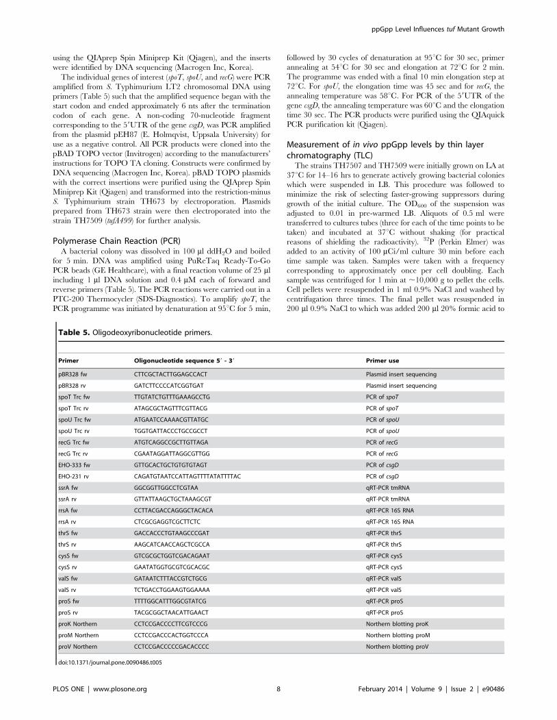
Table 5. Oligodeoxyribonucleotide primers.
Primer Oligonucleotide sequence 59 - 39 Primer use
pBR328 fw CTTCGCTACTTGGAGCCACT Plasmid insert sequencing
pBR328 rv GATCTTCCCCATCGGTGAT Plasmid insert sequencing
spoT Trc fw TTGTATCTGTTTGAAAGCCTG PCR of spoT
spoT Trc rv ATAGCGCTAGTTTCGTTACG PCR of spoT
spoU Trc fw ATGAATCCAAAACGTTATGC PCR of spoU
spoU Trc rv TGGTGATTACCCTGCCGCCT PCR of spoU
recG Trc fw ATGTCAGGCCGCTTGTTAGA PCR of recG
recG Trc rv CGAATAGGATTAGGCGTTGG PCR of recG
EHO-333 fw GTTGCACTGCTGTGTGTAGT PCR of csgD
EHO-231 rv CAGATGTAATCCATTAGTTTTATATTTTAC PCR of csgD
ssrA fw GGCGGTTGGCCTCGTAA qRT-PCR tmRNA
ssrA rv GTTATTAAGCTGCTAAAGCGT qRT-PCR tmRNA
rrsA fw CCTTACGACCAGGGCTACACA qRT-PCR 16S RNA
rrsA rv CTCGCGAGGTCGCTTCTC qRT-PCR 16S RNA
thrS fw GACCACCCTGTAAGCCCGAT qRT-PCR thrS
thrS rv AAGCATCAACCAGCTCGCCA qRT-PCR thrS
cysS fw GTCGCGCTGGTCGACAGAAT qRT-PCR cysS
cysS rv GAATATGGTGCGTCGCACGC qRT-PCR cysS
valS fw GATAATCTTTACCGTCTGCG qRT-PCR valS
valS rv TCTGACCTGGAAGTGGAAAA qRT-PCR valS
proS fw TTTTGGCATTTGGCGTATCG qRT-PCR proS
proS rv TACGCGGCTAACATTGAACT qRT-PCR proS
proK Northern CCTCCGACCCCTTCGTCCCG Northern blotting proK
proM Northern CCTCCGACCCACTGGTCCCA Northern blotting proM
proV Northern CCTCCGACCCCCGACACCCC Northern blotting proV
doi:10.1371/journal.pone.0090486.t005
ppGpp Level Influences tuf Mutant Growth
PLOS ONE | www.plosone.org 8 February 2014 | Volume 9 | Issue 2 | e90486

lyse the cells. This mixture was vortexed and stored at 220uCovernight. Before application to TLC plates, cell debris was
pelleted by centrifugation at 10,000 g for 5 min at 4uC. 5–100 ml
of supernatant (based on the OD600 of the cultures) was applied
drop-wise onto a TLC PEI Cellulose F membrane (Merck). As a
size marker, 0.2 mmol of non-radioactive ppGpp (a gift from Vasili
Hauryliuk, Uppsala) was applied onto the same membrane.
Chromatography was performed in 1.5 M KH2PO4 (pH 3.0) until
the buffer level had reached the top of the membrane. The marker
was visualized under UV-light. The membrane was dried and the
chromatography results were visualized using a PhosphorImager
(Molecular Dynamics) and quantified with the ImageQuant
software, version 4.2a (Molecular Dynamics).
To measure ppGpp as a function of spoT overexpression or relA
inactivation the strains TH7509 (tufA499), TH7964 (tufA499/
pBAD-spoT), and TH7975 (tufA499 relA21::Tn10) were initially
grown as colonies on LA as described above (with ampicillin for
TH7964), and resuspended in LB to an OD600 0.1. For strain
TH7964 the LB was supplemented with ampicillin and a series of
suspensions were prepared containing different concentrations of
L-arabinose (0, 0.025%, 0.05%, 0.1%, 0.2%). Each culture
(0.5 ml) was incubated at 37uC with shaking. After 90 min
incubation 32P was added to an activity of 100 mCi/ml of culture
and incubation was continued for a further 120 min (the OD600
was approximately 0.6). Cultures were harvested, prepared and
applied to TLC plates as described above.
RNA preparation and relative quantification of RNA byreal-time PCR (rtPCR)
Fresh colonies of TH7507, TH7976, TH7509 and TH7975
grown on LA were inoculated into liquid LB medium and grown
for a further 3 cell generations. 0.5 ml samples were extracted and
mixed with 1 ml RNA protect Bacteria Reagent (Qiagen). Total
RNA was isolated using the RNeasy Mini Kit (Qiagen), all steps
according to the manufacturer’s instructions. The quality of the
RNA was assayed visually by gel electrophoresis, and the
concentration of the different samples was measured using a
Nanodrop NO-1000 spectrophotometer (Thermo Scientific). To
remove chromosomal DNA from the RNA preparations the
DNase Turbo DNA-free (Ambion) kit was used according to the
manufacturer’s instructions. 500 ng RNA was converted into
cDNA using the High Capacity Reverse Transcription kit (Applied
Biosystems), with RT buffer, dNTP mix, random primers, and
reverse transcriptase according to the manufacturer’s instruction,
in a total reaction volume of 50 ml. The thermal steps used were
10 min at 25uC and 2 hours at 37uC. For quantitative real-time
PCR reactions, 5 ml cDNA (diluted 1:5), 10 ml PerfeCTa SYBR
Green FastMix (Quanta Biosciences), 1.25 ml of 6 mM forward
and reverse primers, Table 5, (to a final concentration of
0.375 mM), and ddH2O was added to a final reaction volume of
20 ml. The Eco Real-Time PCR System (Illumina) was used for
running the PCR program and for analyzing the data. The gene
ssrA (STM2693), encoding tmRNA, was used as a reference in the
calculations for relative expression.
RNA preparation under acidic conditions and Northernblot measurements of aminoacylation levels
Fresh colonies of TH7507 and TH7509 grown on LA were
inoculated into liquid LB medium and grown to an OD600 of 0.2
(mid-exponential phase). 50 ml of culture were poured into the
same volume of 10% trichloroacetic acid and the tubes were
transferred to ice [40]. The RNA was prepared essentially as
described by [41] and [42]. The culture-TCA mixes were
centrifuged, the pellets resuspended in the last drop of supernatant
and transferred to six microfuge tubes per 50 ml culture. The cells
were pelleted and dissolved in 500 ml NaAc buffer (0.3 M NaAc
pH 4.5, 10 mM Na2EDTA). RNA was extracted by adding 600 ml
phenol (pH 4.3, Sigma Aldrich) to the samples and vortexed
repeatedly for 15 min. After centrifugation at 12000 g for 15 min,
the aqueous phases were carefully removed to new tubes. The
phenol was re-extracted with 250 ml NaAc buffer. RNA was
precipitated by the addition of 450 ml (1 volume) ice-cold 99.5%
ethanol. Samples were kept at 220uC overnight and centrifuged at
12000 g for 30 min. The RNA pellets were washed twice with
300 ml 70% ethanol and dissolved in NaAc, pH 5.0. In total, the
RNA from 50 ml culture was dissolved in 90 ml NaAc.
2.5 mg total RNA was mixed 1:1 with acid urea sample buffer
(0.1 M sodium acetate pH 5.0, 8 M urea, 0.05% bromphenol
blue, 0.05% xylene cyanol FF) and loaded on a polyacrylamide gel
(8% polyacrylamide [19:1 acrylamide/bisacrylamide], 0.1 M
sodium acetate pH 5.0, 8 M urea, 0.15% TEMED, 0.7%
ammonium persulfate). The gel was run at 400 V at 4uC for
approximately 18 h. 0.1 M sodium acetate buffer (pH 5.0) was
used as running buffer. Transfer of the RNA to a nylon membrane
(Hybond-N+, GE Healtcare) was done for 3 h using the Novex
semi-dry blotter (Invitrogen). The transfer was conducted at 3 V,
250 mA, with 40 mM Tris-HCl pH 8.0, 2 mM Na2EDTA as
transfer buffer. RNA was UV-crosslinked to the membrane. The
membranes were pre-hybridized (66SSC, 106 Denhardt’s solu-
tion, 0.5% SDS) for 5 h at 42uC, rolling in a Hyb-Aid oven.
Oligonucleotides were labelled with c-32P-ATP (3000 Ci/mmol,
Perkin Elmer), using T4 polynucleotide kinase (Thermo Fisher).
Excess 32P-ATP was removed by using G50 columns from GE
Healthcare. Hybridization of the RNA to 32P-labeled DNA
oligonucleotides, specific to the tRNA targets (Table 5), was
carried out in hybridization buffer (66SSC, 0.1% SDS, 106 cpm/
ml 32P-labeled probe) for 12 h, with rolling at 42uC. Stringency
washing of the membranes to remove unbound probe was carried
out by 2610 min washes at room temperature in each of 66SSC,
46SSC, and 26SSC. The membranes were visualized using a
Personal Molecular Imager (Bio-Rad) and analyzed using Quan-
tity One software (Bio-Rad).
Supporting Information
Figure S1 Northern blot measurement of proK tRNAaminoacylation. RNA was prepared from mid-log phase
cultures of wild-type (TH7507) and tufA499 mutant (TH7509)
under acidic conditions, run on a polyacrylamide gel and
transferred to nylon membrane. The figure shows a scan of a
representative blot of a membrane hybridized with a 32P-ATP-
labeled probe for the proK tRNA, showing a lower level of acylated
pro-tRNA in the tufA499 mutant relative to the wild-type.
(TIF)
Author Contributions
Conceived and designed the experiments: DH JB DLH. Performed the
experiments: JB DLH. Analyzed the data: DH JB DLH. Wrote the paper:
DH JB DHL.
ppGpp Level Influences tuf Mutant Growth
PLOS ONE | www.plosone.org 9 February 2014 | Volume 9 | Issue 2 | e90486

References
1. Hughes D, Tubulekas I (1993) Ternary complex-ribosome interaction: its
influence on protein synthesis and on growth rate. Biochem Soc Trans 21: 851–857.
2. Tubulekas I, Hughes D (1993) Growth and translation elongation rate aresensitive to the concentration of EF-Tu. Mol Microbiol 8: 761–770.
3. Abdulkarim F, Hughes D (1996) Homologous recombination between the tuf
genes of Salmonella typhimurium. J Mol Biol 260: 506–522.4. Hughes D (1986) The isolation and mapping of EF-Tu mutations in Salmonella
typhimurium. Mol Gen Genet 202: 108–111.5. Hughes D (1990) Both genes for EF-Tu in Salmonella typhimurium are
individually dispensable for growth. J Mol Biol 215: 41–51.
6. Abdulkarim F, Tuohy TM, Buckingham RH, Hughes D (1991) Missensesubstitutions lethal to essential functions of EF-Tu. Biochimie 73: 1457–1464.
7. Abdulkarim F, Liljas L, Hughes D (1994) Mutations to kirromycin resistanceoccur in the interface of domains I and III of EF-Tu.GTP. FEBS Lett 352: 118–
122.8. Hammarlof DL, Hughes D (2008) Mutants of the RNA-processing enzyme
RNase E reverse the extreme slow-growth phenotype caused by a mutant
translation factor EF-Tu. Mol Microbiol 70: 1194–1209.9. Abdulkarim F, Ehrenberg M, Hughes D (1996) Mutants of EF-Tu defective in
binding aminoacyl-tRNA. FEBS Lett 382: 297–303.10. McClelland M, Sanderson KE, Spieth J, Clifton SW, Latreille P, et al. (2001)
Complete genome sequence of Salmonella enterica serovar Typhimurium LT2.
Nature 413: 852–856.11. Xiao H, Kalman M, Ikehara K, Zemel S, Glaser G, et al. (1991) Residual
guanosine 39,59-bispyrophosphate synthetic activity of relA null mutants can beeliminated by spoT null mutations. J Biol Chem 266: 5980–5990.
12. Persson BC, Jager G, Gustafsson C (1997) The spoU gene of Escherichia coli,the fourth gene of the spoT operon, is essential for tRNA (Gm18) 29-O-
methyltransferase activity. Nucleic Acids Res 25: 4093–4097.
13. Kalman M, Murphy H, Cashel M (1992) The nucleotide sequence of recG, thedistal spo operon gene in Escherichia coli K-12. Gene 110: 95–99.
14. Hernandez VJ, Bremer H (1991) Escherichia coli ppGpp synthetase II activityrequires spoT. J Biol Chem 266: 5991–5999.
15. Paul BJ, Barker MM, Ross W, Schneider DA, Webb C, et al. (2004) DksA: a
critical component of the transcription initiation machinery that potentiates theregulation of rRNA promoters by ppGpp and the initiating NTP. Cell 118: 311–
322.16. Potrykus K, Murphy H, Philippe N, Cashel M (2011) ppGpp is the major source
of growth rate control in E. coli. Environ Microbiol 13: 563–575.17. Brown L, Gentry D, Elliott T, Cashel M (2002) DksA affects ppGpp induction of
RpoS at a translational level. J Bacteriol 184: 4455–4465.
18. Magnusson LU, Gummesson B, Joksimovic P, Farewell A, Nystrom T (2007)Identical, independent, and opposing roles of ppGpp and DksA in Escherichia
coli. J Bacteriol 189: 5193–5202.19. Aberg A, Fernandez-Vazquez J, Cabrer-Panes JD, Sanchez A, Balsalobre C
(2009) Similar and divergent effects of ppGpp and DksA deficiencies on
transcription in Escherichia coli. J Bacteriol 191: 3226–3236.20. Kanjee U, Ogata K, Houry WA (2012) Direct binding targets of the stringent
response alarmone (p)ppGpp. Mol Microbiol 85: 1029–1043.21. Rojas AM, Ehrenberg M, Andersson SG, Kurland CG (1984) ppGpp inhibition
of elongation factors Tu, G and Ts during polypeptide synthesis. Molecular &general genetics: MGG 197: 36–45.
22. Potrykus K, Cashel M (2008) (p)ppGpp: still magical? Annu Rev Microbiol 62:
35–51.23. Stead MB, Marshburn S, Mohanty BK, Mitra J, Pena Castillo L, et al. (2011)
Analysis of Escherichia coli RNase E and RNase III activity in vivo using tilingmicroarrays. Nucleic Acids Res 39: 3188–3203.
24. Bremer H, Dennis PP (1996) Modulation of chemical composition and other
parameters of the cell by growth rate. In: Neidhart FC, Curtis III R, Ingraham
JL, Lin ECC, Low KB, Magasanik B, Reznikoff WS, Riley M, Schaechter M
and Umbarger HE, editor. Escherichia coli and Salmonella: cellular and
molecular biology. 2. ed. Washington: ASM Press. pp. 1553–1569.
25. Neidhardt FC, Ingraham JL, Schaechter M (1990) Physiology of the bacterial
cell: a molecular approach. Sunderland, Mass.: Sinauer. xii, 506 p.
26. Magnusson LU, Farewell A, Nystrom T (2005) ppGpp: a global regulator in
Escherichia coli. Trends Microbiol 13: 236–242.
27. Mechold U, Potrykus K, Murphy H, Murakami KS, Cashel M (2013)
Differential regulation by ppGpp versus pppGpp in Escherichia coli. Nucleic
Acids Res 41: 6175–6189.
28. Ross W, Vrentas CE, Sanchez-Vazquez P, Gaal T, Gourse RL (2013) The
magic spot: a ppGpp binding site on E. coli RNA polymerase responsible for
regulation of transcription initiation. Mol Cell 50: 420–429.
29. Perederina A, Svetlov V, Vassylyeva MN, Tahirov TH, Yokoyama S, et al.
(2004) Regulation through the secondary channel–structural framework for
ppGpp-DksA synergism during transcription. Cell 118: 297–309.
30. Vinella D, Potrykus K, Murphy H, Cashel M (2012) Effects on growth by
changes of the balance between GreA, GreB, and DksA suggest mutual
competition and functional redundancy in Escherichia coli. J Bacteriol 194: 261–
273.
31. Murray KD, Bremer H (1996) Control of spoT-dependent ppGpp synthesis and
degradation in Escherichia coli. J Mol Biol 259: 41–57.
32. Stent GS, Brenner S (1961) A genetic locus for the regulation of ribonucleic acid
synthesis. Proc Natl Acad Sci U S A 47: 2005–2014.
33. An G, Justesen J, Watson RJ, Friesen JD (1979) Cloning the spoT gene of
Escherichia coli: identification of the spoT gene product. J Bacteriol 137: 1100–
1110.
34. Sarubbi E, Rudd KE, Xiao H, Ikehara K, Kalman M, et al. (1989)
Characterization of the spoT gene of Escherichia coli. J Biol Chem 264:
15074–15082.
35. Haseltine WA, Block R (1973) Synthesis of guanosine tetra- and pentaphosphate
requires the presence of a codon-specific, uncharged transfer ribonucleic acid in
the acceptor site of ribosomes. Proceedings of the National Academy of Sciences
of the United States of America 70: 1564–1568.
36. Haseltine WA, Block R, Gilbert W, Weber K (1972) MSI and MSII made on
ribosome in idling step of protein synthesis. Nature 238: 381–384.
37. Stent GS, Brenner S (1961) A genetic locus for the regulation of ribonucleic acid
synthesis. Proceedings of the National Academy of Sciences of the United States
of America 47: 2005–2014.
38. Jakubowski H (2012) Quality control in tRNA charging. Wiley interdisciplinary
reviews RNA 3: 295–310.
39. Schmieger H (1971) A method for detection of phage mutants with altered
transducing ability. Mol Gen Genet 110: 378–381.
40. Kruger MK, Sorensen MA (1998) Aminoacylation of hypomodified tRNAGlu in
vivo. J Mol Biol 284: 609–620.
41. Varshney U, Lee CP, RajBhandary UL (1991) Direct analysis of aminoacylation
levels of tRNAs in vivo. Application to studying recognition of Escherichia coli
initiator tRNA mutants by glutaminyl-tRNA synthetase. J Biol Chem 266:
24712–24718.
42. Kohrer C, Rajbhandary UL (2008) The many applications of acid urea
polyacrylamide gel electrophoresis to studies of tRNAs and aminoacyl-tRNA
synthetases. Methods 44: 129–138.
43. Yu D, Ellis HM, Lee EC, Jenkins NA, Copeland NG, et al. (2000) An efficient
recombination system for chromosome engineering in Escherichia coli. Proc
Natl Acad Sci U S A 97: 5978–5983.
44. Datsenko KA, Wanner BL (2000) One-step inactivation of chromosomal genes
in Escherichia coli K-12 using PCR products. Proc Natl Acad Sci U S A 97:
6640–6645.
ppGpp Level Influences tuf Mutant Growth
PLOS ONE | www.plosone.org 10 February 2014 | Volume 9 | Issue 2 | e90486

Related Documents


