doi:10.1136/ard.2007.078246 2008;67;1480-1487; originally published online 12 Nov 2007; Ann Rheum Dis M-J González Quest, U Mandel, E Veerman, M Espinosa, H Clausen, C Leyton, R Romo and C Alliende, Y-J Kwon, M Brito, C Molina, S Aguilera, P Pérez, L Leyton, A F G xerostomia in patients with Sjögren syndrome Reduced sulfation of muc5b is linked to http://ard.bmj.com/cgi/content/full/67/10/1480 Updated information and services can be found at: These include: Data supplement http://ard.bmj.com/cgi/content/full/67/10/1480/DC1 "web only appendix" References http://ard.bmj.com/cgi/content/full/67/10/1480#otherarticles 1 online articles that cite this article can be accessed at: http://ard.bmj.com/cgi/content/full/67/10/1480#BIBL This article cites 31 articles, 14 of which can be accessed free at: Rapid responses http://ard.bmj.com/cgi/eletter-submit/67/10/1480 You can respond to this article at: service Email alerting the top right corner of the article Receive free email alerts when new articles cite this article - sign up in the box at Topic collections (14199 articles) Immunology (including allergy) Articles on similar topics can be found in the following collections Notes http://journals.bmj.com/cgi/reprintform To order reprints of this article go to: http://journals.bmj.com/subscriptions/ go to: Annals of the Rheumatic Diseases To subscribe to on 28 August 2009 ard.bmj.com Downloaded from

Welcome message from author
This document is posted to help you gain knowledge. Please leave a comment to let me know what you think about it! Share it to your friends and learn new things together.
Transcript
doi:10.1136/ard.2007.078246 2008;67;1480-1487; originally published online 12 Nov 2007; Ann Rheum Dis
M-J González Quest, U Mandel, E Veerman, M Espinosa, H Clausen, C Leyton, R Romo and C Alliende, Y-J Kwon, M Brito, C Molina, S Aguilera, P Pérez, L Leyton, A F G
xerostomia in patients with Sjögren syndromeReduced sulfation of muc5b is linked to
http://ard.bmj.com/cgi/content/full/67/10/1480Updated information and services can be found at:
These include:
Data supplement http://ard.bmj.com/cgi/content/full/67/10/1480/DC1
"web only appendix"
References
http://ard.bmj.com/cgi/content/full/67/10/1480#otherarticles1 online articles that cite this article can be accessed at:
http://ard.bmj.com/cgi/content/full/67/10/1480#BIBLThis article cites 31 articles, 14 of which can be accessed free at:
Rapid responses http://ard.bmj.com/cgi/eletter-submit/67/10/1480
You can respond to this article at:
serviceEmail alerting
the top right corner of the article Receive free email alerts when new articles cite this article - sign up in the box at
Topic collections
(14199 articles) Immunology (including allergy) � Articles on similar topics can be found in the following collections
Notes
http://journals.bmj.com/cgi/reprintformTo order reprints of this article go to:
http://journals.bmj.com/subscriptions/ go to: Annals of the Rheumatic DiseasesTo subscribe to
on 28 August 2009 ard.bmj.comDownloaded from
Reduced sulfation of muc5b is linked to xerostomiain patients with Sjogren syndrome
C Alliende,1 Y-J Kwon,1 M Brito,1 C Molina,2 S Aguilera,3 P Perez,1 L Leyton,1
A F G Quest,1 U Mandel,4 E Veerman,5 M Espinosa,1 H Clausen,4 C Leyton,1 R Romo,6
M-J Gonzalez1
c Supplementary table 1 ispublished online only at http://ard.bmj.com/content/vol67/issue10
1 University of Chile, Santiago,Chile; 2 Mayor University,Santiago, Chile; 3 INDISA Clinic-Andres Bello University,Santiago, Chile; 4 University ofCopenhagen, Copenhagen,Denmark; 5 University ofAmsterdam, Amsterdam, TheNetherlands; 6 Santa MarıaClinic, Santiago, Chile
Correspondence to:M-J Gonzalez, Institute ofBiomedical Sciences, Faculty ofMedicine, University of Chile,Casilla 70061, Santiago 7, Chile;[email protected]
The first four authors contributedequally to this work
Accepted 1 November 2007Published Online First12 November 2007
ABSTRACTObjectives: MUC5B contains sulfated and sialylatedoligosaccharides that sequester water required formoisturising the oral mucosa. Xerostomia, in patients withSjogren syndrome, is generally associated with reducedquantities, rather than altered properties, of saliva. Here,we determined the amount of MUC5B (mRNA andprotein) as well as sulfation levels in salivary glands ofpatients with normal or altered unstimulated salivary flow.Localisation of MUC5B and sulfated MUC5B, as well astotal levels sulfated groups were determined andcompared with acini basal lamina disorganisation.Patients and methods: In all, 18 patients with normal oraltered unstimulated salivary flow and 16 controls werestudied. MUC5B mRNA and protein were evaluated insalivary glands by semiquantitative RT-PCR and Westernblot analysis. MUC5B sulfation was determined byWestern blotting. MUC5B and sulfo-Lewisa antigenlocalisation were assessed by immunohistochemistry. Thetotal amount of sulfated oligosaccharides was determinedmicrodensitometrically.Results: No significant differences were detected inMUC5B mRNA and protein levels between controls andpatients, while sulfo-Lewisa antigen levels were lower inpatients. The number of sulfo-Lewisa positive mucousacini was reduced in patients but no correlation wasobserved between lower levels of sulfation and unsti-mulated salivary flow. Microdensitometric data confirmedthe presence of reduced sulfated oligosaccharides levelsin mucous acini from patients with highly disorganisedbasal lamina.Conclusion: Disorganisation of the basal lamina observedin patients with Sjogren syndrome may lead todedifferentiation of acinar mucous cells and, as aconsequence, alter sulfation of MUC5B. These changesare suggested to represent a novel mechanism that mayexplain xerostomia in these patients.
Primary Sjogren syndrome is an autoimmuneexocrinopathy. Secretory activity of salivary andlacrimal glands is highly compromised, and severedryness of the mouth and the eyes are theconsequence.1 Autoantibodies against muscarinic-M3 receptors,2 variations in cytokine levels,3
glandular denervation,4 acinar atrophy,5 redistribu-tion of aquaporin-5 in the acinar cells6 andincreased levels of cholinesterase7 are the mostfrequent molecular and morphological changesassociated with symptoms of dryness. However,only some of these changes are observed in thesepatients. Thus, correlating the observed changeswith specific symptoms, such as dryness, remainsas a controversial issue in the field.7 8
Morphological changes in the secretory granuleshave been consistently observed in all patientsevaluated9 10 and could be considered one of themost reliable indicators for this disease. In non-obese diabetic (NOD) mice, an animal model forSjogren syndrome, changes in secretory granulemorphology and distribution of key effectors ofexocytosis are apparent at 4 weeks after birth,which may be taken to indicate that disease onsetoccurs prior to lymphocyte infiltration.11 A betterunderstanding of events specifically associatedwith early alterations in secretory granule functionshould be insightful.
MUC5B, the predominant mucin in salivarygland high molecular weight mucous glycoprotein1 preparations, is synthesised by mucous acini andglycosylated to varying extents.12 The associatedcarbohydrates are heterogeneous and include neu-tral, sulfated and sialylated oligosaccharides.13
Sulfated and sialylated mucins retain largeamounts of water and contribute thereby togenerating the hydrophilic gel essential for lubrica-tion of the oral epithelium.14
Previous studies revealed high levels of highmolecular weight mucous glycoprotein 1 in restingsaliva of patients with Sjogren syndrome.15 Theseauthors attributed the changes to either thereduced presence of water or, alternatively, to alimitation in the capacity to retain water andsuggested that this might explain xerostomia.15
The objective of this study was to evaluatewhether symptoms of xerostomia were linked toreduced salivary flow or qualitative/quantitativechanges (eg, post-translational modifications) inthe MUC5B produced.
PATIENTS AND METHODSA total of 18 patients (females) with primarySjogren syndrome (mean (SD) age = 50.5 (12.3)years) diagnosed according to the American–European Consensus Group criteria,16 were selectedfor these studies. Several tests were performed toidentify ocular, oral and serological involvement.The focus score was >1 in patients with 50–80% ofremnant parenchyma. Dry eye and mouth symp-toms were observed in all patients. Unstimulatedsalivary flow was lower than 1.5 ml per 15 min in11/18 patients. Additionally, scintigraphy andSchirmer test results were altered. Patients hadneither been treated with corticosteroids, hydro-xychloroquine, or immunosuppressive drugs for atleast 1 month before biopsies were obtained.
Extended report
1480 Ann Rheum Dis 2008;67:1480–1487. doi:10.1136/ard.2007.078246
on 28 August 2009 ard.bmj.comDownloaded from

Additional clinical data are included in the Supplementarymaterial.
The control group was composed of 16 subjects (females)(mean (SD) age = 43.09 (12.18) years) who had consulted theirdoctors because of oral and ocular dryness symptoms over morethan 3 months, but who did not fulfil the criteria for Sjogrensyndrome. Serological, scintigraphy and unstimulated wholesalivary flow data were normal in these patients. Biopsied labialsalivary glands were normal, with scarce and scattereddistribution of mononuclear cells, well-preserved parenchyma,and lack of fibrous or adipose tissue. Control individuals werefree of systemic diseases and did not use drugs that could affectthe function of the exocrine glands. Biopsies were obtainedfollowing informed consent of the control group and patients.No significant differences (p = 0.12) between controls andpatients according to age were detectable. This study wasconducted according to the guidelines of the Ethics Committeeof the Faculty of Medicine, University of Chile.
BiopsiesLabial salivary gland biopsies were obtained in the morning(after at least 2 h of fasting), using the technique described byDaniels.17 Glands were fixed for immunohistochemistry usingtwo different conditions. One half of the gland was treated withalcoholic Bouin fixative (MUC5B and sulfo-Lewisa antigen) andthe other half was fixed in 1% paraformaldehyde (laminin) andthen embedded in paraffin. For RNA and protein preparations,the glands were snap-frozen in liquid nitrogen and storedat –70uC.
RNA extraction/semiquantitative RT-PCR/Preparation of proteinextractsExperiments were performed essentially as previously described18–20 (see also Supplementary material).
Western blottingGland protein extracts were prepared as previouslydescribed.18 19 Aliquots of 25 mg of proteins were separated on6% SDS-polyacrylamide gels under reducing conditions andtransferred to nitrocellulose over 15 h at 60 mA and 4uC.Membranes were incubated with PANH2 (a mouse antibody,recognises partially deglycosylated MUC5B) or F2 (a mouseantibody, recognises the SO3Galb1-3GlcNAc moiety of sulfo-Lewisa antigen, both obtained from the European Consortiumof Mucins), diluted 1:10 or 1:50 in Tris buffered saline/Tween(TBST), respectively, for 2 h at room temperature. Alternatively,membranes were incubated with the anti-actin antibody (MPBiomedicals, Aurora, Ohio, USA) diluted 1:5000 in blockingsolution. Then, blots were incubated with anti-mouse IgG-HRP(Jackson ImmunoResearch Laboratories, West Grove,Pennsylvania, USA) (dilution 1:5000) for 1 h at room tempera-ture, and horseradish peroxidase (HRP) was detected with ECL asdescribed.18 MUC5B and Sulfo-MUC5B bands were analyseddensitometrically (UN-Scan-IT, Silk Scientific Corporation,Orem, Utah, USA) and values obtained were normalised to thoseof b-actin. An amount of 10–70 mg of total protein yielded adensitometric b-actin signal within the linear range.
Immunohistochemistry analysisSections used for MUC5B and sulfo-Lewisa antigen detectionswere blocked to prevent non-specific antibody binding with0.25% casein in phosphate buffered saline (PBS) and incubatedfor 20 h at 4uC with non-diluted PANH2 or F2 diluted 1:50 in
1% bovine serum albumin/PBS, followed by a biotinylatedsecondary antibody and a streptavidin–peroxidase conjugate(Dako, Carpinteria, California, USA). The reaction was devel-oped with 39,39-diaminobenzidine. Mayer haematoxylin wasused for counterstaining. As a negative control, a mouse IgG1(PANH2) or IgM (F2) recognising an epitope not present inmammalian tissues was employed. For additional informationconcerning PANH2 and F2 antibodies, see Supplementarymaterial. Laminin was detected as previously described9 andthese sections were also stained with Alcian blue pH 1.0 for15 min at room temperature, (for details see Supplementarymaterial).
Quantification of the histochemistry reaction of sulfated mucinsGland sections were stained with Alcian blue pH 1.0 to identifysulfated mucins. Staining intensity was measured with a MPM-400 microdensitometer (Carl Zeiss, Jena, Germany). For eachbiopsy, 50 random fields containing parenchyma were analysedand the mean value was expressed in arbitrary units ofabsorbance (AU). The data were evaluated using the Systat9.0 program (SPSS, Chicago, Illinois, USA).
Statistical analysisNormalised data of mRNA, proteins and sulfo-Lewisa antigenwere averaged to calculate mean values and the standarddeviation. A Mann–Whitney U test and non-parametricSpearman correlation were used. Microdensitometric measure-ments of sulfated mucins were compared by unpaired Studentt test analysis. p Values less than 0.05 were consideredsignificant.
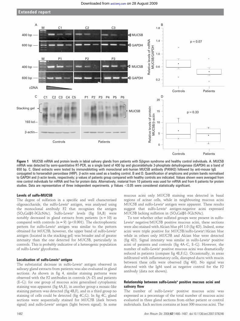
RESULTSmRNA and protein levels of MUC5BAs shown in fig 1A,B, no significant changes in relative mRNAexpression levels were found when comparing samples frompatients (n = 10) with control individuals (n = 9) (p = 0.07);however, a tendency towards decreased mRNA levels wasdetected in patients. The PANH2 antibody, which recognisespartially glycosylated MUC5B, was employed for MUC5Bprotein determination.21 Two broad and diffuse bands wereobserved, one migrating with an apparent molecular weightsomewhat larger than 200 kDa and the second being so largethat it was retained in the stacking gel. The electrophoreticpattern was similar between patients and controls. Nosignificant differences (p = 0.33) were found when bands wereanalysed by scanning densitometry either separately (notshown) or together (fig 1C,D).
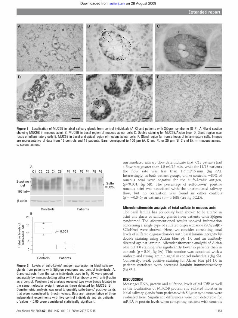
Localisation of MUC5BMUC5B was detected in mucous acini from controls andpatients (fig 2A–F). MUC5B levels were similar in all mucousacinar cells present in the normal gland sections (fig 2A) andlocalised to the basal region of these cells (fig 2B). Doublestaining with PANH2/Alcian blue pH 1.0 revealed sulfatedsecretion products in the apical region of mucous acinar cells incontrols (fig 2C). In patients, MUC5B was detectable in thebasal and apical zones of mucous acinar cells (fig 2D,E).Stronger immunoreactivity was detected in mucous aciniadjacent to foci of inflammatory cells (fig 2D,E), while inregions without foci; the intensity was similar to those observedin controls (fig 2F). In patients, increased immunoreactivity forMUC5B and particularly presence in apical suggests thatMUC5B glycosylation was decreased.
Extended report
Ann Rheum Dis 2008;67:1480–1487. doi:10.1136/ard.2007.078246 1481
on 28 August 2009 ard.bmj.comDownloaded from
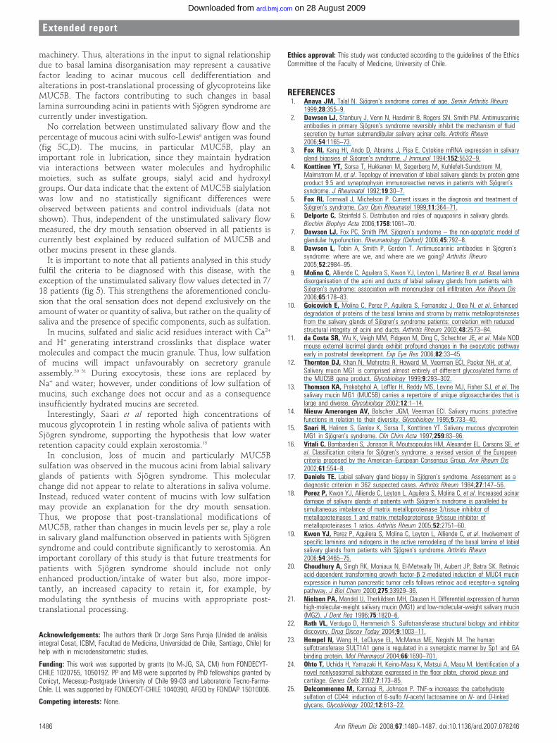
Levels of sulfo-MUC5BThe degree of sulfation in a specific and well characterisedoligosaccharide, the sulfo-Lewisa antigen, was analysed usingthe monoclonal antibody F2 that recognises the antigen(SO3Galb1-3GlcNAc). Sulfo-Lewisa levels (fig 3A,B) werenotably decreased in gland extracts from patients (n = 10) ascompared with controls (n = 9) (p,0.001). The electrophoreticpattern for sulfo-Lewisa antigen was similar to the patternobtained for MUC5B; however, the upper band of sulfo-Lewisa
antigen (located in the stacking gel) was broader and of greaterintensity than the one detected for MUC5B, particularly incontrols. This is probably indicative of a heterogenic populationof sulfo-Lewisa glycoforms.
Localisation of sulfo-Lewisa antigenThe substantial decrease in sulfo-Lewisa antigen observed insalivary gland extracts from patients was also evaluated in glandsections. As shown in fig 4, similar staining patterns wereobserved with the F2 antibodies in controls (A–D) and patients(E–G): for one group of mucous acini generalised cytoplasmicstaining was apparent (fig 4A,E), in another group a mosaic-likestaining pattern was detected (fig 4B,F), and in a third group nostaining of cells could be detected (fig 4C,G). In fig 4C, glandsections were sequentially stained for MUC5B (dark brownsignal) and sulfo-Lewisa antigen (light brown signal). In some
mucous acini only MUC5B staining was detected in basalregions of acinar cells, while in neighbouring mucous aciniMUC5B and sulfo-Lewisa antigen were apparent. These resultssuggest that sulfo-Lewisa antigen-negative acini expressedMUC5B lacking sulfation in (SO3Galb1-3GlcNAc).
To test whether other sulfated groups were present in sulfo-Lewisa negative/MUC5B positive mucous acini, these sectionswere also stained with Alcian blue pH 1.0 (fig 4D). Indeed, someacini were triple positive for MUC5B/sulfo-Lewisa/Alcian bluewhile in others only MUC5B and Alcian blue were detected(fig 4D). Signal intensity was similar in sulfo-Lewisa positiveacini of patients and controls (fig 4A–C, E–G). However, thenumber of sulfo-Lewisa positive mucous acini was dramaticallyreduced in patients (compare fig 4E,F,G). Occasionally, in areasinfiltrated with inflammatory cells, disrupted ducts with mucinbetween these cells were observed (fig 4H). No signal wasdetected with the IgM used as negative control for the F2antibody (data not shown).
Relationship between sulfo-Lewisa positive mucous acini andsalivary flowThe number of sulfo-Lewisa positive mucous acini wasexpressed as a percentage of the total number of mucous acinievaluated in three gland sections from either patient or controlindividuals. Each section contains at least 300 mucous acini. The
Figure 1 MUC5B mRNA and protein levels in labial salivary glands from patients with Sjogren syndrome and healthy control individuals. A. MUC5BmRNA was detected by semi-quantitative RT-PCR, as a single band of 400 bp and glyceraldehyde 3-phosphate dehydrogenase (GAPDH) as a band of650 bp. C. Gland extracts were tested by immunoblotting with monoclonal anti-human MUC5B antibody (PANH2) followed by anti-mouse IgGconjugated to horseradish peroxidase (HRP). b-actin was used as a loading control. B and D. Quantification of amplicons and protein bands normalisedto GAPDH and b-actin levels, respectively; p values of patients group compared with healthy controls are indicated. Values shown were averaged fromnine control individuals for mRNA and five for protein data. Alternatively, material from 10 patients was used for mRNA and from 6 patients for proteinstudies. Data are representative of three independent experiments. p Values ,0.05 were considered statistically significant.
Extended report
1482 Ann Rheum Dis 2008;67:1480–1487. doi:10.1136/ard.2007.078246
on 28 August 2009 ard.bmj.comDownloaded from
unstimulated salivary flow data indicate that 7/18 patients hada flow rate greater than 1.5 ml/15 min, while for 11/18 patientsthe flow rate was less than 1.5 ml/15 min (fig 5A).Interestingly, in both patient groups, unlike controls, ,80% ofmucous acini were negative for the sulfo-Lewisa antigen,(p,0.001; fig 5B). The percentage of sulfo-Lewisa positivemucous acini was associated with the unstimulated salivaryflow, but no correlation was found in either controls(r= 20.346) or patients (r= 0.168) (see fig 5C,D).
Microdensitometric analysis of total sulfate in mucous aciniThe basal lamina has previously been shown to be altered inacini and ducts of salivary glands from patients with Sjogrensyndrome.9 The aforementioned results showed informationconcerning a single type of sulfated oligosaccharide (SO3Galb1-3GlcNAc) were showed. Here, we consider correlating totallevels of sulfated oligosaccharides with basal lamina integrity bydouble staining using Alcian blue pH 1.0 and an antibodydirected against laminin. Microdensitometric analysis of Alcianblue pH 1.0 staining was significantly lower in patients than incontrols (p = 0.04; fig 6A). This reaction was associated with auniform and strong laminin signal in control individuals (fig 6B).Conversely, weak positive staining for Alcian blue pH 1.0 inpatients correlated with decreased laminin immunoreactivity(fig 6C).
DISCUSSIONMessenger RNA, protein and sulfation levels of MUC5B as wellas the localisation of MUC5B protein and sulfated moieties inlabial salivary glands from patients with Sjogren syndrome wereevaluated here. Significant differences were not detectable formRNA or protein levels when comparing patients with controls
Figure 2 Localisation of MUC5B in labial salivary glands from control individuals (A–C) and patients with Sjogren syndrome (D–F). A. Gland sectionshowing MUC5B in mucous acini. B. MUC5B in basal region of mucous acinar cells C. Double staining for MUC5B/Alcian blue. D. Gland region nearfocus of inflammatory cells E. MUC5B in basal and apical region of mucous acinar cells. F. Gland region far from a focus of inflammatory cells. Imagesare representative of data from 16 controls and 18 patients. Bars: correspond to 100 mm (A, D and F), or 20 mm (B, C and E). m: mucous acinus,s: serous acinus.
Figure 3 Levels of sulfo-Lewisa antigen expression in labial salivaryglands from patients with Sjogren syndrome and control individuals. A.Gland extracts from the same individuals used in fig 1C were probedseparately by immunoblotting either with F2 antibody or with anti-b-actinas a control. Western blot analysis revealed two wide bands located inthe same molecular weight region as those detected for MUC5B. B.Densitometric analysis was used to quantify sulfo-Lewisa positive bandsthat were normalised to b-actin values. Data are representative of threeindependent experiments with five control individuals and six patients.p Values ,0.05 were considered statistically significant.
Extended report
Ann Rheum Dis 2008;67:1480–1487. doi:10.1136/ard.2007.078246 1483
on 28 August 2009 ard.bmj.comDownloaded from

(fig 1). Importantly, levels of sulfation were substantiallyreduced in gland extracts obtained from patients (fig 3).Additionally, decreased staining for SO3Galb1-3GlcNAc moi-eties and total sulfate oligosaccharides present in mucous aciniwas observed (figs 5 and 6).
Post-translational processing of mucins, including O/N-glycosylation and sulfation, occur in the Golgi complex.22
Since our current understanding of the regulation of glycosyl-transferases/sulfotransferases is limited,23 pin-pointing thedefect in sulfation pathways that could explain the observedlower levels of sulfated moieties in mucous acinar cells ofpatients is difficult. Lower expression levels and/or lowerenzymatic activity of sulfotransferases, reduced amounts ofsubstrates (eg, oligosaccharide moieties, sulfate groups, etc.)
and/or an increased activity of sulfatases could explain theresults of this study. Although sulfatases are thought to belocated in lysosomes, more recent evidence favours the existenceof a novel non-lysosomal sulfatase pool in the Golgi complex, aswell as at the cell surface.24 Thus, the Golgi complex could bethe organelle responsible for the changes reported in this study;however, further work is necessary to provide a conclusiveexplanation for decreased sulfation observed in patients withSjogren syndrome.
In human myeloid cells, tumour necrosis factor (TNF)astimulates sulfation of GlcNAc present in the cell adhesionglycoprotein CD44, possibly by activating aGlcNAc6sulfotransferase that modulates the degree of sulfationof N- and O-linked glycans.25 By contrast, treatment of cultured
Figure 4 Localisation of sulfo-Lewisa
antigen in labial salivary glands fromcontrol individuals (A–D) and patientswith Sjogren syndrome (E–H). A. mucousacini completely stained. B. mucous aciniwith mosaic staining pattern. C.Sequential staining for MUC5B (darkbrown) and sulfo-Lewisa antigen (lightbrown). D. Triple staining (MUC5B/sulfo-Lewisa/Alcian blue). Mucous acini thatwere either sulfo-Lewisa negative/MUC5Bpositive/Alcian blue positive (insert) orpositive for all three are visible as ayellowish green with dark brown border.E, F, G and H. Sections stained only withF2 antibody. E, F and G. Varyingabundance of mucous acini stainedpositive for sulfo-Lewisa antigen. H. Adamaged duct immersed in a focus ofinflammatory cells. Images arerepresentative of data from 16 controlsand 18 patients. Bars: correspond to50 mm (A, C, E, F and G), 20 mm (B andH) or 100 mm (D).
Extended report
1484 Ann Rheum Dis 2008;67:1480–1487. doi:10.1136/ard.2007.078246
on 28 August 2009 ard.bmj.comDownloaded from

bovine articular synoviocytes with TNFa caused a decrease inGal3sulfotransferase activity,26 suggesting a cell type- andsulfotransferase-specific regulation of sulfation. Additionally,these data indicate that the stimulation of an inflammatoryresponse could alter the ratio of specific sulfated glycanstructures present in a cell. Since TNFa levels are elevated inpatients with Sjogren syndrome, inhibition of some sulfotrans-ferase may provide an explanation for the low levels ofglycoprotein sulfation detected in these patients. However, nocorrelation was observed between reduced sulfation and focusscore (data not shown).
A striking correlation was detected between reduced levels oftotal sulfated oligosaccharides and the degree of basal lamina
disorganisation observed (fig 6). Receptors (integrins) located inthe basolateral plasma membrane interact with basal laminaproteins and thereby trigger a variety of cellular responses,including secretory processes.27 28 Thus, if these membranereceptors are surrounded by a modified environment, changesin the interaction between proteins of both compartmentscould alter receptor functionality, even under circumstanceswhere ligands or agonists are still present.27 28 Previous studieshave reported variations in the organisation and expression ofseveral laminins and nidogens in acini of patients with Sjogrensyndrome. Such changes may affect acinar cell differentiationand polarisation.19 29 Altered signalling events potentially con-tribute to differential engagement of, for instance, the secretory
Figure 5 Relationship between sulfo-Lewisa positive mucous acini and salivaryflow. A. Values for control individuals andpatients with high (q) and low (Q)unstimulated salivary flow (USF) areshown. B. Percentage of sulfo-Lewisa
mucous acini in controls and patients. C.Spearman correlation between sulfo-Lewisa positive mucous acini and salivaryflow in control individuals and patients. D.USF: unstimulated salivary flow, SS:patients with Sjogren syndrome.
Figure 6 Microdensitometric analysis oftotal sulfate in mucous acini of labialsalivary glands from control individualsand patients with Sjogren syndrome. A.Box plot indicating that absorbanceexpressed in AU for Alcian blue positiveareas was higher in control individualsthan in patients. B and C. Double stainingwith Alcian blue pH 1.0 and laminin inmicrophotographs of salivary glandsamples from controls (B) and patients(C). Bars in B and C: 50 mm. AU: arbitraryunits.
Extended report
Ann Rheum Dis 2008;67:1480–1487. doi:10.1136/ard.2007.078246 1485
on 28 August 2009 ard.bmj.comDownloaded from
machinery. Thus, alterations in the input to signal relationshipdue to basal lamina disorganisation may represent a causativefactor leading to acinar mucous cell dedifferentiation andalterations in post-translational processing of glycoproteins likeMUC5B. The factors contributing to such changes in basallamina surrounding acini in patients with Sjogren syndrome arecurrently under investigation.
No correlation between unstimulated salivary flow and thepercentage of mucous acini with sulfo-Lewisa antigen was found(fig 5C,D). The mucins, in particular MUC5B, play animportant role in lubrication, since they maintain hydrationvia interactions between water molecules and hydrophilicmoieties, such as sulfate groups, sialyl acid and hydroxylgroups. Our data indicate that the extent of MUC5B sialylationwas low and no statistically significant differences wereobserved between patients and control individuals (data notshown). Thus, independent of the unstimulated salivary flowmeasured, the dry mouth sensation observed in all patients iscurrently best explained by reduced sulfation of MUC5B andother mucins present in these glands.
It is important to note that all patients analysed in this studyfulfil the criteria to be diagnosed with this disease, with theexception of the unstimulated salivary flow values detected in 7/18 patients (fig 5). This strengthens the aforementioned conclu-sion that the oral sensation does not depend exclusively on theamount of water or quantity of saliva, but rather on the quality ofsaliva and the presence of specific components, such as sulfation.
In mucins, sulfated and sialic acid residues interact with Ca2+
and H+ generating interstrand crosslinks that displace watermolecules and compact the mucin granule. Thus, low sulfationof mucins will impact unfavourably on secretory granuleassembly.30 31 During exocytosis, these ions are replaced byNa+ and water; however, under conditions of low sulfation ofmucins, such exchange does not occur and as a consequenceinsufficiently hydrated mucins are secreted.
Interestingly, Saari et al reported high concentrations ofmucous glycoprotein 1 in resting whole saliva of patients withSjogren syndrome, supporting the hypothesis that low waterretention capacity could explain xerostomia.15
In conclusion, loss of mucin and particularly MUC5Bsulfation was observed in the mucous acini from labial salivaryglands of patients with Sjogren syndrome. This molecularchange did not appear to relate to alterations in saliva volume.Instead, reduced water content of mucins with low sulfationmay provide an explanation for the dry mouth sensation.Thus, we propose that post-translational modifications ofMUC5B, rather than changes in mucin levels per se, play a rolein salivary gland malfunction observed in patients with Sjogrensyndrome and could contribute significantly to xerostomia. Animportant corollary of this study is that future treatments forpatients with Sjogren syndrome should include not onlyenhanced production/intake of water but also, more impor-tantly, an increased capacity to retain it, for example, bymodulating the synthesis of mucins with appropriate post-translational processing.
Acknowledgements: The authors thank Dr Jorge Sans Puroja (Unidad de analisisintegral Cesat, ICBM, Facultad de Medicina, Universidad de Chile, Santiago, Chile) forhelp with in microdensitometric studies.
Funding: This work was supported by grants (to M-JG, SA, CM) from FONDECYT-CHILE 1020755, 1050192. PP and MB were supported by PhD fellowships granted byConicyt, Mecesup-Postgrade University of Chile 99-03 and Laboratorio Tecno-Farma-Chile. LL was supported by FONDECYT-CHILE 1040390, AFGQ by FONDAP 15010006.
Competing interests: None.
Ethics approval: This study was conducted according to the guidelines of the EthicsCommittee of the Faculty of Medicine, University of Chile.
REFERENCES1. Anaya JM, Talal N. Sjogren’s syndrome comes of age. Semin Arthritis Rheum
1999;28:355–9.2. Dawson LJ, Stanbury J, Venn N, Hasdimir B, Rogers SN, Smith PM. Antimuscarinic
antibodies in primary Sjogren’s syndrome reversibly inhibit the mechanism of fluidsecretion by human submandibular salivary acinar cells. Arthritis Rheum2006;54:1165–73.
3. Fox RI, Kang HI, Ando D, Abrams J, Pisa E. Cytokine mRNA expression in salivarygland biopsies of Sjogren’s syndrome. J Immunol 1994;152:5532–9.
4. Konttinen YT, Sorsa T, Hukkanen M, Segerberg M, Kuhlefelt-Sundstrom M,Malmstrom M, et al. Topology of innervation of labial salivary glands by protein geneproduct 9.5 and synaptophysin immunoreactive nerves in patients with Sjogren’ssyndrome. J Rheumatol 1992;19:30–7.
5. Fox RI, Tornwall J, Michelson P. Current issues in the diagnosis and treatment ofSjogren’s syndrome. Curr Opin Rheumatol 1999;11:364–71.
6. Delporte C, Steinfeld S. Distribution and roles of aquaporins in salivary glands.Biochim Biophys Acta 2006;1758:1061–70.
7. Dawson LJ, Fox PC, Smith PM. Sjogren’s syndrome – the non-apoptotic model ofglandular hypofunction. Rheumatology (Oxford) 2006;45:792–8.
8. Dawson L, Tobin A, Smith P, Gordon T. Antimuscarinic antibodies in Sjogren’ssyndrome: where are we, and where are we going? Arthritis Rheum2005;52:2984–95.
9. Molina C, Alliende C, Aguilera S, Kwon YJ, Leyton L, Martinez B, et al. Basal laminadisorganisation of the acini and ducts of labial salivary glands from patients withSjogren’s syndrome: association with mononuclear cell infiltration. Ann Rheum Dis2006;65:178–83.
10. Goicovich E, Molina C, Perez P, Aguilera S, Fernandez J, Olea N, et al. Enhanceddegradation of proteins of the basal lamina and stroma by matrix metalloproteinasesfrom the salivary glands of Sjogren’s syndrome patients: correlation with reducedstructural integrity of acini and ducts. Arthritis Rheum 2003;48:2573–84.
11. da Costa SR, Wu K, Veigh MM, Pidgeon M, Ding C, Schechter JE, et al. Male NODmouse external lacrimal glands exhibit profound changes in the exocytotic pathwayearly in postnatal development. Exp Eye Res 2006;82:33–45.
12. Thornton DJ, Khan N, Mehrotra R, Howard M, Veerman ECI, Packer NH, et al.Salivary mucin MG1 is comprised almost entirely of different glycosylated forms ofthe MUC5B gene product. Glycobiology 1999;9:293–302.
13. Thomson KA, Prakobphol A, Leffler H, Reddy MS, Levine MJ, Fisher SJ, et al. Thesalivary mucin MG1 (MUC5B) carries a repertoire of unique oligosaccharides that islarge and diverse. Glycobiology 2002;12:1–14.
14. Nieuw Amerongen AV, Bolscher JGM, Veerman ECI. Salivary mucins: protectivefunctions in relation to their diversity. Glycobiology 1995;5:733–40.
15. Saari H, Halinen S, Ganlov K, Sorsa T, Konttinen YT. Salivary mucous glycoproteinMG1 in Sjogren’s syndrome. Clin Chim Acta 1997;259:83–96.
16. Vitali C, Bombardieri S, Jonsson R, Moutsopoulos HM, Alexander EL, Carsons SE, etal. Classification criteria for Sjogren’s syndrome: a revised version of the Europeancriteria proposed by the American–European Consensus Group. Ann Rheum Dis2002;61:554–8.
17. Daniels TE. Labial salivary gland biopsy in Sjogren’s syndrome. Assessment as adiagnostic criterion in 362 suspected cases. Arthritis Rheum 1984;27:147–56.
18. Perez P, Kwon YJ, Alliende C, Leyton L, Aguilera S, Molina C, et al. Increased acinardamage of salivary glands of patients with Sjogren’s syndrome is paralleled bysimultaneous imbalance of matrix metalloproteinase 3/tissue inhibitor ofmetalloproteinases 1 and matrix metalloproteinase 9/tissue inhibitor ofmetalloproteinases 1 ratios. Arthritis Rheum 2005;52:2751–60.
19. Kwon YJ, Perez P, Aguilera S, Molina C, Leyton L, Alliende C, et al. Involvement ofspecific laminins and nidogens in the active remodeling of the basal lamina of labialsalivary glands from patients with Sjogren’s syndrome. Arthritis Rheum2006;54:3465–75.
20. Choudhury A, Singh RK, Moniaux N, El-Metwally TH, Aubert JP, Batra SK. Retinoicacid-dependent transforming growth factor-b 2-mediated induction of MUC4 mucinexpression in human pancreatic tumor cells follows retinoic acid receptor-a signalingpathway. J Biol Chem 2000;275:33929–36.
21. Nielsen PA, Mandel U, Therkildsen MH, Clausen H. Differential expression of humanhigh-molecular-weight salivary mucin (MG1) and low-molecular-weight salivary mucin(MG2). J Dent Res 1996;75:1820–6.
22. Rath VL, Verdugo D, Hemmerich S. Sulfotransferase structural biology and inhibitordiscovery. Drug Discov Today 2004;9:1003–11.
23. Hempel N, Wang H, LeCluyse EL, McManus ME, Negishi M. The humansulfotransferase SULT1A1 gene is regulated in a synergistic manner by Sp1 and GAbinding protein. Mol Pharmacol 2004;66:1690–701.
24. Ohto T, Uchida H, Yamazaki H, Keino-Masu K, Matsui A, Masu M. Identification of anovel nonlysosomal sulphatase expressed in the floor plate, choroid plexus andcartilage. Genes Cells 2002;7:173–85.
25. Delcommenne M, Kannagi R, Johnson P. TNF-a increases the carbohydratesulfation of CD44: induction of 6-sulfo N-acetyl lactosamine on N- and O-linkedglycans. Glycobiology 2002;12:613–22.
Extended report
1486 Ann Rheum Dis 2008;67:1480–1487. doi:10.1136/ard.2007.078246
on 28 August 2009 ard.bmj.comDownloaded from
26. Yang X, Lehotay M, Anastassiades T, Harrison M, Brockhausen I. The effect ofTNF-a on glycosylation pathways in bovine synoviocytes. Biochem Cell Biol2004;82:559–68.
27. Parnaud G, Hammar E, Rouiller DG, Armanet M, Halban PA, Bosco D. Blockade of b1integrin-laminin-5 interaction affects spreading and insulin secretion of rat b-cellsattached on extracellular matrix. Diabetes 2006;55:1413–20.
28. Andersson SV, Hamm-Alvarez SF, Gierow JP. Integrin adhesion in regulation oflacrimal gland acinar cell secretion. Exp Eye Res 2006;83:543–53.
29. Laine M, Virtanen I, Salo T, Konttinen YT. Segment-specific but pathologic lamininisoform profiles in human labial salivary glands of patients with Sjogren’s syndrome.Arthritis Rheum 2004;50:3968–73.
30. Espinosa M, Noe G, Troncoso C, Ho SB, Villalon M. Acidic pH and increasing[Ca(2+)] reduce the swelling of mucins in primary cultures of human cervical cells.Hum Reprod 2002;17:1964–72.
31. Perez-Vilar J. Mucin granule intraluminal organization. Am J Respir Cell Mol Biol2007;36:183–90.
Keep up to date: sign up for our alerting services
Find out automatically when an article is published on a specific topic or by a particular author. We canalso alert you when an article is cited or if an eLetter or correction is published. You can also choose tobe alerted when a new issue is published online [and when we post articles Online First]. Check out theNew Content Alerts and Citation tracker from the Online tools section on the home page.
Extended report
Ann Rheum Dis 2008;67:1480–1487. doi:10.1136/ard.2007.078246 1487
on 28 August 2009 ard.bmj.comDownloaded from
Related Documents