Reduced Interhemispheric Connectivity in Schizophrenia- Tractography Based Segmentation of the Corpus Callosum M. Kubicki, M.D, Ph.D 1,2 , M. Styner, Ph.D 3 , S. Bouix, Ph.D 1 , G. Gerig, Ph.D 4 , D. Markant, B.A. 1 , K. Smith, B.A. 1 , R. Kikinis, M.D. 5 , R.W. McCarley, M.D. 2 , and M.E. Shenton, Ph.D. 1,2,5 1 Psychiatry Neuroimaging Laboratory, Department of Psychiatry, Brigham and Women's Hospital, Harvard Medical School 2 Clinical Neuroscience Division, Laboratory of Neuroscience, Boston VA Healthcare System-Brockton Division, Department of Psychiatry, Harvard Medical School, Brockton, MA 3 Departments of Computer Science and Psychiatry, University of North Carolina, Chapel Hill, NC 4 Department of Biomedical Engineering, University of Utah, Salt Lake City, UT 5 Surgical Planning Laboratory, MRI Division, Department of Radiology, Brigham and Women’s Hospital, Harvard Medical School, Boston, MA Abstract Background—A reduction in interhemispheric connectivity is thought to contribute to the etiology of schizophrenia. Diffusion Tensor Imaging (DTI) measures the diffusion of water and can be used to describe the integrity of the corpus callosum white matter tracts, thereby providing information concerning possible interhemispheric connectivity abnormalities. Previous DTI studies in schizophrenia are inconsistent in reporting decreased Fractional Anisotropy (FA), a measure of anisotropic diffusion, within different portions of the corpus callosum. Moreover, none of these studies has investigated corpus callosum systematically, using anatomical subdivisions. Methods—DTI and structural MRI scans were obtained from 32 chronic schizophrenic subjects and 42 controls. Corpus callosum cross sectional area and its probabilistic subdivisions were determined automatically from structural MRI scans using a model based deformable contour segmentation. These subdivisions employ a previously generated probabilistic subdivision atlas, based on fiber tractography and anatomical lobe subdivision. The structural scan was then co- registered with the DTI scan and the anatomical corpus callosum subdivisions were propagated to the associated FA map. Results—Results revealed decreased FA within parts of the corpus interconnecting frontal regions in schizophrenia compared with controls, but no significant changes for callosal fibers interconnecting parietal and temporo-occipital brain regions. In addition, integrity of the anterior corpus was statistically significantly correlated with negative as well as positive symptoms, while posterior measures correlated with positive symptoms only. Corresponding Author: Marek Kubicki, MD, PhD; Psychiatry Neuroimaging Laboratory; 1249 Boylston Street-3rd Floor Boston, MA 02215; TEL: (617) 525-6117; FAX: (617) 525-6150; email: [email protected]. Publisher's Disclaimer: This is a PDF file of an unedited manuscript that has been accepted for publication. As a service to our customers we are providing this early version of the manuscript. The manuscript will undergo copyediting, typesetting, and review of the resulting proof before it is published in its final citable form. Please note that during the production process errors may be discovered which could affect the content, and all legal disclaimers that apply to the journal pertain. NIH Public Access Author Manuscript Schizophr Res. Author manuscript; available in PMC 2009 December 1. Published in final edited form as: Schizophr Res. 2008 December ; 106(2-3): 125–131. doi:10.1016/j.schres.2008.08.027. NIH-PA Author Manuscript NIH-PA Author Manuscript NIH-PA Author Manuscript

Welcome message from author
This document is posted to help you gain knowledge. Please leave a comment to let me know what you think about it! Share it to your friends and learn new things together.
Transcript

Reduced Interhemispheric Connectivity in Schizophrenia-Tractography Based Segmentation of the Corpus Callosum
M. Kubicki, M.D, Ph.D1,2, M. Styner, Ph.D3, S. Bouix, Ph.D1, G. Gerig, Ph.D4, D. Markant, B.A.1, K. Smith, B.A.1, R. Kikinis, M.D.5, R.W. McCarley, M.D.2, and M.E. Shenton, Ph.D.1,2,5
1 Psychiatry Neuroimaging Laboratory, Department of Psychiatry, Brigham and Women's Hospital, HarvardMedical School
2 Clinical Neuroscience Division, Laboratory of Neuroscience, Boston VA Healthcare System-BrocktonDivision, Department of Psychiatry, Harvard Medical School, Brockton, MA
3 Departments of Computer Science and Psychiatry, University of North Carolina, Chapel Hill, NC
4 Department of Biomedical Engineering, University of Utah, Salt Lake City, UT
5 Surgical Planning Laboratory, MRI Division, Department of Radiology, Brigham and Women’s Hospital,Harvard Medical School, Boston, MA
AbstractBackground—A reduction in interhemispheric connectivity is thought to contribute to the etiologyof schizophrenia. Diffusion Tensor Imaging (DTI) measures the diffusion of water and can be usedto describe the integrity of the corpus callosum white matter tracts, thereby providing informationconcerning possible interhemispheric connectivity abnormalities. Previous DTI studies inschizophrenia are inconsistent in reporting decreased Fractional Anisotropy (FA), a measure ofanisotropic diffusion, within different portions of the corpus callosum. Moreover, none of thesestudies has investigated corpus callosum systematically, using anatomical subdivisions.
Methods—DTI and structural MRI scans were obtained from 32 chronic schizophrenic subjectsand 42 controls. Corpus callosum cross sectional area and its probabilistic subdivisions weredetermined automatically from structural MRI scans using a model based deformable contoursegmentation. These subdivisions employ a previously generated probabilistic subdivision atlas,based on fiber tractography and anatomical lobe subdivision. The structural scan was then co-registered with the DTI scan and the anatomical corpus callosum subdivisions were propagated tothe associated FA map.
Results—Results revealed decreased FA within parts of the corpus interconnecting frontal regionsin schizophrenia compared with controls, but no significant changes for callosal fibersinterconnecting parietal and temporo-occipital brain regions. In addition, integrity of the anteriorcorpus was statistically significantly correlated with negative as well as positive symptoms, whileposterior measures correlated with positive symptoms only.
Corresponding Author: Marek Kubicki, MD, PhD; Psychiatry Neuroimaging Laboratory; 1249 Boylston Street-3rd Floor Boston, MA02215; TEL: (617) 525-6117; FAX: (617) 525-6150; email: [email protected]'s Disclaimer: This is a PDF file of an unedited manuscript that has been accepted for publication. As a service to our customerswe are providing this early version of the manuscript. The manuscript will undergo copyediting, typesetting, and review of the resultingproof before it is published in its final citable form. Please note that during the production process errors may be discovered which couldaffect the content, and all legal disclaimers that apply to the journal pertain.
NIH Public AccessAuthor ManuscriptSchizophr Res. Author manuscript; available in PMC 2009 December 1.
Published in final edited form as:Schizophr Res. 2008 December ; 106(2-3): 125–131. doi:10.1016/j.schres.2008.08.027.
NIH
-PA Author Manuscript
NIH
-PA Author Manuscript
NIH
-PA Author Manuscript
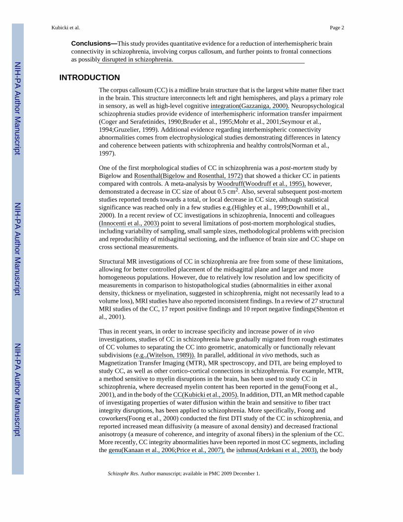
Conclusions—This study provides quantitative evidence for a reduction of interhemispheric brainconnectivity in schizophrenia, involving corpus callosum, and further points to frontal connectionsas possibly disrupted in schizophrenia.
INTRODUCTIONThe corpus callosum (CC) is a midline brain structure that is the largest white matter fiber tractin the brain. This structure interconnects left and right hemispheres, and plays a primary rolein sensory, as well as high-level cognitive integration(Gazzaniga, 2000). Neuropsychologicalschizophrenia studies provide evidence of interhemispheric information transfer impairment(Coger and Serafetinides, 1990;Bruder et al., 1995;Mohr et al., 2001;Seymour et al.,1994;Gruzelier, 1999). Additional evidence regarding interhemispheric connectivityabnormalities comes from electrophysiological studies demonstrating differences in latencyand coherence between patients with schizophrenia and healthy controls(Norman et al.,1997).
One of the first morphological studies of CC in schizophrenia was a post-mortem study byBigelow and Rosenthal(Bigelow and Rosenthal, 1972) that showed a thicker CC in patientscompared with controls. A meta-analysis by Woodruff(Woodruff et al., 1995), however,demonstrated a decrease in CC size of about 0.5 cm2. Also, several subsequent post-mortemstudies reported trends towards a total, or local decrease in CC size, although statisticalsignificance was reached only in a few studies e.g.(Highley et al., 1999;Downhill et al.,2000). In a recent review of CC investigations in schizophrenia, Innocenti and colleagues(Innocenti et al., 2003) point to several limitations of post-mortem morphological studies,including variability of sampling, small sample sizes, methodological problems with precisionand reproducibility of midsagittal sectioning, and the influence of brain size and CC shape oncross sectional measurements.
Structural MR investigations of CC in schizophrenia are free from some of these limitations,allowing for better controlled placement of the midsagittal plane and larger and morehomogeneous populations. However, due to relatively low resolution and low specificity ofmeasurements in comparison to histopathological studies (abnormalities in either axonaldensity, thickness or myelination, suggested in schizophrenia, might not necessarily lead to avolume loss), MRI studies have also reported inconsistent findings. In a review of 27 structuralMRI studies of the CC, 17 report positive findings and 10 report negative findings(Shenton etal., 2001).
Thus in recent years, in order to increase specificity and increase power of in vivoinvestigations, studies of CC in schizophrenia have gradually migrated from rough estimatesof CC volumes to separating the CC into geometric, anatomically or functionally relevantsubdivisions (e.g.,(Witelson, 1989)). In parallel, additional in vivo methods, such asMagnetization Transfer Imaging (MTR), MR spectroscopy, and DTI, are being employed tostudy CC, as well as other cortico-cortical connections in schizophrenia. For example, MTR,a method sensitive to myelin disruptions in the brain, has been used to study CC inschizophrenia, where decreased myelin content has been reported in the genu(Foong et al.,2001), and in the body of the CC(Kubicki et al., 2005). In addition, DTI, an MR method capableof investigating properties of water diffusion within the brain and sensitive to fiber tractintegrity disruptions, has been applied to schizophrenia. More specifically, Foong andcoworkers(Foong et al., 2000) conducted the first DTI study of the CC in schizophrenia, andreported increased mean diffusivity (a measure of axonal density) and decreased fractionalanisotropy (a measure of coherence, and integrity of axonal fibers) in the splenium of the CC.More recently, CC integrity abnormalities have been reported in most CC segments, includingthe genu(Kanaan et al., 2006;Price et al., 2007), the isthmus(Ardekani et al., 2003), the body
Kubicki et al. Page 2
Schizophr Res. Author manuscript; available in PMC 2009 December 1.
NIH
-PA Author Manuscript
NIH
-PA Author Manuscript
NIH
-PA Author Manuscript

(Ardekani et al., 2003;Kubicki et al., 2005;Hubl et al., 2004;Buchsbaum et al., 2006) and thesplenium(Ardekani et al., 2003;Foong et al., 2000;Agartz et al., 2001;Price et al., 2007). Therehave also been negative findings reported(e.g.,(Foong et al., 2002;Price et al., 2005;Sun et al.,2003;Kumra et al., 2004)). Inconsistencies in DTI investigations in schizophrenia mirror thoseof post-mortem as well as structural MR studies, and point to the need for precise, sensitivemethods of measurement.
Unfortunately, similar to structural MRI, DTI methodology has several limitations. The mostpopular analytic method, Voxel Based Morphometry (VBM), turns out to be sensitive to largeintensity variations, rather than to subtle, well localized abnormalities, in addition to notcorrecting very well for shape, or for overall size differences. Region of Interest (ROI) analyses,on the other hand, might be confounded by the type of diffusion measure (e.g., FA, T1W,directional map), and/or by slice thickness or slice orientation of the image used for theirdefinition.
A recent DTI post-processing technique -fiber tractography- shows promise, and twoschizophrenia investigations utilizing this method show its higher sensitivity and specificity,compared to both VBM and ROI approaches(Jones et al., 2005;Kanaan et al., 2006). Sincefiber tractography is capable of visualizing entire fiber tracts, thus following them from onehemisphere to another, it might be well suited for defining anatomical divisions of the CC(Huang et al., 2005);(Hofer and Frahm, 2006). In fact it has been demonstrated(Hofer andFrahm, 2006) that DTI based CC subdivisions reflect anatomy of the callosal connections muchbetter than the traditional geometric subdivisions of CC proposed by Witelson(Witelson,1989).
Diffusion quantification, however, along the entire tracts defined by DTI may nonetheless stillbe problematic, due to the fact that interhemispheric callosal fibers are crossed byintrahemispheric longitudinal fiber bundles in multiple areas of the human brain, which affectsboth the shape and appearance of callosal tracts, making the measurements less reliable. In thecurrent study we try to minimize some of the aforementioned confounding factors. We use acombination of MRI automatic lobar parcellation, and DTI tractography, to segment CC moreprecisely, and we include a template warping procedure for better intersubject reproducibility.Finally, in order to minimize the influence of other fiber bundles on CC, we project ourtractography based parcellation onto the midsagittal plane of the high resolution DTI scans,where all measurements are performed. We predict that CC, particularly in regions connectedto the frontal lobe (regions most frequently indicated in post mortem and genetic studies), willbe more affected in schizophrenia compared with controls, and that these abnormalities willbe related to clinical symptoms of schizophrenia.
METHODSSubject Population and Inclusion-Exclusion Criteria
32 patients with chronic schizophrenia were recruited from in-patient, day treatment, out-patient, and foster care programs at the VA Boston Healthcare System, Brockton, MA. SCID-P interviews were administered by professional staff to make DSM-IV diagnoses. SCID-NPinterviews were completed for the 42 normal comparison subjects. Comparison subjects wererecruited through advertisements, and group-matched to patients on age, sex, handedness, andparental social economic-status (PSES).
Inclusion criteria for all subjects were: right-handedness, ages between 18 and 55 years, nohistory of electroconvulsive shock treatment, no history of neurological illness, no alcohol ordrug dependence in the last 5 years and no abuse in the past year, verbal IQ above 80, nomedication affecting neurological or cognitive functions. In addition, control subjects were
Kubicki et al. Page 3
Schizophr Res. Author manuscript; available in PMC 2009 December 1.
NIH
-PA Author Manuscript
NIH
-PA Author Manuscript
NIH
-PA Author Manuscript

screened to exclude individuals who had a first degree relative with an Axis I disorder. Thestudy was approved by the local IRB committees at the VA and BWH. All the subjects signedinformed consent prior to study participation.
MRI ProtocolFor the DTI data acquisition, we used a 1.5 Tesla GE scanner, with a quadrature head coil. Weused a line scan diffusion imaging protocol (LSDI), which decreases the number of artifactsand distortions compared to other techniques(Gudbartsson et al., 1996; Kubicki et al., 2005;Kubicki et al., 2002). In order to obtain high resolution corpus scans, we acquired 5 sagittaloblique (aligned to the interhemispheric fissure) 1.7 x 1.7 mm in plane, 4 mm thick slices, withEcho Time=70 ms; Repetition Time=2500 ms. We acquired 6 independent diffusion directions(B=1000) and 2 baseline images(B=5), and one NEX (number of excitations, with LSDIsequence gaining signal-to-noise ratios comparable to 16 NEX when single shot EPI is used,but is free from EPI related geometric distortions (Kubicki et al, 2003). In the same session,high resolution structural scans were also acquired, and later co-registered to DTI scans. Theacquisition parameters were as follows: contiguous spoiled gradient-recalled acquisition(SPGR), TR=35ms, TE=5ms, 45 degree flip angle, resulting in 0.9375x0.9375x1.5 mm voxels.
Image AnalysisWe utilized a novel method for the computation of a probabilistic subdivision of CC, which isdescribed in detail in Styner et al. (Styner et al., 2005) and in Cascio et al. (Cascio et al.,2006).
Here, we provide only a brief description, also schematically illustrated in Figure 1. Theprocedure involved four separate steps: 1) Automatic segmentation of the entire corpuscallosum using SPGR data and a statistical, deformable model approach, described in detailin Styner et al. (Styner et al., 2005) (top left of Figure 1); 2) Further separation of the corpusROI (still using structural scan) into 4 separate segments using a previously created atlas (rightside of Figure 1); 3) Co-registration of the structural scan (along with corpus segments) to DTIwas performed via a routine non-rigid b-spline, mutual information based registration method(The Image Registration Toolkit, Rueckert, 1999), which accounts for DTI distortions(although minimal in this brain region and with this pulse sequence), patient motion and FOVdifferences. (left middle part of Figure 1) and 4) After diffusion tensors estimation (using astandard linear square fit, which seems adequate since the principal direction of diffusionin midsagittal plane is quite uniform), the area and mean FA for each corpus segment werecalculated.
The segmentation of CC into 4 smaller anatomical sections(step 3) was performed using aprobabilistic atlas/model(Styner et al., 2005;Cascio et al., 2006), which was generated in aseparate study(Cascio et al., 2006). Its creation is schematically described on the right side ofthe Figure 1, and involved tractography of the entire corpus callosum, its further division usingautomatic lobar segmentation, and back-mapping of these divisions onto midsagittal plane.Last step of this process resulted in midsagittal plane labels of pre-frontal, frontal, parietal andtemporo-occipital callosal segments corresponding to the probability of callosal fibersprojecting to each of these four brain regions. The entire process was automatic and fullyreproducible, i.e., no manual measurements were conducted.
Volume and mean FA for each callosal segment were subjected to group comparison, as wellas Pearson product correlations, where relationship was tested between FA, age, and clinicalsymptoms (derived from Scale for the Assessment of Negative and Positive Symptoms(Andreasen, 1990)).
Kubicki et al. Page 4
Schizophr Res. Author manuscript; available in PMC 2009 December 1.
NIH
-PA Author Manuscript
NIH
-PA Author Manuscript
NIH
-PA Author Manuscript

RESULTSGroups did not differ in age [mean age 40.5±8.8 for controls, and 39.8±9.3 for schizophrenics;P(1,73)=0.78)], parental socioeconomic status P(1.73)=0.33), or gender (all males). Since ageof healthy subjects has been shown to be correlated with FA of anterior CC (Pfefferbaum etal., 2003), we entered age as a covariate into a General Linear Model investigating group effectsfor FA and volume of the four CC subdivisions. When CC volume was analyzed, ICC (intra-cranial content) volume was also entered as a covariate. ANCOVA revealed no significantoverall group effect for area of the CC (F=2.99;P=0.09), but did reveal a significant groupeffect for FA (F=5.77;P=0.02). In addition, we found group by region interaction (F=5.23;P=0.03), with individual ANOVAs showing FA decrease in the entire frontal (F=5.68; P=0.02),but not parietal (F=2.39; P=0.13), or temporo-occipital (F=3.36; P=0.07) callosal connections(see Figure 2). Also, controls, but not schizophrenics, showed a relationship between age andCC integrity in anterior, but not posterior CC regions (rho=−0.35;P=0.02 for frontal segment;and rho=−0.13;P=0.42; rho=−0.16;P=0.31 for parietal and ocipito-temporal segmentsrespectively). Finally, FA for CC regions was statistically significantly correlated with negative(anterior portion), and positive (whole corpus) clinical symptoms (see Table 2 for details).
DISCUSSIONOur investigation revealed decreased fiber tract integrity in patients with schizophrenia,compared with controls, in the corpus callosum, the largest white matter fiber tract of the humanbrain. We did not, however, find volume differences between groups for this structure, whichis consistent with the view that structural MRI might not be sensitive enough to detect subtlewhite matter changes present in schizophrenia(Innocenti et al., 2003;Rossell et al., 2001). Infact, Rossell and colleagues calculated that in order to find statistically significant 0.2 cm2
volume differences between schizophrenics and controls in the corpus callosum, one wouldneed 308 schizophrenia patients. Indeed, our present investigation, as well as other DTI studiesconducted to date, confirms the increased sensitivity of DTI methods to detect subtlemicrostructural changes within the white matter in schizophrenia.
Post-hoc analyses suggested that the callosal tracts providing interhemispheric communicationbetween frontal cortices were the regions strongest affected in schizophrenia. This is in linewith previous studies reporting decreased size of pyramidal neurons in layer 3 of the prefrontalcortex (i.e.(Rajkowska et al., 1998), which is also the region known for its involvement incortico-cortical interhemispheric connections.
Additionally, since regions that we found to be abnormal topographically roughly correspondto the genu and anterior part of the body of the corpus callosum, our results are in line withsome findings obtained recently by other groups that use ROIs and fiber tractography approach(Kanaan et al., 2006;Price et al., 2007). We did not find group differences within the tractsinter-connecting parietal, temporal and occipital lobes, that is fibers projecting into the isthmusand splenium of the CC. Some previous DTI schizophrenia investigations, however, describeabnormalities within these regions. While we can not rule out the possibility that these regionsare still affected, we note that most studies reporting positive findings in this region have usedmethods based on the whole brain registration(Ardekani et al., 2003;Foong et al., 2000;Agartzet al., 2001), which could, in part, be confounded by registration error (affecting controls, butperhaps more profoundly patients with schizophrenia), due to higher inter subject anatomicalvariability existing in posterior parts of the CC(Ozdemir et al., 2007).
FA, in general, is considered to be a measure of axonal integrity, density, thickness andmyelination. Beaulieau (Beaulieu and Allen, 1994) demonstrated that myelination is one ofthe major contributors to diffusion anisotropy. In addition, several recent post-mortem studies
Kubicki et al. Page 5
Schizophr Res. Author manuscript; available in PMC 2009 December 1.
NIH
-PA Author Manuscript
NIH
-PA Author Manuscript
NIH
-PA Author Manuscript

(Uranova et al., 2004), as well as genetic(Hakak et al., 2001) studies, point to myelin, andoligodendrocytes forming the myelin sheath, as possibly being affected in schizophrenia. Also,since the anterior parts of the CC mature earlier than the rest of the CC, and developmentalstudies show size of CC increasing until the late twenties(Giedd et al., 1999;Keshavan et al.,2002), maturation of anterior portions falls roughly into the time period when most patientsdevelop schizophrenia, i.e., early adulthood. Thus the observed CC abnormalities could reflecta breakdown in the developmental process at this particular point of maturation. Additionally,insufficient communication between hemispheres might further affect the developmentalprocess of each individual hemisphere, resulting in the relative lack of asymmetry observed inschizophrenic brains(Innocenti et al., 2003). In addition, our findings regarding the correlationsbetween the integrity within the anterior part of the corpus callosum and age in control subjectsconfirms previously published results by Sullivan and coworkers(Sullivan et al., 2006), whichwere attributed to the fact that the frontal lobes contain thinner and less myelinated fibers,which are more vulnerable to age related breakdown(Aboitiz et al., 1996;Bartzokis et al.,2004). Similar results were also reported by Woodruff et al (Woodruff at el., 1997), whoreported correlations between corpus volume and age in controls, but similar to our results, didnot see these relationships in schizophrenia.
Correlations between FA values and clinical symptoms found here, also confirm the role ofinterhemispheric communication in schizophrenia symptomatology, further suggestinginvolvement of the entire CC in positive symptoms, but only the anterior part in negativesymptomatology. We note, however, that the role of interhemipsheric connectivity providedby the corpus callosum in schizophrenia, and its correlation with clinical symptoms is poorlyunderstood, although we note that similar correlations between CC integrity and clinicalsymptoms have been reported previously in the literature. For example, Downhill (Downhillet al., 2000) reported negative correlations between whole CC size and severity of positivesymptoms, while several other studies suggest the involvement of the anterior part of the CCin negative symptomatology (e.g., Woodruff et al., 1997; Foong et al., 2001). The anteriorportion of the CC is functionally associated with frontal lobe, with its ventral regions heavilyinvolved in emotional and attentional processing and this may, in part, explain why negativesymptom correlations predominate although further investigation is needed to determine thenature of this association.
Our study also included chronic, medicated subjects only. Thus even though we, as well asmany others, did not observe any statistically significant correlations between medicationdosage and diffusion measures, our results could still be confounded by the cumulative effectof medication, or other environmental factors not accounted for in this study.
Methodological issuesOur investigation is one of the first to utilize fiber tractography to subdivide and measure corpuscallosum integrity in schizophrenia. Recent investigations highlight the advantages oftractography over more traditional analytic approaches to DTI data, and show diffusiontractography to be more sensitive than more popular ROI or VBM DTI approaches, in findingabnormalities in schizophrenia(Kanaan et al., 2006;Jones et al., 2006). Tractography basedsegmentation of callosal fibers, has also already been shown to be more precise, and betterreflect the anatomy than the traditional geometric segmentation(Hofer and Frahm, 2006). Thusby using tractography based segmentation in our investigation, we hoped to increase bothsensitivity as well as specificity of our measurements. One of the limitations of our approachis the fact that our DTI data was acquired in only 6 diffusion directions. This limitation,however, affects our results only minimally, since we do not have to use our DTI data togenerate tractography, but instead are mapping an already existing tractography based atlasonto our DTI data. On the other hand, precision of our measurements should be increased
Kubicki et al. Page 6
Schizophr Res. Author manuscript; available in PMC 2009 December 1.
NIH
-PA Author Manuscript
NIH
-PA Author Manuscript
NIH
-PA Author Manuscript

thanks to the sagittal acquisition and high in-plane resolution of the data (1.7 x 1.7 mm in themidsagittal plane), by decreasing partial volume effects associated with thick axial slices usedin most investigations. Finally, our subjects were all medicated males, thus additional studiesneed to be performed on a first episode and mixed gender populations in order to understandfully the meaning of our findings.
CONCLUSIONSDTI is powerful and sensitive tool that demonstrates white matter abnormalities inschizophrenia. Our study demonstrates the utility of using DTI and tractography-based atlasin segmenting and measuring corpus callosum subdivisions in schizophrenia, and points tointerhemispheric communication between frontal lobes as most affected in schizophrenia, andassociated with negative symptoms.
ROLE OF FUNDING SOURCEThis work was funded by the National Alliance for Research on Schizophrenia and Depression(MK), the National Institute of Health (R03 MH068464-01 to MK, K05 MH070047 and R01MH 50747 to MES and R01 MH 40799 to RWM), the Department of Veterans Affairs MeritAwards (MES, RWM), and the VA Schizophrenia Center Grant (RWM/MES), and HarvardMedical School (Milton Award to MK). This work is also part of the National Alliance forMedical Image Computing (NAMIC), funded by the National Institutes of Health through theNIH Roadmap for Medical Research, Grant U54 EB005149 (RK, GG, MES, MS, MK),P50MH080272 (RWM MES, MK,SB). The funding sources had no further role in the studydesign, in the collection, analysis, and interpretation of data; in the writing of the report, andin the decision to submit the paper for publication.
AcknowledgementsWe would like to thank Nancy Maxwell and Jennifer Goodrich for their administrative assistance. The ImageRegistration Toolkit was used under Licence from Ixico Ltd.
ReferencesAboitiz F, Rodriguez E, Olivares R, Zaidel E. Age-related changes in fibre composition of the human
corpus callosum: sex differences. Neuroreport 1996;7:1761–4. [PubMed: 8905659]Agartz I, Andersson JL, Skare S. Abnormal brain white matter in schizophrenia: a diffusion tensor
imaging study. Neuroreport 2001;12:2251–4. [PubMed: 11447344]Andreasen NC. Methods for assessing positive and negative symptoms. Mod Probl Pharmacopsychiatry
1990;24:73–88. [PubMed: 2336066]Ardekani BA, Nierenberg J, Hoptman MJ, Javitt DC, Lim KO. MRI study of white matter diffusion
anisotropy in schizophrenia. Neuroreport 2003;14:2025–9. [PubMed: 14600491]Bartzokis G, Lu PH, Mintz J. Quantifying age-related myelin breakdown with MRI: novel therapeutic
targets for preventing cognitive decline and Alzheimer's disease. J Alzheimers Dis 2004;6:S53–9.[PubMed: 15665415]
Beaulieu C, Allen P. Determinants of Anisotropic Water Diffusion in Nerves. MRM 1994;31:394–400.Bigelow L, Rosenthal R. Schizophrenia and the corpus callosum. Lancet 1972;1:694. [PubMed: 4125202]Bruder G, Rabinowicz E, Towey J, Brown A, Kaufmann CA, Amador X, Malaspina D, Gorman JM.
Smaller right ear (left hemisphere) advantage for dichotic fused words in patients with schizophrenia.Am J Psychiatry 1995;152:932–5. [PubMed: 7755128]
Buchsbaum MS, Friedman J, Buchsbaum BR, Chu KW, Hazlett EA, Newmark R, Schneiderman JS,Torosjan Y, Tang C, Hof PR, Stewart D, Davis KL, Gorman J. Diffusion tensor imaging inschizophrenia. Biol Psychiatry 2006;60:1181–7. [PubMed: 16893533]
Kubicki et al. Page 7
Schizophr Res. Author manuscript; available in PMC 2009 December 1.
NIH
-PA Author Manuscript
NIH
-PA Author Manuscript
NIH
-PA Author Manuscript
Cascio C, Styner M, Smith RG, Poe MD, Gerig G, Hazlett HC, Jomier M, Bammer R, Piven J. Reducedrelationship to cortical white matter volume revealed by tractography-based segmentation of thecorpus callosum in young children with developmental delay. Am J Psychiatry 2006;163:2157–63.[PubMed: 17151168]
Coger RW, Serafetinides EA. Schizophrenia, corpus callosum, and interhemispheric communication: areview. Psychiatry Res 1990;34:163–84. [PubMed: 1962861]
Downhill JE Jr, Buchsbaum MS, Wei T, Spiegel-Cohen J, Hazlett EA, Haznedar MM, Silverman J, SieverLJ. Shape and size of the corpus callosum in schizophrenia and schizotypal personality disorder.Schizophr Res 2000;42:193–208. [PubMed: 10785578]
Foong J, Maier M, Clark CA, Barker GJ, Miller DH, Ron MA. Neuropathological abnormalities of thecorpus callosum in schizophrenia: a diffusion tensor imaging study. J Neurol Neurosurg Psychiatry2000;68:242–4. [PubMed: 10644799]
Foong J, Symms MR, Barker GJ, Maier M, Miller DH, Ron MA. Investigating regional white matter inschizophrenia using diffusion tensor imaging. Neuroreport 2002;13:333–6. [PubMed: 11930133]
Foong J, Symms MR, Barker GJ, Maier M, Woermann FG, Miller DH, Ron MA. Neuropathologicalabnormalities in schizophrenia: evidence from magnetization transfer imaging. Brain 2001;124:882–92. [PubMed: 11335691]
Gazzaniga MS. Cerebral specialization and interhemispheric communication: does the corpus callosumenable the human condition? Brain 2000;123(Pt 7):1293–326. [PubMed: 10869045]
Giedd JN, Blumenthal J, Jeffries NO, Rajapakse JC, Vaituzis AC, Liu H, Berry YC, Tobin M, Nelson J,Castellanos FX. Development of the human corpus callosum during childhood and adolescence: alongitudinal MRI study. Prog Neuropsychopharmacol Biol Psychiatry 1999;23:571–88. [PubMed:10390717]
Gruzelier JH. Functional neuropsychophysiological asymmetry in schizophrenia: a review andreorientation. Schizophr Bull 1999;25:91–120. [PubMed: 10098916]
Gudbartsson H, Maier S, Mulkern R. Line scan diffusion imaging. Magn Reson Med 1996;36:509–519.[PubMed: 8892201]
Hakak Y, Walker JR, Li C, Wong WH, Davis KL, Buxbaum JD, Haroutunian V, Fienberg AA. Genome-wide expression analysis reveals dysregulation of myelination-related genes in chronicschizophrenia. Proc Natl Acad Sci U S A 2001;98:4746–51. [PubMed: 11296301]
Highley JR, Esiri MM, McDonald B, Cortina-Borja M, Herron BM, Crow TJ. The size and fibrecomposition of the corpus callosum with respect to gender and schizophrenia: a post-mortem study.Brain 1999;122(Pt 1):99–110. [PubMed: 10050898]
Hofer S, Frahm J. Topography of the human corpus callosum revisited--comprehensive fiber tractographyusing diffusion tensor magnetic resonance imaging. Neuroimage 2006;32:989–94. [PubMed:16854598]
Huang H, Zhang J, Jiang H, Wakana S, Poetscher L, Miller MI, van Zijl PC, Hillis AE, Wytik R, MoriS. DTI tractography based parcellation of white matter: application to the mid-sagittal morphologyof corpus callosum. Neuroimage 2005;26:195–205. [PubMed: 15862219]
Hubl D, Koenig T, Strik W, Federspiel A, Kreis R, Boesch C, Maier SE, Schroth G, Lovblad K, DierksT. Pathways that make voices: white matter changes in auditory hallucinations. Arch Gen Psychiatry2004;61:658–68. [PubMed: 15237078]
Innocenti GM, Ansermet F, Parnas J. Schizophrenia, neurodevelopment and corpus callosum. MolPsychiatry 2003;8:261–74. [PubMed: 12660799]
Jones DK, Catani M, Pierpaoli C, Reeves SJ, Shergill SS, O'Sullivan M, Golesworthy P, McGuire P,Horsfield MA, Simmons A, Williams SC, Howard RJ. Age effects on diffusion tensor magneticresonance imaging tractography measures of frontal cortex connections in schizophrenia. Hum BrainMapp 2006;27:230–8. [PubMed: 16082656]
Jones DK, Catani M, Pierpaoli C, Reeves SJ, Shergill SS, O'Sullivan M, Maguire P, Horsfield MA,Simmons A, Williams SC, Howard RJ. A diffusion tensor magnetic resonance imaging study offrontal cortex connections in very-late-onset schizophrenia-like psychosis. Am J Geriatr Psychiatry2005;13:1092–9. [PubMed: 16319302]
Kubicki et al. Page 8
Schizophr Res. Author manuscript; available in PMC 2009 December 1.
NIH
-PA Author Manuscript
NIH
-PA Author Manuscript
NIH
-PA Author Manuscript

Kanaan RA, Shergill SS, Barker GJ, Catani M, Ng VW, Howard R, McGuire PK, Jones DK. Tract-specific anisotropy measurements in diffusion tensor imaging. Psychiatry Res 2006;146:73–82.[PubMed: 16376059]
Keshavan MS, Diwadkar VA, DeBellis M, Dick E, Kotwal R, Rosenberg DR, Sweeney JA, Minshew N,Pettegrew JW. Development of the corpus callosum in childhood, adolescence and early adulthood.Life Sci 2002;70:1909–22. [PubMed: 12005176]
Kubicki M, Park H, Westin CF, Nestor PG, Mulkern RV, Maier SE, Niznikiewicz M, Connor EE, LevittJJ, Frumin M, Kikinis R, Jolesz FA, McCarley RW, Shenton ME. DTI and MTR abnormalities inschizophrenia: analysis of white matter integrity. Neuroimage 2005;26:1109–18. [PubMed:15878290]
Kubicki M, Westin CF, Maier SE, Frumin M, Nestor PG, Salisbury DF, Kikinis R, Jolesz FA, McCarleyRW, Shenton ME. Uncinate fasciculus findings in schizophrenia: a magnetic resonance diffusiontensor imaging study. Am J Psychiatry 2002;159:813–20. [PubMed: 11986136]
Kumra S, Ashtari M, McMeniman M, Vogel J, Augustin R, Becker DE, Nakayama E, Gyato K, KaneJM, Lim K, Szeszko P. Reduced frontal white matter integrity in early-onset schizophrenia: apreliminary study. Biol Psychiatry 2004;55:1138–45. [PubMed: 15184032]
Mohr B, Heim S, Pulvermuller F, Rockstroh B. Functional asymmetry in schizophrenic patients duringauditory speech processing. Schizophr Res 2001;52:69–78. [PubMed: 11595393]
Norman RM, Malla AK, Williamson PC, Morrison-Stewart SL, Helmes E, Cortese L. EEG coherenceand syndromes in schizophrenia. Br J Psychiatry 1997;170:411–5. [PubMed: 9307688]
Ozdemir ST, Ercan I, Sevinc O, Guney I, Ocakoglu G, Aslan E, Barut C. Statistical shape analysis ofdifferences in the shape of the corpus callosum between genders. Anat Rec (Hoboken) 2007;290:825–30. [PubMed: 17538981]
Pfefferbaum A, Adalsteinsson E, Sullivan EV. Replicability of diffusion tensor imaging measurementsof fractional anisotropy and trace in brain. J Magn Reson Imaging 2003;18:427–33. [PubMed:14508779]
Price G, Bagary MS, Cercignani M, Altmann DR, Ron MA. The corpus callosum in first episodeschizophrenia: a diffusion tensor imaging study. J Neurol Neurosurg Psychiatry 2005;76:585–7.[PubMed: 15774453]
Price G, Cercignani M, Parker GJ, Altmann DR, Barnes TR, Barker GJ, Joyce EM, Ron MA. Abnormalbrain connectivity in first-episode psychosis: a diffusion MRI tractography study of the corpuscallosum. Neuroimage 2007;35:458–66. [PubMed: 17275337]
Rajkowska G, Selemon LD, Goldman-Rakic PS. Neuronal and glial somal size in the prefrontal cortex:a postmortem morphometric study of schizophrenia and Huntington disease. Arch Gen Psychiatry1998;55:215–24. [PubMed: 9510215]
Rossell SL, Shapleske J, Fukuda R, Woodruff PW, Simmons A, David AS. Corpus callosum area andfunctioning in schizophrenic patients with auditory--verbal hallucinations. Schizophr Res 2001;50:9–17. [PubMed: 11378310]
Rueckert D, Sonoda LI, Hayes C, Hill DL, Leach MO, Hawkes DJ. Non-rigid registration using free-form deformations: Application to breast MR images. IEEE Transactions on Medical Imaging1999;18(8):712–721. [PubMed: 10534053]
Seymour SE, Reuter-Lorenz PA, Gazzaniga MS. The disconnection syndrome. Basic findings reaffirmed.Brain 1994;117(Pt 1):105–15. [PubMed: 8149205]
Shenton ME, Dickey CC, Frumin M, McCarley RW. A review of MRI findings in schizophrenia.Schizophr Res 2001;49:1–52. [PubMed: 11343862]
Styner MA, Oguz I, Smith RG, Cascio C, Jomier M. Corpus callosum subdivision based on a probabilisticmodel of inter-hemispheric connectivity. Med Image Comput Comput Assist Interv Int Conf MedImage Comput Comput Assist Interv 2005;8:765–72. [PubMed: 16686029]
Sullivan EV, Adalsteinsson E, Pfefferbaum A. Selective age-related degradation of anterior callosal fiberbundles quantified in vivo with fiber tracking. Cereb Cortex 2006;16:1030–9. [PubMed: 16207932]
Sun Z, Wang F, Cui L, Breeze J, Du X, Wang X, Cong Z, Zhang H, Li B, Hong N, Zhang D. Abnormalanterior cingulum in patients with schizophrenia: a diffusion tensor imaging study. Neuroreport2003;14:1833–6. [PubMed: 14534430]
Kubicki et al. Page 9
Schizophr Res. Author manuscript; available in PMC 2009 December 1.
NIH
-PA Author Manuscript
NIH
-PA Author Manuscript
NIH
-PA Author Manuscript

Uranova NA, Vostrikov VM, Orlovskaya DD, Rachmanova VI. Oligodendroglial density in the prefrontalcortex in schizophrenia and mood disorders: a study from the Stanley Neuropathology Consortium.Schizophr Res 2004;67:269–75. [PubMed: 14984887]
Witelson SF. Hand and sex differences in the isthmus and genu of the human corpus callosum. Apostmortem morphological study. Brain 1989;112(Pt 3):799–835. [PubMed: 2731030]
Woodruff PW, McManus IC, David AS. Meta-analysis of corpus callosum size in schizophrenia. J NeurolNeurosurg Psychiatry 1995;58:457–61. [PubMed: 7738554]
Kubicki et al. Page 10
Schizophr Res. Author manuscript; available in PMC 2009 December 1.
NIH
-PA Author Manuscript
NIH
-PA Author Manuscript
NIH
-PA Author Manuscript

Figure 1.Schematic Description of processing steps, described in detail in “Styner et al., 2005 and Ciscoet al., 2006), involving automatic segmentation of the corpus callosum into probabilisticsegments using previously generated atlas, co-registration of the corpus segmentation with DTIdata, and extraction of FA values for each segment separately.
Kubicki et al. Page 11
Schizophr Res. Author manuscript; available in PMC 2009 December 1.
NIH
-PA Author Manuscript
NIH
-PA Author Manuscript
NIH
-PA Author Manuscript

Figure 2.Results of group comparison, indicating largest group differences for the anterior parts ofthe corpus callosum.
Kubicki et al. Page 12
Schizophr Res. Author manuscript; available in PMC 2009 December 1.
NIH
-PA Author Manuscript
NIH
-PA Author Manuscript
NIH
-PA Author Manuscript

NIH
-PA Author Manuscript
NIH
-PA Author Manuscript
NIH
-PA Author Manuscript
Kubicki et al. Page 13Ta
ble
1Ta
ble
of c
linic
al c
orre
latio
ns, i
ndic
atin
g si
gnifi
cant
cor
rela
tions
bet
wee
n se
gmen
ts o
f the
cor
pus c
allo
sum
and
clin
ical
scor
es.
Clin
ical
mea
sure
Reg
ion
of C
CN
umbe
r of
subj
ects
Fron
tal
Pari
etal
Occ
ipito
-tem
pora
lG
loba
l SA
NS
33−.
463
(0.0
07)
nsns
Glo
bal A
ttent
ion
33−.
421
(0.0
15)
nsns
Soci
al A
ttent
ion
29−.
446
(0.0
15)
nsns
Glo
bal S
APS
32−.
400
(0.0
23)
−.40
1 (0
.023
)−.
403
(0.0
22)
Glo
bal T
DS
32−.
382
(0.0
31)
−.40
5 (0
.022
)−.
369
(0.0
37)
Dis
tract
ed sp
eech
31−.
492
(0.0
05)
−.42
0 (0
.019
)−.
449
(0.0
11)
Schizophr Res. Author manuscript; available in PMC 2009 December 1.
Related Documents