JOURNAL OF VIROLOGY, Mar. 2005, p. 3038–3051 Vol. 79, No. 5 0022-538X/05/$08.000 doi:10.1128/JVI.79.5.3038–3051.2005 Copyright © 2005, American Society for Microbiology. All Rights Reserved. Reduced Hepatitis B Virus (HBV)-Specific CD4 T-Cell Responses in Human Immunodeficiency Virus Type 1-HBV-Coinfected Individuals Receiving HBV-Active Antiretroviral Therapy J. Judy Chang, 1 Fiona Wightman, 2 Angeline Bartholomeusz, 3 Anna Ayres, 3 Stephen J. Kent, 1,4 Joseph Sasadeusz, 4,5 and Sharon R. Lewin 1,2,4,5 * Department of Microbiology and Immunology, University of Melbourne, 1 Department of Medicine, Monash University, 2 Victorian Infectious Diseases Reference Laboratory, 3 Infectious Diseases Unit, Alfred Hospital, 4 and National Health and Medical Research Council Centre for Clinical Research Excellence in Infectious Diseases, Royal Melbourne Hospital, 5 Melbourne, Australia Received 30 June 2004/Accepted 14 October 2004 Functional hepatitis B virus (HBV)-specific T cells are significantly diminished in individuals chronically infected with HBV compared to individuals with self-limiting HBV infection or those on anti-HBV therapy. In individuals infected with human immunodeficiency virus type 1 (HIV-1), coinfection with HBV is associated with an increased risk of worsening liver function following antiviral therapy and of more rapid HBV disease progression. Total HBV-specific T-cell responses in subjects with diverse genetic backgrounds were charac- terized by using a library of 15-mer peptides overlapping by 11 amino acids and spanning all HBV proteins. The magnitude and breadth of CD4 and CD8 T-cell responses to HBV in peripheral blood were examined by flow cytometry to detect gamma interferon production following stimulation with HBV peptide pools. Chronic HBV carriers (n 34) were studied, including individuals never treated for HBV infection (n 7), HBV-infected individuals receiving anti-HBV therapy (n 13), and HIV-1–HBV-coinfected individuals receiv- ing anti-HBV therapy (n 14). CD4 and CD8 HBV-specific T-cell responses were more frequently detected and the CD8 T-cell responses were of greater magnitude and breadth in subjects on anti-HBV treatment than in untreated chronic HBV carriers. There was a significant inverse correlation between detection of a HBV- specific T-cell response and HBV viral load. HBV-specific CD4 and CD8 T-cell responses were significantly (fivefold) reduced compared with HIV-specific responses. Although, the frequency and breadth of HBV-specific CD8 T-cell responses were comparable in the monoinfected and HIV-1–HBV-coinfected groups, HBV-specific CD4 T-cell responses were significantly reduced in HIV-1–HBV-coinfected individuals. Therefore, HIV-1 infection has a significant and specific effect on HBV-specific T-cell immunity. There are 350 million individuals chronically infected with hepatitis B virus (HBV) worldwide (36, 42, 59). Each year 1 million to 1.5 million carriers die from HBV-related liver dis- ease and liver cancer such as hepatocellular carcinoma (36, 48). In the United States, Europe, and Australia, approxi- mately 6 to 7% of individuals infected with human immuno- deficiency virus type 1 (HIV-1) are also coinfected with HBV (23, 41). Liver disease is now a major comorbidity in HIV-1- infected individuals (11, 66). Although hepatitis C virus (HCV)–HIV-1 coinfection is more common than HBV infec- tion, liver failure occurs more frequently with persistent HBV infection (60, 63). Coinfection of HBV with HIV-1 leads to elevated HBV DNA levels, a lower rate of seroconversion to HBeAg, and lower alanine aminotransferase (ALT) levels than those in HBV monoinfection (21, 31). Spontaneous flares and HBeAg seroconversion occur in 5% of HBV-monoinfected individuals; however, in the setting of HIV-1–HBV coinfec- tion, spontaneous flares or seroconversion to HBeAg is rare (18). In individuals infected with HIV-1 who subsequently ac- quire HBV infection, there is an increased risk of persistent chronic infection (32, 62). On the other hand, HBV does not appear to have a significant impact on AIDS progression or the rate of CD4 T-cell loss (41, 62). Current treatments for HBV include nucleotide and nucle- oside reverse transcriptase inhibitors (RTI) that also suppress HIV-1 replication. The most widely used anti-HBV nucleoside analog, lamivudine (2-deoxy-3-thiacytidine), inhibits both HIV-1 and HBV reverse transcriptases. An increase in HBV- specific CD4 and CD8 T-cell responses in both HBeAg- positive and -negative patients following the administration of lamivudine to HBV-infected individuals has been described (13, 14, 25, 44, 47). Long-term lamivudine therapy (more than 100 weeks) leads to HBeAg seroconversion in 27% of individ- uals but has also been associated with the frequent develop- ment of drug resistance (39, 40). Other nucleotide RTI, such as adefovir [9-(2-phosphonylmethoxyethyl)adenine] and tenofo- vir [9-(R)-(2-phosphonyl-methoxypropyl)adenine], and the nu- cleoside RTI entecavir (formerly BMS-200475, a carbocyclic 2-deoxyguanosine analog) have also been shown to be effec- tive against HBV (1, 6, 23). Tenofovir is often used in combi- nation with lamivudine for effective treatment of HBV in HIV- 1-infected individuals, as both agents are active against HIV-1 as well as HBV. Recent work demonstrates that tenofovir reduces the HBV viral load significantly in HIV-1–HBV-coin- fected individuals infected with lamivudine-resistant HBV * Corresponding author. Mailing address: Infectious Diseases Unit, Alfred Hospital, Commercial Rd., Melbourne, Victoria 3004, Austra- lia. Phone: (613) 9276 3009. Fax: (613) 9276 2431. E-mail: S.Lewin @alfred.org.au. 3038

Welcome message from author
This document is posted to help you gain knowledge. Please leave a comment to let me know what you think about it! Share it to your friends and learn new things together.
Transcript

JOURNAL OF VIROLOGY, Mar. 2005, p. 3038–3051 Vol. 79, No. 50022-538X/05/$08.00�0 doi:10.1128/JVI.79.5.3038–3051.2005Copyright © 2005, American Society for Microbiology. All Rights Reserved.
Reduced Hepatitis B Virus (HBV)-Specific CD4� T-Cell Responses inHuman Immunodeficiency Virus Type 1-HBV-Coinfected Individuals
Receiving HBV-Active Antiretroviral TherapyJ. Judy Chang,1 Fiona Wightman,2 Angeline Bartholomeusz,3 Anna Ayres,3 Stephen J. Kent,1,4
Joseph Sasadeusz,4,5 and Sharon R. Lewin1,2,4,5*Department of Microbiology and Immunology, University of Melbourne,1 Department of Medicine, Monash University,2 Victorian
Infectious Diseases Reference Laboratory,3 Infectious Diseases Unit, Alfred Hospital,4 and National Health and MedicalResearch Council Centre for Clinical Research Excellence in Infectious Diseases, Royal Melbourne Hospital,5
Melbourne, Australia
Received 30 June 2004/Accepted 14 October 2004
Functional hepatitis B virus (HBV)-specific T cells are significantly diminished in individuals chronicallyinfected with HBV compared to individuals with self-limiting HBV infection or those on anti-HBV therapy. Inindividuals infected with human immunodeficiency virus type 1 (HIV-1), coinfection with HBV is associatedwith an increased risk of worsening liver function following antiviral therapy and of more rapid HBV diseaseprogression. Total HBV-specific T-cell responses in subjects with diverse genetic backgrounds were charac-terized by using a library of 15-mer peptides overlapping by 11 amino acids and spanning all HBV proteins.The magnitude and breadth of CD4� and CD8� T-cell responses to HBV in peripheral blood were examinedby flow cytometry to detect gamma interferon production following stimulation with HBV peptide pools.Chronic HBV carriers (n � 34) were studied, including individuals never treated for HBV infection (n � 7),HBV-infected individuals receiving anti-HBV therapy (n � 13), and HIV-1–HBV-coinfected individuals receiv-ing anti-HBV therapy (n � 14). CD4� and CD8� HBV-specific T-cell responses were more frequently detectedand the CD8� T-cell responses were of greater magnitude and breadth in subjects on anti-HBV treatment thanin untreated chronic HBV carriers. There was a significant inverse correlation between detection of a HBV-specific T-cell response and HBV viral load. HBV-specific CD4� and CD8� T-cell responses were significantly(fivefold) reduced compared with HIV-specific responses. Although, the frequency and breadth of HBV-specificCD8� T-cell responses were comparable in the monoinfected and HIV-1–HBV-coinfected groups, HBV-specificCD4� T-cell responses were significantly reduced in HIV-1–HBV-coinfected individuals. Therefore, HIV-1infection has a significant and specific effect on HBV-specific T-cell immunity.
There are 350 million individuals chronically infected withhepatitis B virus (HBV) worldwide (36, 42, 59). Each year 1million to 1.5 million carriers die from HBV-related liver dis-ease and liver cancer such as hepatocellular carcinoma (36,48). In the United States, Europe, and Australia, approxi-mately 6 to 7% of individuals infected with human immuno-deficiency virus type 1 (HIV-1) are also coinfected with HBV(23, 41). Liver disease is now a major comorbidity in HIV-1-infected individuals (11, 66). Although hepatitis C virus(HCV)–HIV-1 coinfection is more common than HBV infec-tion, liver failure occurs more frequently with persistent HBVinfection (60, 63). Coinfection of HBV with HIV-1 leads toelevated HBV DNA levels, a lower rate of seroconversion toHBeAg, and lower alanine aminotransferase (ALT) levels thanthose in HBV monoinfection (21, 31). Spontaneous flares andHBeAg seroconversion occur in 5% of HBV-monoinfectedindividuals; however, in the setting of HIV-1–HBV coinfec-tion, spontaneous flares or seroconversion to HBeAg is rare(18). In individuals infected with HIV-1 who subsequently ac-quire HBV infection, there is an increased risk of persistent
chronic infection (32, 62). On the other hand, HBV does notappear to have a significant impact on AIDS progression or therate of CD4� T-cell loss (41, 62).
Current treatments for HBV include nucleotide and nucle-oside reverse transcriptase inhibitors (RTI) that also suppressHIV-1 replication. The most widely used anti-HBV nucleosideanalog, lamivudine (2�-deoxy-3�-thiacytidine), inhibits bothHIV-1 and HBV reverse transcriptases. An increase in HBV-specific CD4� and CD8� T-cell responses in both HBeAg-positive and -negative patients following the administration oflamivudine to HBV-infected individuals has been described(13, 14, 25, 44, 47). Long-term lamivudine therapy (more than100 weeks) leads to HBeAg seroconversion in 27% of individ-uals but has also been associated with the frequent develop-ment of drug resistance (39, 40). Other nucleotide RTI, such asadefovir [9-(2-phosphonylmethoxyethyl)adenine] and tenofo-vir [9-(R)-(2-phosphonyl-methoxypropyl)adenine], and the nu-cleoside RTI entecavir (formerly BMS-200475, a carbocyclic2�-deoxyguanosine analog) have also been shown to be effec-tive against HBV (1, 6, 23). Tenofovir is often used in combi-nation with lamivudine for effective treatment of HBV in HIV-1-infected individuals, as both agents are active against HIV-1as well as HBV. Recent work demonstrates that tenofovirreduces the HBV viral load significantly in HIV-1–HBV-coin-fected individuals infected with lamivudine-resistant HBV
* Corresponding author. Mailing address: Infectious Diseases Unit,Alfred Hospital, Commercial Rd., Melbourne, Victoria 3004, Austra-lia. Phone: (613) 9276 3009. Fax: (613) 9276 2431. E-mail: [email protected].
3038

(53). As well as being effective in individuals with lamivudine-resistant HBV, tenofovir is also effective in individuals who failalpha interferon (IFN-�) therapy (61). Initiation of antiretro-viral therapy (ART) for HIV-1 can lead to severe acute hep-atitis with or without clearance of HBV (16, 18). Better un-derstanding of how the immune system responds to HBVfollowing ART and how infection with HIV-1 alters this re-sponse is needed. Understanding the pathogenesis of HIV-1and HBV interaction will allow for a rational approach to themanagement of coinfected individuals.
In persistent HBV infection there is a reduction in HBV-specific CD4� and CD8� T cells compared with those in indi-viduals who successfully clear infection. HBeAg has been sug-gested to facilitate HBV persistence by depleting HBeAg- andHBcAg-specific Th1 CD4� T cells via fibroblast-associated(Fas)-mediated apoptosis (51). HBeAg crosses the placentaand therefore may establish tolerance to HBV in newborns,increasing the frequency of persistent HBV infection with ver-tical transmission. The imbalance of Th1/Th2 responses pro-motes suppression of HBeAg/HBcAg-specific CD8� T-cell re-sponses and suppression of Th1 effector cells by production ofanti-inflammatory cytokines such as interleukin-4 (IL-4) andIL-10 (30, 51, 52). A generalized CD4� T-cell hyporesponsive-ness in individuals with chronic HBV infection has also beendescribed, with lower responses to mitogens in untreatedHBV-infected individuals than in HBV-negative controls. Fol-lowing a reduction in HBV viral load after anti-HBV therapy,this hyporesponsiveness is reversed (13). This T-cell hypore-sponsiveness may arise from decreased function in infecteddendritic cells, which have reduced IFN-�, tumor necrosis fac-tor alpha, and IL-12 production and hence reduced stimulationof T-cell responses (5).
In individuals with persistent HBV infection, the HBV-spe-cific T-cell response is significantly diminished when evaluatedby proliferative responses to whole HBV antigens or definedepitopes in HLA-A2-positive chronic carriers (30, 45). In par-ticular, in HLA-A2 HBeAg-positive chronic carriers, CD8� Tcells that recognize an HBV core epitope (in region c18-27)are almost undetectable when measured by tetramers and havediminished ability to produce IFN-�. HBV-specific CD8� Tcells are also found in the liver, where they may cause aninflammatory response but are ineffective in clearing HBVinfection (35, 45).
Examination of HBV-specific T cells by limiting-dilutionassays (7, 56), proliferation assays (13, 29, 30, 35), tetramerstaining (46, 58, 69), and cytokine production has been limitedto responses against predicted HLA-A2 epitopes of core, sur-face, and polymerase proteins and/or whole-antigen stimula-tion (2, 46, 49, 64). In HIV-1 infection the dominant HIV-1-specific T-cell response is often to non-HLA-A2-associatedpeptides as well as to accessory proteins. Therefore, early stud-ies using only HLA-A2 peptides or HIV-1 structural or enve-lope proteins did not accurately reflect the breadth and inten-sity of HIV-1-specific T-cell immunity (10). We thereforedeveloped an overlapping peptide library which allows for de-tection of all HBV-specific CD4� and CD8� T-cell responsesto all gene products to examine the effects of HIV-1 coinfec-tion on the breadth and magnitude of HBV-specific immuneresponses.
MATERIALS AND METHODS
Patient population. Patients with chronic HBV (defined as having detectableHBsAg on two occasions more than 6 months apart) were recruited from theRoyal Melbourne and Alfred Hospitals, Victoria, Australia. Participation wasapproved by the relevant hospital ethics committee, and signed consent wasobtained. Eight uninfected healthy control subjects were also studied for com-parison. Patients (n � 34) were classified into three groups: (i) individuals nevertreated with anti-HBV therapy (n � 7); (ii) HIV-1-uninfected, HBV-infectedindividuals currently or previously treated with one or more anti-HBV agents (n� 13); and (iii) HIV-1-infected, HBV-infected individuals currently or previouslytreated with one or more anti-HBV agents (n � 14).
Peptides. To examine total HBV-specific T-cell responses, an overlappingpeptide library was made by using the consensus sequence for genotype A HBV(68). Genotype A was selected because this is the most common genotype inHIV-1–HBV-coinfected individuals in Australia (23). A total of 394 peptidesconsisting of 15 amino acids (aa) overlapping by 11 amino acids were designedacross the full genome (Mimotopes, Clayton, Australia). Fifteen-mer peptideswere chosen because these can be presented to and recognized by both CD4�
and CD8� T cells (8, 28, 43). A total of 149 extra peptides were synthesized tocover regions of significant variation between genotype A and the three othermain genotypes (B, C, and D) (12, 55) to facilitate the study of HBV-specificimmunity in individuals infected with non-genotype A HBV, resulting in a totalof 543 peptides. Ideally, a complete peptide library should be made for eachgenotype; however, given that this was impractical, the following approach wasused for peptide design: when only one or two amino acids differed from geno-type A, a single 15-mer peptide was synthesized with the variable region usuallyin the center of the peptide, while in regions of greater variability, a limited setof overlapping peptides was made. This allowed for responses to genotypes B toD to be more accurately measured. Each peptide was dissolved in dimethylsulfoxide (DMSO) (Sigma, St. Louis, Mo.) to a final concentration of 100 or 50�g �l�1 (43). The 15-mer peptides were combined to create eight HBV peptidepools, according to the relevant protein, with each peptide represented at equalconcentrations. The peptide pools therefore included precore/core (PC), X pro-tein (X), three surface antigen pools (S1.1, S1.2, and S1.3), and three polymerasepools (Pol1, Pol2, and Pol3). The polymerase and surface antigen peptides weresubdivided into three pools each so that the final DMSO concentration was lowerthan 1% of the total stimulation volume to reduce any toxicity of DMSO. Peptidepool S1.1 covered aa 1 to 131 of the genotype A pre-S1 protein and also the extragenotype B to D peptides corresponding to this same region, S1.2 covered aa 121to 251, and S1.3 covered aa 241 to 400. Peptide pool Pol1 covered aa 1 to 256,Pol2 covered aa 246 to 496, and Pol3 covered aa 486 to 845 of the polymerase.The HBV peptide pools were made with 50 to 100 peptides in each pool, withoutsignificant inhibition of stimulation as previously described (43). The final pep-tide concentration used in stimulating peripheral blood mononuclear cells orwhole blood was 1 �g ml�1 for each peptide. Similar peptide pools were alsomade for HIV-1 peptides (obtained through the AIDS Research and ReferenceReagent Program, Division of AIDS, National Institute of Allergy and InfectiousDiseases, National Institutes of Health), resulting in six peptide pools: for HIV-1polymerase (HIV Pol1 and HIV Pol2), Gag (HIV GAG), and envelope (HIVENV) and HIV-1 accessory proteins Rev, Tat, and Vpu (HIV RTV) and Vif,Vpr, and Nef (HIV VVN).
Measurement of antigen-specific T-cell responses by ICS. Intracellular cyto-kine staining (ICS) was performed as previously described (8, 9, 34, 43) withsome minor modifications. Briefly, whole blood was collected in sodium heparintubes and prepared for peptide stimulation either on the day of collection orafter keeping blood at room temperature overnight. Round-bottomed 96-wellplates were prepared with costimulatory molecules CD28 and CD49d monoclo-nal antibody together with peptides. DMSO with the same costimulatory mole-cules was used as a negative control, and pokeweed mitogen (Sigma) and staph-ylococcal enterotoxin B (Sigma) were used as positive controls. Each stimulationwas performed in duplicate wells with 200 �l of whole blood. The final concen-trations were 1 �g ml�1 for each costimulatory molecule, 1 �g ml�1 for eachpeptide in each pool, and 5 �g ml�1 for each of the positive controls. The plateswere incubated at 37°C for 2 h before brefeldin A (Sigma) was added to a finalconcentration of 10 �g ml�1 and incubated for a further 5 h. The plates eitherwere kept at 4°C overnight or were surface stained immediately with a combi-nation of fluorescently labeled CD3-fluorescein isothiocyanate, CD4-phyco-erythrin, and CD8-peridinin chlorophyll-a protein antibodies (BD Biosciences,San Jose, Calif.). Plates were incubated for 30 min in the dark at room temper-ature, and the contents were then transferred to separate fluorescence-activatedcell sorter (FACS) tubes containing FACS lysing solution (BD Biosciences). Thetubes were incubated for 10 min at room temperature and frozen at �80°C, or
VOL. 79, 2005 HBV-SPECIFIC T-CELL RESPONSES IN HIV-HBV COINFECTION 3039
in some cases ICS was continued without freezing. Keeping plates overnight at4°C prior to staining and freezing in FACS lysing solution has been previouslyshown to have no deleterious effects on detection of T-cell responses (54). Thefrozen tubes were thawed in a 37°C water bath and then washed with phosphate-buffered saline. The cells were resuspended in FACS permeabilizing solution(BD Biosciences) and incubated in the dark at room temperature for 10 min. Thecells were washed once again and then stained intracellularly with anti-IFN-�–allophycocyanin for 1 h in the dark at room temperature. The cells were washedand resuspended in 5% paraformaldehyde.
Flow cytometry analysis. All data was acquired on a FACSCalibur or FACS-can instrument within 8 h of staining and were analyzed by using CellQuest (BDBiosciences). Cells were gated initially on lymphocytes as determined by forwardand side scatter, and, where possible, data from at least 100,000 small lympho-cytes were collected. Activated T cells down regulate surface expression of CD3,CD4, and CD8 due to internalization of these molecules (8, 67). The reducedexpression of CD3, CD4, and CD8 can be accounted for by the use of intracel-lular staining (8, 43). Alternatively, an inverted P gate adapted from that de-scribed by Betts et al. (8) was used to include T cells with both high and low CD3expression. The inverted P gate was set by using blood stimulated with pokeweedmitogen and staphylococcal enterotoxin B and was used for all subsequentanalyses for the particular individual (Fig. 1). For most samples, 10,000 to 20,000CD3� CD4� and CD3� CD8� T cells were collected for analysis. Antigen-specific cells are expressed as the percentage of IFN-�-producing CD8� orCD4� T cells.
Quantification of HBV viral load. HBV viral load was determined with theHBV Digene Hybrid Capture II microplate assay (Roche, Branchburg, N.J.) inaccordance with the manufacturer’s instructions. For the Digene assay, the lowerdetection limit was 0.5 pg (equivalent to 1.42 � 105 copies/ml). If HBV viral loadwas undetectable by Digene assay, the Versant HBV DNA 3.0 assay (BayerHealthCare-Diagnostics, Tarrytown, N.Y.) was used in accordance with themanufacturer’s instructions. The limit of detection for this assay was 2,000copies/ml.
Sequencing of precore/core promoter mutations. HBV DNA was extractedfrom 200 �l of serum, using the QIAamp DNA Minikit (QIAGEN, Chatsworth,Calif.) according to the manufacturer’s instructions. The catalytic domain of thereverse transcriptase/polymerase and the basal core promoter/precore regionswere sequenced as previously described (4, 22). Briefly, the polymerase wasamplified by using internal primers Seq2 (5�-TTG GCC AAA ATT CGC AGTC-3�) (nucleotide [nt] 2122 to 2138) and OS2 (5�-TCT CTG ACA TAC TTTCCA AT-3�) (nt 2798 to 2817). The HBV basal core promoter/precore regionswere amplified by using the same PCR and sequencing conditions but with thesense primer 3026 (5�-GCT GAC GCA ACC CCC ACT ACT-3�) (nt 3007 to3027) and the antisense primer PC2 (5�-GGC AAA AAC GAG AGT AACTC-3�) (nt 540 to 559). A heminested second-round PCR used the sense primerPC5 (5�-TTG CAT GGA GAC CAC CGT GA-3�) (nt 204 to 223). The specificamplified products were purified with PCR purification columns (MO BIOLaboratories Inc., La Jolla, Calif.) and directly sequenced by using a Big DyeTerminator cycle sequencing ready reaction kit (Perkin-Elmer Cetus, Norwalk,Conn.). DNA sequences were compared by using MacVector (Oxford Scientific).
Statistical analysis. Statistical analysis was performed with SPSS for Windowsversion 11.5.0 (LEAD Technologies, Inc.). Comparisons of viral loads and ALTlevels between two groups were done with the Mann-Whitney U test. Differencesin magnitudes of responses between two groups were examined with Student’s ttest. Nominal values were compared by using the Fisher exact test if the samplecontained a subpopulation that was less than 4 or by using the 2 test if allsubpopulation sizes were not less than 4. Correlations were examined by usingSpearman’s rho test for nonparametric values.
RESULTS
Patient characteristics and viral load. Demographic andclinical details of the individuals studied (n � 34) are summa-rized in Tables 1 and 2. All individuals recruited were HBsAg
FIG. 1. Measurement of HBV-specific CD4� and CD8� T-cell responses by flow cytometry. Cells were gated initially on small lymphocytes (topleft) and then on CD3� cells. The percentages of CD4� and CD8� T cells that produced IFN-� were then examined. The response to DMSO wasmeasured for each individual and subtracted from the responses found against the peptide pools. Therefore, the resulting CD4� and CD8� T-cellresponses to peptide pools Pol2 and Pol1 are 0.15 and 0.08%, respectively. APC, allophycocyanin; FITC, fluorescein isothiocyanate; PE,phycoerythrin; PerCP, peridinin chlorophyll-a protein.
3040 CHANG ET AL. J. VIROL.
positive for at least 6 months prior to analysis. Overall therewas no statistical difference between the two groups as definedby the HBeAg and HBeAb status within the preceding 12months of sample collection. There was a trend towards anincrease in HBeAg persistence in HIV-1–HBV-coinfected in-dividuals (P � 0.05; odds ratio [OR] � 0.198; confidence in-terval [CI] � 0.037 to 1.045). This trend has previously beendocumented in studies examining the effects of HIV-1 on thenatural history of HBV, which suggested that coinfected indi-viduals have a lower rate of loss of HBeAg (23, 31). ALT levelsat the time of study were not significantly different between thetwo groups.
There was no significant difference in the HBV viral load
between HBV-monoinfected and HIV-1–HBV-coinfected pa-tients receiving anti-HBV therapy. Sequencing for precore/core promoter mutations for individuals on treatment was lim-ited to those with detectable HBV DNA by nested PCR.Where sequence data were obtained (n � 13), 61.8% of theindividuals were positive for the A1762T and G1754A and/orfor the G1896A mutation, each of which is associated withreduced or lack of HBeAg synthesis. Infection with a precoremutant was significantly reduced in HIV-1–HBV-coinfectedindividuals (P � 0.032; OR � 0.036; CI � 0.002 to 0.741).
The extent of immune suppression in the HIV-1–HBV-coin-fected individuals varied, and the mean CD4 count was 423 304 (range of 76 to 1,155) cells �l�1. A total of 78.6% of the
TABLE 1. Clinical characteristics of individuals studied by using the ICS technique
Group (n) and patient ALT (U/liter)a HBV viral load(copies/ml)b HBsAg HBeAg/
HBeAbcCD4 count,cells/�l (%)
HIV viral load(copies/ml)d
Duration of HBVtherapy (mo)
Duration ofART (mo)f
Therapy-naive chronic HBVcarriers (7)
TN1 118 6.93 � 108 � �/� NAh NA NA NATN2 41 3.81 � 104 � �/� NA NA NA NATN3 30 �2.0 � 103 � UAg/UA NA NA NA NATN4 56 6.47 � 108 � �/� NA NA NA NATN5 19 3.67 � 108 � �/� NA NA NA NATN6 20 2.97 � 104 � �/� NA NA NA NATN7 78 �2.0 � 103 � �/� NA NA NA NA
HBV-monoinfected individualson therapy (13)
TH1 132 5.60 � 105 � �/� NA NA 2.0 NATH2 44 �2.0 � 103 � �/� NA NA 5.5e NATH3 14 �2.0 � 103 � �/� NA NA 7.5 NATH4 16 �2.0 � 103 � �/� NA NA 8.0 NATH5 35 UA � �/� NA NA 8.0 NATH6 17 1.13 � 104 � �/� NA NA 10.0e NATH7 31 1.15 � 108 � �/� NA NA 17.5 NATH8 31 4.66 � 108 � �/� NA NA 19.0 NATH9 25 UA � �/� NA NA 23.0 NATH10 121 UA � UA/UA NA NA 32.5 NATH11 47 �2.0 � 103 � UA/UA NA NA 37.0 NATH12 34 �2.0 � 103 � UA/UA NA NA 46.0 NATH13 19 �2.0 � 103 � �/� NA NA 46.5 NA
HIV-HBV-coinfected individualson therapy (14)
TC1 114 2.52 � 105 � �/� 76 (6) 6.99 � 103 1.0 1.0TC2 136 �2.0 � 103 � �/� 490 (13) 6.99 � 103 2.0 13.0TC3 25 �2.0 � 103 � �/� 200 (22.6) �50 2.5 1.5TC4 73 �2.0 � 103 � �/� 528 (33) �50 4.5 3.0TC5 14 �2.0 � 103 � �/� 1,155 (35) �50 5.0 29.0TC6 70 �2.0 � 103 � UA/UA 92 (14) �50 6.0e 50.5TC7 18 �2.0 � 103 � �/� 292 (17) �50 6.0 6.0TC8 29 2.69 � 106 � �/� 932 (34) �50 10.0 10.0TC9 45 1.96 � 106 � �/� 197 (8) �50 23.5 23.0TC10 41 �2.0 � 103 � UA/UA 593 (19) 400 37.5 36.5TC11 59 UA � UA/UA 308 (18.5) 1.63 � 104 47.0 47.0TC12 64 �2.0 � 103 � �/� 418 (17) 250 49.0 42.0TC13 28 �2.0 � 103 � �/� 390 (38) �400 55.5 55.5TC14 33 UA � �/� 310 (22.4) 400 60.0 60.0
a The normal range is 0 to 40 U/liter.b Measured by HBV Digene Hybrid Capture II microplate assay and Bayer Versant HBV DNA 3.0 assay.c HBeAg and HBeAb status measured within 1 year before sample analysis.d Measured by Roche Amplicor kit reverse transcription-PCR.e Currently off therapy, but previous therapy duration is recorded.f ART is defined as therapy with at least three anti-HIV agents.g UA, unavailable.h NA, not applicable.
VOL. 79, 2005 HBV-SPECIFIC T-CELL RESPONSES IN HIV-HBV COINFECTION 3041

HIV-1–HBV-coinfected individuals had an HIV-1 viral loadequal to or below 400 copies ml�1, with a mean HIV (plus orminus standard deviation) viral load of 2.29 � 103 4.72 � 103
(range of 50 to 1.63 � 104) copies ml�1. All HIV-1–HBV-coinfected patients were on ART, and the mean duration onART was 27.0 21.5 (range of 1 to 60) months. In all cases,ART included an anti-HBV active agent such as lamivudine (n� 6) or both lamivudine and tenofovir (n � 8).
There was no significant difference in duration of anti-HBVtherapy between the monoinfected and HIV-1–HBV-coin-fected individuals. For those individuals who were off therapyat sample time but had previously been on either IFN-�, ad-efovir, or lamivudine (n � 3), the duration of their last treat-ment period was included in the analysis. HBV-monoinfectedindividuals were treated with either lamivudine (n � 9), lami-vudine and entecavir (n � 1), adefovir (n � 2), or IFN-� (n �1).
CD4� and CD8� T-cell responses as measured by intracel-lular cytokine staining. HBV-specific T-cell responses wereanalyzed with fresh blood by using the ICS assay. A response
was calculated as the percentage of CD4� or CD8� T cells thatproduced IFN-� above background levels. Background IFN-�production was determined for each patient by stimulation ofwhole blood with DMSO and the same combination of co-stimulatory molecules. The background level of nonspecificT-cell responses to HBV peptides was determined with bloodfrom HIV-1- and HBV-uninfected volunteers (n � 8). Theoverall means plus two standard deviations of all responses toHBV peptide pools and to HIV-1 peptide pools were both0.03% IFN-�-producing CD4� or CD8� T cells above back-ground. A conservative cutoff level for positive responses of0.05% was therefore used and is consistent with the publishedliterature (8, 28).
IFN-� production from CD4� T cells following stimulationwith all HBV peptide pools was not observed in any of theseven untreated HBV-monoinfected individuals (data notshown). In contrast, there was a significantly higher number ofCD4� T-cell responses in HBV-monoinfected individuals re-ceiving anti-HBV therapy, with 6 of the 13 having detectableHBV-specific CD4� T cells (P � 0.046; OR � 0.467; CI �
TABLE 2. Summary of clinical data for patients examined by using the ICS technique
Parameter
Value for group
HBV monoinfected,on therapy
HIV-HBV coinfectedon therapy
No. of patients 13 14
No. positive/total (% positive)HBsAg 13/13 (100) 14/14 (100)Current HBeAg 2/6 (33.3) 7/12 (58.3)Ever HBeAg 5/13 (38.5) 10/13 (76.9)Current HBeAb 3/6 (50.0) 3/12 (25.0)Ever HBeAb 9/13 (69.2) 4/13 (30.8)Precore mutanta 7/8 (87.5) 1/5 (20.0)
HBV viral load (copies/ml)Mean SD 5.82 � 107 1.48 � 108 4.10 � 105 9.11 � 105
Range 2,000–4.66 � 108 2,000–2.69 � 106
No. with undetectable viral load/total (% withundetectable viral load)
6/10 (60.0) 9/12 (75.0)
ALT (U/liter)Mean SD 43.5 38.3 53.5 35.9Range 14–132 14–136
Current or recent therapy duration (mo)Mean SD 20.2 15.6 22.1 22.6Range 2–46.5 1–60
No. receiving treatment/total (% receiving treatment)Lamivudine 10/13 (76.9) 14/14 (100)Adefovir 2/13 (15.4) (0)Entecavir 1/13 (7.7) (0)Tenofovir (0) 8/14 (57.1)
CD4 count (cells/�l)Mean SD NAb 423 304
CD4 nadir (cells/�l)Mean SD NAb 227 142
HIV-1 viral load Mean (copies/ml SD)Mean SD NAb 2.29 � 103 4.72 � 103
a Precore could be assessed only for individuals with detectable HBV-DNA by PCR, which included only a subset of individuals in the study.b NA, not applicable.
3042 CHANG ET AL. J. VIROL.
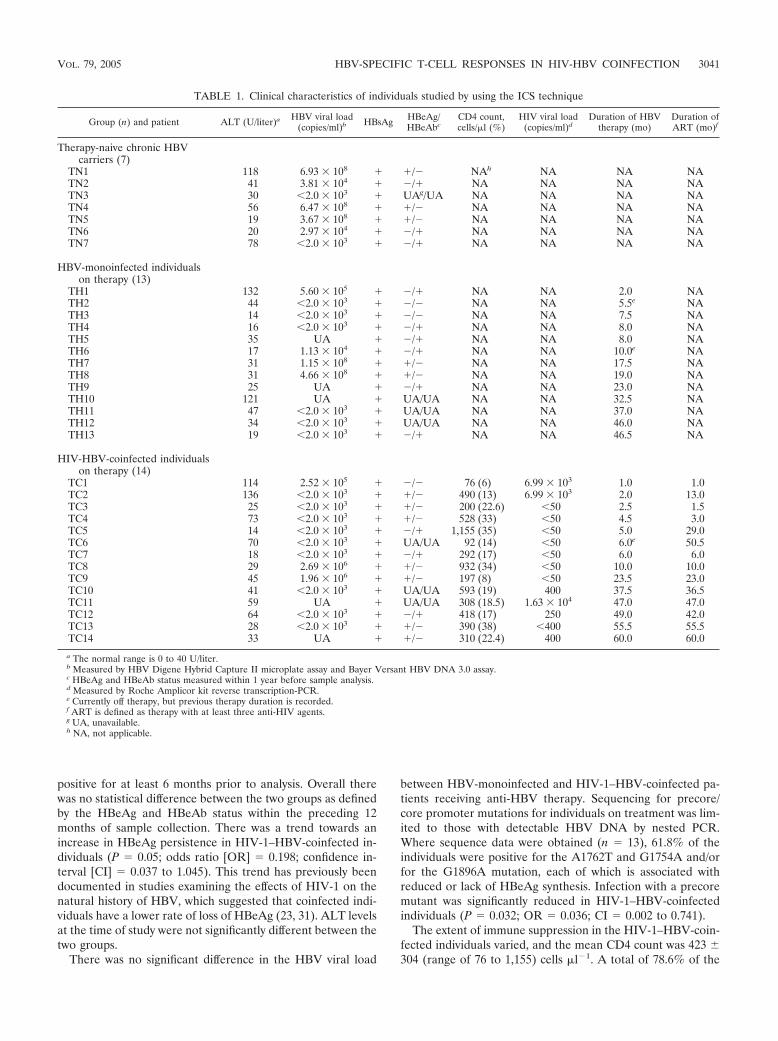
0.272 to 0.802) (Fig. 2a). These HBV-specific CD4� T cellsresponded to several of the HBV peptide pools, with responsesto peptide pool Pol1 most frequently detected (Fig. 2b). IFN-�production in response to one peptide pool was present in foursubjects, and two individuals responded to more than one pool(TH4 to the S1.1 and Pol1 pools and TH10 to the Pol1 andPol2 pools) (Fig. 3a). These data confirm previous reports(assessing proliferation to HBV antigen and peptide stimula-tion) that anti-HBV treatment can lead to the reactivation ofHBV-specific CD4� T-cell responses (13). Although the HIV-1–HBV-coinfected individuals were also on anti-HBV therapy,only 1 of the 14 subjects had a detectable CD4� T-cell re-sponse to one HBV peptide pool (Fig. 4a). Compared withHBV-monoinfected individuals receiving anti-HBV treatment,there were significantly lower number of HIV-1–HBV-coin-fected individuals with an HBV-specific CD4� T-cell response(P � 0.033; OR � 0.90; CI � 0.009 to 0.902) (Fig. 2a). Inindividuals with detectable IFN-� production by CD4� T cells,the duration of anti-HBV therapy was 15.2 14.6 months(mean SD; range, 2 to 37 months).
Overall, in six of the seven individuals in whom IFN-�-producing HBV-specific CD4� T cells were identified, IFN-�-producing CD8� T-cell responses to at least one HBV peptidepool were observed. HBV-specific CD8� T-cell responses wereagain rare in untreated individuals (two of seven), with re-sponses to PC and Pol1 only (data not shown). In 6 of 13
HBV-monoinfected individuals receiving anti-HBV therapy,HBV-specific CD8� T cells were detected in response to atleast one peptide pool, with 3 individuals demonstrating apositive response to more than one peptide pool, includingS1.1 and Pol1 (TH4); X, S1.2, Pol1, and Pol2 (TH10); and X,Pol1, and Pol3 (TH11) (Fig. 3b). Again, Pol1 was the mostimmunogenic of the HBV peptide pools (Fig. 2b). In 8 of the14 HIV-1–HBV-coinfected individuals receiving anti-HBVtreatment, HBV-specific CD8� T cells were observed in re-sponse to stimulation with at least one peptide pool. Of theseeight, CD8� T cells from one individual responded to twopools, those from one individual responded to three pools, andthose from two individuals responded to four pools (Fig. 4b).The magnitude of the HBV-specific CD8� T-cell response forindividual TC13 was significantly higher then that for the restof the group, with a response of 2.72% to Pol3, which was morethan 22-fold higher than the next-highest measured responseagainst Pol3 in this study. No correlation was found betweenanti-HBV treatment duration and magnitude of CD8� T-cellresponses. In individuals with a detectable HBV-specific CD8�
T-cell response, the duration of anti-HBV therapy was 16.7 19.0 months (mean standard deviation) (range, 1 to 55.5months).
For three individuals, we were able to obtain samples frommore than one time point up to 7 months following the initi-ation of anti-HBV therapy. At each time point, ICS was used
FIG. 2. Frequencies of HBV-specific CD4� or CD8� T-cell responses in each clinical group. (Rx), individuals receiving anti-HBV therapy. (a)Percentage of individuals in each group with CD4� (left panel) or CD8� (right panel) T-cell responses to at least one HBV peptide pool. Significantlymore HBV-monoinfected individuals receiving anti-HBV therapy had HBV-specific CD4� T-cell responses then either those not receiving anti-HBVtherapy (P � 0.046) or the HIV-1–HBV-coinfected subjects (P � 0.033) (Fisher exact test). No significant difference in the frequency of HBV-specificCD8� T-cell responses was observed between the three groups. (b) Percentage of individuals in each group who had a CD4� (top panels) or CD8�
(bottom panels) T-cell response to each of the peptide pools. The breadth and frequency of CD8� T-cell responses were similar in the HBV-monoinfected and HIV-1–HBV-coinfected subjects receiving anti-HBV therapy despite the difference in CD4� T-cell responses.
VOL. 79, 2005 HBV-SPECIFIC T-CELL RESPONSES IN HIV-HBV COINFECTION 3043
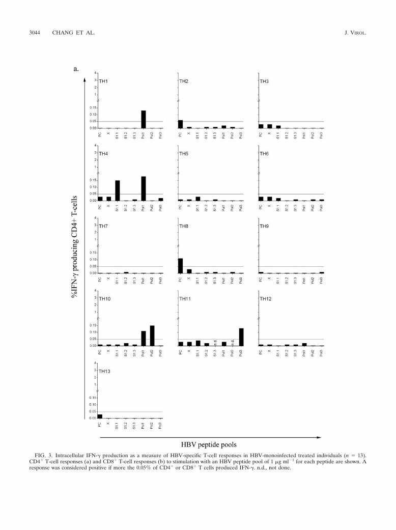
FIG. 3. Intracellular IFN-� production as a measure of HBV-specific T-cell responses in HBV-monoinfected treated individuals (n � 13).CD4� T-cell responses (a) and CD8� T-cell responses (b) to stimulation with an HBV peptide pool of 1 �g ml�1 for each peptide are shown. Aresponse was considered positive if more the 0.05% of CD4� or CD8� T cells produced IFN-�. n.d., not done.
3044 CHANG ET AL. J. VIROL.

to quantify IFN-� production following stimulation with HBVpeptides. In two individuals, we found an initial increase in thenumber of HBV-specific IFN-�-producing T cells followinginitiation of therapy; however, after 7 months on therapy, these
responses were greatly diminished or in some cases disap-peared (data not shown). During this period there was noseroconversion or clearance of HBV in any of the three indi-
FIG. 3—Continued.
VOL. 79, 2005 HBV-SPECIFIC T-CELL RESPONSES IN HIV-HBV COINFECTION 3045

FIG. 4. Intracellular IFN-� production as a measure of HBV-specific T-cell responses in HIV-1–HBV-coinfected treated individuals (n � 14).CD4� T-cell responses (a) and CD8� T-cell responses (b) to stimulation with an HBV peptide pool of 1 �g ml�1 for each peptide are shown tothe left of the vertical bars, and those to stimulation with an HIV peptide pool of 1 �g ml�1 for each peptide are shown to the right of the verticalbars. A response was considered positive if more the 0.05% of CD4� or CD8� T cells produced IFN-�. n.d., not done.
3046 CHANG ET AL. J. VIROL.
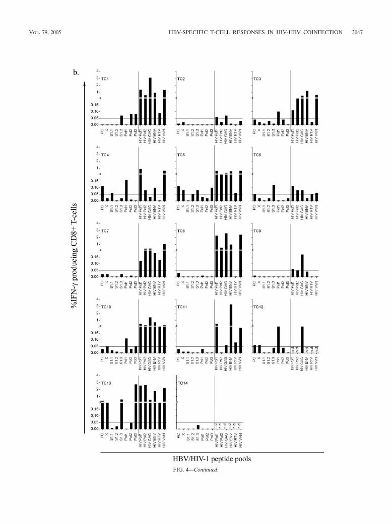
FIG. 4—Continued.
VOL. 79, 2005 HBV-SPECIFIC T-CELL RESPONSES IN HIV-HBV COINFECTION 3047
viduals. These limited studies raise the possibility that HBV-specific T-cell responses fluctuate or perhaps reduce over time.
General T-cell responses to mitogen. IFN-� production fol-lowing stimulation with pokeweed mitogen and staphylococcalenterotoxin B was used as positive controls (data not shown).IFN-� production from CD4� and CD8� T cells was observedfrom all 34 individuals tested. General CD4� T-cell hypore-sponsiveness in persistent HBV infection, as suggested in aprevious study (13), was not observed; however, the magni-tudes of mitogen-induced CD8� T-cell responses were signif-icantly higher in HBV-monoinfected treated individuals(17.84% 8.03%) than in untreated individuals (9.75% 7.55%) (P � 0.037 by t test). Interestingly, this difference wasnot observed between the HIV-1–HBV-coinfected group(14.07% 7.97%) and the untreated group.
T-cell responses to HIV-1 peptide pools. IFN-� productionfollowing stimulation with HIV-1 peptides in HIV-1–HBV-coinfected individuals was assessed to determine if there was aglobal reduction in T-cell responsiveness and to compare re-sponses to two different but persistent viral infections (Fig. 4).All HIV-1–HBV-coinfected individuals tested had detectableCD4� and CD8� T-cell responses to at least one HIV-1 pep-tide pool. The magnitude of the HIV-1-specific CD8� T-cellresponses was significantly higher than that of the HIV-1-specific CD4� T-cell responses (P � 0.028 by paired t test).The proportion of IFN-�� CD8� T cells following stimulationwith the HIV-1 peptide pools was significantly greater (five-fold) than that following stimulation with HBV peptide poolsin this same coinfected group (P � 0.016 by paired t test). Onlyone HIV-1–HBV-coinfected individual had an HBV-specificCD4� T-cell response, so statistical analysis could not be per-formed. In summary, HBV-specific T-cell responses were sig-nificantly diminished compared to those against HIV-1 inHIV-1–HBV-coinfected individuals.
Association of HBV-specific responses, HBV viral load, anddegree of immunosuppression. We next asked whether therewas an association between the detection of HBV-specific T-cell responses and HBV viral load independent of therapystatus and coinfection. Five individuals did not have an HBVviral load measured at sample time and so were not included inthis analysis. HBV viral loads were compared between individ-uals in whom CD4� and/or CD8� HBV-specific T cells to atleast one peptide pool were detected (responders; n � 16) andthose in whom no response was detected (nonresponders; n �13). There were significantly higher HBV viral loads in thenonresponders compared to the responders (P � 0.035 byMann-Whitney U test), with responders having a larger por-tion of individuals with undetectable HBV viral loads (�2,000copies/ml) (P � 0.027; OR � 0.144; CI � 0.027 to 0.774[Fisher’s exact test]). However, there was no significant corre-lation between the magnitude of either CD4� or CD8� T-cellresponses and the HBV viral load (by Spearman’s rho test).Therefore, effective suppression of HBV replication by anti-HBV therapy is a factor in recovery of HBV-specific T-cellresponses. In the coinfected individuals, the duration of ART,the HIV-1 viral load, the CD4 count at sample time, and theCD4 nadir were not significantly different between responders(n � 8) and nonresponders (n � 6).
DISCUSSION
Using a novel HBV peptide library that covers the wholeHBV genome and analysis of T-cell responses by measuringIFN-� production, we demonstrated that HBV-specific T-cellresponses were greater in both HBV-monoinfected and HIV-1–HBV-coinfected individuals on therapy than in chronicHBV carriers not on treatment. T-cell responses to HBV Xand Pol1 peptides, which have not been previously reported,were frequently detected, suggesting that previous methodsmay have underestimated the breadth and magnitude of HBV-specific T-cell responses. Coinfection with HIV-1 was associ-ated with a reduction of HBV-specific CD4� responses despiteclearly detectable HIV-1-specific CD4� and CD8� T-cell re-sponses. Interestingly, despite the low-level HBV-specificCD4� T-cell responses in HIV-1–HBV-coinfected individuals,HBV-specific CD8� T-cell responses were similar to thoseseen in HBV-monoinfected individuals.
Using overlapping peptide libraries covering all HBV pro-teins and the ICS technique, we were able to successfullydetect HBV-specific CD4� and CD8� T-cell production ofIFN-�. The frequency and magnitude of responses to polymer-ase peptides were more readily observed than those of re-sponses to precore/core-derived peptides. Previous studies ofacute HBV infection of HLA-A2-positive individuals demon-strated maximal response to precore/core (45, 46). It is possiblethat treatment of chronic HBV infection may effect the hier-archy of peptides recognized, as described for chronic lympho-cytic choriomeningitis virus infections (71), and, as shown pre-viously, the initiation of therapy may not lead to thereconstitution of all HBV-specific T cells evenly (15). We alsodetected CD8� T-cell responses to X protein, which have notpreviously been documented. These results highlight the in-creased information obtained by using overlapping peptidesthat cover the whole genome to assess HBV-specific T-cellresponses.
The detection of HBV-specific T-cell responses in untreatedindividuals was markedly reduced compared with that intreated individuals, either HBV monoinfected or HIV-1–HBVcoinfected. Boni et al. have previously described the appear-ance of HBV-specific T-cell responses following lamivudinetherapy (13–15). In these studies the T-cell responses weremeasured by CD4� T-cell proliferation and/or CD8� T-cellcytolytic activity. These responses were induced by core and eantigens or peptides (13, 14) and also by previously definedHLA-A2 epitopes in the envelope and polymerase proteins(15). In one study the precore/core-specific IFN-� producing Tcells were also examined by ICS. Both our study and theseprevious studies (13–15) draw the same conclusion that anti-HBV therapy is associated with the detection of HBV-specificT-cell responses. The proliferative and cytolytic responses fol-lowing therapy were more frequent than the IFN-� productionresponses we observed. This discrepancy in T-cell responsive-ness opens the possibility that although HBV-specific T-cellactivity increases following anti-HBV therapy, the responsemay not be fully functional or equivalent to that seen followingsuccessful clearance of acute HBV infection. This may explainwhy long-term clearance of virus following anti-HBV therapyis rarely observed (35).
One possible explanation for the appearance of HBV-spe-
3048 CHANG ET AL. J. VIROL.
cific T cells following antiviral therapy is the reduction in HBVviral load (13, 38). The reduction in HBV viral load and HBVantigen load may lead to a loss of HBV anergy (51). We foundthat individuals with either a CD4� or a CD8� HBV-specificT-cell response had a lower serum HBV viral loads than thosewith no responses. An alternative explanation is that HBV-specific T cells migrate from the liver to the periphery, asrecently suggested by studies using HBV-specific tetramers(47). Although HBV-specific CD8� T cells may not originatefrom the liver, they may traffic to the liver, which acts as a“killing field” for activated CD8� T cells that are usually in-volved in immune tolerance of oral antigens (24, 50).
In addition to the reconstitution of HBV-specific T-cell re-sponses in individuals on anti-HBV therapy, the overall CD8�
T-cell responses to mitogen stimulation were also significantlyhigher in the HBV-monoinfected treated group than in theuntreated group. This supports the hypothesis proposed byBoni et al. that persistent HBV infection is associated with abroad T-cell hyporesponsiveness (13). However, their subse-quent data suggest that this proliferative increase is not long-lasting (14). Clearly, this needs to be further analyzed beforeconclusive results can be obtained. Any HBV-associated sup-pression of T-cell activity may be important in the setting ofcoinfections with other viruses. Interestingly, the number ofIFN-�� CD8� T cells following mitogen stimulation in theHIV-1–HBV-coinfected group was not significantly greaterthan that in the untreated group, most likely due to immunesuppression secondary to HIV-1 infection itself (57).
Overall the magnitude of the HIV-1-specific T-cell responsewas higher than that of the HBV-specific T-cell response. Thismay be due to compartmentalization of HBV infection to theliver hepatocytes, which have reduced HLA expression (33),while HIV-1 infects CD4� T cells (26) and therefore HIV-1antigens may be more readily presented and recognized. Inaddition, functional HBV-specific T cells are likely to localizeto the liver, as has previously been shown (45, 47).
While compartmentalization may account for the overalllower magnitude of HBV-specific T-cell responses comparedto HIV-1-specific responses, it does not explain why there wasa drop in the frequency of HBV-specific CD4� T-cell re-sponses in HIV-1–HBV-coinfected individuals, while the fre-quency of HBV-specific CD8� T-cell responses remained com-parable to that found in HBV-monoinfected treatedindividuals. HIV-1 infection is characterized by a reduction inthe quantity and function of CD4� T cells; however, HIV-1-specific CD4� T-cell responses were clearly detectable. Similarfindings have also been described for persistent HCV infection,where the frequency of HCV-specific CD4� T cells is alsosignificantly reduced in individuals with HIV-1 coinfection(37). An alternative explanation for the absence of HBV-spe-cific CD4� T cells in HIV-1–HBV-coinfected individuals couldbe infection of HBV-specific CD4� T cells with HIV-1 as theystart to proliferate, differentiate, and become activated, giventhat HIV-1-specific CD4� T cells are preferentially infectedwith HIV-1 (27). We think this is an unlikely explanation forthe absence of HBV-specific CD4� T cells in HIV-1–HBVcoinfection, given that robust restoration of CD4� T cells toother organisms such as cytomegalovirus occurs followingART (3, 65). Nevertheless, HIV-1 coinfection impairs the gen-eration of HBV-specific CD4� T-cell responses and may ac-
count for the increased risk of a persistent HBV infection whenHBV infection occurs following HIV-1 infection (32, 62). Pre-vious studies of chronic lymphocytic choriomeningitis virusinfections show that CD4� T-cell help is essential in maintain-ing CD8� T-cell activity as well as for the production of neu-tralizing antibodies (20, 72). Therefore the ability to regener-ate HBV-specific CD4� T-cell responses in the setting of HIV-1–HBV coinfection may be crucial for regulation of HBV-specific CD8� T-cell responses and the development ofimmune restoration hepatitis. Our study was not designed toaddress this, but a longitudinal study examining individualsprior to and following HBV-active ART would be of greatinterest.
Interestingly the specificity of HBV-specific CD8� T-cellresponses differed between HBV monoinfection and HIV-1–HBV coinfection. The S1.3 and PC peptide pools were mostcommonly recognized by CD8� T cells in the coinfected indi-viduals, while peptide pool Pol1 was most commonly recog-nized in HBV-monoinfected individuals. It is possible thatcoinfection with HIV-1 alters the T-cell immunodominance forHBV. Altered recognition of virus-specific epitopes betweenheterologous viruses has been demonstrated in virus coinfec-tion in animal models and may be an important evolutionarymechanism of viral persistence (17, 19, 70).
HIV-1–HBV coinfection has a significant effect on HBV-specific CD4� T-cell function in individuals receiving anti-HBV and anti-HIV-1 therapy. A prospective longitudinalstudy further analyzing T-cell production of IFN-� andchanges in the HBV-specific intrahepatic T cells will be neededto fully characterize the effects of HBV-active ART on HBV-specific immunity.
ACKNOWLEDGMENTS
We acknowledge Scott Bowden from the Victorian Infectious Dis-eases Reference Laboratory for HBV sequencing and viral load mea-surement, Ivan Stratov from the University of Melbourne for assis-tance with the development of the ICS assay, Stephen Locarnini fromthe Victorian Infectious Diseases Reference Laboratory for helpfuldiscussions and review of the manuscript, Joseph Torresi from RoyalMelbourne Hospital and Jenny Hoy from Alfred Hospital for recruit-ment of patients for this study, and the clinical research nurses at bothRoyal Melbourne Hospital and Alfred Hospital for their assistance.
S.R.L. is supported by the National Health and Medical ResearchCouncil of Australia and by the Alfred Research Foundation.
REFERENCES
1. Alberti, A., M. R. Brunetto, M. Colombo, and A. Craxi. 2002. Recentprogress and new trends in the treatment of hepatitis B. J. Med. Virol.67:458–462.
2. Alheim, M., U. Lazdina, D. R. Milich, and M. Sallberg. 2001. Flow cytomet-ric determination of cytokine production and proliferation in hepatitis Bcore antigen specific murine CD4 cells: lack of correlation between numberof cytokine producing cells and cytokine levels in supernatant. J. Immunol.Methods 258:157–167.
3. Autran, B., G. Carcelain, T. S. Li, C. Blanc, D. Mathez, R. Tubiana, C.Katlama, P. Debre, and J. Leibowitch. 1997. Positive effects of combinedantiretroviral therapy on CD4� T cell homeostasis and function in advancedHIV disease. Science 277:112–116.
4. Aye, T. T., A. Bartholomeusz, T. Shaw, S. Bowden, A. Breschkin, J. McMil-lan, P. Angus, and S. Locarnini. 1997. Hepatitis B virus polymerase muta-tions during antiviral therapy in a patient following liver transplantation.J. Hepatol. 26:1148–1153.
5. Beckebaum, S., V. R. Cicinnati, X. Zhang, S. Ferencik, A. Frilling, H.Grosse-Wilde, C. E. Broelsch, and G. Gerken. 2003. Hepatitis B virus-induced defect of monocyte-derived dendritic cells leads to impaired Thelper type 1 response in vitro: mechanisms for viral immune escape. Im-munology 109:487–495.
VOL. 79, 2005 HBV-SPECIFIC T-CELL RESPONSES IN HIV-HBV COINFECTION 3049
6. Benhamou, Y., R. Tubiana, and V. Thibault. 2003. Tenofovir disoproxilfumarate in patients with HIV and lamivudine-resistant hepatitis B virus.N. Engl. J. Med. 348:177–178.
7. Bertoletti, A., C. Ferrari, F. Fiaccadori, A. Penna, R. Margolskee, H. J.Schlicht, P. Fowler, S. Guilhot, and F. V. Chisari. 1991. HLA class I-re-stricted human cytotoxic T cells recognize endogenously synthesized hepa-titis B virus nucleocapsid antigen. Proc. Natl. Acad. Sci. USA 88:10445–10449.
8. Betts, M. R., D. R. Ambrozak, D. C. Douek, S. Bonhoeffer, J. M. Brenchley,J. P. Casazza, R. A. Koup, and L. J. Picker. 2001. Analysis of total humanimmunodeficiency virus (HIV)-specific CD4� and CD8� T-cell responses:relationship to viral load in untreated HIV infection. J. Virol. 75:11983–11991.
9. Betts, M. R., J. P. Casazza, and R. A. Koup. 2001. Monitoring HIV-specificCD8� T cell responses by intracellular cytokine production. Immunol. Lett.79:117–125.
10. Betts, M. R., J. P. Casazza, B. A. Patterson, S. Waldrop, W. Trigona, T. M.Fu, F. Kern, L. J. Picker, and R. A. Koup. 2000. Putative immunodominanthuman immunodeficiency virus-specific CD8� T-cell responses cannot bepredicted by major histocompatibility complex class I haplotype. J. Virol.74:9144–9151.
11. Bica, I., B. McGovern, R. Dhar, D. Stone, K. McGowan, R. Scheib, and D. R.Snydman. 2001. Increasing mortality due to end-stage liver disease in pa-tients with human immunodeficiency virus infection. Clin. Infect. Dis. 32:492–497.
12. Bichko, V., P. Pushko, D. Dreilina, P. Pumpen, and E. Gren. 1985. Subtypeayw variant of hepatitis B virus. DNA primary structure analysis. FEBS Lett.185:208–212.
13. Boni, C., A. Bertoletti, A. Penna, A. Cavalli, M. Pilli, S. Urbani, P. Scog-namiglio, R. Boehme, R. Panebianco, F. Fiaccadori, and C. Ferrari. 1998.Lamivudine treatment can restore T cell responsiveness in chronic hepatitisB. J. Clin. Investig. 102:968–975.
14. Boni, C., A. Penna, A. Bertoletti, V. Lamonaca, I. Rapti, G. Missale, M. Pilli,S. Urbani, A. Cavalli, S. Cerioni, R. Panebianco, J. Jenkins, and C. Ferrari.2003. Transient restoration of anti-viral T cell responses induced by lamivu-dine therapy in chronic hepatitis B. J. Hepatol. 39:595–605.
15. Boni, C., A. Penna, G. S. Ogg, A. Bertoletti, M. Pilli, C. Cavallo, A. Cavalli,S. Urbani, R. Boehme, R. Panebianco, F. Fiaccadori, and C. Ferrari. 2001.Lamivudine treatment can overcome cytotoxic T-cell hyporesponsiveness inchronic hepatitis B: new perspectives for immune therapy. Hepatology 33:963–971.
16. Brau, N., H. L. Leaf, R. L. Wieczorek, and D. M. Margolis. 1997. Severehepatitis in three AIDS patients treated with indinavir. Lancet 349:924–925.
17. Brehm, M. A., A. K. Pinto, K. A. Daniels, J. P. Schneck, R. M. Welsh, andL. K. Selin. 2002. T cell immunodominance and maintenance of memoryregulated by unexpectedly cross-reactive pathogens. Nat. Immunol. 3:627–634.
18. Carr, A., and D. A. Cooper. 1997. Restoration of immunity to chronic hep-atitis B infection in HIV-infected patient on protease inhibitor. Lancet349:995–996.
19. Chen, H. D., A. E. Fraire, I. Joris, M. A. Brehm, R. M. Welsh, and L. K.Selin. 2001. Memory CD8� T cells in heterologous antiviral immunity andimmunopathology in the lung. Nat. Immunol. 2:1067–1076.
20. Ciurea, A., L. Hunziker, P. Klenerman, H. Hengartner, and R. M. Zinker-nagel. 2001. Impairment of CD4(�) T cell responses during chronic virusinfection prevents neutralizing antibody responses against virus escape mu-tants. J. Exp. Med. 193:297–305.
21. Colin, J. F., D. Cazals-Hatem, M. A. Loriot, M. Martinot-Peignoux, B. N.Pham, A. Auperin, C. Degott, J. P. Benhamou, S. Erlinger, D. Valla, and P.Marcellin. 1999. Influence of human immunodeficiency virus infection onchronic hepatitis B in homosexual men. Hepatology 29:1306–1310.
22. Cooley, L., A. Ayres, A. Bartholomeusz, S. Lewin, S. Crowe, A. Mijch, S.Locarnini, and J. Sasadeusz. 2003. Prevalence and characterization of lami-vudine-resistant hepatitis B virus mutations in HIV-HBV co-infected indi-viduals. AIDS 17:1649–1657.
23. Cooley, L., and J. Sasadeusz. 2003. Clinical and virological aspects of hep-atitis B co-infection in individuals infected with human immunodeficiencyvirus type-1. J. Clin. Virol. 26:185–193.
24. Crispe, I. N., T. Dao, K. Klugewitz, W. Z. Mehal, and D. P. Metz. 2000. Theliver as a site of T-cell apoptosis: graveyard, or killing field? Immunol. Rev.174:47–62.
25. Dienstag, J. L., E. R. Schiff, T. L. Wright, R. P. Perrillo, H. W. Hann, Z.Goodman, L. Crowther, L. D. Condreay, M. Woessner, M. Rubin, and N. A.Brown. 1999. Lamivudine as initial treatment for chronic hepatitis B in theUnited States. N. Engl. J. Med. 341:1256–1263.
26. Dimitrov, A. S., X. Xiao, D. S. Dimitrov, and R. Blumenthal. 2001. Earlyintermediates in HIV-1 envelope glycoprotein-mediated fusion triggered byCD4 and co-receptor complexes. J. Biol. Chem. 276:30335–30341.
27. Douek, D. C., J. M. Brenchley, M. R. Betts, D. R. Ambrozak, B. J. Hill, Y.Okamoto, J. P. Casazza, J. Kuruppu, K. Kunstman, S. Wolinsky, Z. Gross-man, M. Dybul, A. Oxenius, D. A. Price, M. Connors, and R. A. Koup. 2002.HIV preferentially infects HIV-specific CD4� T cells. Nature 417:95–98.
28. Draenert, R., M. Altfeld, C. Brander, N. Basgoz, C. Corcoran, A. G. Wurcel,D. R. Stone, S. A. Kalams, A. Trocha, M. M. Addo, P. J. Goulder, and B. D.Walker. 2003. Comparison of overlapping peptide sets for detection of an-tiviral CD8 and CD4 T cell responses. J. Immunol. Methods 275:19–29.
29. Ferrari, C., A. Bertoletti, A. Penna, A. Cavalli, A. Valli, G. Missale, M. Pilli,P. Fowler, T. Giuberti, F. V. Chisari, et al. 1991. Identification of immuno-dominant T cell epitopes of the hepatitis B virus nucleocapsid antigen.J. Clin. Investig. 88:214–222.
30. Ferrari, C., A. Penna, A. Bertoletti, A. Valli, A. D. Antoni, T. Giuberti, A.Cavalli, M. A. Petit, and F. Fiaccadori. 1990. Cellular immune response tohepatitis B virus-encoded antigens in acute and chronic hepatitis B virusinfection. J. Immunol. 145:3442–3449.
31. Gilson, R. J., A. E. Hawkins, M. R. Beecham, E. Ross, J. Waite, M. Briggs,T. McNally, G. E. Kelly, R. S. Tedder, and I. V. Weller. 1997. Interactionsbetween HIV and hepatitis B virus in homosexual men: effects on the naturalhistory of infection. AIDS 11:597–606.
32. Hadler, S. C., F. N. Judson, P. M. O’Malley, N. L. Altman, K. Penley, S.Buchbinder, C. A. Schable, P. J. Coleman, D. N. Ostrow, and D. P. Francis.1991. Outcome of hepatitis B virus infection in homosexual men and itsrelation to prior human immunodeficiency virus infection. J Infect. Dis.163:454–459.
33. Herkel, J., B. Jagemann, C. Wiegard, J. F. Lazaro, S. Lueth, S. Kanzler, M.Blessing, E. Schmitt, and A. W. Lohse. 2003. MHC class II-expressing hepa-tocytes function as antigen-presenting cells and activate specific CD4 Tlymphocytes. Hepatology 37:1079–1085.
34. Hoffmeister, B., F. Kiecker, L. Tesfa, H. D. Volk, L. J. Picker, and F. Kern.2003. Mapping T cell epitopes by flow cytometry. Methods 29:270–281.
35. Jung, M. C., B. Hartmann, J. T. Gerlach, H. Diepolder, R. Gruber, W.Schraut, N. Gruner, R. Zachoval, R. Hoffmann, T. Santantonio, M. Wacht-ler, and G. R. Pape. 1999. Virus-specific lymphokine production differsquantitatively but not qualitatively in acute and chronic hepatitis B infection.Virology 261:165–172.
36. Kane, M. 1995. Global programme for control of hepatitis B infection.Vaccine 13(Suppl. 1):S47–S49.
37. Lauer, G. M., T. N. Nguyen, C. L. Day, G. K. Robbins, T. Flynn, K.McGowan, E. S. Rosenberg, M. Lucas, P. Klenerman, R. T. Chung, and B. D.Walker. 2002. Human immunodeficiency virus type 1-hepatitis C virus coin-fection: intraindividual comparison of cellular immune responses against twopersistent viruses. J. Virol. 76:2817–2826.
38. Lewin, S. R., R. M. Ribeiro, T. Walters, G. K. Lau, S. Bowden, S. Locarnini,and A. S. Perelson. 2001. Analysis of hepatitis B viral load decline underpotent therapy: complex decay profiles observed. Hepatology 34:1012–1020.
39. Liaw, Y. F., R. N. Chien, C. T. Yeh, S. L. Tsai, and C. M. Chu. 1999. Acuteexacerbation and hepatitis B virus clearance after emergence of YMDDmotif mutation during lamivudine therapy. Hepatology 30:567–572.
40. Liaw, Y. F., N. W. Leung, T. T. Chang, R. Guan, D. I. Tai, K. Y. Ng, R. N.Chien, J. Dent, L. Roman, S. Edmundson, C. L. Lai, et al. 2000. Effects ofextended lamivudine therapy in Asian patients with chronic hepatitis B.Gastroenterology 119:172–180.
41. Lincoln, D., K. Petoumenos, and G. J. Dore. 2003. HIV/HBV and HIV/HCVcoinfection, and outcomes following highly active antiretroviral therapy.HIV Med. 4:241–249.
42. Lok, A. S. 2002. Chronic hepatitis B. N. Engl. J. Med. 346:1682–1683.43. Maecker, H. T., H. S. Dunn, M. A. Suni, E. Khatamzas, C. J. Pitcher, T.
Bunde, N. Persaud, W. Trigona, T. M. Fu, E. Sinclair, B. M. Bredt, J. M.McCune, V. C. Maino, F. Kern, and L. J. Picker. 2001. Use of overlappingpeptide mixtures as antigens for cytokine flow cytometry. J. Immunol. Meth-ods 255:27–40.
44. Maini, M. K., and A. Bertoletti. 2000. How can the cellular immune responsecontrol hepatitis B virus replication? J. Viral Hepat. 7:321–326.
45. Maini, M. K., C. Boni, C. K. Lee, J. R. Larrubia, S. Reignat, G. S. Ogg, A. S.King, J. Herberg, R. Gilson, A. Alisa, R. Williams, D. Vergani, N. V. Naou-mov, C. Ferrari, and A. Bertoletti. 2000. The role of virus-specific CD8(�)cells in liver damage and viral control during persistent hepatitis B virusinfection. J. Exp. Med. 191:1269–1280.
46. Maini, M. K., C. Boni, G. S. Ogg, A. S. King, S. Reignat, C. K. Lee, J. R.Larrubia, G. J. Webster, A. J. McMichael, C. Ferrari, R. Williams, D.Vergani, and A. Bertoletti. 1999. Direct ex vivo analysis of hepatitis B virus-specific CD8(�) T cells associated with the control of infection. Gastroen-terology 117:1386–1396.
47. Malacarne, F., G. J. Webster, S. Reignat, J. Gotto, S. Behboudi, A. K.Burroughs, G. M. Dusheiko, R. Williams, and A. Bertoletti. 2003. Trackingthe source of the hepatitis B virus-specific CD8 T cells during lamivudinetreatment. J. Infect. Dis. 187:679–682.
48. Mast, E. E., M. J. Alter, and H. S. Margolis. 1999. Strategies to prevent andcontrol hepatitis B and C virus infections: a global perspective. Vaccine17:1730–1733.
49. Matsumura, S., K. Yamamoto, N. Shimada, N. Okano, R. Okamoto, T.Suzuki, T. Hakoda, M. Mizuno, T. Higashi, and T. Tsuji. 2001. High fre-quency of circulating HBcAg-specific CD8 T cells in hepatitis B infection: aflow cytometric analysis. Clin. Exp. Immunol. 124:435–444.
3050 CHANG ET AL. J. VIROL.

50. Mehal, W. Z., A. E. Juedes, and I. N. Crispe. 1999. Selective retention ofactivated CD8� T cells by the normal liver. J. Immunol. 163:3202–3210.
51. Milich, D. R., M. K. Chen, J. L. Hughes, and J. E. Jones. 1998. The secretedhepatitis B precore antigen can modulate the immune response to the nu-cleocapsid: a mechanism for persistence. J. Immunol. 160:2013–2021.
52. Milich, D. R., S. F. Wolf, J. L. Hughes, and J. E. Jones. 1995. Interleukin 12suppresses autoantibody production by reversing helper T-cell phenotype inhepatitis B e antigen transgenic mice. Proc. Natl. Acad. Sci. USA 92:6847–6851.
53. Nelson, M., S. Portsmouth, J. Stebbing, M. Atkins, A. Barr, G. Matthews, D.Pillay, M. Fisher, M. Bower, and B. Gazzard. 2003. An open-label study oftenofovir in HIV-1 and hepatitis B virus co-infected individuals. AIDS 17:F7–F10.
54. Nomura, L. E., J. M. Walker, and H. T. Maecker. 2000. Optimization ofwhole blood antigen-specific cytokine assays for CD4(�) T cells. Cytometry40:60–68.
55. Okamoto, H., F. Tsuda, H. Sakugawa, R. I. Sastrosoewignjo, M. Imai, Y.Miyakawa, and M. Mayumi. 1988. Typing hepatitis B virus by homology innucleotide sequence: comparison of surface antigen subtypes. J. Gen. Virol.69:2575–2583.
56. Penna, A., M. Artini, A. Cavalli, M. Levrero, A. Bertoletti, M. Pilli, F. V.Chisari, B. Rehermann, G. Del Prete, F. Fiaccadori, and C. Ferrari. 1996.Long-lasting memory T cell responses following self-limited acute hepatitisB. J. Clin. Investig. 98:1185–1194.
57. Piconi, S., D. Trabattoni, M. L. Fusi, F. Milazzo, L. P. Dix, G. Rizzardini, F.Colombo, D. Bray, and M. Clerici. 2000. Effect of two different combinationsof antiretrovirals (AZT�ddI and AZT�3TC) on cytokine production andapoptosis in asymptomatic HIV infection. Antiviral Res. 46:171–179.
58. Pittet, M. J., A. Zippelius, D. E. Speiser, M. Assenmacher, P. Guillaume, D.Valmori, D. Lienard, F. Lejeune, J. C. Cerottini, and P. Romero. 2001. Exvivo IFN-gamma secretion by circulating CD8 T lymphocytes: implications ofa novel approach for T cell monitoring in infectious and malignant diseases.J. Immunol. 166:7634–7640.
59. Protzer, U., and H. Schaller. 2000. Immune escape by hepatitis B viruses.Virus Genes 21:27–37.
60. Puoti, M., M. Airoldi, R. Bruno, B. Zanini, A. Spinetti, C. Pezzoli, A. Patroni,F. Castelli, P. Sacchi, G. Filice, and G. Carosi. 2002. Hepatitis B virusco-infection in human immunodeficiency virus-infected subjects. AIDS Rev.4:27–35.
61. Ristig, M. B., J. Crippin, J. A. Aberg, W. G. Powderly, M. Lisker-Melman, L.Kessels, and P. Tebas. 2002. Tenofovir disoproxil fumarate therapy forchronic hepatitis B in human immunodeficiency virus/hepatitis B virus-coin-fected individuals for whom interferon-alpha and lamivudine therapy havefailed. J. Infect. Dis. 186:1844–1847.
62. Sinicco, A., R. Raiteri, M. Sciandra, C. Bertone, A. Lingua, B. Salassa, andP. Gioannini. 1997. Coinfection and superinfection of hepatitis B virus inpatients infected with human immunodeficiency virus: no evidence of fasterprogression to AIDS. Scand. J. Infect. Dis. 29:111–115.
63. Soriano, V., J. Garcia-Samaniego, E. Valencia, R. Rodriguez-Rosado, F.Munoz, and J. Gonzalez-Lahoz. 1999. Impact of chronic liver disease due tohepatitis viruses as cause of hospital admission and death in HIV-infecteddrug users. Eur. J. Epidemiol. 15:1–4.
64. Szkaradkiewicz, A., A. Jopek, J. Wysocki, M. Grzymislawski, I. Malecka, andA. Wozniak. 2003. HBcAg-specific cytokine production by CD4 T lympho-cytes of children with acute and chronic hepatitis B. Virus Res. 97:127–133.
65. Tanaka, M., C. Yasuoka, I. Genka, N. Tachikawa, Y. Kikuchi, K. Teruya, A.Yasuoka, Y. Hirabayashi, S. Kimura, and S. Oka. 2001. Sustained cytomeg-alovirus-specific CD4� T cell response associated with prevention of recur-rence of cytomegalovirus retinitis without secondary prophylaxis after highlyactive antiretroviral therapy in patients with AIDS. AIDS Res. Hum. Ret-roviruses 17:1749–1756.
66. Thio, C. L., E. C. Seaberg, R. Skolasky, Jr., J. Phair, B. Visscher, A. Munoz,and D. L. Thomas. 2002. HIV-1, hepatitis B virus, and risk of liver-relatedmortality in the Multicenter Cohort Study (MACS). Lancet 360:1921–1926.
67. Trautmann, A., B. Ruckert, P. Schmid-Grendelmeier, E. Niederer, E. B.Brocker, K. Blaser, and C. A. Akdis. 2003. Human CD8 T cells of theperipheral blood contain a low CD8 expressing cytotoxic/effector subpopu-lation. Immunology 108:305–312.
68. Valenzuela, P., M. Quiroga, J. Zaldivar, P. Gray, and W. J. Rutter. 1980. Thenucleotide sequence of the hepatitis B viral genome and the identification ofthe major viral genes, p. 57–70. In B. N. Fields, R. Jaenisch, and C. F. Fox(ed.), Animal virus genetics, vol. 57. Academic Press, New York, N.Y.
69. Webster, G. J., S. Reignat, M. K. Maini, S. A. Whalley, G. S. Ogg, A. King,D. Brown, P. L. Amlot, R. Williams, D. Vergani, G. M. Dusheiko, and A.Bertoletti. 2000. Incubation phase of acute hepatitis B in man: dynamic ofcellular immune mechanisms. Hepatology 32:1117–1124.
70. Welsh, R. M., J. M. McNally, M. A. Brehm, and L. K. Selin. 2000. Conse-quences of cross-reactive and bystander CTL responses during viral infec-tions. Virology 270:4–8.
71. Wherry, E. J., J. N. Blattman, K. Murali-Krishna, R. van der Most, and R.Ahmed. 2003. Viral persistence alters CD8 T-cell immunodominance andtissue distribution and results in distinct stages of functional impairment.J. Virol. 77:4911–4927.
72. Zajac, A. J., J. N. Blattman, K. Murali-Krishna, D. J. Sourdive, M. Suresh,J. D. Altman, and R. Ahmed. 1998. Viral immune evasion due to persistenceof activated T cells without effector function. J. Exp. Med. 188:2205–2213.
VOL. 79, 2005 HBV-SPECIFIC T-CELL RESPONSES IN HIV-HBV COINFECTION 3051
Related Documents