AN ABSTRACT OF THE THESIS OF Patrice L. Prater for the degree of Master of Science in Animal Science presented on November 14. 1990 Title: Physiological Factors Affecting Ovine Uterine Estrogen and Progesterone Receptor Concentrations Redacted for Privacy Abstract Approved: w-A Fredrick Stormshak Two experiments were conducted to determine whether in ewes uterine concentrations of estrogen and progesterone receptors are affected by the presence of a conceptus or by the hormonal milieu associated with extremes in photoperiod to which ewes are exposed. In Exp.1, nine mature ewes were unilaterally ovariectomized by removing an ovary bearing the corpus luteum (CL). The ipsilateral uterine horn was ligated at the external bifurcation and a portion of the anterior ipsilateral uterine horn was removed and assayed for endometrial nuclear and cytosolic concentrations of estrogen receptor (ER) and progesterone receptor (PR) by exchange assays. After a recovery estrous cycle, ewes were bred to a fertile ram. On day 18 of gestation a 10 ml jugular blood sample was collected for measurement of serum concentrations of estradiol -178 (E2) and progesterone by radioimmunoassay. Ewes were

Welcome message from author
This document is posted to help you gain knowledge. Please leave a comment to let me know what you think about it! Share it to your friends and learn new things together.
Transcript
AN ABSTRACT OF THE THESIS OF
Patrice L. Prater for the degree of Master of Science in
Animal Science presented on November 14. 1990
Title: Physiological Factors Affecting Ovine Uterine
Estrogen and Progesterone Receptor Concentrations
Redacted for PrivacyAbstract Approved: w-A
Fredrick Stormshak
Two experiments were conducted to determine whether in
ewes uterine concentrations of estrogen and progesterone
receptors are affected by the presence of a conceptus or by
the hormonal milieu associated with extremes in photoperiod
to which ewes are exposed.
In Exp.1, nine mature ewes were unilaterally
ovariectomized by removing an ovary bearing the corpus luteum
(CL). The ipsilateral uterine horn was ligated at the
external bifurcation and a portion of the anterior ipsilateral
uterine horn was removed and assayed for endometrial nuclear
and cytosolic concentrations of estrogen receptor (ER) and
progesterone receptor (PR) by exchange assays. After a
recovery estrous cycle, ewes were bred to a fertile ram. On
day 18 of gestation a 10 ml jugular blood sample was collected
for measurement of serum concentrations of estradiol -178 (E2)
and progesterone by radioimmunoassay. Ewes were
relaparotomized on day 18 and the remaining uterine tissue was
removed. Endometrium from both the pregnant and nonpregnant
uterine horn was assayed for nuclear and cytosolic ER and PR
concentrations. Nuclear and cytosolic ER concentrations on
day 10 of the cycle were greater than in endometrium of gravid
and nongravid uterine horns on day 18 of gestation (p<.01).
Endometrial nuclear PR levels were also greater on day 10 of
the cycle than in the pregnant (p<.05) and nonpregnant horn
(p<.01) on day 18 of gestation. There were no differences in
nuclear and cytosolic ER and PR concentrations between the
pregnant and nonpregnant uterine horn on day 18. Serum levels
of E2 and progesterone on day 18 of gestation were 16.56 ±
2.43 pg/ml and 1.74 ± 0.57 ng/ml, respectively. These data
suggest that duration of exposure of the uterus to
progesterone and(or) the presence of the conceptus causes a
reduction in uterine concentrations of ER and PR. Further,
an effect of the conceptus, if any, is exerted via a systemic
route.
In Exp. 2, ten mature ewes were bilaterally
ovariectomized in early October. During the onset of the
winter solstice (late December), a 10 ml blood sample was
collected from five ewes for analysis of serum levels of E2
and progesterone. Ewes were then laparotomized and
approximately one-third to one-half of a uterine horn was
removed and assayed for endometrial nuclear and cytosolic ER.
The contralateral horn was ligated at the external bifurcation
and 10 Ag of E2 in 3 ml of physiological saline was injected

into the uterine lumen of the ligated horn. After 48 h, a
jugular blood sample was collected for steroid analysis and
a section of the E2_treated horn was removed and assayed for
endometrial cytosolic and nuclear ER. This procedure was
repeated on the remaining five ewes during the height of the
summer solstice (late June). Endometrial nuclear and
cytosolic concentrations of ER prior to and after exogenous
E2 stimulation were similar during the winter and summer
solstice (p>.05). However, treatment with E2 increased
endometrial nuclear and cytosolic concentrations of ER
compared with those of the nonstimulated uterine horn during
the winter and summer solstice (p<.05 for each). Serum levels
of E2 prior to luminal treatment of ewes with E2 during the
winter and summer solstice did not differ (16.55 ± 4.05 vs
16.00 ± 3.0 pg/ml, respectively, p>.05). Serum levels of E2
48 h after administration of E2 did not differ among ewes at
the winter and summer solstice (18.75 ± 2.4 vs 18.65 ± 1.65
pg/ml, respectively, p>.05). Serum levels of progesterone
were basal (<0.10 ng/ml) and did not differ in ewes prior to
and after E2 treatment at the winter and summer solstice
(p>.05). These data indicate that physiological factors
and(or) hormones such as prolactin and melatonin secreted in
response to extremes in photoperiod do not appear to influence
uterine concentrations of ER in ovariectomized ewes.
Physiological Factors Affecting Ovine Uterine
Estrogen and Progesterone Receptor Concentrations
by
Patrice L. Prater
A THESISsubmitted to
Oregon State University
in partial fulfillment ofthe requirements for the
degree of
Master of Science
Completed November 14, 1990
Commencement June, 1991

APPROVED:
Redacted for PrivacyProfessor of Animal Science and Biochemistry and Biophysicsin charge of the major
Redacted for PrivacyHead of Department of Animal Science
Dean of C
Redacted for Privacy
Date thesis presented: November 14, 1990
Typed for Researcher by: Patrice L. Prater
ACKNOWLEDGEMENTS
I would like to take the opportunity to express my thanks
to all of the individuals who helped make this thesis a
reality. Sincere thanks to Dr. "Stormy" Stormshak for his
guidance and friendship, and most importantly, for teaching
me perseverance to "take the bull by the tail on a uphill
pull" as there will always be worthwhile and attainable goals
to pursue. I would also like to thank Ligaya Tumbelaka, Anna
Cortell-Brown, Ov Slayden, and Sue Leers-Sucheta for their
help and friendship during surgeries and laboratory
tribulations. Appreciation also to the U.S.F.S, LaGrande
research station and Dr. John Stellflug of the U.S. Sheep
Experimental Station, Dubois, Idaho for their contributions
toward this research. Thanks also to Dr. Kenneth Rowe, Oregon
State University, for his statistical guidance.
In hindsight, I realize that without the support and
encouragement of those most dear to me, this degree would not
have been realized. Therefore, I would like to extend a very
special thank you to my family (Moores and Praters) and my
husband Brian for believing in me and for giving me the
foundation of love and encouragement on which to build. An
extra special thank you to Brian, who as a husband and best
friend has shown me that prosperity in life is found through
love and laughter.

TABLE OF CONTENTS
Page
REVIEW OF LITERATURE 1
Introduction 1
ESTROGEN AND PROGESTERONE 4
Origin of Estrogen and Progesterone 4
Physiological Responses to Estrogens 5
Physiological Responses to Progesterone 7
Biosynthesis of Estrogen and Progesterone 7
Steroid Hormone Transport 11
Steroid Hormone Receptors 12
THE ESTROUS CYCLE 17
Follicular Phase 17
Luteal Phase 18
Luteolysis 20
EARLY GESTATION 23
Maternal Recognition of Pregnancy 23
Implantation or Attachment 26
SEASONAL REPRODUCTION 30
Melatonin 31
Prolactin 34
EXPERIMENT 1 38
Introduction 38
Materials and Methods 39
Animals 39
Nuclear Estrogen Receptor Assay 40
Cytosolic Estrogen Receptor Assay 42
Nuclear Progesterone Receptor Assay 43
Cytosolic Progesterone Receptor Assay 44
DNA Determination 45
Serum Progesterone Radioimmunoassay 46
Serum Estradiol Radioimmunoassay 47
Statistical Analysis 49
Results 49
Discussion 52
EXPERIMENT 2 55
Introduction 55
Materials and Methods 56
Animals 56Nuclear and Cytosolic Estrogen Receptor Assay 57

TABLE OF CONTENTS (CONT.)
Page
DNA Determination 57
Serum Progesterone and EstradiolRadioimmunoassay 58
Statistical Analysis 58
Results 58
Discussion 61
BIBLIOGRAPHY 64
LIST OF FIGURES
Figure Page
1. Specific binding of Clflestradio1-178 tonuclear and cytosolic estrogen receptors (mean ±SE) in endometria of ewes on day 10 of the estrouscycle and in endometria of gravid and non-graviduterine horns of ewes on day 18 of gestation. Meannuclear and cytosolic concentrations of estrogenreceptors, day 10 vs day 18 gravid and nongravidhorns differ (p<.05).
2. Specifically bound (3H)R5020 to nuclearprogesterone receptors (mean ± SE) in endometria ofewes on day 10 of the estrous cycle and in endometriaof the gravid and nongravid uterine horns of ewes onday 18 of gestation. Day 10 vs day 18 gravid andnongravid horns differ in concentrations ofprogesterone receptors (p<.05).
3. Specifically bound [3H]estradio1-178 tonuclear and cytosolic estrogen receptors (mean ± SE)in nonstimulated and exogenous estradiol-stimulatedendometria of ewes during the winter and summersolstice. Stimulated vs nonstimulated endometrium;nuclear (p<.05), cytosolic (p<.01).
51
51
60
PHYSIOLOGICAL FACTORS AFFECTING OVINE UTERINEESTROGEN AND PROGESTERONE RECEPTOR CONCENTRATIONS
REVIEW OF LITERATURE
INTRODUCTION
The mechanisms of steroid hormone action have long been
an area of fascination and scientific inquiry. In the female,
estrogen and progesterone play a primary role in promoting the
development of an intrauterine environment necessary for
successful procreation.
Prior to 1960 most research on estrogens and progesterone
entailed studies with various mammalian species to elucidate
the physiological effects of these steroids on the development
of the uterus and mammary glands. During the 1960's interest
in the effects of these hormones shifted from physiological
to biochemical. Indeed, it was during the late 1960's that
experimental evidence was first presented to suggest that the
biological responses evoked by estrogens were the consequence
of this steroid being bound within the target cell by a
macromolecular receptor. Subsequently, research demonstrated
the existence of target tissue receptors for other steroids,
including progesterone.
Because the availability of estrogen and progesterone
receptors directly affects the biological activity of these
steroids, much research has been conducted to quantify
2
estrogen and progesterone receptors in uteri of laboratory
animals. However, comparatively little is known about the
changes in uterine concentrations of estrogen and progesterone
receptors that occur during various reproductive states in
domestic animals.
One of the leading causes of reduced reproductive
efficiency in many species is embryonic mortality. Most
embryos are lost before or soon after implantation. Many
factors influence the ability of an embryo to implant
including adequate production of ovarian hormones and
appropriate uterine responses to these hormones. However,
whether embryo wastage is, in part, attributable to impaired
action of estrogen and progesterone is unknown. Basic
information on estrogen and progesterone receptor
concentrations in the uterus during early gestation could
provide insight about factors that maintain or interfere with
successful pregnancy.
In the northern hemisphere, sheep are seasonal breeders
that exhibit behavioral estrus during the fall and winter and
then become anestrus during the late spring and early summer.
The underlying cause for the annual reproductive cycle of ewes
has been attributed to changes in duration of melatonin
secretion that occurs in response to seasonal changes in
photoperiod. Secretion of melatonin from the pineal gland of
ewes is maximal as hours of daylight decrease and minimal as
hours of daylight increase. In a number of species, daily

3
profiles of secretion of melatonin and prolactin are inversely
related. Indeed, experimental evidence suggests that
melatonin can inhibit the secretion of prolactin from the
adenohypophysis. It is not known whether systemic
concentrations of prolactin or melatonin can alter the
responsiveness of the uterus of ewes to steroids. These
hormones have been implicated as having a direct effect on the
uterus in the mink, pig, hamster and rabbit. Therefore,
research is needed to determine whether uterine concentrations
of steroid receptors in the ewe are influenced by seasonal
fluctuations in the secretion of melatonin and prolactin.
The following review of literature will encompass the
physiological and biochemical responses of the uterus to
estrogen and progesterone. Much of the review will rely on
data acquired from the study of laboratory animals but when
available, information on domestic animals and primates will
be included.
4
ESTROGEN AND PROGESTERONE
ORIGIN OF ESTROGEN AND PROGESTERONE
The female mammal is born with all of the oocytes she
will need throughout her reproductive life. In order for an
oocyte to be ovulated it must first go through several stages
of maturation. In the initial stages of folliculogenesis,
each oocyte is surrounded by a single layer of granulosa
cells, together forming a primordial follicle (Ireland, 1987).
Just prior to or following birth the primordial follicle
enters and remains in a resting state until the appropriate
hormonal signals cue it to resume maturation in preparation
for ovulation or atresia (Ireland, 1987). Follicular growth
and maturation occur in a series of events whereby the
primordial follicle first develops into a primary, then
secondary follicle, followed by formation of the fluid filled
tertiary follicle and finally the ovulatory or Graafian
follicle (Bjersing, 1967; Turnbull et al., 1977). Only
follicles that undergo "recruitment" actually ovulate. Most
follicles instead become atretic and are subsequently replaced
by new follicles (Smeaton and Robertson, 1971). In the
bovine, this maturation process occurs in sequential waves of
development until the ensuing ovulation occurs (Sirois and
Fortune, 1988). Soon after ovulation, the ruptured follicle
is transformed into a corpus luteum (CL).
5
During the estrous or menstrual cycle, ovarian follicles
and the CL are the primary sources of steroid synthesis and
secretion (Falck, 1959; Hafs and Armstrong, 1968; Moor, 1977).
Follicle steroidogenesis begins as the specialized cells that
comprise the follicle wall (granulosa and theca cells) grow
and proliferate. It has been well established that follicles
are the primary source of estrogens in the nonpregnant animal.
Similarly, once the CL has formed it also becomes a major
source of steroid synthesis with progesterone being the major
hormone secreted. During pregnancy, depending on the species,
the developing conceptus and later the placenta may become a
rich source of estrogens and progestins. Estrogens and
progestins are also synthesized and secreted by the adrenal
cortex, however, this source accounts for only a minor
fraction of the total estrogen and progesterone in the
systemic circulation. Without steroid synthesis and secretion
the complex and highly synchronized events that permit
reproduction could not occur.
PHYSIOLOGICAL RESPONSES TO ESTROGENS
Depending upon the mammalian species, there are several
forms of estrogens that are synthesized and circulate
throughout the body. However, the most important and
prevalent of the estrogens are estradio1-17B and estrone.
Estradio1-17B is approximately ten times more potent than
estrone and is the most biologically active of all steroids

6
produced by the ovary (Hisaw, 1959). A third estrogen known
as estriol is found in high concentrations in primates during
gestation. In general, estriol is considered the least potent
of these three estrogenic forms (Hisaw, 1959). During the
estrous or menstrual cycle, estrogens are responsible for
stimulating cellular proliferation and growth of the female
reproductive tract, regulation of utero-ovarian function and
intrauterine environment, and development and maintenance of
secondary sex characteristics. The following discussion will
focus on the role of estrogens in regulating utero-ovarian
function and the intrauterine environment.
Estrogens act on the uterine myometrium to increase both
the frequency and amplitude of contractions, which are
requisite for sperm and accelerated ovum transport, and in
some species, migration and spacing of embryos (Pope et al.,
1982). Estrogens initiate endometrial stromal edema (Astwood,
1938) as a result of increased uterine capillary and venule
permeability (Halkerston et al., 1960; Martin et al., 1973;
Espey, 1980). In response to estrogens, the glands of the
endometrium become increasingly tortuous as the nuclear RNA
polymerases and mRNA activate and facilitate protein
synthesis, cell hypertrophy and cell hyperplasia. Estrogens
are also responsible for increasing uterine blood flow
(Williams, 1948; Greiss and Anderson,1970), which has been
suggested to be a result of hormone-induced vasodilation of
the uterine vasculature (Greiss and Anderson, 1970). The
7
resultant increased mass of both the uterine endometrium and
myometrium as well as increased uterine vascularization is
essential for implantation and development of the embryo.
PHYSIOLOGICAL RESPONSES TO PROGESTERONE
Progesterone is commonly referred to as the hormone of
pregnancy and is the principal secretory product of the CL
(reviewed by Rothchild, 1981). It is absolutely crucial for
normal fetal survival and development in all mammalian species
known to date. Progesterone stimulates secretory changes in
the intrauterine environment that prepares the endometrium for
implantation of the blastocyst and promotes development of
decidual cells that supply nutrients to the early embryo
(Auletta and Flint, 1988; Clark and Markaverich, 1988). In
addition, it decreases the frequency and intensity of uterine
contractions thereby preventing expulsion of the implanted
embryo (reviewed by Clark and Markaverich, 1988). Capacity
of the CL to secrete progesterone and thus to sustain a normal
pregnancy is regulated by hormones synthesized and secreted
by the hypothalamus, pituitary, placenta and by the CL itself.
BIOSYNTHESIS OF ESTROGEN AND PROGESTERONE
As stated previously, the primary source of estrogen and
progesterone synthesis and secretion in the nonpregnant female
mammal is the ovarian follicle and CL, respectively.
Synthesis of these steroids encompasses several factors,

8
namely, follicle maturation, selection and luteinization.
Changes in steroidogenesis associated with folliculogenesis
and luteal development are dependent initially, on the amount
of follicle stimulating hormone (FSH) available to the
maturing follicle, and later the ratio of FSH and luteinizing
hormone (LH) during the preovulatory and luteal phase of the
cycle. Steroid hormones are derivatives of cholesterol of
either dietary or de novo origin. Cholesterol in systemic
blood is generally transported in the form of low density or
high density lipoprotein (LDL or HDL, respectively). Low
density lipoprotein or HDL binds to specific receptors on the
surface of the theca and granulosa cell plasma membranes. The
lipoprotein-receptor complex then enters the cell by way of
endocytosis, becoming enclosed in a coated vesicle. Lysosomes
then enzymatically degrade the coated vesicle containing
lipoprotein-receptor complexes thereby liberating free
cholesterol. This free cholesterol is then available for
conversion into steroids. Excess intracellular cholesterol
is esterified and stored in lipid droplets for future use.
Cholesterol can also be synthesized de novo from small
molecule precursors such as acetate. This pathway of
synthesis is activated when cells are deprived of LDL or HDL.
Cholesterol is transported to the mitochondria where it is
converted to pregnenolone by the cytochrome P - 45O complex
(Ichii et al., 1963; Roberts et al., 1967). Pregnenolone is
then converted to progesterone by 38-hydroxysteroid
9
dehydrogenase in the smooth endoplasmic reticulum and
progesterone or subsequent metabolites of this steroid are
secreted into the extracellular space to ultimately enter the
blood stream (Miller and Turner, 1963; Niswender and Nett,
1988) .
Ovarian steroid synthesis and secretion is dynamically
regulated by the anterior pituitary gonadotropins FSH and LH.
(Kaltenbach et al., 1968b; Hixon and Clegg, 1969; Karsch et
al., 1971; Denamur et al., 1973; Niswender et al., 1976).
These protein hormones regulate the biosynthesis and secretion
of steroids in the ovary by binding to high-affinity, low
capacity plasma membrane receptors. The formation of the
hormone-receptor complex activates the enzyme adenylate
cyclase which in turn catalyzes the formation of cyclic
adenosine-3'5'-monophosphate (cAMP) from adenosine
triphosphate (ATP) (Marsh, 1975). Cyclic AMP then mediates
the activation of specific protein kinases (Ling and Marsh,
1977), which phosphorylate enzymes or other proteins necessary
for the conversion of cholesterol into steroids via the
cholesterol/steroid biosynthetic pathway (Caron et al., 1975;
Caffrey et al., 1979).
Ovarian follicles consist of two predominant and
distinctly different cell types. Theca cells, which are
arranged along the outer surface of the basement membrane of
the follicle, and granulosa cells that lie along the inner
surface of the basement membrane. The interaction of
10
pituitary gonadotropins with the theca and granulosa cells of
the follicle have formed the basis of the two cell-two
gonadotropin model of ovarian steroidogenesis (Falck, 1959;
Short, 1962; Liu and Hsueh, 1986). During the early
follicular stage of development ovarian thecal cells contain
LH but no FSH receptors (Zeleznik et al., 1974; Carson et al.,
1979). Luteinizing hormone is responsible for stimulating the
conversion of cholesterol to pregnenolone in the theca cells
by way of activating the rate limiting cytochrome P-450
cholesterol side chain cleavage (P-450scc) enzyme in
mitochondria (Evans et al., 1981). Luteinizing hormone also
promotes the conversion of pregnenolone to progesterone in the
theca cell endoplasmic reticulum by activating 3B-
hydroxysteroid dehydrogenase (38-HSD) (Ireland, 1987).
Progesterone is then converted to the androgens,
androstenedione and testosterone, through the activation of
17,20 lyase and 17B-dehydrogenase enzymes, respectively (Evans
et al., 1981). The lipid soluble androstenedione and
testosterone can enter the systemic circulation and bind to
carrier protein globulins or they can freely diffuse from the
theca to the granulosa cell and be converted to estrogens by
the granulosa cell aromatase enzyme (Baird, 1977; Dorrington
and Armstrong, 1979; Leung and Armstrong, 1980). In contrast,
prior to the Graafian follicle stage of development, granulosa
cells have only FSH receptors. Follicle stimulating hormone
is the gonadotropin responsible for stimulating the conversion
11
of cholesterol to progesterone in the granulosa cells. Like
LH, FSH activates protein kinases through stimulation of
adenylate cyclase. In addition to stimulating the synthesis
of enzymes necessary for converting cholesterol to
progesterone, the FSH-stimulated protein kinases within the
granulosa cell activate an aromatase enzyme that catalyzes
the conversion of androgens to estrogens (Fortune and
Armstrong, 1978). Estrogens can in turn act back on the
follicle to stimulate further cell proliferation as well as
induce or inhibit (depending on the feedback pathway) the
synthesis of more gonadotropin and steroid receptors.
STEROID HORMONE TRANSPORT
Steroid hormones are, for the most part, transported
throughout the body bound to serum proteins. However, a
relatively smaller fraction may circulate in an unbound or
free form. More specifically, estrogens and androgens are
bound to sex steroid hormone binding globulin (SHBG), while
progesterone is bound to progesterone binding globulin (PBG).
Both of these binding globulins are comparatively high
affinity, low capacity carriers. Alternatively, these
steroids may also circulate throughout the body bound to low
affinity, high capacity serum albumins. Only approximately
2% of steroids actually circulate in the free form. The
mechanism by which the steroid is able to dissociate from its
carrier and thus freely diffuse through the plasma membrane
12
of the target cell is unclear. However, Avvakumov et al.
(1986) found the steroid binding globulin in complex with the
plasma membrane of human decidual endometrial tissue. These
investigators hypothesized that the binding of the protein
binding globulin to the plasma membrane may send some kind of
a signal which then causes dissociation of the steroid and
binding globulin, thus allowing simple diffusion of the
steroid across the plasma membrane. It is also conceivable
that the entire steroid and binding globulin complex may enter
the cell by way of endocytosis where the steroid is then
liberated by lysosomal degradation of the protein globulin.
STEROID HORMONE RECEPTORS
In any biological system, receptors function by
recognizing a specific hormone from all other molecules in
the cellular environment and then upon binding of the hormone,
transmit a signal thereby creating a biological response. The
magnitude of a hormonal response depends on at least three
factors; these include the hormone concentration, the number
of available receptors and the affinity of the hormone for the
receptor. Steroid hormone receptors belong to a family of
regulatory proteins whose ability to control gene expression
is dependent on the binding of their ligand. Binding of the
hormone to its receptor stimulates the synthesis of specific
proteins by altering the rate of transcription of specific
genes (Mueller et al., 1957; Norman and Litwack, 1987).
13
Over the years, the cellular localization of the steroid
hormone receptors has been a source of controversy. It was
once thought and accepted that the unoccupied steroid receptor
was localized in the cytoplasmic fraction of the target cell
(Gorski et al., 1968; Jensen et al., 1968). Upon binding of
the receptor to its specific hormone the extranuclear steroid
hormone-receptor complex was believed to be translocated to
the nuclear compartment where it attached to the DNA to exert
stimulatory effects on gene transcription (Gorski et al.,
1968; Jensen et al., 1968). The foundation for this central
dogma was based on studies in which cells not previously
exposed to a given steroid were homogenized and the nuclear
and cytosolic fractions separated by means of differential
centrifugation. After incubation of these separate fractions
with a radiolabeled steroid the steroid receptors were
predominately found in the cytosol. However, when cells
previously exposed to a radiolabeled steroid were homogenized
and separated by centrifugation, the bound steroid receptors
were found in the nuclear fraction. Recently, studies by
independent laboratories revealed that occupied as well as
unoccupied steroid receptors resided predominantly in the
nucleus. Results of these studies indicated that the presence
of steroid receptors in the cytosolic fraction of cells as
detected in earlier studies was an artifact brought about by
their extraction from the nucleus after cell breakage
(Welshons et al., 1984).
14
With the recent availability of monoclonal antibodies
specific for a number of steroid receptors and the technique
of immunocytochemistry, it is now widely accepted that both
the unoccupied and occupied steroid receptors are
predominately localized in the nucleus (King and Greene, 1984;
Welshons et al., 1984). King and Greene (1984) found that
treatment of cells or tissues in vivo or in vitro with
[3H]estradiol -178 altered the intensity of staining of the
estrogen receptor but not the distribution of the receptor.
After passage across the plasma membrane of its target cell,
the lipophilic steroid hormone may or may not bind to low
affinity binding sites in the cytoplasm (Mercer et al., 1981).
The steroid migrates through the cytoplasm and diffuses across
the nuclear envelope where temperature dependent noncovalent
binding to its specific unoccupied receptor occurs resulting
in the formation of the activated steroid-receptor complex
(Miller et al., 1985; Greene and Press, 1986). The steroid-
receptor complex undergoes a conformational change which
stimulates an increase in its affinity for chromatin and
becomes tightly associated with nuclear acceptor sites on the
chromatin. With respect to the estrogen receptor, ligand
binding and activation may involve the dimerization of two
identical estrogen receptor subunits. The dimerization may
be critically involved in the mechanism by which the estrogen
receptor complexes promote changes in gene transcription
(Sabbah et al., 1989). The binding of the steroid-receptor
15
complex to the chromatin may cause an unwinding of a specific
portion of the DNA strand thus facilitating the binding of
ribonucleic acid (RNA) polymerases, which stimulate
transcription of specific genes into messenger RNA (mRNA)
(reviewed by Yamamato, 1985). The mRNA's then translocate to
the cytoplasm where they bind to ribosomes and are translated
into specific proteins such as enzymes, receptors, low
affinity binders and protein hormones, which in turn
facilitate the cellular and metabolic effects on the target
tissue characteristic of the actions of the steroid hormone
in question.
Interestingly, recent immunocytochemical (Berthois et
al., 1986) and fluorescence (Parikh et al., 1987) studies have
revealed the presence of steroid receptors in the cytoplasm
of steroid target cells. Yamamoto (1985), explained this
occurrence by hypothesizing that the nuclear and cytoplasmic
distribution of steroid receptors may depend solely on genomic
sites. Because of the enormous amount of DNA that exists in
mammalian nuclei this equilibrium would naturally favor that
of a nuclear distribution of receptors in intact cells
including the unbound receptors. Thus the majority of unbound
receptors would reside in the nucleus while only a minor
fraction of the free receptors would be transiently
cytoplasmic. These receptors could bind incoming hormone and
translocate to the nucleus where modulation of gene expression
would then take place.

16
Estrogen and progesterone receptor concentrations are
regulated by both estrogen and progesterone. Specifically,
estrogen stimulates the synthesis of both estrogen and
progesterone receptors thereby enhancing the sensitivity of
target tissues to each of these hormones (Zelinski et al.,
1980; DeHertogh et al., 1986; Ekka et al., 1987). On the
other hand, progesterone causes a down-regulation of both
progesterone and estrogen receptors (Koligian and Stormshak,
1977b; West et al., 1986; Selcer and Leavitt, 1988), by
interfering with the replenishment or de novo synthesis of
the receptor (Hsueh et al., 1976; Pavlik and Coulson, 1976).

17
THE ESTROUS CYCLE
FOLLICULAR PHASE
During the follicular phase of the estrous or menstrual
cycle, FSH and LH-induced maturation of the follicle increases
the production and release of estrogens into the blood (Karsch
et al., 1979; Hansel, 1983). Just prior to ovulation high
levels of estrogen positively feedback to activate the release
of gonadotropin-releasing hormone (GnRH) from the hypothalamus
(Karsch et al., 1979). This in turn stimulates the release
of LH and FSH from the pituitary gland. In the ewe, estradiol
secretion begins to rise as the follicle develops and matures.
An increase in estradiol production of 5 to 10-fold then
occurs over the next few days. Estradiol peaks at the
beginning of the preovulatory LH surge and then rapidly
declines to basal levels (Hauger et al., 1977). Some
researchers suggest that a second estradiol peak occurs in
cows (Glencross et al., 1973; Hansel et al., 1973), and ewes
(Cox et al., 1971). It is presumed this second low amplitude
peak of estrogen stems from renewed follicular development in
preparation for a new cycle. To date, the occurrence and
source of the second estradiol peak remains equivocal. A
surge of LH, along with increased levels of FSH precipitate
ovulation. Initiation of the preovulatory LH surge and
subsequent ovulation requires high frequency, low amplitude
18
LH pulses. These pulses stem from the rising levels of
estrogen that act on the hypothalamus to speed up a GnRH pulse
generator (reviewed by Karsch, 1987). The frequent,
estradiol-enhanced GnRH pulses during the follicular phase
stimulate the LH surge and ovulation. Subsequent to
ovulation, estrogen secretion declines and LH then stimulates
the transformation of the ruptured follicle into a CL as the
thecal and granulosa cells of the follicle undergo rapid
mitosis and hypertrophy.
LUTEAL PHASE
The main function of the CL is to secrete progesterone
in order to establish the intrauterine environment necessary
for successful implantation of an embryo and maintenance of
early pregnancy in the ewe, mare and primate (Casida and
Warwick, 1945; Ginther and First, 1971). The CL is the major
source of progesterone for pregnancy maintenance to term in
the rat, cow, sow and goat (Bazer et al., 1982). Like the
follicle, the CL is composed of two cell types; small and
large luteal cells. Small luteal cells arise from the
luteinization of theca cells and large cells are derivatives
of luteinized granulosa cells (Lemon and Loir, 1977; Uresley
and Leymarie, 1979; Rodgers and O'shea, 1982; Niswender et
al., 1985).
In the cow (Donaldson et al., 1965), ewe (Kaltenbach et
al., 1968), mare (Ginther, 1979) and primate (Moudgal et al.,
19
1971; Mais et al., 1986) LH is the primary luteotropin for
maintaining continued progesterone secretion from the CL.
This was demonstrated when administration of LH to cows on day
5, 10, 15 and 20 of the estrous cycle (Schomberg et al.,
1967), or ewes on day 9 (Niswender et al., 1976) increased the
synthesis and secretion of progesterone. In the ewe, systemic
concentrations of progesterone increase from day 3-4 (first
day of detected estrus = day 0) to a maximum by day 7-8 of the
estrous cycle. Rising levels of progesterone from the CL
block the preovulatory LH surge by suppressing the frequency
of the GnRH pulse generator and thereby reduce the frequency
and amplitude of LH secretory pulses from the pituitary gland
(Yen et al., 1972; Foster et al., 1975; Baird, 1978).
Progesterone levels remain high until day 14-15 of the cycle,
after which time they decline to basal levels 1-2 days prior
to the ensuing estrus as a new cycle is initiated (Baird et
al., 1976; Pant et al., 1977). Concomitant to declining
estrogen and increasing progesterone levels during the luteal
phase, an increase in the follicular protein hormone inhibin
occurs. Inhibin feeds back at the level of the hypothalamus
and pituitary inhibiting further FSH release (Schwartz and
Channing, 1977).
Just as gonadotropins and steroid hormones interact in
dynamic synchrony with one another, so to is the complexity
of interaction between ovarian steroids and uterine
endometrium during the estrous cycle and pregnancy (Stone et
al., 1978).
LUTEOLYSIS
20
In the absence of fertilization of an ovum from the
ruptured follicle, many laboratory and domestic animals will
continue to exhibit estrous cycles. In most non-primate
mammals such as the rat, guinea pig, ruminants and sow, the
uterus exerts a local luteolytic action on the ipsilateral
corpus luteum of the non-pregnant animal to bring about luteal
regression and decreased progesterone secretion (Niswender et
al., 1985). This first became evident when excision of the
uterus was found to prolong luteal maintenance in the sow (du
Mesnil du Buisson, 1961), guinea pig (Fischer, 1965),
pseudopregnant rat (Barley et al., 1966) and ewe (Inskeep and
Butcher, 1966). Upon completion of a previous cycle and
initiation of a new cycle, the uterine endometrium secretes
the luteolysin prostaglandin Fla (McCracken et al., 1970;
Goding, 1974). The secretion of prostaglandin Fla (PGF2a) is
under the influence of ovarian progesterone, estradiol -178,
and oxytocin (Baird, 1978; Sheldrick et al., 1980). In the
ewe, PGF2a levels increase from day 12 of the cycle, reaching
maximum concentrations 1-2 days prior to the subsequent estrus
(Baird et al., 1976). This luteolytic factor enters the ovary
by diffusing across the wall of the uterine vein into the
ovarian artery. The close anatomical proximity of the uterine
veins with the convoluted ovarian artery permits the diffusion

21
of substances such as PGF2a directly into the ovarian artery
by a countercurrent diffusion mechanism. They thus bypass the
general circulation and avoid rapid destruction in the lungs
(McCracken et al., 1970). Once in the ovary PGF2a targets
the progesterone secreting luteal cells of the corpus luteum
to cause luteolysis (McCracken et al., 1970). The mechanism
by which PGF2a exerts its luteolytic action on the CL is not
known. However, Niswender et al. (1975) suggest that the
effects of PGF2a may be via a vascular component because a
significant decrease in capillary blood flow to the CL is
associated with, but does not precede luteolysis. Thus, it
is doubtful that PGF2a causes luteal regression by interfering
with blood flow to the CL.
The protein hormone oxytocin is currently believed to
also play an essential role in the events leading to
luteolysis (Mitchell et al., 1975; Flint and Sheldrick, 1986).
Uterine PGF2a exerts its effect in a local manner, targeting
the ipsilateral ovary and thus causing both inhibition of
luteal progesterone secretion and stimulation of luteal
oxytocin release. This was demonstrated by Walters et al.
(1983) and Sheldrick and Flint, (1983) who found that oxytocin
secretion was stimulated by an intramuscular injection of the
PGF2a analog, cloprostenol in ewes. Similiarly,
Schallenberger et al. (1984) found the same phenomenon to
occur in cows. In ewes, oxytocin triggers the release of more
uterine PGF2a and the positive feedback cycle continues until

22
luteolysis is complete (Mitchell et al., 1975; Flint and
Sheldrick, 1986). Oxytocin concentrations in both ovine
(Sheldrick and Flint, 1983) and bovine (Abdelgadir et al.,
1987) luteal tissue is maximal during the early luteal phase.
Stormshak et al. (1969) found that injection of exogenous
estradiol into ewes on days 10-11 of the estrous cycle caused
premature luteal regression. It is believed that estradiol
also acts to increase uterine PGF2cf secretion thereby
promoting luteal regression (Ford et al., 1975).
The mechanism involved in regression of the primate CL
is not completely understood. Shutt et al. (1976) found that
removal of the primate uterus did not prolong the maintenance
of the CL. It is believed that an intra-ovarian prostaglandin
may be involved, because the ovary is a significant source of
PGF2cc in primates (Shutt et al., 1976). In primates, the
reduction in progesterone secretion that occurs with luteal
regression precipitates atrophy of the endometrial epithelium
and invasion of the endometrium by lymphocytes, causing
stromal degradation, exposure of arterioles and mensus, or
shedding of the uterine endometrial lining (Knobil, 1973).
With a decrease in both progesterone and ovarian secretion of
inhibin, GnRH and the gonadotropins FSH and LH are released
and a new menstrual cycle is initiated.

23
EARLY GESTATION
In order to ensure normal embryonic development and
differentiation a complex communication network between the
embryo and maternal tissues must be established. During the
preimplantation or preattachment stage of gestation conceptus
secretory proteins and steroids as well as hormones of
maternal origin act upon the uterus and ovaries to ensure
maintenance of pregnancy. In particular, factors of conceptus
origin are important for stimulation of vasodilation and
angiogenesis, which are necessary in order to increase uterine
blood flow, stimulate transfer of nutrients into the uterine
lumen, protect the conceptus from immunological rejection, and
maintain luteal function.
MATERNAL RECOGNITION OF PREGNANCY
Moor and Rowson (1966b) found that the critical period
for maternal recognition of pregnancy in the ewe was day 13
of gestation. Maternal recognition of pregnancy occurs when
the presence of an embryo negates the luteolytic action of
the uterus on the CL prior to trophoblastic attachment (Short,
1969). It was determined that this antiluteolytic action
occurred locally, as demonstrated by the fact that a viable
conceptus, confined to one uterine horn was able to maintain
only the ipsilateral corpus luteum in ewes (Moor and Rowson,
24
1966b) .
Following fertilization and implantation, an endocrine
signal, presumably from the conceptus, maintains the function
of the CL by blocking the release of PGF2a from the uterus so
that progesterone secretion continues (Northey and French,
1980; Fincher et al., 1986). In many mammals, including the
rat, sow, cow and goat, continued secretion of progesterone
from the CL is essential for the maintenance of pregnancy to
term (Bazer and First, 1983). In other mammals, such as the
human, mare and ewe, the CL is the primary source of
progesterone initially, but later in gestation (day 56, day
200 and day 60 in the human, mare and ewe, respectively) the
placenta produces progesterone or progestins necessary for
intrauterine maintenance of the embryo (Ginther and First,
1971; Bazer and First, 1983). Godkin et al. (1984b) found
that proteins released by cultured day 15-16 conceptuses
prolonged luteal maintenance when introduced into the uterine
lumen of cyclic ewes. One protein involved in the maternal
recognition of pregnancy in the ewe is ovine trophoblast
protein-1 (Godkin et al., 1984a; Anthony et al., 1988). Ovine
trophoblast protein-1 (oTP-1) is a major polypeptide
synthesized by the ovine conceptus between days 13 and 21 of
gestation. It directly acts on the uterine endometrium to
suppress uterine PGF2a release thereby extending luteal
maintenance. This was demonstrated when oTP-1 infused into
the uterine lumen of ewes prolonged the life span of the CL
25
by reducing the production of uterine PGF2a (Godkin et al.,
1984a). Interestingly, oTP-1 and the antiviral,
immunoregulatory factor a-interferon have been found to be
structurally similar (Farin et al., 1990). Both oTP-1 and a-
interferon in fact bind to endometrial a-interferon receptors
(Stewart et al., 1987). However, it is presently unknown if
oTP-1 also plays an immunoregulatory or antiviral role during
gestation. It is conceivable that oTP-1 constitutes just one
complex, and that other conceptus secretory proteins make up
the remainder of this complex (Martal et al., 1979; Klopper,
1983). A functionally similar and structurally related
antiluteolytic bovine trophoblast protein (bTP-1) also exists
in cattle (Knickerbocker et al., 1986; Anthony et al., 1988).
Prostaglandins may also play a role in mediating the
local endometrial vascular response and transformation of
stromal to decidual cells, both of which are necessary for
embryonic development. Synthesis of prostaglandins by the
endometrium, as well as by the blastocyst, has been reported
for the mouse (Chepenik and Smith, 1980), rabbit (Harper et
al., 1983), ewe (Hyland et al., 1982) and human (Tsang and
Ooi, 1982). Uterine PGF2a as previously discussed, is
responsible for luteal regression (Ford, 1982) during the
estrous cycle. However, uterine and embryonic secretion of
prostaglandin E2 (PGE2) occurs during early gestation (Marcus,
1981; Silvia et al., 1984). Prostaglandin E2 (PGE2) is
vasodilatory and non-luteolytic, and may be responsible for

26
luteal resistance to the luteolytic effect of PGF2a.
Prostaglandin E2 has been demonstrated to protect luteal
tissue from the actions of PGF2cz both in vivo and in vitro.
Treatment of ewes with PGE2 delayed luteal regression in
nonpregnant ewes (Pratt et al., 1977), and increased
progesterone secretion by large luteal cells (Silvia et al.,
1984). It has been suggested that oTP-1 may induce
endometrial synthesis and secretion of PGE2 and thus prevent
luteolysis.
IMPLANTATION OR ATTACHMENT
Among the most critical stages of pregnancy is
implantation or in the case of domestic animals, attachment.
Because the conceptus of the sow, cow and ewe has a non-
invasive trophoblast and diffuse epitheliochorial
placentation, the focus of this discussion will be with
respect to attachment rather than implantation of trophoblast
unless otherwise indicated. In ewes the blastocyst attaches
on day 16 of gestation. Progesterone is essential for
implantation/attachment in most species. In species such as
the mouse and rat, (species that experience a phase of
embryonic diapause or delayed implantation when the mother is
lactating), estrogen has been found to be a requirement for
implantation (Aitken et al., 1977). Successful implantation
in these species requires an estradiol-primed uterus during
proestrus to up-regulate estrogen and progesterone receptors
27
thus sensitizing the uterus to these hormones. This is
followed by progesterone conditioning during the first 4 days
of gestation in order to inhibit myometrial contractility and
embryo expulsion. A preimplantation estrogen peak occurs on
day 4 to establish the endometrial vascular permeability
necessary for implantation and nutrient transport. As
pregnancy progresses secretion of progesterone again increases
and then remains relatively constant until parturition
(Nalbandov, 1971; Aitken, 1979). In species such as the sheep
(Cumming et al., 1974), which do not exhibit embryonic
diapause, progesterone alone will allow attachment to occur
if the proliferative effects of estrogens on the endometrium
have already been achieved. In order for successful
attachment to occur in the ewe, the intrauterine environment
must be primed with estrogen secreted during estrus. This
steroid enhances endometrial and myometrial vascularization
and proliferation of various cell types in the uterus (Moor
and Rowson, 1959; Bindon, 1971; Miller and Moore, 1976).
Progesterone can function successfully if cells of the
reproductive organ have been exposed to estradiol, which
induces the formation of progesterone receptors (Muldoon,
1981). Together with estradiol and the action of anterior
pituitary hormones on the CL, progesterone acts to transform
the uterine endometrium from a proliferative to a secretory
state. Subsequent to estrus, serum concentrations of
progesterone increase as the CL develops accompanied by a
28
decline in serum concentrations of estrogen (Moor and Rowson,
1959; Bindon, 1971; Miller and Moore, 1976). Although
estrogen is not obligatory for implantation in some species,
it has been found to promote increased placental development
(Dalton and Knight, 1983) and litter size (Wilde et al., 1976)
in gilts. Preimplantation blastocysts in the gilt or sow
(Fischer et al., 1985) are able to convert androgens and
progestins to estrogens (Perry et al., 1973; Gadsby et al.,
1980; Fischer et al., 1985), which are essential for migration
and spacing of embryos within the uterine horns. In addition,
estrogen of conceptus origin ensures maintenance of the CL
because in the gilt this steroid is luteotropic (Gardner et
al., 1963). Because of this phenomenon, it is presumed that
estrogen of maternal origin is not necessary for attachment
of embryos in this species. It has been demonstrated that
significant amounts of estrogens are present in the systemic
circulation prior to embryo attachment in the cow (Eley et
al., 1979; Shemesh et al., 1979), ewe (Ellinwood, 1978;
Reynolds et al., 1982) and mare (Zavy et al., 1979). However,
questions remain about the origin of these estrogens in the
indicated species.
Steroid metabolizing enzymes such as 38-hydroxysteroid
dehydrogenase, 1713 -hydroxysteroid dehydrogenase and aromatase
have been detected in rabbit, mouse, rat and hamster
preimplantation blastocysts (George and Wilson, 1978; Singh
and Booth, 1979; Wu, 1985) establishing the possibility for

29
embryonic steroid synthesis. Evidence which supports the
concept that the preimplantation embryo is able to synthesize
and metabolize steroids is complicated by the fact that some
species (rat and mouse) require maternal estrogen for
implantation while other species (pig, sheep, rabbit, hamster
and guinea pig) require only maternal progesterone. These
differences among species may exist because rat and mouse
blastocysts are unable to supply sufficient estrogen compared
with those of the pig, sheep, rabbit, hamster and guinea pig.
Once established, the fetoplacental unit is able to metabolize
and interconvert steroids, and thus possesses the ability to
potentially influence events such as uterine secretory
activity, nutrient transport and uterine blood flow (Singh and
Booth, 1979; Wu, 1985).
30
SEASONAL REPRODUCTION
Reproduction in some species of animals is regulated by
environmental cues such as availability of food or
photoperiod. Animals whose reproductive activity coincides
with changes in photoperiod are commonly referred to as
seasonal breeders. Traditionally these animals have been
referred to as long- or short-day breeders but in actuality
these designations denote the hours of daylight to which the
animal is exposed. Changes in circannual photoperiod
perceived by the animal are transduced into neuroendocrine
signals that ultimately effect gonadal activity. This
photoperiod-induced regulation of reproductive activity
ensures, at least in part, that parturition occurs at a time
when environmental factors are most beneficial to permit
survival of newborn offspring. Species such as hamsters,
ferrets, mink, horses and birds are long-day breeders, with
mating occurring during the spring and summer (Gwinner and
Dorka, 1976; Thorpe and Herbert, 1976). Sheep, deer, and
certain other species of wildlife are short-day breeders and
mate during the fall or winter (Karsch et al., 1984; Bittman
et al., 1985).
31
MELATONIN
Melatonin is a hormone whose secretion is characterized
by circadian and circannual rhythms (Karsch et al., 1984).
This hormone is believed to play an essential role in
regulating seasonal reproduction. Melatonin secretion is
maximal during darkness. Thus, duration of secretion, and
hence, daily quantity of melatonin secreted, is greater during
the seasons of the year when hours of daylight are the
shortest (fall and winter). In essence, the circadian rhythm
of melatonin secretion synchronizes the circannual rhythm of
reproduction.
The mechanism by which photoperiodic signals regulate
melatonin secretion involves various components of the central
nervous system. Light entering the eye stimulates the retina
to produce neurotransmitters, which in turn activate the
retinohypothalamic nerve fibers that terminate in the
suprachiasmatic nucleus (SCN) of the hypothalamus. From the
SCN, nerves descend through the lateral hypothalamus to
synapse in the superior cervical ganglia with post-ganglionic
autonomic nerves that terminate in the pineal gland. The
stimulus evoked by light suppresses the release of a
neurotransmitter (norepinephrine) in the pineal gland (Moore
and Klein, 1974; Stetson and Watson-Whitmyre, 1976; Karsch et
al., 1984). Absence of light results in the release of this
neurotransmitter, which then binds to B-adrenergic receptors

32
located in the plasma membrane of pinealocytes. Binding of
norepinephrine to its receptor promotes activation of
adenylate cyclase, which catalyzes the conversion of ATP to
cAMP. This nucleotide then binds to protein kinase to
activate its catalytic subunit which then stimulates the
activity of N-acetyltransferase, the rate limiting enzyme
involved in the synthesis of melatonin. In species such as
sheep, melatonin is believed to act back upon the hypothalamus
to regulate the pulsatile secretion of LH by modulating the
frequency of the gonadotropin releasing hormone (GnRH) pulse
generator. This action of melatonin is accomplished by
sensitizing the GnRH pulse generator to the feedback effect
of estrogen (Karsch et al., 1980; Goodman et al., 1982; Karsch
et al., 1984).
Anestrus in most breeds of sheep in the northern
hemisphere begins during late winter and extends through late
August or early September. This reproductively quiescent
period is characterized by a comparatively low daily
production of melatonin (due to extended daylight) and reduced
secretion of ovarian estradiol. The limited daily production
of melatonin presumably decreases the sensitivity of the
hypothalamic GnRH pulse generator to the feedback action of
estradiol. Hence, the pulsatile release of GnRH is suppressed
resulting in a corresponding reduction in LH secretion. As
hours of daylight decrease during late summer and fall the
increased daily production of melatonin increases the

33
sensitivity of the hypothalamus to the feedback action of
estradiol. Consequently, increased activity of the GnRH pulse
generator stimulates increased frequency of LH secretion with
a resultant stimulation of additional estradiol production by
the ovarian follicles. This hypothalamic-hypophyseal-gonadal
interrelationship ultimately leads to the preovulatory surge
of LH secretion, which induces the first ovulation of the
breeding season.
In contrast, the pineal gland of long-day photoperiodic
breeders such as the hamster and ferret, secretes low levels
of melatonin during the breeding season and high levels of
the hormone during the anestrous period. In this case
melatonin is inhibitory to the hypothalamic secretion of GnRH
and the subsequent secretion of gonadotropins, thus causing
a decrease in ovarian steroidogenesis and inhibition of
ovulation. Melatonin infusions into male hamsters equally
induced gonadal atrophy in both sham and SCN-lesioned animals;
demonstrating that the SCN may not be an essential component
of the neural circuits that measure the melatonin signal in
long-day photoperiod breeders (Maywood et al., 1990).
Prolactin, another non-ovarian, seasonally regulated
hormone may also be involved in mediating the pivotal
neuroendocrine changes that occur during the breeding and non-
breeding season. In fact, secretion of melatonin and prolactin
have been shown to be inversely related in a number of
species. Melatonin inhibits prolactin release (Clark, 1980;

34
Glass and Lynch, 1983), as demonstrated by an increase in
prolactin secretion after pinealectomy.
PROLACTIN
Prolactin secretion is depressed in hamsters (long-day
breeders) exposed to shortened hours of daylight and exogenous
administration of this hormone can induce gonadal growth and
steroidogenesis. This effect of prolactin may be due to an
increase in LH receptor numbers (Holt et al., 1976; Advis et
al., 1981). Of particular interest with respect to this
thesis is the fact that the secretion of prolactin has been
demonstrated to be influenced by photoperiod in goats,
(Buttle, 1974), hamsters (Bex et al., 1978), heifers (Schams
and Karg, 1974; Peters and Tucker, 1978) rats (Williams et
al., 1978) and ewes (Posner et al., 1974; Webster and
Haresign, 1983; Kennaway et al., 1983). In general, a
positive correlation between increasing daylight and serum
prolactin was detected in each of these species. Because
secretion of prolactin and melatonin occurs in response to
changes in photoperiod it is conceivable that they are
necessary components in regulating seasonal reproduction.
Prolactin is a protein hormone (MW=24,000) whose
structure and activity are similar to those of growth hormone.
Prolactin is secreted from the lactotropes of the
adenohypophysis (Nicoll, 1974). Interestingly, prolactin mRNA
is found in many tissues throughout the body indicating a
35
potential for synthesis. However, in many of these tissues,
synthesis, secretion and the action of this hormone are at
present unknown. The classical function of prolactin is to
promote the development of the mammary gland during gestation
and postpartum lactation (Nicoll, 1974). This stimulation is
essential for milk production and ultimate survival of
offspring.
However, prolactin may also play far reaching and
essential roles in regulating the reproductive cycle of many
mammals through its influence on gonadotropic and
steroidogenic responses. In fact, the presence of prolactin
receptors found widely distributed in a number of non-mammary
tissues from several different species (Posner et al., 1974;
Rolland et al., 1976; Rose et al., 1986) indicates the
potential diversity of actions this hormone may possess.
During pregnancy, prolactin can also be found in the amniotic
fluid and extraembryonic membranes of the developing fetus as
well as the maternal endometrium (Riddick, 1985). The
interaction of prolactin with its specific target cell
receptor triggers a cascade of molecular processes culminating
in a series of diverse responses, including, the synthesis of
its own receptor (Djiane and Durand, 1977; Huhtaniemi and
Catt, 1981; Muldoon, 1981). However, the sequence of
molecular events by which prolactin expresses its action on
target cells is not completely understood.
Estrogen stimulates the synthesis and release of
36
prolactin in rabbits (Tindal, 1969), rats (Neill, 1974; Brann
et al., 1988) and women (Vekemans and Robyn, 1975). It is
therefore not surprising that prolactin concentrations are
greatest during proestrus, estrus, the end of gestation, and
early lactation when estrogen levels are the highest (Sar and
Meites, 1968; Swanson et al., 1972; Boyer et al., 1975;
Webster and Haresign, 1983). In turn, it appears that
prolactin inhibits estrogen production. This occurrence has
been observed in several species including the rat and mink
(Wang et al., 1980; Dorrington and Gore-Langton, 1981; Wang
and Chan, 1982; Slayden, 1990). One explanation of this
phenomenon as demonstrated by Tsai-Morris et al. (1983) is
the ability of prolactin to prevent estrogen synthesis in rats
by inhibiting granulosa cell aromatase activity. Inhibition
of aromatase by prolactin may be due to the ability of the
hormone to stimulate production of intracellular factors that
suppress the activity of the enzyme. Alternatively, such
factors may interfere with the ability of gonadotropins to
maintain normal enzyme activity. Interestingly, Chilton and
Daniel (1987) found that treatment of ovariectomized rabbits
with prolactin caused an increase in the uterine endometrial
surface area as well as stimulation of both uterine estrogen
and progesterone receptors.
Prolactin plays an essential role in promoting luteal
function in rodents (Greenwald and Rothchild, 1968). In these
animals, prolactin is the primary luteotropin responsible for

37
luteinization of granulosa cells necessary for continued
progesterone secretion (Grota and Eik-Nes, 1967). It is
believed that prolactin accomplishes this role by increasing
LH receptor levels (Advis et al., 1981) as well as by
preventing catabolism of newly synthesized progesterone (Advis
et al., 1981). This process enhances the effectiveness of LH
to stimulate the steroidogenic biosynthetic pathway. In
addition prolactin also increases the activity of cholesterol
esterase and 3B-hydroxysteroid dehydrogenase, the enzyme
required for converting pregnenolone to progesterone. The
increased levels of prolactin-stimulated progesterone
secretion (Advis et al., 1981) in turn causes a down-
regulation and inhibition of synthesis of progesterone
receptors in the rabbit, hamster and mink (Djiane and Durand,
1977; Bex et al., 1978; Slayden, 1990, respectively).
Prolactin has also been implicated in stimulating progesterone
secretion in non-rodent species (Advis et al., 1981; Slayden,
1990), however, the mechanism remains unclear.

38
EXPERIMENT 1
INTRODUCTION
There is a paucity of information about the fluctuations
that occur in uterine estrogen and progesterone receptor
concentrations during the estrous cycle and early pregnancy
in the ewe. The presence of a conceptus in one uterine horn
ensures maintenance of the corpus luteum in the ipsilateral
ovary (Moor and Rowson, 1966a), suppresses the synthesis and
secretion of prostaglandin Fla by the uterine endometrium
(Moor and Rowson, 1966b) and undoubtedly alters other aspects
of uterine metabolism and function. However, it has not yet
been determined whether the conceptus influences uterine
estrogen and progesterone receptor concentrations and whether
such an effect of the conceptus would be localized or
systemic.
An understanding of how uterine steroid receptor
concentrations vary between pregnant and nonpregnant animals
may provide insight regarding the role of steroids in
regulating luteolysis during the estrous cycle and the
establishment of maternal recognition of pregnancy. Because
embryonic mortality is one of the most prevalent reasons for
reduced reproductive efficiency it is important that research
be conducted that may explain these embryonic losses.
Certainly, inappropriate levels of estrogen and(or)

39
progesterone receptors during early gestation could result in
desensitization or overstimulation of the intrauterine
environment at inappropriate times thereby causing embryonic
loss.
The present experiment was conducted to determine whether
the presence of a conceptus would alter endometrial
concentrations of estrogen and progesterone receptors in the
gravid and nongravid uterine horns compared with those present
in uteri of nonpregnant ewes.
MATERIALS AND METHODS
Animals
Nine mature Polypay ewes were checked for estrus twice
daily with a vasectomized ram until all had exhibited an
estrous cycle of normal duration (16.3 ± 0.9 days). On day
10 of a subsequent cycle ewes were anesthetized by intravenous
injection of 2% thiamylal sodium and inhalation of a mixture
of oxygen and halothane. A midventral laparotomy was
performed through which the uterus and ovaries were
exteriorized. All ewes were unilaterally ovariectomized by
removing an ovary bearing CL, and the ipsilateral (adjacent)
uterine horn was ligated with #2 surgical silk, 1 cm distal
to the external bifurcation. Approximately one-third to one-
half of the anterior ipsilateral uterine horn was removed and
immediately placed on ice for determination of endometrial
40
concentrations of progesterone and estrogen receptors. Tissue
was transported to the laboratory within 90 min of surgical
removal. All ewes were allowed to complete one full recovery
estrous cycle and at the ensuing estrus, were bred twice to
a fertile ram. The first breeding occurred at the time of
detected estrus (day 0) and the second breeding occurred 8 h
later if estrus was first detected in the morning and 12 h
later if estrus was first detected in the afternoon. On day
18 of gestation 10 ml of blood were collected by jugular
venipuncture for subsequent analyses of serum concentrations
of estrogen and progesterone. Ewes were relaparotomized and
the remaining portion of the ipsilateral and the entire
contralateral uterine horns were removed for analyses of
endometrial estrogen and progesterone receptors. Pregnancy
was verified by the presence of an embryo in saline flushings
of the contralateral uterine horn.
Blood samples that were collected prior to surgeries were
allowed to clot at room temperature (20 °C) for approximately
4 h and then stored overnight at 4°C. Blood samples were then
centrifuged at 500 x g for 15 min at 4°C and the resultant
sera were decanted into vials and stored at -20°C until
assayed for concentrations of estrogen and progesterone.
Nuclear Estrogen Receptor Assay
Intercaruncular uterine endometrium was carefully
dissected from the lumen of the uterine horn. Unless

41
otherwise indicated, all procedures were performed at 4°C
using acid-washed, siliconized glassware. Approximately 300
± 5 mg of endometrium were weighed and homogenized with an all
glass tissue grinder in 2 ml of ice cold TE buffer (0.01M
Tris-0.0015M Na2EDTA, pH 7.4, at 25°C) and centrifuged at 1000
x g for 15 min. The supernatant containing cytosol was
decanted into a tube and saved for analysis of estrogen
receptor concentrations. The remaining nuclear pellet was
washed three times with cold TE buffer and then resuspended
with buffer to an equivalent of 50 mg of endometrium/ml. The
nuclear fraction was vortexed and 1 ml aliquots were pipetted
into each of four tubes. Nuclear concentrations of estrogen
receptor were determined by exchange and binding assay as
described by Koligian and Stormshak (1977a). The exchange was
performed in duplicate using 6,7[3H]- estradiol -17B (specific
activity 1.0 mCi/mmol, NEN Research Products, Boston, MA)
at a concentration of 2 x 10⁻⁸M for determination of total
binding and labeled estradiol -178 plus a 100-fold excess of
diethylstilbestrol (DES) for determination of nonspecific
binding. Samples were incubated for 30 min at 37°C.
Following this exchange period, samples were placed on ice for
10 min to terminate the exchange process, and centrifuged for
10 min at 1000 x g. Nuclear pellets were then washed four
times with 2 ml of cold TE buffer to remove any unbound
ligand. The nuclear pellets were then extracted with 2 ml of
redistilled absolute ethanol overnight at 4°C. Following
42
extraction, the samples were vortexed, centrifuged (1000 x g),
and the ethanolic supernatants were decanted into
scintillation vials. Ten milliliters of scintillation fluid
(0.7% 2,5-diphenyloxazole (PPO) and 0.01% p-bis[2-(5-
phenyloxazoly1)]-benzene (POPOP) dissolved in toluene and
Triton X-100 (2:1)) were added and samples were then counted.
Cytosolic Estrogen Receptor Assay
The cytosolic supernatant that was collected after
homogenization and centrifugation of uterine endometrium was
brought to a volume of 50 mg/ml with cold TE buffer. One-
hundred microliters of dextran-coated charcoal (0.5%
dextran:5% charcoal) were added and incubated for 5 min to
remove any free ligand. The sample was centrifuged for 10
min at 1000 x g and the supernatant decanted into a clean
tube. This procedure was repeated a second time to ensure
all charcoal had been removed. One milliliter aliquots were
pipetted into each of four tubes to which were added 617[3]fl-
estradio1-1713 at a concentration of 2 x 10⁴IM (total binding
tubes) or labeled estradio1-178 plus a 100-fold excess of DES
(nonspecific binding tubes). Samples were incubated for 30
min at 37°C. After completion of the exchange process, the
cytoplasmic fraction was subjected to 500 Al of
hydroxylapatite (60% solution v/v of hydroxylapatite in TE
buffer) for adsorption and incubated at 4°C for 1 h with 15
min intermittent vortexing. Following incubation the

43
hydroxylapatite to which the estrogen-receptor complex was
adsorbed was washed four-times with 2 ml of TE buffer and then
subjected to extraction with 2 ml of redistilled absolute
ethanol at 4°C overnight. After centrifugation the
supernatant was decanted into scintillation vials containing
10 ml of scintillation fluid as described above, and counted.
Nuclear Progesterone Receptor Assay
Aliquots of intercaruncular endometrium were homogenized
in ice cold TEM buffer (10mM Tris-HC1, 1mM Na2EDTA, 12mM
monothioglycerol, 30% v/v glycerol, 5mM Na molybdate, pH 7.4
at 25°C), and centrifuged at 1000 x g for 10 min. The
supernatant was decanted into tubes for assay of cytosolic
concentrations of progesterone receptors. The nuclear pellet
was washed four-times with TEM buffer and then resuspended to
a final volume of 100 mg endometrium/ml of buffer. Samples
were vortexed and aliquots of 1 ml were pipetted into each of
four tubes. The nuclear exchange assay for progesterone
receptors was performed as described by West et al. (1986).
The exchange was carried out by adding [3Hjpromegestone
(17,21-dimethy1-19-nor-4,9-pregnadiene-3,20-dione,alsoknown
as R5020, specific activity 1 mCi/mmol, NEN Research Products,
Boston, MA) at a concentration of 1 x 10⁻⁶M to the total
binding tubes. The synthetic glucocorticoid dexamethasone was
added (2 x 10⁻⁴M) to prevent any progesterone from binding to
the glucocorticoid receptors. Similarly the labeled ligand
44
and dexamethasone were added to non-specific binding tubes as
described above with an additional 100-fold excess of
unlabeled promegestone. Samples were incubated overnight at
4°C to complete the exchange process. After completion of the
exchange, the nuclear pellet was washed four-times with TEM
buffer and extracted with 2 ml of absolute ethanol at 4°C
overnight. Following extraction, the supernatant was decanted
into scintillation vials containing 10 ml of scintillation
fluid and counted.
Cytosolic Progesterone Receptor Assay
After homogenization and centrifugation, the cytosolic
supernatant was also subjected to a progesterone receptor
exchange assay. The supernatant was brought to a final volume
of 100 mg equivalent of endometrium/ml of TEM buffer. One-
hundred microliters of dextran-coated charcoal (0.5%
dextran:5% charcoal in TE buffer) were added to the
supernatant to remove any free ligand, centrifuged (1000 x g)
and decanted into new a tube. This washing procedure was
repeated an additional time to ensure that all charcoal was
removed from the supernatant. The cytosolic sample were then
aliquoted into 1 ml fractions and subjected to the same
binding exchange procedure as described for the nuclear
fraction. After the overnight incubation at 4°C, 500 Al of
dextran-coated charcoal (0.05:0.5% in phosphate buffered
saline, pH 7.4) were added. Samples were vortexed at 5 min
45
intervals during a 10 min incubation period and then
centrifuged at 1500 x g for 10 min. Five-hundred microliters
of supernatant were pipetted into scintillation vials
containing 10 ml of scintillation fluid and counted.
DNA Determination
Deoxyribonucleic acid concentration in the endometrial
tissue were measured using the procedures described by Burton
(1956). In brief, the resultant nuclear pellet from the
exchange assay was stored at 4°C in 0.5M perchloric acid (PCA)
until analyzed for DNA. Standards containing a known amount
of calf thymus DNA (0, 50, 100, 150, 200, 250, 300, 350, 400,
450 and 500 µg /ml dissolved in 5mM sodium hydroxide solution),
were pipetted (100 Al) in duplicate. Bovine serum albumin (1
ml of a 0.1% solution) and 1 ml of 0.5M PCA was then added to
the standard tubes to precipitate the protein and DNA. All
tubes were centrifuged at 1000 x g for 10 min after 1 h of 4°C
incubation and the resultant supernatant was discarded.
Perchloric acid was added to all tubes (2 ml of 0.5M PCA)
which were then capped with marbles and placed in a 90°C
water-bath for 30 min in order to hydrolyze the DNA. Tubes
were then cooled at 4°C for 30 min and subsequently
centrifuged at 1000 x g for 10 min. The resultant supernatant
was pipetted (500 Al) into fresh tubes and colored for 16 h
with 1 ml of diphenylamine reagent (1.5 g diphenylamine
dissolved in 1.5 ml of 96% concentrated sulfuric acid and 100

46
ml of concentrated glacial acetic acid). Samples were then
read using a spectrophotometer set at 600 Am wavelength.
Concentration of estrogen and progesterone receptors were
based on radioactive counts converted to fmol/Ag DNA.
Serum Progesterone Radioimmunoassay
Serum concentrations of progesterone were measured
according to procedures described by Koligian and Stormshak
(1977a). In brief, duplicate 100 Al aliquots of serum were
extracted with 2 ml of benzene:hexane (2:1 v/v) by vortexing.
Separation of organic solvent from the serum was accomplished
by subjecting tubes to -20°C until the serum was frozen and
then decanting the benzene:hexane into tubes. Subsequently
the benzene:hexane was evaporated using a gentle stream of
air. One-hundred microliters of progesterone antibody (#337
antiprogesterone-11-BSA, Gordon Niswender, Colorado State
University) diluted 1:2400 with 0.1% gelatin phosphate
buffered saline (GPBS), were added to tubes containing the
extracted progesterone or quantities of standards (10, 25, 50,
100, 200, 400, 500, 800, 1000 and 2000 pg of progesterone/100
Al) and allowed to equilibrate for >30 min. Following
equilibration, 100 Al of competitor 1,2,6,7[3H]-progesterone
(12 x 103 dpm; 115 Ci/mmol; New England Nuclear [NEN], Boston,
MA), were added to all tubes and the mixture was allowed to
incubate at 4°C overnight. After overnight incubation 1 ml
dextran-coated charcoal (0.25% dextran:2.5% charcoal in 0.01

47
M NaH2PO4(H20) buffered saline) was added and the samples
incubated at 4°C for 15 min to remove any free ligand.
Samples were then centrifuged at 1700 x g for 10 min. The
resultant supernatant was decanted into scintillation vials
and counted. The sensitivity of the assay was 10 pg/assay
tube (100 Al). All serum samples for the progesterone
radioimmunoassay were analyzed in a single assay where the
intraassay coefficient of variation was 8.7 ± 0.37% and the
extraction efficiency was 91.5 ± 3.8%.
Serum Estradiol Radioimmunoassay
Serum estradiol was measured using the extraction
procedures described by Hotchkiss et al. (1971) and the
radioimmunoassay described by Butcher (1977). Briefly, 200
Al of sample serum were pipetted in triplicate into 10 ml
centrifuge tubes with screw caps. Every third tube received
100 Al of 2,4,6,7(3H]- estradiol -178 (diluted to 3500 cpm/200
Al in 0.1% GPBS, NEN) for determination of extraction
efficiency. Anesthetic grade anhydrous diethyl ether (3 ml)
was added and samples were vortexed for 30 sec. In order to
ensure complete separation of organic solvent from the serum,
samples were centrifuged at 1000 x g for 10 min. The sample
tubes were then snap frozen in a dry ice and ethanol bath.
The resultant ether layer was decanted into 12 x 75 mm tubes
and evaporated to dryness under light air in a 37°C water
bath. The frozen pellets from the original tubes were allowed
48
to thaw and the above procedure was repeated. Sample tubes
containing the dried extractant were then stored at 4°C until
the estradiol RIA was conducted.
Standards containing 0, 0.5, 1, 5, 10, 25, 50, 100, 200
and 500 pg estradiol - 17!3/100 Al redistilled absolute methanol
were pipetted in duplicate and dried under light air in a 37°C
water-bath while extractant tubes were allowed to come to room
temperature. To all except blank tubes, 100 Al of 0.3%
gelatin phosphate buffered saline (GPBS) was added [0.3% Knox
gelatin, 0.01M Na2HPO4(H20), 0.15M NaC1, 0.01% merthiolate, pH
7.0]. Blank tubes received 200 Al of 0.3% GPBS. Tubes were
vortexed and allowed to equilibrate for 15 min in order for
the GPBS to take up the ligand. All tubes excluding the
blanks received 100A1 of antiestradiol -178 (anti-E2-
hemisuccinate-BSA diluted to a concentration of 1:10,000 in
0.1% GPBS). In addition, 200 Al of 2,4,6,7[3H)- estradiol -178
(3500 cpm/200 Al) were added to all tubes, blanks included,
and vortexed. Samples were then placed in a 37°C water-bath
for 5 min for the binding process to occur. After incubation
tubes were again vortexed and placed in a 4 °C ice bath for 40
min. Rapidly, 800 Al of ice cold dextran coated charcoal (1%
dextran T-70 from Sigma Chemical, St. Louis, MO, 0.25% Neutral
Norit from Pharmecia Fine Chemicals, Piscataway, NJ) were
added to tubes and vortexed. After 10 min on ice, tubes were
then centrifuged at 2500 x g for 10 min and the resultant
supernatant was decanted into vials containing 10 ml of

49
scintillation fluid and counted. All serum samples for
estradiol radioimmunoassay were analyzed in a single assay,
with a sensitivity of 2.3 pg/tube. The coefficient of
variation was 10 ± 0.43% and extraction efficiency was 95 ±
2%.
Statistical Analysis
Data on nuclear and cytosolic concentrations of estrogen
and progesterone receptors were subjected to log
transformation because of heterogeneity of variances. The
transformed data were analyzed by least squares analysis of
variance for an experiment of completely randomized block,
repeated measures design. Differences among means were tested
for significance by use of Students t-test. For purposes of
presentation, data are presented as nontransformed means.
RESULTS
Endometrial nuclear concentrations of estrogen receptor
in uterine horns of ewes on day 10 of the estrous cycle were
significantly greater (1.21 ± 0.22 fmol/Ag DNA, p<.01) than
in the pregnant and nonpregnant horns of ewes on day 18 of
gestation (0.13 ± 0.23 and 0.22 ± 0.25 fmol/Ag DNA,
respectively; Figure 1). Similarly, cytosolic concentrations
of estrogen receptor in endometrium on day 10 of the estrous
cycle were significantly greater (4.90 ± 0.56 fmol/Ag DNA,
p<.01) than in endometrium of pregnant and nonpregnant horns

50
of ewes on day 18 of gestation (1.25 ± 0.58 and 1.10 ± 0.64
fmol/gg DNA, respectively).
Nuclear concentrations of progesterone receptor in
endometrium of ewes on day 10 of the estrous cycle were
significantly higher (0.63 ± 0.21 fmol/Ag DNA) than in
endometrium of pregnant and nonpregnant uterine horns of ewes
on day 18 of gestation (0.18 ± 0.21, p<.05 and 0.13 ± 0.21
fmol/Ag DNA, p<.01 respectively; Figure 2). Cytosolic
concentrations of progesterone receptor on day 10 of the
estrous cycle were determined to be 1.14 ± 1.03 fmol/Ag DNA.
Data on cytosolic concentrations of progesterone receptor in
both the pregnant and non-pregnant horns of ewes on day 18 of
gestation were invalidated by excessive nonspecific binding
(NSB) of the ligand (77.5 ± 8.6%). Serum concentrations of
estradiol -1713 on day 18 of gestation as determined by
radioimmunoassay were 16.56 ± 2.43 pg/ml. Mean serum
concentration of progesterone on day 18 of gestation was 1.74
± 0.57 ng/ml.

,6.00
4.805
?3.60
2.40
7N 120
0.00
INN NUCLEAR CYTOSOL
d 10 OF CYCLE d18 PREG.HORN d 18 NPREG.HOFts1
Fig. 1. Specific binding of [ flestradiol-178 to nuclear and cytosolic estrogenreceptors (mean ± SE) in endometria of ewes onday 10 of the estrous cycle and in endometriaof gravid and nongravid uterine horns of eweson day 18 of gestation. Mean nuclear andcytosolic concentrations of estrogenreceptors, day 10 vs day 18 gravid andnongravid horns differ (p<.05).
1.00
0.80
3.1
0.60
0.400011.
0.20
A
0.00d10 OF CYCLE d18 PREGHORN d18 NPFEG.HORN
Fig. 2. Specifically bound [ 4]115020 tonuclear progesterone receptors (mean ± SE) inendometria of ewes on day 10 of the estrouscycle and in endometria of the gravid andnongravid uterine horns of ewes on day 18 ofgestation. Day 10 vs day 18 gravid andnongravid horns differ in concentrations ofprogesterone receptors (p<.05).
51
52
DISCUSSION
Results of Exp. 1 indicate that the endometrium of the
nongravid horn of ewes on day 10 of the estrous cycle contains
greater concentrations of nuclear and cytosolic estrogen
receptors than the endometrium of gravid and nongravid horns
of ewes on day 18 of gestation. Similiarly, endometrial
nuclear concentrations of progesterone receptor were greatest
on day 10 of the cycle than in gravid and nongravid uterine
horns of ewes on day 18 of gestation. These changes in
uterine estrogen and progesterone receptor are consistent with
the reported ability of progesterone to down-regulate the
estrogen receptor as well as its own receptor (Koligian and
Stormshak, 1977a; Selcer and Leavitt, 1988). The greater
concentration of these receptors on day 10 of the cycle most
likely reflects the gradual change in the concentrations of
these receptors that occurs from the time of estrus.
Endometrial concentrations of nuclear and cytosolic estrogen
receptors have been reported to be maximal at estrus and to
decrease during the luteal phase of the cycle of the ewe
(Koligian and Stormshak, 1977a; Miller et al., 1977), and cow
(Zelinski et al., 1982) as progesterone secretion increases.
Few data are available regarding uterine concentrations of
steroid receptors during early gestation in ruminants.
However, Senior (1975) reported low levels of uterine
cytosolic estradiol binding sites on day 20 of gestation in

53
cows. Continued production of progesterone by the corpus
luteum whose maintenance is ensured by the conceptus, as well
as low production of ovarian estrogen may be surmised to be
responsible for the reduced concentrations of progesterone and
estrogen receptors observed in this study. Indeed, the low
concentrations of estrogen and progesterone receptors observed
in the uteri of ewes on day 18 of gestation most likely
represent the minimal levels of these receptors necessary to
ensure basal function of the uterus. Logically, greater
concentrations of receptor than detected during early
gestation, especially for estrogen, would not be consistent
with the ability of the uterus to remain in a comparatively
quiescent state necessary for maintenance of pregnancy. It
is possible that some other factor (perhaps of conceptus
origin) besides progesterone may also operate to suppress the
uterine concentrations of steroid receptors. This possibility
is supported in part by the observation that endometrial
concentrations of nuclear and cytosolic estrogen and nuclear
progesterone receptors did not differ between pregnant and
nonpregnant uterine horns on day 18 of gestation. These
latter data indicate that the release of a conceptus secretory
factor may influence uterine concentrations of estrogen and
progesterone receptors in a systemic rather than local manner.
This is further supported by the fact that vascular
connections between horns were blocked by a ligature at the
external bifurcation.
54
Unfortunately, quantification of levels of cytosolic
progesterone receptor concentrations on day 18 of gestation,
as determined by receptor exchange assay, was confounded by
extremely high nonspecific binding. One explanation for this
phenomenon may be the increase in systemic binding proteins
such as progesterone binding globulin (PSG) during early
gestation. Progesterone binding globulin has a high affinity
for progesterone and has been shown to increase in
concentration during gestation in several species (Westphal,
1983). Similiarly, increased levels of systemic binding
globulins that bind estrogens (such as sex steroid hormone
binding globulin) may have contributed to nonspecific binding
in the assay for estrogen receptor, but to a lesser extent.
It is conceivable that the problems encountered by high
nonspecific binding could have been circumvented by using a
substantially greater quantity of unlabeled ligand in the
assay.
These data indicate that the concentrations of estrogen
and progesterone receptors during early gestation in the ewe
are significantly lower than during the midluteal phase of
the estrous cycle when progesterone concentrations are
maximal. Additionally, results of this experiment can be
interpreted as suggesting a systemic effect of the conceptus
on uterine concentrations of estrogen and progesterone
receptors.
55
EXPERIMENT 2
INTRODUCTION
Only recently have researchers identified the hormonal
mechanisms by which varying photoperiod functions to control
seasonal breeding of ewes. In the northern hemisphere,
exposure of ewes to reduced hours of daylight in the fall
results in increased duration of melatonin secretion during
the lengthening hours of darkness (Karsch et al., 1984;
Bittman et al., 1985). It has been proposed that increased
daily secretion of melatonin relieves the sensitivity of the
hypothalamic GnRH pulse regulator to the negative feedback
action of estrogen (Karsch et al., 1980; Goodman et al., 1982;
Karsch et al., 1984). Thus this pineal hormone appears to be
essential for promoting continuity of estrous cycles once
initiated. In addition, melatonin has been found to suppress
the secretion of prolactin in a number of species (Clark,
1980; Glass and Lynch, 1983). This is consistent with data
demonstrating that secretion of prolactin is maximal during
spring and summer anestrus when ewes are exposed to long hours
of daylight. Thus, the patterns of secretion of these two
hormones are inversely related. With the exception of its
effect on mammary gland function, the role(s) of prolactin in
the ewe is not known. During seasons of maximal secretion of
melatonin and prolactin these hormones may act on distant
target organs such as the uterus. Evidence has been presented
56
that melatonin can alter the uterine estrogen receptor
concentrations in the hamster (Danforth et al., 1983) and
human breast cancer cells (Danforth and Lippman, 1981).
However, these conclusions remain equivocal.
This experiment was conducted to determine whether
concentrations of estrogen receptor in control and estradiol-
stimulated uteri of ovariectomized ewes differ between fall
and summer. A detected difference in this uterine
characteristic would suggest the possibility that either
melatonin or prolactin was acting either directly or
indirectly to alter uterine function.
MATERIALS AND METHODS
Animals
Ten mature Polypay ewes were checked twice daily for
estrus to ensure all ewes exhibited an estrous cycle of normal
duration. Ewes were then bilaterally ovariectomized in
October and allowed 4-6 weeks for recovery. At the height of
the winter solstice, a 10 ml blood sample was collected by
jugular venipuncture from each of five ewes for analyses of
serum concentrations of estrogen and progesterone. These five
ewes were then anesthetized and a midventral laparotomy was
performed. Approximately one-third to one-half of a uterine
horn was removed and placed on ice. The contralateral uterine
horn was ligated at the external bifurcation and estradiol-
57
1713 (10 gg/3ml of physiological saline) was injected into the
lumen of the contralateral horn to stimulate synthesis of
estrogen receptors. This dose of estradiol -178 was chosen
because results of a preliminary experiment indicated lower
doses of this steroid to be nonstimulatory. It was assumed
that a dose of 10 leg would not, or at least be minimally
effective, in stimulating release of prolactin. After 48 h
ewes were relaparotomized and a section of the stimulated
uterine horn was removed and placed on ice. Endometrium (50
mg) was assayed within 90 min of removal for concentrations
of estrogen receptors using the estrogen nuclear and cytosolic
exchange methods. The above procedure was repeated during
the height of the summer solstice on the remaining ewes (n=5).
Comparisons were made between the concentration of estrogen
receptors found in the endometrium of ewes during the winter
and summer solstices, as well as between stimulated and
nonstimulated endometrium.
Nuclear and Cytosolic Estrogen Receptor Assay
The assay of nuclear and cytosolic estrogen receptors
was performed as described in experiment 1.
DNA Determination
Deoxyribonucleic acid concentration in the endometrial
tissue was measured as described in experiment 1.

58
Serum Progesterone and Estradiol Radioimmunoassay
Systemic concentrations of progesterone and estradiol-
1713 were measured as described in experiment 1.
Statistical Analysis
Data on endometrial nuclear and cytosolic concentrations
were analyzed by analysis of variance for an experiment of 2
x 2 factorial design. The factors or main effects were season
(winter and summer) and exogenous estradiol (0 and 10 Ag).
RESULTS
Changes in endometrial nuclear and cytosolic
concentrations of estrogen receptor before and after estradiol
treatment at the winter and summer solstice are depicted in
figure 3. Nuclear concentrations of estrogen receptor in the
endometrium of chronically ovariectomized ewes prior to
exogenous estradiol stimulation during the time of the winter
solstice were not different (p>.05) from those prior to
estradiol stimulation during the summer solstice (0.46 ± 0.10
vs 0.46 ± 0.07 fmol/Ag DNA, respectively). Similarly,
cytosolic concentrations of estrogen receptor in endometrium
of ovariectomized ewes prior to exogenous estradiol
stimulation were not different at the winter and summer
solstice (2.64 ± 1.45 vs 3.27 ± 1.08 fmol/Ag DNA,
respectively).

59
However, at both the winter and summer solstice,
treatment of ewes with estradiol resulted in a significant
increase in endometrial concentrations of nuclear and
cytosolic estrogen receptors compared with concentrations
present prior to treatment (p<.05). Nuclear concentrations
of estrogen receptor in endometrium of ovariectomized ewes 48
h after estradiol stimulation were 0.804 ± 0.20 fmol/Ag DNA
during the winter solstice and 1.18 ± 0.46 fmol/Ag DNA during
the summer solstice. These mean nuclear concentrations of
estrogen receptor were not significantly different.
Similarly, there was no significant difference detected in
endometrial cytosolic concentrations of estrogen receptor of
estradiol-treated ewes at the winter and summer solstice (6.09
± 2.22 fmol/Ag DNA vs 8.29 ± 1.17 fmol/Ag DNA; p>.05).
Serum concentrations of estradiol prior to treatment of
ewes with estradiol during the winter solstice and summer
solstice did not differ significantly (16.55 ± 4.05 vs 16.00
± 3.00 pg/ml of serum, respectively; p>.05). Similiarly,
serum levels of estradiol 48 h after administration of
estradiol did not differ significantly among ewes at the
winter and summer solstice (18.75 ± 2.40 vs 18.65 ± 1.65
pg/ml, respectively).
Serum levels of progesterone were basal (<0.10 ng/ml)
confirming successful and complete ovariectomy of ewes.
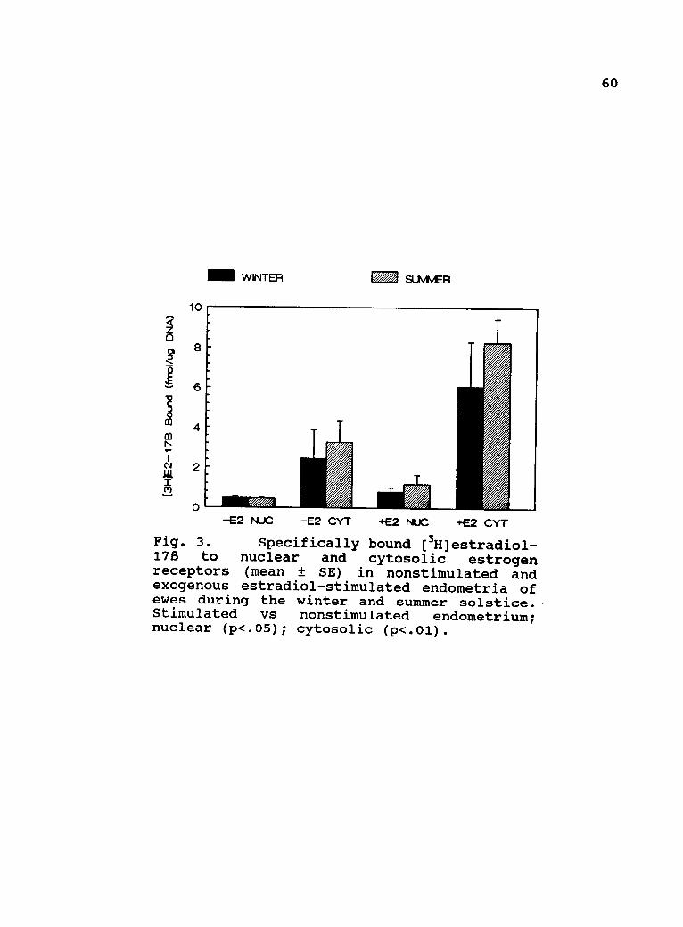
60
ill WINTER SUMMER
-E2 NUC -E2 CYT +E2 NUC +E2 CYT
Fig. 3. Specifically bound Offlestradiol-178 to nuclear and cytosolic estrogenreceptors (mean ± SE) in nonstimulated andexogenous estradiol-stimulated endometria ofewes during the winter and summer solstice.Stimulated vs nonstimulated endometrium;nuclear (p<.05); cytosolic (p<.01).

61
DISCUSSION
Unlike the rabbit uterus, which becomes refractory to
ovarian steroids when the interval between ovariectomy and
hormone replacement is prolonged (Daniel, 1984), results of
this experiment demonstrate that the uterus of the ewe is
responsive to exogenous steroids as long as 9 mo after
ovariectomy. This is reflected by the fact that injection of
10 Ag of estradio1-1713 directly into the uterine lumen at 3
and 9 mo after ovariectomy induced increases in endometrial
estrogen receptors compared with concentrations in control
tissue.
No differences in concentrations of estrogen receptor
were observed between either the estrogen-treated or control
uterine horns of ewes during winter and summer. These data
indicate that there is no nongonadal seasonal effect on
estrogen receptor concentrations in ovariectomized ewes. From
these data, it does not appear that either prolactin (during
summer anestrus), or melatonin (during the fall breeding
season) are able to directly or indirectly alter the
concentration of uterine estrogen receptors in ovariectomized
ewes. This observation is in contrast to those reported by
Danforth et al. (1983) who found that melatonin was able to
induce an increase in estrogen receptor concentrations in
immature hamster uteri in vivo and in vitro. Direct in vitro

62
regulation of estrogen receptor activity by melatonin has also
been shown to occur in human breast cancer cells (Danforth and
Lippman, 1981). Chilton and Daniel (1987) demonstrated that
prolactin treatment of ovariectomized rabbits resulted in an
increase in uterine concentrations of estrogen and
progesterone receptors similar to those detected in the
sexually receptive intact rabbit. Serum concentrations of
estradiol both prior to and after estradiol treatment were not
different from one another indicating that treatment with
estradiol did not increase systemic concentrations of
estradiol and therefore did not likely provoke a release of
prolactin. Serum concentrations of progesterone detected in
the ovariectomized ewes undoubtedly reflect steroid secreted
from the adrenal cortex. Because there was no significant
difference in serum progesterone concentrations among ewes
within season (0 vs 10 Ag estradiol) or between seasons
(winter vs summer) it is unlikely that any slight variation
in progesterone secretion could have accounted for failure to
detect differences in endometrial estrogen receptor
concentrations.
These data indicate that the mechanism by which
seasonality regulates the breeding season is via an indirect
route through the ovary. The secretion of estrogen from the
ovary acts on the uterine endometrium to stimulate the
synthesis of estrogen and progesterone receptors. It appears
that melatonin secreted in greater quantities during the
63
breeding season of the ewe is acting at the level of the
hypothalamus to regulate release of GnRH and subsequently
pituitary LH. Likewise, increased daily secretion of
prolactin during summer anestrus does not appear to act
directly on the uterus to influence synthesis or
concentrations of estrogen receptors and thereby promote this
reproductive state. Collectively, these data indicate that
neither prolactin nor melatonin act directly on the uterine
endometrium to cause fluctuations in the synthesis of estrogen
receptors in the ovariectomized ewe.

64
BIBLIOGRAPHY
Abdelgadir, S.E., Swanson, L.V., Oldfield, J.E. and Stormshak,F. 1987. Prostaglandin Fla- induced release of oxytocinfrom bovine corpora lutea in vitro. Biol. Reprod. 37:550.
Advis, J., Richards, J.S. and Ojeda, S.R. 1981.Hyperprolactinemia-induced precocious puberty: Studieson the mechanism(s) by which prolactin enhances ovarianprogesterone responsiveness to gonadotropins inprepubertal rats. Endocrinology 108:1333.
Aitken, R.J. 1979. The hormonal control of implantation. In:Maternal Recognition of Pregnancy. Ciba Found. Symp., No.64. Excerpta Medica, Amsterdam. p.53.
Aitken, R.J., Bowman, P. and Gauld, I. 1977. The effect ofsynchronous and asynchronous egg transfer on fetal weightin mice selected for large and small body size. J.
Embryol. Exp. Morphol. 37:59.
Anthony, R.V., Helmer, S.D., Sharif, S.F., Roberts, R.M.,Hansen, P.J., Thatcher, W.W. and Bazer, F.W. 1988.Synthesis and processing of ovine trophoblast protein-1and bovine trophoblast protein-1, conceptus secretoryproteins involved in the maternal recognition ofpregnancy. Endocrinology 123:1274.
Astwood, E.B. 1938. A six hour assay for the quantitativedetermination of estrogen. Endocrinology 23:25.
Auletta, F.J. and Flint, A.P.F. 1988. Mechanisms controllingcorpus luteum function in sheep, cows, nonhuman primates,and women especially in relation to the time ofluteolysis. Endocrine Rev. 9:88.
Avvakumov, G.V., Survilo, L.I., Strel'chenok, O.A. 1986.Interaction of serum sex steroid-binding globulin withcell membranes of human decidual tissue. Biochem. Acad.Sci. U.S.S.R. 50:983.
Baird, D.T. 1977. Evidence in vivo for the two-cell hypothesisof oestrogen synthesis by the sheep Graafian follicle.J. Reprod. Fertil. 50:183.
Baird, D.T. 1978. Local utero-ovarian relationships. In:Crighton, D.B., Haynes, G.R., Foxcroft, G.R. and Lamming,G.E. (Eds.) Control of Ovulation. Butterworths, London.pp. 217-232.

65
Baird, D.T., Land, R.B., Sacaramuzzi, R.J. and Wheeler, A.G.
1976. Endocrine changes associated with luteal regression
in the ewe: The secretion of ovarian oestradiol,
progesterone, androstenedione and uterine prostaglandinFez throughout the oestrous cycle. J. Endocrinol. 69:275.
Barley, D.A., Butcher, R.L. and Inskeep, E.K. 1966. Local
nature of utero-ovarian relationships in the
pseudopregnant rat. Endocrinology. 79:119.
Bazer, F.W. and First, N.L. 1983. Pregnancy and parturition.
J. Anim. Sci. 57(Supp1.2):425.
Bazer, F.W., Geisert, R.D., Thatcher, W.W. and Roberts, R.M.1982. The establishment and maintenance of pregnancy. In:
Cole, D.S.A. and Foxcroft, G.R. (Eds.) Control of PigReproduction. Butterworth Scientific, London.
Berthois, Y., Pourreau-Schneider, N., Gandilhon, P., Mittre,
H. and Tubiana, N. 1986. Estradiol membrane binding sites
on human breast cancer cell lines. Use of a fluorescent
estradiol conjugate to demonstrate plasma membranebinding systems. J. Steroid Biochem. 25:963.
Bex, F., Bartke, A., Goldman, B.D. and Dalterio, S. 1978.
Prolactin, growth hormone, luteinizing hormone receptors
and seasonal changes in testicular activities in thegolden hamster. Endocrinology 103:2069.
Bindon, B.M. 1971. The role of progesterone in implantationin the sheep. Aust. J. Biol. Sci. 24:149.
Bittman, E.L., Kaynard, A.H., Olster, D.H., Robinson, J.E.,
Yellon, S.M. and Karsch, F.J. 1985. Pineal melatoninmediates photoperiodic control of pulsatile luteinizinghormone secretion in the ewe. Neuroendocrinology 40:409.
Bjersing, L. 1967. On the morphology and endocrine functionof granulosa cells in ovarian follicles and corporalutea. Acta Endocrinol. 56(Suppl. 125).
Boyar, R.M., Finkelstein, J.W., Kapen, S. and Hellman, L.
1975. Twenty-four hour prolactin (PRL) secretory patternsduring pregnancy. J. Clin. Endocrinol. Metab. 40:1117.
Brann, D.W., Rao, I.M. and Mahesh, V.B. 1988. Antagonism of
estrogen-induced prolactin release by progesterone. Biol.
Reprod. 39:1067.
Burton, K. 1956. A study of the conditions and mechanism of
the diphenylamine reaction for the colorimetricestimation of deoxyribonucleic acid. Biochem. J. 62:315.

66
Butcher, R.L. 1977. Changes in gonadotropins and steroidsassociated with unilateral ovariectomy in the rat.Endocrinology 101:830.
Buttle, H.L. 1974. Seasonal variation of prolactin in plasmaof male goats. J. Reprod. Fertil. 37:95.
Caffrey, J.L., Fletcher, P.W., Diekman, M.A., O'Callaghan,P.L. and Niswender, G.D. 1979. The activity of ovineluteal cholesterol esterase during several experimentalconditions. Biol. Reprod. 21:601.
Caron, M.G., Goldstein, S., Savard, K. and Marsh, J.M. 1975.Protein kinase stimulation of a reconstituted cholesterolside chain cleavage enzyme system in the bovine corpusluteum. J. Biol. Chem. 250:5137.
Carson, R.S., Findlay, J.K., Burger, H.G. and Trounson, A.O.1979. Gonadotropin receptors of the ovine ovarianfollicle during follicular growth and atresia. Biol.Reprod. 21:75.
Casida, L.E. and Warwick, E.J. 1945. The necessity of thecorpus luteum for maintenance of pregnancy in the ewe.J. Anim. Sci. 4:34.
Chepenik, K.P. and Smith, J.B. 1980. Synthesis ofprostaglandins by mouse embryos. Int. Res. Commun. Syst.Med. Sci. 8:783.
Chilton, B.S. and Daniel, Jr., J.C. 1987. Differences in therabbit uterine response to progesterone as influenced bygrowth hormone or prolactin. J. Reprod. Fertil. 79:581.
Clark, J.H. and Markaverich, B.M. 1988. Actions of ovariansteroid hormones. In: Knobil, E., Neill, J.D., Ewing,L.L., Greenwald, G.S., Markert, C.L. and Pfaff, D.W.(Eds.) The Physiology of Reproduction. Raven Press, NewYork. pp. 675-726.
Clark, J.R. 1980. Physiological problems of seasonal breedingin eutherian animals. Oxford Rev. Reprod. Biol. 2:244.
Cox, R.I., Mattner, P.E. and Thorburn, G.D. 1971. Changes inovarian secretion of oestradiol -178 around oestrus in thesheep. J. Endocrinol. 49:345.
Cumming, I.A., Baxter, R. and Lawson, R.A.S. 1974. Steroidhormone requirement for the maintenance of earlypregnancy in sheep : A study using ovariectomized,adrenalectomized ewes. J. Reprod. Fertil. 40:443.

67
Dalton, D.L. and Knight, J.W. 1983. Effects of exogenousprogesterone and estrone on conceptus development in
swine. J. Anim. Sci. 56:1354.
Danforth, Jr., D.N., Tamarkin, L., Do, R. and Lippman, M.E.1983. Melatonin-induced increase in cytoplasmic estrogenreceptor activity in hamster uteri. Endocrinology 113:81.
Danforth, Jr., D.N. and Lippman, M.E. 1981. Melatonin-inducedincrease in estrogen receptor concentration in MCF-7human breast cancer cells. Abstr. 63rd Annu. Mtg.Endocrine Soc. p. 247, Abstr. 660.
Daniel, J.C., Jetton, A.E. and Chilton, B.S. 1984. Prolactinas a factor in the uterine response to progesterone inrabbits. J. Reprod. Fertil. 72:443.
DeHertogh, R., Ekka, E. and Vanderheyden, I. 1986. Estrogendependent induction of low affinity binding sites in thenuclear fraction of rat uterus. J. Steroid Biochem.24:1171.
Denamur, R., Martinet, J. and Short, R.V. 1973. Pituitarycontrol of the ovine corpus luteum. J. Reprod.Fertil.32:207.
Djiane, J. and Durand, P. 1977. Prolactin-progesteroneantagonism in self regulation of prolactin receptors inthe mammary gland. Nature 266:641.
Donaldson, L.E., Hansel, W. and van Vleck, L.D. 1965.Histological study of bovine corpora lutea. J. DairySci. 48:331.
Dorrington, J.H. and Armstrong, D.T. 1979. Effects of FSH ongonadal function. Rec. Prog. Horm. Res. 35:301.
Dorrington, J. and Gore-Langton, R.E. 1981. Prolactin inhibitsoestrogen synthesis in the ovary. Nature 290:600.
du Mesnil du Buisson, F. 1961. Regression unilaterale descorps jaunes apres hysterectomie partielle chez la truie.Ann. Biol. Anim. Biochem. Biophys. 1:105.
Ekka, E., Vanderheyden, I., Glorieux, B. and DeHertogh, R.1987. Estradiol-induced progesterone receptor synthesisin normal and diabetic ovariectomized rat uterus. J.
Steroid Biochem. 28:61.
Eley, R.M., Thatcher, W.W. and Bazer, F.W. 1979. Hormonal andphysical changes associated with bovine conceptusdevelopment. J. Reprod. Fertil. 55:181.
68
Ellinwood, W.E. 1978. Maternal recognition of pregnancy in theewe and the rabbit. Ph.D Dissertation. Colorado StateUniv., Fort Collins.
Espey, L.L. 1980. Ovulation as an inflammatory reaction--ahypothesis. Biol. Reprod. 22:73.
Evans, G., Dobias, M., King, G.J. and Armstrong, D.T. 1981.Estrogen, androgen and progesterone biosynthesis by theca
and granulosa of preovulatory follicles in the pig. Biol.
Reprod. 25:673.
Falck, B. 1959. Site of production of oestrogen in the ratovary as studied by microimplants. Acta Physiol. Scand.
47:94.
Farin, C.E., Imakawa, K., Hansen, T.R., McDonnell, J.J.,
Murphy, C.N., Farin, P.W. and Roberts, R.M. 1990.
Expression of trophoblastic interferon genes in sheep and
cattle. Biol. Reprod. 43:210.
Fincher, K.B., Bazer, F.W., Hansen, P.J., Thatcher, W.W. andRoberts, R.M. 1986. Ovine conceptus secretory proteinssuppress induction of uterine prostaglandin Fla releaseby oestradiol and oxytocin. J. Reprod. Fertil. 76:425.
Fischer, H.E., Bazer, F.W. and Fields, M.J. 1985. Steroidmetabolism by endometrial and conceptus tissues duringearly pregnancy and pseudopregnancy in gilts. J. Reprod.
Fertil. 75:69.
Fischer, T.V. 1965. Local uterine inhibition of the corpusluteum in the guinea pig. Anat. Rec. 151:350.
Flint, A.P.F. and Sheldrick, E.L. 1986. Ovarian oxytocin andthe maternal recognition of pregnancy. J. Reprod. Fertil.
76:831.
Ford, S.P. 1982. Control of uterine and ovarian blood flowthroughout the estrous cycle and pregnancy of the ewe,
sow and cow. J. Anim. Sci. 55(Suppl. 2):32.
Ford, S.P., Weems, C.W., Pitts, R.E., Pexton, J.E., Butcher,R.L. and Inskeep, E.K. 1975. Effects of estradiol -178 andprogesterone on prostaglandin F in sheep uteri anduterine venous plasma. J. Anim. Sci. 41:1407.
Fortune, J.E. and Armstrong, D.T. 1978. Hormonal control of178-estradiol biosynthesis in proestrous rat follicles:Estradiol production by isolated theca versus granulosa.
Endocrinology 102:227.

69
Foster, D.L., Lemons, J.A., Jaffe, R.B. and Niswender, G.D.1975. Sequential patterns of circulating luteinizinghormone and follicle-stimulating hormone in female sheepfrom early postnatal life through the first estrouscycle. Endocrinology 97:985.
Gadsby, J.E., Heap, R.B. and Burton, R.D. 1980. Oestrogenproduction by blastocyst and early embryonic tissue ofvarious species. J. Reprod. Fertil. 60:409.
Gardner, M.L., First, N.L. and Casida, L.E. 1963. Effect ofexogenous estrogen on corpus luteum maintenance in gilts.J. Anim. Sci. 22:132.
George, F.W. and Wilson, J.D. 1978. Estrogen formation in theearly rabbit embryo. Science 199:200.
Ginther, O.J. 1979. Reproductive Biology of the Mare: Basicand Applied Aspects. McNaughton and Gunn, Inc. Ann Arbor,MI.
Ginther, O.J. and First, N.L. 1971. Maintenance of the corpusluteum in hysterectomized mares. Am. J. Vet. Res.32:1687.
Glass, J.D. and Lynch, G.R. 1983. Melatonin: Identificationof sites of antigonadal action in mouse brain. Science.214:821.
Glencross, R.G., Munro, I.B., Senior, B.E. and Pope, G.S.1973. Concentrations of estradio1-1713, oestrone andprogesterone in jugular venous plasma of cows during theoestrous cycle and in early pregnancy. Acta Endocrinol.73:374.
Goding, J.R. 1974. The demonstration that PGF2a is the uterineluteolysin in the ewe. J. Reprod. Fertil. 38:261.
Godkin, J.D., Bazer, F.W. and Roberts, R.M. 1984a. Ovinetrophoblast protein-1, an early secreted blastocystprotein, binds specifically to uterine endometrium andaffects protein synthesis. Endocrinology 114:120.
Godkin, J.D., Bazer, F.W., Thatcher, W.W. and Roberts, R.M.1984b. Proteins released by cultured day 15-16conceptuses prolong luteal maintenance when introducedinto the uterine lumen of cyclic ewes. J. Reprod. Fertil.71:57.

70
Goodman, R.L, Bittman, E.L., Foster, D.L. and Karsch, F.J.1982. Alterations in the control of luteinizing hormone-pulse frequency underlie the seasonal variation in
estradiol negative feedback in the ewe. Biol. Reprod.
27:580.
Gorski, J., Toft, D., Shyamala, G., Smith, D. and Notides, A.
1968. Hormone receptors: Studies on the interaction ofestrogen with the uterus. Rec. Prog. Horm. Res. 24:45.
Greene, G.L. and Press, M.F. 1986. 1. Steroid receptor
structures (including monoclonal antibodies and newmethods of determination). J. Steroid Biochem. 24:1.
Greenwald, G.S. and Rothchild, I. 1968. Formation and
maintenance of corpora lutea in laboratory animals. J.Anim. Sci. 27(Suppl. 1):139.
Greiss, Jr., F.C., and Anderson, S.G. 1970. Effect of ovarianhormones on the uterine vascular bed. Am. J. Obstet.
Gynecol. 107:829.
Grota, L.J. and Eik-Nes, K.B. 1967. Plasma progesteroneconcentrations during pregnancy and lactation in the rat.
J. Reprod. Fertil. 13:83.
Gwinner, E. and Dorka, V. 1976. Endogenous control of annualreproductive rhythms in birds. In: Proc. 16th Int.
Ornithol. Cong. Austr. Acad. of Sci., Canberra. pp.223.
Hafs, H.D. and Armstrong, D.T. 1968. Corpus luteum growth andprogesterone synthesis during the bovine estrous cycle.
J. Anim. Sci. 27:134.
Halkerston, I.D.K., Eichhorn, J., Feinstein, M., Scully, E.
and Hechter, 0. 1960. Early permeability effects ofestradiol on castrate rat uterus. Proc. Soc. Exp. Biol.
Med. 103:796.
Hansel, W., Concannon, P.W. and Lukaszewska, J.H. 1973.
Corpora lutea of the large domestic animals. Biol.
Reprod. 8:222.
Hansel, W. and Convey, E.M. 1983. Physiology of the estrouscycle. J. Anim. Sci. 57(Suppl. 2):404.
Harper, M.J.K., Norris, C.J. and Rajkumar, V. 1983.
Prostaglandin release by zygotes and endometria of
pregnant rabbits. Biol. Reprod. 28:350.
71
Hauger, R.L., Karsch, F.J. and Foster, D.L. 1977. A newconcept for control of the estrous cycle of the ewe based
on the temporal relationships between luteinizinghormone, estradiol and progesterone in peripheral serumand evidence that progesterone inhibits tonic LH
secretion. Endocrinology 101:807.
Hisaw, Jr., F.L. 1959. Comparative effectiveness of estrogenson fluid imbibition and growth of the rat's uterus.Endocrinology 64:276.
Hixon, J.E. and Clegg, M.T. 1969. Influence of the pituitaryon ovarian progesterone output in the ewe: Effects ofhypophysectomy and gonadotropic hormones. Endocrinology84:828.
Holt, J.A., Richards, J.S., Midgley, Jr., A.R. and ReichertJr., L.E. 1976. Effect of prolactin on LH receptor in ratluteal cells. Endocrinology. 98:1005.
Hotchkiss, J., Atkinson, L.E. and Knobil, E. 1971. Time course
of serum estrogen and luteinizing hormone (LH)
concentrations during the menstrual cycle of the rhesusmonkey. Endocrinology 89:177.
Hsueh, A.J.W., Peck, E.J. and Clark, J.H. 1976. Control ofuterine estrogen receptor levels by progesterone.Endocrinology 98:438.
Huhtaniemi, I.T. and Catt, K.J. 1981. Induction and
maintenance of gonadotropin and lactogen receptors inhypoprolactinemic rats. Endocrinology 109:483.
Hyland, J.H., Manns, J.G. and Humphery, W. D. 1982.Prostaglandin production by ovine embryos and endometriumin vitro. J. Reprod. Fertil. 65:299.
Ichii, S., Forchielli, E. and Dorfman, R.I. 1963. In vitroeffect of gonadotropins on the soluble cholesterol sidechain cleaving enzyme system of bovine corpus luteum.
Steroids 2:631.
Inskeep, E.K. and Butcher, R.L. 1966. Local component ofutero-ovarian relationships in the ewe. J. Anim. Sci.
25:1164.
Ireland, J.J. 1987. Control of follicular growth and
development. J. Reprod. Fertil. Suppl. 34:39.
72
Jensen, E.V., Suzuki, T., Kawashima, T., Stumpf, W.E.,Jungblut, P.W. and DeSombre, E.R. 1968. A two-stepmechanism for the interaction of estradiol with ratuterus. Biochemistry 59:632.
Kaltenbach, C.C., Graber, J.W., Niswender, G.D. and Nalbandov,A.V. 1968. Effect of hypophysectomy on the formation andmaintenance of corpora lutea in the ewe. Endocrinology82:753.
Karsch, F.J. 1987. Central actions of ovarian steroids in thefeedback regulation of pulsatile secretion of luteinizinghormone. Ann. Rev. Physiol. 49:365.
Karsch, F.J., Bittman, E.L., Foster, D.L., Goodman, R.L.,Legan, S.J. and Robinson, J.E. 1984. Neuroendocrine basisof seasonal reproduction. Rec. Prog. Horm. Res. 40:185.
Karsch, F.J., Cook, B., Ellicott, A.R., Foster, D.L., Jackson,G.L. and Nalbandov, A.V. 1971. Failure of infusedprolactin to prolong the life span of the corpus luteumof the ewe. Endocrinology 89:272.
Karsch, F.J., Foster, D.L., Legan, S.J., Ryan, K.D. and Peter,G.K. 1979. Control of the preovulatory endocrine eventsin the ewe: Interrelationship of estradiol, progesteroneand luteinizing hormone. Endocrinology 105:421.
Karsch, F.J., Legan, S.J., Ryan, K.D. and Foster, D.L. 1980.Importance of estradiol and progesterone in regulatingLH secretion and estrous behavior during the sheepestrous cycle. Biol. Reprod. 23:404.
Kennaway, D.J., Dunstan, E.A., Gilmore, T.A. and Seamark, R.F.1984. Effects of pinealectomy, oestradiol and melatoninon plasma prolactin and LH secretion in ovariectomizedsheep. J. Endocrinol. 102:199.
King, W.J. and Greene, G.L. 1984. Monoclonal antibodieslocalize oestrogen receptor in the nuclei of targetcells. Nature 307:745.
Klopper, A. 1983. Specific pregnancy proteins. In: Martin, L.and James, V.H.T. (Eds.) The Endocrinology of Pregnancyand Parturition. Academic Press, New York. p. 127.
Knickerbocker, J.J., Thatcher, W.W., Bazer, F.W., Drost, M.,Barron, D.H., Fincher, K.B. and Roberts, R.M. 1986.Proteins secreted by day 16 to 18 bovine conceptusesextend corpus luteum function in cows. J. Reprod. Fertil.77:381.

73
Knobil, E. 1973. On the regulation of the primate corpusluteum. Biol. Reprod. 8:246.
Koligian, K.B. and Stormshak, F. 1977a. Nuclear and
cytoplasmic estrogen receptors in ovine endometriumduring the estrous cycle. Endocrinology 101:524.
Koligian, K.B. and Stormshak, F. 1977b. Progesteroneinhibition of estrogen receptor replenishment in ovineendometrium. Biol. Reprod. 17:412.
Lemon, M. and Loir, M. 1977. Steroid release in vitro by twoluteal cell types in the corpus luteum of the pregnantsow. J. Endocrinol. 72:351.
Leung, P.C.K. and Armstrong, D.T. 1980. Interactions of
steroids and gonadotropins in the control of
steroidogenesis in the ovarian follicle. Ann. Rev.
Physiol. 42:71.
Ling, W.Y. and Marsh, J.M. 1977. Reevaluation of the role ofcyclic adenosine 3',5'-monophosphate and protein kinasein the stimulation of steroidogenesis by luteinizinghormone in bovine corpus luteum slices. Endocrinology
100:1571.
Liu, Y.-X. and Hsueh, A.J.W. 1986. Synergism between granulosa
and theca-interstitial cells in estrogen biosynthesis bygonadotropin-treated rat ovaries: Studies on the two-celltwo-gonadotropin hypothesis using steroid antisera. Biol.
Reprod. 35:27.
Mais, V., Kazer, R.R., Cetel, N.S., Rivier, J., Vale, W. andYen, S.S.C. 1986. The dependency of folliculogenesis and
corpus luteum function on pulsatile gonadotropinsecretion in cycling women using a gonadotropin-releasinghormone antagonist as a probe. J. Clin. Endocrinol.
Metab. 62:1250.
Marcus, G.J. 1981. Prostaglandin formation by the sheep embryoand endometrium as an indication of maternal recognitionof pregnancy. Biol. Reprod. 25:56.
Marsh, J.M. 1975. The role of cyclic AMP in steroidogenesis.Biol. Reprod. 14:30.
Martal, J.M., LaCroix, M.C., Loudis, C., Saunier, M. and
Winterberger-Torres, S. 1979. Trophoblastin, an
antiluteolytic protein present in early pregnant sheep.
J. Reprod. Fertil. 56:63.
74
Martin, L., Hallowes, R.C., Finn, C.A. and West, D.G. 1973.Involvement of the uterine blood vessels in therefractory state of the uterine stroma which followsoestrogen stimulation in progesterone treated mice. J.Endocrinol. 56:309.
Maywood, E.S., Buttery, R.C., Vance, G.H.S., Herbert, J. andHastings, M.H. 1990. Gonadal responses of the male Syrianhamster to programmed infusions of melatonin aresensitive to signal duration and frequency but not tosignal phase nor to lesions of the suprachiasmaticnuclei. Biol. Reprod. 43:174.
McCracken, J.A., Glew, M.E. and Scaramuzzi, R.J. 1970. Corpusluteum regression induced by prostaglandin F2 a. J. Clin.Endocrinol. Metab. 30:544.
Mercer, W.D., Edwards, D.P., Chamness, G.C. and McGuire, W.L.1981. Failure of estradiol immunofluorescence in MCF-7breast cancer cells to detect estrogen receptors. CancerRes. 41:4644.
Miller, B.G. and Moore, N.W. 1976. Effects of progesterone andoestradiol on RNA and protein metabolism in the genitaltract and on survival of embryos in the ovariectomizedewe. Aust. J. Biol. Sci. 29:565.
Miller, M.A., Mullick, A., Greene, G.L. and Katzenellenbogen,B.S. 1985. Characterization of the subunit nature ofnuclear estrogen receptors by chemical cross linking anddense amino acid labeling. Endocrinology 117:515.
Miller, B.G., Murphy, L. and Stone, G.M. 1977. Hormonereceptor levels and hormone, RNA and protein metabolismin the genital tract during the oestrous cycle of theewe. J. Endocrinol. 73:91.
Miller, W.R. and Turner, C.W. 1963. Biosynthesis of steroidsfrom pregnenolone-16-3H by bovine ovarian tissue in
vitro. Steroids. 2:657.
Mitchell, M.D., Flint, A.P.F. and Turnbull, A.C. 1975.Stimulation by oxytocin of prostaglandin F levels inuterine venous effluent in pregnant and puerperal sheep.Prostaglandins 9:47.
Moor, R.M. 1977. Sites of steroid production in ovine Graafianfollicles in culture. J. Endocrinol. 73:143.
Moor, R.M. and Rowson, L.E.A. 1959. Maintenance of pregnancyin ovariectomized ewes by means of progesterone. Nature184:1410.

75
Moor, R.M. and Rowson, L.E.A. 1966a. Local maintenance of thecorpus luteum in sheep with embryos transferred tovarious isolated portions of the uterus. J. Reprod.Fertil. 12:539.
Moor, R.M. and Rowson, L.E.A. 1966b. The corpus luteum of thesheep: Functional relationships between the embryo andthe corpus luteum. J. Endocrinol. 34:233.
Moore, R.Y. and Klein, D.C. 1974. Visual pathways and thecentral neural control of a circadian rhythm in pinealserotonin N-acetyltransferase activity. Brain Res. 71:17.
Moudgal, N.R., Macdonald, G.J. and Greep, R.O. 1971. Effectof hCG antiserum on ovulation and corpus luteum functionin the monkey. J. Clin. Endocrinol. Metab. 32:579.
Mueller, G.C., Herranen, A.M. and Jervell, K.F. 1957. Studieson the mechanism of action of estrogens. Rec. Prog. Horm.Res. 14:95.
Muldoon, T.G. 1981. Interplay between estradiol and prolactinin the regulation of steroid hormone receptor levels,nature, and functionality in normal mouse mammary tissue.Endocrinology 109:1339.
Nalbandov, A.V. 1971. Endocrine control of implantation. In:Blandau, R.J. (Ed.) The Biology of the Blastocyst.University of Chicago Press, Chicago. Chapter 22.
Neill, J.D. 1974. Prolactin: Its secretion and control.Endocrinology 4:469.
Nicoll, C.S. 1974. Physiological actions of prolactin. In:Greep, R.O. and Astwood, E.B. (Eds.) Handbook ofPhysiology. Am. Physiol. Soc. Washington, D.C. pp. 253-292.
Niswender, G.D., Moore, R.T., Akbar, A.M., Nett, T.M. andDiekman, M.A. 1975. Flow of blood to the ovaries ofcycling and pregnant ewes. Biol. Reprod. 13:38.
Niswender, G.D. and Nett, T.M. 1988. The corpus luteum andits control. In: Knobil, E., Neill, J.D. Ewing, L.L.,Greenwald, G.S., Markert, C.L. and Pfaff, D.W. (Eds.) ThePhysiology of Reproduction. Raven Press, New York. pp.489-525.
Niswender, G.D., Reimers, T.J., Diekman, M.A. and Nett, T.M.1976. Blood flow: A mediator of ovarian function. Biol.Reprod. 14:64.
76
Niswender, G.D., Schwall, R.H., Fritz, T.A., Farin, C.E. andSawyer, H.R. 1985. Regulation of luteal function indomestic ruminants: New concepts. Rec. Prog. Horm. Res.41:101.
Norman, A.W. and Litwack, G.L. 1987. Hormones. Academic Press,San Diego. pp. 101-130.
Northey, D.L. and French, L.R. 1980. Effect of embryo removaland intrauterine infusion of embryonic homogenates on thelifespan of the bovine corpus luteum. J. Anim. Sci.
50:298.
Pant, H.C., Hopkinson, C.R.N. and Fitzpatrick, R.J. 1977.Concentration of oestradiol, progesterone, luteinizinghormone and follicle stimulating hormone in the jugularvenous plasma of ewes during the oestrous cycle. J.
Endocrinol. 73:247.
Parikh, I., Ranjendran, K.G., Su, J.L., Lopez, T. and Sar, M.1987. Are estrogen receptors cytoplasmic or nuclear?Some immunocytochemical and biochemical studies. J.
Steroid Biochem. 27:185.
Pavlik, E.J. and Coulson, P.B. 1976. Modulation of estrogenreceptors in four different target tissues: Differentialeffects of estrogen vs progesterone. J. Steroid. Biochem.7:369.
Perry, J.S., Heap, R.B. and Amoroso, E.C. 1973. Steroidhormone production by pig blastocysts. Nature 245:45.
Peters, R.R. and Tucker, H.A. 1978. Prolactin and growthhormone responses to photoperiod in heifers.Endocrinology 103:229.
Pope, W.F., Maurer, R.R. and Stormshak, F. 1982. Survival ofporcine embryos after asynchronous transfer. Proc. Soc.Exp. Biol. Med. 171:179.
Posner, B.I., Kelly, P.A., Shiu, R.P.0 and Friesen, H.G. 1974.Studies of insulin, growth hormone and prolactin binding;tissue distribution, special variation andcharacterization. Endocrinology 95:521.
Pratt, B.R., Butcher, R.L. and Inskeep, E.K. 1977.Antiluteolytic effect of the conceptus and of PGE2 inewes. J. Anim. Sci. 46:784.
77
Reynolds, L.P., Magness, R.R. and Ford., S.P. 1982. Uterineblood flow (UBF) and uterine arterio-venous steroidconcentration on d 11, 13, and 15 of the estrous cycleand pregnancy in ewes. J. Anim. Sci. 55(Suppl. 1):382.
Riddick, D.H. 1985. Regulation and physiological relevance ofnon-pituitary prolactin. In: MacLeod, R.M., Thomer, M.O.and Scapagrini, U. (Eds.) Prolactin Basic and ClinicalCorrelates. V. Liviana Press, Padova. pp. 463-473.
Roberts, K.D., Bandy, L. and Leiberman, S. 1967. Theconversion of cholesterol-3H-sulfate-35S intopregnenolone-3H-sulfate-35S by sonicated bovine adrenalmitochondria. Biochem. Biophys. Res. Commun. 29:741.
Rodgers, R.J. and O'shea, J.D. 1982. Purification, morphologyand progesterone production and content of three celltypes isolated from the corpus luteum of sheep. Aust. J.
Biol. Sci. 35:441.
Rolland, R., Grusalus, G.L. and Hammond, J.M. 1976.Demonstration of specific binding of prolactin by porcinecorpora lutea. Endocrinology 98:1083.
Rose, J., Oldfield, J.E. and Stormshak, F. 1986. Changes inserum prolactin concentrations and ovarian prolactinreceptors during embryonic diapause in mink. Biol.
Reprod. 34:101.
Rothchild, I. 1981. The regulation of the mammalian corpusluteum. Rec. Prog. Horm. Res. 37:183.
Sabbah, M., Redeuilh, G. and Baulieu, E.E. 1989. Subunitcomposition of the estrogen receptor. Involvement of thehormone-binding domain in the dimeric state. J. Biol.Chem. 264:2397.
Sar, M. and Meites, J. 1968. Effects of suckling on pituitaryrelease of prolactin, GH and TSH in post-partum lactatingrats. Neuroendocrinology 4:25.
Schallenberger, E., Schams, D., Bullermann, B. and Walters,D.L. 1984. Pulsatile secretion of gonadotropins, ovariansteroid and ovarian oxytocin during prostaglandin inducedregression of the corpus luteum in the cow. J. Reprod.Fertil. 71:493.
Schams, D. and Karg, G. 1974. Prolactin release in cattle. J.Reprod. Fertil. 39:463.
78
Schomberg, D.W., Coudert, S.P. and Short, R.V. 1967. Effectsof bovine luteinizing hormone and human chorionicgonadotropin on the bovine corpus luteum in vivo. J.Reprod. Fertil. 14:227.
Schwartz, N.B. and Channing, C.P. 1977. Evidence for ovarian"inhibin" suppression of the secondary rise in serumfollicle stimulating hormone levels in proestrous ratsby injection of porcine follicular fluid. Proc. Natl.Acad. Sci. U.S.A. 74:5721.
Selcer, K.W. and Leavitt, W.W. 1988. Progesterone down-regulation of nuclear estrogen receptor: A fundamentalmechanism in birds and mammals. Gen. Comp. Endocrinol.
72:443.
Senior, B.E. 1975. Cytoplasmic oestradiol-binding sites andtheir relationship to oestradiol content in theendometrium of cattle. J. Reprod. Fertil. 44:501.
Sheldrick, E.L. and Flint, A.P.F. 1983. Regression of thecorpora lutea in sheep in response to cloprostenol is notaffected by loss of luteal oxytocin after hysterectomy.J. Reprod. Fertil. 68:155.
Sheldrick, E.L., Mitchell, M.D. and Flint, A.P.F. 1980.
Delayed luteal regression in ewes immunized againstoxytocin. J. Reprod. Fertil. 59:37.
Shemesh, M., Milaguir, F., Ayalon, N. and Hansel, W. 1979.
Steroidogenesis and prostaglandin synthesis by culturedbovine blastocysts. J. Reprod. Fertil. 56:181.
Short, R.V. 1962. Steroids in the follicular fluid and thecorpus luteum of the mare. A two-cell type theory ofovarian steroid synthesis. J. Endocrinol. 24:59.
Short, R.V. 1969. Implantation and maternal recognition ofpregnancy. In: Wolstenholme, G.E.W. and O'Conner, M.
(Eds.) Fetal Autonomy. Churchill, London. pp. 2-26.
Shutt, D.A., Clark, A.H., Fraser, I.S., Goh, P., McMahon,G.R., Saunders, D.M. and Shearman, R.P. 1976. Changes inconcentration of prostaglandin F and steroids in humancorpora lutea in relation to growth of the corpus luteumand luteolysis. J. Endocrinol. 71:453.
Silvia, W.J., Fitz, T.A., Mayan, M.H. and Niswender, G.D.1984. Cellular and molecular mechanisms involved inluteolysis and maternal recognition of pregnancy in the
ewe. Anim. Reprod. Sci. 7:57.

79
Singh, M.M. and Booth, W.D. 1979. Origin of oestrogen inpreimplantation rabbit blastocysts. J. Steroid Biochem.11:723.
Sirois, J. and Fortune, J.E. 1988. Ovarian follicular dynamicsduring the estrous cycle in heifers monitored by real-time ultrasonography. Biol. Reprod. 39:308.
Slayden, 0. 1990. Endocrine Regulation of Uterine Physiologyin Mink. Ph.D. Dissertation. Oregon State Univ.,Corvallis Oregon.
Smeaton, T.C. and Robertson, H.A. 1971. Studies on the growthand atresia of Graafian follicles in the ovary of thesheep. J. Reprod. Fertil. 25:243.
Stetson, M.H. and Watson-Whitmyre, M. 1976. Nucleus supra-chiasmaticus: The biological clock in hamsters? Science
191:197.
Stewart, H.J., McCann, S.H.E., Barker, P.J., Lee, K.E.,Lamming, G.E. and Flint, A.P.F. 1987. Interferon sequencehomology and receptor binding activity of ovine
trophoblast antiluteolytic protein. J. Endocrinol.115:13.
Stone, G.M., Murphy, L. and Miller, B.G. 1978. Hormonereceptor levels and metabolic activity in the uterus ofthe ewe: Regulation by oestradiol and progesterone. Aust.
J. Biol. Sci. 31:395.
Stormshak, F., Kelley, H.E., and Hawk, H.W. 1969. Suppressionof ovine luteal function by 1713 - estradiol. J. Anim. Sci.
29:476.
Swanson, L.V., Hafs, H.D. and Morrow, D.A. 1972. Ovariancharacteristics and serum LH, prolactin, progesterone andglucocorticoids from first estrus to breeding size in
holstein heifers. J. Anim. Sci. 34:284.
Thorpe, P. and Herbert, J. 1976. Studies on the duration ofthe breeding season and photorefractoriness in femaleferrets pinealectomized or treated with melatonin. J.Endocrinol. 70:255.
Tindal, J.S. 1969. Central pathways involved in prolactinsecretion. In: Reynolds, M. and Folley, S. (Eds.)
Lactogenesis: The Initiation of Milk Secretion atParturition. Univ. of Penn. Press, Philadelphia, PA. pp.253-256.
80
Tsai-Morris, C.-H., Ghosh, M., Hirshfield, A.N., Wise, P.M.and Brodie, A.M.H. 1983. Inhibition of ovarian aromataseby prolactin in vitro. Biol. Reprod. 29:342.
Tsang, B.K. and Ooi, T.C. 1982. Prostaglandin secretion byhuman endometrium in vitro. Am. J. Obstet. Gynecol.142:626.
Turnbull, K.E., Braden, A.W.H. and Mattner, P.E. 1977. Thepattern of follicular growth and atresia in the ovineovary. Aust. J. Biol. Sci. 30:229.
Uresley, J. and Leymarie, P. 1979. Varying response toluteinizing hormone of two cell types isolated frombovine corpus luteum. J. Endocrinol. 83:303.
Vekemans, M. and Robyn, C. 1975. The influence of exogenousestrogen on the circadian periodicity of circulatingprolactin in women. J. Clin. Endocrinol. Metab. 40:886.
Walters, D.L., Schams, D., Bullermann, B. and Schallenberger,E. 1983. Pulsatile secretion of gonadotropins, ovariansteroids and ovarian oxytocin during luteolysis in thecow. Biol. Reprod. 28(Suppl. 1):142.
Wang, C. and Chan, V. 1982. Divergent effects of prolactin onestrogen and progesterone production by granulosa cellsof rat Graafian follicles. Endocrinology 110:1085.
Wang, C., Hsueh, A.J.W. and Erickson, G.F. 1980. Prolactininhibition of estrogen production by cultured ratgranulosa cells. Mol. Cell. Endocrinol. 20:135.
Webster, G.M. and Haresign, W. 1983. Seasonal changes in LHand prolactin concentrations in ewes of two-breeds. J.Reprod. Fertil. 67:465.
Welshons, W.V., Lieberman, M.E. and Gorski, J. 1984. Nuclearlocalization of unoccupied oestrogen receptors. Nature307:747.
West, N.B., Hess, D.L. and Brenner, R.M. 1986. Differentialsuppression of progesterone receptors by progesterone inthe reproductive tract of female macaques. J. SteroidBiochem. 25:497.
Westpahl, U. 1983. Corticosteroid-binding globulin. A reviewof some recent aspects. Mol. Cell. Biochem. 55:145.
81
Wilde, D.E., Culver, A.A., Morcom, C.B. and Dukelow, W.R.1976. Effect of administration of progesterone andestrogen on litter size in pigs. J. Reprod. Fertil.
48:209.
Williams, M.F. 1948. The vascular architecture of the ratuterus as influenced by estrogen and progesterone. Am.J. Anat. 83:247.
Williams, G.H., Hammond, J.M., Weisz, J. and Mortel, R. 1978.Binding sites for lactogenic hormone in the rat uterus.Biol. Reprod. 18:697.
Wu, J.T. 1985. Metabolism of progesterone by hamsterblastocysts and the ontogeny of progesterone metaboliccapability. Biol. Reprod. 33:53.
Yamamoto, K.R. 1985. Steroid receptor regulated transcriptionof specific genes and gene networks. Ann. Rev. Genet.
19:209.
Yen, S.C., Tsai, C.C., Naftolin, F., Vanderberg, G. and
Ajabor, L. 1972. Pulsatile patterns of gonadotropinrelease in subjects with and without ovarian function.J. Clin. Endocrinol. Metab. 34:671.
Zavy, M.T., Mayer, R., Vernon, M.W., Bazer, F.W. and Sharp,
D.C. 1979. An investigation of the uterine luminalenvironment of nonpregnant and pregnant pony mares. J.
Reprod. Fertil. Suppl. 27:403.
Zeleznik, A.J., Midgley, Jr., A.R. and Reichert, Jr., L.E.1974. Granulosa cell maturation in the rat: Increasedbinding of human chorionic gonadotropin followingtreatment with follicle stimulating hormone in vitro.Endocrinology 95:818.
Zelinski, M.B., Hirota, N.A., Keenan, E.J. and Stormshak, F.1980. Influence of exogenous estradiol -178 on endometrialprogesterone and estrogen receptors during the lutealphase of the ovine estrous cycle. Biol. Reprod. 23:743.
Zelinski, M.B., Noel, P., Weber, D.W. and Stormshak, F. 1982.Characterization of cytoplasmic progesterone receptors
in the bovine endometrium during proestrus and diestrus.
J. Anim. Sci. 55:376.
Related Documents


![Round results Notices issued by Ofcom under Wireless ... · Airspan [REDACTED] 34 34 EE [REDACTED] 16 16 H3G [REDACTED] 46 46 Telefonica [REDACTED] 37 37 Vodafone [REDACTED] 40 40](https://static.cupdf.com/doc/110x72/5ba4d8c509d3f257608be079/round-results-notices-issued-by-ofcom-under-wireless-airspan-redacted.jpg)