1 Recovery and Identification of Bacteria from Polar and Non-polar Glacial Ice Brent C. Christner 1 , Ellen Mosley-Thompson 2,3 , Lonnie G. Thompson 3,4 , and John N. Reeve 1* . Departments of Microbiology 1 ,Geography 2 and Geological Sciences 4 , Byrd Polar Research Center 3 , Ohio State University, Columbus, OH 43210-1292, USA. *Tel: 1-614-292-2301 Fax: 1-614-292-8120 E-mail: [email protected] Key words: Ice cores, bacteria, longevity, Lake Vostok INTRODUCTION Snowfall accumulates as glacial ice at both poles, and globally at high-altitudes in non-polar regions. Archived chronologically within these glaciers are samples of the atmospheric constituents at the time of snow deposition including particulates of inorganic and biological origin deposited originally on the surface of the snow, often by attachment to snow flakes. Studies of ice cores have established past climate changes and geological events, both globally and regionally, but rarely have these results been correlated with the insects, plant fragments, seeds, pollen grains, fungal spores and bacteria that also are present, and very few attempts have been made to determine the diversity and longevity of viable species entombed in such glacial ice. Fungi, algae, protists, bacteria and viruses have been

Welcome message from author
This document is posted to help you gain knowledge. Please leave a comment to let me know what you think about it! Share it to your friends and learn new things together.
Transcript

1
Recovery and Identification of Bacteria from Polar and Non-polar Glacial Ice
Brent C. Christner1, Ellen Mosley-Thompson2,3, Lonnie G. Thompson3,4, and John N. Reeve1*.
Departments of Microbiology1,Geography2 and Geological Sciences4, Byrd Polar Research
Center3, Ohio State University, Columbus, OH 43210-1292, USA.
*Tel: 1-614-292-2301
Fax: 1-614-292-8120
E-mail: [email protected]
Key words: Ice cores, bacteria, longevity, Lake Vostok
INTRODUCTION
Snowfall accumulates as glacial ice at both poles, and globally at high-altitudes
in non-polar regions. Archived chronologically within these glaciers are samples of the
atmospheric constituents at the time of snow deposition including particulates of inorganic
and biological origin deposited originally on the surface of the snow, often by attachment to
snow flakes. Studies of ice cores have established past climate changes and geological
events, both globally and regionally, but rarely have these results been correlated with the
insects, plant fragments, seeds, pollen grains, fungal spores and bacteria that also are present,
and very few attempts have been made to determine the diversity and longevity of viable
species entombed in such glacial ice. Fungi, algae, protists, bacteria and viruses have been
2
detected and recovered from polar ice cores (Abyzov et al., 1982, 1998; Abyzov 1993;
Dancer et al. 1997; Castello et al. 1999; Willerslev et al., 1999), but there are very few similar
reports describing the microorganisms preserved in non-polar glacial iceof different age and
from different locations. Fortunately, for such studies, we have access to ice cores archived
at the Byrd Polar Research Center (BPRC) at The Ohio State University. These ice cores
have been collected over many years, from globally-distributed sites, and many have already
been subjected to extensive physical and chemical analyses. These, therefore, provide the
opportunity to isolate and to characterize microorganisms from glacial ice formed at defined
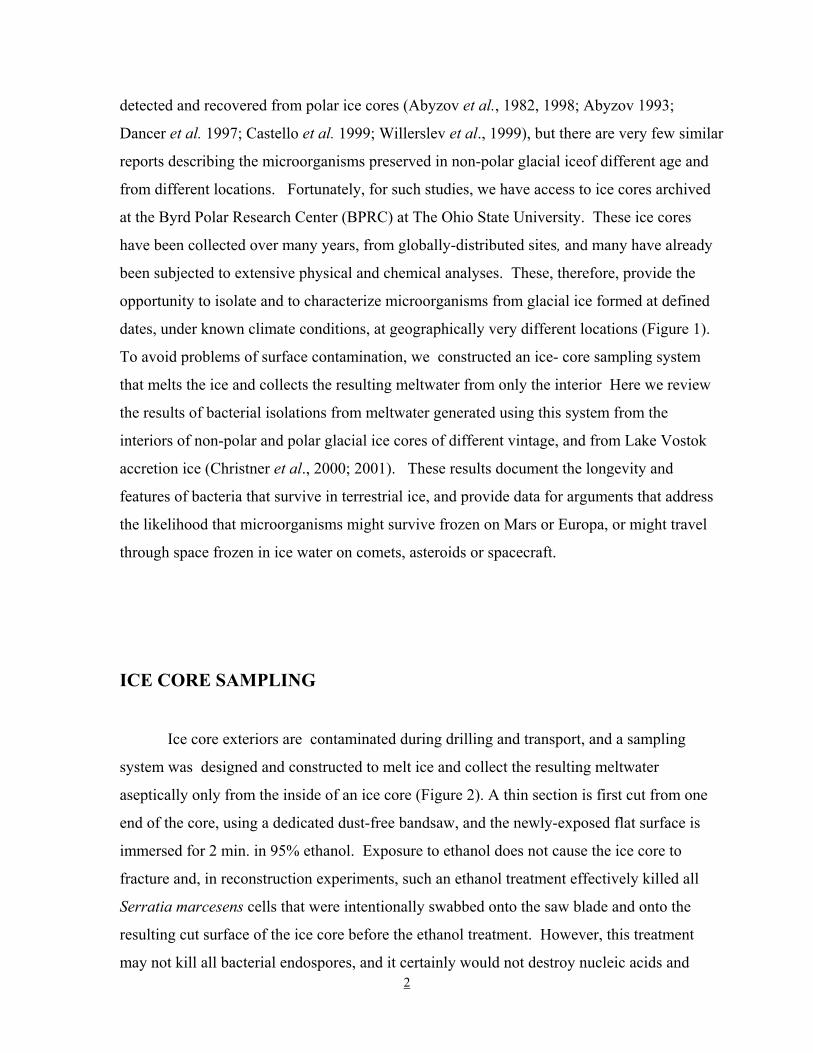
dates, under known climate conditions, at geographically very different locations (Figure 1).
To avoid problems of surface contamination, we constructed an ice- core sampling system
that melts the ice and collects the resulting meltwater from only the interior Here we review
the results of bacterial isolations from meltwater generated using this system from the
interiors of non-polar and polar glacial ice cores of different vintage, and from Lake Vostok
accretion ice (Christner et al., 2000; 2001). These results document the longevity and
features of bacteria that survive in terrestrial ice, and provide data for arguments that address
the likelihood that microorganisms might survive frozen on Mars or Europa, or might travel
through space frozen in ice water on comets, asteroids or spacecraft.
ICE CORE SAMPLING
Ice core exteriors are contaminated during drilling and transport, and a sampling
system was designed and constructed to melt ice and collect the resulting meltwater
aseptically only from the inside of an ice core (Figure 2). A thin section is first cut from one
end of the core, using a dedicated dust-free bandsaw, and the newly-exposed flat surface is
immersed for 2 min. in 95% ethanol. Exposure to ethanol does not cause the ice core to
fracture and, in reconstruction experiments, such an ethanol treatment effectively killed all
Serratia marcesens cells that were intentionally swabbed onto the saw blade and onto the
resulting cut surface of the ice core before the ethanol treatment. However, this treatment
may not kill all bacterial endospores, and it certainly would not destroy nucleic acids and

3
therefore, to monitor for such contamination, the cut surface of each ice core is swabbed after
the ethanol treatment before initiating melting. These swabs are used to inoculate growth
media and are evaluated for the presence of DNA by polymerase chain reaction (PCR)
amplifications using universal 16S rDNA amplification primers. Only very rarely has growth
occurred in a swab-inoculated culture, and no PCR product has yet been generated indicating
that the levels of contamination on the ethanol-treated ice core surfaces are very low, and
below those detectable by standard PCR procedures. Immediately after the exposure to
ethanol, the ice core is positioned vertically in the sampling system with the ethanol-washed
surface placed directly in contact with the sampling unit. The sampling unit is heated
internally and as it melts the ice, it moves upwards through the ice core. The water generated
passes through an orifice in the center of the sampling unit and is collected aseptically into
sterile containers positioned outside the sampling system (Figure 2).
BACTERIA RECOVERED FROM GLACIAL ICE
The numbers and identities of bacteria that form colonies when meltwater is plated
directly on solid media have been determined in ice from Sajama (Bolivia), Guliya (China),
Greenland, and Antarctica. In general, meltwaters from non-polar, low-latitude, high-altitude
glaciers contain greater number, as well as more diversity of colony-forming bacteria than
melt waters from polar ice cores. For example, 180 colony- forming units per ml (cfu/ml)
were present in melt water from a 200-year old sample of Guliya ice whereas water from an
~1,800-year old sample of polar ice from Taylor Dome (Antarctica) contained only ~10
cfu/ml. Even fewer cfus were present in meltwater from ice of a similar vintage from the
Antarctic Peninsula and from the Summit and Dye 2 sites in Greenland. It is important to
note that differences in the amount of annual snowfall, and in the subsequent rates of
compression mean that equal volumes of meltwater from different cores do not necessarily
represent equivalent time periods of microbial deposition. However, these results are
consistent with those of Dancer et al. (1997) who recovered <5 cfu/ml from glacial ice from
the Canadian high arctic after enrichment for coliform bacteria, and other reports of
recovering even fewer bacteria (<1 cfu/ml) from melt waters from polar ice (Abyzov et al.
1982; Hardfield et al. 1992). Logically, these differences arise because non-polar glaciers are

4
closer to major sources of airborne microorganisms such as exposed soils, tropical and sub-
tropical ecosystems. Consistent with this, meltwater from ice from a Taylor Dome site
located at the head of the Taylor Valley in the dry valley complex of Antarctica contained
relatively larger numbers of culturable bacteria (~10 cfu/ml), and microbiological surveys
have documented the abundance of bacteria, fungi and algae in this area despite the very dry
and cold climate (Priscu et al., 1998; Brambilla et al., 2001).
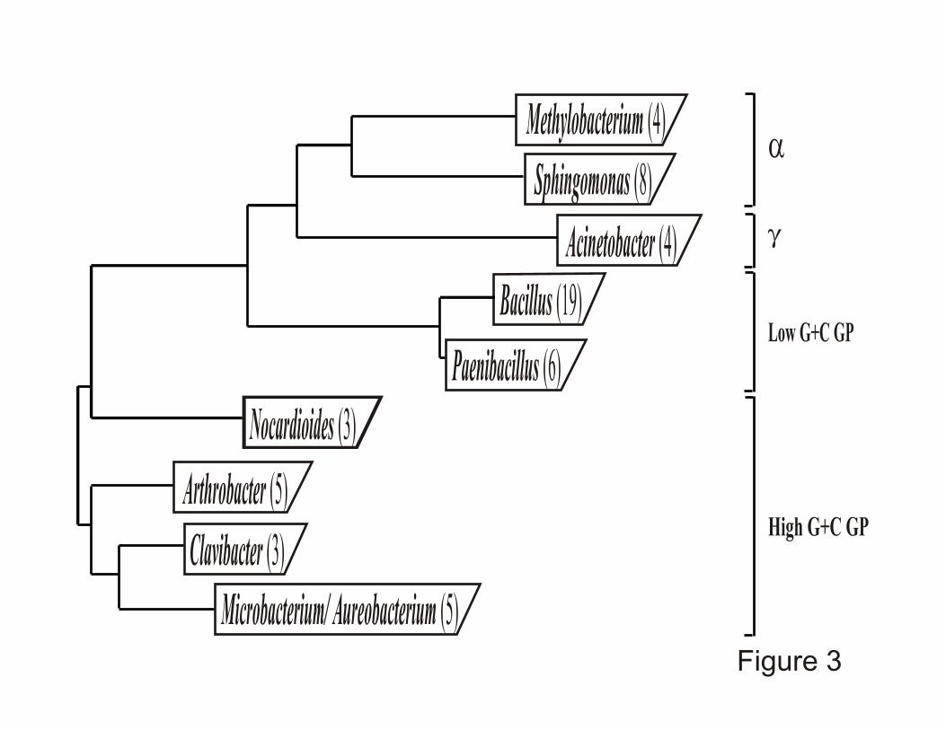
Based on their small-subunit ribosomal RNA encoding sequences (16S rDNAs) most
of the ice-core isolates are members of the non-sporulating Gram-positive, spore-forming
Bacillus, Paenibacillus and Actinobacterias, "- and (-proteobacterial lineages (Figure 3).
Many form colored colonies, consistent with pigment production providing protection from
solar irradiation during airborne transport and subsequent exposure on the glacier surface.
Isolates with 16S rDNA sequences >95% similar to members of the bacterial genera
Acinetobacter, Arthrobacter, Aureobacterium, Bacillus, Cellulomonas, Clavibacter,
Methylobacterium, Microbacterium, Nocardioides, Paenibacillus, and Sphingomonas have
been routinely recovered from both polar and non-polar glacial ices, and based on having 16S
rDNA sequences >98% similar to the 16S rDNA sequences of the type strain, members of the
following bacterial species have been isolated: Acinetobacter radioresistans, Arthrobacter
agilis, Bacillus macroides, Bacillus subtilis, Bacillus thuringiensis, Clavibacter michiganensis
and Sphingomonas paucimobilis.
ISOLATION OF BACTERIA FROM VERY OLD GLACIAL ICE
An ice core that extends over 300 meters below the surface (mbs), to the underlying
bedrock was obtained from the Guliya Ice Cap in Tibet (Figure 1), and based on the
abundance of 36Cl (half life = 301,000 years) the ice at the bottom of this core is >500,000
years old (Thompson et al. 1997). This is the oldest glacial ice recovered to date and provides
an opportunity to evaluate microbial survival in ice on a time scale potentially meaningful for
inter-planetary transport. Aliquots of meltwater from this ice core from 296 mbs were
inoculated into a variety of growth media and, after 30-60 days of aerobic incubation at 4oC,
growth was observed in very dilute nutrient and tryptic soy broths. These media were used at
5
1% of the concentration recommended by the manufacturer (Difco, Inc.). Despite the long
period needed for initial growth, and the primary enrichment cultures being grown under
oligotrophic conditions at 4oC, isolates were subsequently obtained from these cultures that
grew and formed colonies in 2-7 days on nutrient-rich media at 25oC. Long-dormant cells
must eliminate toxic metabolites, such as hydrogen peroxide, superoxide and free radicals,
and repair macromolecular damage that has accumulated before they can grow and divide
successfully (Dodd et al., 1997). The results with the very old Guliya ice are consistent with
this hypothesis, and indicate that successful recovery is facilitated by providing only a very
low level of nutrients initially, sufficient for repair but insufficient to elicit an instant attempt
at growth.
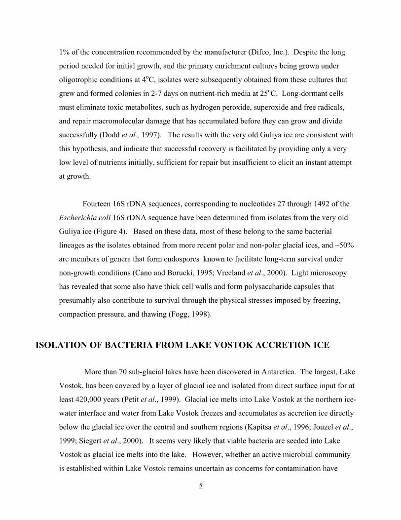
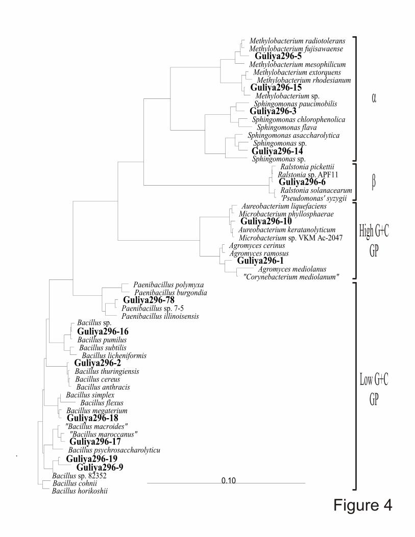
Fourteen 16S rDNA sequences, corresponding to nucleotides 27 through 1492 of the
Escherichia coli 16S rDNA sequence have been determined from isolates from the very old
Guliya ice (Figure 4). Based on these data, most of these belong to the same bacterial
lineages as the isolates obtained from more recent polar and non-polar glacial ices, and ~50%
are members of genera that form endospores known to facilitate long-term survival under
non-growth conditions (Cano and Borucki, 1995; Vreeland et al., 2000). Light microscopy
has revealed that some also have thick cell walls and form polysaccharide capsules that
presumably also contribute to survival through the physical stresses imposed by freezing,
compaction pressure, and thawing (Fogg, 1998).
ISOLATION OF BACTERIA FROM LAKE VOSTOK ACCRETION ICE
More than 70 sub-glacial lakes have been discovered in Antarctica. The largest, Lake
Vostok, has been covered by a layer of glacial ice and isolated from direct surface input for at
least 420,000 years (Petit et al., 1999). Glacial ice melts into Lake Vostok at the northern ice-
water interface and water from Lake Vostok freezes and accumulates as accretion ice directly
below the glacial ice over the central and southern regions (Kapitsa et al., 1996; Jouzel et al.,
1999; Siegert et al., 2000). It seems very likely that viable bacteria are seeded into Lake
Vostok as glacial ice melts into the lake. However, whether an active microbial community
is established within Lake Vostok remains uncertain as concerns for contamination have
6
resulted in a moratorium on direct sampling of Lake Vostok water. Ice core drilling also was
terminated above the ice-water interface although an ice core was retrieved in which the
bottom ~150 meters are accretion ice and therefore represent a sample of Lake Vostok water.
A section of this core from 3591.965 to 3592.445mbs, designated as core section 3593, was
obtained from the National Ice Core Laboratory (Denver, CO), and has been subjected to
microbiological investigation (Christner et al., 2001).
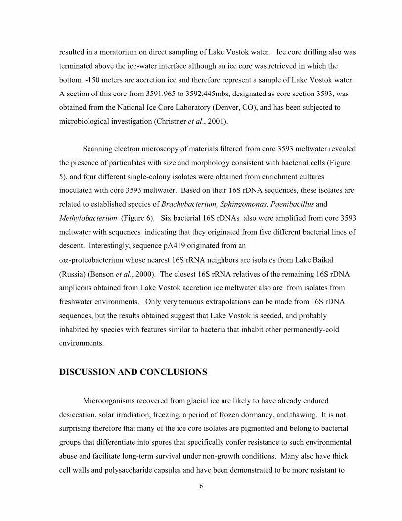
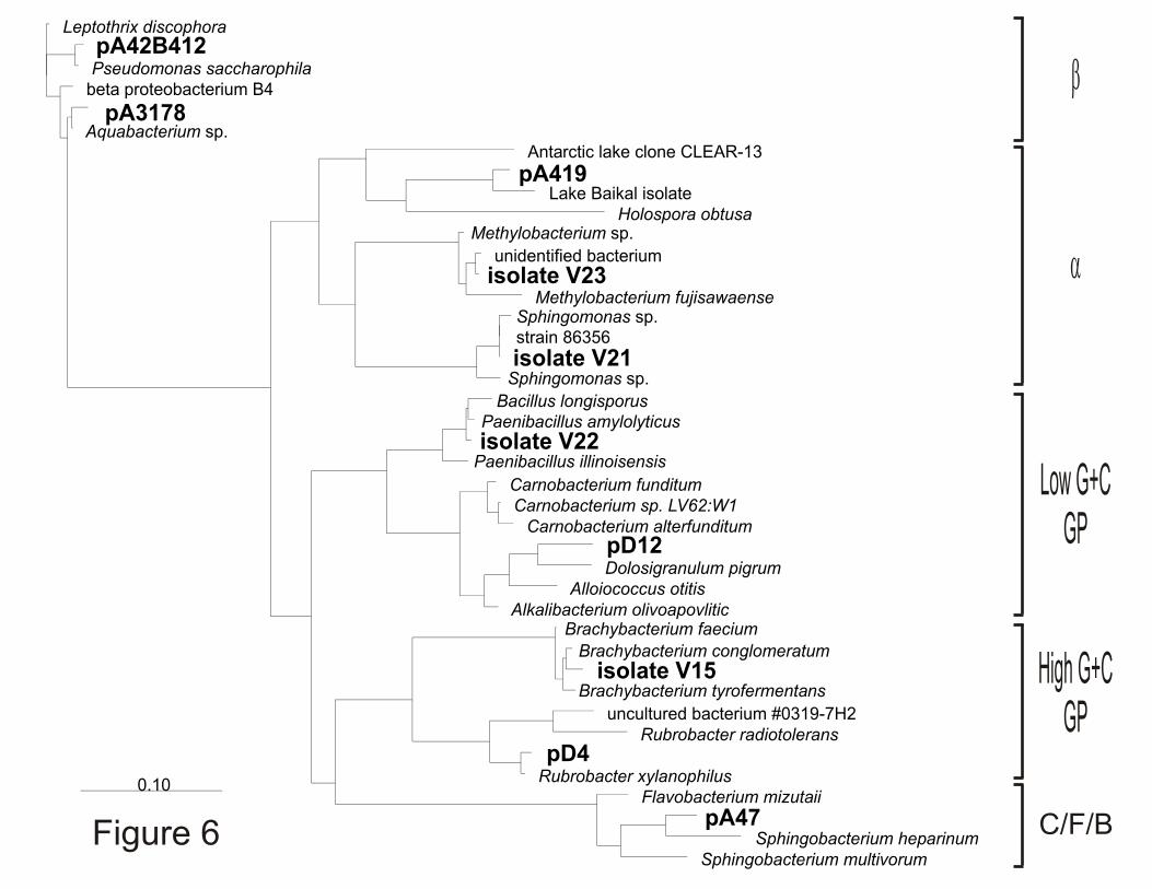
Scanning electron microscopy of materials filtered from core 3593 meltwater revealed
the presence of particulates with size and morphology consistent with bacterial cells (Figure
5), and four different single-colony isolates were obtained from enrichment cultures
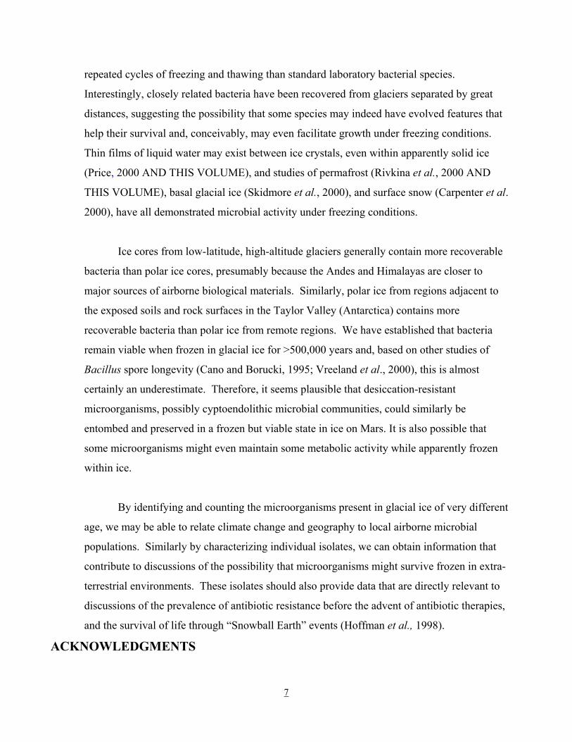
inoculated with core 3593 meltwater. Based on their 16S rDNA sequences, these isolates are
related to established species of Brachybacterium, Sphingomonas, Paenibacillus and
Methylobacterium (Figure 6). Six bacterial 16S rDNAs also were amplified from core 3593
meltwater with sequences indicating that they originated from five different bacterial lines of
descent. Interestingly, sequence pA419 originated from an
"α-proteobacterium whose nearest 16S rRNA neighbors are isolates from Lake Baikal
(Russia) (Benson et al., 2000). The closest 16S rRNA relatives of the remaining 16S rDNA
amplicons obtained from Lake Vostok accretion ice meltwater also are from isolates from
freshwater environments. Only very tenuous extrapolations can be made from 16S rDNA
sequences, but the results obtained suggest that Lake Vostok is seeded, and probably
inhabited by species with features similar to bacteria that inhabit other permanently-cold
environments.
DISCUSSION AND CONCLUSIONS
Microorganisms recovered from glacial ice are likely to have already endured
desiccation, solar irradiation, freezing, a period of frozen dormancy, and thawing. It is not
surprising therefore that many of the ice core isolates are pigmented and belong to bacterial
groups that differentiate into spores that specifically confer resistance to such environmental
abuse and facilitate long-term survival under non-growth conditions. Many also have thick
cell walls and polysaccharide capsules and have been demonstrated to be more resistant to
7
repeated cycles of freezing and thawing than standard laboratory bacterial species.
Interestingly, closely related bacteria have been recovered from glaciers separated by great
distances, suggesting the possibility that some species may indeed have evolved features that
help their survival and, conceivably, may even facilitate growth under freezing conditions.
Thin films of liquid water may exist between ice crystals, even within apparently solid ice
(Price, 2000 AND THIS VOLUME), and studies of permafrost (Rivkina et al., 2000 AND
THIS VOLUME), basal glacial ice (Skidmore et al., 2000), and surface snow (Carpenter et al.
2000), have all demonstrated microbial activity under freezing conditions.
Ice cores from low-latitude, high-altitude glaciers generally contain more recoverable
bacteria than polar ice cores, presumably because the Andes and Himalayas are closer to
major sources of airborne biological materials. Similarly, polar ice from regions adjacent to
the exposed soils and rock surfaces in the Taylor Valley (Antarctica) contains more
recoverable bacteria than polar ice from remote regions. We have established that bacteria
remain viable when frozen in glacial ice for >500,000 years and, based on other studies of
Bacillus spore longevity (Cano and Borucki, 1995; Vreeland et al., 2000), this is almost
certainly an underestimate. Therefore, it seems plausible that desiccation-resistant
microorganisms, possibly cyptoendolithic microbial communities, could similarly be
entombed and preserved in a frozen but viable state in ice on Mars. It is also possible that
some microorganisms might even maintain some metabolic activity while apparently frozen
within ice.
By identifying and counting the microorganisms present in glacial ice of very different
age, we may be able to relate climate change and geography to local airborne microbial
populations. Similarly by characterizing individual isolates, we can obtain information that
contribute to discussions of the possibility that microorganisms might survive frozen in extra-
terrestrial environments. These isolates should also provide data that are directly relevant to
discussions of the prevalence of antibiotic resistance before the advent of antibiotic therapies,
and the survival of life through “Snowball Earth” events (Hoffman et al., 1998).
ACKNOWLEDGMENTS

8
This research was supported by NSF grant OPP-9714206 awarded through the Life in
Extreme Environments Initiative.
LITERATURE CITED
1. Abyzov, S.S., V.Y. Lipenkov, N.E. Bobin, and B.B. Kudryashov.,1982. Microflora of
central Antarctic glacier and methods for sterile ice-core sampling for microbiological
analyses. Biology Bulletin of the Academy of Sciences of the USSR, 9: 304-349.
2. Abyzov, S.S. 1993. Microorganisms in the Antarctic ice. In: Antarctic Microbiology (E.I.
Friedmann, ed.), Wiley-Liss, New York, pp. 265-295.
3. Abyzov, S.S., L.N. Mitskevich, and M.N. Poglazova.1998. Microflora of the deep glacier
horizons of central Antarctica. Microbiology 67: 547-555.
4. Benson, D.A., I. Karsch-Mizrachi,D.J. Lipman,J. Ostell,B.A. Rapp, andD.L.
Wheeler.2000. GenBank. Nucleic Acids Research, 28: 15-18.
5. Brambilla, E., H. Hippe,A. Hagelstein,B.J. Tindall, and E. Stackebrandt.2001. 16S rDNA
diversity of cultured and uncultured prokaryotes of a mat sample from Lake Fryxell,
McMurdo Dry Valleys, Antarctica. Extremophiles, 5: 23-33.
6. Cano, R.J. and M.K. Borucki.1995. Revival and identification of bacterial spores in 25 to
40 million year old Dominican amber. Science, 268: 1060-1064.
7. Carpenter, E.J., S. Lin, and D.G. Capone.2000. Bacterial activity in South Pole snow.
Applied and Environmental Microbiology, 66: 4514-4517.
8. Castello, J.D., S.O. Rogers, W.T. Starmer,C.M. Catranis,L. Ma, G.D. Bachand, Y. Zhao,
and J.E. Smith.1999. Detection of tomato mosaic tobamovirus RNA in ancient glacial
ice. Polar Biology, 22: 207-212.
9. Christner, B.C., E. Mosley-Thompson,L.G. Thompson, V. Zagorodnov, K. Sandman, and
J.N. Reeve.2000. Recovery and identification of viable bacteria immured in glacial
ice. Icarus 144: 479-485.
10.Christner, B.C., E. Mosley-Thompson,L.G. Thompson, and J.N. Reeve.2001. Isolation of
bacteria and 16S rDNAs from Lake Vostok accretion ice. Environmental
Microbiology 3: 570-577.
11.Dancer, S.J., P. Shears,. and D.J. Platt.1997. Isolation and characterization of coliforms
9
from glacial ice and water in Canada’s High Arctic. Journal of Applied Microbiology,
82: 597-609.
12.Dodd, C.E.R., Sharman, R.L., Bloomfield, S.F., Booth, I.R. and Stewart, G.S.A.B. (1997)
Inimical processes: bacterial self-destruction and sub-lethal injury. Trends in Food
Science Technology, 8, 238-241.
13.Fogg, G.E. 1998. The Biology of Polar Habitats, Oxford University Press, Inc., New York,
pp. 33-47
14.Hardfield, M. H.G. Jones, R. Letarte, and P. Simard.1992. Seasonal fluctuation patterns of
microflora on the Agassiz ice cap, Ellesmere Island, Canadian Arctic. The Musk-ox,
39: 119-123.
15.Hoffman, P.F., A.J. Kaufman,G.P. Halverson,and D.P. Schrag.1998. A neoproterozoic
snowball Earth. Science, 281: 1342-1349.
16.Jouzel, J., J.R. Petit,R. Souchez, N.I. Barkov,V.Y. Lifenkov,D. Raymond,M.
Stievenard,N.I. Vassiliev,V. Verbeke,and F. Vimeux.1999. More than 200 meters of
lake ice above subglacial Lake Vostok, Antarctica. Science, 286: 2138-2141.
17.Kapitsa, A.P., J.K. Ridley,G. Robin,M.J. Siegert, and I.A. Zotikov.1996.A large deep
freshwater lake beneath the ice of central East Antarctica. Nature, 381: 684-686.
18.Olsen, G.J., J.H. Natusda,R. Hagstrom, and R.Overbeek.1994. fastDNAml: a tool for
construction of phylogenetic trees of DNA sequences using maximum likelihood.
Computer Applications in the Biosciences, 10: 41-48.
19.Petit, J.R., J. Jouzel,D. Raynaud,N.I. Barkov,J.M. Barnola,I. Basile, M. Benders,J.
Chappellaz,M. Davis,G. Delaygue,M. Delmotte, V.M. Dotlyakov,M. Legrand,V.Y.
Lipendoc,C. Lorius,L. Pepin,C. Ritz,E. Saltzman,and M. Stievenard.1999. Climate and
atmospheric history of the past 420,000 years from the Vostok ice core, Antarctica.
Nature, 399: 429-436.
20.Price, P.B. 2000. A habitat for psychrophiles in deep Antarctic ice. Proceedings of the
National Academy of Sciences, USA, 97:1247-1251.
21.Priscu, J.C., C.H. Fritsen, E.E. Adams, S.J. GiovannoniH.W. Paerl, C.P. McKay,P.T.
Doran,D.A. Gordon,B.D. Lanoil, and J.L. Pinckney.1998. Perennial Antarctic lake ice:
an oasis for life in a polar desert. Science, 280:2095-2098.
22.Rivkina, E.M., E.I. Friedmann,C.P. McKay,. and D.A. Gilichinsky.2000. Metabolic

10
activity of permafrost bacteria below the freezing point. Applied and Environmental
Microbiology 66: 3230-3233.
23.Siegert, M.J., R. Kwok,C. Mayer, and B. Hubbard.2000. Water exchange between
subglacial Lake Vostok and the overlying ice sheet. Nature 403: 643-646.
24.Skidmore, M.L., J.M. Foght, and M.J. Sharp.2000. Microbial life beneath a high Arctic
glacier. Applied and Environmental Microbiology 66: 3214-3220.
25.Strunk, O., O. Gross, B. Reichel,M. May,S. Hermann, N. Struckmann, B. Nonhoff,M.
Lenke,A. Vilbig,T. Ludwig,A. Bode,K.H. Schleifer, and W. Ludwig.1998. ARB: a
software environment for sequence data. Department of Microbiology, Technical
University of Munich, Munich, Germany.
26.Thompson, L.G., T. Yao,M.E. Davis,K.A. Henderson,E. Mosley-Thompson, P.-N. Lin,J.
Beer,H. Synal,J. Cole-Dai,. andJ.F. Bolzan.1997. Tropical climate instability: the last
glacial cycle from a Qinghai-Tibetan ice core. Science 276:1821-1825.
27.Vreeland, R.H., W.D. Rosenzweig, and D.W. Powers.2000. Isolation of a 250 million-year
old halotolerant bacterium from a primary salt crystal. Nature 407: 897-900.
28.Willerslev, E., A.J. Hansen,B. Christensen,J.P. Steffensen, and P.Arctander.1999.
Diversity of Holocene life forms in fossil glacier ice. Proceedings of the National
Academy of Sciences, USA96: 8017-8021.FIGURE LEGENDS
Figure 1. Locations of sampling sites and ice cores available for study at the Byrd Polar
Research Center (BPRC). To date, bacteria have been isolated from ice cores sampled from
glaciers at both poles, in the mountain ranges on the subtropical Tibetan plateau, and in the
tropical Bolivian Andes. In each case, the nearest major ecosystem, and therefore most likely
origin of airborne particulates, is very different.
Figure 2. The ice core sampling system. (A) The sampling system is assembled
completely inside a laminar flow hood that is housed within a –100C walk-in freezer. All
components of the system are autoclaved, dried and exposed to ethylene oxide for 12 h before
use. An ice core is positioned vertically in the sampler with the cut end of the core contacting
(B) the heated sampler head which melts upward (C) through the core and collects the
resulting melt water. In (C), the sampler head is shown disassembled from the main unit to
illustrate its movement through the interior of the ice core.
11
Figure 3. Bacterial genera represented most frequently by ice core isolates. The number
of isolates from both polar and non-polar ice cores, obtained from each of the bacterial genera
shown is listed in parentheses. The phylogenetic relationships illustrated are based on 16S
rDNA sequences. They are not drawn to scale.
Figure 4. Phylogenetic position of 14 bacterial isolates from ice >500,000 years old
from 296 meters below surface of the Guliya ice cap. 16S rDNA sequences (~1400
nucleotides) were obtained from the cells from a single colony of each isolate. They were
aligned based on secondary structures using the ARB software package (Strunk et al. 1998)
and a best fit neighbor-joining tree was constructed. Evolutionary distance is defined as the
number of fixed nucleotide changes per position.
Figure 5. Scanning electron micrographs of materials filtered from meltwater from Lake
Vostok deep ice core section 3593. The particulates shown, apparently bacteria, are retained
on the surface of a 0.2 :m isopore (Millipore) filter.
Figure 6. Phylogenetic analysis of 16S rDNA sequences isolated from bacteria and
directly amplified from meltwater from Lake Vostok core section 3593. Sequences that
correspond to nucleotides 515 through 1392 of the E. coli 16S rDNA were obtained, aligned
and used to construct the figure shown as in Figure 5 (Strunk et al., 1998). A best fit tree was
created using maximum likelihood with a 771 nucleotide mask of unambiguously aligned
positions using fastDNAml (Olsen et al., 1994).
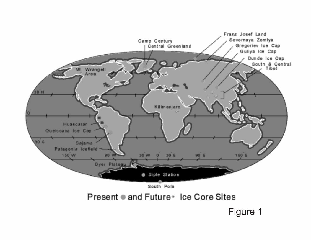
Figure 1

heated watercirculates through
sampling head
melt watercollected
externally
CA B
Figure 2
Microbacterium/ Aureobacterium (5)
Methylobacterium (4)
Sphingomonas (8)
Acinetobacter (4)
Bacillus (19)
Nocardioides (3)
Arthrobacter (5)
Clavibacter (3)
Paenibacillus (6)
a
High G+C GP
Low G+C GP
g
Figure 3
Methylobacterium radiotolerans Methylobacterium fujisawaense
Guliya296-5 Methylobacterium mesophilicum
Methylobacterium extorquens Methylobacterium rhodesianum
Guliya296-15 Methylobacterium sp. Sphingomonas paucimobilis
Guliya296-3 Sphingomonas chlorophenolica
Sphingomonas flava Sphingomonas asaccharolytica
Sphingomonas sp. Guliya296-14 Sphingomonas sp.
Ralstonia pickettii Ralstonia sp. APF11 Guliya296-6 Ralstonia solanacearum 'Pseudomonas' syzygii
Aureobacterium liquefaciens Microbacterium phyllosphaerae Guliya296-10 Aureobacterium keratanolyticum Microbacterium sp. VKM Ac-2047
Agromyces cerinus Agromyces ramosus
Guliya296-1 Agromyces mediolanus
"Corynebacterium mediolanum" Paenibacillus polymyxa Paenibacillus burgondia
Guliya296-78 Paenibacillus sp. 7-5 Paenibacillus illinoisensis
Bacillus sp. Guliya296-16 Bacillus pumilus Bacillus subtilis Bacillus licheniformis
Guliya296-2 Bacillus thuringiensis Bacillus cereus Bacillus anthracis
Bacillus simplex Bacillus flexus
Bacillus megaterium Guliya296-18 "Bacillus macroides"
"Bacillus maroccanus" Guliya296-17 Bacillus psychrosaccharolyticu
Guliya296-19 Guliya296-9
Bacillus sp. 82352 Bacillus cohnii Bacillus horikoshii
0.10
High G+CGP
a
Low G+CGP
b
Figure 4

1 mm1 mm 2 mm1 mm 2 mm1 mm
1 mm 2 mm1 mm
1 mm
Figure 5
Leptothrix discophora pA42B412 Pseudomonas saccharophila
beta proteobacterium B4
pA3178 Aquabacterium sp.
Antarctic lake clone CLEAR-13
pA419 Lake Baikal isolate
Holospora obtusa
unidentified bacterium isolate V23
Methylobacterium fujisawaense
isolate V21
isolate V22
Carnobacterium funditum Carnobacterium sp. LV62:W1
Carnobacterium alterfunditum pD12 Dolosigranulum pigrum
Alloiococcus otitis Alkalibacterium olivoapovlitic
isolate V15
uncultured bacterium #0319-7H2 Rubrobacter radiotolerans
pD4 Rubrobacter xylanophilus
Flavobacterium mizutaii
pA47 Sphingobacterium heparinum
Sphingobacterium multivorum
0.10
Methylobacterium sp.
Sphingomonas sp.
Sphingomonas sp.
strain 86356
Paenibacillus amylolyticus Bacillus longisporus
Paenibacillus illinoisensis
Brachybacterium conglomeratum
Brachybacterium faecium
Brachybacterium tyrofermentans
Low G+CGP
a
b
C/F/B
High G+CGP
Figure 6
Related Documents