Reconstruction of Ancestral Metabolic Enzymes Reveals Molecular Mechanisms Underlying Evolutionary Innovation through Gene Duplication Karin Voordeckers 1,2. , Chris A. Brown 1,2,3,4,5. , Kevin Vanneste 6,7 , Elisa van der Zande 1,2 , Arnout Voet 8 , Steven Maere 6,7 *, Kevin J. Verstrepen 1,2 * 1 VIB Laboratory for Systems Biology, Leuven, Belgium, 2 CMPG Laboratory for Genetics and Genomics, KU Leuven, Leuven, Belgium, 3 Fathom Information Design, Boston, Massachusetts, United States of America, 4 Faculty of Arts and Sciences Center for Systems Biology, Harvard University, Cambridge, Massachusetts, United States of America, 5 Department of Chemistry and Chemical Biology, Harvard University, Cambridge, Massachusetts, United States of America, 6 VIB Department of Plant Systems Biology, Gent, Belgium, 7 Department of Plant Biotechnology and Bioinformatics, Ghent University, Gent, Belgium, 8 Laboratory for Molecular en Structural Biology, KU Leuven, Leuven, Belgium Abstract Gene duplications are believed to facilitate evolutionary innovation. However, the mechanisms shaping the fate of duplicated genes remain heavily debated because the molecular processes and evolutionary forces involved are difficult to reconstruct. Here, we study a large family of fungal glucosidase genes that underwent several duplication events. We reconstruct all key ancestral enzymes and show that the very first preduplication enzyme was primarily active on maltose- like substrates, with trace activity for isomaltose-like sugars. Structural analysis and activity measurements on resurrected and present-day enzymes suggest that both activities cannot be fully optimized in a single enzyme. However, gene duplications repeatedly spawned daughter genes in which mutations optimized either isomaltase or maltase activity. Interestingly, similar shifts in enzyme activity were reached multiple times via different evolutionary routes. Together, our results provide a detailed picture of the molecular mechanisms that drove divergence of these duplicated enzymes and show that whereas the classic models of dosage, sub-, and neofunctionalization are helpful to conceptualize the implications of gene duplication, the three mechanisms co-occur and intertwine. Citation: Voordeckers K, Brown CA, Vanneste K, van der Zande E, Voet A, et al. (2012) Reconstruction of Ancestral Metabolic Enzymes Reveals Molecular Mechanisms Underlying Evolutionary Innovation through Gene Duplication. PLoS Biol 10(12): e1001446. doi:10.1371/journal.pbio.1001446 Academic Editor: Joseph W. Thornton, University of Chicago, United States of America Received February 23, 2012; Accepted October 30, 2012; Published December 11, 2012 Copyright: ß 2012 Voordeckers et al. This is an open-access article distributed under the terms of the Creative Commons Attribution License, which permits unrestricted use, distribution, and reproduction in any medium, provided the original author and source are credited. Funding: S. Maere and K. Vanneste are fellows of the Fund for Scientific Research-Flanders (FWO). Research in the lab of KJV is supported by the Human Frontier Science Program, ERC Starting Grant 241426, VIB, EMBO YIP program, KU Leuven, FWO, IWT and the AB InBev Baillet-Latour foundation. Research in the lab of SM is supported by VIB, Ghent University, FWO and IWT. The funders had no role in study design, data collection and analysis, decision to publish, or preparation of the manuscript. Competing Interests: The authors have declared that no competing interests exist. Abbreviations: AA, amino acid; AIC, Akaike information criterion; anc, ancestral; BEB, Bayes empirical Bayes; BMCMC, Bayesian Markov chain Monte Carlo; df, degree of freedom; EAC, escape from adaptive conflict; GTR, generalized time reversible; IAD, innovation-amplification-divergence; JTT, Jones, Taylor and Thornton; LBA, long branch attraction; LG+I+G, Le and Gascuel+invariable sites+gamma distributed rate heterogeneity; LRT, likelihood ratio test; MalS, maltase; ML, maximum likelihood; Ima, isomaltase; WAG, Whelan and Goldman. * E-mail: [email protected] (SM); [email protected] (KJV) . These authors contributed equally to this work. Introduction In a seminal book, Susumu Ohno argued that gene duplication plays an important role in evolutionary innovation [1]. He outlined three distinct fates of retained duplicates that were later formalized by others (for reviews, see [2,3]). First, after a duplication event, one paralog may retain the ancestral function, whereas the other allele may be relieved from purifying selection, allowing it to develop a novel function (later called ‘‘neofunctionalization’’). Second, differ- ent functions or regulatory patterns of an ancestral gene might be split over the different paralogs (later called ‘‘subfunctionalization’’ [4,5]). Third, duplication may preserve the ancestral function in both duplicates, thereby introducing redundancy and/or increasing activity of the gene (‘‘gene dosage effect’’ [6]). Recent studies have shown that duplications occur frequently during evolution, and most experts agree that many evolutionary innovations are linked to duplication [7–10]. A well-known example are crystallins, structural proteins that make up 60% of the protein in the lenses of vertebrate eyes. Interestingly, paralogs of many crystallins function as molecular chaperones or glycolytic enzymes. Studies suggest that on multiple occasions, an ancestral gene encoding a (structurally very stable) chaperone or enzyme was duplicated, with one paralog retaining the ancestral function and one being tuned as a lens crystallin that played a crucial role in the optimization of eyesight [11,12]. The molecular mechanisms and evolutionary forces that lead to the retention of duplicates and the development of novel functions are still heavily debated, and many different models leading to Ohno’s three basic outcomes have been proposed (reviewed in [2,3,13,14]). Some more recent models blur the distinction between neo- and subfunctionalization [15]. Co-option models, for example, propose that a novel function does not develop PLOS Biology | www.plosbiology.org 1 November 2012 | Volume 10 | Issue 12 | e1001446

Welcome message from author
This document is posted to help you gain knowledge. Please leave a comment to let me know what you think about it! Share it to your friends and learn new things together.
Transcript

Reconstruction of Ancestral Metabolic Enzymes RevealsMolecular Mechanisms Underlying EvolutionaryInnovation through Gene DuplicationKarin Voordeckers1,2., Chris A. Brown1,2,3,4,5., Kevin Vanneste6,7, Elisa van der Zande1,2, Arnout Voet8,
Steven Maere6,7*, Kevin J. Verstrepen1,2*
1 VIB Laboratory for Systems Biology, Leuven, Belgium, 2 CMPG Laboratory for Genetics and Genomics, KU Leuven, Leuven, Belgium, 3 Fathom Information Design,
Boston, Massachusetts, United States of America, 4 Faculty of Arts and Sciences Center for Systems Biology, Harvard University, Cambridge, Massachusetts, United States
of America, 5 Department of Chemistry and Chemical Biology, Harvard University, Cambridge, Massachusetts, United States of America, 6 VIB Department of Plant
Systems Biology, Gent, Belgium, 7 Department of Plant Biotechnology and Bioinformatics, Ghent University, Gent, Belgium, 8 Laboratory for Molecular en Structural
Biology, KU Leuven, Leuven, Belgium
Abstract
Gene duplications are believed to facilitate evolutionary innovation. However, the mechanisms shaping the fate ofduplicated genes remain heavily debated because the molecular processes and evolutionary forces involved are difficult toreconstruct. Here, we study a large family of fungal glucosidase genes that underwent several duplication events. Wereconstruct all key ancestral enzymes and show that the very first preduplication enzyme was primarily active on maltose-like substrates, with trace activity for isomaltose-like sugars. Structural analysis and activity measurements on resurrectedand present-day enzymes suggest that both activities cannot be fully optimized in a single enzyme. However, geneduplications repeatedly spawned daughter genes in which mutations optimized either isomaltase or maltase activity.Interestingly, similar shifts in enzyme activity were reached multiple times via different evolutionary routes. Together, ourresults provide a detailed picture of the molecular mechanisms that drove divergence of these duplicated enzymes andshow that whereas the classic models of dosage, sub-, and neofunctionalization are helpful to conceptualize theimplications of gene duplication, the three mechanisms co-occur and intertwine.
Citation: Voordeckers K, Brown CA, Vanneste K, van der Zande E, Voet A, et al. (2012) Reconstruction of Ancestral Metabolic Enzymes Reveals MolecularMechanisms Underlying Evolutionary Innovation through Gene Duplication. PLoS Biol 10(12): e1001446. doi:10.1371/journal.pbio.1001446
Academic Editor: Joseph W. Thornton, University of Chicago, United States of America
Received February 23, 2012; Accepted October 30, 2012; Published December 11, 2012
Copyright: � 2012 Voordeckers et al. This is an open-access article distributed under the terms of the Creative Commons Attribution License, which permitsunrestricted use, distribution, and reproduction in any medium, provided the original author and source are credited.
Funding: S. Maere and K. Vanneste are fellows of the Fund for Scientific Research-Flanders (FWO). Research in the lab of KJV is supported by the Human FrontierScience Program, ERC Starting Grant 241426, VIB, EMBO YIP program, KU Leuven, FWO, IWT and the AB InBev Baillet-Latour foundation. Research in the lab of SMis supported by VIB, Ghent University, FWO and IWT. The funders had no role in study design, data collection and analysis, decision to publish, or preparation ofthe manuscript.
Competing Interests: The authors have declared that no competing interests exist.
Abbreviations: AA, amino acid; AIC, Akaike information criterion; anc, ancestral; BEB, Bayes empirical Bayes; BMCMC, Bayesian Markov chain Monte Carlo; df,degree of freedom; EAC, escape from adaptive conflict; GTR, generalized time reversible; IAD, innovation-amplification-divergence; JTT, Jones, Taylor andThornton; LBA, long branch attraction; LG+I+G, Le and Gascuel+invariable sites+gamma distributed rate heterogeneity; LRT, likelihood ratio test; MalS, maltase;ML, maximum likelihood; Ima, isomaltase; WAG, Whelan and Goldman.
* E-mail: [email protected] (SM); [email protected] (KJV)
. These authors contributed equally to this work.
Introduction
In a seminal book, Susumu Ohno argued that gene duplication
plays an important role in evolutionary innovation [1]. He outlined
three distinct fates of retained duplicates that were later formalized
by others (for reviews, see [2,3]). First, after a duplication event, one
paralog may retain the ancestral function, whereas the other allele
may be relieved from purifying selection, allowing it to develop a
novel function (later called ‘‘neofunctionalization’’). Second, differ-
ent functions or regulatory patterns of an ancestral gene might be
split over the different paralogs (later called ‘‘subfunctionalization’’
[4,5]). Third, duplication may preserve the ancestral function in
both duplicates, thereby introducing redundancy and/or increasing
activity of the gene (‘‘gene dosage effect’’ [6]).
Recent studies have shown that duplications occur frequently
during evolution, and most experts agree that many evolutionary
innovations are linked to duplication [7–10]. A well-known
example are crystallins, structural proteins that make up 60% of
the protein in the lenses of vertebrate eyes. Interestingly, paralogs
of many crystallins function as molecular chaperones or glycolytic
enzymes. Studies suggest that on multiple occasions, an ancestral
gene encoding a (structurally very stable) chaperone or enzyme
was duplicated, with one paralog retaining the ancestral function
and one being tuned as a lens crystallin that played a crucial role in
the optimization of eyesight [11,12].
The molecular mechanisms and evolutionary forces that lead to
the retention of duplicates and the development of novel functions
are still heavily debated, and many different models leading to
Ohno’s three basic outcomes have been proposed (reviewed in
[2,3,13,14]). Some more recent models blur the distinction
between neo- and subfunctionalization [15]. Co-option models,
for example, propose that a novel function does not develop
PLOS Biology | www.plosbiology.org 1 November 2012 | Volume 10 | Issue 12 | e1001446

entirely de novo but originates from a pre-existing minor function
in the ancestor that is co-opted to a primary role in one of the
postduplication paralogs [2,13]. Examples of such co-option
models include the ‘‘gene sharing’’ or ‘‘Escape from Adaptive
Conflict’’ (EAC) model [5,16–19] and the related ‘‘Innovation,
Amplification and Divergence’’ (IAD) model [20–22]. The IAD
model describes co-option as a neofunctionalization mechanism. A
‘‘novel’’ function arises in the preduplication gene, and increased
requirement for this (minor) activity is first met by gene
amplification (e.g., through formation of tandem arrays). After
this, adaptive mutations lead to divergence and specialization of
some of the duplicate copies. The EAC model, on the other hand,
describes co-option rather as a subfunctionalization mechanism by
which duplication allows a multifunctional gene to independently
optimize conflicting subfunctions in different daughter genes.
Another aspect in which various models differ is the role of
positive selection. Some models emphasize the importance of
neutral drift, while in other models adaptive mutations play an
important role. For example, in the Duplication-Degeneration-
Complementation (DDC) model of subfunctionalization [4],
degenerative mutations (accumulated by neutral drift) lead to
complementary loss-of-function mutations in the duplicates, so
that both copies become essential to perform all of the functions
that were combined in the single preduplication gene. Whereas
this type of subfunctionalization only involves genetic drift [4,8],
other subfunctionalization models, such as the EAC model,
attribute an important role to positive selection for the further
functional optimization of the postduplication paralogs [2,14].
There is a sharp contrast between the large number of detailed
theoretical models of evolution after gene duplication, on the one
hand, and the lack of clear experimental evidence for the various
predictions made by these theories, on the other [2]. The key
problem is the lack of knowledge about the functional properties of
the ancestral, preduplication gene. Since these ancient genes and
the proteins they encode no longer exist, many details in the chain
of events that led from the ancestral gene to the present-day
duplicates remain obscure. In most studies, the activities of the
preduplication ancestor are inferred from unduplicated present-
day outgroup genes that are assumed to have retained similar
functional properties, but this is only an approximation. The
central hurdle to surpass to obtain accurate experimental data on
the evolution of gene duplicates involves rewinding the evolution-
ary record to obtain the sequence and activity of the ancestral
proteins. Recent developments in sequencing and bio-informatics
now enable us to reconstruct ancestral genes and proteins and
characterize them in detail [23–31]. However, most ancestral
reconstruction studies to date did not focus on the mechanisms
that govern evolution after gene duplication.
In this study, we used the yeast MALS gene family as a model
system to gain insight in the molecular mechanisms and
evolutionary forces shaping the fate of duplicated genes. The
MALS genes encode a-glucosidases that allow yeast to metabolize
complex carbohydrates like maltose, isomaltose, and other a-
glucosides [32,33]. Several key features make this family ideal to
study duplicate gene evolution. First, it is a large gene family
encompassing multiple gene duplication events, some ancient and
some more recent. Second, the present-day enzymes have
diversified substrate specificities that can easily be measured
[32]. Third, the availability of MALS gene sequences from many
fungal genomes enabled us to make high-confidence predictions of
ancestral gene sequences, resurrect key ancestral proteins, and
study the selective forces acting throughout the evolution of the
different gene duplicates. Fourth, the crystal structure of one of the
present-day enzymes, Ima1, has been determined [34]. Molecular
modeling of the enzymes’ binding pocket, combined with activity
measurements on reconstructed and present-day enzymes, allowed
us to investigate how mutations altered enzyme specificity and
gave rise to the present-day alleles that allow growth on a broad
variety of substrates. Combining these analyses, we were able to
study the evolution and divergence of a multigene family to an
unprecedented level of detail and show that the evolutionary
history of the MALS family exhibits aspects of all three classical
models of duplicate gene evolution proposed by Ohno (gene
dosage, neo-, and subfunctionalization).
Results
The Present-Day Maltase Enzymes Arose from aFunctionally Promiscuous Ancestor
Some yeast species have evolved the capacity to metabolize a
broad spectrum of natural disaccharides found in plants and fruits
(Figure 1, tree adapted from [35]). The origin of this evolutionary
innovation seems to lie in the duplication and functional
diversification of genes encoding permeases and hydrolases [32].
The common Saccharomyces cerevisiae laboratory strain S288c, for
example, contains seven different MALS genes (MAL12, MAL32,
and IMA1–5), which originated from the same ancestral gene but
allow growth on different substrates [32,33].
To understand how duplications led to functionally different
MalS enzymes, we reconstructed, synthesized, and measured the
activity of key ancestral MalS proteins. We used the amino acid
(AA) sequences of 50 maltases from completely sequenced yeast
species, ranging from Saccharomyces cerevisiae to Pichia and Candida
species, for phylogenetic analysis and ancestral sequence recon-
struction (see Materials and Methods and Dataset S1). A consensus
amino-acid-based phylogenetic tree was constructed using
MrBayes [36] under the LG+I+G model with four rate categories
(see Figure S1, and see Materials and Methods for details). Trees
constructed using MrBayes under other models of sequence
evolution (WAG, JTT) generated largely identical results (unpub-
Author Summary
Darwin’s theory of evolution is one of gradual change, yetevolution sometimes takes remarkable leaps. Such evolu-tionary innovations are often linked to gene duplicationthrough one of three basic scenarios: an extra copy canincrease protein levels, different ancestral subfunctions canbe split over the copies and evolve distinct regulation, orone of the duplicates can develop a novel function.Although there are numerous examples for all thesetrajectories, the underlying molecular mechanisms remainobscure, mostly because the preduplication genes andproteins no longer exist. Here, we study a family of fungalmetabolic enzymes that hydrolyze disaccharides, and thatall originated from the same ancestral gene throughrepeated duplications. By resurrecting the ancient genesand proteins using high-confidence predictions from manyfungal genome sequences available, we show that the veryfirst preduplication enzyme was promiscuous, preferringmaltose-like substrates but also showing trace activitytowards isomaltose-like sugars. After duplication, specificmutations near the active site of one copy optimized theminor activity at the expense of the major ancestralactivity, while the other copy further specialized in maltoseand lost the minor activity. Together, our results revealhow the three basic trajectories for gene duplicates cannotbe separated easily, but instead intertwine into a complexevolutionary path that leads to innovation.
Functional Innovation through Gene Duplication
PLOS Biology | www.plosbiology.org 2 November 2012 | Volume 10 | Issue 12 | e1001446

lished data). To further check the robustness of the AA tree
inferred by MrBayes, we inferred a maximum likelihood (ML) tree
under the LG+I+G model using PhyML (Figure S2) [37]. With the
exception of a few recent splits in the topology, the MrBayes and
PhyML trees agree, increasing our confidence in the constructed
tree. Codon-based tree reconstruction using MrBayes yielded
similar results (see further). Additional tests were performed to
control for potential long branch attraction (LBA) artifacts,
specifically to check the placement of the K. lactis branch as an
outgroup to the Saccharomyces and Lachancea clades (see Text S1 and
Figures S3, S4, S5, S6).
Next, we reconstructed the AA sequence of the ancestral
maltases under several commonly used models of protein evolution
(LG, WAG, JTT; see Materials and Methods). All models support
roughly the same ancestral protein sequences, increasing our
confidence in the reconstructed ancestral sequences. In particular,
all models identified the same residues for variable sites within
10 A of the active center (based on the crystal structure of the
Ima1 protein), which are likely relevant sites with respect to
enzymatic activity. The residues for a few other sites located
further away from the active pocket vary between different models,
but differences generally involve biochemically similar AAs (see
Table S1).
Synthesis of the ancestral enzymes was based on the
reconstructed ancestral sequences obtained with the JTT model.
For ambiguous residues (i.e., sites for which the probability of the
second-most likely AA is .0.2) within 7.5 A of the binding pocket,
we constructed proteins containing each possible AA, while for
Figure 1. Yeast species can grow on a broad spectrum of a-glucosides. Serial dilutions of each species were spotted on medium (Yeast NitrogenBase w/o amino acids) with 2% of each sugar (Me-a-Glu = methyl-a-glucoside). Growth was scored after 3 d incubation at 22uC. +, growth; 2, nogrowth. # MALS genes, the number of maltase genes found in each of these strains. Genotypes are listed in Table S5.doi:10.1371/journal.pbio.1001446.g001
Functional Innovation through Gene Duplication
PLOS Biology | www.plosbiology.org 3 November 2012 | Volume 10 | Issue 12 | e1001446

ambiguous residues outside 7.5 A we considered only the most
likely AA. There is one ambiguous residue close to the active
center in the ancestral proteins ancMalS and ancMal-Ima, namely
residue 279 (based on Saccharomyces cerevisiae S288c Ima1 number-
ing). We therefore synthesized two alternative versions of these
proteins, one having G and one having A at position 279. Whereas
these alternative proteins show different activities for some
substrates, the relative activities are similar and our conclusions
are robust. Sequences for these reconstructed enzymes can be
found in Dataset S2. In the main figures, we show the variant with
the highest confidence. Enzymatic data for all variants can be
found in Table S2.
The activity of all resurrected ancestral enzymes was deter-
mined for different substrates (see Materials and Methods, Text
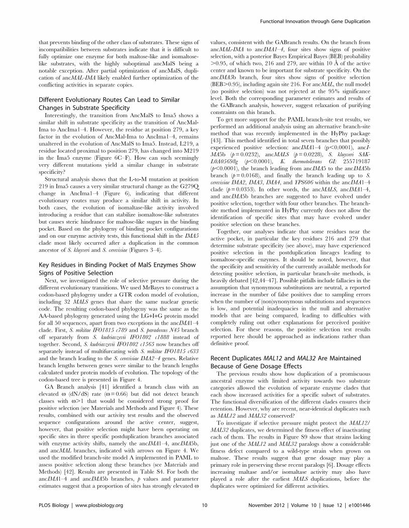
S1, and Figure 2). The results indicate that the very first ancestral
enzyme, denoted as ancMalS, was functionally promiscuous, being
primarily active on maltose-like substrates but also having trace
activity on isomaltose-like sugars. The activity data presented in
Figure 2 show how this promiscuous ancestral protein with
relatively poor activity for several substrates evolved to the seven
present-day enzymes that show high activity for a subset of
substrates, and little or no activity for others. This confirms the
existence of two functional classes of MalS enzymes that originated
from ancient duplication events. First, Mal12 and Mal32 show
activity against maltose-like disaccharides often encountered in
plant exudates, fruits, and cereals, like maltose, maltotriose,
maltulose, sucrose, and turanose (a signaling molecule in plants).
The five MalS enzymes of the second class (Ima1–5), which in fact
result from two independent ancient duplication events giving rise
to the Ima1–4 and Ima5 clades, show activity against isomaltose-
like sugars including palatinose (found in honey [38]) and
isomaltose. Differences in hydrolytic activity between members
of the same (sub)class are more subtle or even absent, which is not
surprising since some of these recent paralogs are nearly identical
(Mal12 and Mal32, for example, are 99.7% identical on the AA
level).
The more recent ancestral enzymes also show a similar split in
activity, with some enzymes (ancMal) showing activity towards
maltose-like substrates, and others (ancIma1–4) towards isomal-
tose-like substrates. Moreover, activity on isomaltose-like sugars
(isomaltose, palatinose, and methyl-a-glucoside) changes in a
coordinate fashion when comparing different enzymes, and the
maltose-like sugars also group together. Careful statistical analysis
reveals that the maltose-like group consists of two subgroups
(maltose, maltotriose, maltulose, and turanose, on one hand, and
sucrose, on the other) that behave slightly different, showing that
the enzymes show quantitative differences in the variation of
specificity towards these substrates (two-way ANOVA analysis
followed by Games-Howell test on log-transformed kcat/Km
values; p values can be found in Table S3).
Interestingly, the most ancient ancestral enzymes do not show a
clear split in activity towards either maltose-like or isomaltose-like
sugars after duplication, and the transition of ancMalS to ancMal-
Ima even shows an increase in activity for all substrates. This
suggests that (slight) optimization for all substrate classes simulta-
neously was still possible starting from ancMalS. A clear
divergence of both subfunctions occurred later, after duplication
of ancMal-Ima, resulting in ancMal and ancIma1–4. AncMal
shows a significant increase in activity on maltose-like sugars
accompanied by a significant drop in activity on isomaltose-like
sugars compared to ancMal-Ima; and the reverse is true for
ancIma1–4 (see also Table S3 for exact p values for each enzyme–
enzyme comparison on the different sugars tested). Together, this
illustrates how, after duplication, the different copies diverged and
specialized in one of the functions present in the preduplication
enzyme.
In two separate instances, a major shift in specificity is observed,
from maltose-like sugars to isomaltose-like sugars (transition from
ancIMA5 to IMA5, and from ancMAL-IMA to ancIMA1–4). The
shift in activity from ancMAL-IMA to ancIMA1–4 is particularly
pronounced. The ancMAL-IMA enzyme hydrolyzes maltose,
sucrose, turanose, maltotriose, and maltulose but has hardly any
measurable activity for isomaltose and palatinose, whereas
ancIMA1–4 can only hydrolyze isomaltose and palatinose (and
also sucrose). For the evolution of the maltase-like activity from the
ancestral MalS enzyme to the present-day enzyme Mal12, we see a
2-fold increase in kcat and a 3-fold decrease in Km for maltose,
indicating an increase in both catalytic power and substrate affinity
for this sugar. For the evolution of isomaltase-like activity in the
route leading to Mal12, kcat decreases more than 3-fold for methyl-
a -glucoside. kcat for isomaltose and palatinose and the affinity for
isomaltose and palatinose are so low that they could not be
measured (see Table S2 for the exact values of kcat and Km for
each enzyme and each sugar; results of two-way ANOVA analysis
followed by Games-Howell test comparing log-transformed kcat/
Km values for different enzymes on each of the sugars can be
found in Table S3).
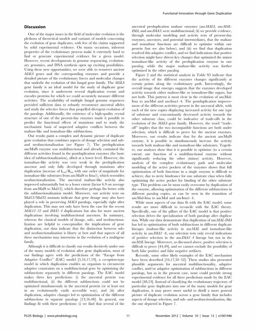
Present-Day Enzymes from Other Yeast Species ShowSimilar Patterns of Functional Diversification
To further explore the evolution of MALS genes and consolidate
the measured activities of the ancestral enzymes, we expressed and
purified additional present-day a-glucosidase alleles from other
yeast species and measured their activities (Figure 3). We focused
primarily on enzymes that are directly related to one of the
ancestral proteins but did not undergo any further duplication
events, and therefore have a higher probability of having retained
a similar activity as their (sub)class ancestor. Indeed, the only
present-day MalS enzyme of the yeast L. elongisporus has a broad
but relatively weak activity comparable to the very first ancestral
MalS enzyme, providing extra support for the accuracy of our
ancestral reconstructions. Also in K. lactis, which contains two Mal
alleles, one of the paralogs retains the broad specificity of
ancMalS. The other paralog (GI:5441460) has a deletion of five
AAs close to the active pocket that likely explains the general lack
of activity of this enzyme (see Materials and Methods and Figure
S7). In contrast, yeasts that show multiple duplication events, like
K. thermotolerans and S. cerevisiae, exhibit specialization, with some
enzymes showing only activity for maltose-like substrates and
others for isomaltose-like substrates. Moreover, the activities
(maltase- or isomaltase-like) of homologs in S. cerevisiae and K.
thermotolerans derived from the same intermediate ancestor are
often similar, except in the IMA5 clade. Here, the K. thermotolerans
and S. cerevisiae homologs have very different substrate specificities,
indicating species-specific evolutionary trajectories and/or recip-
rocal paralog loss in the different species (Figures 3 and 4).
Molecular Modeling and Resurrection of AncestralProteins Identify Residue 279 in the Enzymes’ BindingPocket as a Key Determinant of Substrate Specificity
Next, we investigated which mutations underlie the observed
functional changes. We used the recently resolved crystal structure
of Ima1 (pdb entry 3A4A) [34] as a template to study the
molecular structure of the enzymes’ substrate binding pocket (see
Materials and Methods). All enzymes share a highly conserved
molecular fold, suggesting that changes in activity or substrate
preference are likely caused by mutations in or around the
Functional Innovation through Gene Duplication
PLOS Biology | www.plosbiology.org 4 November 2012 | Volume 10 | Issue 12 | e1001446
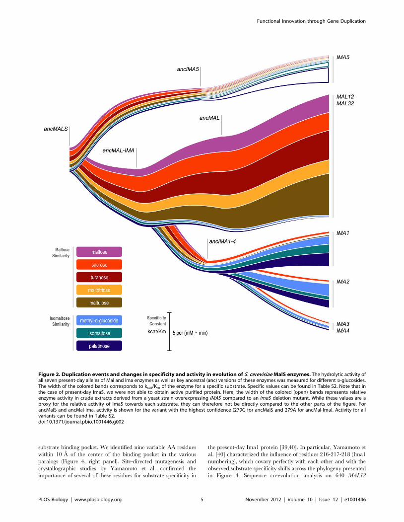
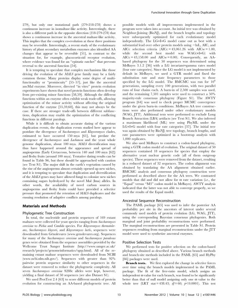
substrate binding pocket. We identified nine variable AA residues
within 10 A of the center of the binding pocket in the various
paralogs (Figure 4, right panel). Site-directed mutagenesis and
crystallographic studies by Yamamoto et al. confirmed the
importance of several of these residues for substrate specificity in
the present-day Ima1 protein [39,40]. In particular, Yamamoto et
al. [40] characterized the influence of residues 216-217-218 (Ima1
numbering), which covary perfectly with each other and with the
observed substrate specificity shifts across the phylogeny presented
in Figure 4. Sequence co-evolution analysis on 640 MAL12
Figure 2. Duplication events and changes in specificity and activity in evolution of S. cerevisiae MalS enzymes. The hydrolytic activity ofall seven present-day alleles of Mal and Ima enzymes as well as key ancestral (anc) versions of these enzymes was measured for different a-glucosides.The width of the colored bands corresponds to kcat/Km of the enzyme for a specific substrate. Specific values can be found in Table S2. Note that inthe case of present-day Ima5, we were not able to obtain active purified protein. Here, the width of the colored (open) bands represents relativeenzyme activity in crude extracts derived from a yeast strain overexpressing IMA5 compared to an ima5 deletion mutant. While these values are aproxy for the relative activity of Ima5 towards each substrate, they can therefore not be directly compared to the other parts of the figure. ForancMalS and ancMal-Ima, activity is shown for the variant with the highest confidence (279G for ancMalS and 279A for ancMal-Ima). Activity for allvariants can be found in Table S2.doi:10.1371/journal.pbio.1001446.g002
Functional Innovation through Gene Duplication
PLOS Biology | www.plosbiology.org 5 November 2012 | Volume 10 | Issue 12 | e1001446

Figure 3. Activities of present-day Mal enzymes in distant fungi correspond well with activities of reconstructed ancestral enzymes.Basic phylogeny of the MALS gene family with different clades, showing the ancestral bifurcation points (indicated by *). Length of the colored bandscorresponds to the measured kcat/Km of the enzyme for a specific substrate. Bands for Ima5 represent relative enzyme activity in crude extractsderived from a yeast strain overexpressing IMA5 compared to an ima5 deletion mutant. For ancMalS and ancMal-Ima, activity is shown for the variantwith the highest confidence (279G for ancMalS and 279A for ancMal-Ima). Error bars represent standard deviations. Activity for all variants and thecorresponding standard deviations can be found in Table S2.doi:10.1371/journal.pbio.1001446.g003
Functional Innovation through Gene Duplication
PLOS Biology | www.plosbiology.org 6 November 2012 | Volume 10 | Issue 12 | e1001446
homologs identified another cluster of three co-evolving residues
among these nine residues (positions 218, 278, and 279 in Ima1),
which we investigate here in detail.
Together with residues 216 and 217, residues 218, 278, and 279
seem to contribute to the activity shift observed in the evolution of
Ima1–4 (see Figures 4–6, Figure S8, and Supplementary
Information for details). Molecular modeling of the mutations at
218-278-279 on the branch leading to ancIma1–4 (see Figure 4)
suggests that the change from alanine to glutamine at residue 279
shifts the binding preference of the pocket from maltose-like to
isomaltose-like sugars (Figure 5B–E). The two co-evolving residues
at positions 218 and 278 are spatially close to AA 279 and cause
subtle structural adaptations that help to better position the Q
residue.
To investigate if changes at all three positions are necessary for
the observed shift in substrate specificity from ancMAL-IMA to
ancIMA1–4 and to investigate the possible evolutionary paths
leading to these three interdependent mutations, we synthesized all
possible intermediate ancIMA1–4 enzyme variants with mutations
at positions 218, 278, and 279. We subsequently expressed,
purified, and measured activity of these enzyme variants. Figure 5F
depicts the results of these enzyme assays and shows that these
residues indeed affect substrate specificity, with the largest shift
depending on the A to Q change at position 279, as expected from
structural analysis. For one mutational path (GVA to GVQ to
SVQ to SMQ), we observe a gradual increase in activity towards
isomaltose and palatinose, demonstrating that there is a muta-
tional path that leads to a consistent increase in isomaltase activity
without traversing fitness valleys. Moreover, in keep with the
stabilizing role of the mutations at positions 218 and 278, the A to
Q change at position 279 along this path takes place before the
two other mutations at positions 218 and 278 (Figure 5F).
Figure 4. Positive selection on residues near binding pocket resulted in distinct subgroups with different substrate preference. Anunrooted codon-based phylogenetic tree of the MALS gene family is shown on the left. Branches are colored according to the v (dN/dS) rate classesinferred from GA Branch analysis [41]. Branches for which branch-site tests for positive selection were performed are indicated by coloredarrowheads. Since v rate classes cannot be inferred reliably for very small branches, branches ,0.01 are not colored. The right part of the figureshows the nine variable AA residues located near the substrate binding pocket of the respective enzymes (numbering based on Ima1 sequence).Sequences of ancestral enzymes are shaded in grey. Subgroups of enzymes that show similar substrate specificity are colored accordingly. Residuesindicated in bold were found to be under positive selection by the branch-site tests. Perfectly co-varying residues are boxed. Substrate preference ofextant and ancestral enzymes was deduced from enzyme assays on S. cerevisiae, K. lactis, K. thermotolerans, L. elongisporus, and reconstructedancestral enzymes (see Figure 3 and Table S4).doi:10.1371/journal.pbio.1001446.g004
Functional Innovation through Gene Duplication
PLOS Biology | www.plosbiology.org 7 November 2012 | Volume 10 | Issue 12 | e1001446

Figure 5. Three co-evolving residues determine the shift in activity observed in the evolution of Ima1–4. (A) Global structure of theMalS proteins with maltose, represented as spheres, bound in the active site. Panels (B–E) show details of the active site, with substrates as sticks(maltose in panels B and C; isomaltose in panels D and E). The variable AAs are shown as spheres. Structural analysis of the binding site suggests thatthe A279Q mutation affects substrate specificity the most. The side chain of Q279 sterically hinders binding of maltose but stabilizes isomaltosebinding through polar interactions. The G218S and V278M changes cause subtle adaptations of the fold, causing Q279 to protrude further into thebinding pocket, which allows optimal interaction with isomaltose. (F) Activity (kcat/Km) of all possible intermediary forms in the evolution of three co-
Functional Innovation through Gene Duplication
PLOS Biology | www.plosbiology.org 8 November 2012 | Volume 10 | Issue 12 | e1001446
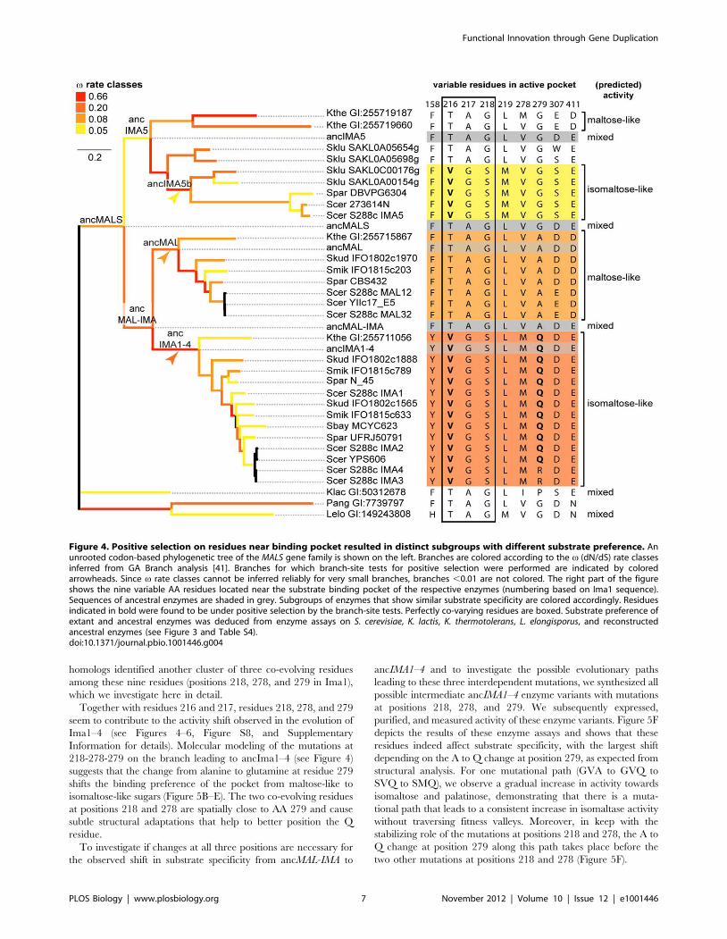
Besides allowing the development of isomaltase activity in the
Ima proteins, duplication also permitted further increase of the
major ancestral function (hydrolysis of maltose-like sugars) in
Mal12 and Mal32. Structural analysis reveals that this increase in
maltase activity, from ancMalS to Mal12/32, is due to mutations
D307E and E411D (Figure 6G–J). These mutations increase the fit
for maltose-like substrates but also completely block the binding of
isomaltose-like substrates (Figure 6). Similar to what is seen for the
evolution of AncMal-Ima to AncIma1–4, changes that increase the
binding stability of one type of substrate cause steric hindrance
evolving residues in AncIma1–4, obtained from enzyme assays performed for all reconstructed proteins. Values for kcat and Km can be found in TableS2.doi:10.1371/journal.pbio.1001446.g005
Figure 6. Evolution of the promiscuous AncMalS enzyme into isomaltose- and maltose-hydrolyzing enzymes. AncMalS is apromiscuous enzyme that hydrolyzes both maltose- and isomaltose-like substrates, whereas the present-day enzymes Ima1,2 and Ima5 preferentiallyhydrolyze isomaltose-like sugars and Mal12–32 preferentially hydrolyzes maltose-like sugars. First, the presence of a Thr or Val residue at position 216affects the binding affinity of the enzyme through changes in the hydrophobic/hydrophilic interactions with the different substrate classes (panels Ato D; see also Figure S8). The case of Ima1,2 and Ima5 (panels C to F) illustrates that an additional shift in substrate specificity can be obtained viadifferent evolutionary routes. In the case of Ima1 and Ima2, the change of G279 to Q279 interferes with binding of maltose-like substrates, but theside chain of Gln can undergo polar interactions with isomaltose (panels C and D). The G218S and V278M changes cause additional subtleadaptations of the protein fold, causing Q279 to protrude further into the binding pocket, allowing optimal interaction with isomaltose (see alsoFigure 5). The evolution of isomaltase activity in Ima5 also occurred via the introduction of steric hindrance in the binding pocket, although in thiscase the change involved was L219M (panels E and F). In ancMalS, residues D307 and E411 allow binding of both maltose- and isomaltose-likesubstrates (panels G and H). In the maltose-specific enzymes Mal12 and Mal32, however, these residues have evolved to E307 and D411 (panels I andJ). These changes not only increase the affinity for maltose-like substrates but also make this site incompatible with isomaltose-like substrates.Subpanels are graphical representations of the binding pocket, with key amino acids depicted as spheres. Maltose and isomaltose are represented assticks.doi:10.1371/journal.pbio.1001446.g006
Functional Innovation through Gene Duplication
PLOS Biology | www.plosbiology.org 9 November 2012 | Volume 10 | Issue 12 | e1001446
that prevents binding of the other class of substrates. These signs of
incompatibilities between substrates indicate that it is difficult to
fully optimize one enzyme for both maltose-like and isomaltose-
like substrates, with the highly suboptimal ancMalS being a
notable exception. After partial optimization of ancMalS, dupli-
cation of ancMAL-IMA likely enabled further optimization of the
conflicting activities in separate copies.
Different Evolutionary Routes Can Lead to SimilarChanges in Substrate Specificity
Interestingly, the transition from AncMalS to Ima5 shows a
similar shift in substrate specificity as the transition of AncMal-
Ima to AncIma1–4. However, the residue at position 279, a key
factor in the evolution of AncMal-Ima to AncIma1–4, remains
unaltered in the evolution of AncMalS to Ima5. Instead, L219, a
residue located proximal to position 279, has changed into M219
in the Ima5 enzyme (Figure 6C–F). How can such seemingly
very different mutations yield a similar change in substrate
specificity?
Structural analysis shows that the L-to-M mutation at position
219 in Ima5 causes a very similar structural change as the G279Q
change in AncIma1–4 (Figure 6), indicating that different
evolutionary routes may produce a similar shift in activity. In
both cases, the evolution of isomaltase-like activity involved
introducing a residue that can stabilize isomaltose-like substrates
but causes steric hindrance for maltose-like sugars in the binding
pocket. Based on the phylogeny of binding pocket configurations
and on our enzyme activity tests, this functional shift in the IMA5
clade most likely occurred after a duplication in the common
ancestor of S. kluyveri and S. cerevisiae (Figures 3–4).
Key Residues in Binding Pocket of MalS Enzymes ShowSigns of Positive Selection
Next, we investigated the role of selective pressure during the
different evolutionary transitions. We used MrBayes to construct a
codon-based phylogeny under a GTR codon model of evolution,
including 32 MALS genes that share the same nuclear genetic
code. The resulting codon-based phylogeny was the same as the
AA-based phylogeny generated using the LG+I+G protein model
for all 50 sequences, apart from two exceptions in the ancIMA1–4
clade. First, S. mikitae IFO1815 c789 and S. paradoxus N45 branch
off separately from S. kudriavzevii IFO1802 c1888 instead of
together. Second, S. kudriavzevii IFO1802 c1565 now branches off
separately instead of multifurcating with S. mikitae IFO1815 c633
and the branch leading to the S. cerevisiae IMA2–4 genes. Relative
branch lengths between genes were similar to the branch lengths
calculated under protein models of evolution. The topology of the
codon-based tree is presented in Figure 4.
GA Branch analysis [41] identified a branch class with an
elevated v (dN/dS) rate (v= 0.66) but did not detect branch
classes with v.1 that would be considered strong proof for
positive selection (see Materials and Methods and Figure 4). These
results, combined with our activity test results and the observed
sequence configurations around the active center, suggest,
however, that positive selection might have been operating on
specific sites in three specific postduplication branches associated
with enzyme activity shifts, namely the ancIMA1–4, ancIMA5b,
and ancMAL branches, indicated with arrows on Figure 4. We
used the modified branch-site model A implemented in PAML to
assess positive selection along these branches (see Materials and
Methods) [42]. Results are presented in Table S4. For both the
ancIMA1–4 and ancIMA5b branches, p values and parameter
estimates suggest that a proportion of sites has strongly elevated v
values, consistent with the GABranch results. On the branch from
ancMAL-IMA to ancIMA1–4, four sites show signs of positive
selection, with a posterior Bayes Empirical Bayes (BEB) probability
.0.95, of which two, 216 and 279, are within 10 A of the active
center and known to be important for substrate specificity. On the
ancIMA5b branch, four sites show signs of positive selection
(BEB.0.95), including again site 216. For ancMAL, the null model
(no positive selection) was not rejected at the 95% significance
level. Both the corresponding parameter estimates and results of
the GABranch analysis, however, suggest relaxation of purifying
constraints on this branch.
To get more support for the PAML branch-site test results, we
performed an additional analysis using an alternative branch-site
method that was recently implemented in the HyPhy package
[43]. This method identified in total seven branches that possibly
experienced positive selection: ancIMA1–4 (p,0.0001), ancI-
MA5b (p = 0.0232), ancMALS (p = 0.0228), S. kluyveri SAK-
L0A05698g (p,0.0001), K. thermotolerans GI: 255719187
(p,0.0001), the branch leading from ancIMA5 to the ancIMA5b
branch (p = 0.0168), and finally the branch leading up to S.
cerevisiae IMA2, IMA3, IMA4, and YPS606 within the ancIMA1–4
clade (p = 0.0353). In other words, the ancMALS, ancIMA1–4,
and ancIMA5b branches are suggested to have evolved under
positive selection, together with four other branches. The branch-
site method implemented in HyPhy currently does not allow the
identification of specific sites that may have evolved under
positive selection on these branches.
Together, our analyses indicate that some residues near the
active pocket, in particular the key residues 216 and 279 that
determine substrate specificity (see above), may have experienced
positive selection in the postduplication lineages leading to
isomaltose-specific enzymes. It should be noted, however, that
the specificity and sensitivity of the currently available methods for
detecting positive selection, in particular branch-site methods, is
heavily debated [42,44–47]. Possible pitfalls include fallacies in the
assumption that synonymous substitutions are neutral, a reported
increase in the number of false positives due to sampling errors
when the number of (non)synonymous substitutions and sequences
is low, and potential inadequacies in the null and alternative
models that are being compared, leading to difficulties with
completely ruling out other explanations for perceived positive
selection. For these reasons, the positive selection test results
reported here should be approached as indications rather than
definitive proof.
Recent Duplicates MAL12 and MAL32 Are MaintainedBecause of Gene Dosage Effects
The previous results show how duplication of a promiscuous
ancestral enzyme with limited activity towards two substrate
categories allowed the evolution of separate enzyme clades that
each show increased activities for a specific subset of substrates.
The functional diversification of the different clades ensures their
retention. However, why are recent, near-identical duplicates such
as MAL12 and MAL32 conserved?
To investigate if selective pressure might protect the MAL12/
MAL32 duplicates, we determined the fitness effect of inactivating
each of them. The results in Figure S9 show that strains lacking
just one of the MAL12 and MAL32 paralogs show a considerable
fitness defect compared to a wild-type strain when grown on
maltose. These results suggest that gene dosage may play a
primary role in preserving these recent paralogs [6]. Dosage effects
increasing maltase and/or isomaltase activity may also have
played a role after the earliest MALS duplications, before the
duplicates were optimized for different activities.
Functional Innovation through Gene Duplication
PLOS Biology | www.plosbiology.org 10 November 2012 | Volume 10 | Issue 12 | e1001446
Discussion
One of the major issues in the field of molecular evolution is the
plethora of theoretical models and variants of models concerning
the evolution of gene duplicates, with few of the claims supported
by solid experimental evidence. On many occasions, inherent
properties of the evolutionary process make it extremely hard to
find or generate experimental evidence for a given model.
However, recent developments in genome sequencing, evolution-
ary genomics, and DNA synthesis open up exciting possibilities.
Using these new opportunities, we were able to resurrect ancient
MALS genes and the corresponding enzymes and provide a
detailed picture of the evolutionary forces and molecular changes
that underlie the evolution of this fungal gene family. The MALS
gene family is an ideal model for the study of duplicate gene
evolution, since it underwent several duplication events and
encodes proteins for which we could accurately measure different
activities. The availability of multiple fungal genome sequences
provided sufficient data to robustly reconstruct ancestral alleles
and study the selective forces that propelled divergent evolution of
the paralogs. Additionally, the existence of a high-quality crystal
structure of one of the present-day enzymes made it possible to
predict the functional effects of mutations and to study the
mechanistic basis of suspected adaptive conflicts between the
maltase-like and isomaltase-like subfunctions.
Our results paint a complex and dynamic picture of duplicate
gene evolution that combines aspects of dosage selection and sub-
and neofunctionalization (see Figure 7). The preduplication
ancMalS enzyme was multifunctional and already contained the
different activities found in the postduplication enzymes (the basic
idea of subfunctionalization), albeit at a lower level. However, the
isomaltase-like activity was very weak in the preduplication
ancestor and only fully developed through mutations after
duplication (increase of kcat/Km with one order of magnitude for
isomaltase-like substrates from ancMalS to Ima1), which resembles
neofunctionalization. The ancestral maltase-like activity also
improved substantially but to a lesser extent (factor 6.9 on average
from ancMalS to Mal12), which therefore perhaps fits better with
the subfunctionalization model. Moreover, our activity tests on
Mal12/Mal32 mutants indicate that gene dosage may also have
played a role in preserving MALS paralogs, especially right after
duplication. This may not only have been the case for the recent
MAL12–32 and IMA3–4 duplications but also for more ancient
duplications involving multifunctional ancestors. In summary,
whereas the classical models of dosage, sub-, and neofunctiona-
lization are helpful to conceptualize the implications of gene
duplication, our data indicate that the distinction between sub-
and neofunctionalization is blurry at best and that aspects of all
three mechanisms may intertwine in the evolution of a multigene
family.
Although it is difficult to classify our results decisively under one
of the many models of evolution after gene duplication, most of
our findings agree with the predictions of the ‘‘Escape from
Adaptive Conflict’’ (EAC) model [5,16,17,19], a co-option-type
model in which duplication enables an organism to circumvent
adaptive constraints on a multifunctional gene by optimizing the
subfunctions separately in different paralogs. The EAC model
makes three key predictions: (i) the ancestral protein was
multifunctional, (ii) the different subfunctions could not be
optimized simultaneously in the ancestral protein (or at least not
in an evolutionarily easily accessible way), and (iii) after
duplication, adaptive changes led to optimization of the different
subfunctions in separate paralogs [13,16,48]. In general, our
findings fit with these predictions: (i) we find that several of the
ancestral preduplication maltase enzymes (ancMALS, ancMAL-
IMA, and ancIMA5) were multifunctional; (ii) we provide evidence,
through molecular modeling and activity tests of present-day
enzymes, ancestors, and potential intermediates, that the maltase
and isomaltase functions are difficult to optimize within one
protein (but see also below); and (iii) we find that duplication
resolved this adaptive conflict, and we find indications that positive
selection might have driven key changes that optimized the minor
isomaltase-like activity of the preduplication enzyme in one
paralog, while the major maltase-like activity was further
optimized in the other paralog.
Figure 2 and the statistical analysis in Table S3 indicate that
the activity of the different enzymes changes significantly at
certain points along the evolutionary path. Interestingly, the
overall image that emerges suggests that the enzymes developed
activity towards either maltose-like or isomaltose-like sugars, but
not both. This pattern is most clear in the evolution of ancMal-
Ima to ancMal and ancIma1–4. The postduplication improve-
ment of the different activities present in the ancestral allele, with
each of the new copies displaying increased activity for one type
of substrate and concomitantly decreased activity towards the
other substrate class, could be indicative of trade-offs in the
evolution of the MALS gene family. However, the word ‘‘trade-
off’’ implies that the two incompatible functions are both under
selection, which is difficult to prove for the ancient enzymes.
Moreover, our results indicate that for the ancient ancMalS
enzyme, it is possible to simultaneously increase the activity
towards both maltose-like and isomaltose-like substrates. Togeth-
er, our analyses show that it is possible to optimize (to a certain
extent) one function of a multifunctional enzyme without
significantly reducing the other (minor) activity. However,
analysis of the complete evolutionary path and molecular
modeling of the active pockets of the enzymes shows that full
optimization of both functions in a single enzyme is difficult to
achieve, due to steric hindrance for one substrate class when fully
optimizing the active pocket for binding of the other substrate
type. This problem can be most easily overcome by duplication of
the enzyme, allowing optimization of the different subfunctions in
different paralog copies, as can be seen in the transition of
ancMal-Ima to ancMal and ancIma1–4.
While most aspects of our data fit with the EAC model, some
results are more difficult to reconcile with the EAC theory.
Specifically, one of the pillars of the EAC model is that positive
selection drives the specialization of both paralogs after duplica-
tion. While our data demonstrate that duplication of ancMAL-IMA
has led to optimization of both subfunctions in different duplicate
lineages (maltase-like activity in ancMAL and isomaltase-like
activity in ancIMA1–4), our selection tests only reveal indications
of positive selection in the ancIMA1–4 lineage but not in the
ancMAL lineage. Moreover, as discussed above, positive selection is
difficult to prove [44,49], and we cannot exclude the possibility of
both false positive and false negative artifacts.
Recently, some other likely examples of the EAC mechanism
have been described [16,17,50–52]. These studies also presented
plausible arguments for ancestral multifunctionality, adaptive
conflict, and/or adaptive optimization of subfunctions in different
paralogs, but as in the present case, none could provide strong
experimental evidence for all three predictions made by the EAC
model [48,53]. Instead of classifying the evolutionary trajectory of
particular gene duplicates into one of the many models for gene
duplication, it may prove more useful to distill a more general
picture of duplicate evolution across a gene family that includes
aspects of dosage selection, and sub- and neofunctionalization, like
the one depicted in Figure 7.
Functional Innovation through Gene Duplication
PLOS Biology | www.plosbiology.org 11 November 2012 | Volume 10 | Issue 12 | e1001446

Our study is the first to investigate multiple duplication events in
the same gene family in detail. Interestingly, we found that
evolution has taken two different molecular routes to optimize
isomaltase-like activity (the evolution of ancMAL-IMA to an-
cIMA1–4 and ancIMA5 to IMA5). In both cases, only a few key
mutations in the active pocket are needed to cause shifts in
substrate specificity. Some of these key mutations exhibit epistatic
interactions. For example, the shift in substrate specificity
occurring on the path from ancMAL-IMA to ancIMA1–4 depends
in part on mutations at three co-evolving positions (218, 278, and
Figure 7. Multiple evolutionary mechanisms contribute to the evolution of the MalS gene family in S. cerevisiae. (A) Overview ofevolutionary mechanisms in the evolution of an ancestral gene with two conflicting activities (major function, red; minor function, blue). Duplicationcan help resolve this ‘‘adaptive conflict’’ by allowing optimization of these activities in two separate copies. Increased requirement for either of theseactivities, for example by changes in the environment, can first be met by duplication of the ancestral gene. Selection for increased gene dosage canhelp to preserve both copies until adaptive mutations optimize the different functions in separate copies. (B) Evolution of the promiscuous ancestralMalS enzyme into the seven present-day MalS alleles shows how different evolutionary forces contribute to the evolution of gene duplicates. Activitytowards isomaltose-like sugars first existed only as a trace activity in the ancestral, preduplication enzyme. The nature of the binding pocketprevented simultaneous optimization of the major and minor function in the ancestral enzyme. Duplication allowed the (full) optimization of the twoconflicting activities of the ancestral enzyme in separate copies. Several key residues in the enzymes’ binding pocket responsible for these shifts insubstrate specificity (shaded in grey) show signs of positive selection (indicated both in red and with red arrows; see also Figure 4). Preservation ofmore recent, highly similar duplicates like Mal12–Mal32 may be mediated through gene dosage effects (see also Figure S9). Sequences above eachenzyme represent the nine variable residues in the binding pocket (numbering based on Ima1 sequence). AA changes that led to improvement ofone of the hydrolyzing activities are shaded in grey.doi:10.1371/journal.pbio.1001446.g007
Functional Innovation through Gene Duplication
PLOS Biology | www.plosbiology.org 12 November 2012 | Volume 10 | Issue 12 | e1001446
279), but only one mutational path (279-218-278) shows a
continuous increase in isomaltase-like activity. Interestingly, there
is also a different path in the opposite direction (218-279-278) that
shows a continuous increase in the ancestral maltase-like activity.
This implies that the complex co-evolution at these three positions
may be reversible. Interestingly, a recent study of the evolutionary
history of plant secondary metabolism enzymes also identified AA
changes that appear to be reversible [51], in contrast to the
situation for, for example, glucocorticoid receptor evolution,
where evidence was found for an ‘‘epistatic ratchet’’ that prevents
reversal to the ancestral function [54].
It is tempting to speculate that complex mechanisms like those
driving the evolution of the MALS gene family may be a fairly
common theme. Many proteins display some degree of multi-
functionality or ‘‘promiscuity’’ [55–57], just like the ancestral
ancMal enzyme. Moreover, directed ‘‘in vitro’’ protein evolution
experiments have shown that novel protein functions often develop
from pre-existing minor functions [58,59]. Although the different
functions within an enzyme often exhibit weak trade-offs, allowing
optimization of the minor activity without affecting the original
function of the enzyme [55,59,60], this may not always be the
case. If there are stronger trade-offs between different subfunc-
tions, duplication may enable the optimization of the conflicting
functions in different paralogs.
While it is difficult to obtain accurate dating of the various
duplication events, the duplication events studied here appear to
postdate the divergence of Saccharomyces and Kluyveromyces clades,
estimated to have occurred 150 mya [61], but predate the
divergence of Saccharomyces and Lachancea and the yeast whole
genome duplication, about 100 mya. MALS diversification may
thus have happened around the appearance and spread of
angiosperms (Early Cretaceous, between 140 and 100 mya [62])
and fleshy fruits (around 100 mya). Tentative dating results can be
found in Table S6, but these should be approached with caution
(see Text S1). The major shift in the earth’s vegetation caused by
the rise of the angiosperms almost certainly opened up new niches,
and it is tempting to speculate that duplication and diversification
of the MALS genes may have allowed fungi to colonize new niches
containing sugars hydrolyzed by the novel Mal (Ima) alleles. In
other words, the availability of novel carbon sources in
angiosperms and fleshy fruits could have provided a selective
pressure that promoted the retention of MALS duplicates and the
ensuing resolution of adaptive conflicts among paralogs.
Materials and Methods
Phylogenetic Tree ConstructionIn total, the nucleotide and protein sequences of 169 extant
maltases were collected for yeast species ranging from Saccharomyces
cerevisiae to Pichia and Candida species. For Kluyveromyces thermotoler-
ans, Saccharomyces kluyveri, and Kluyveromyces lactis, sequences were
downloaded from Genolevures (www.genolevures.org). Sequences
for many of the Saccharomyces cerevisiae and Saccharomyces paradoxus
genes were obtained from the sequence assemblies provided by the
Wellcome Trust Sanger Institute (http://www.sanger.ac.uk/
research/projects/genomeinformatics/sgrp.html). All of the re-
maining extant maltase sequences were downloaded from NCBI
(www.ncbi.nlm.nih.gov/). Sequences with greater than 92%
pairwise protein sequence similarity to other sequences in the
dataset were removed to reduce the phylogenetic complexity. All
seven Saccharomyces cerevisiae S288c alleles were kept, however,
yielding a final dataset of 50 sequences (see also Dataset S1).
We used ProtTest 2.4 [63] to score different models of protein
evolution for constructing an AA-based phylogenetic tree. All
possible models with all improvements implemented in the
program were taken into account. An initial tree was obtained by
Neighbor-Joining (BioNJ), and the branch lengths and topology
were subsequently optimized for each evolutionary model
independently. The LG+I+G model came out as best with a
substantial lead over other protein models using 2lnL, AIC, and
AICc selection criteria (AICc = 43,061.26 with AICw = 1.00,
while the second best model was WAG(+I+G) with
AICc = 43,158.00 and AICw = 0.00). Consequently, an AA-
based phylogeny for the 50 sequences was determined using
MrBayes 3.1.2 [36] with a LG invariant+gamma rates model
(four rate categories). Since the LG model is not implemented by
default in MrBayes, we used a GTR model and fixed the
substitution rate and state frequency parameters to those
specified by the LG model. The BMCMC was run for 106
generations, sampling every 100 generations, with two parallel
runs of four chains each. A burn-in of 2,500 samples was used,
and the remaining 7,501 samples were used to construct a 50%
majority-rule consensus phylogeny (Figure S1). The AWTY
program [64] was used to check proper MCMC convergence
under the given burn-in conditions. MrBayes AA tree construc-
tions were also performed under other evolutionary models
(WAG, JTT). Additional tests were performed to exclude Long
Branch Attraction (LBA) artifacts (see Text S1). We also inferred
a maximum likelihood (ML) tree using PhyML under the
LG+I+G model with four rate categories [37]. The initial tree
was again obtained by BioNJ; tree topology, branch lengths, and
rate parameters were optimized in a bootstrap analysis with
1,000 replicates.
We also used MrBayes to construct a codon-based phylogeny,
using a GTR codon model of evolution. The original dataset of 50
sequences contained 18 sequences for species that employ the
alternative yeast nuclear genetic code (all of them outgroup
species). These sequences were removed from the dataset, resulting
in a reduced dataset of 32 sequences. The codon alignment was
obtained by translating the AA alignment obtained earlier.
BMCMC analysis and consensus phylogeny construction were
performed as described above for the AA trees. We contrasted
models that did and did not allow for v rate variation (i.e., the
‘‘Equal’’ versus ‘‘M3’’ codon model in MrBayes). AWTY analysis
indicated that the latter was not able to converge properly, so we
used the results of the Equal model.
Ancestral Sequence ReconstructionThe PAML package [65] was used to infer the posterior AA
probability per site in the ancestors of interest under several
commonly used models of protein evolution (LG, WAG, JTT),
using the corresponding Bayesian consensus phylogenies. Both
marginal and joint probability reconstructions were performed.
The marginal reconstructions are presented in Table S1. Protein
sequences resulting from marginal reconstructions under the JTT
model were used to synthetize ancestral enzymes.
Positive Selection TestsWe performed tests for positive selection on the codon-based
phylogeny obtained as described above. Various branch methods
and branch-site methods included in the PAML [65] and HyPhy
[66] packages were used.
Branch tests. We first explored the change in selective forces
over time using the branch models implemented in the PAML
package. The fit of the free-ratio model, which assigns an
independent v value for each branch, was found to be significantly
better than that of null model assigning only one v value to the
whole tree (LRT stat = 438.43; df = 60; p,0.0001). This test
Functional Innovation through Gene Duplication
PLOS Biology | www.plosbiology.org 13 November 2012 | Volume 10 | Issue 12 | e1001446
confirms the presence of variability in selection pressure across
branches of the codon tree, but its v estimates are not reliable
because the free-ratio model suffers from overparameterization.
We therefore applied the GABranch method, available as an
extension to the HyPhy package [41,66], as described in [67]. This
method uses a genetic algorithm to search through the space of
possible models and divides the branches of the phylogenetic tree
in subsets of branches that share the same v estimate, reducing
parametric complexity. We used the 012034 GTR nucleotide
model, selected by a HyPhy model selection routine from all 203
available GTR models. We repeated the GABranch procedure on
five replicates and pooled results for postprocessing, after ensuring
that all replicates reached similar solutions. The postprocessing
resulted in a final branch partitioning model with four v rate
categories. Since the GABranch method itself is focused on finding
the best branch-clustering scheme rather than finding the best vestimates, the estimated v values obtained in the GABranch
analysis were further optimized using a HyPhy model optimization
routine that allows for nonsynonymous rate heterogeneity. The net
effect was an increase of the estimated v values for all four rate
categories (see Figure 4).
Branch-site tests. We used the modified branch-site model
A implemented in PAML, which allows v to vary both among sites
in the sequence alignment and across branches on the tree, to
screen for positive selection on sites along specific branches [42].
We used the ancIMA1–4, ancMAL, and ancIMA5b branches
separately as the foreground branch, while the rest of the
phylogeny was considered as the background, and assessed
deviation from the null model (no positive selection) using a
Likelihood Ratio Test following a x12 distribution [68]. A
Bonferroni correction was employed to control for multiple testing
[69], and a posteriori BEB inference was used to identify the sites
that are most likely under positive selection [70].
We also used an alternative branch-site method that was
recently implemented in the HyPhy package [43]. This method
similarly identifies branches that are subject to episodic diversify-
ing selection but differs from the branch-site tests implemented in
PAML in that no background and foreground branches need to be
specified a priori. Instead, the method fits a sequence of
increasingly more complex models to the data, including a model
that permits unrestricted combinations of selective regimes across
sites and branches. Subsequently, all branches with some
proportion of sites with v.1 were tested for positive selection
using a series of LRTs.
Co-Evolving Residue DetectionCo-evolving residues in the MALS gene family were detected
using the framework described by [71]. The NCBI Blast server
was used to collect Saccharomyces cerevisiae S288c MAL12 maltase
homologs, with an E-value ,10e-70, resulting in a set of 1,211
sequences. Proteins were removed that were shorter than 400 AAs,
longer than 800 AAs, and more than 95% similar to another
protein in the dataset. This resulted in a dataset of 640 maltase
homologs with sequence similarity .40% compared to Saccharo-
myces cerevisiae S288c MAL12. These sequences were aligned with
MAFFT and only the most reproducible residue–residue couplings
(present in at least 90% of the splits) were retained.
Statistical AnalysesA two-way ANOVA using log-transformed kcat/Km (to obtain
values that are normally distributed) as the variable and the
different enzymes and sugars as factors was performed using the
aovSufficient function from the HH package in R. This analysis
was followed by pairwise comparisons using the Games-Howell
post hoc test (since samples had unequal variances, as demon-
strated by Levene’s test). Results can be found in Table S3.
Microbial Strains, Growth Conditions, and MolecularTechniques
Ancestral maltase genes were synthesized and cloned into
vectors for overexpression in E. coli host cells by GENEART
(www.geneart.com). Sequences can be found in Table S1 and
Dataset S2. The inferred protein sequences were reverse translated
in order to optimize their codon usage for E. coli. These gene
sequences were synthesized including an N-terminal 6xHis tag
(ATGGGCAGCAGCCATCATCATCATCATCACAGCAGCG-
GCCTGGTGCCGCGCGGCAGCCAT) and 59UTR (TC-
TAGAAATAATTTTGTTTAACTTTAAGAAGGAGA TATA-
CC), cloned into in-house vectors at GENEART, and then
sequenced. Subsequently, the inserts were subcloned into pET-
28(a) vectors (Merck) via XbaI/XhoI sites. All of the overexpres-
sion plasmids were transformed into E. coli strain BL21*. All E. coli
strains were grown under selection in standard LB media+kana-
mycin (Sigma Aldrich). Details on protein expression and
purification can be found in Text S1.
Enzyme Assays and Data AnalysisThe activities of the purified ancestral and present-day enzymes
were determined by measuring glucose release from a-glucosides
(maltose, sucrose, turanose, maltotriose, maltulose, isomaltose,
palatinose, and methyl-a-glucoside) using a standard glucose
oxidase/peroxidase coupled reaction. All sugars were purchased in
their highest available purity. More information on the purchased
sugars as well as a detailed protocol can be found in Text S1.
For each protein and substrate, the reaction velocity (amount of
glucose produced per time unit) was determined. Subsequently,
reaction velocities normalized by enzyme concentration as a function
of substrate concentration were plotted and fitted using a nonlinear
least squares fitting routine (Levenberg-Marquardt algorithm) both
to Michaelis-Menten-style kinetics and Hill-style kinetics:
v
½E�~kcat½S�n
(Km)nz½S�n :
The data fits were compared using an F statistic (i.e., Michaelis-
Menten is a specific case of Hill kinetics with n = 1), and the
Michaelis-Menten model was rejected with a= 5%. From these fits,
errors (standard deviations) were computed by jack-knifing over the
individual substrate concentrations (12 data points in total). For
numerical optimization, code was written in Python using NumPy.
Model parameters of interest, along with their associated errors, were
extracted (i.e., kcat and Km; see Table S2). Processing (http://
processing.org) was used to draw Figure 2 and Figure 5F by writing
code. Enzyme efficiencies were plotted (as vertical lines) at different
points on the tree, and values between were interpolated.
Fitness MeasurementsRelative Malthusian fitness was determined by competing
unlabelled WT (KV1042), mal12 (KV1151), and mal32
(KV1153) strains against a reference strain (KV3261), expressing
GFP from the TDH3p. Details can be found in the Supporting
Information section.
Molecular ModelingAll molecular modeling was performed using the MOE 2010.10
package (The Molecular Operating Environment, The Chemical
Computing Group, Montreal, Canada). The recently released
Functional Innovation through Gene Duplication
PLOS Biology | www.plosbiology.org 14 November 2012 | Volume 10 | Issue 12 | e1001446

crystal structure of the Ima1 protein (pdb entry: 3A4A), with
glucose in the binding pocket, was used as a template to construct
the different MALS homology models, with implementation of the
Amber99 force field. Since the AAs contacting this glucose
molecule are conserved within the different MALS subgroups, this
glucose was used to model the different sugar substrates within the
active sites, using the MOE 2010.10 ligX implementation.
Full methods and any associated references can be found in the
Supporting Information section.
Supporting Information
Dataset S1 MAFFT alignment of the 50 MalS sequences (fasta
format).
(ZIP)
Dataset S2 AA sequences of resurrected enzymes (fasta format).
(ZIP)
Dataset S3 MAFFT alignment of the 50 MalS sequences.
(TXT)
Dataset S4 AA sequences of resurrected enzymes.
(TXT)
Figure S1 Bayesian consensus topology of the 50 MALS genes.
MrBayes consensus tree of the 50 MALS genes (AA-based,
LG+I+G model with four rate categories). Posterior probabilities
are indicated on the branches.
(TIF)
Figure S2 Maximum likelihood phylogeny of the 50 MALS genes.
Maximum likelihood phylogeny of the 50 MALS genes calculated
with PhyML (AA-based, LG+I+G model with four rate categories,
1,000 bootstraps). Bootstrap values are indicated on the branches.
(TIF)
Figure S3 Bayesian consensus topology of the 50 MALS genes
with fast evolving sites removed. MrBayes consensus tree of the 50
MALS genes (AA-based, LG+I+G model with four rate categories).
All AA sites with more than three variable AAs in the outgroup
were stripped from the alignment. Posterior probabilities are
indicated on the branches.
(TIF)
Figure S4 Bayesian consensus topology of the MALS genes
without K. lactis. MrBayes consensus tree of the MALS genes (AA-
based, LG+I+G model with four rate categories). The K. lactis
branch was not included in the tree reconstruction. Posterior
probabilities are indicated on the branches.
(TIF)
Figure S5 Bayesian consensus topology of the MALS genes
without the outgroup. MrBayes consensus tree of the MALS genes
(AA-based, LG+I+G model with four rate categories). The
outgroup branches were not included in the tree reconstruction.
Posterior probabilities are indicated on the branches.
(TIF)
Figure S6 Schematic tree showing inferred orthology–paralogy
relationships between different MALS genes. A schematic version
of the codon-based phylogenetic tree inferred with MrBayes (see
Figure 4) is shown. Duplication events, D; speciation events, S.
Asterisks denote nodes along a segment with ambiguous
speciation/duplication history.
(TIF)
Figure S7 Structural differences between K. lactis [GI:
50312678] and K. lactis [GI:5441460] can explain lack of
glucosidase activity in the latter enzyme. Cartoon representation
of K. lactis [GI: 50312678] (A and C) and K. lactis [GI:5441460] (B
and D) in two different orientations (A and B result in C and D,
respectively, after a 90u rotation) with maltose represented as black
and red spheres. Comparing the sequence of K. lactis [GI:
50312678] and K. lactis [GI:5441460] reveals the absence of five
AAs in the latter protein. Mapping the position of these residues
(the five AAs as well as two flanking residues are shown as yellow
spheres in A and C; in B and D only the flanking residues are
shown) shows that this region is located below the active site of the
enzyme. Its deletion creates a larger cavity. This in turn could be
compensated in the tertiary structure and explain the lack of
activity detected for maltose- and isomaltose-like substrates for K.
lactis [GI:5441460].
(TIF)
Figure S8 Crucial role for the residue at position 216 in
determining substrate affinity. Structural analysis of the active site
reveals a crucial role for position 216 in determining substrate
affinity, by affecting the hydrophobic/hydrophilic interactions
with the different substrate classes. Subpanels are graphical
representations of the binding pocket, with the residue at position
216 shown as spheres. Panels A and B depict an active site with
threonine at position 216, whereas C and D depict an active site
with valine at position 216. Maltose (A and C) and isomaltose (B
and D) are represented as sticks. This structural analysis shows that
threonine is able to form a hydrogen bond with a hydroxyl of the
secondary glucose in maltose (A). The secondary glucose of
isomaltose, however, is positioned in such a way that it causes
unfavorable interactions (B). On the other hand, when residue 216
is a valine, it can form hydrophobic interactions with isomaltose
(D).The hydrophobic side chain of valine is incompatible with the
hydrophilic binding mode of maltose (C).
(TIF)
Figure S9 Strains lacking one of the MAL12/MAL32 paralogs
have a fitness defect on maltose compared to wild type. mal12
(KV1151) and mal32 (KV1153) strains show a significant fitness
defect compared to the wild-type strain (KV1042) on maltose. A
mal12 mal32 double deletion strain does not grow on maltose.
Asterisks indicate significant differences between mutant and wild-
type strains (a= 0.05). Error bars represent 95% confidence
intervals.
(TIF)
Table S1 Results of ancestral sequence reconstruction assuming
different models of protein evolution. Except for the protein
evolution models used, all reconstructions were performed using
the same PAML settings. The JTT reconstruction, which was used
to synthesize the ancestral proteins, was performed using PAML
v4.2a, while the WAG and LG reconstructions were performed
using PAML v4.4c. The residue numbering in the first column
follows the numbering of the S. cerevisiae Mal12 protein, which was
used to fill gaps in the alignment that were treated as missing data
by PAML. Column 2 contains the corresponding S. cerevisiae Ima1
residue numbering, which is used throughout the article.
Subsequent columns labeled JTT, WAG, and LG contain the
results of ancestral state reconstruction using the respective models
of protein evolution and based on the corresponding consensus
phylogenies calculated with MrBayes. For ambiguous ancestral
residues (i.e., if there are multiple residues at a certain site with a
posterior probability .0.20), a list with posterior probabilities is
provided. If an AA has a posterior probability equal to 1 or if the
ancestral AA could not be calculated due to alignment gaps
(Dataset S1), no probabilities are listed.
(XLS)
Functional Innovation through Gene Duplication
PLOS Biology | www.plosbiology.org 15 November 2012 | Volume 10 | Issue 12 | e1001446
Table S2 kcat and Km values for different enzymes on different
sugars. File contains kcat and Km values for each enzyme for the
different sugars tested. Values of Km too high to be measured (due
to the very low affinity of enzyme for a specific substrate) were set
to 10,000. Standard deviations were computed by jack-knifing
over the individual sugar concentrations. Details on measurement
can be found in Text S1.
(XLS)
Table S3 Results of two-way ANOVA analysis on log-
transformed kcat/Km. Activities of the different enzymes on each
sugar were compared using a two-way ANOVA on log-
transformed kcat/Km, followed by the Games-Howell post hoc
test (taking into account the differences in variance between the
different activities, as demonstrated by Levene’s test).
(XLS)
Table S4 Results of PAML branch-site tests. Values in Table S4
show the result of PAML branch-site tests to identify residues that
are under positive selection on three specific branches of the MALS
phylogeny. Branch identifiers follow the nomenclature of Figure 4.
(DOC)
Table S5 Genotypes of yeast strains used in this study.
(DOC)
Table S6 Dating results for key splits in the MALS gene tree.
Mean, median, and geometric mean refer to different average age
estimates obtained from the sampled traces across the different
MCMC chains, and 95% HDP upper and lower can be regarded
as 95% confidence intervals (see BEAST documentation). The
effective sample size (ESS) is a measure of convergence (higher is
better).
(DOC)
Text S1 Full Materials and Methods.
(DOC)
Acknowledgments
The authors thank Bodo Stern, Kevin Foster, Filip Rolland, Stijn Spaepen,
Toon Nicolay, Bram Stynen, and all CMPG members for their help and
suggestions. Statistical analyses were performed by Janick Mathys
(BioInformatics Training and Services (BITS)-VIB).
Author Contributions
The author(s) have made the following declarations about their
contributions: Conceived and designed the experiments: C. Brown, S.
Maere, K. Verstrepen. Performed the experiments: K. Voordeckers, C.
Brown, E. van der Zande. Analyzed the data: K. Voordeckers, C. Brown,
K. Vanneste, A. Voet, S. Maere, K. Verstrepen. Wrote the paper: K.
Voordeckers, C. Brown, K. Vanneste, A. Voet, S. Maere, K. Verstrepen.
Carried out molecular evolution analyses: C. Brown, K. Vanneste, S.
Maere. Carried out structural analyses: A. Voet.
References
1. Ohno S (1970) Evolution by gene duplication, Berlin: Springer-Verlag.
2. Hahn MW (2009) Distinguishing among evolutionary models for the
maintenance of gene duplicates. J Hered 100: 605–617.
3. Taylor JS, Raes J (2004) Duplication and divergence: the evolution of new genes
and old ideas. Annu Rev Genet 38: 615–643.
4. Force A, Lynch M, Pickett FB, Amores A, Yan YL, et al. (1999) Preservation of
duplicate genes by complementary, degenerative mutations. Genetics 151:
1531–1545.
5. Hughes AL (1994) The evolution of functionally novel proteins after gene
duplication. Proc Biol Sci 256: 119–124.
6. Kondrashov FA, Rogozin IB, Wolf YI, Koonin EV (2002) Selection in the
evolution of gene duplications. Genome Biol 3: RESEARCH0008.
7. Cheung J, Wilson MD, Zhang J, Khaja R, MacDonald JR, et al. (2003) Recent
segmental and gene duplications in the mouse genome. Genome Biol 4: R47.
8. Lynch M, Force A (2000) The probability of duplicate gene preservation by
subfunctionalization. Genetics 154: 459–473.
9. Wapinski I, Pfeffer A, Friedman N, Regev A (2007) Natural history and
evolutionary principles of gene duplication in fungi. Nature 449: 54–61.
10. Lynch M, Conery JS (2000) The evolutionary fate and consequences of duplicate
genes. Science 290: 1151–1155.
11. Piatigorsky J, Wistow G (1991) The recruitment of crystallins: new functions
precede gene duplication. Science 252: 1078–1079.
12. Wistow G, Piatigorsky J (1987) Recruitment of enzymes as lens structural
proteins. Science 236: 1554–1556.
13. Conant GC, Wolfe KH (2008) Turning a hobby into a job: how duplicated
genes find new functions. Nat Rev Genet 9: 938–950.
14. Innan H, Kondrashov F (2010) The evolution of gene duplications: classifying
and distinguishing between models. Nat Rev Genet 11: 97–108.
15. He X, Zhang J (2005) Rapid subfunctionalization accompanied by prolonged
and substantial neofunctionalization in duplicate gene evolution. Genetics 169:
1157–1164.
16. Des Marais DL, Rausher MD (2008) Escape from adaptive conflict after
duplication in an anthocyanin pathway gene. Nature 454: 762–765.
17. Hittinger CT, Carroll SB (2007) Gene duplication and the adaptive evolution of
a classic genetic switch. Nature 449: 677–681.
18. Hughes AL (2005) Gene duplication and the origin of novel proteins. Proc Natl
Acad Sci U S A 102: 8791–8792.
19. Rueffler C, Hermisson J, Wagner GP (2012) Evolution of functional
specialization and division of labor. Proc Natl Acad Sci U S A 109: E326–E335.
20. Bergthorsson U, Andersson DI, Roth JR (2007) Ohno’s dilemma: evolution of
new genes under continuous selection. Proc Natl Acad Sci U S A 104: 17004–
17009.
21. Francino MP (2005) An adaptive radiation model for the origin of new gene
functions. Nat Genet 37: 573–577.
22. Hendrickson H, Slechta ES, Bergthorsson U, Andersson DI, Roth JR (2002)
Amplification-mutagenesis: evidence that ‘‘directed’’ adaptive mutation and
general hypermutability result from growth with a selected gene amplification.Proc Natl Acad Sci U S A 99: 2164–2169.
23. Bridgham JT, Carroll SM, Thornton JW (2006) Evolution of hormone-receptorcomplexity by molecular exploitation. Science 312: 97–101.
24. Carroll SM, Ortlund EA, Thornton JW (2011) Mechanisms for the evolution ofa derived function in the ancestral glucocorticoid receptor. PLoS Genet 7:
e1002117. doi:10.1371/journal.pgen.1002117
25. Chandrasekharan UM, Sanker S, Glynias MJ, Karnik SS, Husain A (1996)Angiotensin II-forming activity in a reconstructed ancestral chymase. Science
271: 502–505.
26. Gaucher EA, Govindarajan S, Ganesh OK (2008) Palaeotemperature trend for
Precambrian life inferred from resurrected proteins. Nature 451: 704–707.
27. Jermann TM, Opitz JG, Stackhouse J, Benner SA (1995) Reconstructing theevolutionary history of the artiodactyl ribonuclease superfamily. Nature 374: 57–
59.
28. Malcolm BA, Wilson KP, Matthews BW, Kirsch JF, Wilson AC (1990) Ancestral
lysozymes reconstructed, neutrality tested, and thermostability linked tohydrocarbon packing. Nature 345: 86–89.
29. Thomson JM, Gaucher EA, Burgan MF, De Kee DW, Li T, et al. (2005)
Resurrecting ancestral alcohol dehydrogenases from yeast. Nat Genet 37: 630–635.
30. Zhang J, Dyer KD, Rosenberg HF (2002) RNase 8, a novel RNase Asuperfamily ribonuclease expressed uniquely in placenta. Nucleic Acids Res 30:
1169–1175.
31. Wouters MA, Liu K, Riek P, Husain A (2003) A despecialization step underlyingevolution of a family of serine proteases. Mol Cell 12: 343–354.
32. Brown CA, Murray AW, Verstrepen KJ (2010) Rapid expansion and functionaldivergence of subtelomeric gene families in yeasts. Curr Biol 20: 895–903.
33. Teste MA, Francois JM, Parrou JL (2010) Characterization of a new multigene
family encoding isomaltases in the yeast Saccharomyces cerevisiae, the IMAfamily. J Biol Chem 285: 26815–26824.
34. Yamamoto K, Miyake H, Kusunoki M, Osaki S (2010) Crystal structures ofisomaltase from Saccharomyces cerevisiae and in complex with its competitive
inhibitor maltose. Febs J 277: 4205–4214.
35. Kurtzman CP, Robnett CJ (2003) Phylogenetic relationships among yeasts of the
‘Saccharomyces complex’ determined from multigene sequence analyses. FEMS
Yeast Res 3: 417–432.
36. Huelsenbeck JP, Ronquist F (2001) MRBAYES: Bayesian inference of
phylogenetic trees. Bioinformatics 17: 754–755.
37. Guindon S, Gascuel O (2003) A simple, fast, and accurate algorithm to estimate
large phylogenies by maximum likelihood. Syst Biol 52: 696–704.
38. Low NH, Sporns P (1988) Analysis and Quantitation of Minor Di- andTrisaccharides in Honey, Using Capillary Gas Chromatography. Journal of
Food Science 53: 558–561.
39. Yamamoto K, Miyake H, Kusunoki M, Osaki S (2011) Steric hindrance by 2
amino acid residues determines the substrate specificity of isomaltase fromSaccharomyces cerevisiae. J Biosci Bioeng 112: 545–550.
Functional Innovation through Gene Duplication
PLOS Biology | www.plosbiology.org 16 November 2012 | Volume 10 | Issue 12 | e1001446

40. Yamamoto K, Nakayama A, Yamamoto Y, Tabata S (2004) Val216 decides the
substrate specificity of alpha-glucosidase in Saccharomyces cerevisiae.Eur J Biochem 271: 3414–3420.
41. Pond SL, Frost SD (2005) A genetic algorithm approach to detecting lineage-
specific variation in selection pressure. Mol Biol Evol 22: 478–485.42. Zhang J, Nielsen R, Yang Z (2005) Evaluation of an improved branch-site
likelihood method for detecting positive selection at the molecular level. Mol BiolEvol 22: 2472–2479.
43. Kosakovsky Pond SL, Murrell B, Fourment M, Frost SD, Delport W, et al.
(2011) A random effects branch-site model for detecting episodic diversifyingselection. Mol Biol Evol 28: 3033–3043.
44. Hughes AL (2007) Looking for Darwin in all the wrong places: the misguidedquest for positive selection at the nucleotide sequence level. Heredity (Edinb) 99:
364–373.45. Nozawa M, Suzuki Y, Nei M (2009) Reliabilities of identifying positive selection
by the branch-site and the site-prediction methods. Proc Natl Acad Sci U S A
106: 6700–6705.46. Suzuki Y, Nei M (2001) Reliabilities of parsimony-based and likelihood-based
methods for detecting positive selection at single amino acid sites. Mol Biol Evol18: 2179–2185.
47. Suzuki Y, Nei M (2002) Simulation study of the reliability and robustness of the
statistical methods for detecting positive selection at single amino acid sites. MolBiol Evol 19: 1865–1869.
48. Barkman T, Zhang J (2009) Evidence for escape from adaptive conflict? Nature462: E1; discussion E2–E3.
49. Zhai W, Nielsen R, Goldman N, Yang Z (2012) Looking for Darwin in GenomicSequences–Validity and Success of Statistical Methods. Mol Biol Evol.
50. Deng C, Cheng CH, Ye H, He X, Chen L (2010) Evolution of an antifreeze
protein by neofunctionalization under escape from adaptive conflict. Proc NatlAcad Sci U S A 107: 21593–21598.
51. Huang R, Hippauf F, Rohrbeck D, Haustein M, Wenke K, et al. (2012) Enzymefunctional evolution through improved catalysis of ancestrally nonpreferred
substrates. Proc Natl Acad Sci U S A 109: 2966–2971.
52. Sobreira TJ, Marletaz F, Simoes-Costa M, Schechtman D, Pereira AC, et al.(2011) Structural shifts of aldehyde dehydrogenase enzymes were instrumental
for the early evolution of retinoid-dependent axial patterning in metazoans. ProcNatl Acad Sci U S A 108: 226–231.
53. Des Marais DL, Rausher MD (2008) Reply to: Barkman T. and Zhang J. Nature462: E2–E3.
54. Bridgham JT, Ortlund EA, Thornton JW (2009) An epistatic ratchet constrains
the direction of glucocorticoid receptor evolution. Nature 461: 515–519.
55. Khersonsky O, Tawfik DS (2010) Enzyme promiscuity: a mechanistic and
evolutionary perspective. Annu Rev Biochem 79: 471–505.
56. Copley SD (2003) Enzymes with extra talents: moonlighting functions and
catalytic promiscuity. Curr Opin Chem Biol 7: 265–272.
57. van Hoof A (2005) Conserved functions of yeast genes support the duplication,
degeneration and complementation model for gene duplication. Genetics 171:
1455–1461.
58. Afriat L, Roodveldt C, Manco G, Tawfik DS (2006) The latent promiscuity of
newly identified microbial lactonases is linked to a recently diverged
phosphotriesterase. Biochemistry 45: 13677–13686.
59. Aharoni A, Gaidukov L, Khersonsky O, Mc QGS, Roodveldt C, et al. (2005)
The ‘evolvability’ of promiscuous protein functions. Nat Genet 37: 73–76.
60. Bone R, Silen JL, Agard DA (1989) Structural plasticity broadens the specificity
of an engineered protease. Nature 339: 191–195.
61. Wolfe KH, Shields DC (1997) Molecular evidence for an ancient duplication of
the entire yeast genome. Nature 387: 708–713.
62. Friis EM, Pedersen KR, Crane PR (2005) When Earth started blooming:
insights from the fossil record. Curr Opin Plant Biol 8: 5–12.
63. Abascal F, Zardoya R, Posada D (2005) ProtTest: selection of best-fit models of
protein evolution. Bioinformatics 21: 2104–2105.
64. Nylander JA, Wilgenbusch JC, Warren DL, Swofford DL (2008) AWTY (are we
there yet?): a system for graphical exploration of MCMC convergence in
Bayesian phylogenetics. Bioinformatics 24: 581–583.
65. Yang Z (2007) PAML 4: phylogenetic analysis by maximum likelihood. Mol Biol
Evol 24: 1586–1591.
66. Pond SL, Frost SD, Muse SV (2005) HyPhy: hypothesis testing using
phylogenies. Bioinformatics 21: 676–679.
67. Arnegard ME, Zwickl DJ, Lu Y, Zakon HH (2010) Old gene duplication
facilitates origin and diversification of an innovative communication system–
twice. Proc Natl Acad Sci U S A 107: 22172–22177.
68. Yang Z, Nielsen R (2002) Codon-substitution models for detecting molecular
adaptation at individual sites along specific lineages. Mol Biol Evol 19: 908–917.
69. Anisimova M, Yang Z (2007) Multiple hypothesis testing to detect lineages under
positive selection that affects only a few sites. Mol Biol Evol 24: 1219–1228.
70. Yang Z, Wong WS, Nielsen R (2005) Bayes empirical bayes inference of amino
acid sites under positive selection. Mol Biol Evol 22: 1107–1118.
71. Brown CA, Brown KS (2010) Validation of coevolving residue algorithms via
pipeline sensitivity analysis: ELSC and OMES and ZNMI, oh my! PLoS One 5:
e10779. doi:10.1371/journal.pone.0010779
Functional Innovation through Gene Duplication
PLOS Biology | www.plosbiology.org 17 November 2012 | Volume 10 | Issue 12 | e1001446
Related Documents