Reconstitution of the Platelet Glycoprotein Ib-IX Complex in Phospholipid Bilayer Nanodiscs Rong Yan, †,‡ Xi Mo, ‡ Angel M. Paredes, § Kesheng Dai, † Francois Lanza, ∥ Miguel A. Cruz, ⊥ and Renhao Li* ,‡,@ † Ministry of Health Key Laboratory of Thrombosis and Haemostasis, Jiangsu Institute of Hematology, First Affiliated Hospital of Soochow University, Suzhou, China ‡ Center for Membrane Biology, Department of Biochemistry and Molecular Biology, and § Department of Pathology and Laboratory Medicine, The University of Texas Health Science Center at Houston, Houston, Texas 77030, United States ∥ Etablissement de Transfusion Sanguine, INSERM, Unite311, Strasbourg Cedex, France ⊥ Thrombosis Research Section, Department of Medicine, Baylor College of Medicine, Houston, Texas 77030, United States @ Department of Pediatrics, Division of Hematology/Oncology, Aflac Cancer Center and Blood Disorders Service, Emory University School of Medicine, Atlanta, Georgia 30322, United States * S Supporting Information ABSTRACT: The glycoprotein Ib-IX (GPIb-IX) complex expressed on platelet plasma membrane is involved in thrombosis and hemostasis via the initiation of adhesion of platelets to von Willebrand factor (VWF) exposed at the injured vessel wall. While most of the knowledge of the GPIb-IX complex was obtained from studies on platelets and transfected mammalian cells expressing the GPIb-IX complex, there is not an in vitro membrane system that allows systematic analysis of this receptor. The phospholipid bilayer Nanodisc composed of a patch of phospholipid surrounded by membrane scaffold protein is an attractive tool for membrane protein study. We show here that the GPIb-IX complex purified from human platelets has been reconstituted into the Nanodisc. The Nanodisc-reconstituted GPIb-IX complex was able to bind various conformation-sensitive monoclonal antibodies. Further- more, it bound to VWF in the presence of botrocetin with an apparent K d of 0.73 ± 0.07 nM. The binding to VWF was inhibited by anti-GPIbα antibodies with epitopes overlapping with the VWF-binding site, but not by anti-GPIbβ monoclonal antibody RAM.1. Finally, the Nanodisc-reconstituted GPIb-IX complex exhibited ligand binding activity similar to that of the isolated extracellular domain of GPIbα. In conclusion, the GPIb-IX complex in Nanodiscs adopts a native-like conformation and possesses the ability to bind its natural ligands, thus making a Nanodisc a suitable in vitro platform for further investigation of this hemostatically important receptor complex. C ell adhesion receptors not only mediate cell−matrix interactions and cell−cell contacts through ligand binding of their extracellular domains but also transmit signals into the cell to initiate a response to the change in its surroundings. The activity of many adhesion receptors can be regulated by intracellular signals via an apparent inside-out pathway. While the molecular mechanisms underlying the outside-in and inside-out signaling pathways have been the target of numerous studies, most of which are concentrated on platelet-specific integrin αIIbβ3, 1−3 general principles for the adhesion receptor-mediated transmembrane signaling remain to be elucidated. The glycoprotein Ib-IX (GPIb-IX) complex is expressed in the plasma membrane of platelets and plays a critical role in the initiation of thrombosis and hemostasis. This adhesion receptor complex consists of three type I transmembrane protein subunits, GPIbα, GPIbβ, and GPIX, with a 1:2:1 stoichiom- etry. 4,5 GPIbα connects with two GPIbβ subunits through membrane-proximal disulfide bonds to form a complex called GPIb. 5 GPIX is tightly associated with GPIb through noncovalent forces in both extracellular and transmembrane domains. 4,6−8 In the GPIb-IX complex, the N-terminal leucine- rich repeat domain of GPIbα contains the binding site for its physiological ligand, VWF. 9−11 Upon ligation with the A1 domain of VWF, the GPIb-IX complex transmits inwardly, through the cytoplasmic tail of GPIbα, an activating signal that leads to activation of integrin αIIbβ3 and eventual platelet aggregation. 12,13 A recent study shows that although mouse platelets expressing a PT-VWD mutation in the N-terminal ligand-binding domain of GPIbα spontaneously bind to VWF Received: August 25, 2011 Revised: November 10, 2011 Published: November 14, 2011 Article pubs.acs.org/biochemistry © 2011 American Chemical Society 10598 dx.doi.org/10.1021/bi201351d | Biochemistry 2011, 50, 10598−10606

Welcome message from author
This document is posted to help you gain knowledge. Please leave a comment to let me know what you think about it! Share it to your friends and learn new things together.
Transcript
Reconstitution of the Platelet Glycoprotein Ib-IX Complex inPhospholipid Bilayer NanodiscsRong Yan,†,‡ Xi Mo,‡ Angel M. Paredes,§ Kesheng Dai,† Francois Lanza,∥ Miguel A. Cruz,⊥
and Renhao Li*,‡,@
†Ministry of Health Key Laboratory of Thrombosis and Haemostasis, Jiangsu Institute of Hematology, First Affiliated Hospital ofSoochow University, Suzhou, China‡Center for Membrane Biology, Department of Biochemistry and Molecular Biology, and §Department of Pathology and LaboratoryMedicine, The University of Texas Health Science Center at Houston, Houston, Texas 77030, United States∥Etablissement de Transfusion Sanguine, INSERM, Unite311, Strasbourg Cedex, France⊥Thrombosis Research Section, Department of Medicine, Baylor College of Medicine, Houston, Texas 77030, United States@Department of Pediatrics, Division of Hematology/Oncology, Aflac Cancer Center and Blood Disorders Service, Emory UniversitySchool of Medicine, Atlanta, Georgia 30322, United States
*S Supporting Information
ABSTRACT: The glycoprotein Ib-IX (GPIb-IX) complex expressedon platelet plasma membrane is involved in thrombosis and hemostasisvia the initiation of adhesion of platelets to von Willebrand factor(VWF) exposed at the injured vessel wall. While most of the knowledgeof the GPIb-IX complex was obtained from studies on platelets andtransfected mammalian cells expressing the GPIb-IX complex, there isnot an in vitro membrane system that allows systematic analysis of thisreceptor. The phospholipid bilayer Nanodisc composed of a patch ofphospholipid surrounded by membrane scaffold protein is an attractivetool for membrane protein study. We show here that the GPIb-IXcomplex purified from human platelets has been reconstituted into theNanodisc. The Nanodisc-reconstituted GPIb-IX complex was able tobind various conformation-sensitive monoclonal antibodies. Further-more, it bound to VWF in the presence of botrocetin with an apparent Kd of 0.73 ± 0.07 nM. The binding to VWF was inhibitedby anti-GPIbα antibodies with epitopes overlapping with the VWF-binding site, but not by anti-GPIbβ monoclonal antibodyRAM.1. Finally, the Nanodisc-reconstituted GPIb-IX complex exhibited ligand binding activity similar to that of the isolatedextracellular domain of GPIbα. In conclusion, the GPIb-IX complex in Nanodiscs adopts a native-like conformation andpossesses the ability to bind its natural ligands, thus making a Nanodisc a suitable in vitro platform for further investigation of thishemostatically important receptor complex.
Cell adhesion receptors not only mediate cell−matrixinteractions and cell−cell contacts through ligand binding
of their extracellular domains but also transmit signals into thecell to initiate a response to the change in its surroundings. Theactivity of many adhesion receptors can be regulated byintracellular signals via an apparent inside-out pathway. Whilethe molecular mechanisms underlying the outside-in andinside-out signaling pathways have been the target of numerousstudies, most of which are concentrated on platelet-specificintegrin αIIbβ3,1−3 general principles for the adhesionreceptor-mediated transmembrane signaling remain to beelucidated.The glycoprotein Ib-IX (GPIb-IX) complex is expressed in
the plasma membrane of platelets and plays a critical role in theinitiation of thrombosis and hemostasis. This adhesion receptorcomplex consists of three type I transmembrane proteinsubunits, GPIbα, GPIbβ, and GPIX, with a 1:2:1 stoichiom-
etry.4,5 GPIbα connects with two GPIbβ subunits throughmembrane-proximal disulfide bonds to form a complex calledGPIb.5 GPIX is tightly associated with GPIb throughnoncovalent forces in both extracellular and transmembranedomains.4,6−8 In the GPIb-IX complex, the N-terminal leucine-rich repeat domain of GPIbα contains the binding site for itsphysiological ligand, VWF.9−11 Upon ligation with the A1domain of VWF, the GPIb-IX complex transmits inwardly,through the cytoplasmic tail of GPIbα, an activating signal thatleads to activation of integrin αIIbβ3 and eventual plateletaggregation.12,13 A recent study shows that although mouseplatelets expressing a PT-VWD mutation in the N-terminalligand-binding domain of GPIbα spontaneously bind to VWF
Received: August 25, 2011Revised: November 10, 2011Published: November 14, 2011
Article
pubs.acs.org/biochemistry
© 2011 American Chemical Society 10598 dx.doi.org/10.1021/bi201351d | Biochemistry 2011, 50, 10598−10606

as expected, the platelets were surprisingly inactive and failed torespond to agonist treatment, suggesting that the mutant GPIb-IX complex transmits an inhibitory signal into the platelet.14 Inaddition to mediating outside-in signaling events, the GPIb-IXcomplex undergoes inside-out regulation. For instance, achange in the phosphorylation state of Ser166 in thecytoplasmic domain of GPIbβ can influence binding of VWFto the N-terminal domain of GPIbα.15,16 Moreover, a 27 bpdeletion in the macroglycopeptide-coding region of the GPIBAgene has been identified in certain PT-VWD patients.17 Thedeletion, distal to the ligand-binding domain but proximal tothe transmembrane domain of GPIbα, increased the VWFbinding function of GPIbα that is heterologously expressed inmammalian cells.17 However, our understanding of themechanism by which the GPIb-IX complex transmits signalsin both directions remains limited, partly because of thedifficulty in characterizing multisubunit membrane proteincomplexes and the lack of a suitable in vitro experimentalsystem for this complex.Phospholipid bilayer Nanodiscs are novel model membranes
derived from nascent discoidal high-density lipoproteinparticles.18,19 One disk contains a patch of phospholipid,which is wrapped by two copies of membrane scaffold protein(MSP) derived from human apolipoprotein A-I.19 Because ofthe presence of the protein coat MSP, Nanodiscs are solubleand monodisperse and have well-defined diameters, rangingfrom 9.4 to 12 nm depending on the size of MSP.19,20
Therefore, the membrane proteins placed into the Nanodischave the “native-like” membrane environment. The exposure ofboth extracellular and intracellular domains of a membraneprotein to the same aqueous environment is particularlyamenable to spectroscopic investigation. Indeed, Nanodiscshave been adopted in studies of a variety of membrane proteins,including bacteriorhodopsin and G protein-coupled recep-tors,21−23 bacterial chemoreceptors,24 epidermal growth factorreceptor (EGFR),25 and integrin αIIbβ3.26
We have incorporated the platelet GPIb-IX complex into thephospholipid bilayer Nanodiscs. The GPIb-IX complex adoptsthe native-like conformation in Nanodiscs and is capable ofbinding to VWF with subnanomolar binding affinity. An invitro experimental platform has therefore been established forfuture investigation of the mechanism of signal transmissionthrough the GPIb-IX complex.
■ MATERIALS AND METHODSReagents and Antibodies. POPC was obtained from
Avanti Polar Lipids (Alabaster, AL). Monoclonal antibodiesagainst individual subunits of the GPIb-IX complex, includingWM23, SZ2, AN51, FMC25, AK2, and Gi27, were eitherobtained from the hybridoma cell line or purchased fromBeckman Coulter (Fullerton, CA), Dako (Carpinteria, CA),Millipore (Billerica, MA), AbD Serotec (Raleigh, NC), orChemicon (Temecula, CA). Mouse and rat IgG and HRP-conjugated secondary antibodies were purchased from SantaCruz Biotechnology (Santa Cruz, CA). Human VWF andbotrocetin were obtained from American Diagnostica Inc.(Greenwich, CT) and Centerchem Inc. (Norwalk, CT),respectively. Bovine pancreas DNase I was from Roche AppliedScience (Indianapolis, IN). The MSP1D1 plasmid was obtainedfrom Addgene (Cambridge, MA). Monoclonal antibody RAM.1and the A1A2A3 fragment of VWF were obtained as previouslydescribed.27,28
Purification of the Human Platelet GPIb-IX Complex.Purification of the platelet GPIb-IX complex followed publishedprotocols29,30 with some modifications. Four units of freshconcentrated PRP were obtained from the Gulf Coast RegionalBlood Center (Houston, TX), from which platelets werepelleted in the presence of 5 mM EDTA at 1300g and roomtemperature for 10 min and washed once with CGS buffer [120mM NaCl, 30 mM D-glucose, and 12.9 mM trisodium citrate(pH 6.5)] containing 5 mM EDTA. Pelleted platelets wereresuspended in buffer A [20 mM Tris-HCl, 150 mM NaCl, and10 mM EDTA (pH 7.4)] with 200 mM sucrose, a 1:50 (v/v)dilution of protease inhibitor cocktail (Sigma, St. Louis, MO),and 1 mM PMSF (Amresco, Solon, OH) and sonicated with aBranson (Danbury, CT) Sonifier 250 until the suspensionbecame clear. The platelet membrane portion was extractedfrom the lysate by centrifugation at 180000g and 4 °C for 1 hand then dissolved in ice-cold buffer A containing 1% Triton X-100, 5 mM N-ethylmaleimide (Sigma), a 1:50 dilution ofprotease inhibitor cocktail, and 1 mM PMSF at 4 °C for 2 h.The Triton X-100-soluble fraction was isolated by centrifuga-tion at 180000g and 4 °C for 3 h to remove the cytoskeleton.The soluble fraction was incubated with 2 mg/mL DNase I
on ice for 1.5 h to depolymerize any remaining actin filamentsand then applied to a wheat germ agglutinin (WGA) column(Calbiochem, San Diego, CA). The column was washedthoroughly with buffer B [20 mM Tris-HCl, 150 mM NaCl, 1mM EDTA, and 0.1% Triton X-100 (pH 7.4)], and the boundproteins were eluted with buffer B containing 2.5% N-acetyl-D-glucosamine (Sigma). The eluent was further purified via anionexchange chromatography (Resource Q column, GE Health-care, Piscataway, NJ) using a linear gradient from 0.15 to 1 MNaCl in 20 mM Tris-HCl, 1 mM EDTA, and 0.1% Triton X-100 (pH 7.4). The eluted GPIb-IX complex was concentratedand changed to buffer B using Amicon Ultra 15 mL filters witha 10 kDa molecular mass cutoff (Millipore) before being storedat −80 °C. The concentration of the GPIb-IX complex wasdetermined using a BCA protein assay kit (Pierce Biotechnol-ogy, Rockford, IL).Purification of Platelet Glycocalicin. Glycocalicin, also
known as the soluble extracellular domain of GPIbα, wasisolated from human platelets by a variation of an earliermethod.31 Briefly, platelets were pelleted from 3 units of freshPRP at 1300g for 10 min at room temperature. The pelletedplatelets were washed once with CGS buffer, resuspended in 40mL of TBS buffer [20 mM Tris-HCl and 100 mM NaCl (pH7.4)] containing 2 mM CaCl2, and sonicated to lyse the cells.The mixture was then incubated at 37 °C for 1 h to allow theproteolytic cleavage of GPIbα to generate glycocalicin. PMSF(1 mM) and EDTA (5 mM) were added to quench furtherproteolysis after the incubation. After ultracentrifugation at80000g and 4 °C for 1 h to remove cell components, theglycocalicin-containing supernatant was applied to a WGAcolumn. After being washed with TBS buffer, the boundproteins were eluted with elution buffer [20 mM Tris-HCl and2.5% N-actyl-D-glucosamine (pH 7.4)]. The eluent was furtherloaded onto a Resource Q column with a linear gradient from 0to 1 M NaCl in 20 mM Tris-HCl (pH 7.4) over 40 mL.Glycocalicin was further purified through a Superose 6 10/300GL gel filtration column (GE Healthcare).Reconstitution of the GPIb-IX Complex in Nanodiscs.
MSP1D1 was expressed in Escherichia coli BL21(DE3) cells andpurified by Ni affinity chromatography following the publishedprotocol.18 POPC dissolved in chloroform was dried in a glass
Biochemistry Article
dx.doi.org/10.1021/bi201351d | Biochemistry 2011, 50, 10598−1060610599

vial by a gentle argon stream to a thin film and placed undervacuum overnight to remove residual solvent. The dried lipidwas hydrated with hydration buffer [20 mM Tris-HCl, 100 mMNaCl, 100 mM sodium cholate, and 0.01% NaN3 (pH 7.4)] toa final concentration of 50 mM.Empty Nanodiscs and GPIb-IX complex-containing Nano-
discs (GPIb-IX/ND) were assembled according to thepublished protocols.25,32 Briefly, 300 μL of assembly reactionmixture containing 1 mM POPC, 25 μMMSP1D1, and 16 mMsodium cholate without or with 1 μM GPIb-IX complex wasincubated at room temperature for 45 min and then placed onice for 1 h. The assembly of Nanodiscs was initiated with theaddition of 0.15 g (empty Nanodiscs) or 0.24 g (GPIb-IX/ND)of damp Biobeads SM-2 (Bio-Rad, Richmond, CA), and thereaction complex was gently rocked at 4 °C for 16 h. Afterassembly, the Biobeads were removed by centrifugation at10000g for 1 min and the supernatant was applied to aSuperose 6 10/300 GL column (GE Healthcare) calibrated bymolecular mass standards (Bio-Rad) to analyze the mixture andto purify assembled Nanodiscs.ELISA. Nanodiscs (∼4 μg/mL) or 1% BSA (R&D Systems,
Minneapolis, MN) was immobilized onto a microtiter plate at 4°C overnight, and the wells were blocked with 1% BSA in PBS.To probe the conformation of the GPIb-IX complex,conformation-sensitive monoclonal antibodies against variouscomponents of the GPIb-IX complex were added to Nanodisc-or BSA-coated wells followed by either HRP-conjugated goatanti-mouse IgG or goat anti-rat IgG. To assess binding of VWF,we added various concentrations of VWF with 0.2 unit/mLbotrocetin, followed by rabbit anti-VWF antibody (2 μg/mL)and HRP-conjugated goat anti-rabbit IgG (1:5000). Theincubation was conducted at room temperature for 1 h unlessspecifically stated, and wells were washed four times with PBSbetween every two steps. After incubation of antibodies andstandard washing, substrate tetramethylbenzidine (R&DSystems) was added to each well, and the plates were incubatedfor 20 min. The reaction was stopped by addition of 25 μL of 1M H2SO4 to each well, and the plate was read at 450 nm (withwavelength correction at 570 nm) in a Dynex MRX ELISAplate reader (Dynex Technologies, Chantilly, VA).To compare the ligand binding function of the GPIb-IX
complex versus that of glycocalicin, 4 μg/mL WM23 in PBSwas first immobilized in the microtiter wells at 4 °C overnight.After the wells had been blocked with 1% BSA in PBS, GPIb-IX/ND, glycocalicin, empty Nanodiscs, and 4 μg/mL BSA wereadded to the wells. After capture, full-length VWF or A1A2A3domains with 0.2 unit/mL botrocetin were incubated, and thebinding was detected with rabbit anti-VWF antibody and HRP-conjugated goat anti-rabbit antibody. To compare the efficiencyof capture, the WM23 F(ab′)2 fragment was generated using themouse IgG F(ab′)2 fragment preparation kit (Pierce Bio-technology) and used to coat the wells. The anti-GPIbαmonoclonal antibody and HRP-conjugated goat anti-mouseIgG Fc (Pierce Biotechnology) were used to detect capturedGPIb-IX/ND and glycocalicin.After the background signal had been subtracted, the binding
curve was fitted to the equation
where Y is the specific binding, x the ligand concentration, Bmaxthe binding maximum, and Kd the equilibrium dissociationconstant.
Electron Microscopy of Nanodiscs. Purified GPIb-IX/ND [∼10 μg/mL in 20 mM Tris-HCl (pH 7.4)] was applied toa copper EM grid containing a carbon film support. Thespecimen was stained with 0.7% uranyl acetate. Images weretaken on a model JEM-1200 EX transmission electronmicroscope (JEOL, Peabody, MA) at a magnification of40000× and recorded on a TVIPS F215 2K × 2K CCDcamera (TVIPS, Gauting, Germany).
■ RESULTSAssembly of the Platelet GPIb-IX Complex into
Phospholipid Nanodiscs. The GPIb-IX complex waspurified from human platelet concentrates. Figure 1A shows
the sodium dodecyl sulfate−polyacrylamide gel electrophoresisprofile of the purified GPIb-IX complex. Under nonreducingconditions, GPIb and GPIX bands were present in the SDS gel,constituting more than 95% of the detectable protein. Underreducing conditions, GPIb dissociates into GPIbα and GPIbβsubunits.4,5 To determine the size of the complex before itassembles into Nanodiscs, the purified GPIb-IX complex in theTriton X-100-containing buffer was analyzed by gel filtrationchromatography (Figure 1B). Partly because of the elongatedshape of GPIbα,33 the complex was eluted with a Stokesdiameter of approximately 24 nm.Empty Nanodiscs were first assembled using POPC and
MSP1D1. The POPC:MSP1D1 molar ratio was optimized tominimize protein aggregation and maximize Nanodiscassembly. As shown in Figure 2A, empty Nanodiscs wereeluted with a Stokes diameter of approximately 9.5 nm from thegel filtration column, consistent with a Stokes diameter of 9.5nm for Nanodiscs comprising MSP1D1 and DMPC20 and 9.6
Figure 1. Characterization of the purified GPIb-IX complex in TritonX-100. (A) The GPIb-IX complex was purified from human plateletsand solubilized in 0.1% Triton X-100-containing buffer. The complexwas resolved in a 10% SDS gel under both nonreducing (N.R.) andreducing (R.) conditions and detected by Coomassie blue staining. (B)The purified GPIb-IX complex was applied to a Superose 6 gelfiltration column equilibrated with 0.1% Triton X-100-containingbuffer. Protein elution was monitored by the absorbance at 280 nm(A280). The calibrated Stokes diameter scale and void volume for thiscolumn are shown at the top.
Biochemistry Article
dx.doi.org/10.1021/bi201351d | Biochemistry 2011, 50, 10598−1060610600
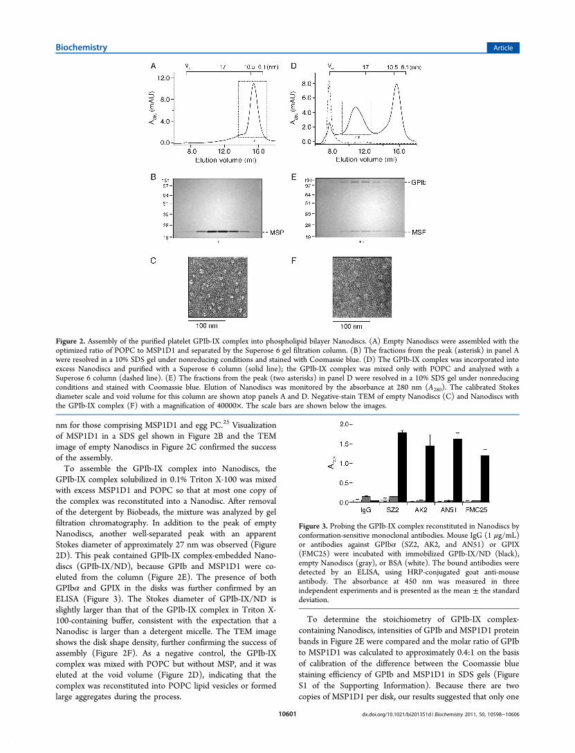
nm for those comprising MSP1D1 and egg PC.25 Visualizationof MSP1D1 in a SDS gel shown in Figure 2B and the TEMimage of empty Nanodiscs in Figure 2C confirmed the successof the assembly.To assemble the GPIb-IX complex into Nanodiscs, the
GPIb-IX complex solubilized in 0.1% Triton X-100 was mixedwith excess MSP1D1 and POPC so that at most one copy ofthe complex was reconstituted into a Nanodisc. After removalof the detergent by Biobeads, the mixture was analyzed by gelfiltration chromatography. In addition to the peak of emptyNanodiscs, another well-separated peak with an apparentStokes diameter of approximately 27 nm was observed (Figure2D). This peak contained GPIb-IX complex-embedded Nano-discs (GPIb-IX/ND), because GPIb and MSP1D1 were co-eluted from the column (Figure 2E). The presence of bothGPIbα and GPIX in the disks was further confirmed by anELISA (Figure 3). The Stokes diameter of GPIb-IX/ND isslightly larger than that of the GPIb-IX complex in Triton X-100-containing buffer, consistent with the expectation that aNanodisc is larger than a detergent micelle. The TEM imageshows the disk shape density, further confirming the success ofassembly (Figure 2F). As a negative control, the GPIb-IXcomplex was mixed with POPC but without MSP, and it waseluted at the void volume (Figure 2D), indicating that thecomplex was reconstituted into POPC lipid vesicles or formedlarge aggregates during the process.
To determine the stoichiometry of GPIb-IX complex-containing Nanodiscs, intensities of GPIb and MSP1D1 proteinbands in Figure 2E were compared and the molar ratio of GPIbto MSP1D1 was calculated to approximately 0.4:1 on the basisof calibration of the difference between the Coomassie bluestaining efficiency of GPIb and MSP1D1 in SDS gels (FigureS1 of the Supporting Information). Because there are twocopies of MSP1D1 per disk, our results suggested that only one
Figure 2. Assembly of the purified platelet GPIb-IX complex into phospholipid bilayer Nanodiscs. (A) Empty Nanodiscs were assembled with theoptimized ratio of POPC to MSP1D1 and separated by the Superose 6 gel filtration column. (B) The fractions from the peak (asterisk) in panel Awere resolved in a 10% SDS gel under nonreducing conditions and stained with Coomassie blue. (D) The GPIb-IX complex was incorporated intoexcess Nanodiscs and purified with a Superose 6 column (solid line); the GPIb-IX complex was mixed only with POPC and analyzed with aSuperose 6 column (dashed line). (E) The fractions from the peak (two asterisks) in panel D were resolved in a 10% SDS gel under nonreducingconditions and stained with Coomassie blue. Elution of Nanodiscs was monitored by the absorbance at 280 nm (A280). The calibrated Stokesdiameter scale and void volume for this column are shown atop panels A and D. Negative-stain TEM of empty Nanodiscs (C) and Nanodiscs withthe GPIb-IX complex (F) with a magnification of 40000×. The scale bars are shown below the images.
Figure 3. Probing the GPIb-IX complex reconstituted in Nanodiscs byconformation-sensitive monoclonal antibodies. Mouse IgG (1 μg/mL)or antibodies against GPIbα (SZ2, AK2, and AN51) or GPIX(FMC25) were incubated with immobilized GPIb-IX/ND (black),empty Nanodiscs (gray), or BSA (white). The bound antibodies weredetected by an ELISA, using HRP-conjugated goat anti-mouseantibody. The absorbance at 450 nm was measured in threeindependent experiments and is presented as the mean ± the standarddeviation.
Biochemistry Article
dx.doi.org/10.1021/bi201351d | Biochemistry 2011, 50, 10598−1060610601

copy of the GPIb-IX complex was incorporated into aNanodisc.VWF Binding Function of the GPIb-IX Complex
Reconstituted in Nanodiscs. To evaluate the quality ofthe GPIb-IX complex reconstituted in Nanodiscs, we firstprobed the conformation of GPIbα and GPIX usingconformation-sensitive monoclonal antibodies. SZ2, AN51,and AK2 recognize distinct three-dimensional epitopes in oraround the N-terminal ligand-binding domain of GPIbα,34,35
whereas FMC25 recognizes only the well-folded GPIXectodomain.30 GPIb-IX/ND, empty Nanodiscs, or BSA wasdirectly coated on the walls of microtiter wells and antibodybinding detected with an ELISA with HRP-conjugatedsecondary antibody. As shown in Figure 3, all the monoclonalantibodies tested bound to wells coated with GPIb-IX/ND butnot to those with empty Nanodisc or BSA, indicating that theGPIb-IX complex reconstituted in Nanodiscs adopts the native-like conformation. Moreover, the conformation of GPIb-IX/ND is stable as it retains binding to conformation-sensitiveantibodies after being stored at 4 °C for 2 weeks (data notshown).Next we determined the affinity of VWF for GPIb-IX/ND in
the presence of botrocetin, which mediates the interactionbetween GPIbα and VWF under static conditions.36 Because itis more difficult to accurately measure the concentration ofGPIb-IX/ND than that of VWF, GPIb-IX/ND was immobi-lized in the ELISA and the amount of bound VWF in themicrotiter well was measured using anti-VWF antibody. Figure4A shows that VWF bound to GPIb-IX/ND in a concentration-dependent and saturable manner, with an apparent dissociationconstant of 0.73 ± 0.07 nM if the molar concentration of VWFwas calculated with a molecular mass of 250 kDa. Themeasured dissociation constant is the same as that observed forfixed human platelets (data not shown) and comparable withthose from previous reports.37,38 The effects of antibodiesagainst each subunit of the GPIb-IX complex on VWF bindingwere also tested. AK2, targeting the ligand-binding domain ofGPIbα,34 abolished botrocetin-induced VWF binding, indicat-ing that the binding is specific to GPIbα. As expected, FMC25,targeting the GPIX extracellular domain, or Gi27, targeting theGPIbβ cytoplasmic domain, had no effect (Figure 4B). Overall,these results showed that the GPIb-IX complex retains itsnormal ligand binding function when reconstituted in Nano-discs.Effect of RAM.1 on Binding of VWF to the GPIb-IX
Complex in Nanodiscs. Although the binding site for VWFis located on the N-terminus of GPIbα, GPIbβ has been shownto regulate the ligand binding function of GPIbα, eitherthrough their cytoplasmic tails15,16 or through the membrane-proximal disulfide bonds.39 RAM.1, a rat monoclonal antibodythat recognizes the extracellular part (Cys68−Cys93) of GPIbβ,hampers botrocetin-induced binding of platelets or GPIb-IXcomplex-transfected cells to VWF as well as their adhesion toVWF under flow conditions.27,40 We tested whether RAM.1can hamper the association of GPIb-IX/ND with VWF in vitro.As shown in Figure 5A, RAM.1 bound to GPIb-IX/ND, but notto empty Nanodiscs or BSA that were immobilized inmicrotiter wells. However, after 10 μg/mL rat IgG or RAM.1was preincubated with immobilized GPIb-IX/ND, no signifi-cant difference in botrocetin-mediated VWF binding wasobserved (Figure 5B). Similar results were observed when ahigher concentration of RAM.1 was used in preincubation,when GPIb-IX/ND was captured by WM23 instead of being
coated directly onto microtiter wells, or when recombinantA1A2A3 tridomains of VWF were used for binding. Thus, theseresults suggest that the effect of RAM.1 on VWF bindingobserved in platelets or GPIb-IX complex-transfected cells maynot be due to direct interference with the VWF−GPIbαinteraction and thus is unlikely to be restricted to theextracellular domain of GPIbβ.Comparison of Ligand Binding between GPIb-IX/ND
and Glycocalicin. To further investigate the role of GPIbβ inthe ligand binding function of GPIbα, binding of full-lengthVWF or A1A2A3 tridomains to GPIb-IX/ND in the presenceof botrocetin was compared to binding to glycocalicin by anELISA. Because glycocalicin could not be recognized by AN51when it was directly coated to microtiter wells, WM23, a mousemonoclonal antibody targeting the macroglycopeptide portionof GPIbα, was used to capture GPIb-IX/ND and glycocalicin inmicrotiter wells. Although results of an ELISA showed thatapparent dissociation constants obtained for interaction withfull-length VWF or A1A2A3 tridomains were indistinguishablebetween GPIb-IX/ND and glycocalicin, the amount of boundVWF or A1A2A3 was different for GPIb-IX/ND andglycocalicin (Figure 6A,B). To explain this difference, SZ2, amonoclonal antibody targeting the N-terminal domain ofGPIbα, was used to detect the amount of GPIb-IX/ND and
Figure 4. Botrocetin-induced binding of VWF to the GPIb-IXcomplex reconstituted in Nanodiscs. (A) Botrocetin-induced bindingof VWF to the GPIb-IX complex in Nanodiscs measured with anELISA. Increasing concentrations of VWF were incubated withimmobilized GPIb-IX/ND (●), empty Nanodiscs (■), or BSA(▲) in the presence of 0.2 unit/mL botrocetin. The binding of VWFwas assessed with rabbit anti-VWF antibody and HRP-conjugated goatanti-rabbit antibody. (B) Effect of monoclonal antibodies againstindividual subunits of the GPIb-IX complex on VWF binding.Antibodies (10 μg/mL) were preincubated with immobilized GPIb-IX/ND before 0.8 nM VWF with 0.2 unit/mL botrocetin was added.The bound VWF was detected by HRP-conjugated anti-VWFantibody. The absorbance at 450 nm was measured in threeindependent experiments and is presented as the mean ± the standarddeviation. For some data points, the error bars are smaller than thesymbols. One hundred percent in panel B is defined as the binding ofVWF to GPIb-IX/ND in the absence of inhibitory antibodies aftersubtraction of the binding to empty Nanodiscs. Groups werecompared using the Student’s t test. **P < 0.01 vs control.
Biochemistry Article
dx.doi.org/10.1021/bi201351d | Biochemistry 2011, 50, 10598−1060610602

glycocalicin immobilized by the WM23 F(ab′)2 fragment inmicrotiter wells. As shown in Figure 6C, significantly moreGPIb-IX/ND than glycocalicin was present in the well,although the same amount of the WM23 F(ab′)2 fragmentwas used for immobilization. Moreover, the difference inWM23’s capturing abilities between GPIb-IX/ND andglycocalicin matches the difference in bound A1A2A3tridomains in ELISAs, indicating that the apparent weakerbinding of A1A2A3 to glycocalicin is due to the lower captureefficiency of glycocalicin by WM23 (Figure 6B). Because full-length VWF is multimeric, the amount of bound VWF may notbe simply correlated with that of the captured receptor. Thus,there appeared to be little difference in the ligand bindingfunction between the GPIb-IX complex reconstituted inNanodiscs and glycocalicin in the presence of botrocetin.
■ DISCUSSION
Compared to detergent micelles, Nanodiscs provide membraneproteins with a real phospholipid bilayer environment thatmimics the cell membrane. Phosphatidylcholine, the mostabundant phospholipid in cell membranes, was used in thisstudy to construct the Nanodisc. Compared to phospholipidvesicles, Nanodiscs also offer several advantages.19 One of theadvantages is the simultaneous access to both extracellular and
cytoplasmic domains of the membrane receptor embedded inNanodiscs. This is particularly useful for mechanistic studies ofreceptor-mediated signal transduction across the mem-brane.24,26 Thus, reconstitution of the functional GPIb-IXcomplex into phospholipid Nanodiscs will provide a usefulplatform for mechanistic investigations of GPIb-IX complex-mediated transmembrane signaling events.In this study, we have reconstituted the purified GPIb-IX
complex into phospholipid Nanodiscs and shown that theGPIb-IX complex is functional in this novel model membrane.Incorporation of the GPIb-IX complex into Nanodiscs wasconfirmed by three lines of evidence. First, MSP1D1 co-eluted
Figure 5. Effect of RAM.1 on the VWF binding activity of the GPIb-IXcomplex reconstituted in Nanodiscs. (A) Binding of RAM.1 to theGPIb-IX complex in Nanodiscs. Rat IgG or RAM.1 (each at 2 μg/mL)was incubated with immobilized GPIb-IX/ND (black) or emptyNanodiscs (gray), or BSA (white), and the binding was detected byHRP-conjugated goat anti-rat antibody. (B) Effect of RAM.1 onbotrocetin-induced binding of VWF to the GPIb-IX complex inNanodiscs. Antibodies at 10 μg/mL, rat IgG (○) and RAM.1 (●),mouse IgG (△), and AK2 (▲), were preincubated with GPIb-IX/ND-coated wells, and then increasing concentrations of VWF with 0.2unit/mL botrocetin was added. The binding was detected by HRP-conjugated anti-VWF antibody. The addition of VWF with botrocetinto empty Nanodisc-coated (■) or BSA-coated (□) wells served as anegative control. The absorbance at 450 nm was measured in threeindependent experiments and is presented as the mean ± the standarddeviation. For some data points, the error bars are smaller than thesymbols.
Figure 6. Comparison of ligand binding activities between GPIb-IX/ND and glycocalicin. WM23 IgG (4 μg/mL) was immobilized ontomicrotiter wells to capture GPIb-IX/ND (●) and glycocalicin (■).Empty Nanodiscs (▲) were also added to the WM23-coated wells asa control. (A) Full-length VWF (0−6.4 nM) or (B) A1A2A3tridomains (0−6.4 nM) were incubated with GPIb-IX/ND, glyco-calicin, or control wells in the presence of 0.2 unit/mL botrocetin. Thebinding was detected by anti-VWF antibody and HRP-conjugatedsecondary antibody. (C) Difference in capturing efficiency. TheWM23 IgG F(ab′)2 fragment (4 μg/mL) was coated to microtiterwells to capture GPIb-IX/ND or glycocalicin. Empty Nanodiscs werealso added to the wells coated with the WM23 F(ab′)2 fragment as acontrol. SZ2 (1 μg/mL) was incubated and detected by HRP-conjugated goat anti-mouse antibody. “Nonspecific binding” is thebinding of SZ2 to the control wells. The absorbance at 450 nm wasmeasured in three independent experiments and is presented as themean ± the standard deviation. For some of the points, the error barsare smaller than the symbols. One hundred percent in panel C isdefined as the binding of SZ2 to GPIb-IX/ND captured by the WM23F(ab′)2 fragment.
Biochemistry Article
dx.doi.org/10.1021/bi201351d | Biochemistry 2011, 50, 10598−1060610603
with GPIb from the gel filtration column after assembly. Co-elution of 25 kDa MSP1D1 with 220 kDa GPIb-IX complexindicates that they are bound together and eluted as a complex.Second, GPIb-IX/ND eluted at a slightly larger Stokes diameterthan the GPIb-IX complex dissolved in the Triton X-100-containing buffer. The average molecular mass of a Triton X-100 micelle is 80 kDa.41 Assuming that there are 160 moleculesof POPC in each Nanodisc, the molecular mass of one emptydisk is 170 kDa.20 The higher molecular mass of a diskcompared to a detergent micelle would explain the slightlylarger Stokes diameter of GPIb-IX/ND. A similar differencewas also observed for EGFR reconstituted in Nanodiscs versusthat dissolved in the Triton X-100-containing buffer.26 Finally,the negative-stain TEM image of GPIb-IX/ND showed the diskshape density, consistent with previous reports.42
Reconstituted in the Nanodisc, the GPIb-IX complex adoptsthe native conformation as it is able to bind all theconformation-sensitive antibodies tested in this study. TheGPIb-IX complex in Nanodiscs is stable for at least 2 weeks(data not shown). Moreover, it shows native-like ligand bindingproperties, having exhibited the same binding affinity for VWFin the presence of botrocetin as in previous reports.37,38 Thebinding affinity measured in this study is also comparable tothose previously reported for the binding of the N-terminalfragment of GPIbα to VWF in the presence of botrocetin.11,43
GPIbβ plays a central role in assembly of the GPIb-IXcomplex. It interacts with GPIbα through a disulfide bond nearthe membrane as well as interactions between their trans-membrane domain.5,44,45 There is also cross-talk between thecytoplasmic tails of these two subunits through the associationwith 14-3-3ζ.15 In a related manner, binding of RAM.1antibody to the GPIbβ extracellular domain leads to thealteration of the cytoplasmic tails of GPIb and at the same timehampers the ligand binding ability of the extracellular domainof GPIbα.40 The molecular mechanism underlying GPIbβmodulation of the VWF binding function of GPIbα is not clear.One possible mechanism may be that the GPIbβ extracellulardomain, with or without RAM.1, directly participates in aninteraction with VWF and thus directly modulates the GPIbα−VWF interaction. To test this possibility, we compared bindingof VWF to glycocalicin versus GPIb-IX/ND. Consistent with aprevious study,38 although glycocalicin and GPIb-IX/ND forsome reason exhibit different immobilization efficiencies withWM23, little difference in binding affinity was observed (Figure6). Furthermore, while RAM.1 exhibited specific binding toGPIb-IX/ND, it did not have any detectable effect on theassociation of VWF with GPIb-IX/ND in the presence ofbotrocetin (Figure 5). These results indicate that the GPIbβextracellular domain may not directly participate in theinteraction with VWF or modulate the GPIbα−VWFinteraction, and that the observed effects of RAM.1 on VWFbinding by platelets and GPIb-IX complex-expressing cells mayrequire other factors inside the cell. One possible scenario isthat RAM.1 binding to the GPIbβ extracellular domain maytransmit a signal into the platelet, through perhaps associationof 14-3-3ζ with cytoplasmic domains of the GPIb-IXcomplex,15,16,46−48 and influence the surface distribution and/or the overall binding activity of the GPIb-IX complex. Furtherinvestigation will be needed to elucidate the apparent trans-subunit effects of RAM.1 on GPIb-IX complex function andsignaling.
■ ASSOCIATED CONTENT
*S Supporting InformationCalibration of GPIb and MSP staining in SDS gels. Thismaterial is available free of charge via the Internet at http://pubs.acs.org.
■ AUTHOR INFORMATION
Corresponding Author*Aflac Cancer Center and Blood Disorders Service, 2015Uppergate Dr., Room 426A, Atlanta, GA 30322. Telephone:(404) 727-8217. Fax: (404) 727-4859. E-mail: [email protected].
FundingSupported by National Institutes of Health Grant HL082808.
■ ACKNOWLEDGMENTSWe thank Ms. Katie Sowa and Limei H. Jones for technicalassistance.
■ ABBREVIATIONSPT-VWD, platelet-type von Willebrand disease; POPC, 1-palmitoyl-2-oleoyl-sn-glycero-3-phosphocholine; HRP, horse-radish peroxidase; PRP, platelet-rich plasma; PMSF, phenyl-methanesulfonyl fluoride; ELISA, enzyme-linked immunosorb-ent assay; BSA, bovine serum albumin; TEM, transmissionelectron microscopy.
■ REFERENCES(1) Takagi, J., Petre, B. M., Walz, T., and Springer, T. A. (2002)Global conformational rearrangements in integrin extracellulardomains in outside-in and inside-out signaling. Cell 110, 599−611.(2) Vinogradova, O., Velyvis, A., Velyviene, A., Hu, B., Haas, T. A.,Plow, E. F., and Qin, J. (2002) A structural mechanism of integrinαIIbβ3 “inside-out” activation as regulated by its cytoplasmic face. Cell110, 587−597.(3) O’Toole, T. E., Mandelman, D., Forsyth, J., Shattil, S. J., Plow, E.F., and Ginsberg, M. H. (1991) Modulation of the affinity of integrinαIIbβ3 (GPIIb-IIIa) by the cytoplasmic domain of αIIb. Science 254,845−847.(4) Du, X., Beutler, L., Ruan, C., Castaldi, P. A., and Berndt, M. C.(1987) Glycoprotein Ib and glycoprotein IX are fully complexed in theintact platelet membrane. Blood 69, 1524−1527.(5) Luo, S.-Z., Mo, X., Afshar-Kharghan, V., Srinivasan, S., Lopez, J.A., and Li, R. (2007) Glycoprotein Ibα forms disulfide bonds with 2glycoprotein Ibβ subunits in the resting platelet. Blood 109, 603−609.(6) Luo, S.-Z., Mo, X., Lopez, J. A., and Li, R. (2007) Role of thetransmembrane domain of glycoprotein IX in assembly of theglycoprotein Ib-IX complex. J. Thromb. Haemostasis 5, 2494−2502.(7) Mo, X., Nguyen, N. X., McEwan, P. A., Zheng, X., Lopez, J. A.,Emsley, J., and Li, R. (2009) Binding of platelet glycoprotein Ibβthrough the convex surface of leucine-rich repeats domain ofglycoprotein IX. J. Thromb. Haemostasis 7, 1533−1540.(8) McEwan, P. A., Yang, W., Carr, K. H., Mo, X., Zheng, X., Li, R.,and Emsley, J. (2011) Quaternary organization of GPIb-IX complexand insights into Bernard-Soulier syndrome revealed by the structuresof GPIbβ and a GPIbβ/GPIX chimera. Blood 118, 5292−5301.(9) Huizinga, E. G., Tsuji, S., Romijn, R. A., Schiphorst, M. E., deGroot, P. G., Sixma, J. J., and Gros, P. (2002) Structures ofglycoprotein Ibα and its complex with von Willebrand factor A1domain. Science 297, 1176−1179.(10) Cauwenberghs, N., Vanhoorelbeke, K., Vauterin, S., andDeckmyn, H. (2000) Structural determinants within plateletglycoprotein Ibα involved in its binding to von Willebrand factor.Platelets 11, 373−378.
Biochemistry Article
dx.doi.org/10.1021/bi201351d | Biochemistry 2011, 50, 10598−1060610604

(11) Miura, S., Li, C. Q., Cao, Z., Wang, H., Wardell, M. R., andSadler, J. E. (2000) Interaction of von Willebrand factor domain A1with platelet glycoprotein Ibα-(1−289). Slow intrinsic binding kineticsmediate rapid platelet adhesion. J. Biol. Chem. 275, 7539−7546.(12) Kroll, M. H., Harris, T. S., Moake, J. L., Handin, R. I., andSchafer, A. I. (1991) von Willebrand factor binding to platelet GpIbinitiates signals for platelet activation. J. Clin. Invest. 88, 1568−1573.(13) Savage, B., Shattil, S. J., and Ruggeri, Z. M. (1992) Modulationof platelet function through adhesion receptors. A dual role forglycoprotein IIb-IIIa (integrin αIIbβ3) mediated by fibrinogen andglycoprotein Ib-von Willebrand factor. J. Biol. Chem. 267, 11300−11306.(14) Guerrero, J. A., Kyei, M., Russell, S., Liu, J., Gartner, T. K.,Storrie, B., and Ware, J. (2009) Visualizing the von Willebrand factor/glycoprotein Ib-IX axis with a platelet-type von Willebrand diseasemutation. Blood 114, 5541−5546.(15) Dai, K., Bodnar, R., Berndt, M. C., and Du, X. (2005) A criticalrole for 14-3-3ζ protein in regulating the VWF binding function ofplatelet glycoprotein Ib-IX and its therapeutic implications. Blood 106,1975−1981.(16) Bodnar, R. J., Xi, X., Li, Z., Berndt, M. C., and Du, X. (2002)Regulation of glycoprotein Ib-IX-von Willebrand factor interaction bycAMP-dependent protein kinase-mediated phosphorylation at Ser 166of glycoprotein Ibβ. J. Biol. Chem. 277, 47080−47087.(17) Othman, M., Notley, C., Lavender, F. L., White, H., Byrne, C.D., Lillicrap, D., and O’Shaughnessy, D. F. (2005) Identification andfunctional characterization of a novel 27-bp deletion in themacroglycopeptide-coding region of the GPIbα gene resulting inplatelet-type von Willebrand disease. Blood 105, 4330−4336.(18) Bayburt, T. H., Grinkova, Y. V., and Sligar, S. G. (2002) self-assembly of discoidal phospholipid bilayer nanoparticles withmembrane scaffold proteins. Nano Lett. 2, 853−856.(19) Nath, A., Atkins, W. M., and Sligar, S. G. (2007) Applications ofphospholipid bilayer nanodiscs in the study of membranes andmembrane proteins. Biochemistry 46, 2059−2069.(20) Denisov, I. G., Grinkova, Y. V., Lazarides, A. A., and Sligar, S. G.(2004) Directed self-assembly of monodisperse phospholipid bilayerNanodiscs with controlled size. J. Am. Chem. Soc. 126, 3477−3487.(21) Bayburt, T. H., Grinkova, Y. V., and Sligar, S. G. (2006)Assembly of single bacteriorhodopsin trimers in bilayer nanodiscs.Arch. Biochem. Biophys. 450, 215−222.(22) Bayburt, T. H., and Sligar, S. G. (2003) Self-assembly of singleintegral membrane proteins into soluble nanoscale phospholipidbilayers. Protein Sci. 12, 2476−2481.(23) Whorton, M. R., Bokoch, M. P., Rasmussen, S. G., Huang, B.,Zare, R. N., Kobilka, B., and Sunahara, R. K. (2007) A monomeric Gprotein-coupled receptor isolated in a high-density lipoprotein particleefficiently activates its G protein. Proc. Natl. Acad. Sci. U.S.A. 104,7682−7687.(24) Boldog, T., Grimme, S., Li, M., Sligar, S. G., and Hazelbauer, G.L. (2006) Nanodiscs separate chemoreceptor oligomeric states andreveal their signaling properties. Proc. Natl. Acad. Sci. U.S.A. 103,11509−11514.(25) Mi, L. Z., Grey, M. J., Nishida, N., Walz, T., Lu, C., and Springer,T. A. (2008) Functional and structural stability of the epidermalgrowth factor receptor in detergent micelles and phospholipidnanodiscs. Biochemistry 47, 10314−10323.(26) Ye, F., Hu, G., Taylor, D., Ratnikov, B., Bobkov, A. A., McLean,M. A., Sligar, S. G., Taylor, K. A., and Ginsberg, M. H. (2010)Recreation of the terminal events in physiological integrin activation. J.Cell Biol. 188, 157−173.(27) Perrault, C., Moog, S., Rubinstein, E., Santer, M., Baas, M. J., dela Salle, C., Ravanat, C., Dambach, J., Freund, M., Santoso, S.,Cazenave, J. P., and Lanza, F. (2001) A novel monoclonal antibodyagainst the extracellular domain of GPIbβ modulates vWF mediatedplatelet adhesion. Thromb. Haemostasis 86, 1238−1248.(28) Auton, M., Cruz, M. A., and Moake, J. (2007) Conformationalstability and domain unfolding of the von Willebrand factor Adomains. J. Mol. Biol. 366, 986−1000.
(29) Fox, J. E. (1985) Linkage of a membrane skeleton to integralmembrane glycoproteins in human platelets. Identification of one ofthe glycoproteins as glycoprotein Ib. J. Clin. Invest. 76, 1673−1683.(30) Berndt, M. C., Gregory, C., Kabral, A., Zola, H., Fournier, D.,and Castaldi, P. A. (1985) Purification and preliminarycharacterization of the glycoprotein Ib complex in the humanplatelet membrane. Eur. J. Biochem. 151, 637−649.(31) Hess, D., Schaller, J., Rickli, E. E., and Clemetson, K. J. (1991)Identification of the disulphide bonds in human platelet glycocalicin.Eur. J. Biochem. 199, 389−393.(32) Denisov, I. G., Baas, B. J., Grinkova, Y. V., and Sligar, S. G.(2007) Cooperativity in cytochrome P450 3A4: Linkages in substratebinding, spin state, uncoupling, and product formation. J. Biol. Chem.282, 7066−7076.(33) Fox, J. E., Aggerbeck, L. P., and Berndt, M. C. (1988) Structureof the glycoprotein Ib-IX complex from platelet membranes. J. Biol.Chem. 263, 4882−4890.(34) Shen, Y., Romo, G. M., Dong, J. F., Schade, A., McIntire, L. V.,Kenny, D., Whisstock, J. C., Berndt, M. C., Lopez, J. A., and Andrews,R. K. (2000) Requirement of leucine-rich repeats of glycoprotein (GP)Ibα for shear-dependent and static binding of von Willebrand factor tothe platelet membrane GP Ib-IX-V complex. Blood 95, 903−910.(35) Ward, C. M., Andrews, R. K., Smith, A. I., and Berndt, M. C.(1996) Mocarhagin, a novel cobra venom metalloproteinase, cleavesthe platelet von Willebrand factor receptor glycoprotein Ibα.Identification of the sulfated tyrosine/anionic sequence Tyr-276-Glu-282 of glycoprotein Ibα as a binding site for von Willebrand factor andα-thrombin. Biochemistry 35, 4929−4938.(36) Andrews, R. K., Booth, W. J., Gorman, J. J., Castaldi, P. A., andBerndt, M. C. (1989) Purification of botrocetin from Bothrops jararacavenom. Analysis of the botrocetin-mediated interaction between vonWillebrand factor and the human platelet membrane glycoprotein Ib-IX complex. Biochemistry 28, 8317−8326.(37) Hoylaerts, M. F., Nuyts, K., Peerlinck, K., Deckmyn, H., andVermylen, J. (1995) Promotion of binding of von Willebrand factor toplatelet glycoprotein Ib by dimers of ristocetin. Biochem. J. 306, 453−463.(38) Meyer, S., Kresbach, G., Haring, P., Schumpp-Vonach, B.,Clemetson, K. J., Hadvary, P., and Steiner, B. (1993) Expression andcharacterization of functionally active fragments of the plateletglycoprotein (GP) Ib-IX complex in mammalian cells. Incorporationof GP Ibα into the cell surface membrane. J. Biol. Chem. 268, 20555−20562.(39) Mo, X., Luo, S.-Z., Munday, A. D., Sun, W., Berndt, M. C.,Lopez, J. A., Dong, J.-f., and Li, R. (2008) The membrane-proximalintermolecular disulfide bonds in glycoprotein Ib influence receptorbinding to von Willebrand factor. J. Thromb. Haemostasis 6, 1789−1795.(40) Perrault, C., Mangin, P., Santer, M., Baas, M. J., Moog, S.,Cranmer, S. L., Pikovski, I., Williamson, D., Jackson, S. P., Cazenave, J.P., and Lanza, F. (2003) Role of the intracellular domains of GPIb incontrolling the adhesive properties of the platelet GPIb/V/IXcomplex. Blood 101, 3477−3484.(41) Biaselle, C. J., and Millar, D. B. (1975) Studies on Triton X-100detergent micelles. Biophys. Chem. 3, 355−361.(42) Tsukamoto, H., Sinha, A., DeWitt, M., and Farrens, D. L.(2010) Monomeric rhodopsin is the minimal functional unit requiredfor arrestin binding. J. Mol. Biol. 399, 501−511.(43) Murata, M., Ware, J., and Ruggeri, Z. M. (1991) Site-directedmutagenesis of a soluble recombinant fragment of platelet glycoproteinIbα demonstrating negatively charged residues involved in vonWillebrand factor binding. J. Biol. Chem. 266, 15474−15480.(44) Mo, X., Lu, N., Padilla, A., Lo pez, J. A., and Li, R. (2006) Thetransmembrane domain of glycoprotein Ibβ is critical to efficientexpression of glycoprotein Ib-IX complex in the plasma membrane. J.Biol. Chem. 281, 23050−23059.(45) Luo, S. Z., and Li, R. (2008) Specific heteromeric association offour transmembrane peptides derived from platelet glycoprotein Ib-IXcomplex. J. Mol. Biol. 382, 448−457.
Biochemistry Article
dx.doi.org/10.1021/bi201351d | Biochemistry 2011, 50, 10598−1060610605

(46) Calverley, D. C., Kavanagh, T. J., and Roth, G. J. (1998) Humansignaling protein 14-3-3ζ interacts with platelet glycoprotein Ibsubunits Ibα and Ibβ. Blood 91, 1295−1303.(47) Mangin, P., David, T., Lavaud, V., Cranmer, S. L., Pikovski, I.,Jackson, S. P., Berndt, M. C., Cazenave, J. P., Gachet, C., and Lanza, F.(2004) Identification of a novel 14-3-3ζ binding site within thecytoplasmic tail of platelet glycoprotein Ibα. Blood 104, 420−427.(48) David, T., Ohlmann, P., Eckly, A., Moog, S., Cazenave, J. P.,Gachet, C., and Lanza, F. (2006) Inhibition of adhesive and signalingfunctions of the platelet GPIb-V-IX complex by a cell penetratingGPIbα peptide. J. Thromb. Haemostasis 4, 2645−2655.
Biochemistry Article
dx.doi.org/10.1021/bi201351d | Biochemistry 2011, 50, 10598−1060610606
Related Documents