Recognizing and Diagnosing Peripheral Arterial Disease (P.A.D.) A Clinical Introduction

Welcome message from author
This document is posted to help you gain knowledge. Please leave a comment to let me know what you think about it! Share it to your friends and learn new things together.
Transcript
Recognizing and
Diagnosing Peripheral
Arterial Disease (P.A.D.)
A Clinical Introduction
page 2
The information provided in this presentation was created at the direction of
and with monetary support from ev3 Endovascular, Inc. All rights to this
presentation are owned by ev3 Endovascular, Inc. Any reproduction or use of
the contents of this presentation without ev3 Endovascular, Inc.’s permission is
strictly prohibited. Should you wish to use or reproduce any portion of this
presentation, please contact [email protected] at ev3
Endovascular, Inc.
This presentation was co-authored with Ofstead & Associates, Inc., Dr Alan
Hirsch and ev3 Inc.
The information provided in this presentation was created with monetary support from ev3 Endovascular, Inc.

page 3
A Clinical Introduction
to P.A.D.
• This presentation covers the following P.A.D. topics:
o Overview
o Risk factors and epidemiology
o Clinical presentation
o Clinical outcomes and comorbid conditions
o Early detection and diagnosis
o Treatment options
o Economic costs
o Specialty concerns
o Call to action
The information provided in this presentation was created with monetary support from ev3 Endovascular, Inc.

page 4
Overview of Peripheral
Arterial Disease (P.A.D.)
• All non-coronary arterial
diseases
• P.A.D.:
o Causes acute and chronic
illness
o Reduces functional capacity
and quality of life
o Causes limb amputations
o Increases risk of death
Lower
extremity
P.A.D.
Licensed from Shutterstock, 2010
The information provided in this presentation was created with monetary support from ev3 Endovascular, Inc.

page 5
P.A.D. Nomenclature
• P.A.D. is Peripheral Arterial
Disease
• P.A.D. is a disease that has
been called many names:
■ PVD (peripheral vascular
disease)
■ PAOD (peripheral arterial
occlusive disease)
■ LEP.A.D. (lower extremity
peripheral arterial disease)
■ Arteriosclerosis obliterans
The information provided in this presentation was created with monetary support from ev3 Endovascular, Inc.
Licensed from Shutterstock, 2010

page 6
Atherosclerosis and
P.A.D.
• Manifestation of a systemic
disease
• Buildup of plaque
■ Cholesterol and other fats
■ Calcium
■ Fibrous tissue
■ Other substances
• Arterial stenosis or occlusion
• Reduced blood flow
• Increased risk of
cardiovascular events and
death
The information provided in this presentation was created with monetary support from ev3 Endovascular, Inc.
Peripheral
Licensed from Nucleus Medical Media, 2010

page 7
Risk Factors for P.A.D.
• Lifestyle ■ Smoking
■ Obesity
• Health conditions ■ Diabetes
■ Cardiovascular disease
■ Erectile dysfunction
■ Chronic kidney disease
■ Hypertension
■ Hyperlipidemia
• Demographics ■ Older age
■ Black race
The information provided in this presentation was created with monetary support from ev3 Endovascular, Inc.
More than half of the attributable
risk of P.A.D. is due to smoking
and diabetes
Licensed from Shutterstock, 2010

page 8
Smoking and P.A.D.
• More than 80% of persons
with P.A.D. are current or
former smokers
• Smoking increases the risk
of P.A.D. 4-fold
• P.A.D. in smokers:
o Develops 10 years earlier
o More likely to progress
o Worse outcomes
■ Double the risk of amputation
■ Poor survival rates
The information provided in this presentation was created with monetary support from ev# Endovascular, Inc.
“Smoking is the single most
important modifiable risk factor
for prevention of P.A.D.”
Licensed from Shutterstock, 2010

page 9
Smoking and P.A.D.
Smoking introduces lead and cadmium into the body
Higher levels of these metals increase the risk of P.A.D. almost 3 times
The information provided in this presentation was created with monetary support from ev# Endovascular, Inc.
Licensed from Shutterstock, 2010

page 10
Smoking and P.A.D.
The risk of P.A.D. is dose-dependent
Risk and severity of P.A.D. increase with the number
of cigarettes and years smoked
The information provided in this presentation was created with monetary support from ev3 Endovascular, Inc.
Licensed from Shutterstock, 2010

page 11
P.A.D. and Current
Smokers
30%-40% of persons with P.A.D. are current smokers
80%-90% of persons with P.A.D. who require revascularization
are current smokers
The information provided in this presentation was created with monetary support from ev3 Endovascular, Inc.
Licensed from Shutterstock, 2010

page 12
Diabetes and P.A.D.
• 25%-40% of persons with P.A.D. have diabetes
• Risk of P.A.D. is 2-4 times higher
• Risk increases in proportion to diabetes severity and duration
• P.A.D. in persons with diabetes: o Commonly asymptomatic
o More severe and progresses rapidly
o Worse outcomes ■ Ulceration and gangrene
■ Amputation
■ Cardiovascular events
The information provided in this presentation was created with monetary support from ev3 Endovascular, Inc.
Licensed from Shutterstock, 2010

page 13
Diabetes and P.A.D.
1 in 3 persons over age 50 with diabetes is likely to have P.A.D.
The information provided in this presentation was created with monetary support from ev3 Endovascular, Inc.
Licensed from Shutterstock, 2010

page 14
Diabetes, P.A.D., and
Amputation
• Diabetes alone does not
cause amputation—it
increases the risk of P.A.D.
• P.A.D. and diabetes are the
leading cause of non-
traumatic, lower limb
amputations
• P.A.D. patients with diabetes
have a 7-15 times higher risk
of amputation
The information provided in this presentation was created with monetary support from ev3 Endovascular, Inc.
Licensed from Mediscan, 2010

page 15
Prevalence of P.A.D.
and Cancer in the U.S.
P.A.D. affects the same
number of Americans as
cancer
Cancer
11 million
P.A.D
. 8-12 million
The information provided in this presentation was created with monetary support from ev3 Endovascular, Inc.
Licensed from Shutterstock, 2010
Licensed from Shutterstock, 2010

page 16
5-Year Mortality Rates for
P.A.D. and Breast Cancer in
the U.S.
The information provided in this presentation was created with monetary support from ev3 Endovascular, Inc.
P.A.D.
15%-30%
Breast Cancer
11%
Licensed from Shutterstock, 2010

page 17
Prevalence of P.A.D.
Among Older Adults
• The prevalence of P.A.D.
increases dramatically
with age
• 12%-20% of persons
aged 65 or older have
P.A.D.
The information provided in this presentation was created with monetary support from ev3 Endovascular, Inc.

page 18
Expansion of the
Older Population
The information provided in this presentation was created with monetary support from ev3 Endovascular, Inc.
Millions of Americans aged 65 and older by year
Orange indicates millions with P.A.D.
2000
35.0
2050
88.5 81.2
2040
72.0
2030
54.8
2020
40.2
2010
Millio
ns

page 19
Clinical Presentation of
P.A.D.
The information provided in this presentation was created with monetary support from ev3 Endovascular, Inc.
P.A.D. Status Rate of Clinical
Presentation (%)
Asymptomatic
No leg pain 20%-50%
Atypical leg pain
Leg discomfort with exertion 40%-50%
Claudication
Leg muscle discomfort with exertion 10%-35%
Critical limb ischemia (CLI)
Chronic leg pain at rest
Nonhealing ulcers and gangrene
1%-2%
Acute limb ischemia (ALI)
Sudden onset of leg pain NA

page 20
Asymptomatic P.A.D.
• More than 50% do not have
classical signs or symptoms
• Asymptomatic patients:
■ Subtle impairments of limb
function
■ Risk factors and comorbidities
comparable to symptomatic
patients
• Symptoms may not occur in
patients who do not perform
sufficient activity to produce
them
The information provided in this presentation was created with monetary support from ev3 Endovascular, Inc.
Licensed from Shutterstock, 2010

page 21
Claudication and
P.A.D.
• Claudication is the most common
symptom of P.A.D.
■ Cramping, aching, fatigue, weakness, or
pain
■ Involving the muscles of the buttocks,
legs, or feet
■ Occurs with activity
■ Quickly relieved by rest
• Present in only about 10% of P.A.D.
patients
• Claudication alone does not define
the presence or absence of P.A.D.
The information provided in this presentation was created with monetary support from ev3 Endovascular, Inc.
Licensed from Shutterstock, 2010

page 22
Clinical Signs of Limb
Ischemia
Licensed from Custom Medical Stock Photo & Mediscan, 2010
The information provided in this presentation was created with monetary support from ev3 Endovascular, Inc.

page 23
P.A.D. Patient are at
Increased Risk
• Impaired function and
quality of life
• Progressive disease
severity
• Amputation
• Cardiovascular ischemic
events
• Cardiovascular mortality
The information provided in this presentation was created with monetary support from ev3 Endovascular, Inc.
Licensed from Shutterstock, 2010

page 24
Loss of Functional
Independence with P.A.D.
Independence is valued in all stages of life and in all cultures
P.A.D. limits physical activity and can result in isolation
The information provided in this presentation was created with monetary support from ev3 Endovascular, Inc.
Licensed from Shutterstock, 2010

page 25
Comorbid Conditions
Associated with P.A.D.
• Atherosclerotic diseases:
o Coronary artery disease (CAD;
MI)
o Cerebrovascular disease (CVD;
stroke)
o Aortic aneurysmal disease
(rupture)
o Erectile dysfunction
• Chronic kidney disease (CKD)
• Diabetes
The information provided in this presentation was created with monetary support from ev3 Endovascular, Inc.
CAD
CVD
P.A.D.
Licensed from Shutterstock, 2010

page 26
Cardiovascular
Disease and P.A.D.
• Coprevalence of cardiovascular disease among P.A.D.
patients:
The information provided in this presentation was created with monetary support from ev3 Endovascular, Inc.
25%-50%
50%-80%
25%-40%
CVD
CAD
Renal
Licensed from Shutterstock, 2010

page 27
Cardiovascular Events
and P.A.D.
• P.A.D. patients have:
o 40% increased risk of a
cerebrovascular event
(stroke)
o 20%-60% increased risk of a
heart attack (MI)
o 2-6-fold increased risk of
death due to coronary events
The information provided in this presentation was created with monetary support from ev3 Endovascular, Inc.
Licensed from Shutterstock, 2010

page 28
Mortality Among P.A.D.
Patients
70%-80% of P.A.D. patients die of
cardiovascular causes
The information provided in this presentation was created with monetary support from ev3 Endovascular, Inc.
P.A.D. Status Annual mortality
rate
All patients
with P.A.D. 4%-6%
Acute limb
ischemia (ALI) 15%-20%
Critical limb
ischemia (CLI) 20-25%
CLI & amputation 45%
Licensed from Shutterstock, 2010

page 29
Importance of Early
Detection
• P.A.D. is underdiagnosed
■ Over ⅔ are asymptomatic or
have atypical symptoms
■ ½ have not yet suffered a major
cardiovascular event
• Early detection can identify
individuals:
■ Without claudication
■ With atypical leg symptoms
■ At high cardiovascular risk
• Initiate risk reduction
treatment
The information provided in this presentation was created with monetary support from ev3 Endovascular, Inc.
Licensed from Shutterstock, 2010

page 30
Target Your Efforts
The information provided in this presentation was created with monetary support from ev3 Endovascular, Inc.
Identify Persons at High Risk
Age >70 years
Lifestyle Smokers
• >50 years
Comorbidities Diabetes
• >50 years
• Other risk factors
Cardiovascular disease
Chronic kidney disease
Symptoms Leg pain with exertion
Leg pain at rest
Walking impairment
Nonhealing wounds Licensed from Shutterstock, 2010

page 31
Clinical Assessment
for P.A.D.
• Clinical History & Vascular Review
■ Vascular history
■ Limb symptoms
■ Atherosclerotic risk factors
■ Comorbid conditions
• Physical examination of the legs,
feet, and toes
■ Weak or absent peripheral pulses
■ Signs of limb ischemia
• Laboratory testing and ABI
The information provided in this presentation was created with monetary support from ev3 Endovascular, Inc.
Licensed from Shutterstock, 2010

page 32
Noninvasive Diagnostic
Tests for P.A.D.
• Universally indicated
diagnostic tests:
o Ankle-brachial index (ABI)
o Toe-brachial index (TBI)
■ Substitute or supplement for ABI
• Reimbursement for the ABI
depends on using
appropriate:
o Equipment
o Coding
The information provided in this presentation was created with monetary support from ev3 Endovascular, Inc.
Licensed from Mediscan, 2010

page 33
Measuring the ABI
To perform the ABI,
use a 10-12 cm blood
pressure cuff and a
handheld 5- or 10-
mHz Doppler probe
Sources: Hirsch et al. (2006) ACC/AHA Practice Guidelines for P.A.D.; Norgren et al (2007) TASC II Guidelines for P.A.D.
1. Left arm
2. Right arm
Left ankle:
3. Dorsalis pedis
4. Posterior tibial
Right ankle:
5. Dorsalis pedis
6. Posterior tibial
Systolic
blood
pressure
(mm Hg)
1. Take 6
measurements
with patient in
supine position
2. Select higher
values for
calculating ABI
Licensed from Mediscan, 2010
Licensed from Shutterstock, 2010
The information provided in this presentation was created with monetary support from ev3 Endovascular, Inc.

page 34
Calculating and
Interpreting the ABI
ABI Interpretation
(Arterial Status)
>1.30 Noncompressible
1.00-1.29 Normal
0.91-0.99 Borderline (equivocal)
0.41-0.90 Mild to Moderate
P.A.D.
0.00-0.40 Severe P.A.D.
P.A.D. is defined as an ABI of ≤0.90
Sources: Hirsch et al. (2006) ACC/AHA Practice Guidelines for P.A.D.; Norgren et al. (2007) TASC II Guidelines for P.A.D.
ABI Calculation
Right ABI:
Left ABI:
Higher
left ankle
pressure
Higher
arm
pressure
= ÷
Higher
right ankle
pressure
Higher
arm
pressure
= ÷
Licensed from Shutterstock, 2010
The information provided in this presentation was created with monetary support from ev3 Endovascular, Inc.

page 35
Value of the ABI Test
• Detects P.A.D. at all stages
• 95% sensitive and nearly 100% specific
• Confirms the diagnosis of P.A.D.
• Lower ABIs:
■ Higher cardiovascular risk
■ Greater disease severity
■ Worse prognosis for limb and life
• Most cost-effective tool for P.A.D. detection
The information provided in this presentation was created with monetary support from ev3 Endovascular, Inc.
The ABI is the gold standard
for diagnostic P.A.D. testing
Licensed from Shutterstock, 2010

page 36
ACC/AHA and TASC II
Guidelines on P.A.D.
• Current guidelines
endorsed by the American
Heart Association (AHA),
the American College of
Cardiology (ACC), and
international vascular
societies recommend:
o ABI testing for all patients
with a history or exam
indicative of P.A.D. (i.e., high
risk patients)
Patients at High Risk for P.A.D.
Age >70 years
Lifestyle Smokers
• >50 years
Comorbid
conditions
Diabetes
• >50 years
• Other risk factors
Cardiovascular disease
Chronic kidney disease
Symptoms Leg pain with exertion
Leg pain at rest
Walking impairment
Nonhealing wounds
The information provided in this presentation was created with monetary support from ev3 Endovascular, Inc.

page 37
Noninvasive Diagnostic
Tests for P.A.D.
• Supportive diagnostic tests to
determine anatomy, physiology,
or functional status:
o Segmental pressure
measurements
o Pulse volume recordings (PVR)
o Doppler waveform
measurements
o Transcutaneous oxygen tension
o Exercise ABI testing
o Vascular imaging
■ Duplex ultrasound
■ Angiography (CTA, MRA)
The information provided in this presentation was created with monetary support from ev3 Endovascular, Inc.
Licensed from Shutterstock & Custom Medical Stock Photo, 2010

page 38
Treatment of P.A.D.
• Treatment goals are to:
■ Reduce the risk of death and cardiovascular events
■ Prevent limb loss
■ Relieve symptoms
■ Improve function and quality of life
• Cardiovascular risk reduction therapy is indicated for all patients
■ Risk factor modification
■ Antiplatelet therapy
• Symptomatic treatment is individualized
The information provided in this presentation was created with monetary support from ev3 Endovascular, Inc.
Only 20%-30% of patients with
P.A.D. are receiving treatment
Licensed from Shutterstock, 2010

page 39
Lifestyle Modifications
to Treat P.A.D.
• Risk reduction:
o Smoking cessation
o Risk factor modification:
■ Lipid control
■ Blood pressure control
■ Diabetes control
■ Weight reduction
o Exercise
o Nonatherogenic diet
• Lifelong treatment
The information provided in this presentation was created with monetary support from ev3 Endovascular, Inc.
Licensed from Shutterstock, 2010

page 40
Medications for
Treating P.A.D.
• Risk reduction
o Statins
o ACE inhibitors
o Antiplatelet therapy
■ Aspirin
■ Clopidogrel
• Symptom relief
o Claudication
■ Cilostazol
o CLI
■ Pain medication
■ Antibiotics
The information provided in this presentation was created with monetary support from ev3 Endovascular, Inc.
Lifelong antiplatelet therapy is
recommended for patients with P.A.D.
You need to decide what is best for your
patient.
Licensed from Shutterstock, 2010

page 41
Exercise Therapy to
Treat P.A.D.
• Exercise program
■ Walking is most effective
■ Exercise-rest-exercise
• Sessions performed for:
■ Minimum of 30-45 minutes
■ At least 3 times per week
■ Minimum of 3 months
• Walking outcomes:
■ Relief from claudication
■ Increase in walking ability and
daily activity
■ Risk reduction
The information provided in this presentation was created with monetary support from ev3 Endovascular, Inc.
Licensed from Shutterstock, 2010

page 42
Revascularization and
P.A.D.
• Indications:
■ Failure with exercise and drug therapy
■ Lifestyle-limiting symptoms and function
■ Nonhealing wound
■ Risk of amputation
• Requires a favorable risk/benefit ratio
• Less invasive endovascular
procedures:
■ Preferred over surgery
■ Preserve options for fall-back surgical
procedures
The information provided in this presentation was created with monetary support from ev3 Endovascular, Inc.
Licensed from Shutterstock, 2010

page 43
Endovascular P.A.D.
Treatment –Angioplasty
• Mechanism:
o Catheter-guided balloon
o Balloon dilation
o Plaque displacement into the
artery wall
o Vessel stretch and expansion
The information provided in this presentation was created with monetary support from ev3 Endovascular, Inc.
Licensed from A.D.A.M., 2010

page 44
Endovascular P.A.D.
Treatment – Stents and
Stent-Grafts
• Mechanism:
o Balloon-expandable or self-
expanding
o Plaque displacement into the
artery wall
o Vessel stretch and expansion
• Indications:
o Prevent recoil of the artery wall
o Repair complications resulting
from angioplasty
The information provided in this presentation was created with monetary support from ev3 Endovascular, Inc.
Licensed from A.D.A.M. & Nucleus Medical Media, 2010

page 45
Endovascular P.A.D.
Treatment – Atherectomy
• Mechanism:
o Debulk plaque
■ Cut
■ Pulverize
■ Shave
o Remove or excise plaque
• Types:
o Directional or excisional
o Rotational or orbital
o Photoablative (excimer laser)
Source: Garcia et al. (2009)
The information provided in this presentation was created with monetary support from ev3 Endovascular, Inc.

page 46
Surgical Treatment for
P.A.D.
• Types:
o Surgical bypass
■ Venous or synthetic bypass
graft
o Endarterectomy
■ Surgical removal of plaque
o Intra-operative hybrid
procedure
• Not recommended as
prophylactic therapy
• Increased risk of operative
mortality
The information provided in this presentation was created with monetary support from ev3 Endovascular, Inc.
Licensed from Shutterstock, 2010
page 47
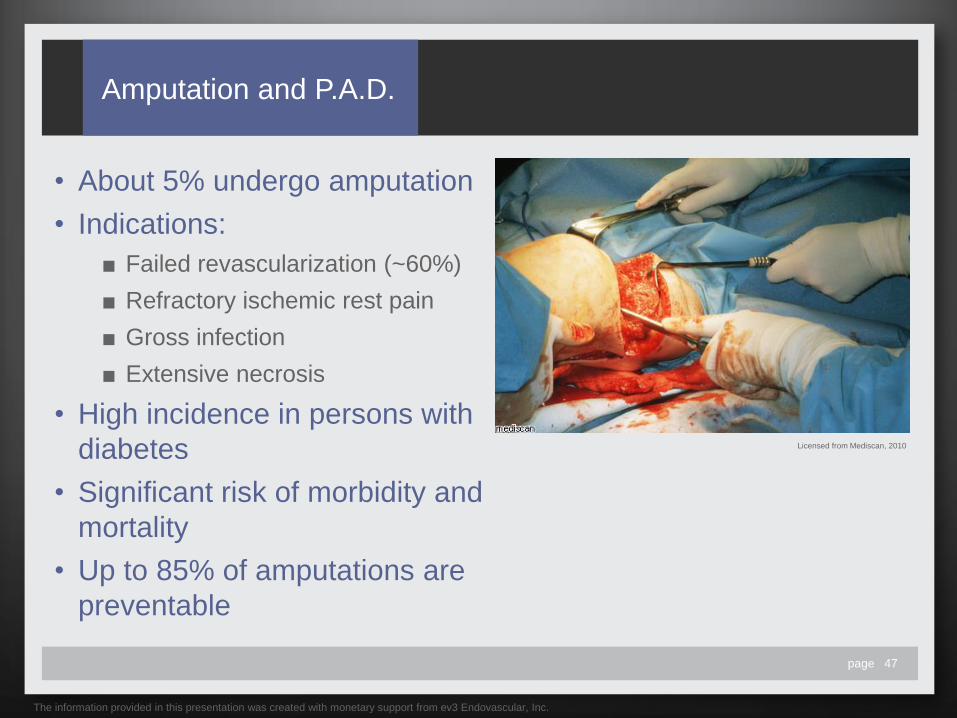
Amputation and P.A.D.
• About 5% undergo amputation
• Indications:
■ Failed revascularization (~60%)
■ Refractory ischemic rest pain
■ Gross infection
■ Extensive necrosis
• High incidence in persons with
diabetes
• Significant risk of morbidity and
mortality
• Up to 85% of amputations are
preventable
The information provided in this presentation was created with monetary support from ev3 Endovascular, Inc.
Licensed from Mediscan, 2010

page 48
Personal Costs of
Major Amputation
• Less than half of amputees
regain the ability to walk
• 15% require amputation of
the other limb within 2 years
• Amputees have a 20%-35%
risk of MI, stroke, and
infection
• Less than half of amputees
survive more than 2-3 years
The information provided in this presentation was created with monetary support from ev3 Endovascular, Inc.
Licensed from Shutterstock, 2010

page 49
Economic Costs of
Major Amputation
• Annual costs associated with
amputation are $10-20 billion in
the U.S.
• Post-amputation care costs
$50,000 per patient annually
• Nursing home care costs
$100,000 per patient
The information provided in this presentation was created with monetary support from ev3 Endovascular, Inc.
Licensed from Shutterstock, 2010
page 50
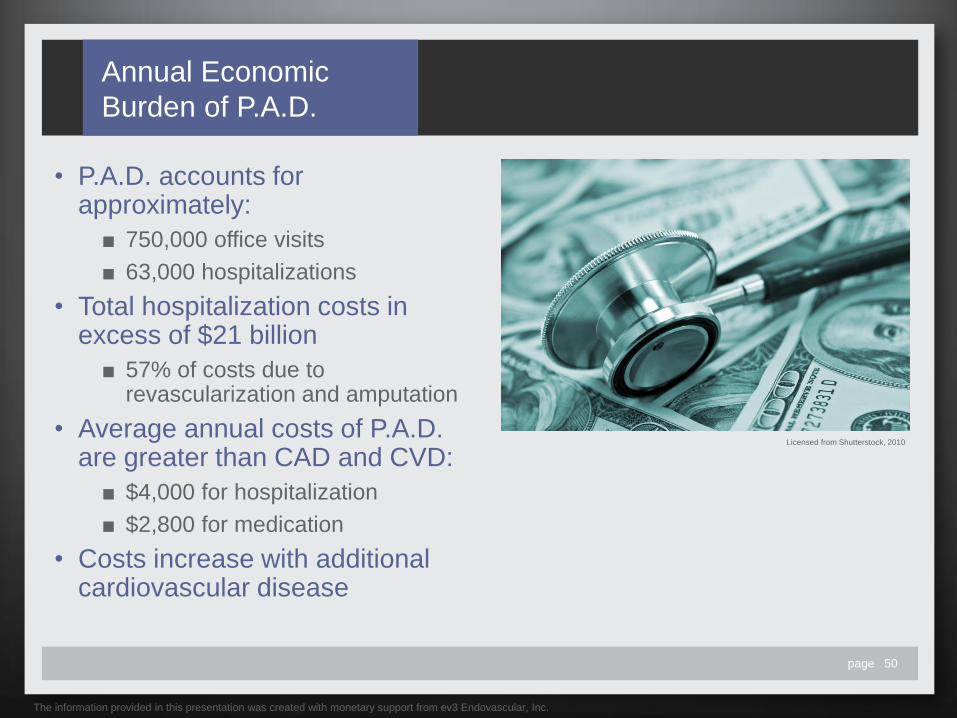
Annual Economic
Burden of P.A.D.
• P.A.D. accounts for approximately:
■ 750,000 office visits
■ 63,000 hospitalizations
• Total hospitalization costs in excess of $21 billion
■ 57% of costs due to revascularization and amputation
• Average annual costs of P.A.D. are greater than CAD and CVD:
■ $4,000 for hospitalization
■ $2,800 for medication
• Costs increase with additional cardiovascular disease
The information provided in this presentation was created with monetary support from ev3 Endovascular, Inc.
Licensed from Shutterstock, 2010
page 51
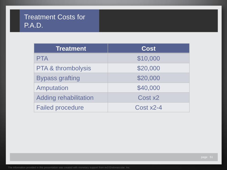
Treatment Costs for
P.A.D.
Treatment Cost
PTA $10,000
PTA & thrombolysis $20,000
Bypass grafting $20,000
Amputation $40,000
Adding rehabilitation Cost x2
Failed procedure Cost x2-4
The information provided in this presentation was created with monetary support from ev3 Endovascular, Inc.

page 52
P.A.D. Costs and
Medicare
• 98% of U.S. adults over age 65 are covered by Medicare
• 6.8% of beneficiaries received P.A.D. treatment
■ Accounts for only 1/3 of estimated P.A.D. population
• Medicare expenditures for P.A.D.:
■ $1,868 average annual treatment cost per patient
■ 88% of costs due to inpatient care
■ 2.3% of total Medicare budget
• $4.37 billion in treatment costs
The information provided in this presentation was created with monetary support from ev3 Endovascular, Inc.
Licensed from Shutterstock, 2010

page 53
Medicare Expenditures
for Disease Care
The information provided in this presentation was created with monetary support from ev3 Endovascular, Inc.
Billions in Medicare Expenditures
2.7
Cardiac
dysrhythmias
3.7
Cerebrovascular
disease
3.9
Congestive
heart failure
3.9
P.A.D.
Bil
lio
ns
page 54
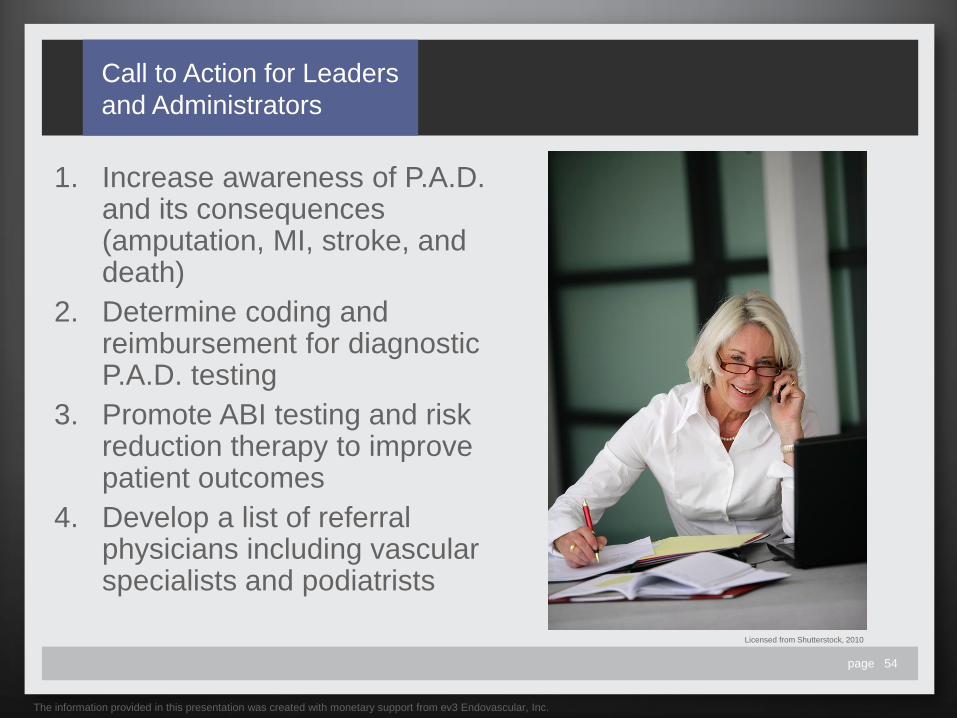
Call to Action for Leaders
and Administrators
1. Increase awareness of P.A.D. and its consequences (amputation, MI, stroke, and death)
2. Determine coding and reimbursement for diagnostic P.A.D. testing
3. Promote ABI testing and risk reduction therapy to improve patient outcomes
4. Develop a list of referral physicians including vascular specialists and podiatrists
Licensed from Shutterstock, 2010
The information provided in this presentation was created with monetary support from ev3 Endovascular, Inc.

page 55
Call to Action for
Clinicians
1. Use medical history and
recognize risk factors
2. Perform ABI testing on high
risk patients to increase
early diagnosis
3. Manage risk factors
promptly and aggressively
4. Implement multidisciplinary
care or make the
appropriate referrals
5. Maintain the continuity of
care
The information provided in this presentation was created with monetary support from ev3 Endovascular, Inc.
Peripheral artery disease (P.A.D.) is
underrecognized, underdiagnosed,
and undertreated in the U.S.
Licensed from Shutterstock, 2010

page 56
References
• Alberts JA, Bhatt DL, Mas J, Magnus-Ohman E, et al. Three-year follow-up and event rates in the international reduction of atherothrombosis for continued health registry. European Heart Journal. Aug 2009;30:2318–2326.
• American Cancer Society. Cancer Facts & Figures 2010. American Cancer Society. Atlanta: American Cancer Society; 2010. • American Diabetes Association. Peripheral arterial disease in people with diabetes. Diabetes Care. Dec 2003;26(12):3333-3341. • American Diabetes Association. Standards of medical care in diabetes – 2010. Diabetes Care. Jan 2010;33(1 Suppl);S11-S61. • American Heart Association. Heart Disease & Stroke Statistics – 2010 Update At-A-Glance. American Heart Association. Dallas, Texas: American Heart
Association; 2010. • American Heart Association. Performance Measures. American Heart Association. Available at:
http://www.americanheart.org/print_presenter.jhtml;jsessionid=XPSTL5KBFBTOUCQFCXPSCZQ?identifier=3012904. Accessed July 13, 2010. • American Podiatric Medical Association. Arterial disease and amputation. American Podiatric Association; Oct 2008. Available at:
http://www.apma.org/MainMenu/News/Campaigns/DIAFA_2009/Archives20022008/2008-Elect-to-Save-Your-Feet/PeripheralArterialDiseaseP.A.D.FactSheet.aspx. Accessed July 29, 2010.
• American Podiatric Medical Association. Study Finds Foot and Ankle Exams Critical to Peripheral Arterial Disease Detection. Press Release. American Podiatric Medical Association; Oct 2008. Available at: http://www.apma.org/MainMenu/News/NewsReleases/NewsReleaseArchives/2007/ResearchShowsFootandAnkleExamsCriticaltoDetectionofPeripheralArterialDisease.aspx. Accessed July 29, 2010.
• Beckman JA, Jaff MR,Creager MA. The United States Preventive Services Task Force recommendation statement on screening for peripheral arterial disease: more harm than benefit? Circulation. 2006;114:861-866.
• Bell D. Peripheral arterial disease overview: Here are some guidelines for prevention and treatment of this disease. Pod Mgmt. Apr/May 2009:210-220. • Bennet PC, Silverman S, Gill PS, Lip GYH. Ethnicity and peripheral artery disease. Q J Med. 2009; 102:3–16. • Bevilacqua NJ, Rogers LC, Andros G. Understanding the effects of P.A.D. on the diabetic foot. Podiatry Today. May 2010;23(5). Available at:
http://www.podiatrytoday.com/understanding-the-effects-of-pad-on-the-diabetic-foot. Accessed July 14, 2010. • Chan AW. Expanding roles of the cardiovascular specialists in panvascular disease prevention and treatment. Can J Cardiol. Apr 2004;20(5):535-544. • Cherr GS, Zimmerman PM, Wang J, Dosluoglu HH. Patients with depression are at increased risk for secondary cardiovascular events after lower extremity
revascularization. J Gen Intern Med. Feb 2008;23(5):629–34. • Cleveland Clinic. Erectile Dysfunction. Cleveland Clinic Center for Continuing Education. Available at:
http://www.clevelandclinicmeded.com/medicalpubs/diseasemanagement/endocrinology/erectile-dysfunction/. Accessed July 29, 2010. • De Vinuesa SG, Ortega M, Martinez P. Subclinical peripheral arterial disease in patients with chronic kidney disease: Prevalence and related risk factors.
Kidney International. 2005;67(93 Suppl): S44–S47. • DeLoach SS, Mohler ER. Peripheral arterial disease: A guide for nephrologists. Clin J Am Soc Nephrol.2007;2:839–846. • Fridman R, Bar-David T, Dayal R, et al. Multidisciplinary peripheral arterial disease. Foot Ankle Spec. 2010;3(1):35-39. • George B, Cebioglu M, Yeghiazaryan. Inadequate diabetic care: global figures cry for preventive measures and personalized treatment. EPMA Journal.
2010;1:13–18.
The information provided in this presentation was created with monetary support from ev3 Endovascular, Inc.

page 57
References
• Hebert K, Lopez B, Michael C, et al. The prevalence of peripheral arterial disease in patients with heart failure by race and ethnicity. Congest Heart Fail. May 2010;16(3):118-121. Hetzel L, Smith A. The 65 years and over population: 2000. Census 2000 Brief. Oct 2001. Available at: http://www.uscensus.gov/population/www/cen2000/briefs.html. Accessed: July 14, 2010.
• Hiatt WR, Goldstone J, Smith SC et al. Atherosclerotic peripheral vascular disease symposium II: Nomenclature for vascular diseases. Circulation. 2008;118:2826-2829.
• Hirsch AT, Cirqui MH, Treat-Jacobson D, et al. Peripheral arterial disease detection, awareness, and treatment in primary care. JAMA. 2001;286(11):1317-1324.
• Hirsch AT, Hartman L, Town RJ, Virnig BA. National health care costs of peripheral arterial disease in the Medicare population. Vasc Med. 2008;13:209-215. • Hirsch AT, Haskal ZJ, Hertzer NR, et al. ACC/AHA 2005 Guidelines for the Management of Patients With Peripheral Arterial Disease (Lower Extremity,
Renal, Mesenteric, and Abdominal Aortic): A Collaborative Report from the American Association for Vascular Surgery/Society for Vascular Surgery, Society for Cardiovascular Angiography and Interventions, Society for Vascular Medicine and Biology, Society of Interventional Radiology, and the ACC/AHA Task Force on Practice Guidelines. J Am Coll Cardiol. 2006;47:e1-e192.
• Hirsch AT, Treat-Jacobson D, Lando HA, Hatsukami DK. The role of tobacco cessation, antiplatelet and lipidlowering therapies in the treatment of peripheral arterial disease. Vasc Med. 1997;2:243-251.
• Jaff MR. Referall-based, reimbursable screening: A new program offers a unique opportunity to improve patient care by making the primary physician your partner in detecting peripheral arterial disease at its early stages. Endovasc Today. Nov/Dec 2004:67-70.
• Janov J. Seven Keys To Preventing Malpractice Lawsuits. Podiatry Today. Aug 2007;20(8). Available at: http://www.podiatrytoday.com/article/7490/. Accessed July 14, 2010.
• Jeremias A, Grunberg L, Patel J, et al. Effect of peripheral arterial disease on in-hospital outcomes after primary percutaneous coronary intervention for acute myocardial infarction. Am J Cardiol. 2010 May 1;105(9):1268-71.
• Lumsden AB, Davies MG, Peden EK. Medical and Endovascular Management of Critical Limb Ischemia. J Endovasc Ther. 2009;16(Suppl II):II31–II62. • Mahoney EM, Wang K, Cohen DJ, et al. One-Year Costs in Patients With a History of or at Risk for Atherothrombosis in the United States. Circ Cardiovasc
Qual Outcomes. 2008;1:38-45. • Mehlsen J, Wiinberg N, Joergensen BS, Schultz-Larsen P. High prevalence of peripheral arterial disease in patients with previous cerebrovascular or
coronary event. Blood Press. May 2010. Available at: http://www.ncbi.nlm.nih.gov/pubmed/20504243. Accessed on July 29, 2010. • Mohler ER, Treat-Jacobson D, Reilly MP, et al. Utility and barriers to performance of the ankle-brachial index in primary care practice. Vasc Med. 2004;9:253-
260. • Muir RL. Peripheral arterial disease: Pathophysiology, risk factors, diagnosis, treatment, and prevention. J Vasc Nurs. 2009;27:26-30. • Muntner P, Wildman RP, Reynolds K, et al. Relationship between HbA1c Levels and peripheral arterial disease. Diab Care. Aug 2005;28(8):1981-1987. • National Heart Lung and Blood Institute Disease Index. Peripheral Arterial Disease. National Heart Lung and Blood Institute. Available at:
http://www.nhlbi.nih.gov/health/dci/Diseases/pad/pad_all.html. Accessed on July 14, 2010. • National Kidney Foundation. KDOQI Clinical Practice Guidelines for Cardiovascular Disease in Dialysis Patients. National Kidney Foundation. Available at:
http://www.kidney.org/professionals/kdoqi/guidelines_cvd/guide10.htm. Accessed July 29, 2010.
The information provided in this presentation was created with monetary support from ev3 Endovascular, Inc.

page 58
References
• Norgren L, Hiatt WR, Dormandy JA, et al. Inter-Society Consensus for the Management of Peripheral Arterial Disease (TASC II). J Vasc Surg. Jan 2007;45(1
Suppl S);S5A-S67A.
• Polonsky TS, Taillon LA, Sheth H, et al. The association between erectile dysfunction and peripheral arterial disease as determined by screening ankle-
brachial index testing. Atherosclerosis. 2009;207:440–444.
• Redberg RF, Benjamin EJ, Bittner V. ACCF/AHA 2009 Performance measures for primary prevention of cardiovascular disease in adults: A report of the
American College of Cardiology Foundation/ American Heart Association task force on performance measures. J Am Coll Cardiol. Sep 2009;54(14):1364-
1405.
• Rowe VL, Weaver FA, Lane JS, Etzioni DA. Racial and ethnic differences in patterns of treatment for acute peripheral arterial disease in the United States,
1998-2006. J Vasc Surg. Apr 2010;51(4 Suppl):21S-26S.
• Ruo B, Liu K, Tian L, et al. Persistent Depressive Symptoms and Functional Decline Among Patients with Peripheral Arterial Disease. Psychosomatic
Medicine. 2007;69:415–424.
• Sanders LJ. The Challenge of Peripheral Arterial Disease in People with Diabetes: A Call to Action. J Am Podiatric Med Assn May/Jun 2005;95(3):307-319.
• Sheehan P. Peripheral Arterial Disease in People With Diabetes: Consensus Statement Recommends Screening. Clin Diab. Nov 2004;22:179-180.
• Smolderen KG, Aquarius AE, de Vries J. Depressive symptoms in peripheral arterial disease: a follow-up study on prevalence, stability, and risk factors. J
Affect Disord. Sep 2008;110(1-2):27-35.
• Snyder RJ, Kirsner RS, Warriner RA, et al. Consensus recommendations on advancing the standard of care for treating neuropathic foot ulcers in patients
with diabetes. Ostomy Wound Mgmt. 2010;56(4 Suppl):S1-S24.
• Stehouwer CD, Clement D, Davidson C, et al. Peripheral arterial disease: a growing problem for the internist. Eur J Intern Med. Mar 2009;20(2):132-138.
• Sumpio BE, Armstrong DG, Lavery LA, Andros G. The role of interdisciplinary team approach in the management of the diabetic foot: A joint statement from
the Society for Vascular Surgery and the American Podiatric Medical Association. J Vasc Surg. 2010;51:1504-1506.
• U.S. Preventive Services Task Force. Screening for Peripheral Arterial Disease: Recommendation Statement. U.S. Preventive Services Task Force. August
2005. Available at: http://www.ahrq.gov/clinic/uspstf05/pad/padrs.htm. Accessed on July 14, 2010.
• Veerana V, Froelich J, Eagle KA. Treatment Approach to Patients with Combined Peripheral and Coronary Artery Disease. Vasc Dis Mgmt. 2010;7:E135–
E141.
• Vincent GK, Velkoff VA. The Next Four Decades, The Older Population in the United States: 2010 to 2050. Current Population Reports. P25-1138. U.S.
Census Bureau 2010. Washington, DC.
• Washington, DC.Wang Y, Beydoun MA, Liang L, Caballero B, Kumanyika SK. Will All Americans Become Overweight or Obese? Estimating the Progression
and Cost of the US Obesity Epidemic. Obesity 2008;16:2323–2330.
• Wattanakit K, Folsom AR, Selvin E, et al. Kidney Function and Risk of Peripheral Arterial Disease: Results from the Atherosclerosis Risk in Communities
(ARIC) Study. J Am Soc Nephrol. 2007;18: 629–636.
The information provided in this presentation was created with monetary support from ev3 Endovascular, Inc.

page 59
Copyright Information for
Images
• Images licensed from Shutterstock may be used for up to 250,000 viewings.
13981702 Image Copyright Sebastian Kaulitzki, 2010 43400224 Image Copyright SomeSun, 2010
43424656 Image Copyright gaga, 2010 26780440 Image Copyright Igor Shikov, 2010
1499081 Image Copyright aaleksander, 2010 35904019 Image Copyright Lisa F. Young, 2010
46449469 Image Copyright ansar80, 2010 26939917 Image Copyright Monkey Business Images, 2010
40740250 Image Copyright xjbxjhxm123, 2010 40667596 Image Copyright Nagy Melinda, 2010
53033977 Image Copyright -=MadDog=-, 2010 39285835 Image Copyright Yuri Arcurs, 2010
2431461 Image Copyright Slobodan Djajic, 2010 12499432 Image Copyright Monkey Business Images, 2010
28665994 Image Copyright 0833379753, 2010 3542291 Image Copyright Kuzma, 2010
35840110 Image Copyright GWImages, 2010 22490029 Image Copyright Alexander Raths, 2010
35955373 Image Copyright Vikulin, 2010 26945080 Image Copyright Monkey Business Images, 2010
1774401 Image Copyright Rob Byron, 2010 10504795 Image Copyright kalewa, 2010
3448453 Image Copyright Dwight Smith, 2010 39735553 Image Copyright Yuri Arcurs, 2010
17586670 Image Copyright Cleo, 2010 39735565 Image Copyright Yuri Arcurs, 2010
27533122 Image Copyright Oliver Klimek, 2010 37050526 Image Copyright Smit, 2010
15728149 Image Copyright Monkey Business Images, 2010 400404 Image Copyright Jacqueline Shaw, 2010
3679666 Image Copyright Brittany Bastian, 2010 55305025 Image Copyright auremar, 2010
3541137 Image Copyright Alexander Raths, 2010 39734968 Image Copyright Yuri Arcurs, 2010
17103757 Image Copyright Monkey Business Images, 2010 18502465 Image Copyright AVAVA, 2010
1104141 Image Copyright Lou Oates, 2010 9543043 Image Copyright Diego Cervo, 2010
13981699 Image Copyright Sebastian Kaulitzki, 2010 25406743 Image Copyright StockLite, 2010
53297275 Image Copyright Sebastian Kaulitzki, 2010 26939812 Image Copyright Monkey Business Images, 2010
53297290 Image Copyright Sebastian Kaulitzki, 2010 45768595 Image Copyright Bork, 2010
53297266 Image Copyright Sebastian Kaulitzki, 2010 7806397 Image Copyright Patricia Hofmeester, 2010
11129668 Image Copyright ariadna de raadt, 2010 39873484 Image Copyright Monkey Business Images, 2010
13714876 Image Copyright Oscar C. Williams, 2010 39734932 Image Copyright Yuri Arcurs, 2010
6339859 Image Copyright Alexander Raths, 2010 15922855 Image Copyright Gary Blakeley, 2010
The information provided in this presentation was created with monetary support from ev3 Endovascular, Inc.

page 60
Copyright Information for
Images
• Images licensed from Shutterstock may be used for up to 250,000 viewings.
5859715 Image Copyright Rey Kamensky, 2010 14464525 Image Copyright Monkey Business Images, 2010
53990452 Image Copyright gemphotography, 2010 2741657 Image Copyright carlosseller, 2010
12485275 Image Copyright Svanblar, 2010 20290003 Image Copyright Yuri Arcurs, 2010
22875925 Image Copyright Sebastian Kaulitzki, 2010 2321781 Image Copyright Stefan Ataman, 2010
2997394 Image Copyright Jorge Salcedo, 2010 16691659 Image Copyright Jeff Banke, 2010
17636893 Image Copyright aceshot1, 2010 13643485 Image Copyright Sebastian Kaulitzki, 2010
26945083 Image Copyright Monkey Business Images, 2010 26945005 Image Copyright Monkey Business Images, 2010
52799650 Image Copyright mangostock, 2010 22262242 Image Copyright Picsfive, 2010
9585979 Image Copyright Andi Berger, 2010 43351369 Image Copyright GONUL KOKAL, 2010
45287170 Image Copyright Andy Dean Photography, 2010 3347258 Image Copyright Baloncici, 2010
49361347 Image Copyright Sean Prior, 2010 34065367 Image Copyright Alexander Raths, 2010
14464558 Image Copyright Monkey Business Images, 2010 56860867 Image Copyright ruzanna, 2010
41903698 Image Copyright vadim kozlovsky, 2010 3455975 Image Copyright beerkoff, 2010
42439225 Image Copyright Sean Prior, 2010 43524862 Image Copyright Yuri Arcurs, 2010
46389259 Image Copyright Morgan Lane Photography, 2010
• Images licensed from Mediscan may be used through August 1, 2015.
007379 Image Copyright Mediscan, 2010 001982 Image Copyright Mediscan, 2010
021413 Image Copyright Mediscan, 2010 025661 Image Copyright Barry Slaven/Mediscan, 2010
000245 Image Copyright Mediscan, 2010
The information provided in this presentation was created with monetary support from ev3 Endovascular, Inc.

page 61
Copyright Information for
Images
• Images licensed from Custom Medical Stock Photo may be used for the life of
this presentation.
Z801-J-3157 Image Copyright Science Photo Library – Custom Medical Stock Photo, 2010
Z050-HH-62 Image Copyright Ribotsky D.P.M. – Custom Medical Stock Photo, 2010
Z801-J-3156 Image Copyright Science Photo Library – Custom Medical Stock Photo, 2010
Z801-J-3041 Image Copyright Simon Fraser/Science Photo Library – Custom Medical Stock Photo, 2010
Z800-L-3344 Image Copyright Science Photo Library – Custom Medical Stock Photo, 2010
Z050-HH-63 Image Copyright Ribotsky D.P.M. – Custom Medical Stock Photo, 2010
Z801-S-2248 Image Copyright Science Photo Library – Custom Medical Stock Photo, 2010
Z050-HH-16 Image Copyright Ribotsky D.P.M. – Custom Medical Stock Photo, 2010
Z109-E-344 Image Copyright John Smith – Custom Medical Stock Photo, 2010
Z264-S-1997 Image Copyright Wedgworth – Custom Medical Stock Photo, 2010
• Additional licensed images
58219 Image Copyright PhotoDisc® Volume 58—Mature Lifestyles 2, 1999
103002, 103035-44, 103046-48, 103054, 103057, 103062-63, 103067, 103087-88, 103091 Image Copyright PhotoDisc® Volume 103—Senior
Lifestyles, 2001
347009, 374079 Image Copyright digitalvision®—Autograph Series: Young At Heart
The information provided in this presentation was created with monetary support from ev3 Endovascular, Inc.

page 62
Copyright Information
for Illustrations
• Illustrations (3792_300EWT, 3794_300EWT) licensed from A.D.A.M. Images
may be used through June 25, 2011.
• Illustrations (BY00008, si55551835) licensed from Nucleus Medical Media may
be used through June 25, 2012.
The information provided in this presentation was created with monetary support from ev3 Endovascular, Inc. 115100-001
Related Documents


