HAL Id: tel-00716332 https://tel.archives-ouvertes.fr/tel-00716332 Submitted on 10 Jul 2012 HAL is a multi-disciplinary open access archive for the deposit and dissemination of sci- entific research documents, whether they are pub- lished or not. The documents may come from teaching and research institutions in France or abroad, or from public or private research centers. L’archive ouverte pluridisciplinaire HAL, est destinée au dépôt et à la diffusion de documents scientifiques de niveau recherche, publiés ou non, émanant des établissements d’enseignement et de recherche français ou étrangers, des laboratoires publics ou privés. Recherche et caractérisation de glycosyltransférases impliquées dans la biosynthèse des polysaccharides de la paroi chez Arabidopsis thaliana Sumaira Kousar To cite this version: Sumaira Kousar. Recherche et caractérisation de glycosyltransférases impliquées dans la biosynthèse des polysaccharides de la paroi chez Arabidopsis thaliana. Autre [q-bio.OT]. Université de Grenoble, 2011. Français. NNT : 2011GRENV077. tel-00716332

Welcome message from author
This document is posted to help you gain knowledge. Please leave a comment to let me know what you think about it! Share it to your friends and learn new things together.
Transcript

HAL Id: tel-00716332https://tel.archives-ouvertes.fr/tel-00716332
Submitted on 10 Jul 2012
HAL is a multi-disciplinary open accessarchive for the deposit and dissemination of sci-entific research documents, whether they are pub-lished or not. The documents may come fromteaching and research institutions in France orabroad, or from public or private research centers.
L’archive ouverte pluridisciplinaire HAL, estdestinée au dépôt et à la diffusion de documentsscientifiques de niveau recherche, publiés ou non,émanant des établissements d’enseignement et derecherche français ou étrangers, des laboratoirespublics ou privés.
Recherche et caractérisation de glycosyltransférasesimpliquées dans la biosynthèse des polysaccharides de la
paroi chez Arabidopsis thalianaSumaira Kousar
To cite this version:Sumaira Kousar. Recherche et caractérisation de glycosyltransférases impliquées dans la biosynthèsedes polysaccharides de la paroi chez Arabidopsis thaliana. Autre [q-bio.OT]. Université de Grenoble,2011. Français. �NNT : 2011GRENV077�. �tel-00716332�

THÈSE Pour obtenir le grade de
DOCTEUR DE L’UNIVERSITÉ DE GRENOBLE Spécialité : BIOLOGIE VEGETALE Arrêté ministériel : 7 août 2006 Présentée par
Sumaira KOUSAR Thèse dirigée par Christelle BRETON et codirigée par Olivier LEROUXEL préparée au sein du Laboratoire Centre de Recherches sur les Macromolécules Végétales dans l'École Doctorale Chimie Science du Vivant
Recherche et Caractérisation de glycosyltransférases impliquées dans la biosynthèse des polysaccharides de la paroi chez Arabidopsis thaliana Thèse soutenue publiquement le « 4 Novembre 2011 », devant le jury composé de :
Mme Marie-Christine RALET Directeur de Recherche, INRA, Nantes, Rapporteur M. Jérôme PELLOUX Professeur, Université de Picardie Jules Verne, Amiens, Rapporteur M. Hervé CANUT Chargé de Recherche, CNRS, Toulouse, Examinateur M. Stéphane RAVANEL Directeur de Recherche, INRA, Grenoble, Examinateur Mme Christelle BRETON Professeur, Université de Grenoble, Grenoble, Examinatrice M. Olivier LEROUXEL Maître de Conférences, Université de Grenoble, Grenoble, Examinateur
Acknowledgements
1
ACKNOWLEDGEMENTS
I carried out my thesis under the auspices of CERMAV, University of Grenoble France. I can say without exaggeration that the three years spent in my PhD make the most memorable time of my life. During this span of time, I have observed tremendous positive changes in my personal and professional development. I am of the opinion that everyone that we come across in life influences us one way or the other. The bunch of people that I came to know during my PhD was simply wonderful. While I remain the sole responsible for imprecisions and omissions, there are a lot of people to whom I am indebted.
My utmost gratitude goes to my supervisor Dr. Christelle Breton. Her cool and calm personality had been a source of inspiration for me. During this period, Dr. Christelle not only provided all the lab material necessary for carrying out day to day research but also spent a lot of time for the analysis of the results. I have greatly benefitted from her scientific competence and had a chance of fruitful scientific discussions with her.
I am highly indebted to Dr. Olivier Lerouxel. He has always been cool, encouraging, kind and patient. He will always remain special for me for the special reason that he showed confidence in me when I needed it the most. I take great pride in having worked with him who is the specialists of his field. I have learnt a lot from him. I am grateful to him for having always kept his office door open for answering to all my questions: sometime stupid. Without doubt, he showed great patience to have scientific, religious and social discussions with me. He has always been a source of hope, courage and confidence for me during the ups and downs of my professional and personal life. He invested a lot of time for the explanation of the theoretical aspects of experiments. Above all it was a pure pleasure to work and discuss with him. I am extremely short of words to pay gratitude to you Olivier for all you have done for me. I wish you a life of unlimited pleasures and success.
I am highly thankful to Azeddine Durioch and Marie-Laure Follet-Gueye for allowing and helping me to carry out microscopy experiments under their kind supervision at Glycomev, University of Rouen.
I am extremely grateful to the members of my jury who gave their consent to analyze and consequently validate my work. I feel honored that my work got approved by such well renowned specialists.
I feel greatly indebted to all other researchers Anne Imberty, Annabelle varrot and Aline Thomas at CERMAV for nice working environment and company. I would like to thanks to all my colleagues Geraldine, Gaelle, Joanna and Aymeric who made my life colorful. I feel lucky to have known such gentle and nice people. I am also grateful to Valerie Chazalet for technical assistance during the thesis. I am thankful to Vincent Grassot M2R student who helped me a lot during his internship and he taught me French language too. I also say thanks to Anita Sarker for her time to time help to resolve the computer problems.
I am also thankful to Higher Education Commission Pakistan for providing me the scholarship of 4 years for getting higher education in France.

Acknowledgements
2
Along with the wonderful professional life, I have experienced four best years of my
personal life during my stay in France. I dedicate all this to my friends especially „Grenobloise‟
(I would like to write the individual names but the list is very long) whose company or contact had been a source of enjoyment, pleasure and peace of mind.
I am thankful to Mr Nawazish Hameed, my beloved husband for his patience and support all along the period of my thesis. I am grateful to my parents, brothers and sisters and friends in Pakistan for their best wishes, prayers and love.
Above all I am thankful to Almighty Allah for providing me the wit and health without which nothing would have been possible.

Abstract
3
ABSTRACT
The plant cell wall not only defines the unique biology of the plants but also have practical applications as feedstock for biomaterials and for the production of biofuels. Plant primary cell wall is mainly composed of cellulose, hemicelluloses and pectins. Significant progress has been made recently in identifying the enzymes involved in plant cell wall biosynthesis, but only a handful of those have been involved in pectin biosynthesis. With the aim of identifying new putative glycosyltransferases (GTs), in lab Hansen et al 2009 designed a bioinformatic strategy and identified a new group of 24 genes called “NGT” for (Novel Glycosyltransferase) which were considered “strong” candidates for putative glycosyltransferase activities. In order to determine the putative role of these NGT genes in plant cell wall biosynthesis, we designed a functional genomics strategy, analysing in parallel Arabidopsis T-DNA mutant lines and performing heterologous expression of candidate genes.
I have characterized 15 homozygous mutant lines among the group of 24 putative NGT genes through PCR. We analysed the homozygous mutants for phenotypic alteration such as dwarfing or organ malformation and found that some of mutant lines have narrow leaves as compared to Wild type plants. In parallel I have carried out the cell wall chemical analysis of 12 homozygous mutant lines and did not get any strong difference in neutral monosaccharide composition. The detailed and complete analysis (chemical, expression and microscopic analysis) of all the above mentioned genes could have been time consuming and an overwhelming work, so I focused on At5g28910 (named NGT1) which harbours a fucosyltransferase peptide signature and on At5g14550 (named P), a gene belonging to the DUF266 gene family.
Homozygous T-DNA mutant lines ngt1-1 and ngt1-2 lines were analyzed and showed a reduced growth phenotype (leaf area). Leaf area was quantified at various development stages using ImageJ, and showed a 38% reduction in mutants. Additionally, biochemical characterization of the cell wall was performed showing a reduction in neutral monosaccharide contents, like arabinose, rhamnose and galactose in mutant cell wall. Furthermore glycosyl linkage analysis of mutant lines ngt1-1 and ngt1-2 has shown that 5-Arabinofuranose (5-Araf) and 3,5-Arabinofuranose (3,5-Araf ) contents were decreased as compared to Wild type Col0 cell wall. These results were also confirmed by immunolabeling of stem cross section of mutant and wild type plants. The complementation of the mutant plants through Agrobacterium transformation resulted in the complete restoration of plant phenotype. Taken together, these data suggest that NGT1 could be an arabinosyltransferase. In order to characterize its biochemical activity, the NGT1 protein was heterologously expressed in Pichia pastoris. The recombinant protein was used to perform in vitro activity tests, but we were unable to demonstrate any neither fucosyltransferase (on the basis of peptide signature) nor arabinosyltransferase activity. In parallel to this study, I contributed to the heterologous expression and characterization of two biochemically characterized Arabidopsis GTs involved in xyloglucan synthesis: the fucosyltransferase (AtFUT1) and xylosyltransferase (AtXT1). I have successfully expressed a truncated and active form of AtFUT1, which represents an essential step for further structural studies that will be undertaken in the lab.

Résumé
4
Résumé
La paroi végétale assure des fonctions biologiques majeures définissant la singularité des plantes ; elle est également à l’origine de multiples applications en tant que ressource agro-alimentaire, source de biomatériaux ou encore pour la production de biocarburants. Malgré cette importance fondamentale et pratique de la paroi végétale, la connaissance de sa biosynthèse apparaît à ce jour toujours très limitée. En effet, la faible abondance des glycosyltransférases (GTs) responsables de sa biosynthèse, l’absence de substrat spécifique et les difficultés à obtenir certains nucléotides-sucres nécessaires aux tests enzymatiques, a souvent rendu difficile les approches de biochimie classiques. Cependant, le séquençage de génomes (Arabidopsis thaliana, Oryza sativa, Poplar populus), la création de banques de mutants d’insertion et la classification des activités glycosyltransférases dans la base de données CAZy (www.cazy.org) sont autant d’outils récents ayant permis des avancées significatives vers la compréhension de la biosynthèse de la paroi des végétaux.
Le CERMAV a participé à ce type d’avancée en 2009, en publiant une liste de 24 gènes candidats, nommés « NGT » pour « Nouvelles GlycosylTransférases », présentant des signatures caractéristiques des glycosyltransférases. Afin de démontrer l’implication des gènes NGT dans les processus d’édification de la paroi végétale, nous avons développé une approche de génomique fonctionnelle, analysant en parallèle des lignées mutantes d’Arabidopsis altérées pour les gènes NGT et testant l’activité GT de ces protéines exprimées en systèmes hétérologues. Durant mes travaux de thèse j’ai pu caractériser 15 lignées mutantes à l’état homozygote pour 7 des 24 gènes NGT. Ces lignées homozygotes ont été criblées afin de rechercher un phénotype d’altération du développement ou de la composition en sucres de leur paroi qui soit corrélé à l’altération des gènes NGT. Ce travail de criblage a conduit à s’intéresser plus particulièrement aux mutants ngt1-1 et ngt1-2 altérés pour le gène NGT1 (At5g28910).
La caractérisation des lignées mutantes ngt1-1 et ngt1-2 a permis de quantifier un phénotype de croissance foliaire réduit de 38%, par comparaison au développement des feuilles de la plante sauvage. Par ailleurs, la caractérisation biochimique de la paroi des mutants a révélé des réductions significatives et quantitatives de l’arabinose, du galactose et du rhamnose dans la paroi des mutants, ainsi que des modifications qualitatives marquées principalement des arabinanes. L’altération des arabinanes a d’ailleurs pu être confirmée par microscopie après immuno-marquage de sections d’hypocotyle de mutants à l’aide des anticorps monoclonaux LM6 et LM13 dirigés contre des épitopes α-1,5-arabinanes. Il a pu être montré également que la complémentation des mutants par une construction 35S::NGT1 permet de restaurer un phénotype sauvage à ces mutants. Par ailleurs, de façon à tester l’activité glycosyltransférase de la protéine NGT1, nous avons réalisé son expression en système hétérologue. A ce jour, malgré des résultats préliminaires encourageants, il n’a pas été possible de déterminer des conditions de tests permettant d’observer une activité glycosyltransférase suffisante et reproductible pour la protéine NGT1, que ce soit une activité fucosyltransférase (correspondant à la signature de la séquence du gène) ou bien une activité arabinosyltransférase (correspondant au phénotype biochimique des mutants ngt1).

Préface
5
Préface
Mes travaux de thèse ont été financés principalement par la “ Higher education
commission of Pakistan » et supportés par le CNRS. Cette bourse de thèse m’a été allouée afin
de poursuivre un master2, puis une thèse dans l’université de mon choix. J’ai choisi de rejoindre
l’Université de Grenoble qui offrait l’assurance d’une excellente formation scientifique, dans le
cadre d’une université habituée à recevoir un nombre important d’étudiants étrangers, ceci afin
de faciliter mon intégration. La mission de la « Higher Education Commission” (HEC) est
dédiée à faciliter la constitution au Pakistan d’une base de personnels très qualifiés, qui
serviront le développement socio-économique national. Afin de poursuivre les objectifs de
HEC, j’ai décidé de conduire des travaux de biologie végétale au sein de l’équipe Glycobiologie
Moléculaire du CERMAV-CNRS (UPR 5301).
L’équipe Glycobiologie Moléculaire s’intéresse à deux groupes de protéines
particulièrement importantes en glycobiologie, les glycosyltransférases qui synthétisent les
structures glucidiques complexes et les lectines qui reconnaissent ces structures glucidiques.
Mes travaux de recherche, présentés dans ce manuscrit de thèse s’inscrivent dans la thématique
de l’identification et la caractérisation de nouvelles glycosyltransférases qui seraient impliquées
dans la biogénèse de la paroi végétale. La paroi végétale est une structure complexe, tant du
point de vue de sa composition biochimique, que de la variété de fonctions physiologiques
essentielles qui sont assurées par cette matrice extracellulaire, telle que la régulation de
l’élongation cellulaire ou bien la participation aux mécanismes de défenses contre les
phytopathogènes. La composition biochimique de la paroi végétale est maintenant bien
documentée pour quelques plantes modèles (Arabidopsis thaliana, Physcomitrella patens,
Poplar, etc…) ainsi que de plusieurs espèces d’intérêt agronomique (Oryza sativa, Zea mays,
…) ; et si de nombreuses variations existent entre-espèces et même entre différents organes
d’une espèce, il se dégage de l’ensemble de ces études un modèle de la paroi composé de deux
réseaux interdépendant de polysaccharides. Le premier réseau serait composé de microfibrilles
de cellulose associées les unes aux autres par l’intermédiaire de molécules d’hémicelluloses
adsorbées à leur surface. Ce premier réseau confèrerait la majeure partie des propriétés
mécaniques de la paroi, et serait interpénétré par un second réseau composé des polymères
pectiques. Ce second réseauserait lui plutôt impliqué dans la cohésion intercellulaire. En effet, il
est généralement admis que parmi les pectines, les homogalacturonanes participent à la
réticulation des pectines, via la formation de ponts calciques tout comme les molécules de
rhamnogalacturonanes II qui forment des ponts inter-moléculaires via des atomes de bore.
Préface
6
Ainsi, la paroi végétale assure des fonctions biologiques majeures définissant la singularité des
plantes ; elle est également à l’origine de multiples applications en tant que ressource agro-
alimentaire, source de biomatériaux ou encore pour la production de biocarburants.
Malheureusement, malgré cette importance fondamentale et pratique de la paroi végétale, la
méconnaissance de sa biosynthèse limite le développement de la valorisation de la biomasse
notamment en tant que ressource énergétique. En effet, on estime qu’environ 10% du génome
d’Arabidopsis thaliana (plus de 2000 gènes) serait impliqué dans l’édification, l’assemblage et
le maintien de la cette paroi végétale ; mais à ce jour seule une poignée a pu être caractérisée.
A titre d’exemple, à la vue de la diversité des liaisons entre les unités
monosaccharidiques qui constituent les polysaccharides de la paroi, on estime que plus d’une
centaine de glycosyltransférases seraient impliquées ; cependant moins d’une dizaine a pu être
caractérisée de façon biochimique à ce jour. Mes travaux de thèse s’inscrivent dans ce contexte
difficile de caractérisation d’activités glycosyltransférases de la paroi végétale : la faible
abondance de glycosyltransférases dans les cellules, l’absence de molécule acceptrice spécifique
de chaque GTs, la variété de monosaccharides qui composent la paroi et les difficultés
d’obtention de certains nucléotides-sucres donneurs ont rendu extrêmement difficile les
approches « classiques » de caractérisation biochimique de ces enzymes. Cependant, le
séquençage de génomes, la création de banques de mutants d’insertion et la classification des
activités glycosyltransférases dans la base de données CAZy (www.cazy.org) sont autant
d’outils récents ayant permis des avancées significatives vers la compréhension de la
biosynthèse de la paroi des végétaux en suivant une approche de génétique inverse. C’est cette
stratégie de génomique fonctionnelle qui constitue la clef de voute de mes travaux de thèse.
Mes travaux se basent donc sur la caractérisation de gènes codant de nouvelles
glycosyltransférases végétales potentielles chez Arabidopsis que l’on a nommés gènes NGT
pour « New GlycosylTransferase ». Ces 24 gènes NGT ont été préalablement identifiés au cours
de la thèse de doctorat de Sara Fasmer Hansen dans l’équipe Glycobiologie Moléculaire du
CERMAV, à l’aide d’une approche bio-informatique originale (Ph.D. Université Grenoble,
2009). J’ai donc entrepris une étude de génomique fonctionnelle axée d’une part sur la
caractérisation de mutants d’Arabidopsis pour ces gènes et d’autre part sur l’expression de ces
gènes en systèmes hétérologues afin de caractériser l’activité potentielle de ces
glycosyltransférases.
Le manuscrit de thèse est divisé en 4 chapitres.
Le chapitre I est une introduction générale sur l’état de l’art de mon sujet de recherche. Ce
chapitre commence par une description de la paroi végétale et de son importance chez la plante

Préface
7
modèle en physiologie végétale, Arabidopsis thaliana. Ce chapitre décrit ensuite les
caractéristiques des enzymes qui synthétisent les polysaccharides dans le vivant, nommées
glycosyltransférases, en s’attachant à leurs mécanismes d’action. Enfin, un dernier paragraphe
recense les acteurs moléculaires actuellement identifiés comme étant impliqués dans la
biosynthèse des différents polysaccharides de la paroi végétale, pour finalement conclure sur
l’objectif de mes travaux de thèse.
Le chapitre II décrit principalement la première année de mes travaux de thèse, durant laquelle
je me suis attachée à caractériser un maximum de lignées mutantes d’Arabidopsis concernant
les gènes NGT. Ce travail a été entrepris afin de rechercher à l’aide d’études de phénotypes,
qu’ils soient développementaux ou bien biochimiques, les mutants et donc les gènes qui
sembleraient impliqués dans la mise en place de la paroi végétale. Cette partie des travaux m’a
conduit à identifier par PCR 35 lignées T-DNA affectant 16 gènes NGT différents. Ce travail
préalable, relativement fastidieux, m’a permis par la suite d’étudier le développement en serre
de ces 35 mutants, ainsi que d’analyser leur composition en sucres, afin de choisir sur la base de
ce criblage de caractériser deux lignées mutantes affectant le gène At5g28910 (renommé
NGT1). Le chapitre II se poursuit et termine finalement sur les clonages que j’ai effectués de 6
gènes de la famille NGT, dont le gène At5g28910, afin de démontrer une activité
glycosyltransférase in-vitro pour la protéine NGT1 exprimée en système hétérologue.
Le chapitre III est l’utilisation de la génomique fonctionnelle pour démontrer l’implication du
gène NGT1 dans les processus de biosynthèses de la paroi végétale. Ce chapitre débute par la
caractérisation de deux lignées mutantes nommées ngt1-1 et ngt1-2 qui sont respectivement
altérés dans le premier exon et dans la région 5’- non traduite du gène NGT1. Ces deux lignées
mutantes présentent un phénotype qui se traduit par un développement ralenti des feuilles. Ce
phénotype peut être restauré par la transformation de nos lignées mutantes par le gène NGT1
sous contrôle d’un promoteur fort, ce qui permet de corréler de façon certaine ce phénotype à
l’altération du gène NGT1 chez nos mutants. Par ailleurs, la caractérisation de la paroi des
mutants ngt1-1 et ngt1-2 a révélé des diminutions significatives de certains sucres présents
notamment au niveau des pectines, ce qui nous a conduit à étudier plus finement la composition
de la paroi des mutants et à démontrer l’implication du gène NGT1 dans l’élaboration de la
paroi. Le chapitre III se termine sur de nombreux tests de caractérisation in-vitro de l’activité
glycosyltransférase de la protéine NGT1, qui malgré des résultats parfois encourageants n’ont
pas permis à ce jour de définir de façon indiscutable le type d’activité qui serait catalysée par le
gène NGT1.

Préface
8
Le chapitre IV décrit une partie annexe de mes travaux de thèse, durant laquelle je me suis
intéressée à exprimer de façon hétérologue deux glycosyltransférases impliquées dans la
biosynthèse du xyloglucane (une xylosyltransférase et une fucosyltransférase), afin de
permettre une étude structurale d’une première glycosyltransférase végétale par diffraction aux
rayons-X. Des versions tronquées (délétées de leur partie N-terminale) de la xylosyltransférase
(AtXT1) ainsi que de la fucosyltransférase (AtFUT1) ont pu être exprimées en système
hétérologue mais seule AtFUT1 a été démontrée active et caractérisée. Ces travaux ouvrent la
perspective d’une purification prochaine de cette glycosyltransférase afin de cribler des
conditions permettant sa cristallisation et son étude structurale.
Le chapitre V est une conclusion générale des résultats marquants de mes travaux qui ouvre sur
une discussion des perspectives à court terme, notamment du point de vue expérimental,
concernant l’étude des gènes NGT.
Finalement, les trois derniers chapitres du manuscrit sont respectivement la partie « matériels et
méthodes », la partie « bibliographie » et une partie « annexes » de mes travaux de thèses. Il est
à noter que cette partie « annexes » présente entre-autres documents un projet de revue intitulé
« Golgi-mediated synthesis and secretion of matrix polysaccharides of the primary cell wall of
higher plants » qui sera publiée prochainement.

Table of contents
9
Chapter 1 .............................................................................................. 7
1 Introduction .................................................................................. 16
1.1 Importance of the plant cell wall .................................................................................. 16 1.2 Arabidopsis thaliana as a model plant to study cell wall biosynthesis ........................ 18 1.3 Plant cell wall components ........................................................................................... 21
1.3.1 Cellulose ................................................................................................................. 21 1.3.2 Hemicelluloses ....................................................................................................... 24 1.3.3 Pectins .................................................................................................................... 27 1.3.4 Callose .................................................................................................................... 32 1.3.5 Lignin ..................................................................................................................... 32 1.3.6 Cell wall proteins ................................................................................................... 33
1.4 Glycosyltransferases ..................................................................................................... 35 1.4.1 Classification .......................................................................................................... 35 1.4.2 Mechanism ............................................................................................................. 36 1.4.3 Structure ................................................................................................................. 37 1.4.4 Localization of glycosyltransferases ...................................................................... 39
1.5 Biosynthesis of cell wall polysaccharides .................................................................... 40 1.5.1 Cellulose biosynthesis in plants ............................................................................. 41 1.5.2 Hemicellulose biosynthesis .................................................................................... 44 1.5.3 Pectin biosynthesis ................................................................................................. 53
1.6 Callose biosynthesis ..................................................................................................... 56 1.7 The objective of thesis work ........................................................................................ 62
2 Developing functional genomics on putative “Novel
Glycosyltransferase”genes .................................................................. 66
2.1 Introduction .................................................................................................................. 66 2.2 Identification of homozygous T-DNA lines ................................................................. 69
2.2.1 Selection of homozygous mutant lines ................................................................... 69 2.2.2 Phenotypic characterization of homozygous mutant lines ..................................... 74 2.2.3 Biochemical characterization of homozygous mutant lines ................................... 76
2.3 Characterization of At5g14550 (P) mutant T-DNA lines ............................................ 78 2.3.1 Phenotypic characterization of P mutant line P ..................................................... 80 2.3.2 Neutral monosaccharide quantification of cell wall from p2-1 mutant line through GC-MS 82
2.4 Cloning of DUF 266 cDNA for heterologous expression in Pichia pastoris .............. 85 2.5 Conclusion .................................................................................................................... 87

Table of contents
10
3 The role of NGTI in the biosynthesis of cell wall of Arabidopsis
thaliana ............................................................................................... 89
3.1 Introduction .................................................................................................................. 89 3.2 Protein sequence analysis ............................................................................................. 90 3.3 Characterization of T-DNA insertion lines ngt1-1 and ngt1-2 .................................... 92 3.4 Phenotypic characterization of mutant lines ngt1-1 and ngt1-2 ................................... 97 3.5 Quantification of neutral monosaccharide of cell wall from ngt1-1 and ngt1-2 using gas chromatography ............................................................................................................... 101 3.6 Glycosyl linkage analysis of ngt1-1 and ngt1-2 mutant cell walls ............................ 103 3.7 Immunolabeling of ngt1-1, ngt1-2 and wild type hypocotyls .................................... 106 3.8 Complementation of ngt1-1 and ngt1-2 mutant lines ................................................. 109 3.9 Heterologous expression of NGT1 in Pichia pastoris ............................................... 113 3.10 Free sugar assay using T7:NGT1 microsomes ........................................................... 117 3.11 Fucosyltransferase assay using ngt1-1 mutant cell wall as an acceptor ..................... 120 3.12 Arabinosyltransferase assay using microsomal fraction of Pichia-NGT1 and NGT1-∆69 produced in Hi-5 cells ..................................................................................................... 123 3.13 Conclusion .................................................................................................................. 126
4 Heterologous expression of Arabidopsis thaliana xylosyltransferase
(AtXT1) and fucosyltransferase (AtFUT1) for structural
characterization ................................................................................. 128
4.1 Introduction ................................................................................................................ 128 4.2 AtXT1 ......................................................................................................................... 130
4.2.1 Expression of truncated AtXT1-Δ140 in insect cells ........................................... 130 4.2.2 Xylosyltransferase assay for AtXT1-Δ140 .......................................................... 133 4.2.3 Expression of AtXT1-Δ44 in insect cells ............................................................. 134 4.2.4 Xylosyltransferase assay for AtXT1-Δ44 ............................................................ 135
4.3 AtFUT1 ...................................................................................................................... 136 4.3.1 Expression of truncated AtFUT1-Δ160 in insect cells ......................................... 136 4.3.2 Fucosyltransferase assay for AtFUT1-Δ160 ........................................................ 138 4.3.3 Expression of AtFUT1-Δ68 in insect cells ........................................................... 139 4.3.4 Fucosyltransferase activity test for AtFUT1-Δ68 protein produced in insect cells 140 4.3.5 Cloning and expression of truncated AtFUT1-Δ68 in Pichia pastoris ................ 141 4.3.6 Fucosyltransferase activity test for AtFUT1-Δ68 protein produced in Pichia pastoris 142
4.4 Enzyme kinetics of AtFUT1-Δ68 ............................................................................... 145

Table of contents
11
4.4.1 Initial rate analysis of AtFUT1-Δ68 ..................................................................... 145 4.4.2 Determination of Km and Vmax of AtFUT1-Δ68 ................................................... 146
4.5 Development of a non-radioactive activity assay for AtFUT1-Δ68 .......................... 147 4.5.1 Fluorophore-assisted polyacrylamide carbohydrate gel electrophoresis (FACE) 147 4.5.2 Matrix Assisted Laser Desorption Ionization Time Of Flight (MALDI-TOF MS) analysis 149 4.5.3 Conclusion ............................................................................................................ 151
5 General Discussion and perspectives ......................................... 153
6 Material and methods ................................................................. 160
6.1 Materials ..................................................................................................................... 160 6.2 Methods ...................................................................................................................... 161
6.2.1 Methods for heterologous expression of proteins ................................................ 161 6.2.2 Cloning of AtFUT1-Δ68 for heterologous expression in Pichia pastoris ........... 167 6.2.3 Cloning of AtXT1-Δ140, AtXT1-Δ44 and AtFUT1-Δ68 for heterologous expression in insect cells .................................................................................................... 169
6.3 Methods for protein separation and identification ..................................................... 171 6.3.1 Microsomes preparation from Pichia pastoris to test activity ............................. 171 6.3.2 Protein extraction from Pichia pastoris ............................................................... 171 6.3.3 Protein quantification ........................................................................................... 171 6.3.4 Protein analysis by electrophoresis ...................................................................... 172
6.4 Methods to test protein activities ............................................................................... 173 6.4.1 Radioactivity test .................................................................................................. 173 6.4.2 Non-radioactive activity test for AtFUT1-Δ68 protein ........................................ 175 6.4.3 Arabinosyltransferase activity of microsomal protein from Pichia expressing NGT1 using MALDI-TOF MS .......................................................................................... 176
6.5 Methods for T-DNA mutants identification ............................................................... 177 6.6 Methods for cell wall analysis .................................................................................... 184
6.6.1 Cell wall preparation ............................................................................................ 184 6.6.2 Monosaccharide composition ............................................................................... 186 6.6.3 Glycosyl linkage composition analysis ................................................................ 188
6.7 Phenotypic characterization of T-DNA lines ............................................................. 190 6.7.1 Leaf area measurement using image J .................................................................. 190 6.7.2 Sample preparation for microscopy ..................................................................... 190
7 BIBLIOGRAPHY ...................................................................... 194
8 Annexes ...................................................................................... 212

List of Abbreviations
12
List of Abbreviations 2-AB 2-Aminobenzamide 3D Three-dimensional aa Amino acid ABRC Arabidopsis Biological Resource Center AGA ApioGAlacturonan AGP Arabinogalactan protein ANTS 8-Aminonaphthalene-1,3,6-TriSulfonate Api Apiose APS Ammonium Per Sulphate Ara Arabinose AtFUT1 A. thaliana Fucosyltransferase1 AtpFut A. thaliana putative Fucosyltransferase AtXT1 A. thaliana Xylosyltransferase1 BAR Bio-Array Resource BSA Bovine serum albumin BMMY Buffered Methanol-complex Medium BMGY Buffered Glycerol-complex Medium CalS Callose synthase CATMA The Complete Arabidopsis Transcriptome MicroArray CAZy Carbohydrate Active EnZyme CesA Cellulose-synthase CMP Cytidine monophosphate Col0 Columbia ecotype CSB.DB Comprehensive Systems-Biology Database
CSC Cellulose synthase complex Csl Cellulose-synthase-like CTAB Hexa-decyl-trimethyl ammonium bromide Dha 3-deoxy-D-lyxo-2-heptulosaric acid DTT Di-thio-threitol EDTA Ethylene Diamine Tetra-Acetic acid ER Endoplasmic reticulum EST Expressed sequence tag EXTs Extensins FTIR Fourier-transformed infrared spectroscopy Fuc Fucose FACE Fluorophore-Assisted Carbohydrate Electrophoresis Gal Galactose GalNAc N-Acetylgalactosamine GATL GAlacturonic acid Transferase-Like GAUT GAlactUronic acid Transferase GAX Glucurono ArabinoXylan GDP Guanidine diphosphate GH Glycosylhydrolase Glc Glucose

List of Abbreviations
13
GlcA Glucuronic acid GlcNAc N-Acetylglucosamine GPI glycosylphosphatidylinositol GSL Glucan synthase-like GT Glycosyltransferase GUS β-glucuronidase HCA Hydrophobic Cluster Analysis HG Homogalacturonan HPLC High-Performance Liquid Chromatography HRGP Hydroxyproline-rich glycoproteins HRP Horseradish peroxidase HyP Hydroxyproline irx Irregular xylem IHF Integration Host Factor Int Integrase Kdo 2-keto-3-deoxy-D-manno-octulosonic acid Man Mannose ManS Mannan synthase MALDI-TOF Matrix Assisted Laser Desorption Ionisation-Time Of Flight MLG Mixed Linked Glucans MS Mass Spectrometer NASC Nottingham Arabidopsis Stock Center NDP Nucleotide diphosphate NeuAC N-acetyl neuraminic acid NGT Novel GlycosylTransferase NMR Nuclear Magnetic Resonance OGA OligoGalacturonides ORF Open reading frame PAGE Polyacrylamide gel electrophoresis PBS Phosphate buffered saline PCR Polymerase chain reaction PCW Plant cell wall PME Pectine methylesterase PRPs Proline-rich proteins QUA Quasimodo RG I Rhamnogalacturonan I RG II Rhamnogalacturonan II Rha Rhamnose rsw Radial swelling mutant SDS Sodium dodecyl sulfate TAIR The Arabidopsis Information Resource TBE Tris-borate-EDTA buffer TBS Tris buffered saline TFA Tri-Fluoroacetic Acid TMS Tri-Methyl Silylation

List of Abbreviations
14
TMD Transmembrane domaine Tris Tris (hydroxymethyl)aminomethane UDP Uridine diphosphate UGTs UDP-glycosyltransferases WT Wild-type XET/XTH Xyloglucan Endotransglycosylases XGA Xylogalacturonan Xis Excisionase XyG Xyloglucan Xyl Xylose YPD Yeast Peptone Dextrose YPDS Yeast Peptone Dextrose Sorbitol
Chapter 1
15
Chapter 1
Introduction

Chapter 1
16
1 Introduction
1.1 Importance of the plant cell wall
Plant cells are enclosed by a dynamic multilayered structure, which is a unique and
characteristic feature of plants, called cell wall that differentiates them from animals. Plant cell
wall receives a lot of attention as it serves multiple purposes for the plant physiology and
development, but also because of the many applications it has for human uses. For the plant, cell
wall plays a central role in determining plant shape, growth, development, provides tensile
strength and mechanical support. In addition, it has a significant role in plant defense against
pathogens and responses to environmental stresses. Cell wall is also involved in other processes
like cell adhesion, cell signaling and cell-cell interaction (Carpita & Gibeaut 1993, Gibeaut &
Carpita 1994, Vorwerk et al., 2004). The plant cell wall has many commercial uses, it serves for
example as a raw material in wood, paper, textile and food industries (Farrokhi et al., 2006) but
it is also envisioned as a major source of renewable biomass for sustainable biofuel production .
The structural and functional properties of cell wall depend on polysaccharides, proteins,
lignin and some other compounds like suberin and cutin that make up the plant cell wall (Figure
1.1). Owing to the diversity of cell shapes and functions, the molecular composition and
arrangement of cell wall exhibits a great diversity. Based on ultrastructural observation and
biochemical composition the plant cell wall consists of three types of layers in higher plants.
The first layer, called middle lamella, is the outermost layer to the cell and is mostly made up of
pectic polysaccharides. The middle lamella is found at the interface of two adjacent cells (which
develop from the cell plate present at division) and hold them together thanks to divalent cations
bridging anionic pectic polysaccharides from each cell. Primary walls (the 2nd layer) are formed
in developing, growing and enlarging cells. They are composed of 90% of polysaccharides and
10% of proteins (McNeil et al., 1984, Showalter 1993; 2001). Primary cell wall mainly provides
mechanical support and dynamic strength to allow cell expansion. Depending on the
composition, two different types of primary cell walls (type I and type II) are found in
angiosperm (or flowering plants) (Carpita & Gibeaut 1993). Dicots and non-commelinoids have
type I primary cell walls that consists of cellulose microfibrills interconnected by xyloglucan
(XyG) polysaccharides in a network (Carpita & Gibeaut 1993, Yokoyama & Nishitani 2004).
Then, this cellulose-XyG network is embedded in a pectic network consisting of

Chapter 1
17
homogalacturonan (HGA), rhamnogalacturonan I (RG-I) and rhamnogalacturonan II (RG-II)
(Carpita & Gibeaut 1993, Carpita & McCann 2000). The percentage composition of different
components of type I primary cell walls (on a dry weight basis) is typically cellulose-XyG
~50%, Pectin ~30% and structural proteins are ~20%.
Type II walls are found in commelinoid monocotyledons, i.e. in cereals such as rice,
wheat, oat and barley (Carpita & Gibeaut 1993). They are organized like type I walls except that
they have lower amount of XyG and pectin (Carpita & Gibeaut 1993). The major hemicellulose
is glucuronoarabinoxylans (GAX) and mixed linked glucans (MLG). The percentage
composition of different components of type II primary cell walls (on a dry weight basis) is
cellulose ~30%, GAX ~30%, MLG ~30%, Pectin ~5%, XyG ~4% and structural proteins are
almost 0.5% (Fry & Stephen 1988) .
Figure 1.1: Scale model of the polysaccharides organisation in an Arabidopsis leaf cell wall. The amount of the various polymers is shown based approximately on their ratio to the amount of cellulose. The amount of cellulose shown was reduced, for clarity. Because of the exaggerated distance between microfibrils, the hemicellulose cross-links [shown in dark orange (xyloglucan, XyG) or light orange (glucoronoarabinoxylan, GAX)] are abnormally extended (Somerville et al., 2004).
Secondary cell wall is formed when the cells have ceased enlarging and fully expanded
and is laid down between primary cell wall and plasma membrane. Secondary cell walls provide

Chapter 1
18
strength and contribute to specialized functions related to specific cell types such as xylem
fibers, tracheids and sclerides. It is mainly composed of cellulose but also have some other
polysaccharides like hemicelluloses. In addition, it has lignin and glycoproteins which are
responsible for mechanical strength (Carpita & McCann 2000). Pectins and structural proteins
or enzymes may be absent in secondary cell walls.
1.2 Arabidopsis thaliana as a model plant to study cell wall
biosynthesis
Arabidopsis is a small flowering plant that completes its life cycle in six weeks. In
addition, this plant is of very small size which makes it easy to cultivate in a small space in labs.
Individual plant can produce several thousand seeds. It has one of the smallest plant genome,
estimated at 26735 genes spreads over five chromosomes and encoding approximately 31392
proteins (http://genome.jgi.doe.gov/), and was the first plant genome to be completely
sequenced by the Arabidopsis genome Initiative in 2000. All these features lead to Arabidopsis
thaliana as a unique model in plant biology, in order to unravel genetics mechanisms underlying
many plant traits. Although Arabidopsis was one of the first lands plant species whose genome
sequencing project was completed it was followed by the sequencing of the genome of many
other plant species like Medicago truncatula, Oryza sativa, Zea mays, Citrus sinensis etc.
Genome sequencing of the plants is an important genetic tool which facilitates the scientific
community to a greater extent for the following reasons:
• Genome sequencing not only provides sequence information of all the genes but also
provides sequence information of the regulatory regions outside the genes.
• This sequence information can be very useful to predict the function of these genes by
homology with already characterized genes of the related species.
• Last but not least, genome sequencing has helped to reduce the time needed for the
molecular/genetic characterization of the plant species and to identify genes for crop
improvement.
Data regarding the sequenced plant genomes are freely available to the scientific
community all over the world on the genome data base phytozome
(http://www.phytozome.net/). This database is powered by the joint project of the Department
of Energy's Joint Genome Institute and the Center for Integrative Genomics

Chapter 1
19
(http://www.jgi.doe.gov/). It facilitates comparative genomic studies amongst green plants. Till
date 25 nuclear genomes (Arabidopsis thaliana, Medicago truncatula, Oryza sativa, zea mays,
citrus sinensis, chlaymydomonas reinhardtii etc) have been completely sequenced and
annotated. Some of the plant species whose genome have been sequenced serve as model plants
for other genus of the same species i.e. their genome is representative of the genomes of other
genera.
There are many databases which provide the molecular, genetic and physiological
information about the genes of different species. For example, Bio-Array Resource” (BAR)
which can be used to explore large scale data sets available from microarrays of Arabidopsis
and other species is currently serving a scientist community. It comprises of various tools that
facilitates the community of researchers by providing information about the expression of a
gene, co-expression of genes, interaction of other proteins with your gene of interest,
localization of the gene, and much other useful information
(http://bar.utoronto.ca/affydb/BAR_instructions.html). Another database, publicly available, is
“Genevestigator” (https://www.genevestigator.ethz.ch/). It contains gene expression data
available from many transcriptome experiments and gives information about the regulation of
gene expression i.e. spatial and temporal localization, response to stimuli, drug treatment,
disease or genetic modification. An Arabidopsis specific database for gene sequence tags
(GSTs) is CATMA. CATMA stands for (The Complete Arabidopsis Transcriptome
MicroArray) (http:// www.catma.org/). It contains gene model sequences for over 70% of the
predicted genes in the Arabidopsis thaliana genome as well as primer sequences for GSTs
amplification and a wide range of supplementary information. The Comprehensive Systems-
Biology Database (CSB.DB) is hosted at the Max Planck Institute of Molecular Plant
Physiology, Golm, Germany. It presents the biostatistical analyses on numeric gene expression
data which is associated with current biological knowledge. It also provides Co-Response
Databases of various model organisms, like Escherichia coli, Saccharomyces cerevisiae and
Arabidopsis thaliana.
After the completion of genome sequencing for many plant species, the important work
was to assign function to identified genes. Then the scientists have focused attention to the
functional genomics. An important tool while doing functional genomics is insertional
mutagenesis which is used to disrupt the gene function to obtain knock out mutants. It provides
direct route to determine gene function. T-DNA of Agrobacterium tumefaciens is commonly

Chapter 1
20
used as mutagen to create knock-out mutants. Hundreds of thousands T-DNA insertion mutants
are available at ABRC (Arabidopsis Biological Resource Center) and NASC (Nottingham
Arabidopsis Stock Center) that are helpful to link DNA sequence to its phenotype. The main
knock-out mutant resources are SALK (http://signal.salk.edu/cgi-bin/tdnaexpress), SAIL
(Syngenta Arabidopsis Insertion Line, available on SALK website), GABI-KAT
(http://www.mpiz-koeln.mpg.de/GABI-Kat/) and FLAG (http://flagdb-genoplante-
info.infobiogen.fr/).
It has been estimated that in Arabidopsis almost 10% of the genes are involved in
different aspects of plant cell wall metabolism like polysaccharides biosynthesis, their transport
and deposition and remodeling and regulation of these processes (McCann & Carpita 2008).
Arabidopsis is one good model plant for cell wall studies because its cell wall is similar to many
other crop plants and trees (Liepman et al., 2010). In order to determine the putative function of
candidate genes involved in cell wall biosynthesis, a functional genomics strategy is then
commonly used. This strategy consists of two approaches (Figure 1.2).
1- First approach is the characterization and identification of the T-DNA mutant and
then determination of the alterations in phenotype and chemotype of the mutants to
find out the putative role of the gene.
2- Second is the cloning of gene of interest for heterologous expression of protein and
then to perform activity test in vitro to find out its function.

Chapter 1
21
Figure 1.2: A functional genomic strategy for the characterization of putative genes involved in plant cell wall
biosynthesis. This approach consists of parallel identification of the protein activity using hetereologous expression
and activity tests and characterization of cell wall features from altered mutants.
1.3 Plant cell wall components 1.3.1 Cellulose
Cellulose is mostly synthesized by vascular plants, but many species from algae to
bacteria, including the animal tunicate are naturally able to produce cellulose. As cellulose is
one of the major components of plant cell wall, it is also the world’s most abundant
macromolecule found in nature (Somerville 2006). Up to one third of the total dry mass of many
plants is often contributed by cellulose alone. It is the major load bearing component of plant
cell wall. It not only provides the strength to resist the turgor pressure in plant cell walls but also
has a very important role in maintaining the size, shape and division/differentiation potential of
most plant cells and ultimately determines the direction of plant growth.
Chemically, cellulose is a simple linear polymer of β-(1→4) linked glucose residues.
The repetitive building block in cellulose is cellobiose which consists of a pair of glucose linked
in β-(1→4), where successive glucosyl units are rotated of 180° with respect to the other (Figure
1.3). The flat conformation of the glucopyranose ring and the linkage pattern provide a ribbon
shape and semi-rigid properties to the cellulose, finally permitting the molecules to crystallize
into rods named microfibrils.
Figure 1.3: Schematic representation of cellulose composition and organization to form a microfibril.
(Modified from http://genomics.energy.gov)

Chapter 1
22
• Different cellulose substructures
Cellulose appears a simple structure but its physical properties vary remarkably in term
of degree of crystallinity and molecular weight, because of the diversity of the living source
from which it could be obtained. Cellulose can be found in nature as noncrystalline, crystalline
form I (cellulose I) and crystalline form II (cellulose II). Cellulose I is found in higher plants
and characterized with glucan chains parallel to each other and packed to form the microfibril.
NMR and X-ray studies showed that cellulose I exists in two different forms named allomorph
Iα and allomorph Iβ (Brown et al., 1976, Brown 1996). These two allomorphs of cellulose differ
in their physical properties because of different molecular conformation, their crystal packing
and hydrogen bonding but can still co-exist together within a microfibril (Nishiyama et al.,
2003). Cellulose from plant (cotton fiber) was shown to be enriched in cellulose Iβ whereas
bacteria and algae were rich in cellulose Iα. Cellulose II, the most thermodynamically stable
form of cellulose, is rarely found in nature and was studied using Acetobacter Xylinum mutants.
• Biogenesis of cellulose I
The substrate for cellulose synthesis is UDP-Glucose which is channeled through a
plasma-membrane localized enzymatic complex named cellulose synthase complex (CSC).
Although the detailed mechanism of the polymerization of glucose units into a linear cellulose
chain have not been established (regarding how successive glucosyl units will be flipped by
180° from its neighbor unit), it remains that cellulose I is synthesized processively with the non-
reducing end (growing end) of the glucan chains attached with the catalytic enzyme of the CSC
(Koyama et al., 1997). Parallel chains are then synthesized and held together by hydrogen
bonding to form crystalline microfibrils. Cellulose microfibrils vary in width from 25-30 nm in
Valonia and other green algae to approximately 5-10 nm in most of the plants (Herth 1983, Ha
et al., 1998). The secondary cell wall has higher molecular weight cellulose with the degree of
polymerization of 14000-15000 units (Brett 2000) whereas low molecular weight cellulose is
present in primary cell wall with a degree of polymerization of 8000 units (Brown 2004).
Freeze fracture electron microscopy showed that CSC harbors a hexagonal structure with
a six-fold symmetry, also named rosette or terminal complex, which is present at the plasma
membrane surface in algae and vascular plants (Mueller et al., 1976, Giddings et al., 1980).
This hexagonal structure is believed to contain 6 rosette subunits, each subunit being formed of
the assembly of 6 cellulose synthase (CESA) catalytic polypeptide chains (the products of three

Chapter 1
23
different CesA genes). This hypothetical organization is deduced from immunogold labelling
assays using an antibody raised against cotton CESA (Figure 1.4; (Kimura et al., 1999). This
organization suggests that a rosette would be responsible for the simultaneous elongation of 36
β-(1 4)-glucan chains that would co-crystallize to form a microfibril (Delmer 1999).
Figure 1.4: The cellulose-synthesizing machinery of the cell wall. A: Immunogold labeling shows that CESA is localized to hexameric 'particle rosettes' in the plasma membrane (Kimura et al., 1999). The black circles represent gold nanoparticles that are attached to antibody against CESA. The smallest subunit in the particle rosette is believed to be made of six CESA proteins. Particle rosettes are sometimes found attached to cellulose microfibrils. Scale bar, 30 nm. B: This model of a hexameric particle rosette shows how three different CESA proteins (shown in three different colours: , orange; , brown; , green) might be organized into rosette subunits and then into a hexameric synthase complex (Doblin et al., 2002).CESA assembly into rosette subunits C: A model of how CESA complexes synthesize a cellulose microfibril. Each CESA protein can synthesize a single - (1 4)-linked-D-glucan chain. Cellulose is formed as a crystalline ribbon that is composed of many such glucans. In this model, 36
-D-glucan chains are formed by a particle rosette, which is composed of a hexamer of CESA hexamers.
This number of 36 chains was actually compatible with the lateral size of the
microfibrils isolated from primary cell wall of most of the plants (Delmer 1999). However,
others studies propose the presence of 18 glucan chains or even less per microfibril (Chanzy
1978, Chanzy et al., 1979, Ha et al., 1998, Thimm et al., 2002, Kennedy et al., 2007). Actually
the determination of the number of glucan chains depend on the number of active catalytic
subunits per rosette, but how many active enzyme molecules are present per rosette have not
been experimentally demonstrated (Guerriero et al., 2010). Although the precise composition of

Chapter 1
24
the cellulose synthase complex, the way cellulose is synthesized and the number of glucan
chains within a microfibril are still under debate, our understanding of cellulose biosynthesis
has moved forward thanks to genetics using Arabidopsis mutants impaired for cellulose
biosynthesis. This aspect of the “genetic” of cellulose biosynthesis, using Arabidopsis mutants,
will be developed in paragraph 1.5.1.
1.3.2 Hemicelluloses
Hemicelluloses are a heterogeneous group of polysaccharides present in various
proportions in the cell wall, depending on plants. Their composition is also variable in quantity
between primary and secondary walls, between species and even within different plant organs
(O’Neill & York 2003). They are grouped into xyloglucan, xylans, mannans and glucomannans,
and mixed-linked β-(1→3, 1→4)-glucans (but mixed-linked glucans are present only in cereals
and grasses and absent in Arabidopsis cell wall).
• Xyloglucans
Xyloglucan (XyG) is one of the principal hemicellulose in the primary cell walls of dicots
and present in almost all vascular plant species but has not been found in charophytes (Popper
& Fry 2003, Moller et al., 2007, Popper 2008). The XyG interacts with cellulose microfibrills
through hydrogen bonding between xyloglucan backbone and the cellulose chain and this
network is considered a major load bearing element in plant primary cell wall (Somerville et al.,
2004). Xyloglucan is composed of a backbone of β-(1→4)-linked glucose residues most of
which are substituted by α-(1→6)-linked xylose side chains. These xylosyl residues can bear β-
D-galactosyl (1→2) at O-2 position and some of which are further substituted by α-L-fucosyl
(1→2) units (Figure 1.5) (McNeil et al., 1984, Fry 1989a; b). Previous studies showed that XyG
is not fucosylated in grasses (Hayashi 1989). But later on fucosylated XyG has been found in
Festuca arundinaceae (McDougall & Fry 1994) and low xyloglucan amount was also detected
in rice (Pena et al., 2008). This indicates that, at least at early stages of XyG synthesis, fucose
would be present but may be removed at late stages, for example during deposition into the cell
wall. Xyloglucan has mainly two structural arrangements XXGG and XXXG where G
represents unsubstituted glucosyl residues and X represents a glucosyl residue substituted with a
xylosyl residue (Fry et al., 1993). Most common among plant is the XXXG-type characteristic
gymnosperm and found in dicots, like Arabidopsis (Fry et al., 1993, Lerouxel et al., 2002,
Cavalier et al., 2008). The XXGG-type is characteristic of some plant species like Poaceae

Chapter 1
25
(monocots) and Solanaceae where xylosyl residues can be also substituted by α-L-Araf. For
these species XyG represents only 1-5% of the primary cell wall whereas it represents up to
20% in dicots cell wall (Scheller & Ulvskov 2010; Bonin et al., 1997).
Table 1: Nomenclature for Xyloglucan Oligosaccharides. This table is based on the nomenclature (Fry et al., 1993); modified with (Ray et al., 2004) in which each of the differently substituted β-D-glucosyl residues is indicated by a single letter. For commodity, the pattern of xyloglucan substitution of each glucose residue is represented using a single letter nomenclature corresponding to the outermost substitution. Reducing glucose residues that have been converted to alditol moieties are indicated by the code "Gol". Xyloglucan oligosaccharides are unambiguously named by listing the code letters for each glucosyl residue, starting with the non-reducing end.
Figure 1.5: Schematic representation of xyloglucan structure. Xyloglucan glucan backbone is branched with
xylose, galactose and fucose. (According to previous nomenclature this polysaccharide would be coded as: XXLG-
XXFG-XLFG.)

Chapter 1
26
Xyloglucan is currently described as a cross-linking polymer bridging cellulose
microfibrils, and thus forming a load-bearing network responsible for most cell wall stiffness.
This interaction between xyloglucan and cellulose was firstly based upon the observation that
the two molecules remain associated during extraction procedures but could also be modelled at
the atomic scale. Molecular dynamics simulations indicated that xyloglucan can interact with
cellulose through its side chains as well as through its backbone (Hanus & Mazeau 2006). It
has been observed that in case of less substituted XXXG direct interaction of all its residues
occur due to flat conformation of xyloglucan and cellulose as it is difficult for XXLG and
almost impossible for highly substituted XXFG to adopt a flat conformation that make the
interaction difficult for all of the residues with the cellulose surface. These results are in
accordance with experimental data as NMR experiments on tamarind xyloglucan and cellulose
have shown that for the interaction of XXFG fragment, all backbone and side chain residues are
in the close proximity of cellulose (Hanus & Mazeau 2006).
• Xylans
Xylans are the main hemicellulosic polysaccharides in the secondary wall of dicots.
They consist of β-(1→4)-D-xylosyl residues for the backbone that could be substituted by
arabinose, glucuronic acid and 4-O-methyl glucuronic acid residues, depending on plant
species. Xylans are involved in the cross-linking of cellulose microfibrils and lignin (Awano et
al., 2002). When xylan backbone is substituted with arabinofuranose (Araf) they are called
arabinoxylans and glucuronoarabinoxylans (Figure 1.6). Arabinoxylans are more common in
primary wall of grasses where they may be acetylated on C-2 and C-3 position of the GlcA
residue (McNeil et al., 1984, Ebringerova & Heinze 2000, Teleman et al., 2000).
Figure 1.6: Schematic representation of glucuronoarabinoxylan structure. Xylose residues are substituted with
Glucuronic acid and Arabinose.

Chapter 1
27
• Galacto-gluco-mannans
Galacto-gluco-mannans are hemicellulosic polysaccharides with a backbone of β-
(1→4)-linked mannosyl and β-(1→4)-linked glucose residues, which backbone is substituted
with α-(1→6)-linked galactosyl residues. They are present in seeds, as a storage carbohydrate,
in different plant species like legumes and palms but also exist in cell wall harbouring a
structural role, specifically demonstrated in secondary cell wall (Buckeridge et al., 2000; Maeda
et al., 2000). Other types of mannans are galactomannans and glucomannans mainly present in
secondary cell walls; depending on plant type (Heredia et al., 1995). The absence of the major
glucomannan synthase in seeds of Arabidopsis results in a severe embryo lethal phenotype
(Goubet et al., 2003) which confirms the importance of mannan for seeds development.
• Mixed-Linked Glucans
Mixed linked β-(1→3, 1→4)-glucans (MLG) are found in poaceae (grasses) (Smith &
Harris 1999) but not in dicots. MLG is composed of β-D-(1→4) linked glucans with
interspersed β-D-(1→3)-linkages. In primary cell walls they are involved in cell expansion but
their quantity is variable at different stages of growth (Obel et al., 2002, Gibeaut et al., 2005).
1.3.3 Pectins
Pectin is one of the major components of plant primary cell wall and middle lamella. Like
other polysaccharides pectin has many commercial uses and approximately 40,000 tons of
pectins are produced every year to be used in food industry mainly as a gelling agent, thickening
agent and stabilizer. Some pectic polymers are even studied as pharmaceuticals for prostate
cancer treatment (Jackson et al., 2007). The cell wall of Arabidopsis leaves contains
approximately 50% of pectin but the content varies according to the environment, tissue and
species (Zablackis et al., 1995). Pectin have a very important role for plant growth and
development, cell wall strength, defense, morphogenesis, signaling, cell expansion, pollen tube
growth, leaf abscission, seed hydration and fruit development (Ridley et al., 2001, Willats et
al., 2001, Mohnen 2008). Pectins are also involved in defence mechanisms as they can detect
pathogen attack and trigger signaling pathways that induce defence responses in the plants.
Plant pathogens cause degradation of cell wall by releasing cell wall degrading enzymes. It has
been suggested that degradation of homogalacturonan produce oligogalacturonides (OGA)
which act as elicitors to trigger plant defences. Notably, it has been established that plants

Chapter 1
28
treated with OGA produce reactive oxygen species (ROS) and plant defence hormones like
ethylene (ET) and jasmonic acids (JA) (Moscatiello et al., 2006). In addition modifications of
pectic polymer also affect the plant growth and development. Peaucelle and his colleagues
showed that pectin de-methyl-esterification plays an important role in the formation of flower
primordia in the Arabidopsis shoot apical meristem (Peaucelle et al., 2008). Pectin is a
structurally complex molecule with high heterogeneity that could be subdivided in five
(different classes, i.e. homogalacturonan (HG), xylogalacturonan (XGA), apiogalacturonan
(AGA), rhamnogalacturonan-I (RG-I) and rhamnogalacturonan-II (RG-II), all having in
common the presence of a high content of galacturonic acid.
• Homogalacturonan
HG is the most abundant polysaccharide, constituting about 65% of the total pectin
(Mohnen 2008), is a linear polymer of α-(1→4)- linked D-galacturonic acid (GalA) residues
(Figure 1.7) that are often methyl-esterified at the C-6 carboxyl position and possibly
acetylated at the O-2 and O-3 of the GalA residues but degree of acetylation varies a lot among
species (Carpita & Gibeaut 1993).
Figure 1.7: Schematic representation of homogalacturonan backbone substituted at the C-6 carboxyl position with
methyl ester groups.
• Xylogalacturonan and Apiogalacturonan
Xylogalacturonan (XGA) has a backbone of GalA residues like HG but it is substituted
with a single D-xylose residues at the C-3 of the GalA backbone residues (Schols et al., 1990,
Nakamura et al., 2002, O'Neill & York 2003) but additional Xyl residues can be attached to the
first Xyl with β-(1→4) linkage (Figure 1.8(Zandleven et al., 2006). XGA is mostly abundant
in reproductive tissues but to some extent present in other tissues, such as Arabidopsis leaves
(Zandleven et al., 2007).

Chapter 1
29
Figure 1.8: Schematic representation of xylogalacturonan (XGA), HG backbone substituted with xylose residues.
Apiogalacturonan is also similar to HG except that it is substituted with D-apiose residues at
the C-2 or C-3 of GalA backbone residues. It has been described so far in aquatic plants such as
duck weeds and the marine sea grasses (Hart & Kindel 1970, Ovodov et al., 1971). Sometimes
substitution can also occur with apiose, with the disacharide of apiose (Apif-(1 3)-Apif-(1-
found in lemna walls (O’Neill & York,2003)
• Rhamnogalacturonan-I
Rhamnogalacturonan-I (RG-I) has a different backbone from other pectic
polysaccharides. It is made up of repeating disaccharide units of [α-(1→4)-GalA-α-(1→2)-Rha].
The rhamnose residues are often substituted with galactan, arabinan and type I arabinogalactan
(Figure 1.9). Galactans are linear chains of β-(1→4)-linked galactose residues, while arabinans
are chains of α-(1→5)-linked arabinofuranose residues that are mostly branched at C-3 and
sometimes at C-2. RG-I is often acetylated at O-3 position of galacturonic acid (Ishii 1995;
1997). Type I arabinogalactans (AGs) are associated with RG-I. Type I AG has a β-(1→4)-
linked linear chain of D-galactose which is substituted with single arabinose unit or shorter
chains of L-arabinose units while type II AGs are highly branched chains with backbones of
variously linked α-D-galactose units, which are terminated by L-arabinose residues and they are
found in association with arabinogalactan proteins and xylans.

Chapter 1
30
Figure 1.9: Schematic representation of substituted rhamnogalacturonan I (RG-I) with arabinan and
arabinogalactan side chains
• Rhamnogalacturonan-II
Rhamnogalacturonan-II (RG-II) has a backbone of GalA residues but it is substituted at
the C-2 and C-3 with four complex side chains (A to D), composed of 12 different types of
glycosyl residues including some unique sugars like 2-O-methyl-xylose, 2-O-methyl-fucose,
aceric acid (AceA), 2-keto-3-deoxy-D-lyxo heptulosaric acid (Dha) and 2-keto-3-deoxy-D-
manno-octulosonic acid (kdo) (Figure 1.10). These glycosyl residues are linked together at least
with 22 different glycosidic linkages, but despite of its complex nature the structure of RG-II is
highly conserved among vascular plants (Matsunaga et al., 2004, O'Neill et al., 2004) which
suggest that it has an important role in wall integrity and functions.

Chapter 1
31
Figure 1.10: Schematic representation of rhamnogalacturonan II (RG-II) backbone which is substituted with four
side chains. These side chains harbor rare and specific sugars.
The important characteristic of RG-II is its ability to dimerize with another RG-II
molecule. In planta, 95 % of RG-II molecules exist in dimers where two apiosyl residues of side
chain A are cross-linked by a borate diester bond between (O'Neill et al., 2001). RG-II
dimerization was first demonstrated in vitro by NMR spectroscopy in sugar beet cell wall, but
its importance for plant development was demonstrated later thanks to Arabidopsis mutant
studies (Ishii & Matsunaga 1996; O’Neill et al., 2001). Indeed, characterization of RG-II from
mur1 mutant had shown that the lack of fucose in RG-II side chain B ultimately led to the
alteration of RG-II overall structure and ability to dimerize (O'Neill et al., 2001). While boron
has been referenced as an essential micronutriment in plant physiology since the fifties, only

Chapter 1
32
recently the analysis of Arabidopsis mutants altered for RG-II has been able to suggest that
boron is essential for cell wall integrity and required for cell-cell adhesion and plant
development (O'Neill et al., 2001).
1.3.4 Callose
Callose is a plant polysaccharide, present at particular stages of growth and
differentiation in cell walls or cell wall-associated structures (Bruce & Clarke 1992). It is
composed of linear β-(1→3)-linked glucose residues and sometimes has β-(1→6) branches.
Callose is laid down at plasmodesmata, at cell plates during cytokinesis and is involved
in pollen development (Bruce & Clarke 1992). It is also produced in response to wounding,
infection by pathogens, aluminium, abscissic acid and other physiological stresses (Bruce &
Clarke 1992). Callose is involved in multiple aspects of plant growth and development and
response to biotic and abiotic stress (Figure 1.11).
Figure 1.11: Callose is involved in multiple aspects of plant growth and development and response to biotic and abiotic stress (Chen & Kim 2009).
1.3.5 Lignin
Lignin is the second most abundant biopolymer on earth after cellulose. Lignin is found
in plant mostly in secondary cell wall. It has a complex chemical structure based on the
association of three monolignol monomers, i.e. ρ-coumaryl alcohol, coniferyl alcohol and
sinapyl alcohol. It performs many biological functions, e.g. it provides mechanical strength to
the cell wall by cross-linking different plant polysaccharides as it is covalently linked to
hemicellulose and cellulose and makes a lignin polysaccharide complex (Sarkanen 1998b,
Sarkanen 1998c, Whetten et al., 1998, Anterola & Lewis 2002, Boerjan et al., 2003). It fills the

Chapter 1
33
spaces in the cell wall between cellulose, hemicellulose and pectin components especially in
tracheids, sclereids and xylem cells. It helps to conduct water in plant stems. It has some
economic values as lignified wood serves as raw material for many applications and it can also
be used as fuel. It also provides resistance to insects and pathogens.
1.3.6 Cell wall proteins
Proteins account usually for 10% of dry weight of plant cell wall and ubiquitous
proteoglycans on the cell surface in plants. These proteins play a structural role, provide
strength, control rate of cell growth and prevent or protect the cells from pathogen attack. They
are involved in all aspects of plant development such as cell division and differentiation, pollen
recognition and fertilization, flower organ formation etc (Wu et al., 2001). Arabidopsis plant
cell wall contains a super-family of hydroxyproline-rich glycoproteins which includes hyperglycosylated arabinogalactan proteins (AGPs), moderately glycosylated extensins (EXTs),
and lightly glycosylated proline-rich proteins (PRPs).
The main structural protein in the cell wall of higher plants is extensin which is a class of
hydroxyproline-rich glycoproteins (HRGP) (Showalter 1993). These proteins provide rigidity
and strength to the wall by cross-linking with themselves or to other cell wall components and
involved in the process of cell extension (Brady et al., 1996, MacDougall et al., 2001).
Interestingly, one gene XEG113 belonging to GT family 77 was identified through forward
chemical genetic approach which could putatively encode extensin arabinosyltransferase (Gille
et al., 2009). As analysis of T-DNA insertional mutant xeg113 showed that etiolated hypocotyls
are more elongated as compared to WT plants, so it provides the genetic evidence that extensins
play an important role in the process of cell elongation and moreover the reduction of arabinose
in xeg113 have shown that extensin arabinosylation is important for normal plant growth and
development (Gille et al., 2009).
Arabinogalactan proteins (AGPs) consist of a core protein backbone (10%) that is
decorated by arabinogalactan polymer chains and arabinoside oligomers as their carbohydrate
components (90%) (Showalter, 2001). Most of the carbohydrate chains contain β-(1→3)-linked
galactan and β-(1→6)-linked galactan chains those are connected to each other with (1→3,
1→6)-linked at O-3 and O-6 positions with side chains mainly composed of arabinose residues
but sometimes also contain glucuronic acid, rhamnose, xylose, fucose (Figure 1.12) (Gaspar et
al., 2001, Seifert & Roberts 2007). Interestingly, many AGPs have been described to have a

Chapter 1
34
glycosylphosphatidyinositol (GPI) lipid anchor, leading to suggest a role in signaling this
molecule.
Figure 1.12: Schematic representation of arabinogalactan protein (AGP). The glycan is linked to hydroxyprolin of
the peptide chain
They are classified into classical and non-classical AGPs. Classical AGPs consist of a
central domain rich in Proline, Alanine, Serine, Threonine flanked by an N-terminal signal
peptide and C-terminal GPI anchor, whereas non-classical AGPs contains Lysine-rich domains
and Fasciclin-like AGPs (Gaspar et al., 2001, Showalter 2001). Arabidopsis genome has 47
genes that encode AGP protein backbones and most of them are predicted to be GPI anchored
out of which 13 are classical AGPs, three AGPs containing Lys-rich region, approximately 21
fasciclin-like AGPs and approximately 10 arabinogalactan peptides consisting of only 10-17

Chapter 1
35
amino acid residues (Schultz et al., 2002). They have a role in plant growth and development
and recent studies proposed their role in many other biological processes like cell proliferation
and survival and in plant pathogen interaction.
Plant cell wall also contains expansins which refer to a family of closely-related non-
enzymatic proteins, which play important roles in plant cell growth, fruit softening, abscission,
and emergence of root hairs, pollen tube invasion of the stigma and style, and other
developmental processes where cell wall loosening occurs (Cosgrove 2000). It has been
suggested that it disrupts the hydrogen bonding between xyloglucan and cellulose and believe to
regulate cell expansion.
1.4 Glycosyltransferases
The biosynthesis of oligosaccharides, polysaccharides and glycoproteins requires the
existence of a specific class of enzymes that catalyse the formation of the glycosidic bond by
transferring a monosaccharide unit from a donor to a specific acceptor substrate. These enzymes
are named glycosyltransferases (GTs). GTs commonly use activated donor sugar substrates like
nucleoside diphosphate sugars that contain a phosphate leaving group (e.g., GDP-Fuc, UDP-
Gal) but also some other donor molecules like nucleoside monophosphate sugars (CMP-
NeuAc), or dolichol phosphate sugars (Lairson et al., 2008). GTs that use nucleotide sugars as
donors are called Leloir enzymes in honor of Luis F. Leloir who discovered the first nucleotide
sugar. The acceptor molecules more commonly used by GTs are sugars but can also be some
other molecules like lipids, proteins, nucleic acid, antibiotic or any other small molecule
(Lairson et al., 2008). These enzymes are present in both eukaryotes and prokaryotes and show
exquisite specificity for both the glycosyl donor and the acceptor substrate.
1.4.1 Classification
A classification has been proposed that groups GTs into families based on amino acid
sequence similarities. It is kept updated in the Carbohydrate-Active enZyme database (CAZy,
available at http://www.cazy.org/). The database currently comprises over 75000 GT sequences
that have been divided over 94 GT families. The GT families are created based on an
experimentally proven protein function of at least one GT, and then populated by sequences of
significant sequence similarity that are extracted from public databases. Within each family, a
similar three-dimensional structure should be expected. The vast majority of enzymes in these

Chapter 1
36
+acceptordonor
families remain biochemically uncharacterized ORFs. Plants tend to have far more GT genes
(and other carbohydrate-active enzymes) than any other organism sequenced to date. More than
450 Arabidopsis GT genes have already been listed in CAZy that are spread into 42 families but
less than 20% of these genes have been biochemically characterized. By comparison, less than
230 GT genes have been identified in the human genome, of which more than 80% are
annotated. Multiplicity of GT genes in plants is mainly attributed to the highly complex network
of cell wall polysaccharides that requires the participation of numerous GTs (Lerouxel et al.,
2006, Mohnen et al., 2008), and to the large number of glycosylated secondary metabolites
(Lim & Bowles 2004).
1.4.2 Mechanism
Glycosylation reactions proceed either through processive transfer reactions with
multiple addition of the same monosaccharide (e.g., cellulose synthesis), or through non-
processive single transfer reactions, as it is observed in the synthesis of most of the
glycoconjugates. The transfer of saccharides by GTs is regiospecific and stereospecific.
Glycosyltransferase reactions follow two mechanistically distinct pathways which result in
either inversion or retention of the anomeric configuration of the transferred sugar (Figure 1.13).
In the case of an inverting enzyme, a monosaccharide α-linked to its donor becomes β-linked in
the final product (or vice-versa) whereas there is no change in the anomericity of the transferred
sugar with a retaining enzyme.
Figure 1.13: GT reactions occurring with either inversion or retention of configuration at the anomeric center of the donor sugar.

Chapter 1
37
Inverting reactions are believed to follow a single displacement mechanism that involves
nucleophilic attack of the OH-group of the acceptor on the anomeric center of the donor sugar
(Lairson et al., 2008). Structural and enzymatic studies strongly suggest that inverting GTs
utilize a single SN2-like displacement mechanism that involves nucleophilic attack of the OH-
group of the acceptor on the anomeric center of the donor sugar (Lairson et al., 2008). Reaction
occurs with the formation of an oxocarbenium-ion transition state and is concomitant with
departure of the nucleotide leaving group. The mechanism of retaining GTs is less clear.
Although some members of this class may utilize a SN2-like double-displacement mechanism
that leads to the formation of a covalent glycosyl-enzyme intermediate (Monegal & Planas
2006, Soya et al., 2011), a SNi-like mechanism involving the formation of a short-lived ion pair
intermediate has also been proposed that seems likely for the majority of retaining GTs (Lairson
et al., 2008, Wilson et al., 2008, Errey et al., 2010).
1.4.3 Structure
In contrast to other classes of enzymes like glyosylhydrolases which have wide variety
of folds, nucleotide-sugar-dependent glycosyltransferases solved to date have revealed only two
structural folds called GT-A and GT-B (or variants) (Breton et al., 2006).
• GT-A fold
The first 3D X-ray crystal for GT-A fold was identified for SpsA, an inverting enzyme
from Bacillus subtilis (Charnock & Davies 1999). The GT-A fold consists of a α/β/α sandwich
(a mixed seven-stranded β-sheet with 3214657 topology where strand 6 is antiparallel to the
rest) that resembles the Rossmann fold. The central β-sheet is flanked by a smaller one, and the
association of both creates the active site (Figure 1.14). The first region mostly corresponds to
the nucleotide binding domain, encompassing the first 100-120 residues, and that is usually
terminated by a characteristic Asp-X-Asp (often referred to as DxD motif) (Breton et al., 1998,
Breton & Imberty 1999) where X represents any aminoacid. The DxD motif is a degenerated
sequence that is shown in all crystal structures to interact primarily with the phosphate groups of
the nucleotide donor through the coordination of a divalent cation, typically Mn2+. However one
example of GT-A enzyme was shown recently to not possess this characteristic DxD motif (Pak
et al., 2006). The C-terminal part of GT-A shows more structural variability than the N-terminal
and is responsible for the recognition of acceptor molecules (Breton et al., 2006).

Chapter 1
38
The two sialyltransferases of family GT-42 showed almost the same canonical GT-A
topology but with different order of β-strands, hence these structures were considered either as a
new fold or as a variant of the GT-A fold (Chiu et al., 2004, Chiu et al., 2007). These enzymes
utilize a nucleotide monophosphosugar (CMP-NeuAc) as a donor and do not have DxD motif.
• GT-B fold
The first 3-D structure reported was for the GT-B fold enzyme, a DNA modifying β-
glucosyltransferase from bacteriophage T4 (Vrielink et al., 1994). It was found to be
structurally homologous to glycogen phosphorylase. The GT-B fold is characterized by two
separate Rossmann-type domains with a connecting linker region and a catalytic site located
between the domains. Both domains show a α/β/α structure formed by a central parallel β-sheet
with the topology 321456 (Figure 1.14). Members of GT-B family have a structurally conserved
C-terminal domain which is responsible for binding the nucleotide sugar donor substrate
whereas the N-terminal domain has more pronounced variations in the loops and α-helices
which is responsible for the recognition of the wide range of sugar acceptors. GT-B are metal-
ion independent and do not possess a DXD motif.
The 3D structure of a fucosyltransferase from Helicobacter pylori revealed a different
fold type that can be considered as variant of GT-B fold because it exhibited the same 2-domain
architecture as other GT-B members but they have some differences in the connectivity of β-
strands (Sun et al., 2007). A second fucosyltransferase that has been crystallized is the human
α−(1 6)-fucosyltransferase (Ihara et al., 2007). This enzyme displays an unusual modular
architecture, consisting of a coiled N-terminal coil region, a catalytic domain, and a SH3 domain
at the C-terminal. The catalytic domain is formed by two sub-structures, an open α/β sheet
structure and a Rossmann domain.
• Other GT folds
Very recently, completely different folds have been observed for glycosyltransferases
that utilize lipid-phosphate activated donor substrates. The crystal structures of the
peptidoglycan glycosyltransferase domains (GT51) from Staphylococcus aureus and Aquifex
aeolicus display an intriguing structural similarity to the bacteriophage λ-lysozyme (Lovering et
al., 2007; Yuan et al., 2007). The STT3 catalytic subunit of oligosaccharyltransferase (GT66)
from Pyrococcus furiosus shows very different and modular protein architecture (Igura et al.,
2008). These enzymes are not constrained by the need to bind a nucleotide, which might explain
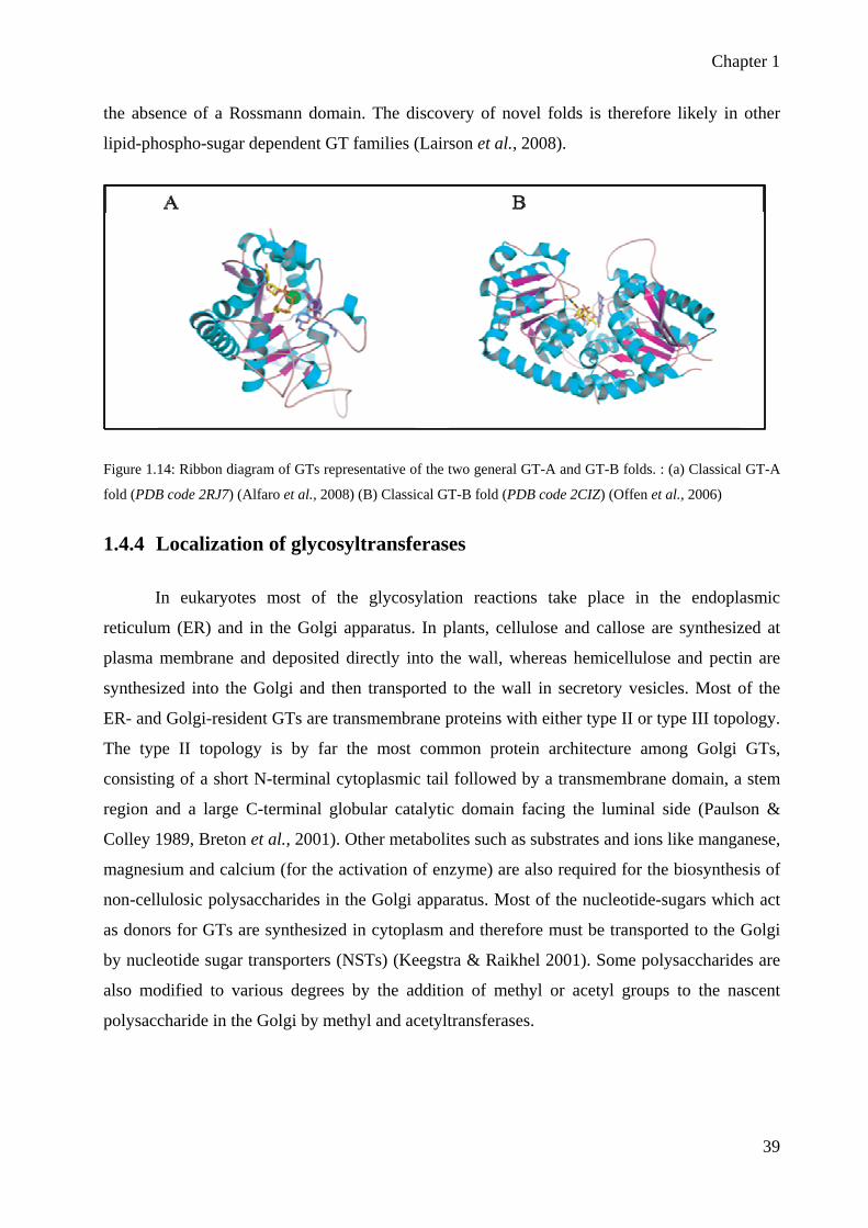
Chapter 1
39
the absence of a Rossmann domain. The discovery of novel folds is therefore likely in other
lipid-phospho-sugar dependent GT families (Lairson et al., 2008).
Figure 1.14: Ribbon diagram of GTs representative of the two general GT-A and GT-B folds. : (a) Classical GT-A
fold (PDB code 2RJ7) (Alfaro et al., 2008) (B) Classical GT-B fold (PDB code 2CIZ) (Offen et al., 2006)
1.4.4 Localization of glycosyltransferases
In eukaryotes most of the glycosylation reactions take place in the endoplasmic
reticulum (ER) and in the Golgi apparatus. In plants, cellulose and callose are synthesized at
plasma membrane and deposited directly into the wall, whereas hemicellulose and pectin are
synthesized into the Golgi and then transported to the wall in secretory vesicles. Most of the
ER- and Golgi-resident GTs are transmembrane proteins with either type II or type III topology.
The type II topology is by far the most common protein architecture among Golgi GTs,
consisting of a short N-terminal cytoplasmic tail followed by a transmembrane domain, a stem
region and a large C-terminal globular catalytic domain facing the luminal side (Paulson &
Colley 1989, Breton et al., 2001). Other metabolites such as substrates and ions like manganese,
magnesium and calcium (for the activation of enzyme) are also required for the biosynthesis of
non-cellulosic polysaccharides in the Golgi apparatus. Most of the nucleotide-sugars which act
as donors for GTs are synthesized in cytoplasm and therefore must be transported to the Golgi
by nucleotide sugar transporters (NSTs) (Keegstra & Raikhel 2001). Some polysaccharides are
also modified to various degrees by the addition of methyl or acetyl groups to the nascent
polysaccharide in the Golgi by methyl and acetyltransferases.

Chapter 1
40
1.5 Biosynthesis of cell wall polysaccharides
The polysaccharide synthesis process is divided mainly into four different stages (1) production
of activated nucleotide-sugar donors, (2) initiation of polymer synthesis, (3) polymer
elongation, and (4) termination of polymer synthesis (Delmer & Stone 1988). The processes of
production of nucleotide-sugar donors and the polymer elongation have been well studied but
the mechanism of initiation and termination of polymer synthesis is not known. The
biosynthesis of cellulose and callose takes place directly at the plasma membrane through
polysaccharide synthases (Kudlicka & Brown 1997, Delmer 1999) whereas other non-cellulosic
and pectic polysaccharides are synthesized by using both polysaccharide synthases and
glycosyltransferases within the endoplasmic reticulum (ER) and Golgi apparatus (Figure 1.15)
(Fincher & Stone 1981, Gibeaut & Carpita 1994). Then these polysaccharides are transported to
the cell surface via Golgi-derived secretory vesicles and deposited into the cell wall.
Figure 1.15: Schematic representation of the key events in cell wall biosynthesis. Cellulose biosynthesis occurs at the plasma membrane in large complexes visualized as rosettes. The synthesis of matrix polysaccharides and glycoproteins occurs in the Golgi where the products accumulate in the lumen before transport to the cell wall via vesicles. The regulation of these biosynthetic events is an important issue that needs more study. Abbreviations used in the figure: CesA, cellulose synthase proteins that form the rosette; NDP-sugar, nucleotide sugars that act as

Chapter 1
41
donors for the sugars that go into polysaccharides; Csl, cellulose synthase-like proteins that are known to be involved in hemicellulose biosynthesis (Keegstra 2010).
1.5.1 Cellulose biosynthesis in plants
The catalytic subunit of plant cellulose synthases are encoded by CesA genes which are
involved in cellulose biosynthesis. These genes were first identified in a cotton fiber cDNA
library by weak homology with bacterial cellulose synthase (Pear et al., 1996) and revealed the
presence of three peptides conserved regions with respect to the proteins encoded by bacterial
celA genes. Interestingly, the two putative CesA genes identified were expressed at high levels
during secondary cell wall cellulose synthesis in cotton fibers. The completion of Arabidopsis
genome sequence revealed that Arabidopsis has 10 CesA genes on the basis of sequence
similarity (Holland et al., 2000, Richmond 2000). In other plant species, maize has at least 12
(Appenzeller et al., 2004), rice 9, barley has at least 8 (Burton et al., 2004) and poplar has at
least 7 (Joshi et al., 2004).
• Genetic characterization of cellulose biosynthesis in Arabidopsis
Molecular characterization of mutants with defects in cell wall biogenesis has confirmed
the participation of the CesA proteins in cellulose synthesis (Arioli et al., 1998b, Taylor et al.,
1999, Fagard et al., 2000). First direct evidence for the involvement of cellulose synthase in
cellulose biosynthesis came from the analysis of rsw1 (radial swelling 1) mutants due to
mutation in AtCesA1. Mutation in CesA1 gene caused the disassembly of cellulose synthase
and rosettes dissociation into individual lobes so it showed that CESA1 proteins were required
for proper cellulose synthesis in Arabidopsis (Arioli et al., 1998b).
Tissue specific expression of various CesA genes, combined with knowledge of the
mutant phenotypes, has led to the proposal that at least three different CesA proteins are
required for the formation of a functional CesA complex (Taylor et al., 2000, Perrin et al.,
2001, Gardiner et al., 2003). The current view emphasizes two triplexes of CesAs, one
consisting of CesA1, 3 and 6 and the other of CesA4, 7 and 8, which are active during primary
and secondary cell wall formation, respectively.
In Arabidopsis AtCesA1, AtCesA3 and AtCesA6 would be required for primary cell
wall cellulose synthesis as the mutation in these genes affected cellulose production in primary
Chapter 1
42
walls. (Sarkanen 1998, Fagard et al., 2000, Scheible et al., 2001, Burn et al., 2002a). Mutation
in CesA1 and CesA3 resulted in severely retarded growth phenotypes which showed that both
are non-redundant (Arioli et al., 1998b, Beeckman et al., 2002, Gillmor et al., 2002) whereas
mutation in CesA6 exhibited anisotropic cell swelling phenotypes and a growth phenotype for
etiolated seedling but not in light-grown condition (Fagard et al., 2000). This observation led to
the hypothesis that other CesA subunits may be functionally redundant to CesA6. Later on
double mutants of other CesAs like CesA6, 2, 5 and 9 showed redundancy in their functions and
suggested that CESAs 2, 5 and also 9 might be able to substitute for the CESA6 sub-unit
(Desprez et al., 2007, Persson et al., 2007b). Recently the null mutation in cellulose synthase 9
(CesA9) resulted in 25% reduction in cellulose contents in seeds but unaltered composition of
cellulose in leaves and stems (Stork et al., 2010). Scanning electron micrograph studies have
shown that in cesa9 mutant seeds, the epidermal layer is de-shaped and moreover irregular seed
coat size, shape and internal angle uniformity was observed. So these results showed that CesA9
plays a non-redundant role for secondary cell wall biosynthesis in radial cell walls of epidermal
seed coat and important for seed morphogenesis (Stork et al., 2010).
The occurrence of triplet of CesA proteins involved in cellulose biosynthesis was
biochemically demonstrated using co-immunoprecipitation and bimolecular fluorescence
confocal microscopy. Because mutation in three genes AtCesA4, AtCesA7 and AtCesA8
resulted in the impairment of cellulose formation in secondary cell wall, it was hypothesized
that CesA4, CesA7 and CesA8 work together in a complex and demonstrated later on thanks to
co-immuno precipitation experiments (Turner & Somerville 1997, Taylor et al., 1999, Taylor et
al., 2000, Taylor et al., 2003). Similar results were obtained for primary cell wall CESAs by the
co-immuno precipitation of CesA1, CesA3and CesA6 (Desprez et al., 2007). It was also showed
that CesA3 and CesA6 are expressed in the same cell at the same time in dark growing
seedlings when fused to GFP and have a similar cellular distribution. Time-lapse spinning disk
microscopy showed that both CesA3 and CesA6 proteins migrate at the cell surface with the
same velocities along linear trajectories (Desprez et al., 2007). These results provided strong
evidence for the presence of three distinct catalytic subunits in plants primary cell wall cellulose
synthase complex, with two positions being invariably occupied by CesA1 and CesA3 whereas
there is competition between CesA6, CesA2 and CesA5 isoforms for the third position, because
of partial functional redundancy among these genes.

Chapter 1
43
Recent advances in visualizing the Cellulose synthase complex (CsC), using a functional
YFP (Yellow Fluorescent Protein)-tagged CesA6, have greatly improved our understanding for
how primary wall cellulose is synthesized (Paredez et al., 2006). Time average images of the
fluorescently labelled primary wall CesA complexes in vivo lead to the observation that these
complexes move with an average velocity estimated at 300nm/min. This corresponds to the
addition of 300 to 1000 glucose molecules/min, assuming that the microfibril is immobilized in
the cell wall. In addition, Paredez et al. (2006) also showed that the migrating primary wall
CesA complexes are aligned with the microtubules in rapidly expanding cells (Chan et al.,
2010).
Very recently, Sullivan and his colleagues have shown that in Arabidopsis CesA5 is
involved in the biosynthesis of cellulose in seed adherent mucilage (Sullivan et al., 2011). They
observed that birefringent microfibrils are absent from adherent mucilage hence crystalline
cellulose is reduced in cesa5 mutant seeds. Furthermore, labeling experiments of adherent
mucilage residues indicated that cesa5 mutant seeds have less cellulose and less pectin methyl
esterification of HG was observed (Sullivan et al., 2011).
Figure 1.16: Cartoon showing a cellulose synthase complex that is moving inside the plasma membrane leaving a cellulose microfibril in its wake. This particular cellulose synthase became active while unattached to a microtubule. It then bumped into a microtubule and followed it further. Abbreviations: CMF, cellulose microfibril; CMT, cortical microtubule; CSC, cellulose synthase complex; PM, plasma membrane. (Emons et al., 2007)
• Other genes involved in cellulose biosynthesis in Arabidopsis
In addition to CesA proteins, some other proteins were found to be involved in cellulose
biosynthesis but their specific functions still remain to be identified. Mutations in the
KORRIGAN (KOR) gene which encodes an endo-β-(1→4) glucanases, resulted in lateral organ
swelling, and reduced cellulose production (Zuo et al., 2000). Another gene, COBRA, was

Chapter 1
44
found out in a screen of mutants that encodes a small protein present on the plasma membrane
(Benfey et al., 1993). Cob mutants showed defects in cellulose microfibrils orientations but
exact role of this protein is still unclear in polymerization or cell wall deposition (Roudier et al.,
2002, Roudier et al., 2005). Arabidopsis genome has 11 COBRA-LIKE (COBL) genes. Like
other CesA proteins COBL4 is involved in secondary cell wall biosynthesis because the cobl4
mutants showed secondary cell wall defects (Brown et al., 2005). COBL6 has a role in the
biosynthesis of cellulose in pollens and anthers like CesA9 (Persson et al., 2007a). Some other
cellulose deficient mutants like kobito, pom1, rsw3, fragile fibre1 (fra1) and fragile fiber2 (fra2)
were studied but for none of them clear functional mechanism for cellulose biosynthesis was
identified (Hauser et al., 1995, Burk et al., 2001, Burn et al., 2002b, Pagant et al., 2002, Zhong
et al., 2002, Mouille et al., 2003).
Recently, one non-CesA, cellulose synthase-interactive protein 1(CSI1) was identified
by using a two-hybrid screen system. This protein was found to interact with cellulose synthase
isoforms that are involved in primary cell wall (Gu et al., 2011). Mutation in CSI1 reduced
cellulose contents and caused defects in cell elongation in hypocotyls and roots. Red fluorescent
fusion protein showed that CSI1 is associated with CESA complex in the plasma membrane.
1.5.2 Hemicellulose biosynthesis
• Backbone biosynthesis: The CSL hypothesis
Plants contain a large number of genes encoding cellulose synthase-like (Csl) proteins
that share sequence similarity with CesA proteins. It has been hypothesized that the backbone of
non-cellulosic polysaccharides including glucuronoarabinoxylan, xyloglucan,
galactoglucomannan, and mixed-linked glucan may be synthesized by Csl proteins that are
predicted to be Golgi-resident proteins (Richmond & Somerville 2001). This assumption was
proposed based on the observation that cellulose backbone was structurally related to the
backbone of hemicellulose β-(1 4)-linked sugar. These Csl genes are members of GT2 family
in CAZy database. The Arabidopsis genome contains multiple Csl genes that have been
subdivided into six groups (CslA, B, C, D, E, and G) (Richmond & Somerville 2001). But some
other groups like CslF and CslH are also present in grasses. Characterization of mutant plants
and recombinant enzymatic activities provided valuable information about the functions of these
Csl genes. Many groups have shown that members of CslA family are involved in the
biosynthesis of either mannan and glucomannan backbone depending on the type of substrate

Chapter 1
45
provided to the heterologously expressed proteins (Dhugga et al., 2004, Liepman et al., 2005,
Suzuki et al., 2006, Liepman et al., 2007b, Goubet et al., 2009). Heterologous expression of rice
CslF gene in Arabidopsis which lacks mixed linked glucans (MLGs) in its wall, showed
detectable MLGs in transformed plants and they confirmed their presence in epidermal cells by
using specific MLG monoclonal antibody (Burton et al., 2006). Similarly Doblin and colleagues
have shown that in barley MLG polysaccharides are synthesized by another CslH gene
subfamily. They cloned CSLH gene from barley and expressed it in Arabidopsis and showed
that transgenic plants have detectable MLGs in their walls (Doblin et al., 2009). Moreover their
presence was confirmed through immunoelectron microscopy with the use of specific MLG
antibody. Later on Bernal and colleagues studied the CslD group of Arabidopsis and
demonstrated that CslD2, CslD3 and CslD4 are localized in Golgi when their N-terminus was
fused with yellow fluorescent protein (YFP) (Bernal et al., 2008).
• Xyloglucan biosynthesis
Many of the biosynthetic enzymes involved in XyG biosynthesis have been determined
by using different approaches. Its biosynthesis requires at least four different
glycosyltransferase activities like α-(1→2)-fucosyltransferase, β-(1→2)-galactosyltransferase,
β-(1→4)-glucan synthase and α-(1→6) xylosyltransferase (Faik et al., 2000). The Arabidopsis
CELLULOSE SYNTHASE-LIKE C4 (CslC4) gene may encode β-(1→4)-glucan synthase that
is involved in XyG backbone biosynthesis (Cocuron et al., 2007) but characterization of XyG in
cslc4 mutant plants is still lacking.
XyG fucosyltransferase was one of the first XyG biosynthetic enzymes to be identified
(Perrin et al., 1999). It was purified from the pea epicotyls using biochemical purification
techniques (Perrin et al., 1999). It was named as PsFUT1.Then the amino acid sequence was
used to find out and characterize the homologous gene (AtFUT1) in Arabidopsis thaliana (Faik
et al., 2000) which belongs to GT37 family in CAZy database. In in vitro assays AtFUT1
catalyzes the addition of L-fucose at the 2-position of galactose residue into XyG in the presence
of non-fucosylated xyloglucan acceptor by using GDP-L-fucose as a donor. Afterwards a
genetic screen of Arabidopsis mutants showed that mur2 was also affected in the same AtFUT1
gene (Vanzin et al., 2002). The mur2 plants were completely lacking L-fucose in cell wall and
showed 99% reduction in xyloglucan fucosylation thus indicating that AtFUT1 is the only
fucosyltransferase responsible for XyG fucosylation, at least in Arabidopsis (Reiter et al., 1997,
Vanzin et al., 2002, Perrin et al., 2003).

Chapter 1
46
The Arabidopsis genome has nine AtFUT1-like genes (named AtFUT2 to AtFUT10) on
the basis of amino acid sequence similarities. Wu and colleagues have shown recently that
AtFUT4 and AtFUT6 genes putatively encode α-(1→2)-fucosyltransferases (FUTs) that are
responsible for the fucosylation of arabinogalactan proteins (Wu et al., 2010b).
From the same collection of mutants, some other fucose-deficient mutants like mur1 and
mur3 were identified (Reiter et al., 1997). mur1 plants were defective in the interconversion of
GDP-D-mannose to GDP-L-fucose and were completely deficient in cell wall fucose content
(Bonin et al., 1997),whereas mur2 and mur3 plants were identified having a 50% reduction in
cell wall fucose content (Reiter et al., 1997). MUR3 protein was shown to encode a XyG β-
(1→2)-galactosyltransferase (Madson et al., 2003). The mutant plants have altered XyG
structure because α-L-fucose (1→2) β-D-galactosyl side chains were completely absent
(Lerouxel et al., 2002, Madson et al., 2003). This galactosyltransferase is specific for the
addition of the third galactose in the repeating XXXG unit in XyG. This galactose is often
fucosylated explaining why the mutants are also fucose-deficient (Scheller & Ulvskov 2010).
Ten predicted coding regions in the Arabidopsis genome are closely related to the MUR3 XyG
galactosyltransferase and one of them has been proposed to add a galactose onto the second
position of the repeating unit, but the final evidence is still lacking (Li et al., 2004).
Seven Arabidopsis genes belonging to family GT34 were previously annotated as
putative galactosyltransferases on the basis of sequence similarity to the previously
characterized fenugreek galactomannan α-(1→6)-galactosyltransferase (Faik et al., 2002). All
of these candidate genes were expressed into Pichia pastoris to evaluate this hypothesis. As a
result of this heterologous expression one of the candidate genes showed xylosyltransferase
activity in the presence of cellopentaose acceptor substrate. This gene was first named as AtXT1
but was recently renamed XXT1. Later on, a second gene displaying 85% similarity to the
XXT1 was shown to also encode an α-(1 6)-xylosyltransferase activity when expressed into
insect cells and was named XXT2 (Cavalier & Keegstra 2006). Both XXT1 and XXT2 were
capable of transferring Xyl from UDP-Xyl onto cellopentaose and cellohexaose acceptor
substrates. In fact in the presence of cellohexaose acceptor both enzymes catalyzed the addition
of second xylose residues next to the first one forming dixylosylated cellohexaose. Later on by
the use of functional genomics it was proved that both XXT1 and XXT2 genes are involved in
XyG biosynthesis. Single T-DNA insertion mutant xxt1 and xxt2 did not show remarkable

Chapter 1
47
phenotype and almost no reduction in xyloglucan contents, whereas xxt1 xxt2 double mutant
showed a severe root hair phenotype and lacked detectable xyloglucan (Cavalier et al., 2008).
A third GT34 enzyme was found to be involved in XyG biosynthesis named as XXT5
(Zabotina et al., 2008). It is believed to encode α-(1→6) xylosyltransferase and T-DNA
insertion showed that mutants have shorter root hairs and less xyloglucan quantity in cell wall.
But no in vitro xylosyltransferase activity was observed when XXT5 protein was heterologously
expressed either in Pichia pastoris or Sf21 insect cells.
• Xylan Biosynthesis
Xylans are structurally diverse plant polysaccharides with a backbone of β-(1→4)-linked
D-Xylosyl residues which are often substituted with glucuronic acid and 4-O-methyl glucronic
acid (glucuronoxylan) and with arabinose (arabinoxylans). The backbone substitution is greatly
dependent on plant species and tissues of origin. Glucuronoxylans are major hemicelluloses in
secondary cell wall of dicotylenous plants whereas arabinoxylans are major components of cell
wall of grasses. At least five types of enzymes are required for Xylan backbone and side chain
biosynthesis and for their modifications including xylosyltransferase (XylT),
glucuronosyltransferase (GlcATs), arabinosyltransferase (AraT), acetyltransferase and
methyltransferase.
• Backbone Biosynthesis
Despite a lot of work, very little was known till recently about the mechanism
underlying xylan biosynthesis. To date, a number of putative glycosyltransferase genes have
been identified that are involved in the synthesis of either xylan backbone, side chains or the
reducing end sequence which are summarized in figure 1.17. Because of the structural
similarity of xylan to the β-(1→4)-linked backbones of other hemicelluloses, it has been
assumed that members of the Csl protein group might probably be involved in xylan backbone
synthesis. However, heterologous expression of various Csl genes failed to identify any xylan
synthase activity (Richmond & Somerville 2001). However some genetic evidences about xylan
biosynthesis arose from the characterization of mutants harbouring irregular xylem (irx)
structure, and altered for genes named irx9, irx14, I9H (irx9-LIKE), I14H (irx14-LIKE), irx10
and irx10-LIKE (which are discussed in detail in the next paragraphs) (Bauer et al., 2006,
Brown et al., 2007, Lee et al., 2007a, Brown et al., 2009, Wu et al., 2009). All of these genes

Chapter 1
48
encode putative glycosyltransferases belonging to families GT43 and GT47 (Scheller &
Ulvskov 2010). They are expressed in cells undergoing secondary cell wall biosynthesis and
their encoded proteins are targeted to the Golgi where xylan is synthesized (Jensen et al., 2011,
Zhong et al., 2005). It has been shown experimentally for IRX9 to be localized in Golgi (Pena
et al., 2007).
Analysis of mutants in each of these genes indicates that IRX9, IRX10, and IRX14
encode enzymes that function as xylosyltransferases in the synthesis of the β-1-4-xylan
backbone while IRX8, FRA8, and PARVUS appear to be involved in synthesis of the reducing
end tetrasaccharide structure (for a review see Scheller and Ulvskov 2010). However, till now
no xylan synthase activity has been observed for any of these proteins when heterologously
expressed, which leads to the suggestion that the enzymes may not work alone and require other
proteins in a protein functional complex for xylan synthesis (Brown et al., 2007, Pena et al.,
2007). Previous studies have already proposed that glucuronoxylan (GX) biosynthesis requires
the cooperative actions of XylT and GlcAT (Baydoun et al., 1989). Interestingly, very recently
in wheat, a Glucuronoarabionoxylan (GAX) synthase complex was identified by using
proteomic and transcriptomic approches (Zeng et al., 2010). This complex contains three
putative glycosyltransferases (from GT43, GT47 and GT75 CAZy families), a
xylosyltransferase (XylT), an arabinosyltransferase (AraT), and a glucuronosyltransferase
(GlcAT) which would be required for the biosynthesis of GAX.
Mutation in the Arabidopsis irx9 gene, which belongs to family GT43, resulted in plants
with decreased amounts of wall GX contents, suggesting that this gene is required for GX
synthesis (Brown et al., 2005, Bauer et al., 2006) and further characterization of irx9 mutants
have shown the decrease in chain length of glucuronoxylan (GX) (Pena et al., 2007). Later on
Lee and his colleagues showed that the irx9 mutant is deficient in xylan xylosyltransferase
activity (Lee et al., 2007a). They first measured the XylT and GlcAT activities from the
microsomal extraction of the stems of wild-type Arabidopsis rich in xylan in the presence of
exogenous acceptor (1→4)-linked β-D-xylooligomers. Then they have shown that XylT activity
was substantially reduced in the irx9 mutant compared with the wild type but GlcAT remained
unchanged in the irx9 mutants (Lee et al., 2007a). These observations confirmed the previous
results and showed that IRX9 is a xylosyltransferase that is responsible for the elongation of the
xylan backbone. Another mutation in IRX14, homologous of IRX9 and also belonging to family
GT43 has shown the reduction in GX contents in mutant cell wall (Brown et al., 2007) and

Chapter 1
49
biochemical assays have shown that irx14 is defective in the incorporation of radiolabelled
UDP- 14C-xylose onto β-(1→4) xylooligosaccharides which proposed that IRX14 would be
needed for the elongation of xylan back bone.
Arabidopsis GT43 family has four GT members, two of them IRX9 and IRX14 have
been previously characterized but recently two independent groups (Lee et al., 2010, Wu et al.,
2010) have studied two other members of this family and have used different nomenclature for
the same genes, for example Lee et al called them I9H (homolog of IRX9, also called IRX9-L)
and I14H (homolog of IRX14, also called IRX14-L) by Wu et al. (2010). Genetic analysis
showed that both genes are expressed in cells undergoing secondary wall thickening and
predicted to be Golgi-localized. Lee et al (2010) have shown that defects in GX synthesis
caused by irx9 mutation can be rescued by the overexpression of I9H but not by either IRX14 or
IRX14-L. Similarly, overexpression of I14H complemented the defects caused by irx14
mutation but not by either IRX9 or IRX9-L. The functional redundancy of this gene was
actually confirmed by the analysis of double mutants which showed a severe reduction in GX
contents and loss of secondary wall thickening in fibre cells as compared to single mutants. To
summarize, IRX9, IRX9-L and IRX14, IRX14-L genes are involved in GX backbone
elongation, but complementation studies showed that there exist two functionally non-redundant
groups in this GT43 family. Wu and colleagues, confirmed these results through complementary
studies and showed that IRX9 are IRX14 are more important and major genes while IRX9-L are
IRX14-L are less important and minor genes that are involved in GX synthesis (Wu et al.,
2010).
Two other Arabidopsis genes belonging to the family GT47, IRX10 and IRX10-LIKE
have been identified that are involved in xylan backbone elongation (Wu et al., 2009). Single
mutation did not show any visible phenotype but the double mutants showed a severe phenotype
as the mutants have shorter rosettes and infertile inflorescence. NMR (Nuclear Magnetic
Resonance) analysis showed that degree of polymerization of the xylan backbone was decreased
in mutant plants as compared with the wild type. Very recently, two articles were published
studying the DUF579 domain in Arabidopsis and they found that 5 genes out of 10 are co-
expressed with genes involved in secondary cell wall biosynthesis (Brown et al., 2011). They
showed that two members of the DUF579 gene family named as IRREGULAR XYLEM 15
(IRX15) and IRREGULAR XYLEM 15 LIKE (IRX15L) are essential for normal xylan
deposition during secondary cell wall biosynthesis. Sugar composition analysis of double

Chapter 1
50
mutant irx15/irx15l exhibited reduction in xylan content as compared to wild type and
furthermore the xylem vessels are distorted like other xylan deficient mutants. Localization
experiments with the use of fluorescent fusion proteins have shown that both proteins are
located into Golgi apparatus (Brown et al., 2011).
Figure1.17: Basic structure of glucuronoxylan indicating reducing end sequence, backbone elongation and side
chains. The genes involved in the biosynthesis of different parts of glucuronoxylan are represented in red colour.
• Side chains biosynthesis
The side chains of xylans should require the action of α-glucuronsyltransferases and α-
arabinofuranosyltransferases. In dicots xylan is substituted with glucuronic acid (GlcA) and 4-
O-methylglucuronic acid (MeGlcA), and recently two putative glycosyltransferases named
GUX1and GUX2 (GlucUrono acid substitution of Xylan) were identified to add GlcA and
MeGlcA to xylan backbone in Arabidopsis stem cell walls (Mortimer et al., 2010). Double
mutants of gux1 gux2 have shown no change in xylan backbone contents but GlcA and MeGlcA
contents were almost completely absent. Moreover, the glucuronosyltransferase activity was
also strongly diminished in double mutants because the stem microsomes were unable to
transfer GlcA from UDP-GlcA onto xylooligosaccharide acceptors. These results suggest that
GUX1 and GUX2 are responsible for the substitution of xylan backbone in Arabidopsis stem
glucuronoxylan.
• Reducing end oligosaccharide synthesis

Chapter 1
51
Xylans have a reducing end with a unique structure β-D-Xylp-(1→4)-β-D-Xylp-(1→3)-
α-L-Rhap-(1→2)-α-D-GalpA-(1→4)-D-Xylp (Shimizu et al., 1976, Pena et al., 2007)
mostly in dicots. A number of mutations in different genes IRX8, PARVUS, FRA8 (IRX7), and
F8H (homolog of FRA8) have been demonstrated to be involved in the biosynthesis of the
reducing end of GX (Brown et al., 2007, Lee et al., 2007b, Pena et al., 2007). FRA8, IRX8 and
F8H all are Golgi localized proteins except PARVUS that is located in endoplasmic reticulum.
Arabidopsis, IRX8 also known as GAUT12, a close homolog of GAUT1 belongs to
family GT8 that encodes an α-(1→4) galacturonosyltransferase activity, possibly involved in
the biosynthesis of the backbone of pectic homogalacturonan (Sterling et al., 2006). Mutation in
IRX8 resulted in significant reduction in secondary cell wall thickness and moreover the plant
height was reduced. Cell wall analysis of mutants showed that level of xylan and
homogalacturonan were significantly reduced which was further confirmed by immuno-
histochemical analysis. Structural fingerprinting of cell wall polymers revealed the remarked
reduction in glucuronoxylan chain length or almost loss of GX reducing end tetrasaccharide
sequence but it did not cause a complete disruption in GX biosynthesis. Double mutants of
irx9/irx8 exhibited collapsed xylem vessels and reduction in xylose and cellulose in cell wall as
compared to irx8 and irx9 single mutants (Persson et al., 2007b). Protein encoded by PARVUS
is co-localized with endoplasmic reticulum and belongs to GT8 family. It is thought to be
involved in the initiation of biosynthesis of the GX reducing end tetrasaccharide sequence by
catalyzing the transfer of the reducing Xyl residues onto an unknown acceptor in the
endoplasmic reticulum (Lee et al., 2007a). Mutation of the PARVUS gene resulted in secondary
wall thickening and reduced GX contents. NMR analysis showed the absence of tetrasaccharide
primer sequence at the reducing end of GX in mutants and loss of glucuronic acid side chain in
GX. These data proposed the putative role of PARVUS in the initation of biosynthesis of the
GX tetrasaccharide primer sequence.
Arabidopsis thaliana fragile fiber8 (fra8) mutant was shown to have reduced levels of
xylan and resulted in fiber wall thickness and decreased in stem strength (Zhong et al., 2005).
FRA8 was suggested to encode glucuronosyltansferase on the basis of amino acid sequence
similarity to tobacco (Nicotiana plumbaginifolia) pectin glucuronosyltransferase which belongs
to family GT47 (Zhong et al., 2005). Structural analysis of cell wall of fra8 showed the absence
of substituted glucuronic acid but the presence of only 4-O-methylglucuronic acid. These data
suggested that FRA8 encodes a glycosyltransferase that is involved in the addition of glucuronic

Chapter 1
52
acid residues onto the backbone of xylan. Recently a close homolog of FRA8, F8H was
identified in Arabidopsis and was shown to be involved in GX biosynthesis (Lee et al., 2009).
The Gus reporter gene expression indicated that the F8H expression is associated with
secondary cell walls in xylem cells which are rich in GX and F8H protein is located in the Golgi
where GX is synthesized. To check that either these homologs are functionally redundant,
mutants of fra8 were complemented with F8H construct and its overexpression completely
rescued the mutant phenotypes i.e. reduction in secondary cell wall thickness and a deformation
of xylem vessels (Zhong et al., 2005). NMR spectrometry also showed the recovery of
structural defects in fra8 mutant that was the loss of GlcA side chains in GX and reduction in
the GX reducing end tetrasaccharide structure. Double mutant of fra8/fh8 has shown more
severe defects that include deformation of vessels and retarded growth as compared to single
mutant of fra8. All these findings proposed that FR8 and F8H are functional paralogs and
functionally redundant in GX biosynthesis.
P. Ulvskov has proposed that reduction of α-D-GlcA in fra8 could be an indirect effect
of mutation as all the GTs in family 47 are inverting enzymes whereas a xylan specific α-
glucuronosyltransferase would be a retaining enzyme (Ulvskov 2011). So if it is true that FRA8
is involved in xylan biosynthesis, another alternative might be that FRA8 encodes β-1,3-
xylosyltransferase involved in synthesis of reducing end oligosaccharide (Scheller & Ulvskov,
2010).
• Galacto-glucomannan Biosynthesis
Galacto-gluco-mannans (GGM) are polysaccharides found in many tissues and cell types
in Arabidopsis (Handford et al., 2003, Moller et al., 2007, Liepman et al., 2007a). It has been
demonstrated by Liepman and coll. (2007) that GGM are synthesized by CslA protein from
GDP-mannose and GDP-glucose activated-sugar. A second enzyme at least is required for
GGM biosynthesis, an α-(1→6)-galactosyltransferase adding side chains to backbone (Edwards
et al., 1999). The study of GGM biosynthesis could be considered as land mark, as the first
proof of a CslA gene to be responsible for the biosynthesis of hemicellulose backbone, was the
characterization of a guar Csl gene encoding a mannan synthase synthesizing activity (Dhugga
et al., 2004). This work actually validates a long standing hypothesis of Cellulose-synthase-like
genes family to be involved in hemicelluloses biosynthesis. As all members of Csl genes family,
CslA genes involved in GGM biosynthesis are grouped in family GT2. Heterologous expression
studies have shown that CslA is responsible for the synthesis of mannan and glucomannan

Chapter 1
53
polysaccharides in Arabidopsis but the type of polysaccharide synthesized depend on the type of
substrate used (Dhugga et al., 2004, Liepman et al., 2005, Suzuki et al., 2006, Liepman et al.,
2007b). Mutation in CslA7 resulted in embryo lethality and reduced transmission of pollens
thus demonstrating that CslA7 is necessary for embryogenesis and pollen tube growth (Goubet
et al., 2003). Later on to determine the role of other CslA proteins involved in glucomannan
synthesis in Arabidopsis, insertional single, double and triple mutants were characterized. Out
of which one single csla7 and triple csla2-csla3-csla9 mutants have reduced glucomannan
which confirmed the role of CslA in glucomannan synthesis (Goubet et al., 2009).
1.5.3 Pectin biosynthesis
Because of complexity of pectic polysaccharides, it has been proposed that at least 67
different glycosyltransferases, acetyltransferases, and methyltransferases (Mohnen 2008) are
required for its biosynthesis. Till now very little is known about its biosynthesis and only a few
pectic biosynthetic enzymes have been identified unambiguously. Because of the complex
nature of pectic polysaccharides it has also been hypothesized that GTs that are involved in
pectin biosynthesis work in supramolecular complexes. However some of the genes have been
identified that could be involved in pectin biosynthesis.
• HG biosynthesis
Study of HG biosynthesis in vitro has been a challenging field for researchers: starting in
the sixties, (Villemez et al., 1965) had first shown the synthesis of the D-galacturonic acid chain
of pectin with a cell free enzyme preparation from mung beans but it finally took a very long
time for the first characterization of a HG galacturonosyltransferase (GAUT1; GT8 family)
enzyme from Arabidopsis (Sterling et al., 2006). Before characterization of GAUT1 activity,
some evidence came from the screening of Arabidopsis mutants that GT8 gene family could be
responsible for homogalacturonan biosynthesis. Indeed, in 2002, Bouton and coll. characterized
an Arabidopsis mutant quasimodo1 (qua1) with pectin alteration phenotypes including a large
decrease in homogalacturonan contents (HGA), a reduction in cell adhesion and stunted plant
growth. Interestingly, scanning electron microscopy also confirmed cell adhesion defect,
showing the rupture lines and gaps between epidermal cells by comparison to wild type plants.
These findings suggested that QUASIMODO1 could encode a glycosyltransferase that is
involved in pectin biosynthesis. However the activity of QUA1/GAUT8 has not been
determined but the qua1 mutants showed a 23% decreased in HG galacturonosyltransferase

Chapter 1
54
activity (Orfila et al., 2005), consistent with QUA1 protein being involved in pectin
biosynthesis. However, the authors also noticed a 40% decrease in activity for β-(1 4)-D-
xylan synthesis in qua1 mutant stem as compared to WT. So, QUA1 activy remains to be
determined unambigously.
Later on, in Arabidopsis, the first pectin biosynthetic enzyme, the HG α-(1→4)-
Galacturonosyltransferase was identified using a traditional biochemical approach (Sterling et
al., 2006). GalAT activity was partially purified from Arabidopsis suspension-cultured cells and
bioinformatics together with peptide sequence data were used to identify two putative GalATs
that were named GAUT1 and GAUT7. Biochemical characterization of GAUT1 (At3g61130)
provides compelling evidence that this protein is a HG:GalAT when expressed in human kidney
cells (HEK293 cells). GAUT7 did not show GalAT activity when expressed in HEK293 cells,
but recent results suggest that both proteins interact with each other and are present in the same
pectic biosynthetic complex in vivo (Mohnen, 2008).
GAUT1 and GAUT7 are members of a multigenic family of 25 genes in Arabidopsis
that classifies into CAZy family GT8, and which comprises 15 GAUT genes and 10 GAUT-like
(GATL) with, respectively, 56-84 and 42-53% amino acid sequence similarity to GAUT1
(Sterling et al., 2006). In order to determine the function of these genes in Arabidopsis, 26
homozygous T-DNA insertion mutants for 13 out of 15 GAUT genes were studied (Caffall &
Mohnen 2009). Some gaut mutants like gaut6, 8, 9, 10, 11, 12, 13 and 14 showed a significant
reduction in different cell wall glycosyl residues composition like galacturonic acid, xylose,
rhamnose, galactose and arabinose. The observed phenotypes for different gaut mutants allowed
proposing a role in pectin or xylan biosynthesis for GAUT genes. GAUT 11 has been found to
be implicated in the synthesis of mucilage polysaccharides because mutants have reduced
mucilage expansion and less GalA contents in extracted mucilage and in testa (Caffall &
Mohnen 2009).
Another functional homolog of QUA1, in Arabidopsis referred as QUA2 putatively
encodes a pectin methyltransferase because it has no sequence similarity to glycosyltransferases
but having a known methyltransferase domain (Mouille et al., 2007). qua2-1 mutant has
revealed a 50% reduction in HG contents as compared to wild type but no change was found in
other cell wall polysaccharides.

Chapter 1
55
Recently Miao and colleagues have shown that QUA3 is a Golgi localized type II
integral membrane protein. It encodes a putative homogalacturonan methyltransferase which
plays an important role in controlling the pectin methylation and as well as cell wall
biosynthesis in Arabidopsis suspension cultured cells (Miao et al., 2011).
• RG-I Biosynthesis
The biosynthesis of RG-I requires multiple glycosyltransferase activities to synthesize a
backbone of [→2)-α-L-Rhap-(1,4)-α-D-GalpA-(1→] disaccharide repeats that are branched at
the C-4 of approximately half of the rhamnose residues by 5-linked and 3,5-linked arabinan, 4-
linked and 4,6-linked galactan, as well as Type-I and Type-II arabinogalactans (Caffall &
Mohnen 2009). Potentially 34 specific activities may be required to synthesize RG-I. Only two
GTs have been identified as involved in RG-I biosynthesis, namely ARAD1 (ARABINAN
DEFICIENT 1) and XGD1 (XYLOGALACTURONAN DEFICIENT 1) (Harholt et al., 2006).
Both enzymes belong to family GT47 and were shown to be located in the Golgi. ARAD1 is a
putative arabinosyltransferase as suggested by the analysis of arad1 mutant showing a reduced
amount of Ara in the cell wall. These assays showed that T-DNA mutants leaves and stems have
75% and 46% less arabinose contents as compared to wild type and showed decrease in
arabinan when labeling was done with the LM6 anti-arabinan antibody (Harholt et al., 2006).
They further confirmed these results by linkage analysis which also exhibited reduction in
arabinan contents in mutants as compared to wild type and this phenotype was restored with the
complementation of wild type gene ARAD1 under the 35S promoter. These results suggested
that ARAD1 is an α-(1→5)- arabinosyltransferase but all attempts to demonstrate AraT activity
by in vitro or in vivo assays have so far been unsuccessful. The Arabidopsis genome has seven
homologs of ARAD1; the closest homolog to ARAD1 is referred as ARAD2. ARAD1 and
ARAD2 are not redundant in function because expression of ARAD2 in arad1 mutants does not
complement the arabinan defiency. Immunohistochemical analysis also showed different
patterns of labeling in single arad1, arad2 and double arad1 arad2 mutants when compared to
wild type reviewed in (Harholt et al., 2010b).
In pectic arabinan and galactan, arabinose is almost exclusively present in the furanose
configuration. Recently it was demonstrated that plants have mutases that convert UDP-
arabinopyranose to UDP-arabinofuranose. (Konishi et al., 2007; Konishi et al., 2010).

Chapter 1
56
XGD1 is thought to be involved in pectin biosynthesis since sugar composition and
linkage analysis of loss-of-function mutants showed a substantially decreased content of XGA
(Jensen et al., 2008). Transient expression of XGD1 in N. benthamiana cells and in vitro assays
showed transfer of xylose from UDP α-D-xylose onto homogalacturonan oligogalacturonides
and endogeneous acceptors, thereby confirming the function of XGD1 as a xylogalacturonan β-
(1 3)-xylosyltransferase.
• RG-II biosynthesis
RG-II is the most structurally complex polysaccharide in the plant cell wall so it has
been hypothesized that at least 24 biosynthetic activities are required, but its biosynthesis has
been poorly studied. Three genes RGXT1, RGXT2 and RGXT3 belonging to family GT77 were
found to be involved in its biosynthesis (Egelund et al., 2006, Egelund et al., 2008). These
proteins encode α-(1→3) Xylotransferases and can transfer Xyl from UDP-Xyl onto Fucose.
This glycosidic linkage is only present in RG-II so suggesting that these proteins are involved in
RG-II biosynthesis. However the mutants of rgxt1 and rgxt2 do not exhibit any changes in sugar
cell wall composition, so the lack of visible phenotype could be attributed to the functional
redundancy among the members of RGXT family (Egelund et al., 2006, Harholt et al., 2010a).
The RGXT family in Arabidopsis has 4 members, and the fourth member, designated RGXT4,
has been very recently characterized (Fangel et al., 2011, Liu et al., 2011). When expressed in
Pichia pastoris, RGXT4 exhibited xylosyltransferase activity and transferred xylose onto fucose
(Liu et al., 2011).
A cell adhesion mutant in Nicotiana plumbaginifolia was a decade ago reported to be
deficient in GlcA residues of RG-II, suggesting that the protein (named NpGUT1) was involved
in RG-II biosynthesis (Iwai et al., 2002). However recent results tend to demonstrate that the
effect on RG-II in the Nicotiana plumbaginifolia mutant was a pleiotropic effect and that
NpGUT1 is not directly involved in RG-II biosynthesis (Wu et al., 2009). NpGUT1 that is an
ortholog of IRX10/IRX10L is expected to be involved in xylan biosynthesis. No gene encoding
RG-II acetyltransferase and methyltransferase has been identified to date.
1.6 Callose biosynthesis
Callose synthases are encoded by multigene families, and different isoforms have
different locations and functions in the plant. The enzymes which are responsible for callose

Chapter 1
57
biosynthesis are called callose synthase and they use UDP-glucose as a substrate. In
Arabidopsis, twelve genes encoding putative callose synthases have been identified as a result
of work of two independent groups. They were named as CalS (Callose synthase), AtCalS1-
AtCalS12 according to Desh Verma group and named as GSL (Glucan synthase-like), and
AtGSL1 to AtGSL12 by Somerville group. These genes encode proteins with a molecular mass
of 200 kDa which is larger than most plant genes (Verma & Hong 2001, Enns et al., 2005).
These proteins contain multiple transmembrane domains that are grouped into N-terminal and
C-terminal regions. Most of the genes were identified through mutational analysis of mutants of
callose synthase genes in Arabidopsis.
AtGSL1 and AtGSL5 are found to be involved in pollen development and fertility (Enns
et al., 2005). Both of these genes are responsible for the synthesis of callose deposition at the
primary wall of meiocytes, tetrads and microspores, and these genes also play a role in pollen
grain germination. Mutation in GSL2 (Cals5) resulted in remarked reduction in fertility.
However this mutation resulted in male sterility but the female gametes were not affected and
were fertile. Callose were almost absent in meiocytes, tetrads, microspores and mature plloen of
these mutants (Dong et al., 2005).
Mutants of other two GSL family members like GSL8 and GSL10 resulted in lethal male
gametophytic phenotype so no homozygous mutants were obtained for either of these genes
(Toller et al., 2008). Mutation leads to failure of microspores division symmetry and as well
entry of microspores into mitosis. Further the authors have suggested their indirect functions
through interaction with other proteins, rather than through their catalytic activity alone. After
that it has been found that amino terminus of AtGSL6 could interact with lectin-containing
receptor-like kinase so it can be hypothesized that GSL8 and GSL10 may interact with receptor-
like kinase for the regulation of pollen entry during mitosis.

Chapter 1
58
Gene AGI no Name Function Mutant Mutant phenotypes References
Cellulose biosynthesis ( CESA genes ) At4g32410 AtCesA1 Primary cell wall cellulose
synthase catalytic subunit rsw1 Reduced cellulose in primary cell wall (Arioli et al., 1998b)
At4g39350 AtCesA2 Cellulose synthase subunit cesa2 Shorter hypocotyl of dark grown seedlings (Arioli et al., 1998a, Desprez et al., 2007)
At5g05170 AtCesA3 Primary cell wall cellulose synthase catalytic subunit
ixr1 Reduced growth and cellulose contents (Scheible & Pauly 2004)
At5g64740 AtCesA6 Primary cell wall cellulose synthase catalytic subunit
procuste1/ixr2
Growth defects, reduced cellulose contents (Fagard et al., 2000, Desprez et al., 2002)
At5g44030 AtCesA4 Secondary cell wall cellulose synthase subunit
Irx5 Reduced cellulose and irregular xylem (Taylor et al., 2003)
At5g17420 AtCesA7 Secondary cell wall cellulose synthase subunit
Irx3 Collapsed xylems (Taylor et al., 1999)
At4g18780 AtCesA8 Secondary cell wall cellulose synthase subunit
irx1 Reduced cellulose and collapsed xylem (Zhong et al., 2003)
(Taylor et al., 2000) At2g21770 AtCesA9 Secondary cell wall synthesis
in seed coat cesa9 25% reduction in cellulose contents in
seeds (Stork et al., 2010)
Cellulose biosyhthesis ( Non-CESA genes ) At5g49720 KORRIGAN Endo-1,4-β-D-glucanase kor1 Lateral organ swelling and reduction in
secondary cell wall cellulose (Zuo et al., 2000, Lane et al., 2001)
At3g08550 KOBITO 1 Cellulose biosynthesis during
cell expansion kobito1 Reduced cellulose content (Pagant et al., 2002)
At5g49720 COBRA Cellulose microfibril organization
Cob defects in cellulose microfibrils orientation and reduction in crystalline cellulose in roots
(Schindelman et al., 2001, Roudier et al., 2002, 2005)
At2g22125 CSI1 Primary cell wall cellulose biosynthesis
csi1 reduced cellulose content ; defects in cell elongation in hypocotyls and roots
(Gu et al., 2010)

Chapter 1
59
Gene AGI no Name Function Mutant Mutant phenotypes References Pectin biosynthesis genes
At3g25140 AtQUA1 putative α-1,4-galacturonosyltransferase qua1 Dwarf, reduced cell adhesion, 25%
reduction in cell wall GalA content (Bouton et al., 2002)
At3g61130 AtGAUT1 α -1,4-galacturonosyltransferase None None (Sterling et al., 2006)
At4g01770 AtRGXT1 α -1,3-xylosyltransferase rgxt1 RGII from mutant (but not from WT) is an acceptor for a-1,3-xylosyltransferase acitvity
(Egelund et al., 2006)
At4g01750 AtRGXT2 α -1,3-xylosyltransferase rgxt2 RGII from mutant (but not from WT) is an acceptor for a-1,3-xylosyltransferase acitvity
(Egelund et al., 2006)
At1g56550 AtRGXT3 α -1,3-xylosyltransferase None None (Egelund et al., 2008)
At4g01220 AtRGXT4 α -1,3-xylosyltransferase mgp4 Defect in pollen tube growth (Fangel et al., 2011, Liu et al., 2011)
At2g35100 AtARAD1 α -1,5-arabinosyltransferase arad1 Cell wall composition altered, decrease in
RGI arabinose content (Harholt et al., 2006)
At3g55830 AtEPC1 Unknown epc1 Reduced growth habit, defects in vascular formation and reduced cell-cell adhesion in hypocotyls
(Singh et al., 2005)
At1g78240 AtQUA2 putative methyltransferase qua2 Dwarf, reduced cell adhesion, 50% reduction in HG content (Mouille et al., 2007)
At5g33290 AtXGD1 Xylogalacturonan xylosyltransferase xgd1 Reduction in pectic xylogalacturonan (Jensen et al., 2008)
At1g18580 AtGAUT11 unknown gaut11 reduced mucilage expansion and less GalA contents in extracted mucilage and in testa (Caffall & Mohnen 2009)
Gene AGI no Name Function Mutant Mutant phenotypes Refrences
Hemicelluloses (Xyloglucan syhthesis genes) At2g03220 AtFUT1 Xyloglucan α-1,2-fucosylT mur2 99% reduction in fucosylation of
xyloglycan (Faik et al., 2002 ; Vanzin et al., 2002a)

Chapter 1
60
At2g20370 Mur3 Xyloglucan β-1,2 galactosyltransferase mur3 50% reduction in fucose contents and α-L-fucose (1→2) β-D-galactosyl side chains absent
(Lerouxel et al., 2002{Madson et al., 2003)
At3g62720 XXT1 α-1,6- xylosyltransferase xxt1 Decreased Xyloglucan contents (Cavalier & Keegstra 2006)
At4g02500 XXT2 α-1,6-xylosyltransferase xxt2 Decreased Xyloglucan contents (Cavalier & Keegstra 2006)
At1g74380 XXT5 Putative α-1,6-xylosyltransferase xxt5 shorter root hairs and less xyloglucan contents
(Zabotina et al., 2008)
Xylan syhthesis genes At2g37090 IRX9 Putative β-1,4-xylosyltransferase
(elongation of xylan backbone)
irx9 Shorter xylan chain length, less activity and irregular xylem
(Lee et al., 2007a, Pena et al., 2007))
At4g36890 IRX14 Putative β-1,4-xylosyltransferase
(elongation of xylan backbone)
irx14 Defective in the incorporation of radiolabelled UDP- 14C-xylose
(Brown et al., 2007)
At1g27600 IRX9-L/ I9H Elongation of glucuronoxylan backbone Irx9-l Reduction in GX contents and loss of secondary wall thickening
(Lee et al., 2010, Wu et al., 2010a)
At5g67230 IRX14-L/
I14H Elongation of glucuronoxylan backbone Irx14-l Reduction in GX contents and
loss of secondary wall thickening
(Wu et al., 2010a)
(Lee et al., 2010) AT1G27440 IRX10 Elongation of glucuronoxylan backbone Irx10 irx10-
like Double mutants have shorter rosettes and infertile inflorescence
(Wu et al., 2009)
IRX10-LIKE Elongation of glucuronoxylan backbone Irx10 irx10-like
Double mutants: shorter rosettes and infertile inflorescence
(Wu et al., 2009)
AT3G50220 IRX15 Xylan synthesis irx15 irx15l Double mutants: less xylan contents and irregular xylem vessels
(Brown et al., 2011)

Chapter 1
61
AT5G67210 IRX15-LIKE Xylan synthesis irx15 irx15l Double mutants: less xylan contents and irregular xylem vessels
(Brown et al., 2011)
AT3G18660 GUX1 α-glucuronsyltransferases (glucuronoxylan side chains synthesis)
gux1 gux2 Absent of side chains (GlcA and MeGlcA contents reduced)
(Mortimer et al., 2010)
AT4G33330 GUX2 α-glucuronsyltransferases (glucuronoxylan side chains synthesis)
gux1 gux2 Absent of side chains (GlcA and MeGlcA contents reduced)
(Mortimer et al., 2010)
AT5G54690 IRX8/GAUT12
Putative α-(1→4) galacturonosyltransferase
irx8 Shorter plants, loss of GX reducing end tetrasaccharide sequence
(Persson et al., 2007a)
AT2G28110 FRA8 Glucuronosyltransferase fra8 fiber wall thickness , decreased in stem strength and absence of substituted glucuronic acid
(Zhong et al., 2005)
AT5G22940 F8H Glucuronoxylan biosynthesis fra8/fh8 Deformation of vessels and retarded growth
(Lee et al., 2009)
Chapter 1
62
1.7 The objective of thesis work
Cell walls play many important roles in defining the unique biology of plants; they also
have practical applications as a feedstock for biomaterials and for the production of biofuels
and are important sources of human and animal nutrition. Despite the fundamental and
practical importance of cell walls, little is known about the biosynthesis of the
macromolecular components that comprise them. This is difficult because only mutational
studies are not sufficient as cell wall related genes expressed at low levels between cells of
different tissues (Sarria et al., 2001) and secondly because of gene redundancy and
compensatory mechanism to maintain cell wall function in the absence of a particular gene
(Somerville et al., 2004). Thus, complexity of cell wall polysaccharides required a
combination of various tools in order to fully understand the cell wall structure and its
function
Plant primary cell wall is mainly composed of polysaccharides like cellulose,
hemicelluloses and pectins. Glycosyltransferases (GTs) are the enzymes which are involved in
cell wall polysaccharides biosynthesis. These enzymes are classified into 94 families on the
basis of amino acid sequence similarities into CAZy database. With the completion of
Arabidopsis genome project, approximately 450 GT Arabidopsis sequences have already been
identified. Significant progress has been made recently in identifying the enzymes involved in
plant cell wall biosynthesis through bioinformatics and biochemical approaches but only a
handful of those are involved in polysaccharide biosynthesis. As the plant cell wall
polysaccharide network is very complex, it can be expected that this number of genes remain
incomplete.
So in our lab a bioinformatic strategy has been developed to search new genes
encoding putative GT sequences in the Arabidopsis genome that could be responsible for
polysaccharide synthesis and their modification. As most of the GTs display only two
structural folds, named GT-A and GT-B folds, so bioinformatic strategy was designed to
search the Arabidopsis translated genome or proteom for new candidates GT genes. This
strategy was based on the combination of several bioinformatics tools. First, the profile
Hidden Markov Model (HMM) method, which acts at 1D level, was used to find out remote
homologues in the Arabidopsis proteome based on amino acid sequences comparison with the
known GT families. Secondly the use of PSI-BLAST programme linked to structural overlap

Chapter 1
63
approach (Sov), which acts at 2D level, and thirdly ProFit, a fold recognition program which
acts at 3D level, were used. These methods helped to find out more than 150 putative GT
candidate genes out of which 24 were considered “strong” candidates after extensive sequence
analysis. Among these genes, At5g28910 showed structural similarity to mammalian
fucosyltransferase that belongs to family GT23 of CAZy database. In addition, three
conserved motifs were identified that are specific for the large fucosyltransferase family
(Figure 1.18). The other 23 genes belong to Domain of Unknown Function 266 (DUF266)
family (Hansen et al., 2009).
Figure1.18: The three conserved peptide regions of the large fucosyltransferase superfamily (Hansen et al.,
2009).
The objective of my thesis work at CERMAV is to assign function to these candidate
genes that encode putative glycosyltransferases that could be possibly involved in the
biosynthesis of plant cell wall polysaccharides, particularly in pectin biosynthesis which are
not reported in CAZy database. One of the selected genes At5g28910 is unique in the
Arabidopsis genome and presents the characteristic features of known fucosyltransferases,
making this gene a likely candidate to be involved in RGII biosynthesis, named as NGT1
(Novel Glycosyltransferase 1) and 23 other selected genes belong to a new gene family, so far
annotated with a “Domain of Unknown Function 266” (DUF266) for which implication in
polysaccharides / pectin biosynthesis is hypothesized (Hansen et al., 2009). Pectins have
different classes of cell wall polymers, namely homogalacturonan, rhamnogalacturonan I and
Chapter 1
64
rhamnogalacturonan II and xylogalacturonan that are essential for plant development and
physiology, but also have economical interest as commonly used in food industry and recently
developed as pharmaceuticals (Jackson et al., 2007). Because of the diversity and complexity
of pectic polysaccharides it has been determined that at least 67 different glycosyltransferases
are expected to be required for its biosynthesis (Caffall & Mohnen 2009).
Thus complexity of cell wall polysaccharides required a combination of various tools
in order to fully understand the cell wall structure and its function. So in our lab, functional
genomic strategy was developed to unravel the function of putative fucosyltransferase
At5g28910. This strategy mainly consists of two tasks to be done in parallel. First is the
characterization of T-DNA mutants of gene of interest and then determination of phenotype
and chemotype of these mutants. We have used GC‐MS linkage analysis of permethylated
sugars derived from the cell wall to screen each mutant for pectin and cell wall defects. This
technique will provide us a “two‐dimensional biochemical characterization” of the cell wall,
as it will be informative about the type of sugar and the type of linkage binding units. Second
is the cloning of gene of interest for heterologous expression of protein and then to perform
activity test with different acceptors and donors available at Cermav Glycolibrary.
During first year of my PhD at CERMAV, I characterized homozygous mutant lines
for putative GT identified displaying a fucosyltransferase signature and for some of the genes
belonging to DUFF-266 family and cloning of these genes was done into donor and
expression vector for expressing them into heterologous expression system.
More specifically, I focused during my thesis on the characterization of putative
fucosyltransferase named as NGT1 for “Novel GlycosylTransferase 1” and evaluated for
being a putative GT involved in plant cell wall biosynthesis.
In parallel, I also worked on the expression of ATFUT1 (Arabidopsis
fucosyltransferase) which is responsible for the fucosylation of xyloglucan in Arabidopsis
thaliana, for its structural characterization. Truncated version of AtFUT1 (without
transmembrane domain) was expressed in insect cells and in pichia pastoris to test its activity
in-vitro and to undergo enzyme purification.

Chapter 2
65
Chapter 2
Developing functional genomics on putative “Novel Glycosyltransferase” genes
Chapter 2
66
2 Developing functional genomics on putative
“Novel Glycosyltransferase”genes
2.1 Introduction
Plant cell wall is a complex network of polysaccharides and proteins for which
biosynthesis remains poorly understood. One likely hypothesis for the lack of information
about the genes involved in polysaccharide biosynthesis inferred that some of these genes have been
missed during Arabidopsis genome annotation, and thus not referenced in CAZy database. In our lab,
a bioinformatics strategy was developed to search new candidate Glycosyltransferase genes
(GTs) from the Arabidopsis proteome, in order to identify novel GTs responsible for
polysaccharide synthesis and their modification (Hansen et al., 2009). It is interesting to note
that such strategy was undertaken in the past and led to the characterization of several novel
non-CAZy glycosyltransferases which are involved in RG II biosynthesis (Egelund et al.,
2004, Egelund et al., 2006). Our bioinfomatics strategy to identify novel glycosyltransferases
was expected to be complementary as the criteria for the identification of genes of interests
were different. Taking advantage of this approach, more than 150 candidate genes have been
identified (Hansen et al., 2009). Among them, a new group of genes called “NGT” for (Novel
Glycosyltransferase) has been identified. It comprises 24 genes that are considered likely
candidates to encode GT activity on the basis of clear GT signatures that could be identified in
their sequences with high prediction.
Out of these 24 genes, At5g28910 gene was unique and contained a specific signature,
different from the 23 others genes all belonging to one family of gene called DUF266
(Domain of Unknown Function 266). The At5g28910 gene was named “NGT1” (Novel
Glycosyltransferase1) during the course of our study. Interestingly, NGT1 gene harbors a
fucosyltransferase signature thus providing a testable function to that candidate gene. The
presence of the fucosyltransferase signature makes this gene, for example, a possible
candidate to be involved in pectin RGII biosynthesis as this complex cell wall polysaccharide
contains both fucose and 2-O-Me-fucose. Moreover, the other fucosyltransferase involved in
N-glycan, arabinogalactan proteins and xyloglucan biosyntheses are already known and have
been characterized (Perrin et al., 1999, Vanzin et al., 2002b, Wu et al., 2010). On the

Chapter 2
67
contrary, the fucosyltransferases which are responsible for the addition of fucose and 2-O-Me-
fucose during pectin RG-II biosynthesis remains unknown. This expectation of NGT1 to
encode a fucosyltransferase activity involved in RG-II biosynthesis based on the protein
sequence signature strengthens our interest in the functional characterization of this gene
among others. It is noteworthy that this gene has a close homolog At5g28960 in Arabidopsis
genome but the analysis of comprehensive database revealed this gene has no EST (Hansen et
al., 2009).
Additionally, a second set of genes was identified, comprising 23 members belonging
to the Domain of Unknown Function 266 family (DUF266 family). This DUF266 family
contains 23 genes with overall amino acid sequence similarity ranging from 15-80% and can
be clustered into 3 clades as represented in phylogenetic tree (Figure 2.1). We used an
alphabetical one letter code (A V) to reference them in the lab, to facilitate ourselves and
data handling. It has been suggested that these 23 selected genes could encode GTs (as NGT1)
involved in polysaccharide biosynthesis (Hansen et al., 2009).
Figure 2.1: Phylogenetic tree of DUF266 genes. 23 genes grouped into 3 different clades. Each gene was
annotated with a one letter code with respect to its AGI number. Genes circled in purple were studied for mutant
characterization.

Chapter 2
68
A functional genomics approach was then developed in the lab in order to determine if the
selected genes could encode GTs. This approach mainly consists of two tasks currently
performed in parallel (as shown in figure 2.2).
Figure 2.2: Functional genomics strategy for the characterization of putative genes involved in plant cell wall
biosynthesis.
- First task is the characterization of homozygous T-DNA mutants of gene of interest.
Gene knockout (or null) mutants are very useful in reverse genetics because they provide
direct information about the gene function in vivo. There are many ways to introduce targeted
mutagenesis into specific gene of interest. Out of these, insertional mutagenesis is a very
common and valuable tool for studying function of a gene in Arabidopsis thaliana. This is
mainly based on the insertion of foreign DNA into the gene of interest which disrupts the gene
function and then the effect of this deficiency on the plant is investigated. During my thesis,
out of these 24 putative glycosyltransferases, I have selected 13 genes of interest (A-
At1g10280, C- At1g11940, D- At1g51770, E- At1g62305, I- At2g19180, J- At3g52060 M-
At4g31350, N-At4g32290, O-At5g11730, P- At5g14550, R- At5g22070 and V- At5g57270
from the DUF266 gene family and W-At5g28910 for NGT1) on the basis of mutant seed
stock availability and ordered Arabidopsis mutant seeds for one or two alleles. Over the 13
genes selected were representatives of the three different clades of DUF266 family shown in
figure 18. It was our choice to try to characterize various Arabidopsis mutants over the three

Chapter 2
69
different clades expecting that among each clade these genes may have evolved related but
distinct functions.
-The characterization of mutant lines for 13 genes (12 genes from DUF266 family and
NGT1) represented a labour intensive work, but it offered the opportunity to compare
phenotype and cell wall composition of plants having various but related genotypes in a
comprehensive manner, and ultimately to draw hypothesis about their implication in cell wall
biogenesis.
‐ Second task is the cloning of gene of interest into a vector for its heterologous
expression in the yeast Pichia pastoris or insect cells. For this purpose cDNAs were ordered
for the DUF266 family genes (A, C, D, E, I, J, L, M, N, O, P, R and V) and for the putative
fucosyltransferase NGT1. These cDNAs were cloned into Gateway cloning system. This
versatile cloning system offers the advantage of expressing the proteins into multiple
expression systems like Pichia pastoris and insect cells. Heterologously expressed proteins
could then be used to perform radioactive activity test in vitro, with different acceptors and
donors currently available at CERMAV glycolibrary (comprising commercial and home-made
oligosaccharides prepared at CERMAV). Ultimately, if a radioactive sugar transfer is assessed
from the nucleotide-sugar donor onto the acceptor, a product characterization would be
performed.
2.2 Identification of homozygous T-DNA lines
The main objective of this work was to address the following questions for the 13 selected
genes. 1- Is there any specific developmental phenotype related to impairment of the selected
gene?
2- What is the effect of T-DNA knockout mutation of the selected gene on plant cell wall
composition?
2.2.1 Selection of homozygous mutant lines
For the evaluation and determination of the putative biological function of candidate
genes, 28 T-DNA knockout mutant lines for 13 candidate genes (in order to characterize 2 or

Chapter 2
70
more different alleles of a gene mutation in parallel when possible) were ordered from
Nottingham Arabidopsis Stock Centre (NASC). Table 2.1: List of alleles of each mutant and additional information i.e. gene name, letter code, order, T-DNA
category and T-DNA line status. W is the one letter code used for NGT1.
Gene name
Letter code Order TDNA category PCR screening
At1g10280 A1 N613287 SALK_113287 Yes A2 N566646 SALK_066646 Yes A3 N859542 SALK_066024 YesAt1g10880 B1 N25026 SALK_014515 No B2 N517312 SALK_017312 NoAt1g11940 C1 N801531 SAIL_31_G08 YesAt1g51770 D1 N511445 SALK_011445 Yes D2 N837648 SAIL_842_F01 YesAt1g62305 E1 N813237 SAIL_286_A08 Yes E2 N517871 SALK_017871 YesAt1g68380 F1 N25148 SALK_107582 No F2 N65220 SALK_047206c NoAt1g68390 G1 not ordered NoAt1g73810 H1 Discussion NoAt2g19160 I1 N879626 SAIL_666_F10 YesAt3g21310 not ordered NoAt3g52060 J1 N613224 SALK_113224 Yes J2 N25017 SALK_012392 Yes J3 N824019 SAIL_565_F07 YesAt4g25870 K1 N613513 SALK_113513 No K2 N813460 SAIL_290_E10 NoAt4g30060 L1 not ordered NoAt4g31350 M1 N522360 SALK_022360 Yes M2 N528175 SALK_028175 Yes M3 N643591 SALK_143591 YesAt4g32290 N1 N25154 SALK_116524 Yes N2 N844989 SAIL_1229_G05 Yes N3 N654154 SALK_039708c YesAt5g11730 O1 N826474 SAIL_617_H01 YesAt5g14550 P1 N841069 SAIL_912_D02 Yes P2 N828444 SAIL_655_C04 YesAt5g16170 Q1 not ordered NoAt5g22070 R1 N816396 SAIL_353_G08 Yes R2 N816395 SAIL_353_G07 YesAt5g25330 S1 not ordered NoAt5g25970 T1 not ordered NoAt5g25990 U1 not ordered NoAt5g57270 V1 N516363 SALK_016363 Yes V2 N819542 SAIL_423_E07 Yes
At5g28910 W1 / ngt1-1 N585839 SALK_085839
Yes
W2 N585848 SALK_085848 Yes
W3 / ngt1-2 N638819 SALK_138819
Yes

Chapter 2
71
In the table given above 2.1, the alleles of each mutant and additional information
(gene name, letter code, order, T-DNA category and T-DNA line status) are given (whether or
not PCR screening of the T-DNA line was attempted is indicated).
The seeds were received from NASC and grown in vitro. Ten days seedlings were
transferred to sterilised soil for seed amplification. The seeds thus obtained represented our F1
generation. For the selection of resistant lines for T-DNA insertion, F1 generation seeds were
grown in vitro and checked for segregation. Approximately after one week of growth,
resistant plants were scored for the expected segregation of a recessive mendelian trait (Figure
2.3). Based on segregation mutant lines were annotated homozygous, heterozygous or wild-
type and further checked by PCR.
Figure 2.3: Selection of homozygous mutant plants on MS media containing kanamycin. Healthy plants were
selected and genotyped by PCR
As mentioned earlier, in order to distinguish the homozygous, heterozygous and wild type
lines for the T-DNA insertion we used PCR amplification technique which is explained in the
figure 2.4 below. For each putative homozygous mutant line, DNA was prepared from eight
individual plants and used for PCR genotyping.

Chapter 2
72
Figure 2.4: Schematic representation of the gene structure of I gene (At2g19160) and the position of insertion of
T-DNA in the first intron (corresponding to “i1” mutant). The binding site of primers used in PCR reaction is
indicated by arrows. Gene (I) has 10 exons represented by blue boxes.
We used 3 different pairs of primers called LB1 (LBa1 + RPI1), LB2 (LB1.3 + RPI1)
and geno (LPI1 + RPI1) as shown in figure 2.4 with red arrows. The first two pairs of primers
LB1 and LB2 are specific to the presence of T-DNA insertion at predicted loci and the last
pair of primers (genomic) will give a product only if insertion at this predicted locus is
missing. Thus homozygous lines are predicted to be LB1 (+), LB2 (+) and genomic (-),
whereas heterozygous lines will be positive for all three reactions (LB1, LB2 and genomic).
Figure 2.5: T-DNA mutant characterization through PCR. In figure A, B and C primers LB1 and LB2 are left
border T-DNA specific primers while geno are genomic forward and reverse gene specific primers. Col0 DNA is
used as a control. A) Homozygous plants selected by PCR for genes P, I and J. Primers genoP, genoI, genoJ
represent genomic gene specific primers. B) Heterozygous plants selected by PCR for genes C, D and V. Primers
Chapter 2
73
genoC, genoD and genoV represent genomic gene specific primers. C) Wild type plants found by PCR for genes
R and E. Primers genoR2, genoR1 and genoE represent genomic gene specific primers.
The region corresponding to T-DNA insertion of genes of interest belonging to DUF
266 was amplified using T-DNA specific left border primer with the gene specific right
primer represented in figure LB1 and LB2. Similarly the region corresponding to genomic
DNA for DUF 266 genes was obtained by using gene specific primers (“geno”) represented in
figure 2.5 as well as left and right border primers. In the figure 2.5 A, for example i1-1, i1-2,
j3-8 and Col0 represent different DNA stocks isolated from mutant and wild type plants that
were checked by PCR for T-DNA insertion using three sets of primers, two amplifying a
product in the presence of T-DNA at the expected locus, one being specific to the WT version
of the gene at this locus (Figure 2.5A). In the figure 2.5A, lanes 2 and 3 show a PCR product
indicating insertion of a T-DNA at I locus i1-1 in plants by the use of primers (LB1, LB2),
while the lack of product in lane 4 (primers geno) from i1-1DNA confirmed that the
Arabidopsis line i1-1is homozygous for T-DNA insertion at I locus. Similar results were
obtained for i1-2 (lane 5 to 7) and for j3-8 (lane 8 to 10) indicating that these mutant lines are
also homozygous. In contrast, in case of Col0 genomic DNA the presence of a PCR product in
lane 11 to 13 confirmed that the DNA prepared from Col0 plants was wild type for I and J
locus.
Eventually all this PCR reaction products were sequenced to confirm unambiguously their
identity, but also to determine precisely the size of DNA sequence deletion (from genomic or
T-DNA) that occurs in consequence of the T-DNA insertion into the Arabidopsis genome.
The figure 2.5A shows that the i1-1, i1-2 and j3-8 mutants are homozygous. The complete list
of homozygous mutant alleles is given in the table 2.2, while the figures related to other
homozygous alleles are given in the Annexe figure 8.1. The Figure 2.5B shows that the c1-4,
d2-9 and v2-5, mutants are heterozygous as all of these lines are positive for all the sets of
primers used for PCR amplification (lanes 2 to 10). The complete list of heterozygous mutant
alleles is also given in the table 2.2, while the figures related to other heterozygous alleles are
given in the Annexe figure 8.2.The figure 2.5C shows that the mutants lines (r2-1,r2-5, r1-3,
e1-2 and e1-5 do not have T-DNA insertion and behaved like wild type as all the lines are
negative for LB1 (lanes 2, 5, 8,11 and 14) and LB2 (lanes 3, 6, 9, 12 and 15) set of primers
while all of them are positive for “geno” set of primers (lane 4, 7, 10, 13 and 16) while the

Chapter 2
74
figures related to other wild type alleles are given in the Annexe figure 8.3. In the table 2.2,
the results of the PCR screening are summarised.
Table 2.2: The results of PCR screening strategy
Gene name
Letter code Order TDNA category Ho TDNA lines
At1g10280 A1 N613287 SALK_113287 Homozygous A2 N566646 SALK_066646 Homozygous A3 N859542 SALK_066024 HomozygousAt1g11940 C1 N801531 SAIL_31_G08 HomozygousAt1g51770 D2 N837648 SAIL_842_F01 HomozygousAt1g62305 E2 N517871 SAIL_286_A08 Wild type At2g19160 I1 N879626 SAIL_666_F10 Homozygous At3g52060 J2 N25017 SALK_012392 Homozygous J3 N824019 SAIL_565_F07 HomozygousAt4g31350 M1 N522360 SALK_022360 Wild type M2 N528175 SALK_028175 Homozygous M3 N643591 SALK_143591 HomozygousAt4g32290 N1 N25154 SALK_116524 Homozygous N2 N844989 SAIL_1229_G05 Wild type N3 N654154 SALK_039708c HomozygousAt5g11730 O1 N826474 SAIL_617_H01 Homozygous At5g14550 P2 N828444 SAIL_655_C04 HomozygousAt5g22070 R1 N816396 SAIL_353_G08 Wild type R2 N816395 SAIL_353_G07 Wild typeAt5g57270 V2 N819542 SAIL_423_E07 Heterozygous
At5g28910 W1 / ngt1-1 At5g28910 SALK_085839 Homozygous
W3 / ngt1-2 SALK_138819 Homozygous
From the above mutant characterization, 15 mutant lines with a T-DNA insertion have been
characterized unambiguously homozygous, 1 heterozygous mutant line for 1 gene only and 5
lines behaved like WT.
2.2.2 Phenotypic characterization of homozygous mutant lines
In order to check that either one of these genes of interest would be responsible for any
developmental or growth defect, homozygous mutants were grown (two plants for each
genotype) and checked for phenotype alteration such as dwarfing or organ malformation.

Chapter 2
75
Figure 2.6: 23 days old Arabidopsis wild type and mutant plants grown on soil for phenotypic studies Mutant
plants of genes N1, W3, W1, J2, C1, J3, O1, N3 and P2 have narrower leaves as compared to wild type Col0
plants. Scale bar = 1cm
We did not observe any striking effect of mutation on plant growth as observed earlier
for other cell wall mutants like qua1 and qua2 involved in pectin HG biosynthesis (Bouton et
al., 2002, Mouille et al., 2007). However subtle differences can be observed in 23 day-old
mutant plants as compared to wild type (Col0) (Figure 2.6). These differences are more
pronounced in some mutant lines (e.g. N1, W1, and J3). More accurate methods would be
required to evidence growth defect phenotypes. There can be many factors which can explain
the observed subtle growth phenotype. One possible reason for the lack of a clear visible
phenotype for the genes belonging to DUF266 could be the redundancy of gene function

Chapter 2
76
among this gene family (or from genes belonging to other families) as it has been already
observed for GAUT proteins which are involved in pectin biosynthesis (Caffall & Mohnen
2009). Alternatively, it is possible that these genes may not directly be involved in cell wall
biosynthesis or may have small contribution in plant growth and development. For some
allelic mutants we got only heterozygous and wild type plants (e.g. E2, M1, N2, R1, R2 and
V2 in table 2.2) and we were not able to get any homozygous plants which may suggest that
mutants have embryo lethal phenotypes.
2.2.3 Biochemical characterization of homozygous mutant lines
The detailed and complete analysis (chemical, expression and microscopic analysis) of
all the above mentioned genes could have been time consuming and an overwhelming work.
Therefore I carried out the cell wall composition analysis of only 12 homozygous mutant lines
in order to get an indication about possible alteration in cell wall sugar composition (A1, A3,
C1, D2, J3, I1, I2, M3, O3, W1, W3 and P2.1 but the results are shown for 6 lines in figure
2.7.
For this purpose, Arabidopsis mutant seedlings A1, A3, C1, D2, J3 and I1 were grown
in dark condition over 6 days, then harvested and treated with ethanol (cf materials and
methods 6.6.1) to obtain an Alcohol Insoluble Residue (AIR) that would be hydrolyzed with
an acid to access its sugar composition. Neutral monosaccharides including L-rhamnose
(Rha), L-fucose (Fuc), L-arabinose (Ara), , D-xylose (Xyl), D-mannose (Man), and D-glucose
(Glc) were quantified after hydrolysis of AIR polysaccharides to monosaccharides.
Monosaccharide were then converted to alditol acetates and quantified by GC (cf materials
and methods 6.6.2.1). This method offers the advantage of being efficient, quantitative, and
enables the analysis of large series of samples that need to be processed at the same time to
get an idea about alteration of cell wall composition in a mutant by comparison to the wild
type analysed in the same conditions. Results obtained from sugar quantification of AIR in
different lines are displayed in the graph below (Figure 2.7).

Chapter 2
77
Figure 2.7: Neutral monosaccharide quantification from AIR of wild type Col0 and from different mutant lines
through GC-MS. Galactose content was not integrated in order to avoid the contamination from the culture
medium.
The quantification of neutral monosaccharides showed alterations in almost all the
homozygous lines tested but did not provide any strong clue about the specific biochemical
alteration in mutant cell wall. At this step we decided to focus our work on NGT1
(At5g28910) which harbours a fucosyltransferase signature and on At5g14550 gene (a
DUF266 gene named as P, see phylogenetic tree in Figure 2.1). The latter gene was selected
for the following reasons:
1- Recently Zhou and coll. have shown that BC10 rice gene which contained a domain
of unknown function DUF266 is involved in cell wall biosynthesis. BC10 is a close homolog
of Arabidopsis thaliana At5g14550 (P) and shares 82 % sequence similarity. Mutation in
BC10 gene resulted in impaired cellulose and arabinogalacatan proteins (Zhou et al., 2009).
Moreover the mutant plant phenotype showed reduction of mechanical strength in aerial
organs (sclerenchyma cells and vascular bundles), resulting in brittle culms and leaves that
could be broken easily by bending compared to wild type plants (Zhou et al., 2009). So it
provided us evidence that P gene belonging to DUF266 family could be involved in cell wall
biosynthesis in Arabidopsis. In addition, Zhou and colleagues have found that BC10 has a low
but significant glycosyltransferase activity when expressed in Chinese hamster ovary (CHO)

Chapter 2
78
cells. So this data supports the validity of our bioinformatics approach which was used to
select the 24 new GT candidate genes in Arabidopsis (Hansen et al., 2009).
2- Initial cell wall composition analysis of 10 genes out of 23 showed that mutant
plants of P gene present some alteration in cell wall neutral monosaccharides contents as
compared to that in wild type plants. So it would be interesting to characterize in detail for
further advancement.
3- Phenotypic studies of P mutant plants have shown that the mutant leaves are smaller
as compared to wild type plants which phenotype could be related to a defect in cell wall
biosynthesis in this mutant.
In order to test its putative role in Arabidopsis cell wall synthesis we decided to focus
our work on the At5g14550 gene (P gene). Identification and characterization of homozygous
plants for At5g28910 (NGT1) which encode a putative fucosyltransferase gene will be
discussed in chapter 3.
2.3 Characterization of At5g14550 (P) mutant T-DNA lines
In order to evaluate if the candidate P gene is involved in plant cell wall biosynthesis,
two allelic T-DNA knock out mutant lines were ordered from Nottingham Arabidopsis Stock
Centre (NASC). These two mutant lines are N841069 (SAIL_912_D02) termed as “p1” and,
N828444 (SAIL_655_C04) termed as “p2,” which have T-DNA insertion in 5’UTR region and in
the first exon, respectively (Figure 2.8).
Figure 2.8: Schematic representation of the gene structure of At5g14550 (P) and the position of insertion of T-
DNA for p1 and p2. The binding site of primers used in PCR reaction is indicated by arrows in red color.
Arabidopsis mutants received from NASC were first amplified on soil without
selection pressure and afterwards the progeny was checked for segregation of the NPT II
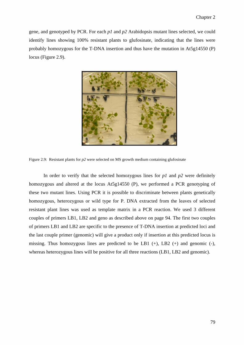
Chapter 2
79
gene, and genotyped by PCR. For each p1 and p2 Arabidopsis mutant lines selected, we could
identify lines showing 100% resistant plants to glufosinate, indicating that the lines were
probably homozygous for the T-DNA insertion and thus have the mutation in At5g14550 (P)
locus (Figure 2.9).
Figure 2.9: Resistant plants for p2 were selected on MS growth medium containing glufosinate
In order to verify that the selected homozygous lines for p1 and p2 were definitely
homozygous and altered at the locus At5g14550 (P), we performed a PCR genotyping of
these two mutant lines. Using PCR it is possible to discriminate between plants genetically
homozygous, heterozygous or wild type for P. DNA extracted from the leaves of selected
resistant plant lines was used as template matrix in a PCR reaction. We used 3 different
couples of primers LB1, LB2 and geno as described above on page 94. The first two couples
of primers LB1 and LB2 are specific to the presence of T-DNA insertion at predicted loci and
the last couple primer (genomic) will give a product only if insertion at this predicted locus is
missing. Thus homozygous lines are predicted to be LB1 (+), LB2 (+) and genomic (-),
whereas heterozygous lines will be positive for all three reactions (LB1, LB2 and genomic).
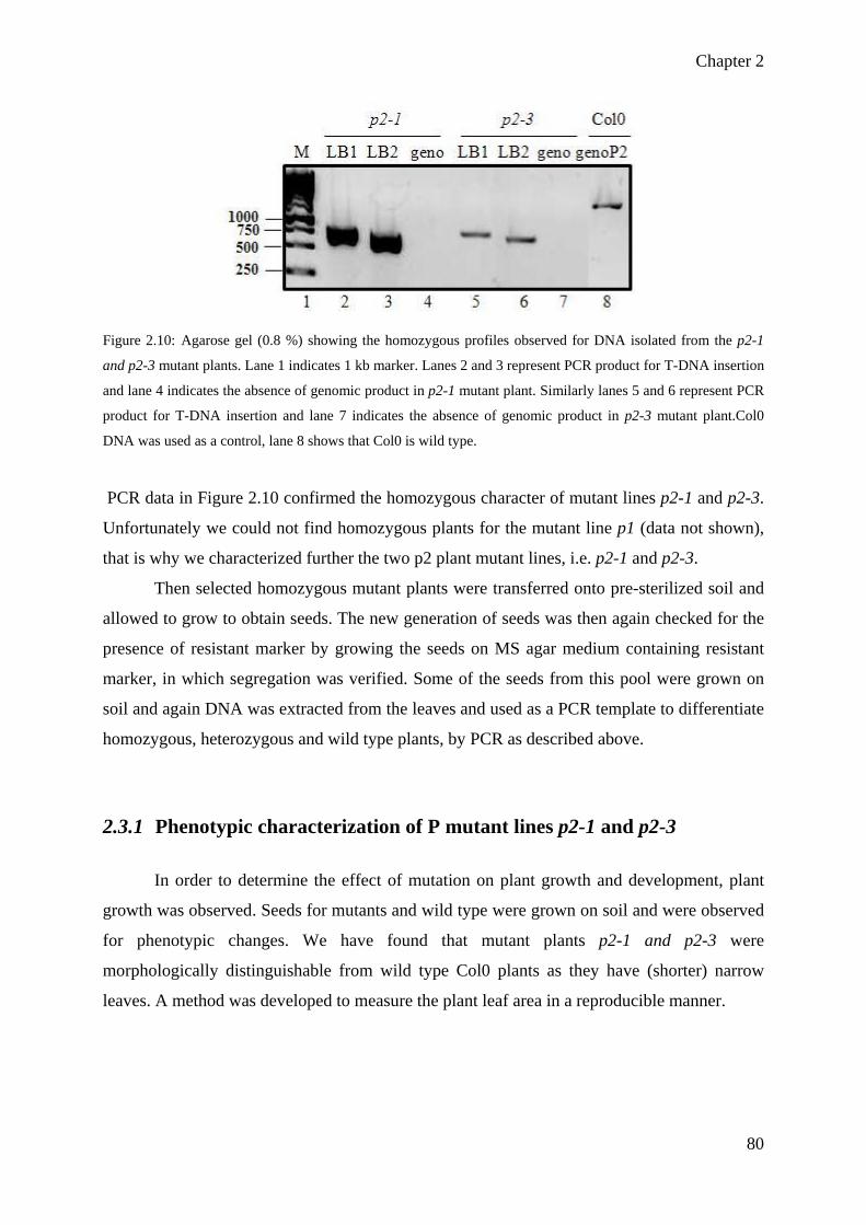
Chapter 2
80
Figure 2.10: Agarose gel (0.8 %) showing the homozygous profiles observed for DNA isolated from the p2-1
and p2-3 mutant plants. Lane 1 indicates 1 kb marker. Lanes 2 and 3 represent PCR product for T-DNA insertion
and lane 4 indicates the absence of genomic product in p2-1 mutant plant. Similarly lanes 5 and 6 represent PCR
product for T-DNA insertion and lane 7 indicates the absence of genomic product in p2-3 mutant plant.Col0
DNA was used as a control, lane 8 shows that Col0 is wild type.
PCR data in Figure 2.10 confirmed the homozygous character of mutant lines p2-1 and p2-3.
Unfortunately we could not find homozygous plants for the mutant line p1 (data not shown),
that is why we characterized further the two p2 plant mutant lines, i.e. p2-1 and p2-3.
Then selected homozygous mutant plants were transferred onto pre-sterilized soil and
allowed to grow to obtain seeds. The new generation of seeds was then again checked for the
presence of resistant marker by growing the seeds on MS agar medium containing resistant
marker, in which segregation was verified. Some of the seeds from this pool were grown on
soil and again DNA was extracted from the leaves and used as a PCR template to differentiate
homozygous, heterozygous and wild type plants, by PCR as described above.
2.3.1 Phenotypic characterization of P mutant lines p2-1 and p2-3
In order to determine the effect of mutation on plant growth and development, plant
growth was observed. Seeds for mutants and wild type were grown on soil and were observed
for phenotypic changes. We have found that mutant plants p2-1 and p2-3 were
morphologically distinguishable from wild type Col0 plants as they have (shorter) narrow
leaves. A method was developed to measure the plant leaf area in a reproducible manner.

Chapter 2
81
Figure 2.11 : Arabidopsis thaliana 23 days old wild type (Col0) and mutant plants (p2-1, p2-3). The leaves of
mutant p2-1, p2-3 are narrow than wild type plants. Scale bar = 2cm
Leaf area was measured at different developmental stages of plant growth by using
publicly available software ImageJ as explained in materials and methods (cf materials and
methods 6.7.1). Briefly, we measured the leaf area by taking a picture of individual plant for
each p2-1, p2-3 and Col0, each having a reference scale bar (Figure 2.11). Area of the leaf
was calculated with the help of reference scale, and reported as histograms.

Chapter 2
82
Figure 2.12: Analysis of leaf area of wild type Col0 and mutant plants p2-1, p2-3. Leaf area was measured
quantitatively. Each value is an average determined from the measurement of leaf area of six plants in two
independent experiments performed under the same conditions. The error bars represent the standard deviation
calculated for all values of two experiments. At 42 day of plant growth, the leaves of the mutant plants p2-1, p2-
3 are smaller than leaves of wild Col0 plants.
This phenotypic study has shown that mutant plants of both lines have smaller leaves
as compared to wild type plants but p2-1 and p2-3 behaved differently when compared to each
other. This is little bit surprising but it could be possible because of difference in growth
condition as this data is calculated from 2 leaves of 3 individual plants from p2-1 and p2-3
mutant plants. The results showed that mutant leaves were very smaller at 14 days of growth
and this difference was remarkable even at 42 days of growth of plant with a difference of
52% for p2-1 and 30% for p2-3 as compared to wild type Col0 plants (Figure 2.12).
2.3.2 Neutral monosaccharide quantification of cell wall from p2-1 mutant
line through GC-MS
As plant cell wall consists of 90% carbohydrates, the quantification of neutral
monosaccharide in both wild type and mutant plants can give us an idea about the implication
of P gene in cell wall biosynthesis. For this purpose AIR residues were prepared from dark
grown 6 days old seedlings from mutant and wild type plants. Neutral monosaccharides were
quantified by first hydrolyzing the polysaccharides to their monosaccharide constituents with
strong acid (TFA) and then converting them to alditol acetates by reduction with sodium
borohydride to the corresponding alditol, followed by acetylation of the hydroxyls on each
alditol. The resultant alditol acetates were quantified by GC-MS. Seven major
monosaccharides were quantified from cell wall including rhamnose (Rha), fucose (Fuc),
arabinose (Ara), galactose (Gal), xylose (Xyl), mannose (Man), and glucose (Glc).
Quantification analysis of sugars has shown that almost all the neutral sugars present
in cell wall are affected and mutant line p2-1 has significant changes in cell wall glycosyl
residues as compared to wild type because of mutation in gene P as shown in figure 2.13. A
reduction is observed in the quantity of sugars in mutant cell wall as compared to wild type
Col0 cell wall.
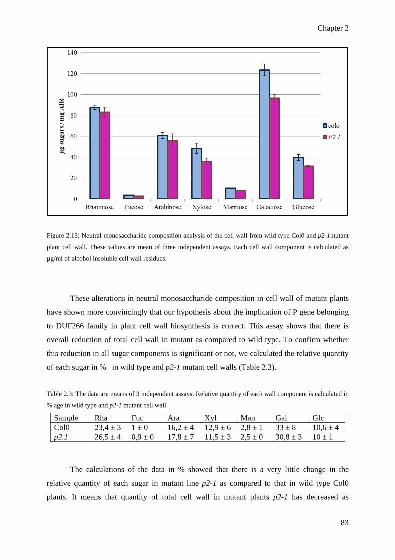
Chapter 2
83
Figure 2.13: Neutral monosaccharide composition analysis of the cell wall from wild type Col0 and p2-1mutant
plant cell wall. These values are mean of three independent assays. Each cell wall component is calculated as
µg/ml of alcohol insoluble cell wall residues.
These alterations in neutral monosaccharide composition in cell wall of mutant plants
have shown more convincingly that our hypothesis about the implication of P gene belonging
to DUF266 family in plant cell wall biosynthesis is correct. This assay shows that there is
overall reduction of total cell wall in mutant as compared to wild type. To confirm whether
this reduction in all sugar components is significant or not, we calculated the relative quantity
of each sugar in % in wild type and p2-1 mutant cell walls (Table 2.3).
Table 2.3: The data are means of 3 independent assays. Relative quantity of each wall component is calculated in
% age in wild type and p2-1 mutant cell wall
Sample Rha Fuc Ara Xyl Man Gal Glc Col0 23,4 ± 3 1 ± 0 16,2 ± 4 12,9 ± 6 2,8 ± 1 33 ± 8 10,6 ± 4 p2.1 26,5 ± 4 0,9 ± 0 17,8 ± 7 11,5 ± 3 2,5 ± 0 30,8 ± 3 10 ± 1
The calculations of the data in % showed that there is a very little change in the
relative quantity of each sugar in mutant line p2-1 as compared to that in wild type Col0
plants. It means that quantity of total cell wall in mutant plants p2-1 has decreased as

Chapter 2
84
compared to wild type cell wall. As this neutral monosaccharide quantification provided us an
idea about the biochemical changes in mutant cell wall but did not give us information which
particular polysaccharide is modified, so in order to get this more precise information
glycosidic linkage analysis after permethylation of cell wall samples and extraction of
individual cell wall components is required. Zhou et al., (2009) have quantified the neutral
monosaccharides in rice alcohol insoluble residues and have shown that glucose is 20%
decreased in mutant bc10 as compared to wild type Col0 but all other sugars like xylose,
galactose, arabinose and rhamnose are increased as shown in Table 2.4. This analysis did not
provide them any strong evidence about the alteration of specific sugar and in turn about the
function of BC10 gene.
Table 2.4. The data are means of five independent assays. Each wall component was calculated as mg/g of
alcohol-insoluble cell-wall residue (Zhou et al., 2009).
Sample Glc Xyl Man Gal Ara Rha
Col0 395 ± 6.5 49.2 ± 0.7 7.49 ± 0.1 14.1 ± 0.1 10.3 ± 0.2 4.74 ± 0.1
bc10 316 ± 6.3 62.8 ± 1.0 7.72 ± 0.2 16.6 ± 0.6 16.7 ± 0.3 5.58 ± 0.2
They performed immuno-labelling experiments which showed a reduction of AGPs
labelling in the mutant. In order to confirm this observation, they isolated the AGPs and
observed 70% reduction in AGPs content in bc10 as compared to wild type plants. Similarly
we have observed that in Arabidopsis cell wall all the neutral monosaccharides are changed
quantitatively but no sugar changed dramatically in p2-1 mutants which could be linked to its
function. In order to determine its function, it needs further detailed analysis like
permethylation linkage analysis and immuno-labelling assays that will provide information
which particular polysaccharide is affected because of mutation in p2-1 plants.
Because of the unavailability of the cDNAs for P gene we have not yet carry out its
heterologous expression. Heterologous expression would be very useful for the functional
characterization of the P gene, as well as the preliminary study of Arabidopsis cell wall
characterization for P gene that we started in the lab.

Chapter 2
85
2.4 Cloning of DUF 266 cDNA for heterologous expression in
Pichia pastoris
The results of the mutant screening and biochemical analysis provided some clues
about the changes in plant phenotype and cell wall composition. We decided to confirm these
results by another functional genomics approach, i.e. the heterologous expression of candidate
genes. Gateway Cloning Technology is a powerful methodology that greatly facilitates protein
expression, cloning of PCR products and analysis of gene function with site-specific
recombination. This technology provides a versatile system for transferring DNA segments
between vectors. Once in the system, DNA segments can be transferred from an Entry Clone
into numerous vectors (e.g., for protein expression). For the expression of our candidate genes
it was an ideal system as it gives the possibility to use different expression systems.
In order to clone candidate genes, cDNAs available at that time were ordered from
SALK. We ordered cDNA for 15 genes belonging to DUF 266 family. They were amplified
with a 2 step PCR reaction as shown in figure 2.14. T7 N-terminal tag was added to facilitate
the protein identification through western blot and purification of protein. The genes of
interest were flanked by attB1 and attB2 gateway border sites which recombine with donor
vector attP1 and attP2 sites to create any entry vector. These sites were added through PCR
amplification of a gene (see materials and methods 6.2.1.1.1).
In order to find out the role of the proteins in the cell wall composition/biosynthesis
and in the growth and development of the plant, we decided to clone the genes for which
homozygous mutants were found and cell wall composition analysis was performed. Here in
the figure 8, amplification has been shown for genes like A, C, I, J, N and W by two step PCR
reaction (See materials and methods 6.2.1.1.1). Figure 2.14A represents the 1st PCR reaction
while figure 2.14B represents the 2nd PCR reaction.

Chapter 2
86
Figure 2.14: A) Agarose gel (0.8 %) showing the 1st PCR reaction for amplification of cDNA of genes A, C, I, J,
N and W. B) Agarose gel (0.8 %) showing the 2nd PCR reaction for genes A, C, I, J, N and W
These PCR products were cloned into pDONR207 to create entry vectors through BP cloning
reaction. Correct sequence of the reading frame was confirmed through sequencing. In an LR
reaction this entry vector pENTR207 is integrated into destination vector pPICZ to make an
expression clone. After LR reaction the plasmids were also sent for sequencing to confirm the
correct sequence of gene of interests.
Figure 2.15: The histogram showing the number of genes whose cDNAs were cloned into entry vector
pDONR207 and pPICZ expression vector

Chapter 2
87
Out of total 23 genes, for 14 genes cDNAs were available at the time of ordering. I
cloned 10 genes out of 14 into entry vector and expression vector through gateway cloning
and sequenced (Figure 2.15). Functional characterization of all the cloned genes would have
been time consuming and laborious work, so I focused only on the expression of two genes:
gene E (having no fucosyltransferase signature, used as a control) which belongs to DUF266
family and gene At5g28910 (W) which has fucosyltransferase signature and will be discussed
in next chapter 3.
2.5 Conclusion
The work presented in this chapter resulted in the identification of 16 homozygous
mutants among the 35 allelic mutant lines for 23 genes belonging to DUFF266 family. It is a
valuable work for the forthcoming researchers and students in the lab as it will save their time
for antibiotic selection and mutant characterization through PCR based screening strategy.
Along with the above gene P, during my PhD I focused more on characterization and
functional analysis of W (NGT1) gene because of following reasons.
1-As Hansen et al. (2009) have shown that this gene is unique in Arabidopsis genome
and is distantly related to the known fucosyltransferases present in different GT families
(GT11, GT23, GT37, GT65 and GT68) it could possibly be involved in cell wall biosynthesis
particularly in pectin RG-II biosynthesis.
2-Allelic mutant of NGT1 gene showed developmental defects during homozygous
mutant characterization (discussed in the next chapter). Preliminary data showed that mutant
plants have smaller leaf area as compared to wild type plants which will be discussed in detail
in chapter 3.
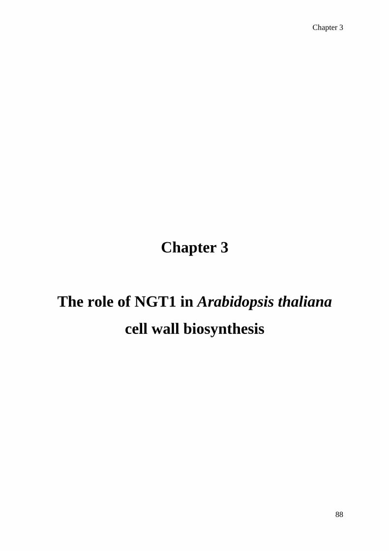
Chapter 3
88
Chapter 3
The role of NGT1 in Arabidopsis thaliana
cell wall biosynthesis

Chapter 3
89
3 Role of NGT1 in A.thaliana cell wall biosynthesis
3.1 Introduction
A previous study developed in the lab uses bio-informatics to screen the Arabidopsis
genome for the presence of new genes harbouring GT signatures; this study ultimately led to
the identification of 24 genes without predicted functions as being putative
glycosyltransferases (Hansen et al., 2009). The objective of my research work was to
functionally characterize one of these 24 newly identified genes, in order to validate
experimentally whether or not these genes encode GTs involved in plant cell wall
biosynthesis. Interestingly, among these 24 candidate genes, 23 genes belong to the DUF266
gene family and one was unique. The first part of my work (chapter 2) was mostly devoted to
the molecular characterization of Arabidopsis mutants T-DNA lines from the DUF266 gene
family, in order to perform a phenotypic analysis of these mutants as well as a biochemical
characterization of their cell wall content, seeking for specific phenotypes related to the
alteration of DUF266 genes. Accordingly, I undertook the characterization of 35 T-DNA
insertion lines, phenotyped 15 mutants and analysed cell wall content for 10 mutants.
Unfortunately, no specific trait could reliably be detected that would link genetic alteration in
gene family DUF 266 and plant development or cell wall composition. In the present chapter,
I will present data obtained for the gene At5g28910 that was not related to the DUF266 gene
family. This gene was actually unique and highlighted by our bioinformatics approach as
being a putative fucosyltransferase. Over the course of that work some confidence was gained
that this gene (At5g28910) would be of particular interest, especially because the T-DNA
lines altered for this locus appears to carry a slower growth development, but also because
gene sequence showed characteristic features with known fucosyltransferases, thus providing
a testable hypothesis to develop GT activity tests. This genetic locus At5g28910 was then
named NGT1 for Novel GlycosylTransferase 1. Two Arabidopsis mutant T-DNA lines altered
for At5g28910 locus were characterized and named ngt1-1 and ngt1-2. In order to determine
the function of NGT1, we used the functional genomics approaches described in chapter 2,
i.e., heterologous expression and T-DNA knock out mutants characterization of candidate
gene (NGT1). Heterologous expression of candidate gene shall help to assign biochemical
function of NGT1 by analysing its role through activity test. While the T-DNA knock-out
mutant lines will offer the opportunity to unravel the role of candidate gene NGT1 in cell wall
biosynthesis through phenotypic characterization and chemical analysis of mutant cell wall.

Chapter 3
90
3.2 Protein sequence analysis
There are two gene models/annotations for gene At5G28910 (NGT1) in TAIR database
which are represented as At5G28910.1 and At5G28910.2. According to these models there
are two possible initiation sites for the gene translation, one giving a protein of 408 amino-
acids, and the other gives a 535 amino-acid protein. We characterized the gene model
At5G28910.2 which will result in the synthesis of longer protein of 535 amino-acids and its
nucleotide and translated sequence of the coding region is shown in figure 3.1.
atgagcatgaagtcattagaaagagtggtttcagagagagcattaaaacttggaaattca M S M K S L E R V V S E R A L K L G N S tttccatgtcaaatatgtgtagttgggtttctatgtggaatctgtctcacttcactcttc F P C Q I C V V G F L C G I C L T S L F ttagctgctctcacttctcttggcaccttcgaattcgccgccttctccttcacctcctct L A A L T S L G T F E F A A F S F T S S tcctctgtttttcctccttgcaattcctccacctctcacatcatcaatatggttgcaagt S S V F P P C N S S T S H I I N M V A S atagaccggaaactgaaatggaagaacaaagttgagatagaagaagaagatgaagtgaaa I D R K L K W K N K V E I E E E D E V K cttttggtctctgcttgggataatttattactaaatgaagaagacttcttgaagaaggta L L V S A W D N L L L N E E D F L K K V ggtattaacaaatccgatgtaccaaatggtccacatttggagaattgtgaggagaaggct G I N K S D V P N G P H L E N C E E K A cgagttagggagcgtttggatacacgtttggcgaactggacacttcctccttggatcagt R V R E R L D T R L A N W T L P P W I S ggaggagatgaagagaattatccgttaacgaggagagtgcaaagagatatatggattcat G G D E E N Y P L T R R V Q R D I W I H cagcatcctttggattgcggaaacaagagtctcaagttccttgtagctgattgggaaaca Q H P L D C G N K S L K F L V A D W E T cttcctggttttggaataggagctcagatagctggaatgactggtctactcgcgatagct L P G F G I G A Q I A G M T G L L A I A ataaatgaaaaccgagtgcttgttgcaaattactacaaccgagcagatcatgatggttgc I N E N R V L V A N Y Y N R A D H D G C aaaggttcttttcgtggaaactggtcttgctattttctacaggaaacgtcagaagagtgt K G S F R G N W S C Y F L Q E T S E E C cgaaaacgagcctttgcgattgtgaagaagagagaagcgtgggagagtgggattgttaca R K R A F A I V K K R E A W E S G I V T gggaaacaaaattatagcacaaaggagatttgggctggggctataccaaagcaatggggt G K Q N Y S T K E I W A G A I P K Q W G aagccttggagttatatgaagccaactacagaaatcaacggaagtttaatctccaatcat K P W S Y M K P T T E I N G S L I S N H cggaaaatggatcggagatggtggagagcacaagcagtgagatacttgatgagatatcaa R K M D R R W W R A Q A V R Y L M R Y Q acagaatacacttgcggtttgatgaacattgctcgaaattccgcgtttggaaaagaagct T E Y T C G L M N I A R N S A F G K E A gccaagattgttctttcagctggagattggagaaagaagaataagaagatgaggacagag A K I V L S A G D W R K K N K K M R T E attgaggaacaggtgtggtcggatcacaagccgtggcttccaaggccaatgctgagtgtt I E E Q V W S D H K P W L P R P M L S V cacgtacggatgggagacaaagcatgcgagatgagagtcgcagctttagaagagtacatg H V R M G D K A C E M R V A A L E E Y M catttagctgatcggatcagagatcggtttccagagctcaacaggatctggctctctaca H L A D R I R D R F P E L N R I W L S T gagatgaaggaagtggtggacagaagtaaagattatgctcactggagattctattacacg E M K E V V D R S K D Y A H W R F Y Y T gaagtggcaagacaagtcggtaataagtcgatggctgagtatgaagcgagcctcgggaga E V A R Q V G N K S M A E Y E A S L G R gagatgagcacaaactatcctctggttaacttcttaatggcgtcagaagctgatttcttc E M S T N Y P L V N F L M A S E A D F F gtcggagcattgggttccacttggtgtttcctcatcgatggtatgaggaatacgggtggg V G A L G S T W C F L I D G M R N T G G aaagtcatgtctggttatctcagtgtcaataaagatcggttctggtaa K V M S G Y L S V N K D R F W -

Chapter 3
91
Figure 3.1. Nucleotide sequence and translated sequence of the coding region of At5g28910.2. Predicted TMD is marked in turquoise, N-glycosylation sites with green letters underlined. The β-strands and α-helices, marked in yellow and magenta, respectively, were predicted by the consensus secondary structure prediction tool at the NPSA server (http://npsa-pbil.ibcp.fr/cgi-bin/npsa_automat.pl?page=/NPSA/npsa_seccons.html).
NGT1 protein is predicted to have one transmembrane helix at its N-terminus
(http://www.cbs.dtu.dk/services/TMHMM/) (Figure 3.2). Thus, NGT1 displays the typical
type II membrane protein topology that is observed for most of the Golgi-resident GTs,
consisting in a short cytoplasmic N-terminal region, a trans-membrane domain spanning the
amino acids 26-47, a stem region and a large globular catalytic C-terminal domain facing the
luminal side of the Golgi.
Figure 3.2: Prediction of trans-membrane domain of NGT1 protein using TMHMM. The probability of regions
being transmembrane (red), inside (i.e. cytoplasmic, blue) or outside (i.e. Golgi lumen, magenta) are plotted as a
function of amino acid residue number.
Seven putative N-glycosylation sites were predicted by the use of N-glycosylation
prediction server (at http://www.cbs.dtu.dk/) at position 68, 123, 152, 188, 247, 284 and 313
of amino acids sequence (Figure 3.3).
Figure 3.3: Predicted structure of NGT1 protein. NGT1contains one transmembrane domain represented by
square at its N-terminal and has seven putative N-glycosylation sites shown by triangles with the positions of
amino acids

Chapter 3
92
3.3 Characterization of T-DNA mutants ngt1-1 and ngt1-2
For this study, we hypothesized that NGT1 is a glycosyltransferase involved in plant
cell wall biosynthesis, and we analyzed cell wall from NGT1 deficient Arabidopsis mutants,
expecting quantitative or qualitative variation in sugar content by comparison to the wild-type
cell wall. More specifically, particular attention was payed to the fucose content in the cell
wall of ngt1-1 and ngt1-2 mutant lines. Fucose is present over various polymers in
Arabidopsis, such as N-glycans in glycoproteins, arabinogalactan proteins (AGPs) and cell
wall polysaccharides like pectins and xyloglucan. A few Arabidopsis genes encoding
fucosyltransferase activities have already been identified and characterized. These genes are
involved in the fucosylation of XG, AGPs and protein N-glycosylation and they classify into
GT37 and GT10 families in CAZy database (www.cazy.org). However, no fucosyltransferase
gene has been identified to date for the fucosylation of RG-II in pectins,
In order to evaluate if NGT1 is involved in pectin biosynthesis, two allelic T-DNA
knock out mutant lines were ordered from Nottingham Arabidopsis Stock Centre (NASC).
These two mutant lines N585839 (SALK_085839) termed as ngt1-1 has insertion in first exon
(downstream +217 bp from the translation initiation codon ) and N638819 (SALK_138819)
termed as ngt1-2 has insertion in the 5’ UTR (upstream -23 bp from the translation initiation
start codon) of the same gene NGT1 (Figure 3.4).
Figure 3.4: Schematic representation of the gene structure of NGT1 and the position of insertion of T-DNA for
ngt1-1 and ngt1-2. The binding site of primers used in PCR reaction is indicated by red arrows. Blue boxes
represent exons of NGT1 gene, red boxes labelled “FS” correspond to the genomic flanking sequence of the T-
DNA according to NASC seed stock center.

Chapter 3
93
These two SALK lines ngt1-1 and ngt1-2 harbour an alteration of NGT1 that is caused
by the integration of a T-DNA cassette containing the neomycin phosphotransferase II (NPT
II) gene providing resistance to the antibiotic kanamycin. Arabidopsis mutants received from
NASC were first amplified on soil without selection pressure and afterwards the progeny was
checked for segregation of the NPT II gene, and genotyped by PCR. For each ngt1-1 and
ngt1-2 Arabidopsis mutant lines selected, we could identify lines showing 100% resistant
plants to kanamycin, indicating that the lines were probably homozygous for the T-DNA
insertion and thus have the mutation in NGT1 locus (Figure 3.5). In order to verify that the
selected homozygous lines for ngt1-1 and ngt1-2 were homozygous and altered at the NGT1
locus we performed a PCR genotyping of these two mutant lines. DNA extracted from the
leaves of selected resistant plant lines was prepared and genotyped. Different sets of primers
were used referenced as primer 1, 2, 3, 4, 5 and 6 in figure 3.6. Primers 1 and 2 are left border
(LBa1 and LB1.3) T-DNA specific primers which will give two products of different sizes for
T-DNA and genomic hybrid product while (3 + 4) and (5 + 6) are gene specific right and left
primers (RPW1 and LPWI), (RPW3+ LPW3) for ngt1-1 and ngt1-2, respectively.
Figure 3.5: On the left, ngt1-2 T-DNA mutant altered for At5g28910 (NGT1) gene is resistant to kanamycin
whereas Wt Col0 plants are (on the right).
ngt1 resistant plants Wt Col0 Sensitive plants

Chapter 3
94
The region corresponding to T-DNA insertion of NGT1 gene was amplified using T-
DNA specific left border primer 1 and 2 (LBa1 and LB1.3) with the gene specific primers 3
and 5 for ngt1-1 and ngt1-2, respectively. Similarly the region corresponding to genomic
DNA for NGT1 gene was obtained by using gene specific left border and right border primers
(3+ 4) and (5+6) for ngt1-1 and ngt1-2, respectively. In the figure 3.6, for example, ngt1-1,
ngt1-2 and Col0 represent different DNA stocks isolated from mutant and wild type plants
that were checked by PCR for T-DNA insertion using three sets of primers: two sets
amplifying a product in the presence of T-DNA at the expected locus, one set being specific
of the WT version of the gene at this locus (Figure 3.6). Lanes 1 and 2 show a PCR product
indicating insertion of a T-DNA at NGT1 locus in ngt1-1 plants by the use of primers (1+3,
2+3), while the lack of product in lane 3 (primers 3+4) from ngt1-1 DNA confirmed the
Arabidopsis line ngt1-1 is homozygous for T-DNA insertion at NGT1 locus. Similar results
were obtained for ngt1-2 (lanes 4 to 6) indicating that this allelic line of ngt1-2 was also
homozygous. In contrast, in case of Col0 genomic DNA the lack of product with primers
(1+3, 2+3; specific of the T-DNA) in lanes 8 and 9 and the presence of a PCR product in lanes
10 and 11 confirmed that the DNA prepared from Col0 plants was wild type for NGT1 locus.
All this PCR reaction products were sequenced to unambiguously confirm their identity, but
also to precisely determine deletion of DNA sequences (from genomic or T-DNA) that occurs
in consequence of the T-DNA insertion into the Arabidopsis genome.
Figure 3.6: Agarose gel (0.8 %) showing the homozygous profiles observed for DNA isolated from the ngt1-1
and ngt1-2 lines. Lane 1 indicates 1 kb marker. Lanes 2 and 3 represent PCR products for T-DNA insertion and
lane 4 indicates the absence of genomic product in ngt1-1 mutant. Similarly lanes 5 and 6 represent PCR
products for T-DNA insertion and lane 7 indicates the absence of genomic product in ngt1-2 mutant. Col0 DNA
was used as a control, lanes 8 and 9 show the absence of T-DNA.

Chapter 3
95
The right border of the insertion was studied using identical strategy to the left border
but using right border specific primers. Despite numerous efforts we were unsuccessful to
obtain a hybrid PCR product bearing sequence from the right border and the genomic near the
insertion. The most likely hypothesis would be a deletion of the T-DNA right border sequence
occurring during insertion into the genome of the mutants. We overcome that difficulty by
carrying out an extensive amplification of the T-DNA including the genomic sequence near
the left border. This PCR reaction resulted in the amplification of two products over 2kb
which confirmed that at least 2 kb of T-DNA are inserted in our gene of interest for both
ngt1-1 and ngt1-2 mutant lines. For mutant line ngt1-1, the size of the PCR product was 1490
bp (Figure 3.7, lane 2) with the set of primers (RPW1+OL99) while the second set of primers
(RPW1+OL100) generated the product of 2049 bp (lane 3). Similarly two products of T-DNA
were obtained by PCR for line ngt1-2 of the size of 1646 bp (OL99+RPW3, lane 4) and 2205
bp (OL100+RPW3, lane 5). PCR amplicon was sequenced and showed to be a hybrid product
of our gene of interest NGT1 and left border of the T-DNA by sequencing. In addition, we
carried out the phenotypic studies on successive progenies which confirmed that the observed
phenotype was genetically linked to the T-DNA insertion across generation. The phenotypic
studies are described in the next paragraphs.
Figure 3.7: Agarose gel (0.8%) showing the left border and genomic sequence amplificon of ngt1-1 and ngt1-2
lines by PCR. Lane 1 shows the molecular markers. Lane 2 and 3 shows the PCR amplified product for ngt1-1
through the use of two set of primers (RPW1+OL99) and (RPW1+OL100) respectively while lane 4 and 5
represent the left border PCR product of line ngt1-2 by the use of two set of primers (OL99+RPW3) and
(OL100+RPW3), respectively.

Chapter 3
96
• Characterization of NGT1 mutants by RT-PCR
The T-DNA insertion sites were characterized through PCR using left border and
genomic primers for both ngt1-1 and ngt1-2 mutant lines (cf materials and methods 6.5.6).
This analysis confirmed disruption of NGT1 in the first exon of ngt1-1 and insertion in the
5’UTR in ngt1-2 mutant line. Both mutant lines were then further analyzed by RT-PCR
seeking the presence of NGT1 transcript (or hybrid transcript T-DNA:NGT1), thus in order to
find out whether ngt1-1and ngt1-2 mutants were null mutants (NGT1 not expressed at mRNA
level). For this purpose total RNAs were extracted from 2 weeks old wild type Col0, ngt1-1
and ngt1-2 mutant seedlings. cDNAs were prepared and analyzed for the presence of NGT1
transcripts using PCR. Five different set of primers, namely “GS1”, “GS2”, “UBQ”, “T-DNA
ngt1-1”, “T-DNA ngt1-2” were used first for Col0 cDNA. (Ubiquitin specific primers were
used as a control for determining the RT-PCR amplification efficiency among the different
samples).The first two set of primers GS1 (Gene Specific primer 1) and GS2 (Gene Specific
primer 2) are specific primers for NGT1 cDNA and expected to give a product of 210 bp and
389 bp, respectively. Accordingly, these two PCR products, indicative of the presence of
NGT1 cDNA, were positively amplified using Col0 cDNA library but not using cDNA
libraires prepared from ngt1-1 and ngt1-2 mutants (figure 3.8, lanes 2 and 6). The lack of
amplification for GS1 and GS2 product in both mutant lines indicate that no full length NGT1
transcript is present in any mutant lines. On the contrary, two set of primers specific for the
presence of the T-DNA insertion at ngt1-1 or ngt1-2 loci were used in order to seek the
presence of T-DNA: NGT1 hybrid product in both mutant lines. As a control Col0 cDNA
library was used as matrix for such reaction and as expected did not provide any amplification
as no T-DNA is inserted at NGT1 locus in WT Col0 (figure 3.8 lanes 4 and 5). However, such
T-DNA: NGT1 hybrid product would be possibly transcribed in ngt1-1 or ngt1-2 mutants.
Figure 3.8: RT-PCR analysis of transcripts from the wild type Col0 and homozygous mutant lines ngt1-1 and
ngt1-2. NGT1 transcript is detected in Col0 cDNA library whereas no such transcript is detected from
homozygous mutant lines ngt1-1 and ngt1-2.

Chapter 3
97
It is important to note that a hybrid product was characterized from the ngt1-1 cDNA
library. Analysis of the T-DNA:NGT1 hybrid product (780 pb; lane 9 fig 3.8) obtained for
ngt1-1 indicates the presence of the first 219 bp from NGT1 sequence which would represent
the first 73 amino acids of NGT1 protein sequence if translated. Because the first 73 amino
acids at the N-ter of the sequence do not comprise the expected catalytic site for NGT1
protein, we conclude that functional NGT1 protein would be absent in ngt1-1, thus being a
null mutant. Finally, in case of ngt1-2 mutant, neither gene specific nor T-DNA:NGT1 product
were amplified using PCR indicating such mutant is null for NGT1 transcript (figure 3.8, lanes
13 and 14).
3.4 Phenotypic characterization of mutant lines ngt1-1 and ngt1-2
As mentioned in the introduction, normal plant development is, among other
parameters, dependent of plant cell wall biosynthesis. Indeed, in order to grow properly, a
plant like Arabidopsis will need to synthesize polysaccharides necessary to maintain cell wall
integrity during cell elongation and expansion. Research program trying to decipher plant cell
wall biogenesis actually took advantage of this tight correlation between polysaccharide
biosyntheses and plant development, while developing a strategy where mutants having an
altered development (i.e. short hypocotyl in dark-grown culture condition) were suspected to
be altered for cellulose biosynthesis. Accordingly, as we expected NGT1 locus to be involved
in cell wall biosynthesis, we observed the effect (if any) of mutation at NGT1 locus on
Arabidopsis growth. Seeds for allelic ngt1-1, ngt1-2 mutants and wild type Col0 were grown
on soil and observed for phenotypic changes during 8 weeks. The first observation was the
lack of strong phenotype altering organ development as sometimes observed for some pectin
altered mutants such as quasimodo1 and quasimodo2 (Bouton et al., 2002, Mouille et al.,
2007). However, during the course of their growth, we have found that ngt1-1 and ngt1-2 mutants were morphologically distinguishable from wild type Col0 plants as they have
“narrow” leaves and a pale green color which is a classical sign of chlorosis (Fig 3.9).
Although this observation of pale color appears consistent over plant cylce, and was
reproducible from generation to generation of ngt1-1 and ngt1-2 lines, it remains difficult to
quantify precisely. Thus, we focused on the developmental phenotype (narrow leaves) and
sought to develop a method in order to measure the plant leaf area in a reproducible manner
with the aim of shifting from a qualitative observation to a quantified trait. Leaf area was then
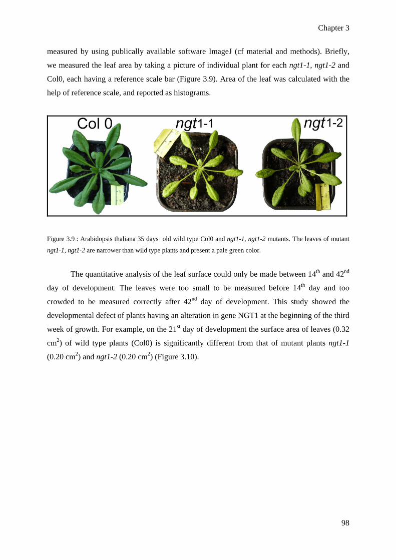
Chapter 3
98
measured by using publically available software ImageJ (cf material and methods). Briefly,
we measured the leaf area by taking a picture of individual plant for each ngt1-1, ngt1-2 and
Col0, each having a reference scale bar (Figure 3.9). Area of the leaf was calculated with the
help of reference scale, and reported as histograms.
Figure 3.9 : Arabidopsis thaliana 35 days old wild type Col0 and ngt1-1, ngt1-2 mutants. The leaves of mutant
ngt1-1, ngt1-2 are narrower than wild type plants and present a pale green color.
The quantitative analysis of the leaf surface could only be made between 14th and 42nd
day of development. The leaves were too small to be measured before 14th day and too
crowded to be measured correctly after 42nd day of development. This study showed the
developmental defect of plants having an alteration in gene NGT1 at the beginning of the third
week of growth. For example, on the 21st day of development the surface area of leaves (0.32
cm2) of wild type plants (Col0) is significantly different from that of mutant plants ngt1-1
(0.20 cm2) and ngt1-2 (0.20 cm2) (Figure 3.10).

Chapter 3
99
Figure 3.10: Analysis of leaf area of wild type Col0 and mutant lines ngt1-1 and ngt1-2. Leaf area was measured
quantitatively. Each value is an average, determined from the measurement of leaf area of six plants in two
independent experiments performed under the same conditions. The error bars represent the standard deviation
calculated for all values of two experiments. At 42 day of plant growth, the leaves of the mutant lines ngt1-1 and
ngt1-2 are 34% and 36% smaller than leaves of wild Col0 plants.
The developmental difference observed among the wild type and mutant plants are
more significant on 35th and 42nd days of growth. As on 42nd day, the leaves of ngt1-1 and
ngt1-2 are 34 % and 36 % smaller as compared to that of wild type plant leaves. Although the
quantitative analysis shows that leaf area of ngt1-1 was different from that of ngt1-2 but this
difference was not relevant. On contrary, the leaf area of mutants was significantly different
from that of wild type plants from the 21st day of development.

Chapter 3
100
Figure 3.11: Comparative analysis of leaf growth rate of wild type (Col0) and mutant lines ngt1-1 and ngt1-2 followed during 4 weeks of growth. Rate of growth of ngt1-1 and ngt1-2 mutants is weaker than Col0 plants only before 4th week of growth
The quantification of the leaf surface area made it possible to measure the growth rate
[(Leaf area of the week W+1) - (Leaf area of the week W)] / (Leaf area of the week W)] of
different plants during the phenotypic studies. The growth rate profiles could only be
calculated between 14th and 21st day, 21st and 28th day etc. Significant growth rate difference
was observed among the wild type and mutant plants only between 14th and 21st day of
development (Figure 3.11). From 21st to 35th day of development, the plants seemed to have
the tendency of similar growth. This observation was confirmed from the growth rate of 35th
and 42nd day. These results indicate that the mutation in NGT1 alters the growth rate only
during early developmental stage and has no or little effect on the growth rate during late
development.
This phenotypic difference showed that NGT1 gene plays a role in controlling leaf
development and particularly slows down normal growth. The next step we envisioned was
then to evaluate whether or not the polysaccharide content of the cell wall would be modified
in mutants. Accordingly, we performed biochemical analysis of the cell wall from ngt1-1 and
ngt1-2, in order to account for its composition.

Chapter 3
101
3.5 Quantification of neutral monosaccharide of cell wall from
ngt1-1 and ngt1-2 using gas chromatography
As the plant cell wall consists of roughly 90% carbohydrates, one would expect that
the alteration of a glycosyltransferase activity involved in cell wall biosynthesis would lead to
a deficiency in carbohydrate that could be determined in a quantitative manner. It is
noteworthy that this speculation has led researchers to screen Arabidopsis plants for changes
in the neutral monosaccharide composition of mutant cell walls, and successfully identify
Arabidopsis mutants altered for cell wall biosynthetic enzymes (Reiter et al., 1997). Reiter
and his co-workers used gas chromatography to analyze the neutral sugar content of 5,200
plants, successfully identifying eleven mutants (mur1 to mur11) among which 5 have been
characterized (mur1, mur 4, mur2, mur3 and mur10; (Burget & Reiter 1999, Pauly et al.,
2001, Vanzin et al., 2002b, Madson et al., 2003, Tamura et al., 2005, Bosca et al., 2006).
Based on this observation we decided to perform the analysis of ngt1-1 and ngt1-2 cell walls
by gas chromatography, quantifying sugar contents after hydrolysis of the polysaccharides to
monosaccharides and their reduction to the corresponding alditol acetates (cf materiel and
methods 6.6.2.1). Arabidopsis mutants ngt1-1 and ngt1-2 (along with Col0 WT for
comparison) were grown during 6 days in dark-grown condition not only to standardize
culture conditions but also to avoid starch accumulation during photosynthesis and
consequently to ensure that the quantified glucose would be mostly from cell wall content.
After 6 days, Arabidopsis hypocotyls, cotyledons and roots were heated several times in
ethanol (70%) and seedlings were ground using a glass homogenizer. The pellet obtained after
low speed centrifugation corresponded to all macromolecules insoluble in 70% ethanol and
was referenced as AIR (Alcohol Insoluble Residue). Briefly AIR was analyzed in order to
quantify neutral monosaccharides, after hydrolysis of polysaccharides with strong acid (TFA)
and conversion of resultant monosaccharides to alditol acetates by reduction with sodium
borohydride. Finally alditols were further derivatized followed by acetylation of the free
hydroxyls forming alditols acetates molecules. As plant cell wall is particularly rich in seven
monosaccharides, namely L-rhamnose (Rha), L-fucose (Fuc), L-arabinose (Ara), D-galactose
(Gal), D-xylose (Xyl), D-mannose (Man), and D-glucose (Glc), the resultant alditol acetates for
those seven neutral monosaccharides were quantified by gas chromatography.

Chapter 3
102
Figure 3.12: Neutral monosaccharide composition analysis of AIR isolated from ngt1-1, ngt1-2 and wild type
Col0 through GC-MS. Rhamnose, arabinose and galactose contents are significantly decrease in ngt1-1, ngt1-2
as compared to wild type plant cell wall. The statistical differences were evaluated by ANOVA test followed by
LSD test. The comparison between a/b, b/c and a/c indicate the significant difference while a/a and b/b indicate
that non-significant difference among cell wall neutral sugars in wild type Col0 and ngt1-1, ngt1-2 mutant
plants. The mean difference is significant at the 0.05 value. Bar indicates standard errors where n = 10
These values are mean of ten independent series of samples. Results are expressed as
mean of quantity of each sugar + standard error between wild type and mutant plants.
Statistical analysis of data was performed by using a Shapiro’s tests for normality of data and
then ANOVA. The mean comparison test was a Least Significant Difference test (LSD)
carried out with 95% confidence interval. Changes in neutral sugars were detected for almost
all monosaccharides present in the cell wall while the amount of arabinose was significantly
reduced in both allelic mutant ngt1-1 and ngt1-2 (68% and 72% of wild type plants
respectively) (Figure 3.12). This decrease in arabinose contents has already been reported for
Arabidopsis mutant lines of ARAD1 which is a putative arabinosyltransferase involved in
pectin biosynthesis (Harholt et al., 2006). Similarly the amounts of galactose and rhamnose
were also decreased significantly in mutant cell wall as compared to WT plants. These results
have shown that mutation in NGT1 affected the cell wall polysaccharides biosynthesis: more
specifically the pectin representative sugars like arabinose, rhamnose and galactose. These

Chapter 3
103
defects in cell wall composition showed more convincingly that our hypothesis about the
implication of NGT1 in plant cell wall biosynthesis is correct.
Arabinose and galactose sugars are mainly present in arabinogalactan proteins,
arabinan and rhamnoglacturonan I while rhamnose is mainly present in pectic components,
rhamnoglacturonan I and rhamnoglacturonan II. This quantification of neutral
monosaccharide provided us important information about the biochemical changes in mutant
cell wall but could hardly be informative about which particular polysaccharide is modified,
as monosaccharides can be constitutive of various polysaccharides. In order to get more
information about cell wall alteration in the mutant lines, we performed glycosidic linkage
analysis after permethylation of cell wall samples.
3.6 Glycosyl linkage analysis of ngt1-1 and ngt1-2 mutant cell
walls
As quantitative variations of the monosaccharide contents were observed in the two
allelic ngt1-1 and ngt1-2 by comparison to the WT cell wall composition, we sought to get a
comprehensive overview of the polysaccharides content in the cell wall using linkage
analysis. Indeed, glycosyl linkage analysis performed onto cell wall samples permits to
resolve all neutral monosaccharide from a sample with respect to the position these
monosaccharides had in the polysaccharide. Firstly, the polysaccharides are derivatized to
form acid-stable methyl ethers by converting free OH groups to methyl ethers, and afterwards
hydrolysed with trifluoroacetic acid (TFA). Then samples are peracetylated, and analyzed by
gas chromatography coupled to mass spectrometer detector (GC-MS). For each peak resolved
on the chromatogram, a mass profile is generated from the fragmentation of the
monosaccharide derivative forming the peak. The mass spectra generated from the molecules
fragmentation, is characteristic of the type of sugar (pentose, hexose, deoxyhexose...) and the
way the sugar were linked in the polysaccharide. Analysis of cell wall samples using these
techniques enables to detect differences between cell wall samples that would be missed by a
single composition of the monosaccharides.
First of all composition of wild type cell wall was determined through permethylation
linkage analysis and 29 molecules corresponding to major monosaccharide sugars present in

Chapter 3
104
Arabidopsis cell wall were identified. A typical chromatogram obtained for the Col0 cell wall
by GC-MS is shown in figure 3.13.
Figure 3.13: Example of glycosidiclinkage analysis of cell wall from WT Col0 with GC-MS. Chromatogram is
shown with an inner box representing the mass spectra of daughter ions derived from 4-Glcp peak, where m/z at
118 and 233 mass units are characteristic fragments for a 4-linked hexose molecule.
One milligram of cell wall from 6-days old dark grown seedlings was treated to
determine structural changes of the polymers in mutant ngt1-1, ngt1-2 and wild type Col0. We
observed that 5-Arabinofuranose (5-Araf) and 3,5-Arabinofuranose (3,5-Araf ) were
decreased in both allelic mutant lines ngt1-1, ngt1-2 as compared to wild type. The 3,5-Araf
were reduced in both lines ngt1-1, ngt1-2 (51% and 25%, respectively) while 5-Araf was
reduced 50% in both mutant lines as compared to wild type (Figure 3.14). Linkage analysis of
cell wall showed the reproducible differences between the two mutants and the wild type cell
wall. Interestingly we observed the increase in terminal galactopyranose (T-Galp) and 2,5-
Arabinofuranose in both mutant lines as compared to wild type cell wall (Figure 3.14 and
table 3.5). We have also calculated the mol % of each structural molecule (Table 3.5). From
these results we have concluded that mutant lines harbour alteration in their arabinan and
galactan contents and thus that NGT1 locus is related to normal cell wall biosynthesis either
directly or indirectly. The decrease in the amount of the molecule of 4,6-Glcp seems an
indirect effect of the mutation in NGT1 gene. Indeed, the molecule 4,6-Glcp is a characteristic
part of xyloglucan polymer, and the genes involved in its biosynthesis in plants have been
identified (Lerouxel et al., 2006, Cavalier et al., 2008). This would indicate that the alteration
of the NGT1 gene indirectly influence the content in other polymers of the wall through
Retention
Arbitrary units

Chapter 3
105
indirect (or compensatory) mechanisms, as for many cell wall mutants (Scheible & Pauly
2004).
Figure 3.14: Permethylation linkage analysis of AIR of wild type Col0, ngt1-1 and ngt1-2 mutants through GC-
MS. Three linkages which are rectangled in red colour are significantly less in both allelic mutants as compared
to wild type. Histogram is made by using the data from table 3.1 which is made without integration of
4,Glucopyranose peak.

Chapter 3
106
Table 3.1: Molar % of glycosydic linkages present in Col0 and ngt1-1 and ngt1-2 cell wall
As the sequence alignment has shown that NGT1 could be a fucosyltransferase but linkage
analysis data strongly provide us with an alternative hypothesis. Indeed from the analysis of
glycosidic linkage analysis where arabinan-related linkages were decreased, we alternatively
postulate that NGT1 could encode an arabinosyltransferase involved in pectin RG-I
biosynthesis. As we did not observe any decrease in fucose content, it appears essential to
decipher the NGT1 activity in vitro.
3.7 Immunolabeling of ngt1-1, ngt1-2 and wild type hypocotyls
Monosaccharide composition and glycosyl linkage analysis demonstrated that the cell
wall composition was altered in a similar way for both ngt1-1 and ngt1-2, and eventually
suggested that arabinan or galactan containing polymers were affected in both ngt1-1, ngt1-2

Chapter 3
107
mutant lines. Several polymers related to the cell wall, like pectin and arabinogalactan
proteins, contain arabinose and galactose which were quantitatively and qualitatively different
in the mutant lines. Hence, in order to confirm the biochemical phenotypes observed in the
cell wall from both mutants, we sought to characterize them using antibodies directed against
various cell wall epitopes specific of pectins and AGPs. The immunolabelling experiments
were performed on hypocotyl cross-sections prepared from 6 day-old dark grown seedlings
from mutants and WT plants. Etiolated seedlings were used for these experiments, mostly
because the biochemical glycosidic linkage analysis was performed on cell wall derived from
such plants but also because the analysis of leaf development showed differences of the
growth rate at early stages. Several different but specific antibodies against plant cell wall
polymers were tested in order to get a comprehensive overview of polymer alteration in
mutant lines. To assess differences in pectin RG-I related epitopes, the stem cross section of
mutant and wild type seedlings were labelled with LM-6 antibody which recognizes α-(1 5)-
arabinan (Willats et al., 1998) as well as LM13 antibody which recognizes linearised α-
(1 5)-arabinan (Moller et al., 2008, Verhertbruggen et al., 2009) and LM5 antibody specific
for β-(1 4)-galactans associated with side chains of RG-I (Jones et al., 1997).
Homogalacturonan (HG) specific antibodies were also tested, like JIM5 which
recognizes relatively low esterified HG and JIM7 which recognizes highly esterified HG. As
galactan and arabinan are also present in arabinogalactan proteins (AGPs), we used antibodies
against AGPs: LM2 (Smallwood et al., 1996, Yates et al., 1996) and JIM16 (Knox et al.,
1991, Yates et al., 1996) to test the hypocotyls cross-sections. Control incubations were
performed by skipping the incubation with the primary antibodies described above, and
incubating only the secondary antibody. These controls permit to assess background
fluorescence due to the use of secondary antibody alone. After immunolabelling of stem cross
sections, samples were observed with an epifluroscence microscope.

Chapter 3
108
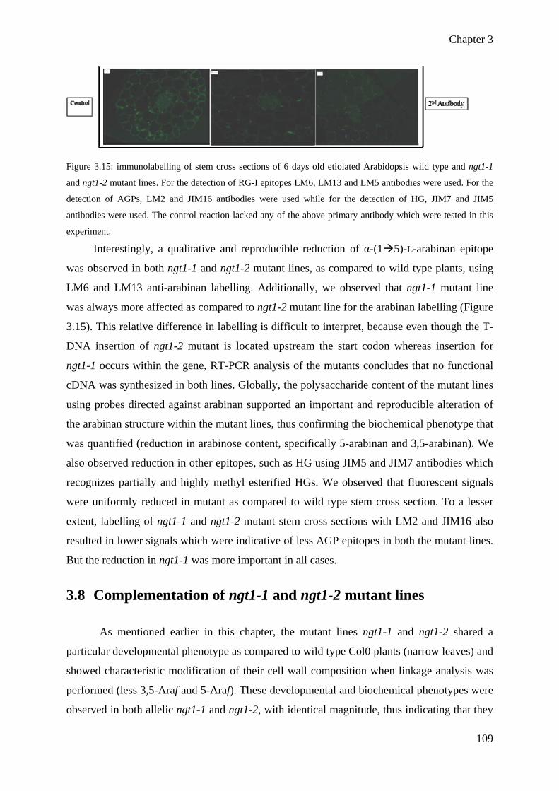
Chapter 3
109
Figure 3.15: immunolabelling of stem cross sections of 6 days old etiolated Arabidopsis wild type and ngt1-1
and ngt1-2 mutant lines. For the detection of RG-I epitopes LM6, LM13 and LM5 antibodies were used. For the
detection of AGPs, LM2 and JIM16 antibodies were used while for the detection of HG, JIM7 and JIM5
antibodies were used. The control reaction lacked any of the above primary antibody which were tested in this
experiment.
Interestingly, a qualitative and reproducible reduction of α-(1 5)-L-arabinan epitope
was observed in both ngt1-1 and ngt1-2 mutant lines, as compared to wild type plants, using
LM6 and LM13 anti-arabinan labelling. Additionally, we observed that ngt1-1 mutant line
was always more affected as compared to ngt1-2 mutant line for the arabinan labelling (Figure
3.15). This relative difference in labelling is difficult to interpret, because even though the T-
DNA insertion of ngt1-2 mutant is located upstream the start codon whereas insertion for
ngt1-1 occurs within the gene, RT-PCR analysis of the mutants concludes that no functional
cDNA was synthesized in both lines. Globally, the polysaccharide content of the mutant lines
using probes directed against arabinan supported an important and reproducible alteration of
the arabinan structure within the mutant lines, thus confirming the biochemical phenotype that
was quantified (reduction in arabinose content, specifically 5-arabinan and 3,5-arabinan). We
also observed reduction in other epitopes, such as HG using JIM5 and JIM7 antibodies which
recognizes partially and highly methyl esterified HGs. We observed that fluorescent signals
were uniformly reduced in mutant as compared to wild type stem cross section. To a lesser
extent, labelling of ngt1-1 and ngt1-2 mutant stem cross sections with LM2 and JIM16 also
resulted in lower signals which were indicative of less AGP epitopes in both the mutant lines.
But the reduction in ngt1-1 was more important in all cases.
3.8 Complementation of ngt1-1 and ngt1-2 mutant lines
As mentioned earlier in this chapter, the mutant lines ngt1-1 and ngt1-2 shared a
particular developmental phenotype as compared to wild type Col0 plants (narrow leaves) and
showed characteristic modification of their cell wall composition when linkage analysis was
performed (less 3,5-Araf and 5-Araf). These developmental and biochemical phenotypes were
observed in both allelic ngt1-1 and ngt1-2, with identical magnitude, thus indicating that they

Chapter 3
110
were most probably genetically related to the alteration of NGT1 gene in these lines.
However, in order to confirm that assumption we carried out the complementation of both
ngt1-1 and ngt1-2 mutants with a 35S::NGT1 construct in vector pH2GW7, and transformed
Col0 WT with the same construct (control). Transformation was performed using A.
tumefaciens previously transformed with the 35S::NGT1 cassette by dipping 4-weeks-old
Arabidopsis plant in the bacterial suspension, plants were allowed to recover and develop, and
transformants were selected on hygromycin (see materials and methods section 6.5.7.3).
Figure 3.16: Screening of positive transformants for 35S::NGT1. Seeds harvested from Col0, ngt1-1 and ngt1-2
plants transformed with Agrobacterium carrying 35S::NGT1 were plated on MS growth medium containing
50µg/mL hygromycin. Sensitive plants harbour a stunt development (small greenish leaves, no roots) whereas
35S::NGT1 transformed at the bottom of the picture developed almost normally.
Through this selection, we could identify plants harbouring hygromycin resistance, indicative
of plants complemented with 35S::NGT1 gene (Figure 3.16). These resistant plants were
transferred onto soil and analysed by PCR to confirm their complementation.

Chapter 3
111
Figure 3.17: Genotyping of transformed plants through PCR. Lanes 1 and 12 represent the 1 Kb marker. The
presence of product in lane 2, 6 and 9 indicates that plants are successfully transformed. Other PCR reactions
were performed to check genetically each line regarding the occurrence (for ngt1-1 and ngt1-2) or not (for Col0)
of T-DNA insertion at specific NGT1 locus.
DNA was extracted from individual complemented plants and used as a template in a
PCR reaction. Different set of primers GS (cDNA NGT1 gene specific), T-DNA ngt1-1
(LBa1 + RPW1 for ngt1-1), T-DNA ngt1-2 (LBa1 + RPW3 for ngt1-2) and Geno (LPW1+
RPW1 or LPW3 or RPW3 gene specific primers) were used for amplification.
A product of 981pb is positively amplified with GS primers using Col0 (lane 2), ngt1-
1(lane 6) and ngt1-2 (lane 9) DNA which indicates that wild type Col0 and mutant lines ngt1-
1, ngt1-2 are transformed with 35S::NGT1 construct (Figure 3.17). The presence of PCR
product (lanes 7 and 10) using T-DNA left border primers in combination with the lack of
amplification in lanes 8 and 11 in both ngt1-1 and ngt1-2 lines indicates that in these two
lines, NGT1 gene contains a T-DNA insertion. No amplification was observed in control
reactions of Wt col0 while using ngt1-1 and ngt1-2 T-DNA specific set of primers
After genotyping the plants, we collected the seeds from complemented plants. We
grew the seeds for phenotypic studies, i.e. the measurement of leaf area between 14th and 35th
day of development as previously done to genotype Col0 and ngt1-1 and ngt1-2 mutant
plants. We examined the phenotype of the transformed plants of the first generation and at the
same time we grew the non-transformed wild type and mutant plants (control). This analysis
has shown that mutant phenotype is fully restored after the complementation with
35S::NGT1. We have observed that at 14th day of development, ngt1-1+ 35s::NGT1 and ngt1-

Chapter 3
112
2+35s::NGT1 have same leaf area as compared to that of Col0_35s::NGT1 plants. It indicates
that the expression of NGT1 gene in the mutant background fully rescue the mutant
phenotype. Whereas for non-transformed control mutant plants we confirmed our previous
results of the narrow leaf phenotype for both mutant lines ngt1-1 and ngt1-2 (described on
page 116.)
Figure 3.18: Analysis of leaf area of wild type Col0, mutant lines ngt1-1 and ngt1-2 and Col0+35s:: NGT1, ngt1-
1+ 35s::NGT1, and ngt1-2+35s::NGT1. Leaf area was quantified using ImageJ. Each value is an average
determined from the measurement of leaf area of three individual plants. The error bars represent the standard
deviation. This data shows that mutant phenotype is restored after the complementation.

Chapter 3
113
Figure 3.19: Analysis of leaf area of wild type Col0, mutant lines ngt1-1 and ngt1-2 and Col0+35s::NGT1, ngt1-
1+ 35s::NGT1 and ngt1-2+35s::NGT1 .Leaf area was measured quantitatively at 14TH 28TH AND 35TH DAY OF
developmental. Each value is an average determined from the measurement of leaf area of three plants. The error
bars represent the standard deviation. This data shows that mutant phenotype is restored even after the 35th day of
growth with the complementation of NGT1.
We measured the leaf area on 28th and 35th day of growth also. Here we have presented the
data about only ngt1-1+ 35s::NGT1 and not for ngt1-2+35s::NGT1 because we lost the
transformed plants during manipulation. We did not reported the values for 21st day of
development, because the transfer of seedlings from the Petri dishes to the soil, occurred
between 14th to 21st day, and we anticipated that this set of data (21st day) would be impaired
by the stress corresponding to transfer to new growth conditions. It is clear from the above
figure that the mutant (not complemented) has narrow leaf area as compared to that of wild
type and the rescued-mutant line, and this at all observed developmental stages.
3.9 Heterologous expression of NGT1 in Pichia pastoris
Heterologous expression is a key element in the assignment of function to a protein,
but successful heterologous expression and demonstration of enzymatic activity of plant cell

Chapter 3
114
wall glycosyltransferases are rare and difficult. In this study we used Pichia pastoris as a host
system for the expression of NGT1 protein. As an eukaryote, Pichia pastoris offers many
advantages for the expression of plant proteins, for protein processing, protein folding and
post-translational modifications. Furthermore, it is relatively easy to manipulate and less
costly compared to other expression systems like insect and mammalian cells. Original cDNA
of NGT1 was ordered from SALK institute and provided subcloned into a pUNI plasmid.
Overlapping PCR was then used, in order to add to NGT1 cDNA specific sequences coding
for the T7 tag epitope and Gateway border sequences necessary for the recombination of the
PCR product in the pDONR207 entry plasmid (BP cloning; cf materials and methods
6.2.1.1.2). Recombination of the synthesized PCR product into pDONR207 vector led to the
formation of novel plasmid, named pENTR207_T7:NGT1 that was checked by sequencing.
pENTR207_T7:NGT1 corresponds to an entry vector that can be used to transfer the gene
sequence into various destination vectors such as pPICZα expression (vector for expression in
Pichia). Such reaction was performed forming an expression vector (named
pPICZ_T7:NGT1) containing the bacterial pUC origin of replication for propagation in E. coli
and AOX1 (Alcohol Oxidase) promoter which is used to enhance transcriptional regulation of
NGT1 in the presence of methanol inducer for recombinant protein expression in Pichia
pastoris. After Pichia transformation with pPICZ_T7:NGT1 plasmid, Pichia cells were
streaked on zeocin selection medium and 12 positive clones were selected. We selected 12
clones for characterization of protein expression because we anticipated this parameter may
vary between clones. Checking 12 clones for protein expression will provide the opportunity
to select the higher expressing and productive clone. Pichia_T7:NGT1 clones were grown
(only 6 are shown in Figure 3.20A and 3.20B) on BMMY medium and protein expression was
induced for 4 days by adding methanol 0.5 % final to the medium. Microsomal proteins were
prepared by breaking the Pichia cells (see materials and methods 6.3), protein content was
quantified through Bradford assay and first checked by SDS-PAGE (Fig 3.20A). No
differential expression pattern could actually be determined between transformed
Pichia_T7:NGT1 and wild-type line GS115, at the expected size of 61 kDa. This observation
indicates that if T7_NGT1 is produced in our clones, it is below the threshold of Coomassie
blue staining.

Chapter 3
115
Figure 3.20:A. SDS-PAGE analysis of total proteins in six NGT1 clones (lanes 1-6) and WT Pichia cells as a
control. Coomasie blue staining of 12% polyacrylamide gel. M: molecular weight markers B. Immunoblot
detection of NGT1 protein in clones 1 to 6. Expression of different clones was compared to select highly
productive clone for NGT1 and wild type Pichia strain (WT) used as a control. T7 was used as a positive control
with the molecular weight markers (M) and its band is visible below 37 kDa.
The expression of T7:NGT1 protein was further verified using western blot analysis
and the T7-tag antibody (Figure 3.20B). The lack of immunodetection in the lane of the wild-
type indicates that there was no cross reactivity of the T7 antibody with microsomal proteins
in Pichia GS-115 WT. For clones 1 to 6 (Figure 3.20B) a signal was picked up by the
antibody indicating all this 6 clones were actually expressing a protein recognized by the
antibody, most probably T7:NGT1. We observed a difference in migration between the lanes
1-6 because of migration parameters. By visual comparison, clone #2 was selected for
expression optimization and large scale production in order to produce enough microsomal
proteins for radioactive activity test. However, prior to large scale production of microsomes
from our selected clone#2, we spent some effort to determine whether or not the band (smear)
immunodetected actually corresponded to the expression of T7:NGT1 protein. Indeed, the
T7:NGT1 protein was apparently produced at a higher molecular weight than expected,
corresponding to a smear spread around 60-75 kDa when subjected to immunoblot analysis
(Fig 3.20B). One explanation would be that T7:NGT1 protein is produced but hyper-
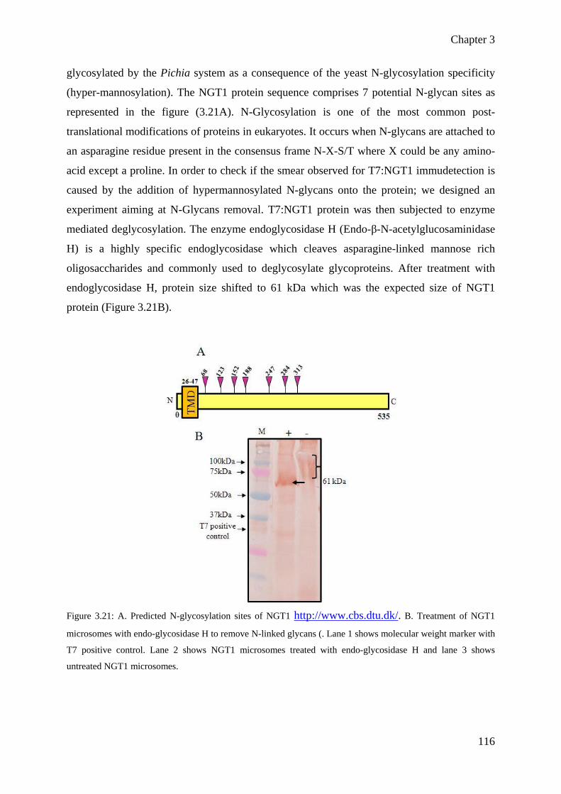
Chapter 3
116
glycosylated by the Pichia system as a consequence of the yeast N-glycosylation specificity
(hyper-mannosylation). The NGT1 protein sequence comprises 7 potential N-glycan sites as
represented in the figure (3.21A). N-Glycosylation is one of the most common post-
translational modifications of proteins in eukaryotes. It occurs when N-glycans are attached to
an asparagine residue present in the consensus frame N-X-S/T where X could be any amino-
acid except a proline. In order to check if the smear observed for T7:NGT1 immudetection is
caused by the addition of hypermannosylated N-glycans onto the protein; we designed an
experiment aiming at N-Glycans removal. T7:NGT1 protein was then subjected to enzyme
mediated deglycosylation. The enzyme endoglycosidase H (Endo-β-N-acetylglucosaminidase
H) is a highly specific endoglycosidase which cleaves asparagine-linked mannose rich
oligosaccharides and commonly used to deglycosylate glycoproteins. After treatment with
endoglycosidase H, protein size shifted to 61 kDa which was the expected size of NGT1
protein (Figure 3.21B).
Figure 3.21: A. Predicted N-glycosylation sites of NGT1 http://www.cbs.dtu.dk/. B. Treatment of NGT1
microsomes with endo-glycosidase H to remove N-linked glycans (. Lane 1 shows molecular weight marker with
T7 positive control. Lane 2 shows NGT1 microsomes treated with endo-glycosidase H and lane 3 shows
untreated NGT1 microsomes.

Chapter 3
117
3.10 Free sugar assay using T7:NGT1 microsomes
A T7- tag version of NGT1 protein was successfully expressed and produced in Pichia
pastoris. We pursued the functional characterization of NGT1 gene, taking advantage of
NGT1 annotation to test a Pichia line expressing NGT1 for fucosyltransferase activity.
Microsomes were prepared (for details see materials and methods 6.3) from this T7:NGT1
expressing pichia pastoris and used for activity test, after induction of protein expression.
Enzyme activity tests were developed using radioactive GDP-[14C]-fucose taking advantage
of the high sensitivity of radioactive assays particularly adapted to decipher
glycosyltransferase functions. Indeed, using radioactive test, picomolar transfer of
radiolabelled fucose onto an acceptor can be quantified with certainty. One main disadvantage
in the use of radioactivity test to assess a glycosyltransferase activity, is the limited
availability of both radioactive nucleotide-sugar and relevant acceptors. This is particularly
true when one is studying a glycosyltransferase potentially involved in cell wall biogenesis
because of the high degree of diversity and complexity that can be found throughout cell wall
structure, specific acceptor of each cell wall structure can hardly be found. In an attempt to
overcome that difficulty we adapt a system called “free sugar assay” where the acceptor
molecules would be a monosaccharide present at high concentration (half molar).
Figure 3.22: Schematic representation of principle for free sugar assay
The rationale of the assay (is based on the principle shown in figure 3.22) being to
force the enzyme to transfer a radioactive sugar onto a monosaccharide even though this
monosaccharide is obviously not the preferred acceptor of the enzyme, and this in order to
gain knowledge about which monosaccharide the enzyme is able to work with. Then, a second
step would permit to check the plant cell wall for possible acceptor molecule comprising the
monosaccharide onto which the enzyme is active. Thus, in order to determine the putative
activity of T7:NGT1, we used the microsomal protein fractions (enriched for Golgi
membranes) prepared from Pichia pastoris. We checked membrane fractions for the ability
of the T7:NGT1 protein to catalyze the transfer of [14C]-labelled UDP donor sugars onto

Chapter 3
118
acceptors. We tested 3 different types of NDP-sugars as donors taking advantage of their
availability in the laboratory (UDP-Gal, UDP-Glc and GDP-Fuc) and 6 different
monosaccharides as acceptors (Arabinose, Galactose, Glucose, Mannose, Rhamnose and
Xylose). We provided equal quantity (50 nCi) of UDP-Glc [14C] (control) and GDP-Fuc [14C]
(as the NGT1 protein is a putative fucosyltransferase). First we observed that whatever the
acceptor the transfer of Glc from UDP-Glc appears highly stable (4420 +/-212 cpm/h)
whereas the values of transfered radioactivity, even if low, were fluctuating in the case we use
GDP-[14C]-Fuc (8305 +/-1641cpm/h). This indicates that regarding UDP-[14C]-Glc, all
monosaccharides behave identically, none of them being a substrate for the enzyme. In the
case of GDP-Fuc the observation that the standard deviation is high may indicate that some
acceptors have value away from the mean which would be expected in case of a transfer.
Noticeably, the transfer of GDP-[14C]-Fuc onto different acceptor sugars is variable (Fig
3.23), and particularly high onto arabinose as an acceptor sugar. However the level of
transferred observed either using UDP-[14C]-Glc and GDP-[14C]-Fuc as donors are just
around the background level because approximately 250,000 cpm (5nCi) were provided to the
assay and 4,000 to 10,000 were recovered at best. The only conclusion that could be drawn is
that the enzyme would better accommodate GDP-Fuc compared to UDP-Glc in its catalytic
domain, explaining why values would more vary while using GDP-Fuc compared to UDP-
Glc. This last remark is consistent with NGT1 being a putative fucosyltransferase although no
data collected from this free sugar assay would really show enough transfer to strengthen
unambiguously this hypothesis.
Figure 3.23: Free sugar assay for NGT1 activity with UDP-Glc and GDP-Fuc donors. Half molar concentration of different monosaccharides like arabinose, galactose, glucose, mannose, rhamnose and xylose were used as acceptor substrate.

Chapter 3
119
We also tested UDP-Gal [14C] as a donor but provided a smaller quantity (5 nCi). This
assay demonstrates that transfer level of [14C]-Gal onto monosaccharide acceptor the same on
all the tested monosaccharide acceptors, no strong transfer of the donor sugar (Gal) was
observed, as observed when UDP-Glc was used (Figure3.24).
Figure 3.24: Free sugar assay for NGT1activity with UDP-Gal as donor. Half molar concentration of different monosaccharides like arabinose, galactose, glucose, mannose, rhamnose and xyloses were used an acceptor
From the above free sugar assays we have concluded that GDP-[14C]-Fuc would be a
better donor for T7:NGT1 protein compared to UDP-Glc and UDP-Gal, and that the level of
transfer of radioactivity is higher for arabinose, and to a lesser extent galactose, as acceptors.
However the level of transfer detected was too low to worth any product characterization
using TLC separation. We repeated the free sugar assay using Arabinose and Galactose
because these two sugars showed some ability to accept transfer of Fucose (from GDP-[14C]-
Fuc ) in the presence of T7:NGT1. We included another sugar (Rhamnose) that did not show
particular level of transfer of Fucose onto it, and was thus served as a control. Another control
reaction was the assay lacking acceptor sugars, and the assay was performed using heat-
inactivated microsomes. Interestingly this assay confirms that Arabinose and Galactose had a
small but reproducible capability to accept transfer of [14C]-Fucose whereas Rhamnose would
not (Figure 3.25). Unfortunately, the level of transfer observed were again too small too worth
any product characterization, most probably because the acceptor and or the donor sugars

Chapter 3
120
provided to the T7:NGT1 protein were too different from the actual specificity of the reaction
that the protein would catalyzed.
Figure3.25: Free sugar assay for NGT1activity with GDP-Fuc as donor. Different monosaccharides like arabinose, galactose and rhamnose were used as putative acceptors. Control reaction lacks any acceptor.
3.11 Fucosyltransferase assay using ngt1-1 mutant cell wall as an
acceptor
The free sugar assay experiment demonstrates that microsomal membranes expressing
T7:NGT1 had preference to accommodate GDP-Fuc compared to others nucleotide-sugars
tested and to transfer them, to a small extent, onto Arabinose or Galactose. In order to confirm
this observation and get better understanding of the role of T7:NGT1, we carried out a
fucosyltransferase activity test using mutant cell wall as the acceptor substrate. This strategy
has been successfully used by Egelund and his colleagues for the characterization of RGXT1
and RGXT2 xylosyltransferases involved in pectin rhamnogalacturonan II biosynthesis
(Egelund et al., 2006). The rationale of the assay is based on the principle (shown in
figure3.26) that the mutation in NGT1 gene will cause an alteration of the cell wall in ngt1-1
mutant and consequently makes its cell wall deficient for specific polysaccharide structure.
Adding the T7:NGT1 protein and the correct (radioactive) NDP-sugar, this cell wall structure
could be used as an acceptor. As a control we would use cell wall prepared from the wild-type

Chapter 3
121
Col0 for which we do not expect the structure synthesized by NGT1 to be deficient, providing
a negative control to this test. Another control was provided thanks to the use of microsomal
preparation of membranes expressing At1g62305 protein. This gene (named E in Figure 2.1
of chapter 2) belongs to DUF266 family and do not show any fucosyltransferase signature.
Figure 3.26: Schematic representation of mechanism of NGT1 putative fucosyltransferase
Figure 3.27: Fucosyltransferase activity assay for NGT1 activity with mutant cell wall as an acceptor. Transfer of
fucose is observed onto mutant cell wall in presence of NGT1 microsomes while there is no transfer of fucose
onto mutant cell wall in the presence of At1g62305 microsomes which are used as a control
This assay shows that T7:NGT1 catalyzes the transfer of [14C]-Fuc to the mutant cell
wall as there is more transfer of Fucose in the ngt1-1 mutant as compared to the wild type
Col0 cell wall (Figure 3.27). In case of control microsomes expressing another protein
At1g62305 no significant transfer of fucose was observed onto mutant cell wall as compared
to wild type. From this experiment, we concluded that cell wall from ngt1-1 mutant appears a
good acceptor of fucose as compared to the wild-type cell wall, but again the level of transfer
observed was low compared to the amount of radioactivity included in the test. This indicates
Acceptor
Microsomes NGT1
Acceptor-Fuc*
GDP-Fuc* GDP

Chapter 3
122
that the reaction studied is probably not the actual (true) activity of the T7:NGT1 protein. Cell
wall prepared from the ngt1-1 mutant should offer a large variety of molecules to be
acceptors, hence the lack of strong transfer in our assay could be interpreted as the donor
sugar of the reaction (GDP-Fuc) not being the proper one, if we assume that the numerous
potential acceptor of the cell wall were evenly accessible.
We did another assay by using the microsomes of NGT1 and GS-115 wild type pichia
strain as a control. We observed that in the presence of NGT1 microsomes and GDP-Fuc [14C
] donor, there is transfer of fucose onto two allelic mutants ngt1-1 and ngt1-2 cell wall as
compared to wild type cell wall (Figure3.28). In contrast there is less transfer of fucose in the
presence of wild type Pichia GS-115 microsomes.
Figure 3.28: Fucosyltransferase assay from Pichia microsomes expressing NGT1. This assay used cell wall extracted from wild type Col0, ngt1-1 and ngt1-2 mutants as acceptors. Transfer of fucose is observed onto mutant cell wall in presence of Pichia microsomes expressing NGT1 while only a background transfer of fucose onto mutant cell wall when using WT GS-115 microsomes (negative control).
From all above assays we concluded that NGT1 protein has the ability to transfer
fucose onto mutant cell wall as compared to wild type cell wall but this level of transfer is
very low. We were not able to purify and characterize the reaction product in result of free
sugar assay and fucosyltransferase activity tests with mutant cell wall acceptor for further
chemical analysis.Radioactive fucosyltransferase assay data did not prove NGT1
fucosyltransferase enzyme activity. So we envisioned to develop an assay to alternatively test

Chapter 3
123
arabinosyltransferase activity as the biochemical analysis of ngt1-1 and ngt1-2 mutants cell
wall shown the reduction in arabinose contents.
3.12 Arabinosyltransferase assay using microsomal fraction of
Pichia-NGT1 and NGT1-∆69 produced in Hi-5 cells
In order to test putative arabinosyltransferase activity from heterologously expressed
NGT1 or NGT1-∆69 protein, we carried out this non-radioactive assay for NGT1, that is why
when it was not possible to characterize NGT1 using radioactive assays because of
unavailability of commercial radioactive UDP-Araf (donor molecule). UDP-Araf was
provided by Dr Richard Daniellou (ENS Rennes). We developed an assay using
arabinotetraose or arabinohexaose acceptors (4mM), UDP-Arabinofuranose (0.4mM) in
buffer containing 50mM Hepes-KOH pH 7 supplemented with MnCl2 5mM. Putative
transfers of Arabinofuranose onto acceptors was later evaluated by MALDI-TOF MS at it was
previously done with success to characterize activity of AtFUT1-∆68 (cf section 6.4.2). In a
first assay we used NGT1 expressing Pichia microsomes and wild type strain GS-115
microsomes (as control) in the presence of arabinotetraose and arabinohexaose (not shown).
We provided the cold UDP-Araf to the enzyme (for details see materials and methods section
6.4.3) and the reaction was incubated for 2h. Acceptor molecules were then labelled with a 2-
aminobenzamide (2-AB) fluorophore (to increase sensitivity) and analyzed by mass
spectrometry (MALDI-TOF-MS) (Figure 3.29A, 3.29B, 3.30C and 3.30D). MALDI-TOF-MS
analysis of 2-AB labeled neutral oligosaccharides allowed the detection of [M + H] +, [M +
Na] + and [M + K] + in the positive-ion mode.

Chapter 3
124
Figure 3.29: MALDI-TOF MS analysis of NGT1 for arabinosyltransferase activity. A. Mass spectra of
Arabinotetraose in the presence of WT Pichia GS-115 microsomes. This control reaction (WT microsomes)
shows that the assay contain a small proportion of Arabinopentaose (m/z=821), but in relatively low amount as
compared to arabinotetraose (m/z= 689). B.MALDI-TOF-MS analysis of the assay including Arabinotetraose
(m/z=689) and in the presence of NGT1 microsome: No addition of arabinose was observed in the presence of
NGT1 microsomes as the relative quantity of Arabinopentaose peak (m/z=821) remains unchanged.
We observed one peak with a mass of 821.38 (Figure 3.29B) corresponding to mass of
arabinopentaose that was expected in case of transfer of Arabinose onto acceptor, but the
presence of this peak (at the same intensity) in the control reaction is indicative of the lack of
arabinosyltransferase activity of microsome prepared from NGT1 expressing Pichia. Indeed
the control indicates a lack of purity of the arabinotetraose acceptor that was used in the study
and the fact that the arabinopentaose peak has the same intensity (relatively to the
arabinotetraose peak) in both reaction confirmed the lack of transfer, and thus the lack of
arabinosyl transferase activity in the assay using NGT1 microsomes.

Chapter 3
125
Figure 3.30: MALDI-TOF MS analysis of NGT1 for fucosyltransferase activity. C. Mass spectra of Arabino-
tetraose in the presence of culture medium of NGT1. This reaction shows that the assay contain a small
proportion of Arabinopentaose (m/z=821), but in relatively low amount as compared to arabinotetraose (m/z=
689). No mass increment was observed in the peaks. D. Mass spectra of Arabino-hexaose in the presence of
culture medium of NGT1. This reaction shows that the assay contains a small proportion of Arabinoheptaose
(m/z=1085), but in relatively low amount as compared to arabinohexaose (m/z= 953). No addition of arabinose
was observed in the presence of culture medium of NGT1 enzyme.
As in the above described experiment we did not observe the NGT1 activity; this
might be because of the membranous form of NGT1 protein extracted from the pichia
microsomes. In order to obtain the soluble form, the truncated NGT1 protein was expressed in
insect cells. In this assay we used arabinotetraose (not shown) and arabinohexaose acceptor
molecules. Similarly, Arabinopentaose molecule (821.33) could be identified in both the
reaction with NGT1-∆69 and the control, and at the same relative intensity which indicates
consequently a lack of arabinosyltransferase activity.

Chapter 3
126
3.13 Conclusion
In conclusion, phenotypic studies have shown that both mutant lines ngt1-1 and ngt1-2
have narrow leaves at 14th and 21st day of development. These results indicate that the
mutation in NGT1 alters the growth rate only during early developmental stage so it could be
hypothesized that NGT1 gene is essential at early stages of plant development, possibly in the
biosynthesis of cell wall. Neutral monosaccharide quantification has shown that mutant cell
wall has less arabinose as compared to that of wild type Col0. In addition biochemical
analysis of mutant cell wall through GC-MS has shown that both mutant lines ngt1-1 and
ngt1-2 have 50% less 3,5-Araf and 5-Araf which indicates that NGT1 is involved directly or
indirectly in arabinan chain biosynthesis. This reduction in arabinan polymer was additionally
observed when immunolabelling was performed on cross section of stem labelled with LM6
and LM13 anti-arabinan antibodies.
NGT1 gene was expressed hetrologously in Pichia pastoris and microsomes were used
to determine the catalytic activity of NGT1 protein. Free sugar assay has shown that GDP-Fuc 14C would be a better donor for T7:NGT1 protein as compared to UDP-Glc and UDP-Gal. A
fucosyltransferase assay was also carried out by using mutant cell wall as the acceptor
substrate. This assay resulted in the transfer of 14C-Fuc to the mutant cell wall in the presence
of T7:NGT1 protein but the level of transfer observed was low indicating that the reaction
studied was not the actual activity of the T7:NGT1 protein.
Because the mutational studies of NGT1 altered mutant lines suggested that NGT1
protein could encode an arabinan-arabinosyltransferase, this later hypothesis was tested in a
non-radioactive activity test using MALDI-TOF MS detection but unfortunately no
Arabinosyltransferase activity could be shown so far using Arabinotetraose and
arabinohexaose acceptor molecules. Whether NGT1 protein would encode a GT activity
remains unproved, so far.

Chapter 4
127
Chapter 4
Heterologous expression of Arabidopsis
thaliana xylosyltransferase (AtXT1) and
fucosyltransferase (AtFUT1) for structural
characterization
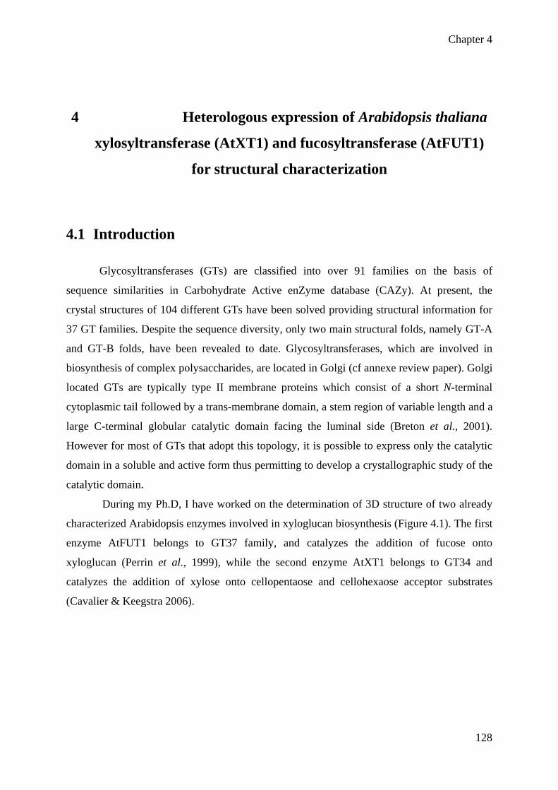
Chapter 4
128
4 Heterologous expression of Arabidopsis thaliana
xylosyltransferase (AtXT1) and fucosyltransferase (AtFUT1)
for structural characterization
4.1 Introduction
Glycosyltransferases (GTs) are classified into over 91 families on the basis of
sequence similarities in Carbohydrate Active enZyme database (CAZy). At present, the
crystal structures of 104 different GTs have been solved providing structural information for
37 GT families. Despite the sequence diversity, only two main structural folds, namely GT-A
and GT-B folds, have been revealed to date. Glycosyltransferases, which are involved in
biosynthesis of complex polysaccharides, are located in Golgi (cf annexe review paper). Golgi
located GTs are typically type II membrane proteins which consist of a short N-terminal
cytoplasmic tail followed by a trans-membrane domain, a stem region of variable length and a
large C-terminal globular catalytic domain facing the luminal side (Breton et al., 2001).
However for most of GTs that adopt this topology, it is possible to express only the catalytic
domain in a soluble and active form thus permitting to develop a crystallographic study of the
catalytic domain.
During my Ph.D, I have worked on the determination of 3D structure of two already
characterized Arabidopsis enzymes involved in xyloglucan biosynthesis (Figure 4.1). The first
enzyme AtFUT1 belongs to GT37 family, and catalyzes the addition of fucose onto
xyloglucan (Perrin et al., 1999), while the second enzyme AtXT1 belongs to GT34 and
catalyzes the addition of xylose onto cellopentaose and cellohexaose acceptor substrates
(Cavalier & Keegstra 2006).

Chapter 4
129
Figure 4.1: Schematic view of a typical xyloglucan structure (type XXFG). Glycosyltransferases enzymes
involved in its biosynthesis are indicated with arrows.
It was demonstrated that, in vitro, AtFUT1 catalyzes the addition of L-fucose at the 2-
position of galactose residue into XyG, using non-fucosylated xyloglucan acceptor and
radiolabelled GDP-L-[14C] fucose as the donor (Faik et al., 2000). Afterwards, a genetic
screen isolated mur2 Arabidopsis mutants showing a decrease in fucose content in the cell
wall and fine analysis of the mutant cell wall revealed a 99 % reduction in xyloglucan
fucosylation for mur2 plants. Later, mutation in mur2 was mapped and revealed a mutation in
the AtFUT1 gene, demonstrating that AtFUT1 encodes a xyloglucan specific
fucosyltransferase (Vanzin et al., 2002b). Moreover, the strong reduction in xyloglucan
fucosylation observed in the analysis of mutants (99%) indicates that AtFUT1 is the only
fucosyltransferase responsible for xyloglucan fucosylation in Arabidopsis (Reiter et al., 1997,
Vanzin et al., 2002b, Perrin et al., 2003). No crystal structure is currently available for
AtFUT1 or for any GT belonging to GT37, but fold recognition studies predicted a similar
fold (at least for the nucleotide binding region) to the human FUT8 and bacterial NodZ
protein, which are α−(1−−>6)-fucosyltransferases belonging to family GT23 (Breton et al.,
2006).
The second plant GT selected for this study was the Arabidopsis xylosyltransferase
(AtXT1) characterized to be involved in xyloglucan biosynthesis (Cavalier & Keegstra 2006,
Cavalier et al., 2008). Heterologous expression of the AtXT1 in Pichia pastoris showed an α-

Chapter 4
130
(1 )6-Xylosyltransferase activity in vitro, using UDP-Xyl and cellopentaose as donor and
acceptor substrates, respectively (Faik et al., 2002). AtXT1 belongs to a small gene family
(comprising seven members in Arabidopsis) and which classifies into family GT34. Another
gene AtXT2 belonging to this family having 85 % similarity to AtXT1 has been expressed
into Pichia pastoris and was shown to also encode a α-(1 6)-Xylosyltransferase activity
(Cavalier & Keegstra 2006). Recently, an Arabidopsis double mutant KO for both AtXT1 and
AtXT2 genes was named “xxt1-xxt2”, and demonstrated to lack detectable amount of
xyloglucan within its cell wall. The double mutant “xxt1-xxt2” showed a severe root hair
phenotype (bulging) and lacked detectable xyloglucan at the whole plant level. This study
demonstrated that the two genes are partially redundant and required for xyloglucan
biosynthesis in Arabidopsis. More recently, a T-DNA insertion in another gene, named
AtXT5 was characterized. AtXT5 deficient mutant was consequently named “xxt5”, and study
showed that xxt5 mutant had shorter root hairs and less xyloglucan quantity in cell wall.
Accordingly, authors conclude that AtXT5 encodes another xylosytransferase activity
involved in xyloglucan biosynthesis (Zabotina et al., 2008b). Although fold prediction
suggested that GT34 enzymes could adopt a GT-A fold, no crystal structure is currently
available for enzymes belonging to this GT family. It is therefore challenging to get a 3D
structure of at least one enzyme of this family. I carried out the expression of the truncated
proteins of both the enzymes: AtFUT1 and AtXT1 in order to produce soluble proteins for the
determination of 3D crystal structure of these enzymes.
4.2 AtXT1 4.2.1 Expression of truncated AtXT1-Δ140 in insect cells
Our aim was to express a truncated and soluble form of AtXT1. Protein sequence
analysis allowed to delineate the different protein domains of AtXT1 and to define the best
truncation site. The use of fold recognition programs such as Phyre
(http://www.sbg.bio.ic.ac.uk/~phyre/) and the hydrophobic Cluster analysis method
(Gaboriaud et al., 1987) predicted that the catalytic domain would encompass the region [140-
461]. The baculovirus/insect cell heterologous expression system was chosen as it was in the
past demonstrated to work well with eukaryotic GTs, particularly of plant origin.
A DNA fragment lacking the first 420 bp within the coding region of AtXT1 was
generated by PCR using pUNI51-AtXT1 from NASC stock center as a template, comprising
the full length cDNA of Arabidopsis thaliana AtXT1 gene. The restriction sites Pst1 and

Chapter 4
131
Kpn1 were added to the amplified sequence through primers at the 5’ and 3’ end, respectively
(Figure 4.2), and used for cloning into insect cells expression vector (pVT-Bac-His1) (Tessier
et al., 1991). Using this plasmid, the AtXT1 gene was cloned in frame with the melittin
peptide signal, thus allowing the secretion of the recombinant protein in the culture
supernatant. In addition, the protein harbors at its N-terminus a His-tag and X-Press tag useful
for immunodetection and/or purification of the protein. As shown in Figure 4.2, the truncated
protein comprises only one putative N-glycosylation site (at position 137 of amino acid
sequence). atgatagagaagtgtataggagcgcatcggtttcggagattacagagattcatgcgtcaa M I E K C I G A H R F R R L Q R F M R Q gggaaagtgacgattctttgtctcgttctcaccgtcatcgtcttacgtggcacaatcgga G K V T I L C L V L T V I V L R G T I G gccggtaagtttggtacgccggagaaagatatcgaggagatccgtgagcatttcttctac A G K F G T P E K D I E E I R E H F F Y acgcgtaaacgcggcgagcctcaccgtgtcctcgtcgaggtctcttccaaaacgacgtcg T R K R G E P H R V L V E V S S K T T S tccgaagacggaggaaatggtggtaacagctacgagaccttcgatatcaacaagctattc S E D G G N G G N S Y E T F D I N K L F gttgatgaaggagacgaagagaaatctcgagaccggactaataaaccttattctcttggt V D E G D E E K S R D R T N K P Y S L G cccaagatctctgattgggatgagcagagacgtgattggctcaaacaaaaccctagcttc P K I S D W D E Q R R D W L K Q N P S F AAACTGCAGcctaatttcgtggcgccaaac cctaatttcgtggcgccaaacaagcctagggttcttcttgtcacaggttcagctcctaaa P N F V A P N K P R V L L V T G S A P K ccgtgtgagaatcctgtaggagaccattacctcttgaaatcgattaagaacaaaatcgat P C E N P V G D H Y L L K S I K N K I D tactgtagaatacacggaatcgagatcttctacaacatggcgttgctcgatgctgagatg Y C R I H G I E I F Y N M A L L D A E M gctggattctgggctaagcttccgttgattaggaagttactcttgtcacatcctgagatt A G F W A K L P L I R K L L L S H P E I gagtttctatggtggatggatagtgatgccatgttcacggacatggtgttcgagcttcca E F L W W M D S D A M F T D M V F E L P tgggagaggtacaaagattacaacttggtgatgcatggttggaacgagatggtttatgac W E R Y K D Y N L V M H G W N E M V Y D cagaagaattggattggtctcaacacgggaagtttcttgctcaggaactcacagtggtcg Q K N W I G L N T G S F L L R N S Q W S cttgatcttcttgacgcttgggctcctatgggcccaaaagggaagatccgagaagaagcg L D L L D A W A P M G P K G K I R E E A ggtaaagtcttgacccgggaacttaaagaccgacccgctttcgaagctgacgatcaatcg G K V L T R E L K D R P A F E A D D Q S gcgatggtttatctgctggcgacggagagagagaaatggggaggcaaagtttatctagag A M V Y L L A T E R E K W G G K V Y L E agtggttattacttgcacggttattgggggattttggtagaccggtacgaggagatgatt S G Y Y L H G Y W G I L V D R Y E E M I gagaatcataaaccgggttttggagaccatcggtggccattggttacgcatttcgtcggg E N H K P G F G D H R W P L V T H F V G tgtaaaccgtgcgggaaatttggagattatccggtggaacggtgtctacggcagatggat C K P C G K F G D Y P V E R C L R Q M D agagcgtttaatttcggagacaatcagatccttcaaatgtatggtttcacgcataaatcg R A F N F G D N Q I L Q M Y G F T H K S cttgggagccggcgcgtgaaacccacgcgcaatcagacggataggccgctcgatgccaag L G S R R V K P T R N Q T D R P L D A K gacgagtttgggctgcttcatccgccgttcaaagcggccaagcttagtacgacgacgacgtga D E F G L L H P P F K A A K L S T T T T -
catgctgctgctgcactCCATGGGGC
Figure 4.2: Nucleotide sequence and translated sequence of the coding region of AtXT1. Predicted TMD is marked in turquoise, N-glycosylation sites with green letters underlined. Forward and reverse primers are added in red and the bold letters in the DNA sequences represent their respective annealing sites. DNA sequence was translated at ExPASy (http://www.expasy.ch/tools/dna.html).

Chapter 4
132
Cloned sequences were further confirmed by sequencing and used for transformation. After
transformation and three repeated amplifications in Sf9 insect cells, a titer of 2x10⁷ pfu/ml
was estimated for the AtXT1 virus stock. This stock was used for the production of
recombinant proteins in Hi-5 cells infected with the recombinant virus at MOI of 5 (5
pfu/cell). Hi-5 cells were typically grown at 27°C and supernatants collected after 4 days post
infection. The collected supernatants were clarified by centrifugation and stored frozen until
use.
Protein was produced in two different media of production, namely Express FiveM and
Excell-405 serum-free media. The recombinant protein was trapped using either UDP-
Fractogel or Ni-agarose beads. Bound proteins were analysed by SDS-PAGE and western
blot. The calculated size of AtXT1- is 39 kDa. Two protein bands were observed in western
blot from the protein production in Hi5-Express medium that could correspond to the non-
glycosylated and glycosylated isoforms (Figure 4.3A). Protein yield was higher in Hi5 Excell-
405 medium, where we observed a more diffuse band, but two bands can also be clearly seen,
particularly when protein is trapped using Ni-agarose beads (as observed in figure 4.3B)..
Figure 4.3: SDS-PAGE and Western blotting of recombinant AtXT1- Δ140 produced into two different serum-
free media, Express-Five medium (A) and Excell-405 (B). Left panels correspond to a Coomassie blue staining
of 10 % polyacrylamide gel and right panels to the detection of the recombinant protein using anti-Xpress
antibodies. Lane M represents molecular weight markers, lanes Fr and Ni indicate protein bound onto UDP-
Fractogel or Ni-agarose beads, respectively.

Chapter 4
133
4.2.2 Xylosyltransferase assay for AtXT1-Δ140
In order to determine the activity of truncated AtXT1-Δ140, xylosyltransferase activity
test was performed as described previously for full length AtXT1 (Faik et al., 2002, Cavalier
& Keegstra 2006). We tested cellohexaose and celloheptaose as acceptor substrates and UDP-14C Xyl as donor in this assay. Typically, enzyme assays were carried out at 30°C for 1h, in
presence of MnCl2 5 mM. We performed the assay using a “HEPES-Triton-X100” buffer as
previously described by Cavalier and colleagues, albeit the use of Triton detergent necessary
in their study (expressing a full length AtXT1with transmembrane domain) was probably not
necessary in our study, while expressing a truncated version of AtXT1. We observed that
heterologously expressed AtXT1-Δ140 did not catalyze the transfer of xylose onto
cellohexaose and cellopentaose acceptor substrates (data not shown).
In order to check if the lack of transfer of xylose onto cellohexaose and cellopentaose
was due to our experimental conditions, we tested other buffer conditions like Hepes- pH 7
buffer or replace buffer with water, but again no transfer of xylose was detected (Figure 4.4).
Figure 4.4: Xylosyltransferase assay for truncated AtXT1-Δ140 in the presence of Hepes buffer or water.
Cellohexaose and cellopentaose are used as potential acceptors of [14C]-xylose transfer whereas xyloglucan is
used as a negative control. The red bar named as ‘control represent the total radioactivity (CMP/hr) present in the
reaction.

Chapter 4
134
This assay shows that AtXT1-Δ140 is not able to transfer [14C]-xylose onto
cellohexaose and cellopentaose acceptor substrate whatever were the experimental conditions
(Triton-X100, Hepes buffer or water) (Figure 4.4). From the above results we hypothesized
that AtXT1 is not functional after the removal of 140 amino acids because this region may
comprise part of the catalytic site or an additional domain necessary for the transfer reaction.
In order to check our hypothesis, we analyzed the activity of a less truncated AtXT1-Δ44
protein consisting in the removal of only 44 amino acids which correspond to the cytoplasmic
domain and transmembrane region.
4.2.3 Expression of AtXT1-Δ44 in insect cells
In order to generate an AtXT1- Δ44 protein, we amplified a DNA fragment lacking the
first 132 bp within the coding region of AtXT1 by PCR, using pVT-Bac1-AtXT1as a template
comprising the full length cDNA of Arabidopsis thaliana XXT1 gene. The restriction sites
Pst1 and EcoR1 were added to the amplified sequence through primers at the 5’ and 3’ end
during the PCR reaction. PCR product was cleaved with the restriction endonucleases Pst1
and EcoR1. For further steps of cloning and expression, the same procedure was adopted as
described in the section 4.4.1 of this chapter. AtXT1-Δ44 protein was produced in Excell-405
serum-free media. The recombinant protein was trapped using Ni-agarose beads. Bound
proteins were analyzed by SDS-PAGE. Western blot analysis was done to check the
expression of protein (Figure 4.5). Protein was detected by using the anti-histidine antibody.
The calculated size for the protein is 50 kDa. Protein was produced in media of production,
Excell-405 serum-free media which is shown in figure 4.5.

Chapter 4
135
Figure 4.5: SDS-PAGE and Western blotting of recombinant AtXT1-Δ44 produced into two different serum-
free media, Express-Five medium (A) and Excell-405 (B). Left panels correspond to a coomassie blue staining of
10 % polyacrylamide gel and right panels to the detection of the recombinant protein using anti-Xpress
antibodies. Lane M represents molecular weight markers, lanes Fr and Ni indicate protein bound onto UDP-
fractogel or Ni-agarose beads, respectively.
4.2.4 Xylosyltransferase assay for AtXT1-Δ44
In order to determine the activity of truncated AtXT1-Δ44, xylosyltransferase activity
test was performed as described previously for full length AtXT1 (Faik et al., 2002, Cavalier
& Keegstra 2006). We tested cellohexaose and celloheptaose as acceptor substrate and UDP-
[14C]-Xyl as a donor in this assay. We used the Hepes buffer pH7 and MnCl2 in the assay.
We observed that heterologously expressed AtXT1-Δ44 did not catalyze the transfer of [14C]-
xylose onto cellohexaose and celloheptaose acceptor substrates (Figure 4.6). The activity level
was same as compared to control reaction which lacked the acceptor. We repeated the same
experiment and again no activity was observed for AtXT1-Δ44 truncated protein.

Chapter 4
136
Figure 4.6: Xylosyltransferase assay for AtXT1-Δ44 in the presence of Hepes buffer. Cellohexaose and
celloheptaose are used an acceptors. Control reaction lacked the acceptor substrate.
Several explanations could explain the lack of activity for AtXT1-Δ44 and AtXT1-
Δ140 proteins: either the full-length form and/or a correct location in the microsomal fraction
is required or alternatively a cofactor that could be necessary for activity is missing. Upon the
lack of success in the obtention of an active truncated form of AtXT1, we pursue the effort of
expressing a plant GT for crystallographic study by expressing truncated form of AtFUT1.
4.3 AtFUT1 4.3.1 Expression of truncated AtFUT1-Δ160 in insect cells
A soluble form of the AtFUT1 catalytic domain was produced. As for AtXT1, the
truncation site was determined through protein sequence analysis. HCA, secondary structure
prediction and fold recognition analysis suggested a truncation site at a position around 160.
This resulted in the deletion of cytoplasmic and a trans-membrane domain as well as a large
portion of what is expected to correspond to the stem region. This truncation site also
eliminates two putative N-glycosylation sites.
A DNA fragment lacking the first 480 bp (160 amino acids) within the coding region
of AtFUT1 was generated by PCR using pENTR-AtFUT1 as a template comprising the full
length cDNA of Arabidopsis thaliana FUT1 gene. The restriction sites Pst1 and Kpn1 were
added to the amplified sequence through primers at the 5’ and 3’ end during the PCR reaction
(Figure 4.7). PCR product was cleaved with the restriction endonucleases Pst1 and Kpn1. For

Chapter 4
137
further steps of cloning and expression, the same procedure was adopted as described above
for AtXT1.
atggatcagaattcgtacaggagaagatcgtctccgatcagaaccactaccggcggttca M D Q N S Y R R R S S P I R T T T G G S aagtccgttaatttctccgaactacttcaaatgaagtatctcagctccggtacgatgaag K S V N F S E L L Q M K Y L S S G T M K ctcacgagaaccttcactacttgcttgatagtcttctctgtactagtagcattctcaatg L T R T F T T C L I V F S V L V A F S M atctttcaccaacacccatctgattcaaatcggattatgggtttcgccgaagctagagtt I F H Q H P S D S N R I M G F A E A R V ctcgacgccggagttttcccaaatgttactaacatcaattctgataagcttctcggaggg L D A G V F P N V T N I N S D K L L G G ctacttgcttctggttttgatgaagattcttgccttagtaggtaccaatcagttcattac L L A S G F D E D S C L S R Y Q S V H Y cgtaaaccttcaccttacaagccatcttcttatctcatctctaagcttagaaactacgaa R K P S P Y K P S S Y L I S K L R N Y E aagcttcacaagcgatgtggtccgggtactgaatcttacaagaaagctctaaaacaactt K L H K R C G P G T E S Y K K A L K Q L AAACTGCAGgatcaagaacatattgatggtgatggtgaatgc gatcaagaacatattgatggtgatggtgaatgcaaatatgttgtgtggatttcttttagc D Q E H I D G D G E C K Y V V W I S F S ggcttagggaacaggatactttctctagcctcggtttttctttacgcgcttttaacggat G L G N R I L S L A S V F L Y A L L T D agagtcttgcttgttgaccgagggaaagacatggatgatctcttttgcgagccgtttctc R V L L V D R G K D M D D L F C E P F L ggtatgtcgtggttgctacctttagatttccctatgactgatcagtttgatggattaaat G M S W L L P L D F P M T D Q F D G L N caagaatcatctcgttgttatggatatatggtgaagaatcaggtgattgatactgaggga Q E S S R C Y G Y M V K N Q V I D T E G actttgtctcatctttatcttcatcttgttcatgattatggagatcatgataagatgttc T L S H L Y L H L V H D Y G D H D K M F ttctgtgaaggagaccaaacattcatcgggaaagtcccttggttgattgttaaaacagac F C E G D Q T F I G K V P W L I V K T D aattactttgttccatctctgtggttaataccgggtttcgatgatgaactaaacaagcta N Y F V P S L W L I P G F D D E L N K L ttcccacagaaagcgactgtctttcatcacttaggtaggtatctttttcacccaactaac F P Q K A T V F H H L G R Y L F H P T N caagtatggggcttagtcactagatactacgaagcttacttatcgcatgcggatgagaag Q V W G L V T R Y Y E A Y L S H A D E K attgggattcaagtaagagttttcgatgaagacccgggtccatttcagcatgtgatggat I G I Q V R V F D E D P G P F Q H V M D cagatttcatcttgtactcaaaaagagaaacttctacctgaagtagacacactagtggag Q I S S C T Q K E K L L P E V D T L V E agatctcgccatgttaatacccccaaacacaaagccgtgcttgtcacatctttgaacgcg R S R H V N T P K H K A V L V T S L N A ggttacgcggagaacttaaagagtatgtattgggaatatccgacatcaactggagaaatc G Y A E N L K S M Y W E Y P T S T G E I atcggtgttcatcagccgagccaagaaggttatcagcagaccgaaaaaaagatgcataat I G V H Q P S Q E G Y Q Q T E K K M H N ggcaaagctcttgcggaaatgtatcttttgagtttgacagataatcttgtgacaagtgct G K A L A E M Y L L S L T D N L V T S A tggtctacatttggatatgtagctcaaggtcttggaggtttaaagccttggatactctat W S T F G Y V A Q G L G G L K P W I L Y agacccgaaaaccgtacaactcccgatccttcgtgtggtcgggctatgtcgatggagcct R P E N R T T P D P S C G R A M S M E P tgtttccactcgcctccattctatgattgtaaagcgaaaacgggtattgacacgggaaca C F H S P P F Y D C K A K T G I D T G T ctagttcctcatgtgagacattgtgaggatatcagctggggacttaagctagtatga L V P H V R H C E D I S W G L K L V - cgacccctgaattcgatcatactCCATGGGGC Figure 4.7: Nucleotide sequence and translated sequence of the coding region of AtFUT1. Predicted TMD is marked in turquoise, N-glycosylation sites with green letters underlined. Forward and reverse primers are added in red and the bold letters in the DNA sequences represent their respective annealing sites. DNA sequence was translated at ExPASy (http://www.expasy.ch/tools/dna.html).
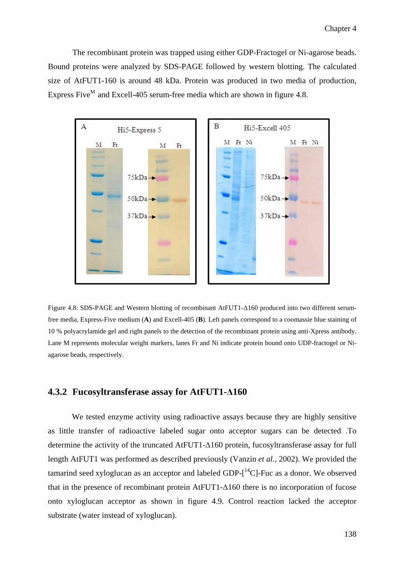
Chapter 4
138
The recombinant protein was trapped using either GDP-Fractogel or Ni-agarose beads.
Bound proteins were analyzed by SDS-PAGE followed by western blotting. The calculated
size of AtFUT1-160 is around 48 kDa. Protein was produced in two media of production,
Express FiveM and Excell-405 serum-free media which are shown in figure 4.8.
Figure 4.8: SDS-PAGE and Western blotting of recombinant AtFUT1-Δ160 produced into two different serum-
free media, Express-Five medium (A) and Excell-405 (B). Left panels correspond to a coomassie blue staining of
10 % polyacrylamide gel and right panels to the detection of the recombinant protein using anti-Xpress antibody.
Lane M represents molecular weight markers, lanes Fr and Ni indicate protein bound onto UDP-fractogel or Ni-
agarose beads, respectively.
4.3.2 Fucosyltransferase assay for AtFUT1-Δ160
We tested enzyme activity using radioactive assays because they are highly sensitive
as little transfer of radioactive labeled sugar onto acceptor sugars can be detected .To
determine the activity of the truncated AtFUT1-Δ160 protein, fucosyltransferase assay for full
length AtFUT1 was performed as described previously (Vanzin et al., 2002). We provided the
tamarind seed xyloglucan as an acceptor and labeled GDP-[14C]-Fuc as a donor. We observed
that in the presence of recombinant protein AtFUT1-Δ160 there is no incorporation of fucose
onto xyloglucan acceptor as shown in figure 4.9. Control reaction lacked the acceptor
substrate (water instead of xyloglucan).

Chapter 4
139
Figure 4.9: Fucosyltransferase assay using GDP-[14C]-Fucose for AtFUT1-Δ160 protein produced in insect
cells. Control reaction lacked the acceptor substrate, whereas xyloglucan 1 and xyloglucan 2 represent two
independent reactions with the same protein production
This assay showed that truncated AtFUT1-Δ160 was not able to catalyze the transfer
of GDP-[14C]-Fuc onto xyloglucan acceptor substrate, hence AtFUT1-Δ160 is not active.
Again, several hypotheses can be drawn to explain the lack of activity for AtFUT1-Δ160, one
likely explanation would be the removal of part of catalytic site of the enzyme while
designing the truncation. We hypothesize that part of the catalytic site of the AtFUT1 lies in
the first 160 amino acids of the N-terminus as an explanation for why no activity was
observed. In order to confirm this hypothesis, we prepared a second (less truncated) version of
AtFUT1 protein which lacked only 68 amino acids from the N-terminal side (named AtFUT1-
Δ68).
4.3.3 Expression of AtFUT1-Δ68 in insect cells
In order to express AtFUT1-Δ68 in insect cells, a DNA fragment lacking the first 204
bp within the coding region of AtFUT1 was generated by PCR using pVT-Bac1-AtFUT1 (as a
template comprising the full length cDNA of Arabidopsis thaliana AtFUT1 gene. The
restriction sites Pst1 and EcoR1 were added to the amplified sequence through primers at the
5’ and 3’ end during the PCR reaction. PCR product was cleaved with the restriction

Chapter 4
140
endonucleases Pst1 and EcoR 1. For the further steps of cloning and expression of truncated
AtFUT1- Δ68, the same procedure was followed as described in section 2 of this chapter. The
calculated size of AtFUT1-68 is around 58 kDa. Protein was produced in Excell-405 serum-
free media. The recombinant protein was trapped using Ni-agarose beads. Bound proteins
were analyzed by SDS-PAGE. Western blot analysis was done to check the expression of
protein using the anti-histidine antibody (Figure 4.10).
Figure 4.10: Quantitative and qualitative analysis of AtFUT1-Δ68 by PAGE and western blot. (A) Coommasie
staining of PAGE, and (B) western blot analysis of truncated AtFUT1-Δ68 produced in Excell-405 media. Lane
M represent molecular weight marker, lane Ni indicates protein bound onto Ni-agarose beads.
4.3.4 Fucosyltransferase activity test for AtFUT1-Δ68 protein produced in
insect cells
In order to check whether truncated AtFUT1-Δ68 protein produced in insect cells is
active, fucosyltransferase assay was performed as discussed above. We used the crude protein
for this activity assay because the radioactive assay is very sensitive. We observed that
AtFUT1-Δ68 was able to transfer fucose from GDP-[14C]-Fuc onto xyloglucan acceptor
(Figure 4.11). We performed two independent reactions one for xyloglucan acceptor and a
second for galactoglucomannan (GGM). In control reaction which lacked the acceptor, no
transfer of fucose was observed. Similarly for galactoglucomannan which is not the right

Chapter 4
141
acceptor for fucosyltransferase enzyme, no activity was observed. This assay confirmed that
truncated protein AtFUT1-Δ68 which lacked transmembrane domain is active.
Figure 4.11: Fucosyltransferase assay for truncated AtFUT1-Δ68 produced in insect cells. xyloglucan acceptor
substrate was used for this assay whereas galactoglucomannan is used as a control which is not a good acceptor
for fucosyltransferase enzyme. Control reaction lacked the acceptor substrate. Two independent reactions were
done with xyloglucan and galactoglucomannan acceptor substrates which are indicated by XyG1 and 2 and
GGM1 and 2 respectively. In order to confirm these results, we performed activity test of AtFUT1-Δ68 while
expressing the protein in Pichia pastoris, another expression system which is not expensive
and easy to carry out for the expression of recombinant proteins.
4.3.5 Cloning and expression of truncated AtFUT1-Δ68 in Pichia pastoris
Pichia pastoris revealed to be also an excellent heterologous expression system for
plant GTs. We therefore decided to turn on this system that was recently set up in the
laboratory. A truncated form of AtFUT1 (AtFUT1-Δ68) was produced by PCR using pENTR-
AtFUT1 as a template which comprises the full length cDNA of Arabidopsis thaliana
fucosyltransferase gene. This construct AtFUT1-Δ68 lacks the nucleotides encoding the first
68 amino acids of AtFUT1.

Chapter 4
142
The 1575 bp coding region corresponding to the truncated soluble form of AtFUT1-
Δ68 was obtained using the forward and reverse primers. These primers were designed to
generate the restriction sites EcoR1 and Not1 at the 5’ and 3’ of the amplified sequence,
respectively.PCR product was cleaved with the restriction endonucleases EcoR1 and Not1.
DNA was isolated and inserted by ligation into the pPICZαHis flag vector, digested with the
same enzymes. Afterwards the ligated plasmid was transformed into Pichia pastoris GS115.
Plasmid insertion was checked by PCR by using gene specific primers and was further
confirmed by sequencing. Protein was produced in BMMY media and expression was
induced for 5 days with methanol. Recombinant protein was secreted into media and analyzed
by SDS-PAGE gel following Coomasie blue staining.
4.3.6 Fucosyltransferase activity test for AtFUT1-Δ68 protein produced in
Pichia pastoris
AtFUT1-Δ68 protein was expressed in Pichia pastoris, recombinant protein was
secreted into the culture media (for details see materials and methods) and collected from two
AtFUT1-Δ68 expressing Pichia pastoris clones, named as AtFUT1-A and AtFUT1-B.
Activity test was performed as previously described for full length AtFUT1 (Vanzin et al.,
2002). We collected the secreted protein after 3 days and 5 days of expression from Pichia
pastoris cells. In the fucosyltransferase assay, we have observed that in the presence GDP-
[14C]-Fuc donor, AtFUT1-Δ68 catalyzed the transfer of fucose onto tamarind xyloglucan
acceptor (Figure 4.12), whereas no transfer of fucose was observed in the presence of
galactoglucomannan as an acceptor. Similarly no transfer was observed in the control reaction
which lacked the acceptor.

Chapter 4
143
Figure 4.12: Fucosyltransferase assay for truncated AtFUT1-Δ68 in the presence of xyloglucan acceptor
substrate whereas galactoglucomannan is used as a negative control for fucosyltransferase activity. A second
control reaction lacking the acceptor substrate was also included. Activity was checked for two AtFUT1-Δ68
expressing Pichia clones after 3 days and 5 days of protein induction.
We observed that the extension of the induction from 3rd to 5th day may increase
protein activity (Figure 4.12, clone Pichia-AtFUT1-B), possibly as a consequence of protein
accumulation. It is noteworthy that AtFUT1-Δ68 protein secreted into media was present but
diluted in an important volume of medium. To overcome this difficulty, we used Vivaspin
column to concentrate 15 mL of medium to 1mL. This concentrated medium was then used to
test activity and we observed a 10 fold increase in the activity of AtFUT1-Δ68, as expected
(Figure 4.13). Accordingly, no activity was observed from the filtrate medium and no transfer
of fucose was observed in the presence of galactoglucomannan acceptor (negative control).
Another negative control was performed with an assay lacking the acceptor, no activity was
observed. So these results confirmed that truncated AtFUT1-Δ68 catalyzed the incorporation
of [14C]-fucose onto xyloglucan acceptor substrate.

Chapter 4
144
Figure 4.13: Fucosyltransferase assay for “concentrated” AtFUT1-Δ68 protein containing medium, in the
presence of xyloglucan (acceptor ) and galactoglucomannan is ( negative control) for fucosyltransferase activity.
Another control reaction lacked the acceptor (water). Protein AtFUT1-Δ68 was isolated after 5 days of
expression in Pichia pastoris and used for activity assays.
In summary, the AtFUT1-Δ68 protein is active and able to transfer fucose onto
xyloglucan acceptor substrate. However, when western blot analysis was carried out and total
proteins were transferred, for unknown reasons, no signal was detected at the expected size of
AtFUT1-Δ68. One possible explanation could be a degradation of the histidine tag during
protein accumulation.

Chapter 4
145
4.4 Enzyme kinetics of AtFUT1-Δ68 4.4.1 Initial rate analysis of AtFUT1-Δ68
After demonstrating the activity of AtFUT1-Δ68, we analyzed the kinetic parameters
of fucosyltransferase regarding the xyloglucan acceptor. Biochemical characterization of
AtFUT1-Δ68 activity was realized using AtFUT1-Δ68 expressed in insect cells. We
determined the effects of changing time of incubation on fucose addition onto xyloglucan
acceptor substrate through radioactive assays. Radioactive assays involve the incorporation of
radioactivity to measure the amount of product made over time. We determined the initial rate
of reaction by drawing the graph from two independent series values. We checked the effect
of time on product formation each 5 minutes. We observed that the maximum product is
formed after 30 minutes and is in linear range indicative of initial rate conditions. We selected
the initial rate of reaction 30 minutes because the incubation time should be long enough to
permit a moderate amount of product to be formed and to make the error in timing
insignificant and reproducible.
Figure 4.17: Initial rate incorporation of GDP-[14C]-Fuc into tamarind XyG using culture medium from Hi5cells
expressing AtFUT1-Δ68, with fixed concentration of GDP-Fuc (20µM) and XyG (1mg/mL). Data from 2 series
of identical experiment were plotted.

Chapter 4
146
4.4.2 Determination of Km and Vmax of AtFUT1-Δ68
Michaelis-Menten Km and maximal reaction rates Vmax were determined for AtFUT1-
Δ68 express in insect cells, by varying the concentration of acceptor xyloglucan substrate with
a fixed concentration of GDP-Fucose. Results showed that the AtFUT1-Δ68 has a Km value of
0.72 mg/mL (~ 0.62 µM) in good accordance with the Km (0.46 mg/mL) value obtained by
Faik and coll. (2000) while studying activity of full length XyG fucosyltransferase from pea
(PsFUT1). It is intersting to note that in case of XyG acceptor, Km is relatively low because of
numerous acceptor sites available on the acceptor molecule. Vmax of AtFUT1-Δ68 produced in
insect cells was determined at 12,2mM/h/mg of total protein, which appeared significatively
lower than PsFUT1 (200mM/h/mg) but could be explained by various factors such as the
protein truncation or the xyloglucan fucosyltransferase from Arabidopsis being less active
than its counter-part in Pisum sativum. Efforts have been undertaken to purify AtFUT1-Δ68,
and such kinetics study could then be completed unambiguously.
Fig 4.18: Double-reciprocal plot of the initial rate reaction with tamarind xyloglucan as the varied substrate. Data presented are 2 series of identical experiment using culture medium from Hi5cells expressing AtFUT1-Δ68 protein with fixed concentration of GDP-Fuc (100µM). XyG concentration were as followed (0.05 ; 0.1 ; 0.2 ; 0.4 ; 0.8 ; 1 and 2 mg/mL).
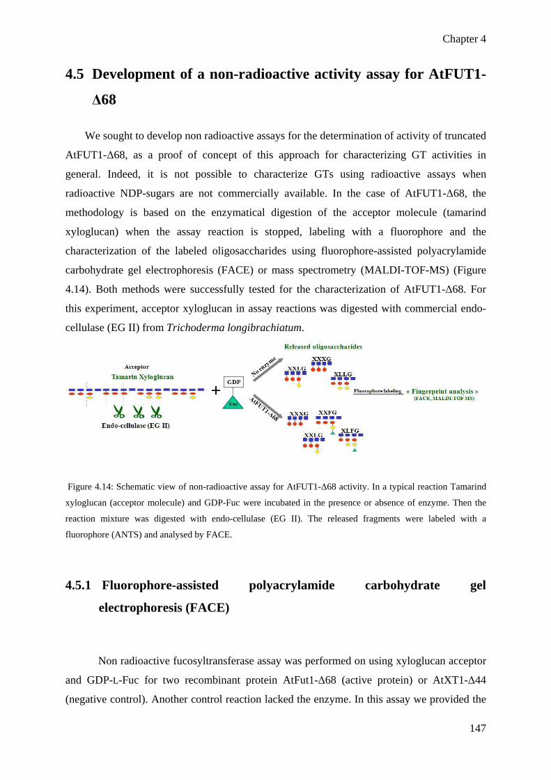
Chapter 4
147
4.5 Development of a non-radioactive activity assay for AtFUT1-
Δ68
We sought to develop non radioactive assays for the determination of activity of truncated
AtFUT1-Δ68, as a proof of concept of this approach for characterizing GT activities in
general. Indeed, it is not possible to characterize GTs using radioactive assays when
radioactive NDP-sugars are not commercially available. In the case of AtFUT1-Δ68, the
methodology is based on the enzymatical digestion of the acceptor molecule (tamarind
xyloglucan) when the assay reaction is stopped, labeling with a fluorophore and the
characterization of the labeled oligosaccharides using fluorophore-assisted polyacrylamide
carbohydrate gel electrophoresis (FACE) or mass spectrometry (MALDI-TOF-MS) (Figure
4.14). Both methods were successfully tested for the characterization of AtFUT1-Δ68. For
this experiment, acceptor xyloglucan in assay reactions was digested with commercial endo-
cellulase (EG II) from Trichoderma longibrachiatum.
Figure 4.14: Schematic view of non-radioactive assay for AtFUT1-Δ68 activity. In a typical reaction Tamarind
xyloglucan (acceptor molecule) and GDP-Fuc were incubated in the presence or absence of enzyme. Then the
reaction mixture was digested with endo-cellulase (EG II). The released fragments were labeled with a
fluorophore (ANTS) and analysed by FACE.
4.5.1 Fluorophore-assisted polyacrylamide carbohydrate gel
electrophoresis (FACE)
Non radioactive fucosyltransferase assay was performed on using xyloglucan acceptor
and GDP-L-Fuc for two recombinant protein AtFut1-Δ68 (active protein) or AtXT1-Δ44
(negative control). Another control reaction lacked the enzyme. In this assay we provided the

Chapter 4
148
cold GDP-L-Fuc to the enzyme (for details see materials and methods 6.4.2). The reaction was
performed in the presence of Hepes and MnCl2. After 2 hours of incubation, 2µL of
endoglucanase was added and put at 30°C for 1h. Samples were derivatized after
concentration under N2 (see materials and methods). Depending on labeling intensities on a
first ANTS (8-Aminonaphthalene-1,3,6-TriSulfonate) gel, loading quantity was adjusted for
the second gel.
Figure 4.15: FACE profile of endoglucanase generated xyloglucan fragments. Lane M shows the maltodextrine
ladder which is derivatized using ANTS. Other lanes show the XyG fragments obtained upon incubation of XyG
in the presence of AtFut1-Δ68 protein, AtXT1-Δ44 protein and a negative control without the enzyme. Similarly
lane 3 which lacked the AtFut1-Δ68 no activity was observed.
We use maltodextrine ladder (dp5 to dp11) which was also derivatized using ANTS.
The data shows that in the presence of AtFUT1-Δ68 an extensive fucosylation of xyloglucan
occurred (at the level of detection). Lanes 2 and 3 show characteristics mobility from tamarind
XyG oligosaccharides whereas lane 1 shows the presence of fucosylation (higher mobility)
which is shown in figure 4.15 by arrows. We observed that in the reaction containing AtFut1-
Δ68 enzyme, xyloglucan fragments that were released correspond to XXXG, XLXG, and
XLLG fragments characteristics of non-fucosylated XyG from tamarind seeds, but
additionally two new structures with reduced electrophoretic mobility were identified which
corresponds to XXFG and XLFG fragments. This assay confirmed that truncated AtFUT1-
Δ68 is able to transfer fucose onto xyloglucan acceptor substrate.
Chapter 4
149
4.5.2 Matrix Assisted Laser Desorption Ionization Time Of Flight
(MALDI-TOF MS) analysis
MALDI-TOF MS is a fast and reproducible qualitative technique which could be used
to determine the proportion of different oligosaccharides present in a mixture. The reaction
mixture which contained cold GDP-Fuc (donor), xyloglucan substrate (acceptor) and
AtFUT1-Δ68 enzyme was incubated for two hours for the fucosyltransferase reaction to take
place. In parallel, a control reaction which lacked AtFUT1-Δ68 enzyme and another control
reaction which contained AtXT1-Δ44 in place of AtFUT1-Δ68 enzyme were also performed.
All the above described reactions were subjected to endoglucanase enzymatic digestion.
Endoglucanase cleaves the glucosidic bond of the xyloglucan and results in the production of
oligosacharides. After digestion, the released oligosaccharide fragments were labeled with 2-
aminobenzamide (2-AB, see material and methods) easier to desorb and easily detectable by
MALDI-TOF-MS. Mass spectra of the labeled xyloglucan fucosylated and unfucosylated
(control reaction) oligosacharides were performed. As MALDI-TOF-MS analysis of 2-AB
labeled neutral oligosaccharides allowed the detection of [M + Na] + in the positive-ion mode. As expected from FACE characterization of AtFut1-Δ68 enzyme assay, MS analysis
showed two new peaks, one with a mass of 1513.63 where fucose is added onto XXLG then
producing XXFG fragment, and a second new peak with the mass of 1675.68 where fucose is
added onto XLLG, then producing XLFG fragment in the presence of AtFut1-Δ68 enzyme
(Figure 4.16A). This mass exactly corresponds to the mass which is calculated after the
addition of fucose. This assay showed us that AtFut1-Δ68 is able to transfer fucose onto
xyloglucan acceptor substrate. This is the definitive proof of its activity. While in the absence
of enzyme, only peaks for XXXG, XLXG and XLLG were observed and no peaks were
observed for fucosylated xyloglucan fragments (Figure 4.16B). Similarly no peaks for
fucosylated xyloglucan fragments were observed in the control reaction in the presence of
AtXT1-Δ44 (Figure 4.16C).

Chapter 4
150

Chapter 4
151
Figure 4.16: MALDI-TOF MS analysis of endoglucanase digested fragments of xyloglucan for
fucosyltransferase activity. A. Mass spectra of xyloglucan in the presence of AtFUT1-Δ68 enzyme. The mass
increment was observed in the peaks due to the addition of fucose. B. Mass spectra of xyloglucan in the absence
of AtFUT1-68 enzyme. No addition of fucose was observed when the reaction lacked the enzyme. C. Mass
spectra of xyloglucan in the presence of AtXT1-Δ44 which is used as a control. Similarly in the control reaction,
the addition of fucose was not observed.
4.5.3 Conclusion
In this chapter we have carried out the expression of truncated versions of both
AtFUT1 and AtXT1 proteins in order to determine the 3D-structure of these GTs using x- ray
crystallography. Unfortunately, none of the truncated version of AtXT1 proteins (both
AtXT1-Δ140 and AtXT1-Δ44) that were expressed in insect cells happened to be active. The
lack of activity for AtXT1-Δ44 and AtXT1-Δ140 could have numerous reasons; from the lack
of the transmembrame domain important for protein folding and activity or the lack of a
necessary cofactor that would be missing in our assays.
On the other hand, AtFUT1-Δ68 was successfully expressed either in insect cells or
Pichia pastoris, and was shown active in both systems. Thus, it was possible to determine the
kinetic parameter of AtFUT1-Δ68, and to compare these parameters with the one previously
obtain for the characterization of the pea xyloglucan fucosyltransferase (PsFUT1). AtFUT1-
Δ68 was additionally demonstrated functional over non-fucosylated xyloglugan
oligosaccharides using a non-radioactive assay (FACE). Analysis of the Fluorophore-assisted
polyacrylamide carbohydrate gel electrophoresis (FACE) experiments by mass spectrometry
(MALDI-TOF-MS) offer the opportunity to do product characterization of the fucosylated
XyG oligosaccharides, and thus confirm the protein activity. Actually, efforts are underway to
purify the AtFUT1-Δ68 protein using affinity chromatography on nickel column, in order to
crystallize the protein and perform a 3D-structure study of a first plant GT.

Chapter 5
152
Chapter 5
General discussion and perspectives

Chapter 5
153
5 General Discussion and perspectives
Cell walls play many important roles in defining the unique biology of plants; they
also have practical applications as a feedstock, for biomaterials and for the production of
biofuels. Cell walls also have strong economical interests: i.e. 40,000 tons of pectins are
produced every year to be used in food industry, and some pectic polymers are studied as
pharmaceuticals for prostate cancer treatment (Jackson et al., 2007). Despite the fundamental
and practical importance of cell walls, far too little is known about the biosynthesis of the
macromolecular components that comprise them. Unfortunately, this lack of knowledge
prevents any manipulation of the plant biosynthetic pathway responsible for cell wall
elaboration by plant, and thus any optimization of the biomass products for human
applications. However as stated recently by Pauly and Keegstra (2008): “the natural
variability in wall compositional quantity and quality suggests that there is an opportunity for
altering the abundance of specific wall components without compromising the life cycle of a
plant”. Thus a comprehensive understanding of polysaccharides biosyntheses by
glycosyltransferases (GTs) is required for the development of plant with optimized biomass
contents for human uses.
My Ph.D. work at CERMAV fits this goal, as we aim to understand how
polysaccharides are synthesized by GTs, delivered to the cell surface, incorporated into the
plant cell wall matrix, and how these processes are regulated. We postulated that one likely
hypothesis for the lack of success in the identification of pectin biosynthetic enzymes relates
to the fact that all plant glycosyltransferases have not been annotated yet in plant genome, and
thus could not be studied. Accordingly, Hansen and Coll. (2009) developed a novel method
where they have used bioinformatics, seeking Arabidopsis genome for novel
glycosyltransferases candidate genes and identified a new family of genes called “NGT”
(Novel Glycosyltransferase) by using bio-informatics strategy (Hansen et al., 2009). This
method led to the identification of more than 150 candidate genes. Among them, 24 were
considered as strong candidates that should be further investigated since known GT signatures
were clearly identified. More specifically, one putative GT identified displayed a
fucosyltransferase signature (and was named NGT1 for “Novel GlycosylTransferase 1”),

Chapter 5
154
whereas the 23 other gene were related and belong to the “Domain of Unknown Function
266” (DUF 266) family (Hansen et al., 2009).
The first work I took over in the lab was the characterization of T-DNA Arabidopsis
mutants from the NGT family that were ordered. The genetic characterization of 35 mutant
lines was carried out and permit after PCR screening the isolation of 16 homozygous mutant
lines from NGT family. These homozygous mutant lines have been used in a developmental
study seeking abnormal growth characteristics or unusual organ development that could be
indicative of defect in the way cell wall was laid down in these mutants. Peculiar but tenuous
phenotype could be identified for some of the lines such as narrow leaves during early
development for ngt1-1 and ngt1-2 mutants (or mutants altered for At5g14550 gene; coded
“P” in this manuscript). However, many homozygous mutants that have been characterized
during the first part of my work have not been analysed specifically for this trait which open
perspectives for other students in the lab as the methodology developed for leaf area
quantification can easily be duplicated to all the NGT mutant lines.
The NGT family comprises 24 genes that are considered strong candidates to encode
GT activity on the basis of clear GT signatures that have been identified in their sequences
with high similarity; but, 24 candidate genes appeared an overwhelming number of genes to
characterize. As this project aim was mainly to determine whether or not some (or none) of
the gene identified using bioinformatics encode a GT activity, and thus validate our strategy
of GTs identification, I focused on the characterization of one gene At5g28910 (NGT1).
The identification of homozygous mutants for the DUF266 gene family was not
carried out with much detail during the course of my Ph.D. but it would be a valuable work
for the forthcoming researchers and students in the lab. Noticely, the extensive
characterization of T-DNA mutant lines that I carried out should save time in the future for
such work to be achieved. Heterologous expression would also be very useful for the
functional characterization of the At5g14550 gene but it was not carried as we focused on
NGT1 activity characterization and because the unavailability of At5g14550 cDNA would
have delayed such study. However, At5g14550 cDNA was recently added to the NASC, and
heterologous expression of this DUF266 gene should be carried out to test GT activity for the
protein.

Chapter 5
155
After, the characterization of T-DNA lines for NGT family, we decided to focus our
functional genomic study on NGT1 gene for which a putative function was assigned,
providing a testable hypothesis for protein activity tests. Moreover, as two homozygous
mutant lines ngt1-1 and ngt1-2 were characterized, we were able for this locus to study the
impact of NGT1 alteration on cell wall polysaccharides content. During the course of our
study, several lines of evidences have shown that NGT1 gene was either directly or indirectly
involved in cell wall biosynthesis. Phenotypic studies have shown that leaves of mutant plants
ngt1-1 and ngt1-2 are smaller than WT plants. This observation is interesting as cell wall
mutant plants have often shown various developmental growth defects (Scheible and Pauly,
2004). In order to check the growth defects in our mutant lines, leaf area of both lines were
measured. This analysis has shown that mutant lines have narrow leaves at the beginning of
the third week of growth which remained significant till 6TH week of growth. This reduction
in leaf area of mutant plants has shown that mutant plants have faced some difficulty to grow
and have changed properties like elasticity and rigidity of cell wall. Interestingly, significant
growth rate difference was observed among the wild type and mutant plants only between 14th
and 21st day of development (Figure 3.11). From 21st to 35th day of development, the plants
seemed to have the tendency of similar growth. These results indicate that the mutation in
NGT1 alters the growth rate only during early developmental stage and has no or little effect
on the growth rate during late development.
In order to check this hypothesis, expression profile of NGT1 during different
developmental stages was studied by using Genevestigator (www.genevestigator.com). It
showed that NGT1 is highly expressed at germination stage, in young leaves and in mature
silics but their expression is decreased in mature leaves and at inflorescence stage. In
conclusion, finally this phenotypic growth defect in ngt1-1 and ngt1-2 mutant plants have
shown that NGT1 gene is involved at early stages of plant development in the biosynthesis of
cell wall.
Second evidence for the involvement of NGT1 gene in cell wall biosynthesis came
from the biochemical analysis of cell wall of mutant plants. Neutral monosaccharide
quantification through GC-MS has shown that ngt1 mutant plant cell wall has significantly
less arabinose, galactose and rhamnose as compared to that in wild type col0 cell wall (Figure
3.12). This data confirmed the link between the biochemical composition and phenotype
which is due to the alteration in NGT1 gene. These results have shown that mutation in NGT1

Chapter 5
156
affected the cell wall polysaccharides biosynthesis: more specifically the pectin representative
sugars like arabinose, rhamnose and galactose. These defects in cell wall composition showed
more convincingly that our hypothesis about the implication of NGT1 in plant cell wall
biosynthesis is correct but in order to get detailed idea, which specific polymer is modified,
permethylation linkage analysis was performed.
This biochemical analysis has shown that cell wall from both ngt1-1 ngt1-2 mutant
cell wall was significatively impaired for 3 molecules namely 3,5-Araf, 5-Araf and 4,6-Glcp
as compared to wild type content(Figure 3.14). The reduction of two linkages 3,5-Araf, 5-Araf
(50%) constitutive of arabinan was notice, as the neutral monosaccharide quantification
results showed the decrease in total arabinose content in mutant cell wall (as compared to
WT). Arabinan pectic polymer actually contains these 2 linkages (3,5-Araf, 5-Araf), and this
observation suggested us to study the possibility that NGT1 would encode a GT activity
responsible for the biosynthesis of arabinan, as an alternative to the putative
fucosyltransferase activity predicted through gene sequence homology. The decrease in the
amount of the molecule of 4,6-Glcp was ruled out as a direct effect of NGT1 activity as this
linkage is present in xyloglucan due to xylostransferase activities, and because such GTs have
been already characterized in GT family 34. We then hypothesized that decrease in 4,6-Glcp
was an indirect effect of the NGT1 as a compensatory mechanisms responding to the
alteration of other polysaccharide of the cell wall, such as arabinan. Interesting information
were gathered while analysing cell wall of etiolated seedling with permethylation to unravel
glycosidic linkages between polysaccharides; however this study could be completed by a
sequential extraction of plant cell wall polysaccharides in order to confirm and strengthen
observation that were made. Indeed, a detailed analysis of the pectin fraction would be
informative of the quality of arabinan in ngt1-1 and ngt1-2 mutants. Interestingly, we
performed immunolabelling of stem cross section of etiolated seedlings, showed a qualitative
and reproducible reduction of -α-(1 5)-L-arabinan epitope in ngt1-1 and ngt1-2 mutant lines,
using LM6 and LM13 anti-arabinan antibodies (Fig 32). Thus these results confirmed the
biochemical phenotype identified for both mutants, even though subtle differences in labelling
intensity were found between ngt1-1 and ngt1-2. It is noteworthy that results obtained from
ngt1 Arabidopsis mutants characterization directly challenge our hypothesis of a
fucosyltransferase activity supported by the analysis of the gene sequence homologies. This
indicates how careful one should be over the course of a functional genomic study, especially
in the case of the functional anaylysis of a new gene family for which no activity has been
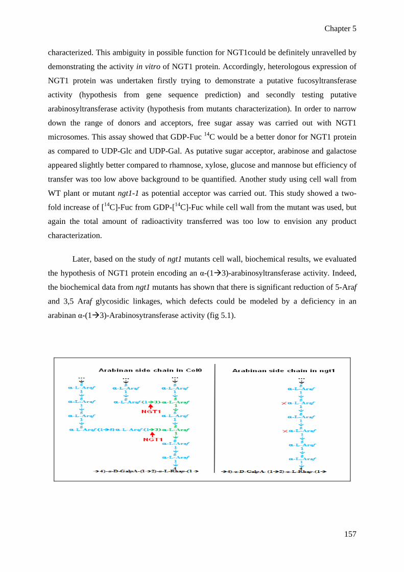
Chapter 5
157
characterized. This ambiguity in possible function for NGT1could be definitely unravelled by
demonstrating the activity in vitro of NGT1 protein. Accordingly, heterologous expression of
NGT1 protein was undertaken firstly trying to demonstrate a putative fucosyltransferase
activity (hypothesis from gene sequence prediction) and secondly testing putative
arabinosyltransferase activity (hypothesis from mutants characterization). In order to narrow
down the range of donors and acceptors, free sugar assay was carried out with NGT1
microsomes. This assay showed that GDP-Fuc 14C would be a better donor for NGT1 protein
as compared to UDP-Glc and UDP-Gal. As putative sugar acceptor, arabinose and galactose
appeared slightly better compared to rhamnose, xylose, glucose and mannose but efficiency of
transfer was too low above background to be quantified. Another study using cell wall from
WT plant or mutant ngt1-1 as potential acceptor was carried out. This study showed a two-
fold increase of [14C]-Fuc from GDP-[14C]-Fuc while cell wall from the mutant was used, but
again the total amount of radioactivity transferred was too low to envision any product
characterization.
Later, based on the study of ngt1 mutants cell wall, biochemical results, we evaluated
the hypothesis of NGT1 protein encoding an α-(1 3)-arabinosyltransferase activity. Indeed,
the biochemical data from ngt1 mutants has shown that there is significant reduction of 5-Araf
and 3,5 Araf glycosidic linkages, which defects could be modeled by a deficiency in an
arabinan α-(1 3)-Arabinosytransferase activity (fig 5.1).

Chapter 5
158
Figure 5.1: Hypothesis of function of NGT1 protein in Arabidopsis cell wall. In green: the residue 3,5-Araf; in
blue: the residue 5-Araf; in red: linkage made by NGT1
We then develop a non-radioactive arabinosyltransferase activity assay with the
heterlogously produced NGT1 protein. We used the arabinotetraose, arabinohexaose and
arabinan as acceptors in the presence of UDP-Araf sugar donor in an assay, but no transfer of
arabinose onto these two putative acceptors was monitored after analysis of the acceptor
oligosaccharide by MALDI-TOF MS. Meanwhile, advance further in understanding the role
of NGT1, an activity test in in-vitro is required. One possibility for future work would be to
use purified α-(1 5)-arabinan polysaccharides free of α-(1 3)-arabinose side chain and to
use them as acceptor in an assay. We envisioned that if NGT1 activity is to create branching
in arabinan polymer, then the α-(1 5)-arabinan provided in the assay would be modified and
that an increase in Terminal-Arabinose could be quantified using GC-MS as a proof of NGT1
α-(1 3)-arabinosyltransferase activity.
Finally, another study that would require attention in future work is the subcellular
localization of NGT1 within the plant cell. Indeed, It has been well studied that non-cellulosic
and pectic polysaccharides are synthesized within the Golgi apparatus (Fincher & Stone,
1981; Gibeaut & Carpita, 1994). Using protein features software, it has been predicted that
NGT1 adopt a type II membrane protein topology, typical of Golgi located GTs but it would
be of importance to confirm experimentally this prediction. For this purpose Arabidopsis
plants have been transformed with a GFP version of NGT1 protein, where GFP was fused N-
terminal to NGT1. Generating plant harbouring stable expression of GFP:NGT1 should
enable the characterization of protein subcellular localization using YFP marker available for
various plant cell compartment, and thus provide support for NGT1 to be involved in
polysaccharide biosynthesis if Golgi-located.

Chapter 6
159
Chapter 6 MATERIALS AND METHODS

Chapter 6
160
6 Material and methods
6.1 Materials
• Biological materials
T-DNA knockout mutant lines were ordered from Nottingham Arabidopsis Stock
Centre (NASC). Pichia pastoris strain GS115 was purchased from Invitrogen and E.Coli (XLI
Blue) strain was obtained from Agilent. Agrobacterium strain C58C1 was kindly provided by
Dr Gabrielle Tichtinsky (LPCV, CEA Grenoble).
• Antibodies used
For western blotting of NGT1 protein, T7 monoclonal antibody was ordered from
Novagen, Penta-Histidine N-ter antibody for the detection of AtFUT1-Δ68 was obtained from
QIAGEN and secondary Anti-Xpress antibody was purchased from Invitrogen.
Antibodies used for the immunolabelling of plant stem cross section, LM13
monoclonal anti- (1→5)-α-L-arabinan was purchased from Plant Probes Leeds, UK. Other
antibodies like LM6, LM2, LM5, JIM7, JIM5 and JIM16 was provided by Azeddine Driouich
(Glycomev lab, University of Rouen). Secondary antibody for immunolabeling of stem cross
section, Anti-rat IgG-FITC produced in goat was purchased from Sigma.
• Specific materials or reagents used for biochemistry
Reagents 2,5-Dihydroxybenzoic acid (2,5-DHB), trifluoroacetic acid (TFA),
trimethylsylil reagent (TMS), methanolic-HCl, NaBD4 , 2-aminobenzamide (2-AB), 8-amino-
1,3,6-naphtalene trisulfonic acid (ANTS) were purchased from Sigma. AEC 101 staining kit
used for developing the western blot membrane was purchased from Sigma. Endocellulase
was obtained from Megazyme. Endo-H and protease inhibitor tablets were provided by
Roche. Radioactive sugar donors GDP-Fuc 14C (specific activity: 925Bq/µL) and UDP-Xyl 14C (specific activity: 925Bq/µL) were purchased from GE Healthcare. Protease inhibitor
tablet (EDTA-free) were from Roche (11836153001). GDP-Fucose, UDP-Galactose, UDP-
Glucose were from Kyowa Hakko Kyoga LTD (Japan). UDP-Xylose was from Carbosources
(ww.ccrc.uga.edu).
• Specific material for molecular biology
Hot Star high fidelity polymerase and MiniElute PCR purification kit were obtained
from QIAGEN. Go Taq DNA polymerase and DNase treatment kit of RNAs were purchased
from Promega. For RT-PCR, cDNAs were made by using the iScript kit obtained from Bio-
Rad.

Chapter 6
161
BP Cloning kit, LR Cloning kit and Plasmids pPICZ gatewayable, pPICZ α,
pDONR207, pDONR221 and LB culture media were ordered from Invitrogen. Nucleospin
Plasmid kit extraction and Nucleotrap kit were purchased from Macherey-Nagel. Gel red for
DNA gel staining was purchased from Interchim. Silwet-L77 was ordered from Lehle Seeds.
Plant gateway vectors pH2GW7 and ph7WGF2 for Agrobacterium transformation were
obtained from the department of Plant Systems Biology (http://gateway.psb.ugent.be/).
Nitrocellulose membrane used for western blot was ordered from PALL membranes,
BioTrace™ NT.
• Specific materials for microscopy
Osmium tetraoxide was purchased from Sigma. LR white, CFM-1 and Eukitt
mounting medium were ordered from Electron Microscopy Science (EMS).
6.2 Methods 6.2.1 Methods for heterologous expression of proteins
6.2.1.1 Gateway cloning for heterologous expression in Pichia pastoris
Gateway Cloning Technology is a powerful method that allows the subcloning of a
gene of interest into a pDONR vector to produce a versatile pENTR from which gene can
easily transferred into various destination vector depending on the system selected for protein
expression in the course of the study. It basically consists of two reactions i.e. BP reaction and
LR reaction. BP reaction transfers the gene of interest (between attB sites) into the pDONR
vector (containing attP sites) to produce a new Entry vector (attL sites). This reaction is
catalyzed by the BP Clonase mix of two enzymes integrase (Int) and integration host factor
(IHF) necessary for recombination. The LR Reaction is a recombination reaction between an
Entry Clone and a Destination Vector mediated by the LR Clonase mix containing three
different enzymes integrase (Int), excisionase (Xis) and integration host factor (IHF). This
reaction transfers DNA segments (e.g. cDNA, genomic DNA, or gene sequences) from the
Entry vector to the Destination vector to create an expression vector (Figure 6.1).

Chapter 6
162
Figure 6.1: Schematic representation of the two steps gateway cloning recombination system. A) First step, the
PCR product carrying appropriate flanking sequences is integrated into the pDONR207 vector by recombination
forming a new vector named pENTR207-T7-GOI in our study (GOI: Gene Of Interest). B) Second step, the
gene of interest (T7-GOI) is transferred from the pENTR207-T7-GOI to a gatewayable version of pPICz
destination vector, forming pPICz-T7-GOI used for Pichia transformation.
6.2.1.1.1 PCR amplification of gene of interests
Amplification of our gene of interest required a two-round PCR reaction in order to
add at 5’ and 3’ of the gene sequence the coding sequence of the T7 tag and the gateway
border (Figure 6.2). Each PCR reaction was carried out in a final volume of 25 μl containing 1
μl of DNA, and following final concentration of 1X hot star PCR buffer (containing dNTPs
and MgSO4), 0.4 µM of each (forward and reverse) primer and 0.1 units of hot star high
fidelity Polymerase. DNA template was amplified with gene specific primers in the first PCR
reaction and then T7 tag and gateway border sequences were added by the second PCR
reaction. The sequence of primers are given in table 6.1.

Chapter 6
163
Figure 6.2: Schematic presentation of the procedure of PCR for the addition of gateway borders and T7 tag.
attB1 and attB2 represent gateway border recombination sites. GOI (Gene of Interest).
Table 6.1: Primers used for amplification of the indicated genes for gateway cloning Gene Primer Name Sequence (5’ 3’) At1g10280 (A) Forward T7_A_for GGTGGACAGCAAATGGGTATGGCGAGAGGAGGGAAA Reverse B2_A_rev GTACAAGAAAGCTGGGTTTCAAAACCCCAGGACAGT At1g11940 (C) Forward T7_C_for GGTGGACAGCAAATGGGTATGACGAAGAAATCACAG Reverse B2_C_rev GTACAAGAAAGCTGGGTTTTATAAGGTTTTGTTTGC At1g62305 (E) Forward T7_E_for GGTGGACAGCAAATGGGTATGCCGCGGCTTCCGTCG Reverse B2_E_rev GTACAAGAAAGCTGGGTTCTAAAAGGTTGTTGTATC At1g68380 (F) Forward T7_F_for GGTGGACAGCAAATGGGTATGCCGACCCAAAAGGAC Reverse B2_F_rev GTACAAGAAAGCTGGGTTTCAGAAATACATGACGAT At2g19160 (I) Forward T7_I_for GGTGGACAGCAAATGGGTATGAAGGCAATTAAGGGA Reverse B2_I_rev GTACAAGAAAGCTGGGTTCTACACGATGCTTGTGTA At3g52060 (J) Forward T7_J_for GGTGGACAGCAAATGGGTATGTTCTCATCTTCTACA Reverse B2_J_rev GTACAAGAAAGCTGGGTTTCAATCCCTAAAGATCAC At4g30060 (L) Forward T7_L_for GGTGGACAGCAAATGGGTATGAAGGCAGTTAAAAGA Reverse B2_L_rev GTACAAGAAAGCTGGGTTTCACAAATTCCAAGTGAC At4g32290 (N) Forward T7_N_for GGTGGACAGCAAATGGGTATGGCGAACCCCAACGAG Reverse B2_N_rev GTACAAGAAAGCTGGGTTCTATGATTTCTCTTGGGT At5g11730 (O) Forward T7_O_for GGTGGACAGCAAATGGGTATGCAGAATAGGATTGTA Reverse B2_O_rev GTACAAGAAAGCTGGGTTCTATGATTTCTCTTGGGT At5g22070 (R) Forward T7_R_for GGTGGACAGCAAATGGGTATGTTCTCGAATCAGTTT Reverse B2_R_rev GTACAAGAAAGCTGGGTTTTAATCTCTGAAAATCAC At5g25330 (S) Forward T7_S_for GGTGGACAGCAAATGGGTATGTTGGCCTTATATTTC Reverse B2_S_rev GTACAAGAAAGCTGGGTTTTACACAAGAGCCGAATC At5g57270 (V) Forward T7_V_for GGTGGACAGCAAATGGGTATGGGAGAGAATCTGCAT Reverse B2_V_rev GTACAAGAAAGCTGGGTTAATGACCACTGTGCTTGT At5g28910 (W) Forward T7_W_for GGTGGACAGCAAATGGGTATGAGCATGAAGTCATTA Reverse B2_W_rev GTACAAGAAAGCTGGGTTTTACCAGAACCGATCTTT
6.2.1.1.2 BP cloning
AttB-flanked PCR product after second PCR reaction was used to create entry clone.
Following components; attB-PCR product (=10 ng/µl; final amount ~15-150 ng) 1-7 µl,
Donor vector pDONR207 (150 ng/µl), 1 µl TE buffer (pH 8.0) to final volume of 8 µl were
added to a 1.5 ml tube at room temperature and mixed. The BP Clonase™ II enzyme mix was
thawed on ice for about 2 min. The BP Clonase™ II enzyme mix was vortexd briefly twice (2
seconds each time). 2 µl of BP Clonase™ II enzyme mix was added to the reaction and gently
mixed with two brief vortex pulses. Reaction mixture was incubated at 25°C for 1h. Then
Chapter 6
164
reaction was stopped by adding 1 µl of the Proteinase K solution, vortexed briefly and
samples were incubated at 37°C for 10 min.
6.2.1.1.3 Transformation and plasmid extraction
One microliter of BP reaction was used to transform 50 µL of XL1 blue competent
cells transformation. Cells were incubated on ice for 30 min with BP reaction, heat-shocked
for 45s at 42°C and put back on ice before 250 µl of S.O.C medium is added. Cells were then
allowed to recover 1 h at 37°C with shaking, plated on LB agar (1% tryptophane; 0.5% yeast
extract, 0.5% NaCl and 1.5% bacto-agar) supplemented with kanamycin 50 µg/ml and placed
overnight in an incubator at 37°C.
Plasmid extraction from bacteria was carried out using Nucleospin Plasmid kit
(Macherey-Nagel). First, single colony was precultured over night at 37°C in 2 ml LB
supplemented with appropriate antibiotic. Preculture was then centrifuged at 11,000 g for 1
min at room temperature (RT) and processed for plasmid DNA extraction following
manufacturer protocol. This includes briefly cells lysis, neutralisation of the first buffer, DNA
binding onto a silica membrane, wash and elution of DNA with deionised water or TE buffer.
Obtention of Entry vector carrying gene of interest was confirmed by PCR using gene specific
primers and sequencing.
6.2.1.1.4 LR cloning into pPICZ
Gene of interest was transferred from a Gateway pDONR207 entry clone to pPICZ
destination vector by the following protocol. Following components; Entry clone (~15-150
ng) 1-7 µl, Destination vector pPICZ (150 ng/µl), 1 µl TE buffer (pH 8.0) to final volume of
8 µl were added to a 1.5 ml tube at room temperature and mixed. Then the LR Clonase II
enzyme mix was thawed on ice for about 2 min. The LR Clonase II enzyme mix was vortexed
briefly twice (2 sec each time). To each sample (Step 1, above), 2 µl of LR Clonase™ II
enzyme mix was added to the reaction and was mixed well by vortexing briefly twice
following brief table-top centrifugation. Reaction mixture was incubated at RT for 1h, then
stopped by adding 1 µl of the Proteinase K solution, vortexed briefly and incubated at 37°C
for 10 min. Plasmid DNA was extracted as described above in paragraph 6.2.1.1.3. Obtention
of destination vector carrying gene of interest was confirmed by sequencing PCR products.
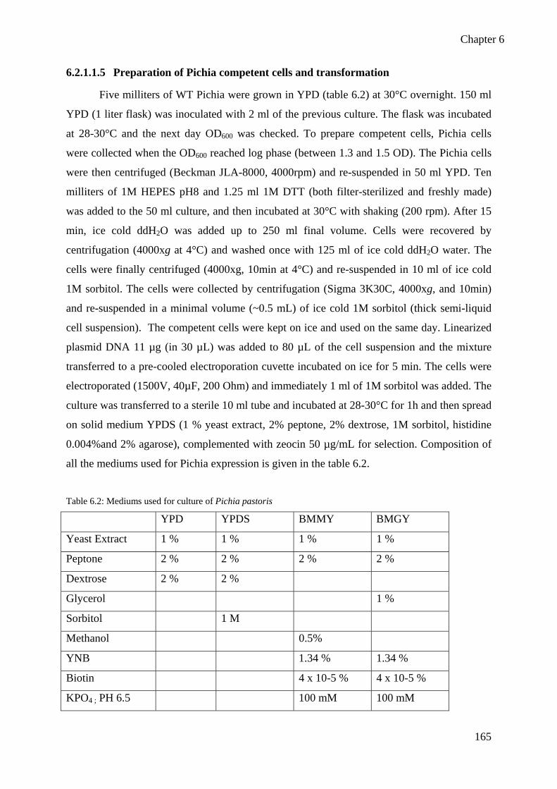
Chapter 6
165
6.2.1.1.5 Preparation of Pichia competent cells and transformation
Five milliters of WT Pichia were grown in YPD (table 6.2) at 30°C overnight. 150 ml
YPD (1 liter flask) was inoculated with 2 ml of the previous culture. The flask was incubated
at 28-30°C and the next day OD600 was checked. To prepare competent cells, Pichia cells
were collected when the OD600 reached log phase (between 1.3 and 1.5 OD). The Pichia cells
were then centrifuged (Beckman JLA-8000, 4000rpm) and re-suspended in 50 ml YPD. Ten
milliters of 1M HEPES pH8 and 1.25 ml 1M DTT (both filter-sterilized and freshly made)
was added to the 50 ml culture, and then incubated at 30°C with shaking (200 rpm). After 15
min, ice cold ddH2O was added up to 250 ml final volume. Cells were recovered by
centrifugation (4000xg at 4°C) and washed once with 125 ml of ice cold ddH2O water. The
cells were finally centrifuged (4000xg, 10min at 4°C) and re-suspended in 10 ml of ice cold
1M sorbitol. The cells were collected by centrifugation (Sigma 3K30C, 4000xg, and 10min)
and re-suspended in a minimal volume (~0.5 mL) of ice cold 1M sorbitol (thick semi-liquid
cell suspension). The competent cells were kept on ice and used on the same day. Linearized
plasmid DNA 11 µg (in 30 µL) was added to 80 µL of the cell suspension and the mixture
transferred to a pre-cooled electroporation cuvette incubated on ice for 5 min. The cells were
electroporated (1500V, 40µF, 200 Ohm) and immediately 1 ml of 1M sorbitol was added. The
culture was transferred to a sterile 10 ml tube and incubated at 28-30°C for 1h and then spread
on solid medium YPDS (1 % yeast extract, 2% peptone, 2% dextrose, 1M sorbitol, histidine
0.004%and 2% agarose), complemented with zeocin 50 µg/mL for selection. Composition of
all the mediums used for Pichia expression is given in the table 6.2.
Table 6.2: Mediums used for culture of Pichia pastoris
YPD YPDS BMMY BMGY
Yeast Extract 1 % 1 % 1 % 1 %
Peptone 2 % 2 % 2 % 2 %
Dextrose 2 % 2 %
Glycerol 1 %
Sorbitol 1 M
Methanol 0.5%
YNB 1.34 % 1.34 %
Biotin 4 x 10⁻⁵ % 4 x 10⁻⁵ %
KPO4 ; PH 6.5 100 mM 100 mM

Chapter 6
166
6.2.1.1.6 Screening of transformants
Transformants were selected on YPDS plates complemented with zeocin 50 µg/ml;
fifty transformants have been patched on a new plate using sterilized toothpicks. Plates were
incubated at 28-30°C for two days and putative transformants were selected for PCR
screening.
• Yeast DNA miniprep
To determine whether the gene of interest has been integrated into Pichia genome,
genomic DNA was isolated from 12 transformants by using the following protocol: Cells
were grown in 2 ml of YPD culture medium overnight at 28-30°C. One milliter of cells was
centrifuged at 11,000xg for 1 min and medium was discarded. Cells were washed with 1 ml of
water and centrifuged again. Cells were re-suspended in 200 µl of lysis buffer and then boiled
for 5 min in a water bath. Centrifugation was done and liquid phase was recovered into
another tube. 100 µl of 7 M ammonium acetate pH 7.0 was added and incubated at 65°C for 5
min and then 5 min on ice. 200 µl of chloroform was added, vortexed and centrifuged for 2
min. Supernatant was taken into a new tube and DNA was precipitated with 250 µl
isopropanol. It was incubated for 5 min at RT and then centrifugation was done for pelleting
the DNA at 13,000 g for 10 min. The pellet was washed with 70 % EtOH, dried under
vacuum and dissolved in 40 μl sterile water.
• PCR analysis of integrants
After isolation of genomic DNA, PCR was done to amplify the gene of interest either
using gene specific primers or 5’ AOX1 forward primer (5’-ATGTTGAGTTGTCCTTGC TTC-3’)
paired with 3’ AOX1 reverses primer (5’-GATAAGACCAACTTTCAAGGAG-3’). The reaction was
carried out in a final volume of 25 μl containing 2 μl of DNA, and following final
concentration of 1X Go Taq DNA polymerase buffer, 2 mM MgSO4 , 0.2 µM of dNTPs mix,
0.2 µM of each (forward and reverse) primer and 0.03 units of Taq DNA Polymerase enzyme.
Denaturation of double stranded DNA was done at 94°C for 2 min and it was followed by 25
cycles of amplification which included 10 sec at 94°C, 50 sec at Ta (Tm-5°C) and 1min45s at
72°C. Samples were analyzed on a 1X TBE, 0.8 % agarose gel. The integration of gene of
interest in Pichia was further confirmed by purifying and sequencing PCR product.

Chapter 6
167
6.2.1.2 Heterologous expression of proteins into Pichia pastoris
• Expression of full length NGT1
Pre-culture was started from a single colony in 5 ml YPD complemented with Zeocin
(50 µg/ml) overnight at 28-30°C. 40 ml of BMGY (Buffered Glycerol-complex Medium)
were inoculated with 50 µl of pre-culture and cells were grown at 28-30°C in a shaking
incubator until culture reached an OD600 = 2-6 (~16-18h). The cells would be in log-phase
growth. The cells were harvested by centrifugation at 3000 x g for 5 min at RT. Supernatant
was removed and cell pellet re-suspended to an OD600 of 1.0 in BMMY (Buffered Methanol-
complex Medium to induce expression (approximately 100-200 ml). Culture was placed in a 1
litre baffled flask and the flask was covered with 2 layers of sterile gauze or cheesecloth and
returned to incubator to continue growth. 100 % methanol was added to a final concentration
of 0.5 % methanol every 24h to maintain induction. The expression of clones was stopped
after 4 days. Cells were centrifuged at 5000xg for 5 min, and pellet was stored at -20°C to
check the protein expression by Coomassie stained SDS-PAGE and western blot.
6.2.2 Cloning of AtFUT1-Δ68 for heterologous expression in Pichia
pastoris
6.2.2.1 Obtention of AtFUT1-Δ68 PCR product
A truncated form of gene AtFUT1-Δ68 was designed and amplified from the plasmid
pENTR207 harbouring the full length cDNA of AtFUT1 with gene specific primers and
primers include EcoR1 and Not1 restriction endonucleases sites. PCR reaction was carried out
in 25 µl with final concentration of 50 ng of DNA, 1X hot star PCR buffer (containing dNTPs
and MgSO4), 0.4 µM of each (forward and reverse) primer and 0.1units of hot star high
fidelity Polymerase. The volume was made to 25 µl by adding water. The PCR programme
consisted of initial denaturation at 95°C for 5 min, followed by 30 cycles of 94°C for 15 sec,
annealing for 1 min at a 45°C temperature specific to the primer pairs and elongation at 72°C
for 1min40sec. The programme ended with an extension at 72°C for 10 min. The PCR
products were visualized on a 0.8 % agarose gel stained with Gel red. PCR product was
purified by using MiniElute PCR purification kit following manufacturer protocol and DNA
was eluted by adding 40 µl ddH2O.
Chapter 6
168
6.2.2.2 Cloning AtFUT1-∆68 into PpicZαhis flag
PCR product (1730 ng) and plasmid pPICZαhis flag (2.5 ng) was digested and
linearized using 1.5 µl of EcoR1 and 1.5 µl of Not1 in the presence of buffer React 3 and
water was added up to 34 µL final volume. Reaction mix was incubated for 3h at 37°C, and
digested PCR product was purified using SureClean kit. For this purpose, an equal volume of
SureClean solution was added to the digestion and mixed thoroughly. Mixture was incubated
for 10 min at RT. Centrifugation was done at 14,000xg for 10 min and supernatant was
removed. Precipitation was done by the addition of 2 vol of 70 % ethanol and then vortexed
for 10 sec. Centrifugation was done again, removed supernatant and the pellet was air dried to
ensure complete removal of ethanol. Pellet was re-suspended in 6 µl of sterilized water.
6.2.2.3 Gel extraction and purification of plasmid
After DNA electrophoretical separation on an agarose gel, plasmid purification from
the gel was carried out. First of all the DNA fragment with the correct size was identified
under UV light. DNA fragment was cut with the use of clean scalpel and purified by using
Nucleotrap kit. DNA gel slice was weighed and for 100 mg of agarose 300 µl of buffer NT1
was added. Nucleotrap suspension was vortexed thoroughly, and incubated at 50°C until the
gel slices were dissolved. Centrifuged for 30 sec at 10,000 g and supernatant was removed.
Silica matrix was washed by adding 500 µl of buffer NT2, centrifuged again for 30s at 10,000
g and supernatant was removed completely. Then the silica matrix was washed a second time
by adding 500 µl of buffer NT3, vortexed, centrifuged and supernatant was removed and third
wash was repeated with NT3 buffer. After washing, silica matrix was dried at RT and DNA
was eluted with 50 µl of water by adding to the silica matrix, pellet was resuspended through
vortexing and then centrifugation was done for 30 sec at 10,000xg and DNA was transferred
to clean tube.
• Ligation
Ligation of digested vector and insert was done by using the standard protocol. Two
tubes were prepared: the first tube for the ligation of insert into the vector contained 1µl of
digested insert (117ng/µl), 4 µl of vector (45ng/µl) and 5 µl of ligation mixture whereas the
second tube used water instead of insert to control the vector for self-ligation. Tubes were
incubated for 5 min at 25°C. 2 µl of reaction was used for transformation of XL1-blue cells,
plasmid was extracted and insertion checked after digestion with EcoR1 and Not1
endonucleases.

Chapter 6
169
6.2.3 Cloning of AtXT1-Δ140, AtXT1-Δ44 and AtFUT1-Δ68 for
heterologous expression in insect cells
6.2.3.1 PCR for cloning of AtXT1-Δ140, AtXT1-Δ44 and AtFUT1-Δ68 in insect cells
A truncated form of gene AtXT1 was designed and named AtXT1-∆140. This gene
sequence encodes the AtXT1-∆140 protein lacking 140 aminoacids at the N-terminus of
AtXT1 (including the transmembrane domain) but conserving the predictive catalytic
sequence at the C-ter of the protein. AtXT1-∆140 gene sequence was amplified from the
pUN51-XT1 plasmid (U14458) template using forward primer (5’-
AAACTGCAGCCTAATTTCGTGGCGCCAAAC-3') and the reverse primer (5’-
CGGGGTACCTCACGTCGTCGTCGTAC-3'). These primers were designed to include the cloning
restriction sites PstI and KpnI at the 5’end and 3’end, respectively of the amplified sequence.
Similarly, a truncated form AtXT1-∆44 was amplified using the forward primer (5’-
GGTACGCCGGAGAAAGATATCG-3’) and reverse primer (5’-GCTTAGTACGACGACGACG-3’).
These primers were designed to include the restriction sites PstI and EcoRI at the 5’end and
3’end, respectively of the amplified sequence.
A truncated form AtFUT1-∆160 was amplified from pENTR-AtFUT1 as template
comprising the full length cDNA of the A. thaliana AtFUT1 gene. The 1197 bp coding region
corresponding to the desired truncated soluble form of AtFUT1 was obtained using the
forward primer (5'-AAACTGCAGGATCAAGAACATATTGATGGTGATGG-TGAATGC-3') and the
reverse primer (5'-CGGGGTACCTCATACTAGCTTAAGTCCCCAGC-3'). These primers were
designed to include the restriction sites PstI and KpnI at the 5’end and 3’end, respectively of
the amplified sequence
A truncated form of gene AtFUT1-∆68 was amplified from the plasmid pENTR 207
harbouring the full length cDNA of AtFUT1. The 1473 bp coding region corresponding to the
desired truncated soluble form of AtFUT1 was obtained using the forward primer (5’-
AAACTGCAGTCAAATCGGATTATGGGTTTCG-3’) and the reverse primer (5’-
CCGGAATTCTCATACTAGCTTAAGTCCCC-3’). These primers were designed to include the
restriction sites PstI and EcoRI at the 5’end and 3’end, respectively of the amplified sequence.
For PCR reaction and cloning of all these constructs, same steps were followed as explained
in sections 6.2.2 .1 to 6.2.2.2.

Chapter 6
170
6.2.3.2 Transformation and expression of AtXT1-Δ140, AtXT1-Δ44 and AtFUT1-Δ68
in insect cells
• Transformation and amplification in Sf9 cells
Spodoptera frugiperda cells (Sf9 cells from Invitrogen) derived from ovarian tissue
were used for the production and amplification of recombinant baculoviruses. Sf9 cells were
grown in monolayer culture at 27°C in Grace’s medium (Invitrogen) supplemented with 10%
fetal bovine serum (Sigma) and 50 µg/ml gentamycin (Sigma). Cotransfection of the cells
with baculovirus DNA (7 ng BaculoGold linear DNA) and pVT-Bac-His1 containing the
insert (30 ng) was done according to the manufacturer’s instructions (Pharmingen BD
Dickinson, see annex Protocol 1). High-titer viral stocks around 107 to 108 plaque- forming
units/ml (pfu) of recombinant viruses were obtained by three repeated transfection procedures
of Sf9 cells (annex protocol 2). After the third amplification, culture medium containing the
recombinant virus was collected by centrifugation of the infected cell culture and was stored
at 4°C. A titration of the virus stock was done to estimate the pfu/ml.
• Protein Expression in hi-5 cells
The production of recombinant proteins was done in Trichoplusia ni Hi-5 (High
Five™, Invitrogen) cells, cultivated in serum-free medium (Express Five™ from Invitrogen or
Excell-405 from Sigma), and supplemented with 16mM L-glutamine and 50 µg/ml
gentamycin (Sigma). Cells were seeded at 70% confluency and allowed to attach for 30 min
to the plastic culture flasks before addition of the virus. Cells were typically infected with the
recombinant virus at a MOI (multiplicity of infection) of 5 (5 pfu/cell), and then grown at
27°C for 4 days. The culture supernatants (containing the secreted recombinant protein) were
collected, clarified by centrifugation (8000xg for 5 min), and stored at -70°C or used directly
for the protein isolation (For details see annex protocol 3).
• Protein Isolation and Visualization
Commercial or homemade affinity matrices (Nucleotide-Fractogel beads, Nucleotide-
agarose beads, Ni-agarose beads) were used to concentrate the secreted recombinant protein.
Samples of crude hi5 supernatants were incubated for 1h at 4°C, under stirring, with affinity
adsorbents (50 µl of bead suspension for 2 ml of supernatant) to trap the recombinant protein
(annex protocol 4). The resin matrix was afterwards centrifuged, washed twice with 1 ml
PBS, and then resuspended in Laemmli denaturing buffer. Samples were heated at 100°C for
5 min, sonicated for 2 min, and proteins were then separated on SDS-PAGE gels (annex

Chapter 6
171
protocol 5). The gels were either stained using Coomassie Brilliant blue (Protocol 6), or
electro-transferred to a nitrocellulose membrane (annex protocol 7), using a semi-dry system
(BioRad) for Western blotting. The nitrocellulose membrane was probed by incubation with
mouse anti-Xpress antibody (1:4000, Invitrogen) directed against the Xpress epitope,
followed by peroxidase-conjugated goat anti-mouse secondary antibody (1:2000, Sigma), and
developed using the AEC staining kit (Sigma). (See annex protocol 8).
6.3 Methods for protein separation and identification 6.3.1 Microsomes preparation from Pichia pastoris to test activity
Cells were broken by using cell disruptor. Samples were re-suspended in a volume of
2 ml to 7 ml of cold Extraction buffer (50 mM Hepes-KOH pH 7.5, 0.4 M Sucrose, 10 mM
MgCl2 and 1/20 ml protease inhibitor cocktail), and broken at 2.5 kbar pressure.
6.3.2 Protein extraction from Pichia pastoris
After breaking the cells for NGT1, extraction buffer was added up to final volume of
20 ml. For the preparation of microsomes, samples were centrifuged at 9’600 g for 15 min at
4°C and supernatant was taken into ultracentrifuge tubes. Tubes were balanced by adding
extraction buffer. Ultracentrifugation was performed at 100,000xg for 1h at 4°C. The pellet
was re-suspended in 250 µl of extraction buffer using a 2 ml homogeneiser (all steps were
performed at 4°C). After the 3rd and 5th days of expression of AtFUT1-Δ68 protein was
collected from the supernatant as it was secreted into the medium instead of breaking the
Pichia cells.
6.3.3 Protein quantification
The protein quantification was carried out using the Bradford assay. 200 µl of
Bradford reagent was added to 800 µL of standards and samples containing from 0 to 10µg
proteins. Tubes were mixed gently by inversion, covered with aluminum paper and reacted for
15 min before reading absorbance at 595 nm. Data were plotted between absorbance of the
standards vs. their concentration, and extinction coefficient was computed to determine the
concentrations of the unknown samples.

Chapter 6
172
6.3.4 Protein analysis by electrophoresis
6.3.4.1 SDS-PAGE
The gel apparatus was set, resolving gel was poured and let to polymerize for 30 min
before stacking gel was poured in gel cassette. The composition of resolving and stacking gels
are given in the below table 6.3. Resolving gel was 12 % and run for 1-2h at 150 V using
migration buffer (0.12 M Tris base, 1.25 M Glycine 10 % SDS and water).
Table 6.3: Composition of SDS-PAGE resolving 12 % and stacking gel 5 %
Sloutions Resolving gel (12 %) for 10 ml Stacking gel (5 %) for 3 ml
Acrylamide//bis-acrylamide
(30/1; p/v)
3.3 ml 500 µl
Tris-HCl 1.5 M pH 8.8 2.5 ml
Tris-HCl 1.0 M pH 6.8 380 µl
10 % SDS 100 µl 30 µl
10 % ammonium persulfate 100 µl 30 µl
TEMED 4 µl 3 µl
Water 4 ml 2.1 ml
6.3.4.2 Coomassie blue staining
Classically, two SDS-PAGE gels were run in parallel. One for Coomassie blue
staining and verification of the protein contents of samples and a second for western blot. Gel
was stained in coomassie blue solution for 15-30 min, before de-staining using solution of
7.5% acetic acid and 5% ethanol in water.
6.3.4.3 Western blot
For electric transfer of the proteins, the nitrocellulose membrane and 6 whatman
papers at the same size of the respective gel were cut. Two buffer solutions namely cathode
buffer (25 mM Tris-HCl, pH 9.4, Glycine 30 mM) and anode buffer (Tris 0.3 M pH 10.4)
were prepared. Membrane was dipped in anode buffer for 5 min. Three whatman papers were
dipped in cathode buffer and three in anode buffer. Three whatman papers soaked in anode
buffer were stacked on graphite electrode. A clean plastic rod was rolled over the stack to
remove any bubbles trapped between them and soaked nitrocellulose membrane was put on
these papers. Polyacrylamide gel was placed on the top of the transfer stack. Three whatman

Chapter 6
173
papers soaked in cathode buffer were put on the gel. A clean plastic rod was rolled again over
the stack to remove any bubbles trapped between them. The current flows from cathode to
anode, so the proteins being negatively charged due to SDS move from the gel (cathode side)
to the membrane (anode side) and are trapped in the membrane. Transfer was done for 2h at
0.8 mA/cm2.
The membrane was stained in “Red Ponceau” to confirm protein transfer efficiency.
After transfer, the nitrocellulose membrane was washed in 20 ml TBS (10 mM Tris-HCl (pH
8), 150 mM NaCl) for 5 min. Saturation of non-specific sites was carried out with a solution
of 1 % gelatin in a total of 20 ml TBS for 45 min with agitation at room temperature.
Membrane was washed twice for 5 min in 20 ml TBS-T (10 mM Tris-HCl (pH 8), 150 mM
NaCl and 0.05 % Tween20). The membrane was incubated with a mouse primary antibody
directed against T7 tag (1:4000) in TBS-T for 90 min. Membrane was washed three times for
5 min in 20 ml TBST. The membrane was again incubated with secondary antibody Gam-per
(1:2000, Sigma, goat anti-mouse conjugated to peroxidase) for 90 min at RT in TBS-T. The
membrane was washed four times for 5 min in 20 ml TBST and once with 20 ml TBS for 5
min. Blot was revealed using the AEC 101 kit (Sigma).
6.3.4.4 Enzymatic removal of N-glycans using Endo-H
Reaction was carried out in 40 µl containing 100 µg of protein extract which was
mixed with 1% SDS (2 µl), 1.5 M (1.2 µl) 2-mercaptoethanol, 10 µl of Extraction buffer (50
mM Hepes-KOH pH 7.5, 0.4 M Sucrose, 10 mM MgCl2 and 1/20 ml protease inhibitor) and
volume was made up to 40 µl with distilled water. Reaction mixture was heated at 50°C for
15 min. 300 mM (3.5 µl) of sodium acetate (pH 5.8) was added. Samples were put at 37°C for
15 min. 5 mU (1.5µl) of EndoH was added and incubated at 37°C for 18h. 15 µl of sample
containing (50 µg of protein) was mixed with DB 5X buffer (3 µl) for western blotting.
6.4 Methods to test protein activities 6.4.1 Radioactivity test
• Free sugar assay for NGT1 microsomes
Free sugar radioactive assay was carried out by using NGT1 microsomes from Pichia
pastoris cells. Reaction was carried out in final volume of 50 µl which containing 100µg of
microsomes, 10 mM MnCl2, 10 mM MgCl2, 30 µM of Cold GDP-Fuc, 3 µM (50 nci ) of
GDP-[14C]-Fuc, and 0.5 M of each monosaccharide acceptor substrate like arabinose,

Chapter 6
174
galactose, glucose, mannose, rhamnose, xylose and mannitol, 25 mM of buffer Hepes-KOH
(pH 6.5). Reactions were incubated at 30°C for 1h and stopped by addition of 500 µL of AG
1-X8 ion-exchange resin resuspended in water (1g: 4mL resin in ddH2O). Resin has been
prepared at least 1h in advance to equilibrate in water. Free NDP-sugars will bind to resin
which quench the reaction. Tubes were spun in a microcentrifuge at 5000g for 3 min. 600µL
of supernatant is transferred into scintillation counter vials and resin is then washed again with
600 µL of ddH2O, centrifuged and supernatant is pooled to the same vial. 4.0 ml of
scintillation counting liquid (BCS scintillation cocktail; GE Health Care) is added and
radioactivity is measured on a Beckman Tricarb 1600 TR scintillation counter.
• Fucosyltransferase activity test for NGT1 using cell wall as acceptor
Fucosyltransferase activity test was carried out by using NGT1 microsomes from Pichia
pastoris cells. Reaction was carried out in final volume of 50 µl containing 100µg of
microsomes, 10 mM MnCl2, 10 mM MgCl2, 30 µM of Cold GDP-Fuc, 3 µM (50 nci,
44’000cpm) of GDP-[14C]-Fuc, and 500µg of cell wall prepared from either WT or ngt1-1
mutant (acceptor), 25 mM of buffer Hepes-KOH (pH 6.5). Reactions were incubated at 37°C
for 1h, terminated, processed and counted as described above.
• Xylosyltransferase activity test for AtXT1-Δ44 and AtXT1-Δ140
The assay was performed as described previously by Cavalier and coll. (2006).
Xylosyltransferase radioactive assay was carried out by using cell extracts from Hi 5 cells
expressing AtXT1-∆140 or AtXT1-∆44. For each assay 40 µl of culture medium was mixed
with 1 mM UDP-xylose, 50,000 cpm UDP-[14C]-xyl, and 4 mM acceptor substrate
(cellopentaose or celloheptaose), 50 mM Hepes-triton 1% (pH 7) buffer and 5mM MnCl2. In
some experiments, we also used the Hepes buffer without Triton-X100 or water. Reactions
were incubated at 30°C for 1h, terminated, processed and counted as described above.
• Fucosyltransferase activity test for AtFUT1-Δ160 and AtFUT1-Δ68
The assay was performed as described previously by Vanzin and Coll. (2002).
Fucosyltransferase radioactive assay was carried out using either cell extracts /supernatants
from Hi 5 cells or Pichia pastoris cells expressing AtFUTI-∆160 and AtFUT1-∆68. Each
reaction contained 40µl of cell extract mixed with 1µl of 20 µM GDP-Fuc, 2µl of GDP-[14C]-
Fuc (45,000 cpm), and 2µl of (0.4µg/µl) tamarind xyloglucan acceptor substrate, 5 µl of 0.5
M Hepes-KOH buffer, pH 7, and 50 mM MnCl2. Reactions were incubated at 30°C for 1h,
terminated, processed and counted as described above.

Chapter 6
175
6.4.2 Non-radioactive activity test for AtFUT1-Δ68 protein
• AtFUT1-Δ68 Fucosyltransferase activity test
60 µl of total reaction mixture containing 50 mM Hepes-KOH 0.5 M, pH7, 50 mM
MnCl2, 166 µM cold GDP-Fucose, 0.83 µg/µl of xyloglucan tamarind and 39 µl of culture
medium or microsomal protein of AtFUT1-∆68 was added. The mixture was incubated for
reaction during 2h at 30°C. Then 2 µl of endoglucanase was added to the mixture to cleave
xyloglucan in fragments and reaction was incubated at 30°C for 1h. 700µl of AG 1x8 resin
was added to bind NDP-sugar and thus stopped the reaction. Reaction mix was centrifuged
and 400 µl of supernatant (avoiding resin) was uptaken and concentrated using evaporator.
Endoglucanase digested xyloglucan fragments (up to 500 µg) were lyophilized in a
microreaction tube with heating temperature not exceeding 45°C.
• Non-radioactive analysis of AtFUT1-Δ68 activity using ANTS labeling of
Xyloglucan oligosaccharides
ANTS labelling of oligosaccharides enables easy and sensitive detection (less than 25
nmol) of derived oligosaccharides. Labelled molecules can be analyzed on acrylamide gel
after electrophoresis, chromatography with fluorescence detector or alternatively using mass
spectrometry (Lerouxel et al., 2002). In this study, endoglucanase digested xyloglucan
fragments were labelled using addition of 5 µl of ANTS (0.15M ANTS in acetic acid: water
3:17, v/v) solution and 5 µl of 1M sodium cyanoborohydride in dimethylsulfoxide and
incubating at 37°C for 16h. The reaction mixture was dried from 1h to 4h at 45°C, dissolved
in 100 µl of glycerol:ddH2O (1:4, v/v) and analysed by fluorophore-assisted carbohydrate
electrophoresis (FACE). For FACE analysis, the gel system consisted of two discontinuous
gel, a resolving gel (8 ml acrylamide/bis-acrylamide (37.5/1), 1.94 ml stock gel buffer
solution 10 µl Temed and 50 µl APS 10 %) and a stacking gel (1 ml acryl/bis-acryl (37.5/1;
v/v), 1.9 ml stock gel buffer solution 10 µl TEMED, 100 µl APS 10 % and 7 ml water . 10 ml
of resolving gel was prepared and poured into the gel cassette. 1 ml of water (to make 1 cm
layer) was added to it to keep the surface levelled and let it polymerized. After 30 min water
was removed with the help of whatman paper and 10 ml of stacking gel solution was pipetted
between to glass plates on the top of the solid resolving gel. The comb of required size was
placed on it. After 30 min the comb was removed and the samples and molecular weight
marker were loaded to it. The gel was run in a Tris-Glycine migration buffer (25 mm Tris-HCl

Chapter 6
176
and 192 mm Gly, pH 8.5) for 1-2h at 15 mA until excess of unreacted ANTS reagent run out
of the gel and then fluorogramme was analyzed by using UV.
• Non-radioactive analysis of AtFUT1-Δ68 activity using MALDI-TOF MS
For MALDI-TOF analysis, Endo-glucanase digested xyloglucan fragments were
lyophilized and then labelled by 2-aminobenzamide. Assay was resuspended in 100 µl of
freshly made solution prepared by mixing 64 mg of sodium cyanoborohydride, 41 mg of 2-
AB, 700 µl of DMSO and 300 µl of acetic acid. Labeling was done overnight (16h) at 37°C
and samples were dried using evaporator at 50°C under N2 stream. Excess of reagent was
removed using successive resuspension and evaporation of 75 % methanol and 85 %
methanol. Samples were resuspended in 300 µl of water and basicity was adjusted by adding
300 µl of 2 M NH4OH, pH 10. Then excess of reagent was extracted with 500 µl of
dicholoromethane (8X) and aqueous phase was loaded on a conditioned Sep-pak C18 column.
C18 column was conditioned as following: 4 ml methanol, then 4 ml water, and then 4 ml
acetonitrile and 4 ml water were successively added. Aqueous phase was loaded and allowed
to pass through the column. Column was washed with 5 ml of water and oligosaccharides
were eluted using 80 % acetonitrile (in water). Acetonitrile was evaporated using N2 and
resulting sample was freeze dried. For analyzing the samples by MALDI –TOF,
oligosaccharides were resuspended in 300 µl of MeOH and 0.1 % formic acid. They were
analyzed by MALDI-TOF MS (Autoflex Bruker). Mass spectra were obtained in the
reflectron mode using 2,5-dihydroxybenzoic acid (50 mg / mL) of matrix mixed with the
solubilized sugars 1:1 (v/v).
6.4.3 Arabinosyltransferase activity of microsomal protein from Pichia
expressing NGT1 using MALDI-TOF MS
50 µl of total reaction mixture containing 50 mM HEPES-KOH 0.5 M, pH 7, 50 mM
MnCl2, 0.4 mM cold UDP-Araf, 4 mM of acceptor (arabinotetraose, arabinohexasoe or
arabinan) and 39 µl of microsomes containing NGT1 protein were added. The mixture was
incubated for 1h at 30°C. Tubes were dried using evaporator and labelled with 2-AB reagent,
as described above.

Chapter 6
177
6.5 Methods for T-DNA mutants identification 6.5.1 Sterilization of seeds
T-DNA knockout mutant lines were ordered from Nottingham Arabidopsis Stock
Centre (NASC). The Arabidopsis seeds of Wild type Columbia (Wt), and ngt1-1 (N585839)
and ngt1-2 (N638819) mutants were surface sterilised before in vitro cultivation. Surface
sterilisation of the seeds was carried out in a 50 ml solution of 0.75 % hypochlorite and 95 %
Ethanol. Seeds were dipped into the solution for 5 min and continuously agitated by inverting
the 50 ml Falcon tube manually. The seeds were washed twice with absolute Ethanol and kept
in a hood for at least 10h or overnight until complete drying.
6.5.2 Plant culture in Petri dishes
The sterilized seeds were cultivated in Petri dishes on 25 ml of a growth medium MS/2
containing 2.2 g MS salt (SIGMA); 8 g of bacto agar per liter without sucrose. Stratification
of the seeds was done at 4°C during 48h in darkness in order to remove dormancy and to have
a synchronised germination. Then the seeds were transferred to a growth chamber with a cycle
of 16h light and 8h darkness for germination and growth. The light intensity in the growth
chamber was 10,000 lumens/cm2 and temperature was set at 22°C and 16°C for light and dark
period, respectively. Petri dish lids were scotched with urgopore scotch to avoid
contamination but to ensure passage of air for respiration of growing seedlings. Seeds were
exposed 4h to light in growth chamber, wrapped in aluminium foil and cultured during 5 days
in dark conditions. Etiolated seedlings were used for cell wall extraction. If required,
antibiotics were added to the medium providing selection.
6.5.3 Plant culture on soil
To measure the plant leaf area, the seeds were spread over the surface of the
autoclaved soil, and covered with a plastic film to avoid dryness. Plastic film was removed
after the first pair of leaves was detectable. Seeds were stratified for two days at 4°C in
darkness. The plants were grown in a growth room (22°C, light intensity 10’000 lumens/cm2,
16h light/8h dark photoperiod).
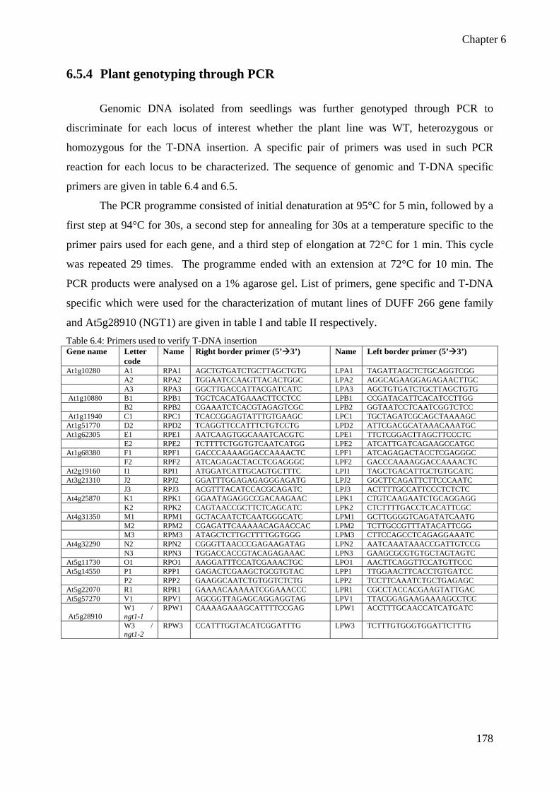
Chapter 6
178
6.5.4 Plant genotyping through PCR
Genomic DNA isolated from seedlings was further genotyped through PCR to
discriminate for each locus of interest whether the plant line was WT, heterozygous or
homozygous for the T-DNA insertion. A specific pair of primers was used in such PCR
reaction for each locus to be characterized. The sequence of genomic and T-DNA specific
primers are given in table 6.4 and 6.5.
The PCR programme consisted of initial denaturation at 95°C for 5 min, followed by a
first step at 94°C for 30s, a second step for annealing for 30s at a temperature specific to the
primer pairs used for each gene, and a third step of elongation at 72°C for 1 min. This cycle
was repeated 29 times. The programme ended with an extension at 72°C for 10 min. The
PCR products were analysed on a 1% agarose gel. List of primers, gene specific and T-DNA
specific which were used for the characterization of mutant lines of DUFF 266 gene family
and At5g28910 (NGT1) are given in table I and table II respectively. Table 6.4: Primers used to verify T-DNA insertion Gene name Letter
code Name Right border primer (5’ 3’) Name Left border primer (5’ 3’)
At1g10280 A1 RPA1 AGCTGTGATCTGCTTAGCTGTG LPA1 TAGATTAGCTCTGCAGGTCGG A2 RPA2 TGGAATCCAAGTTACACTGGC LPA2 AGGCAGAAGGAGAGAACTTGC A3 RPA3 GGCTTGACCATTACGATCATC LPA3 AGCTGTGATCTGCTTAGCTGTG At1g10880 B1 RPB1 TGCTCACATGAAACTTCCTCC LPB1 CCGATACATTCACATCCTTGG B2 RPB2 CGAAATCTCACGTAGAGTCGC LPB2 GGTAATCCTCAATCGGTCTCC At1g11940 C1 RPC1 TCACCGGAGTATTTGTGAAGC LPC1 TGCTAGATCGCAGCTAAAAGC At1g51770 D2 RPD2 TCAGGTTCCATTTCTGTCCTG LPD2 ATTCGACGCATAAACAAATGC At1g62305 E1 RPE1 AATCAAGTGGCAAATCACGTC LPE1 TTCTCGGACTTAGCTTCCCTC E2 RPE2 TCTTTTCTGGTGTCAATCATGG LPE2 ATCATTGATCAGAAGCCATGC At1g68380 F1 RPF1 GACCCAAAAGGACCAAAACTC LPF1 ATCAGAGACTACCTCGAGGGC F2 RPF2 ATCAGAGACTACCTCGAGGGC LPF2 GACCCAAAAGGACCAAAACTC At2g19160 I1 RPI1 ATGGATCATTGCAGTGCTTTC LPI1 TAGCTGACATTGCTGTGCATC At3g21310 J2 RPJ2 GGATTTGGAGAGAGGGAGATG LPJ2 GGCTTCAGATTCTTCCCAATC J3 RPJ3 ACGTTTACATCCACGCAGATC LPJ3 ACTTTTGCCATTCCCTCTCTC At4g25870 K1 RPK1 GGAATAGAGGCCGACAAGAAC LPK1 CTGTCAAGAATCTGCAGGAGG K2 RPK2 CAGTAACCGCTTCTCAGCATC LPK2 CTCTTTTGACCTCACATTCGC At4g31350 M1 RPM1 GCTACAATCTCAATGGGCATC LPM1 GCTTGGGGTCAGATATCAATG M2 RPM2 CGAGATTCAAAAACAGAACCAC LPM2 TCTTGCCGTTTATACATTCGG M3 RPM3 ATAGCTCTTGCTTTTGGTGGG LPM3 CTTCCAGCCTCAGAGGAAATC At4g32290 N2 RPN2 CGGGTTAACCCGAGAAGATAG LPN2 AATCAAATAAACCGATTGTCCG N3 RPN3 TGGACCACCGTACAGAGAAAC LPN3 GAAGCGCGTGTGCTAGTAGTC At5g11730 O1 RPO1 AAGGATTTCCATCGAAACTGC LPO1 AACTTCAGGTTCCATGTTCCC At5g14550 P1 RPP1 GAGACTCGAAGCTGCGTGTAC LPP1 TTGGAACTTCACCTGTGATCC P2 RPP2 GAAGGCAATCTGTGGTCTCTG LPP2 TCCTTCAAATCTGCTGAGAGC At5g22070 R1 RPR1 GAAAACAAAAATCGGAAACCC LPR1 CGCCTACCACGAAGTATTGAC At5g57270 V1 RPV1 AGCGGTTAGAGCAGGAGGTAG LPV1 TTACGGAGAAGAAAAGCCTCC
At5g28910 W1 / ngt1-1
RPW1 CAAAAGAAAGCATTTTCCGAG LPW1 ACCTTTGCAACCATCATGATC
W3 / ngt1-2
RPW3 CCATTTGGTACATCGGATTTG LPW3 TCTTTGTGGGTGGATTCTTTG

Chapter 6
179
Table 6.5 : T-DNA insertion primers Lines Name Left border primer of the T-DNA insertion (5’ 3’)
SALK LBb1 GCGTGGACCGCTTGCTGCAACT SALK LBb1.3 ATTTTGCCGATTTCGGAAC SALK LBa1 TGGTTCACGTAGTGGGCCATCG SAIL LB1 GCCTTTTCAGAAATGGATAAATAGCCTTGCTTCC SAIL LB2 GCTTCCTATTATATCTTCCCAAATTACCAATACA SAIL LB3 TAGCATCTGAATTTCATAACCAATCTCGATACAC
6.5.5 PCR product sequencing
PCR products were purified and send for sequencing to GATC for determining the exact
position of insertion in T-DNA mutants.
6.5.6 RT PCR analysis of ngt1-1 and ngt1-2
6.5.6.1 RNA preparation
Plants were grown in vitro for 2 weeks (500 mg fresh weight) for Wild type Col0, ngt1-1 and
ngt1-2 mutant lines were collected and frozen into liquid nitrogen which ensures inactivation
of ribonucleases. Then the entire plants (roots, stems and leaves) were ground into a fine
powder in liquid nitrogen with the help of pestle and mortar. All the necessary measures were
taken to avoid melting of frozen powder as it activates ribonucleases which in turn cause
degradation of RNAs. 2.5 ml of Plant RNA reagent extraction solution (from Fisher) was
added and mixture was vortexed until the sample is thoroughly resuspended. Tubes were
incubated for 5 min at room temperature. Centrifugation was carried out at 4°C for 15 min at
26,000xg. Aqueous phase was taken into a new microreaction tube and 400 µl of 5 M NaCl
and 1.2 ml of chloroform were added. Centrifugation was carried out at 4°C for 10 min at
12,000xg. Aqueous phase was transferred to an RNase-free tube and its volume was
measured. 0.9 volume of isopropyl alcohol was added, mixed well and kept at room
temperature for 10 min. Centrifugation was carried out at 4°C at 12,000xg for 10 min and
supernatant was removed. 5ml of 75% EtOH was added to the pellet and centrifuged at 4°C
for 5 min at 12,000xg. Supernatant was removed and pelleted RNAs were dissolved in
RNase-free water. RNAs were quantified by a spectrophotometer and was run on a 1%
agarose gel to check the quality of RNAs. RNA aliquots were stored at –20°C.

Chapter 6
180
• Treatment of RNAs with DNase
In order to efficiently remove DNA (as required for RT-PCR reaction) Promega kit was
used. Reaction was carried out in a final total volume of 10 µl containing 2 µg of RNA mixed
with (1-8 µl of water), 1 µl of RQ1 RNase-free DNase 10X reaction buffer and 1U of RQ1
RNase-free DNase. Reaction was incubated at 37°C for 30 min. 1 µl of RQ1 DNase stop
solution was added to terminate the reaction and incubated at 65°C to inactivate the DNase.
Sterile water was added to increase the volume to 100 μl and phenol extraction was done with
one volume of phenol followed by chloroform extraction. The aqueous phase was removed
into a new tube and precipitated with isopropanol overnight at -20°C followed by
centrifugation at 13000 rpm at 4°C for 25 min. The pellet was washed by 70% ethanol and
dried using vacuum dryer. RNAs were quantified by spectrophotometer and gel
electrophoresis.
• RT-PCR detection of a “T-DNA:NGT1” hybrid mRNA
After DNase treatment, cDNAs were synthesized using the iScript kit. The reaction
was carried out in a final volume of 30 µl containing 2 µg of RNAs (11 µl of above reaction),
8 µl of 5x iScript reaction mix, 2 µl of iScript reverse transcriptase and 9 µl of Nuclease-free
water. The reaction mix was incubated at 25°C for 5 min, at 42°C for 30 min and 85°C for 5
min. 3 μl of cDNA is utilized to carry out the PCR reaction. Five different set of primers,
namely “GS1 (RT1FUTs + RT2FUTs)”, “GS2 (ATG-FUTs + RPW3)”, “UBQ (UBQ10- For
+ UBQ10-Rev)”, “T-DNA ngt1-1 (LBa1 + RPW1)”, “T-DNA ngt1-2 (LBa1 + RPW3)” were
used. Ubiquitin specific primers were used as a control. Sequences of primers are given in
table 6.6. Table 6.6: sequence of primers used for RT-PCR
Set of primers
Name
Forward primer (5’ 3’) Name Reverse primer (5’ 3’)
GS1 RT1-FUTs
CAAGTTCCTTGTAGCTGATTGGG RT2-FUTs CAGGAAACGTCAGAAGAGTGTCG
GS2 ATG-FUTs
GGCTTGACCATTACGATCATC RPW3 CCATTTGGTACATCGGATTTG
UBQ UBQ10-For
GGCCTTGTATAATCCCTGATGAATAAG
UBQ10- Rev
AAAGAGATAACAGGAACGGAAACATAGT
T-DNA ngt1-1
LBa1 TGGTTCACGTAGTGGGCCATCG RPW1 CAAAAGAAAGCATTTTCCGAG
T-DNA ngt1-2
LBa1 TGGTTCACGTAGTGGGCCATCG RPW3 CCATTTGGTACATCGGATTTG

Chapter 6
181
The reaction was carried out in a final volume of 25 µl containing 3 µl of cDNA and
following final concentration of 1 X Go Taq buffer, 0.34 µM of dNTP, 0.4 µM of each
forward and reverse primer, 1 µM of MgSO4 and 0.03 units of Go Taq DNA polymerase and
water was added to a final volume of 25 µl. Denaturation of double stranded DNA was done
at 94°C for 2 min and it was followed by 29 cycles of amplification which included 30 sec at
94°C, 50 sec at Ta (Tm-5°C) and 1 min at 72°C. Samples were analyzed on a 1X TBE, 0.8 %
agarose gel.
6.5.7 ngt1-1 and ngt1-2 mutants complementation with 35S::NGT1
6.5.7.1 Cloning 35S::NGT1 construct for complementation
Entry vector containing NGT1 gene as made in section 6.2.2.1.2 was used in a LR
reaction using either pH2GW7 (to complement the mutant phenotype) or pH7WGF2 (with the
GFP construct for localization experiment) as followed: Entry clone (~15-150 ng) 1-7 µl,
destination vector pH2GW7 and pH7WGF2 (150 ng/µl), 1 µl TE buffer (pH 8.0) to final
volume of 8 µl were added to a 1.5 ml tube at RT and mixed. Then the LR Clonase II enzyme
mix was thawed on ice for about 2 min. The LR Clonase II enzyme mix was vortexed briefly
twice (2s each time). To each sample (Step 1, above), 2 µl of LR Clonase™ II enzyme mix
was added to the reaction and was mixed well by vortexing briefly twice following brief table-
top centrifugation. Reaction mixture was incubated at RT for 1h, then stopped by adding 1 µl
of the Proteinase K solution, vortexed briefly and incubated at 37°C for 10 min. Procedure for
plasmidic DNA extract is the same as decribed above in paragraph 6.2.1.1.3. Integration of
gene into destination vectors was confirmed by PCR product sequencing.
6.5.7.2 ngt1-1 and ngt1-2 mutants transformation with pH2GW7_NGT1 and
pH7GWF2_NGT1
• Preparation of Agro-bacterium competent cells
Agro-bacterium strain C58C1 was grown in 2 ml YEP medium containing rifampicin
(50 µg/ml) and gentamycin (50 µg/ml) overnight at 30°C. Next day it was transferred to 200
ml YEP flask and shaked at 250 rpm until the OD600 is 0.3 (4-5hrs). Cells were centrifuged
at 5000 rpm for 10 min, supernatant was removed and pellet was re-suspended in 20 ml ice
cold 1 mM Hepes buffer ( pH 7 ) and centrifuged again. This step was repeated twice and
pellet was re-suspended in 2 ml ice cold 10 % sterile glycerol and then immediately

Chapter 6
182
distributed in aliquots of 40 µl in pre-chilled sterile microreaction tubes, frozen in liquid
nitrogen and stored at -80°C.
• Agrobacterium electroporation
1µl of DNA from plasmid pH2GW7-NGT1 and pH7GWF2-NGT1 with a
concentration of 10-50 ng/µl was added into completely thawed agrobacterium competent
cells strain C58C1, kept on ice for 60 seconds and then quickly transferred in chilled 2 mm
cuvette. Cells were electroporated at a voltage of 1800 V, 500 µl of LB was added and then
the mixture was transferred in 1.5 ml microtubes and incubated with shaking at 30°C for 2h.
The bacteria were evenly spread on LB plates with appropriate antibiotics (spectinomycin,
rifampicin and gentamycin at 50 µg/ml). Plates were incubated at 30°C overnight or till the
colonies appear (one more day).
• Agrobacterium DNA miniprep
As plant transformation and transformant selection is time consuming, Agrobacterium
positive clones were checked for the presence of gene construct prior to plant transformation.
For that matter, a culture was started with a single colony in 5 ml LB with antibiotics (see
above), and incubated overnight at 30°C with shaking. 2 ml of cells were centrifuged,
supernatant was removed and 2 ml of cells were added again to the tubes and centrifuged
again. The composition of buffers used in this prep MPS1, MPS2 and MPS3 is given in the
table 6.7 below. The supernatant was removed and cells were re-suspended in 200 µl MPS1
solution, and tubes were incubated at room temperature for 5 min. 20 µl of 20 mg/ml
zymolase solution was added, briefly vortexed and incubated at 37°C for 15 min. 300 µl of
freshly prepared MPS2 solution. The mix was agitated gently by inverting the tubes 3-4 times
and tubes were incubated on ice for 5 min. Then 200 µl of MPS3 solution were added. Tubes
were vortexed at least for 10 seconds and incubated for 5 min on ice. Centrifuged for 5 min
and supernatant was removed to new tubes. 400 µl of phenol/chloroform/isoapropanol
(25:24:1) was added, vortexed, centrifugation was done for 5 min at 15000 rpm and
supernatant was removed to new tubes. This step was repeated again and then this step was
repeated with chloroform alone. 300 µl of isopropanol was added and incubated on ice for 10
min. centrifugation was done for 5 min and then pellet was washed with 70 % EtOH, dried
and re-dissolved in 40 µl water. 2 ml of DNA was used for PCR.

Chapter 6
183
Table 6.7: composition of buffers used for agrobacterium DNA miniprep
MPS1 for 5 ml
(Final concentration)
MPS2 for 10 ml MPS3 for 100 ml
Glucose
(1M)
2.5 ml (50 mM) NaOH
(10N)
200 µl (0.2
N)
Potassium
acetate 5M
60 ml
EDTA (0.5
M)
1 ml (10 mM) SDS 10 % 1 ml (1 %) Glacial acetic
acid
11.5 ml
Tris pH 8.0
(1M)
1.25 ml (25mM) H2O 8.8 ml H2O 28.5 ml
• Agrobacterium-mediated transformation of Arabidopsis thaliana using floral dip
method
Sterilized seeds of Col0, ngt1-1 and ngt1-2 mutants were sown on soil and kept in
darkness for 2 days to optimize seed germination. Plants were grown in a growth chamber
until the plants begin to bolt and produce floral inflorescence and siliques. Agrobacterium
strain transformed with pH2GW7_NGT1 or pH7GWF2-NGT1 were grown by inoculating a
single colony into 5 ml liquid LB medium containing the appropriate antibiotics (see above)
for binary vector selection and the culture was incubated at 28°C overnight. This pre-culture
was used to inoculate 500 ml liquid LB medium (with antibiotics) and cultured at 28°C for
16-24h (till 1.5<OD<2). Transformation of Agrobacterium with plasmids was confirmed by
PCR with a gene specific and a primer specific of plasmid antibiotic resistance gene. Cells
were collected by centrifugation at 4,000xg for 10 min at room temperature and cells were
gently re-suspended in 1 volume of freshly made 5 % (w/v) sucrose solution with a stirring
bar. Silwet-L77 detergent was added to a final concentration of 0.02 % (100 µl per 500 ml of
solution) and mixed well, immediately before dipping and then transferred to a 500 ml beaker.
Plants were covered with plastic film to avoid contamination from the soil. Plants were
inverted to dip the aerial parts of the plant including the inflorescence and the rosettes to soak
shorter secondary inflorescence in the Agrobacterium cell suspension for 10s with gentle
agitation. Dipped plants were removed from the solution and drained the treated plants for 3-5
sec to remove the excessive solution to avoid damage to the flower buds which can occur if
they are soaked in solution for too long. Plants were covered with plastic film and laid down
the treated plants on their sides for 16-24h to maintain high humidity. Next day the cover was

Chapter 6
184
removed and plants were put back to the growth chamber and grew then normally. Seeds were
harvested after siliques maturation.
6.5.7.3 Plant transformants selection
Surface sterilisation of the seeds was carried out in a 50 ml solution of 0.75 %
hypochlorite and 95 % ethanol. Seeds were dipped into the solution for five min and
continuously agitated by inverting the 50 ml Falcon tube. They were washed twice with
absolute Ethanol. They were kept in a hood for at least 10h or overnight for drying. The
sterilized seeds were cultivated in Petri dishes on 25 ml of a growth medium MS/2 containing
2.2 g MS salt (Sigma); 8 g of bacto agar per liter without sucrose and appropriate antibiotics
(cefotaxime 250 µg/ml and hygromycin 50 µg/ml). Stratification of the seeds was done at 4°C
for 48h in darkness in order to remove dormancy and to have a synchronised germination.
Then the seeds were transferred to a growth chamber with a cycle of 16h light and 8h
darkness for germination and growth. The light intensity in the growth chamber was 10,000
lumens/cm2 and temperature was 22°C. Petri dishes were wrapped with urgopore scotch to
avoid contamination but to ensure passage of air for respiration of growing seedlings. After 7-
10 days, transformants were readily distinguished as seedlings have healthy green cotyledons
and true leaves. Then potential transformants were transferred to water saturated soil and tray
was covered with plastic film to maintain high humidity. Tray was put in growth chamber and
plants were grown for seed collection.
6.6 Methods for cell wall analysis 6.6.1 Cell wall preparation
For the preparation of alcohol insoluble residues (AIR), cell wall was extracted from 5
days old dark grown seedlings and for sub-fractionation of polysaccharides cell wall was
prepared from 8 weeks old light grown plants to get sufficient quantity of material.
6.6.1.1 Alcohol insoluble residues preparation
Cell wall was prepared from 5 day-old dark-grown seedlings, culture on top of a nylon
mesh membrane. Seedlings were harvested with the spatula by sliding over nylon mesh.
Seedlings were washed with 15 ml of 70 % EtOH and placed at 70°C for 15min for
inactivation of enzymes and to wash intracellular contents (Fry, 1988). Centrifugation was
done for 10 min at 5,000xg to remove ethanol, organic phase and seedlings were transferred
into tissue homogeniser for grinding. They were ground in 5 ml of 70 % EtOH. Wash two

Chapter 6
185
times seedlings with 5 ml EtOH 70 %, 15 min at 70°C, followed by centrifugation as did
previously. Supernatant was removed and the pellet was dried at room temperature or at 40°C
with nitrogen evaporator.
Cell wall material was weighted and re-suspended in water (containing 0.02 % NaN3 to avoid
contamination) up to the required dilution (i.e. 50µg/ml).
6.6.1.2 Cell wall preparation for sub-fractionation
Leaves were harvested from 35 days old Arabidopsis thaliana plants from Col0 (WT) and
ngt1-1 and ngt1-2 mutants. Ten grams of plant material was ground by mortar and pestle
using liquid N2 for each cell wall preparation. Plant material was extracted once during 4h
with 50 mL of buffer (Hepes 20mM, pH7.5, SDS 0.5%, sodium metabisulfite 3 mM, one drop
of octanol), centrifuged to pellet insoluble material and pellet is then rinsed twice with 50 mL
ddH2O and once with 50 mL EtOH 70%. All supernatants (230 mL for each sample) were
pooled and stored at 4°C overnight to precipitate most SDS, and then centrifuged 30 min at
13,000xg at 4°C. As all materials did not totally pellet down, the suspension was further
filtrated on nylon cloth to obtain clear supernatants. Supernatants are then mixed with EtOH
96 % (1 / 4; v/v) in order to precipitate polymer contents and centrifuged 45 min at 12,000xg.
Pellets (ngt1-1 and ngt1-2) from each SDS extraction were resuspended in 30 mL phosphate
buffer containing α-amylase (phosphate buffer 100 mM, pH6, 0.02% NaN3, 2500 units α-
amylase) and let overnight at 30°C. Then 15mL of EtOH absolute was added to quench the
reaction and suspension was spun 8,000xg for 30min at 10°C. Pellets were resuspended and
washed twice EtOH 70% spun 20 min at 8’000g and at 10°C. All supernatants were
combined. Remaining pellets were washed once again with acetone, spun 20 min at 8,000xg
at 10°C and dried. After drying, pellet was ground using mortar and pestle and weight. Pellets
were considered cell wall material and ready for further extraction.Typical yields for Col0 and
mutants cell wall material were around 100-130 mg Col0 (112 mg), NGT1-1 (110 mg),
NGT1-2 (129 mg).
Cell wall from Col0, ngt1-1 and ngt1-2 was extracted with 15+5 mL ammonium oxalate
0.5% pH 4.5 (2h, 90°C) with a previous homogenization using glass potter, then centrifuged
9,000xg for 15 min at 15°C. Supernatants were saved and pellets were extracted once more
with 20 mL ammonium oxalate 0.5% pH 4.5 (1h, 90°C). Suspension was centrifuged
(swinging, 15 min, 15°C, 4,000xg). Supernatants were combined to the first one, transferred
to dialysis tube (MWCO 3500Da, snakeskin), dialysed 48h against ddH2O (5L) at 4°C with
water changes each 12h. Pellet was washed once with EtOH 70 % (15 mL), centrifuged and

Chapter 6
186
considered ready for hemicelluloses extraction. Cell wall remaining from each oxalate
fraction were extracted overnight with 20 mL KOH 1N containing 0.1% NaBH4 at RT,
centrifuged (swinging ) 10 min at 15°C 4,000xg, and pellet was extracted once again for 4h
with 20mL KOH 1N containing 0.1% NaBH4 at RT then centrifuged 10 min, 15°C 4,000xg.
Supernatants were pooled and neutralised (using concentrated CH3COOH 17N) previously to
dialysis. Sample volumes were measured before dialysis (Col0 KOH 1N extract 40mL, ngt1-1
KOH 1N extraction was 42mL and ngt1-2 KOH 1N extraction was 38mL). Supernatants
KOH 1N were combined to the first one, transferred to dialysis tube (MWCO 3500Da,
snakeskin), dialysed 48h against ddH2O (5L) at 4°C with water changes each 12h. After
dialysis sample was freeze-dried. Cell wall remaining from each KOH 1N fraction were
extracted overnight with 20 mL KOH 4N containing 0.1% NaBH4 at RT. Centrifuged 10 min
at 15°C 4,000xg. Pellets were extracted once again for 4h with 20 mL KOH 4N containing
0.1% NaBH4 at RT, then centrifuged 10 min, 15°C 4,000xg and supernatants were pooled and
neutralised previous to dialysis, using concentrated CH3COOH. Supernatants KOH 4N were
combined to the first one, transfer to dialysis tube (MWCO 3500 Da, snakeskin), dialysed 48h
against ddH2O (5L) at 4°C with water changes each 12h. After dialysis sample was freeze-
dried. Pellet was rinsed once with 10mL ddH2O, combined to KOH 4N supernatant, and once
with 20 mL acetone. Acetone is discarded after centrifugation 10min, 15°C 4,000g. Residual
pellet is dried and weighted. All fractions were analysed using alditol acetates procedure for
sugar content quantification.
6.6.2 Monosaccharide composition
6.6.2.1 Alditol acetates
This method is used to obtain a quantification of the neutral sugar composition from a
cell wall sample. In order to decompose sugar polymers to monomers, sample is hydrolyzed
with trifluoroacetic acid (TFA), then sample is reduced to open the ring structure of the sugar
monomer and in final step sample is acetylated. The derivative molecules are volatile and can
be separated on Gas Chromatography (GC)
• TFA hydrolysis
2.5 mg of cell wall material was placed and dried in a clean glass tube. 500 μl of TFA 2N
was added to each tube and tubes were placed at 121 ºC (250 ºF) for 2h and tubes were let 10
min on the bench to cool down. 25 µg of inositol was added in each tube, and tubes were
dried using nitrogen evaporator. 250 μl of NH4OH 2M were added to neutralize the residues

Chapter 6
187
and tube contents were dried (Heating Block could be setup around 50ºC). 250 μl of NH4OH
0.2M were added to neutralize the residues and tube content was dried. Now the samples were
ready for reduction step.
• Derivatization of sugar into corresponding alditol acetates
For reduction of samples, residues were re-suspended in 200 μl of NH4OH 2M (could be
vortexed or sonicate to help dissolution). 200 µL of NaBH4 50 mg/ml in NH4OH 2M were
added (this solution has to be made fresh). Tube was quickly vortexed for homogenisation and
placed for 90 min at 40 ºC. 10 μl of acetic acid was added (check for the freezing), to destroy
excess of reductive agent and tube content was dried under N2. If no feezing, the reduction
step was done once again. 200 μl of a MeOH/Acetic acid (9/1) were added and samples were
dried under N2. 500 μl of MeOH were added, residues re-suspended and dried again. This was
repeated two times again and samples were reduced and were ready for acetylation
300 μl of 1-Methyl-imidazole (catalyst) was added to residues and residues were re-
suspended in the catalyst (by strong vortexing). 2 ml of anhydride acetic was added and tubes
were vortexed quickly for homogenisation with catalyst and then let stand 10 min at room
temperature. 4 ml of ddH2O were added to destroy the excess of anhydride acetic acid. 1 ml of
dichloromethane was added and tubes were vortexed. Centrifugation was done for 1 min at
1000 rpm to separate the two phases. Organic phase was washed with 4 ml ddH2O, recovered
(at least 500 μl) into GC auto sampler vials, and injected on GC-MS.
6.6.2.2 Trimethylsilyl derivative (TMS)
• Methanolysis
Tubes containing 1 mg of dried cell wall (with 20 µg of inositol as an internal standard)
and dried standard mixture (100 µg of each sugar with 20 µg of inositol) were taken. 500 µl of
1M HCL in MeOH was added and tubes were heated at 80°C for about 16h. Tubes were
checked after 10 min to ensure the caps are tightly closed. Methanolic HCl was evaporated at
40°C with the stream of N2. 250 µl of MeOH was added and dried. This step is repeated once.
• TMS derivatization
200 µl of Silylation reagent was added, tubes were sealed or closed and heated at 80°C for
20 min. Samples were allowed to cool, and reagent was evaporated with the N2 until dried but
not too long. 1 ml of cyclohexane was added, vortexed and centrifuged. Cyclohexane was
transferred to GC vials and samples were analyzed by GC.

Chapter 6
188
6.6.3 Glycosyl linkage composition analysis
This method is used to get linkage information about the polysaccharides present in a
sample, by comparison of retention time with a standard library. In this method
polysaccharides or cell wall is treated for methylation then sample is hydrolyzed with TFA, in
order to decompose sugar polymers to monomers. Sample is reduced to open the ring
structure of sugar monomer. Sample is acetylated, and then the derivative molecules are
volatile and can be separated on gas chromatography (Figure 6.3).
• Methylation
1 mg of cell wall material was placed and dried in clean glass tube. Sample was re-
suspended in 300 µl of DMSO-NaOH (1g of NaOH ground in 4 ml DMSO), vortexed and
stirred at least 2h (RT) with a small magnetic stirrer in the tube. Tubes were placed on ice and
500 µL of CH3I were added. Tubes were put back to RT and stirred for 1h. 500 µl of CH3I
were added again, and the slurry was stirred for 1h. 2 ml of ddH2O were added and tubes were
stirred. 2 ml of dichloromethane were added; tubes were vortexed and centrifuged 2 min at
1,500 rpm to separate the phases. Aqueous phase was transferred to a glass tube and washed
with 1 ml of CH2Cl2; this organic wash was pooled to organic phase. Organic phase was
recovered, washed twice with 5 ml of ddH2O and tubes were vortexed. Centrifugation was
done again for 2 min at 1,500 rpm to separate the phases. Aqueous phase was discarded and
organic phase was dried under N2 (with heating 40°C). The partially methylated
polysaccharides were treated with TFA for hydrolysis, reduced with NaBD4 and acetylation
was performed as in section 2.5.2.1.

Chapter 6
189
Figure 6.3: Scheme explaining the different steps of per-methylation and per-acetylation of polysaccharides. In
red: O-methylation; in blue: O-acetylation and green colour indicates the free hydroxyl groups due to TFA
• Analysis by GC-MS
Gas chromatography-mass spectrometry (GC-MS) is a method that combines the features
of gas chromatography and mass spectrometry to identify different substances within a test
sample. Derivatized monosaccharide resulting from treatment of cell wall polysaccharides are
separated using a gas chromatograph (Agilent 6850) equipped with a capillary column SP-
2380 (length 30 m, internal diameter 0.25 mm, 0.20 µm phase thickness, Sigma-Aldrich) and
analyzed by mass spectrometry with Electron Ionization detector. Injector was heated at
240°C, solvent delay was set at 4 min and MS transfer line was set at 150°C. Oven program
for alditol acetates started at 160°C with a 2.5°C/min ramp till 220°C, then 5°C/min till
240°C. Oven program for partially methylated alditols acetates was a linear ramp from 140°C
to 220°C at 1.5°C min, then 5°C/min till 240°C.

Chapter 6
190
6.7 Phenotypic characterization of T-DNA lines 6.7.1 Leaf area measurement using image J
To measure the area of leaves in a precise and reproducible manner, a method has been
developed in the lab. The photo of each pot was taken, including the plant to be measured and
a reference (a mark of 2 cm on paper) at different stages of development. These photos were
then analyzed using the free software ImageJ ( http://rsbweb.nih.gov/ij/ ), as follows: a line is
drawn over the mark of 2 cm to calculate the pixel/cm. Ellipses are then drawn around two or
three leaves ( depending on the stage of development ) and leaf area of outermost leaves are
measured. With over pixel/cm, the leaf area in cm2 can be calculated. The data are then
analyzed using Excel software (Microsoft ©).
6.7.2 Sample preparation for microscopy
Tissue fixation was done in 2.5 % glutaraldehyde in 0.1 M sodium cacodylate buffer at
room temperature (pH 7.2) during 2h and then washed in 0.1 M sodium cacodylate buffer pH
7.2. Post fixation was done in 1 % osmium tetraoxide at 4°C in 0.1M sodium cacodylate
buffer during 1h. For embedding in LR white, dehydration of tissues was done with 25 %,
50%, 70% and 80% ethanol during 20 min for 2 times with each concentration of ethanol.
Afterwards for impregnation for embedding in LR White, tissues were dipped into a mixture
of 1vol of LR white and 1vol of 80 % alcohol for 20 min. They were dipped into a mixture of
2vol of LR white and 1vol of 80 % alcohol for 20 min. Dipping was done again into pure LR
White two times for 30 min. Incubation was done overnight into pure LR White at 4°C and
they were embedded in closed, labelled, gelatine capsules with fresh resin and let them
polymerised at 60oC for 72h. Several sections were cut at a thickness of 1 micron using
diamond knife and an ultramicrotome. The sections were deposited onto a glass slide and
dried on a hot plate at 60°C. The sections were then stained with 1 % toluidine blue in 1 %
borax solution for 1 min at 60°C. The slides were rinsed off with distilled water, dried and the
sections were covered with a glass coverslip using a Eukitt mounting medium.
• Immunolabeling of stem cross sections
For immunofluorescence staining technique, we used the semi-thin sections of sample
without post-fixation by osmium tetraoxide. The slides were incubated in PBS buffer
containing 5 % BSA for 1h to block the non-specific sites and to avoid background. Sections

Chapter 6
191
were incubated with specific primary antibody at 1/5 dilution in PBS-B (PBS with 1 % BSA),
at 4°C overnight. The sections were washed three times with PBS-T (PBS with 0.1 % Tween
20) for 5 min, then incubated with secondary antibody at 1/50 dilution in PBS-B for 1h at RT
in dark. Sections were again washed three times with PBS-T and finally washed twice with
ddH2O for 5 min. Sections were allow to dry and covered with a glass coverslip using
glycerol-phosphate buffered saline based mounting solution (CFM-1). After immunolabelling
of stem cross sections, they were observed with epifluorescence microscope at 5030 ms
acquisition time.

Chapter 6
192

Bibliography
193
BIBLIOGRAPHY

Bibliography
194
7 BIBLIOGRAPHY
Alfaro, J. A., R. B. Zheng, M. Persson, J. A. Letts, R. Polakowski, Y. Bai, S. N. Borisova, N. O. L. Seto, T. L. Lowary, M. M. Palcic, and S. V. Evans. 2008. ABO(H) blood group A and B glycosyltransferases recognize substrate via specific conformational changes. Journal of Biological Chemistry 283: 10097-10108.
Anterola, A. M., and N. G. Lewis. 2002. Trends in lignin modification: a comprehensive analysis of the effects of genetic manipulations/mutations on lignification and vascular integrity. Phytochemistry 61: 221-294.
Appenzeller, L., M. Doblin, R. Barreiro, H. Y. Wang, X. M. Niu, K. Kollipara, L. Carrigan, D. Tomes, M. Chapman, and K. S. Dhugga. 2004. Cellulose synthesis in maize: isolation and expression analysis of the cellulose synthase (CesA) gene family. Cellulose 11: 287-299.
Arioli, T., L. Peng, A. S. Betzner, J. Burn, W. Wittke, W. Herth, C. Camilleri, H. Hofte, J. Plazinski, R. Birch, A. Cork, J. Glover, J. Redmond, and R. E. Williamson. 1998a. Molecular analysis of cellulose biosynthesis in Arabidopsis. Science 279: 717-20.
Arioli, T., L. C. Peng, A. S. Betzner, J. Burn, W. Wittke, W. Herth, C. Camilleri, H. Hofte, J. Plazinski, R. Birch, A. Cork, J. Glover, J. Redmond, and R. E. Williamson. 1998b. Molecular analysis of cellulose biosynthesis in Arabidopsis. Science 279: 717-720.
Awano, T., K. Takabe, and M. Fujita. 2002. Xylan deposition on secondary wall of Fagus crenata fiber. Protoplasma 219: 106-115.
Bauer, S., P. Vasu, S. Persson, A. J. Mort, and C. R. Somerville. 2006. Development and application of a suite of polysaccharide-degrading enzymes for analyzing plant cell walls. Proceedings of the National Academy of Sciences of the United States of America 103: 11417-11422.
Baydoun, E. A. H., K. W. Waldron, and C. T. Brett. 1989. The interaction of xylosyltransferase and glucuronyltransferase involved in glucuronoxylan synthesis in pea (pisum-sativum) epicotyls. Biochemical Journal 257: 853-858.
Beeckman, T., G. K. H. Przemeck, G. Stamatiou, R. Lau, N. Terryn, R. De Rycke, D. Inze, and T. Berleth. 2002. Genetic complexity of cellulose synthase A gene function in Arabidopsis embryogenesis. Plant Physiology 130: 1883-1893.
Benfey, P. N., P. J. Linstead, K. Roberts, J. W. Schiefelbein, M. T. Hauser, and R. A. Aeschbacher. 1993. Root development in arabidopsis - 4 mutants with dramatically altered root morphogenesis. Development 119: 57-70.
Bernal, A. J., C. M. Yoo, M. Mutwil, J. K. Jensen, G. Hou, C. Blaukopf, I. Sorensen, E. B. Blancaflor, H. V. Scheller, and W. G. T. Willats. 2008. Functional Analysis of the Cellulose Synthase-Like Genes CSLD1, CSLD2, and CSLD4 in Tip-Growing Arabidopsis Cells. Plant Physiology 148: 1238-1253.
Boerjan, W., J. Ralph, and M. Baucher. 2003. Lignin biosynthesis. Annual Review of Plant Biology 54: 519-546.
Bonin, C. P., I. Potter, G. F. Vanzin, and W. D. Reiter. 1997. The MUR1 gene of Arabidopsis thaliana encodes an isoform of GDP-D-mannose-4,6-dehydratase, catalyzing the first step in the de novo synthesis of GDP-L-fucose. Proceedings of the National Academy of Sciences of the United States of America 94: 2085-2090.
Bosca, S., C. J. Barton, N. G. Taylor, P. Ryden, L. Neumetzler, M. Pauly, K. Roberts, and G. J. Seifert. 2006. Interactions between MUR10/CesA7-dependent secondary cellulose biosynthesis and primary cell wall structure. Plant Physiology 142: 1353-1363.

Bibliography
195
Bouton, S., E. Leboeuf, G. Mouille, M. T. Leydecker, J. Talbotec, F. Granier, M. Lahaye, H. Hofte, and H. N. Truong. 2002. Quasimodo1 encodes a putative membrane-bound glycosyltransferase required for normal pectin synthesis and cell adhesion in Arabidopsis. Plant Cell 14: 2577-2590.
Brady, J. D., I. H. Sadler, and S. C. Fry. 1996. Di-isodityrosine, a novel tetrameric derivative of tyrosine in plant cell wall proteins: A new potential cross-link. Biochemical Journal 315: 323-327.
Breton, C., and A. Imberty. 1999. Structure/function studies of glycosyltransferases. Current Opinion in Structural Biology 9: 563-571.
Breton, C., J. Mucha, and C. Jeanneau. 2001. Structural and functional features of glycosyltransferases. Biochimie 83: 713-718.
Breton, C., E. Bettler, D. H. Joziasse, R. A. Geremia, and A. Imberty. 1998. Sequence-function relationships of prokaryotic and eukaryotic galactosyltransferases. Journal of Biochemistry 123: 1000-1009.
Breton, C., L. Snajdrova, C. Jeanneau, J. Koca, and A. Imberty. 2006. Structures and mechanisms of glycosyltransferases. Glycobiology 16: 29R-37R.
Brett, C. T. 2000. Cellulose microfibrils in plants: Biosynthesis, deposition, and integration into the cell wall, pp. 161-199. In K. W. Jeon [ed.], International Review of Cytology - a Survey of Cell Biology, Vol 199.
Brown, D., Raymond Wightman1, Zhinong Zhang2, Leonardo D. Gomez3, Ivan Atanassov1,§, John-Paul Bukowski1,–,, and S. J. M.-M. Theodora Tryfona2, Paul Dupree2 and Simon Turner. 2011. Arabidopsis genes IRREGULAR XYLEM (IRX15) and IRX15L encode DUF579-containing proteins that are essential for
normal xylan deposition in the secondary cell wall. The Plant Journal. Brown, D. M., L. A. H. Zeef, J. Ellis, R. Goodacre, and S. R. Turner. 2005. Identification of
novel genes in Arabidopsis involved in secondary cell wall formation using expression profiling and reverse genetics. Plant Cell 17: 2281-2295.
Brown, D. M., Z. N. Zhang, E. Stephens, P. Dupree, and S. R. Turner. 2009. Characterization of IRX10 and IRX10-like reveals an essential role in glucuronoxylan biosynthesis in Arabidopsis. Plant Journal 57: 732-746.
Brown, D. M., F. Goubet, W. W. A. Vicky, R. Goodacre, E. Stephens, P. Dupree, and S. R. Turner. 2007. Comparison of five xylan synthesis mutants reveals new insight into the mechanisms of xylan synthesis. Plant Journal 52: 1154-1168.
Brown, R. J., J. H. Willison, and C. L. Richardson. 1976 Cellulose biosynthesis in Acetobacter xylinum: visualization of the site of synthesis and direct measurement of the in vivo process. Proc Natl Acad Sci U S A 73: 4565-9.
Brown, R. M. 1996. The biosynthesis of cellulose. Journal of Macromolecular Science-Pure and Applied Chemistry A33: 1345-1373.
Brown, R. M. 2004. Cellulose structure and biosynthesis: What is in store for the 21st century? Journal of Polymer Science Part a-Polymer Chemistry 42: 487-495.
Bruce, A. S., and A. E. Clarke. 1992. Chemistry and Biology of (1-3)-β-D-Glucans. Victoria, Australia: La Trobe University Press.
Buckeridge, M. S., H. P. dos Santos, and M. A. S. Tine. 2000. Mobilisation of storage cell wall polysaccharides in seeds. Plant Physiology and Biochemistry 38: 141-156.
Burget, E. G., and W. D. Reiter. 1999. The mur4 mutant of arabidopsis is partially defective in the de novo synthesis of uridine diphospho L-arabinose. Plant Physiology 121: 383-389.
Burk, D. H., B. Liu, R. Q. Zhong, W. H. Morrison, and Z. H. Ye. 2001. A katanin-like protein regulates normal cell wall biosynthesis and cell elongation. Plant Cell 13: 807-827.

Bibliography
196
Burn, J. E., C. H. Hocart, R. J. Birch, A. C. Cork, and R. E. Williamson. 2002a. Functional analysis of the cellulose synthase genes CesA1, CesA2, and CesA3 in Arabidopsis. Plant Physiology 129: 797-807.
Burn, J. E., U. A. Hurley, R. J. Birch, T. Arioli, A. Cork, and R. E. Williamson. 2002b. The cellulose-deficient Arabidopsis mutant rsw3 is defective in a gene encoding a putative glucosidase II, an enzyme processing N-glycans during ER quality control. Plant Journal 32: 949-960.
Burton, R. A., N. J. Shirley, B. J. King, A. J. Harvey, and G. B. Fincher. 2004. The CesA gene family of barley. Quantitative analysis of transcripts reveals two groups of co-expressed genes. Plant Physiology 134: 224-236.
Burton, R. A., S. M. Wilson, M. Hrmova, A. J. Harvey, N. J. Shirley, B. A. Stone, E. J. Newbigin, A. Bacic, and G. B. Fincher. 2006. Cellulose synthase-like CslF genes mediate the synthesis of cell wall (1,3;1,4)-beta-D-glucans. Science 311: 1940-1942.
Caffall, K. H., and D. Mohnen. 2009. The structure, function, and biosynthesis of plant cell wall pectic polysaccharides. Carbohydrate Research 344: 1879-1900.
Carpita, N. C., and D. M. Gibeaut. 1993. Structural models of primary-cell walls in flowering plants - consistency of molecular-structure with the physical-properties of the walls during growth. Plant Journal 3: 1-30.
Carpita, N. C., and M. McCann. 2000. The cell wall. In Biochemistry and Molecular Biology of Plants.
Cavalier, D. M., and K. Keegstra. 2006. Two xyloglucan xylosyltransferases catalyze the addition of multiple xylosyl residues to cellohexaose. Journal of Biological Chemistry 281: 34197-34207.
Cavalier, D. M., O. Lerouxel, L. Neumetzler, K. Yamauchi, A. Reinecke, G. Freshour, O. A. Zabotina, M. G. Hahn, I. Burgert, M. Pauly, N. V. Raikhel, and K. Keegstra. 2008. Disrupting two Arabidopsis thaliana xylosyltransferase genes results in plants deficient in xyloglucan, a major primary cell wall component. Plant Cell 20: 1519-1537.
Chan, J., E. Crowell, M. Eder, G. Calder, S. Bunnewell, K. Findlay, S. Vernhettes, H. Hofte, and C. Lloyd. 2010. The rotation of cellulose synthase trajectories is microtubule dependent and influences the texture of epidermal cell walls in Arabidopsis hypocotyls. Journal of Cell Science 123: 3490-3495.
Chanzy, H. 1978. Electron diffraction from primary wall of cotton fibres. Chanzy, H., K. Imada, A. Mollard, R. Vuong, and F. Barnoud. 1979.
CRYSTALLOGRAPHIC ASPECTS OF SUB-ELEMENTARY CELLULOSE FIBRILS OCCURRING IN THE WALL OF ROSE CELLS CULTURED INVITRO. Protoplasma 100: 303-316.
Charnock, S. J., and G. J. Davies. 1999. Structure of the nucleotide-diphospho-sugar transferase, SpsA from Bacillus subtilis, in native and nucleotide-complexed forms. Biochemistry 38: 6380-6385.
Chen, X. Y., and J. Y. Kim. 2009. Callose synthesis in higher plants. Plant Signal Behav 4: 489-92.
Chiu, C. P. C., L. L. Lairson, M. Gilbert, W. W. Wakarchuk, S. G. Withers, and N. C. J. Strynadka. 2007. Structural analysis of the alpha-2,3-sialyltransferase cst-i from Campylobacter jejuni in apo and substrate-analogue bound forms. Biochemistry 46: 7196-7204.
Chiu, C. P. C., A. G. Watts, L. L. Lairson, M. Gilbert, D. Lim, W. W. Wakarchuk, S. G. Withers, and N. C. J. Strynadka. 2004. Structural analysis of the sialyltransferase CstII from Campylobacter jejuni in complex with a substrate analog. Nature Structural & Molecular Biology 11: 163-170.

Bibliography
197
Cocuron, J. C., O. Lerouxel, G. Drakakaki, A. P. Alonso, A. H. Liepman, K. Keegstra, N. Raikhel, and C. G. Wilkerson. 2007. A gene from the cellulose synthase-like C family encodes a beta-1,4 glucan synthase. Proceedings of the National Academy of Sciences of the United States of America 104: 8550-8555.
Cosgrove, D. J. 2000. Loosening of plant cell walls by expansins. Nature 407: 321-326. Delmer, D. P. 1999. Cellulose biosynthesis: Exciting times for a difficult field of study.
Annual Review of Plant Physiology and Plant Molecular Biology 50: 245-276. Delmer, D. P., and B. A. Stone. 1988. Biosynthesis of plant cell walls. In The Biochemistry of
Plants 14: 373-420. Desprez, T., S. Vernhettes, M. Fagard, G. Refregier, T. Desnos, E. Aletti, N. Py, S. Pelletier,
and H. Hofte. 2002. Resistance against herbicide isoxaben and cellulose deficiency caused by distinct mutations in same cellulose synthase isoform CESA6. Plant Physiology 128: 482-490.
Desprez, T., M. Juraniec, E. F. Crowell, H. Jouy, Z. Pochylova, F. Parcy, H. Hofte, M. Gonneau, and S. Vernhettes. 2007. Organization of cellulose synthase complexes involved in primary cell wall synthesis in Arabidopsis thaliana. Proceedings of the National Academy of Sciences of the United States of America 104: 15572-15577.
Dhugga, K. S., R. Barreiro, B. Whitten, K. Stecca, J. Hazebroek, G. S. Randhawa, M. Dolan, A. J. Kinney, D. Tomes, S. Nichols, and P. Anderson. 2004. Guar seed beta-mannan synthase is a member of the cellulose synthase super gene family. Science 303: 363-366.
Doblin, M. S., I. Kurek, D. Jacob-Wilk, and D. P. Delmer. 2002. Cellulose biosynthesis in plants: from genes to rosettes. Plant and Cell Physiology 43: 1407-1420.
Doblin, M. S., F. A. Pettolino, S. M. Wilson, R. Campbell, R. A. Burton, G. B. Fincher, E. Newbigin, and A. Bacic. 2009. A barley cellulose synthase-like CSLH gene mediates (1,3;1,4)-beta-D-glucan synthesis in transgenic Arabidopsis. Proceedings of the National Academy of Sciences of the United States of America 106: 5996-6001.
Dong, X. Y., Z. L. Hong, M. Sivaramakrishnan, M. Mahfouz, and D. P. S. Verma. 2005. Callose synthase (CalS5) is required for exine formation during microgametogenesis and for pollen viability in Arabidopsis. Plant Journal 42: 315-328.
Ebringerova, A., and T. Heinze. 2000. Xylan and xylan derivatives - biopolymers with valuable properties, 1 - Naturally occurring xylans structures, procedures and properties. Macromolecular Rapid Communications 21: 542-556.
Edwards, M. E., C. A. Dickson, S. Chengappa, C. Sidebottom, M. J. Gidley, and J. S. G. Reid. 1999. Molecular characterisation of a membrane-bound galactosyltransferase of plant cell wall matrix polysaccharide biosynthesis. Plant Journal 19: 691-697.
Egelund, J., M. Skjot, N. Geshi, P. Ulvskov, and B. L. Petersen. 2004. A complementary bioinformatics approach to identify potential plant cell wall glycosyltransferase-encoding genes. Plant Physiology 136: 2609-2620.
Egelund, J., I. Damager, K. Faber, C. E. Olsen, P. Ulvskov, and B. L. Petersen. 2008. Functional characterisation of a putative rhamnogalacturonan II specific xylosyltransferase. Febs Letters 582: 3217-3222.
Egelund, J., B. L. Petersen, M. S. Motawia, I. Damager, A. Faik, C. E. Olsen, T. Ishii, H. Clausen, P. Ulvskov, and N. Geshi. 2006. Arabidopsis thaliana RGXT1 and RGXT2 encode Golgi-localized (1,3)-alpha-D-xylosyltransferases involved in the synthesis of pectic rhamnogalacturonan-II. Plant Cell 18: 2593-2607.
Emons, A. M. C., H. Hofte, and B. M. Mulder. 2007. Microtubules and cellulose microfibrils: how intimate is their relationship? Trends in Plant Science 12: 279-281.

Bibliography
198
Enns, L. C., M. M. Kanaoka, K. U. Torii, L. Comai, K. Okada, and R. E. Cleland. 2005. Two callose synthases, GSL1 and GSL5, play an essential and redundant role in plant and pollen development and in fertility. Plant Molecular Biology 58: 333-349.
Errey, J. C., S. S. Lee, R. P. Gibson, C. M. Fleites, C. S. Barry, P. M. J. Jung, A. C. O'Sullivan, B. G. Davis, and G. J. Davies. 2010. Mechanistic Insight into Enzymatic Glycosyl Transfer with Retention of Configuration through Analysis of Glycomimetic Inhibitors. Angewandte Chemie-International Edition 49: 1234-1237.
Fagard, M., T. Desnos, T. Desprez, F. Goubet, G. Refregier, G. Mouille, M. McCann, C. Rayon, S. Vernhettes, and H. Hofte. 2000. PROCUSTE1 encodes a cellulose synthase required for normal cell elongation specifically in roots and dark-grown hypocotyls of arabidopsis. Plant Cell 12: 2409-2423.
Faik, A., N. J. Price, N. V. Raikhel, and K. Keegstra. 2002. An Arabidopsis gene encoding an alpha-xylosyltransferase involved in xyloglucan biosynthesis. Proceedings of the National Academy of Sciences of the United States of America 99: 7797-7802.
Faik, A., M. Bar-Peled, A. E. DeRocher, W. Q. Zeng, R. M. Perrin, C. Wilkerson, N. V. Raikhel, and K. Keegstra. 2000. Biochemical characterization and molecular cloning of an alpha-1,2-fucosyltransferase that catalyzes the last step of cell wall xyloglucan biosynthesis in pea. Journal of Biological Chemistry 275: 15082-15089.
Fangel, J. U., B. L. Petersen, N. B. Jensen, W. G. T. Willats, A. Bacic, and J. Egelund. 2011. A putative Arabidopsis thaliana glycosyltransferase, At4g01220, which is closely related to three plant cell wall-specific xylosyltransferases, is differentially expressed spatially and temporally. Plant Science 180: 470-479.
Farrokhi, N., R. A. Burton, L. Brownfield, M. Hrmova, S. M. Wilson, A. Bacic, and G. B. Fincher. 2006. Plant cell wall biosynthesis: genetic, biochemical and functional genomics approaches to the identification of key genes. Plant Biotechnology Journal 4: 145-167.
Fincher, G. B., and B. A. Stone. 1981. Metabolism of noncellulosic polysaccharides. In ‘Encyclopedia of plant physiology.NewSeries, plant carbohydrates II’.
(Eds W Tanner, FA Loewus) 68-132. Fry, and Stephen. 1988. The growing plant cell wall: Chemical and metabolic analysis. Essex,
UK: Longman Scientific and Technical. Fry, S. C. 1989a. The structure and functions of xyloglucan. Journal of Experimental Botany
40: 1–11. Fry, S. C. 1989b. The structure and functions of xyloglucan. Journal of Experimental Botany
40: 1-11. Fry, S. C., W. S. York, P. Albersheim, A. Darvill, T. Hayashi, J. P. Joseleau, Y. Kato, E. P.
Lorences, G. A. Maclachlan, M. McNeil, A. J. Mort, J. S. G. Reid, H. U. Seitz, R. R. Selvendran, A. G. J. Voragen, and A. R. White. 1993. An unambiguous nomenclature for xyloglucan-derived oligosaccharides. Physiologia Plantarum 89: 1-3.
Gaboriaud, C., V. Bissery, T. Benchetrit, and J. P. Mornon. 1987. HYDROPHOBIC CLUSTER-ANALYSIS - AN EFFICIENT NEW WAY TO COMPARE AND ANALYZE AMINO-ACID-SEQUENCES. Febs Letters 224: 149-155.
Gardiner, J. C., N. G. Taylor, and S. R. Turner. 2003. Control of cellulose synthase complex localization in developing xylem. Plant Cell 15: 1740-1748.
Gaspar, Y., K. L. Johnson, J. A. McKenna, A. Bacic, and C. J. Schultz. 2001. The complex structures of arabinogalactan-proteins and the journey towards understanding function. Plant Molecular Biology 47: 161-176.
Gibeaut, D. M., and N. C. Carpita. 1994. Biosynthesis of plant-cell wall polysaccharides. Faseb Journal 8: 904-915.

Bibliography
199
Gibeaut, D. M., M. Pauly, A. Bacic, and G. B. Fincher. 2005. Changes in cell wall polysaccharides in developing barley (Hordeum vulgare) coleoptiles. Planta 221: 729-738.
Giddings, T. H., D. L. Brower, and L. A. Staehelin. 1980. VISUALIZATION OF PARTICLE COMPLEXES IN THE PLASMA-MEMBRANE OF MICRASTERIAS-DENTICULATA ASSOCIATED WITH THE FORMATION OF CELLULOSE FIBRILS IN PRIMARY AND SECONDARY CELL-WALLS. Journal of Cell Biology 84: 327-339.
Gille, S., U. Hansel, M. Ziemann, and M. Pauly. 2009. Identification of plant cell wall mutants by means of a forward chemical genetic approach using hydrolases. Proceedings of the National Academy of Sciences of the United States of America 106: 14699-14704.
Gillmor, C. S., P. Poindexter, J. Lorieau, M. M. Palcic, and C. Somerville. 2002. alpha-glucosidase I is required for cellulose biosynthesis and morphogenesis in Arabidopsis. Journal of Cell Biology 156: 1003-1013.
Goubet, F., A. Misrahi, S. K. Park, Z. N. Zhang, D. Twell, and P. Dupree. 2003. AtCSLA7, a cellulose synthase-like putative glycosyltransferase, is important for pollen tube growth and embryogenesis in Arabidopsis. Plant Physiology 131: 547-557.
Goubet, F., C. J. Barton, J. C. Mortimer, X. L. Yu, Z. N. Zhang, G. P. Miles, J. Richens, A. H. Liepman, K. Seffen, and P. Dupree. 2009. Cell wall glucomannan in Arabidopsis is synthesised by CSLA glycosyltransferases, and influences the progression of embryogenesis. Plant Journal 60: 527-538.
Gu, Y., N. Kaplinsky, M. Bringmann, A. Cobb, A. Carroll, A. Sampathkumar, T. I. Baskin, S. Persson, and C. R. Somerville. Identification of a cellulose synthase-associated protein required for cellulose biosynthesis. Proceedings of the National Academy of Sciences of the United States of America 107: 12866-12871.
Gu, Y., N. Kaplinsky, M. Bringmann, A. Cobb, A. Carroll, A. Sampathkumar, T. I. Baskin, S. Persson, and C. R. Somerville. 2010. Identification of a cellulose synthase-associated protein required for cellulose biosynthesis. Proceedings of the National Academy of Sciences of the United States of America 107: 12866-12871.
Guerriero, G., J. Fugelstad, and V. Bulone. 2010. What Do We Really Know about Cellulose Biosynthesis in Higher Plants? Journal of Integrative Plant Biology 52: 161-175.
Ha, M. A., D. C. Apperley, B. W. Evans, M. Huxham, W. G. Jardine, R. J. Vietor, D. Reis, B. Vian, and M. C. Jarvis. 1998. Fine structure in cellulose microfibrils: NMR evidence from onion and quince. Plant Journal 16: 183-190.
Handford, M. G., T. C. Baldwin, F. Goubet, T. A. Prime, J. Miles, X. L. Yu, and P. Dupree. 2003. Localisation and characterisation of cell wall mannan polysaccharides in Arabidopsis thaliana. Planta 218: 27-36.
Hansen, S. F., E. Bettler, M. Wimmerova, A. Imberty, O. Lerouxel, and C. Breton. 2009. Combination of Several Bioinformatics Approaches for the Identification of New Putative Glycosyltransferases in Arabidopsis. Journal of Proteome Research 8: 743-753.
Hanus, J., and K. Mazeau. 2006. The xyloglucan-cellulose assembly at the atomic scale. Biopolymers 82: 59-73.
Harholt, J., A. Suttangkakul, and H. V. Scheller. 2010a. Plant Physiology 153: 384-395. Harholt, J., A. Suttangkakul, and H. V. Scheller. 2010b. Biosynthesis of Pectin. Plant
Physiology 153: 384-395. Harholt, J., J. K. Jensen, S. O. Sorensen, C. Orfila, M. Pauly, and H. V. Scheller. 2006.
ARABINAN DEFICIENT 1 is a putative arabinosyltransferase involved in biosynthesis of Pectic Arabinan in Arabidopsis. Plant Physiology 140: 49-58.

Bibliography
200
Hart, D. A., and P. K. Kindel. 1970. Isolation and partial characterization of apiogalacturonans from cell wall of lemna-minor. Biochemical Journal 116: 569-&.
Hauser, M. T., A. Morikami, and P. N. Benfey. 1995. Conditional root expansion mutants of arabidopsis. Development 121: 1237-1252.
Hayashi, T. 1989. Xyloglucans in the primary cell wall. Annu Rev Plant Physiol Plant Mol Biol 40: 139-168.
Heredia, A., A. Jimenez, and R. Guillen. 1995. Composition of plant-cell walls. Zeitschrift Fur Lebensmittel-Untersuchung Und-Forschung 200: 24-31.
Herth, W. 1983. ARRAYS OF PLASMA-MEMBRANE ROSETTES INVOLVED IN CELLULOSE MICROFIBRIL FORMATION OF SPIROGYRA. Planta 159: 347-356.
Holland, N., D. Holland, T. Helentjaris, K. S. Dhugga, B. Xoconostle-Cazares, and D. P. Delmer. 2000. A comparative analysis of the plant cellulose synthase (CesA) gene family. Plant Physiology 123: 1313-1323.
Igura, M., N. Maita, J. Kamishikiryo, M. Yamada, T. Obita, K. Maenaka, and D. Kohda. 2008. Structure-guided identification of a new catalytic motif of oligosaccharyltransferase. Embo Journal 27: 234-243.
Ihara, H., Y. Ikeda, S. Toma, X. C. Wang, T. Suzuki, J. G. Gu, E. Miyoshi, T. Tsukihara, K. Honke, A. Matsumoto, A. Nakagawa, and N. Taniguchi. 2007. Crystal structure of mammalian alpha 1,6-fucosyltransferase, FUT8. Glycobiology 17: 455-466.
Ishii, T. 1995. PECTIC POLYSACCHARIDES FROM BAMBOO SHOOT CELL-WALLS. Mokuzai Gakkaishi 41: 669-676.
Ishii, T. 1997. O-acetylated oligosaccharides from pectins of potato tuber cell walls. Plant Physiology 113: 1265-1272.
Ishii, T., and T. Matsunaga. 1996. Isolation and characterization of a boron-rhamnogalacturonan-II complex from cell walls of sugar beet pulp. Carbohydrate Research 284: 1-9.
Iwai, H., N. Masaoka, T. Ishii, and S. Satoh. 2002. A pectin glucuronyltransferase gene is essential for intercellular attachment in the plant meristem. Proceedings of the National Academy of Sciences of the United States of America 99: 16319-16324.
Jackson, C. L., T. M. Dreaden, L. K. Theobald, N. M. Tran, T. L. Beal, M. Eid, M. Y. Gao, R. B. Shirley, M. T. Stoffel, M. V. Kumar, and D. Mohnen. 2007. Pectin induces apoptosis in human prostate cancer cells: correlation of apoptotic function with pectin structure. Glycobiology 17: 805-819.
Jensen, J. K., H. Kim, J. C. Cocuron, R. Orler, J. Ralph, and C. G. Wilkerson. The DUF579 domain containing proteins IRX15 and IRX15-L affect xylan synthesis in Arabidopsis. Plant Journal 66: 387-400.
Jensen, J. K., S. O. Sorensen, J. Harholt, N. Geshi, Y. Sakuragi, I. Moller, J. Zandleven, A. J. Bernal, N. B. Jensen, C. Sorensen, M. Pauly, G. Beldman, W. G. T. Willats, and H. V. Scheller. 2008. Identification of a xylogalacturonan xylosyltransferase involved in pectin biosynthesis in Arabidopsis. Plant Cell 20: 1289-1302.
Jones, L., G. B. Seymour, and J. P. Knox. 1997. Localization of pectic galactan in tomato cell walls using a monoclonal antibody specific to (1->4)-beta-D-galactan. Plant Physiology 113: 1405-1412.
Joshi, C. P., S. Bhandari, P. Ranjan, U. C. Kalluri, X. Liang, T. Fujino, and A. Samuga. 2004. Genomics of cellulose biosynthesis in poplars. New Phytologist 164: 53-61.
Keegstra, K. 2010. Plant Cell Walls. Plant Physiology 154: 483-486. Keegstra, K., and N. Raikhel. 2001. Plant glycosyltransferases. Current Opinion in Plant
Biology 4: 219-224.

Bibliography
201
Kennedy , C. J., G. J. Cameron , A. Šturcová , D. C. Apperley , C. Altaner , and T. J. Wess 2007. Microfi bril diameter in celery collenchyma
cellulose: X-ray scattering and NMR evidence. Cellulose 14: 235 – 246. Kimura, S., W. Laosinchai, T. Itoh, X. J. Cui, C. R. Linder, and R. M. Brown. 1999.
Immunogold labeling of rosette terminal cellulose-synthesizing complexes in the vascular plant Vigna angularis. Plant Cell 11: 2075-2085.
Knox, J. P., P. J. Linstead, J. Peart, C. Cooper, and K. Roberts. 1991. DEVELOPMENTALLY REGULATED EPITOPES OF CELL-SURFACE ARABINOGALACTAN PROTEINS AND THEIR RELATION TO ROOT-TISSUE PATTERN-FORMATION. Plant Journal 1: 317-326.
Konishi, T., Y. Miyazaki, S. Yamakawa, H. Iwai, S. Satoh, and T. Ishii. 2010. Purification and Biochemical Characterization of Recombinant Rice UDP-Arabinopyranose Mutase Generated in Insect Cells. Bioscience Biotechnology and Biochemistry 74: 191-194.
Konishi, T., T. Takeda, Y. Miyazaki, M. Ohnishi-Kameyama, T. Hayashi, M. A. O'Neill, and T. Ishii. 2007. A plant mutase that interconverts UDP-arabinofuranose and UDP-arabinopyranose. Glycobiology 17: 345-354.
Koyama, M., W. Helbert, T. Imai, J. Sugiyama, and B. Henrissat. 1997. Parallel-up structure evidences the molecular directionality during biosynthesis of bacterial cellulose. Proceedings of the National Academy of Sciences of the United States of America 94: 9091-9095.
Kudlicka, K., and R. M. Brown. 1997. Cellulose and callose biosynthesis in higher plants .1. Solubilization and separation of (1->3)- and (1->4)-beta-glucan synthase activities from mung bean. Plant Physiology 115: 643-656.
Lairson, L. L., B. Henrissat, G. J. Davies, and S. G. Withers. 2008. Glycosyltransferases: Structures, functions, and mechanisms. Annual Review of Biochemistry 77: 521-555.
Lane, D. R., A. Wiedemeier, L. C. Peng, H. Hofte, S. Vernhettes, T. Desprez, C. H. Hocart, R. J. Birch, T. I. Baskin, J. E. Burn, T. Arioli, A. S. Betzner, and R. E. Williamson. 2001. Temperature-sensitive alleles of RSW2 link the KORRIGAN endo-1,4-beta-glucanase to cellulose synthesis and cytokinesis in arabidopsis. Plant Physiology 126: 278-288.
Lee, C., Q. Teng, W. L. Huang, R. Q. Zhong, and Z. H. Ye. 2010. The Arabidopsis Family GT43 Glycosyltransferases Form Two Functionally Nonredundant Groups Essential for the Elongation of Glucuronoxylan Backbone. Plant Physiology 153: 526-541.
Lee, C. H., M. A. O'Neill, Y. Tsumuraya, A. G. Darvill, and Z. H. Ye. 2007a. The irregular xylem9 mutant is deficient in xylan xylosyltransferase activity. Plant and Cell Physiology 48: 1624-1634.
Lee, C. H., Q. Teng, W. L. Huang, R. Q. Zhong, and Z. H. Ye. 2009. The F8H Glycosyltransferase is a Functional Paralog of FRA8 Involved in Glucuronoxylan Biosynthesis in Arabidopsis. Plant and Cell Physiology 50: 812-827.
Lee, C. H., R. Q. Zhong, E. A. Richardson, D. S. Himmelsbach, B. T. McPhail, and Z. H. Ye. 2007b. The PARVUS gene is expressed in cells undergoing secondary wall thickening and is essential for glucuronoxylan biosynthesis. Plant and Cell Physiology 48: 1659-1672.
Lerouxel, O., D. M. Cavalier, A. H. Liepman, and K. Keegstra. 2006. Biosynthesis of plant cell wall polysaccharides - a complex process. Current Opinion in Plant Biology 9: 621-630.
Lerouxel, O., T. S. Choo, M. Seveno, B. Usadel, L. Faye, P. Lerouge, and M. Pauly. 2002. Rapid structural phenotyping of plant cell wall mutants by enzymatic oligosaccharide fingerprinting. Plant Physiology 130: 1754-1763.

Bibliography
202
Li, X. M., I. Cordero, J. Caplan, M. Molhoj, and W. D. Reiter. 2004. Molecular analysis of 10 coding regions from arabidopsis that are homologous to the MUR3 xyloglucan galactosyltransferase. Plant Physiology 134: 940-950.
Liepman, A. H., C. G. Wilkerson, and K. Keegstra. 2005. Expression of cellulose synthase-like (Csl) genes in insect cells reveals that CslA family members encode mannan synthases. Proceedings of the National Academy of Sciences of the United States of America 102: 2221-2226.
Liepman, A. H., D. M. Cavalier, O. Lerouxel, and K. Keegstra. 2007a. Cell Wall Structure, Biosynthesis and Assembly. In Plant Cell Separation and Adhesion (Roberts, J. and Gonzalez-Carranza, Z., eds). Oxford: Blackwell Publishing: 8-39.
Liepman, A. H., R. Wightman, N. Geshi, S. R. Turner, and H. V. Scheller. 2010. Arabidopsis - a powerful model system for plant cell wall research. Plant Journal 61: 1107-1121.
Liepman, A. H., C. J. Nairn, W. G. T. Willats, I. Sorensen, A. W. Roberts, and K. Keegstra. 2007b. Functional genomic analysis supports conservation of function among cellulose synthase-like a gene family members and suggests diverse roles of mannans in plants. Plant Physiology 143: 1881-1893.
Lim, E. K., and D. J. Bowles. 2004. A class of plant glycosyltransferases involved in cellular homeostasis. Embo Journal 23: 2915-2922.
Liu, X. L., L. F. Liu, Q. K. Niu, C. A. Xia, K. Z. Yang, R. Li, L. Q. Chen, X. Q. Zhang, Y. H. Zhou, and D. Ye. 2011. MALE GAMETOPHYTE DEFECTIVE 4 encodes a rhamnogalacturonan II xylosyltransferase and is important for growth of pollen tubes and roots in Arabidopsis. Plant Journal 65: 647-660.
Lovering, A. L., L. H. de Castro, D. Lim, and N. C. J. Strynadka. 2007. Structural insight into the transglycosylation step of bacterial cell-wall biosynthesis. Science 315: 1402-1405.
MacDougall, A. J., G. M. Brett, V. J. Morris, N. M. Rigby, M. J. Ridout, and S. G. Ring. 2001. The effect of peptide-pectin interactions on the gelation behaviour of a plant cell wall pectin. Carbohydr Res 335: 115-26.
Madson, M., C. Dunand, X. M. Li, R. Verma, G. F. Vanzin, J. Calplan, D. A. Shoue, N. C. Carpita, and W. D. Reiter. 2003. The MUR3 gene of Arabidopsis encodes a xyloglucan galactosyltransferase that is evolutionarily related to animal exostosins. Plant Cell 15: 1662-1670.
Maeda, Y., T. Awano, K. Takabe, and M. Fujita. 2000. Immunolocalization of glucomannans in the cell wall of differentiating tracheids in Chamaecyparis obtusa. Protoplasma 213: 148-156.
Matsunaga, T., T. Ishii, S. Matsumoto, M. Higuchi, A. Darvill, P. Albersheim, and M. A. O'Neill. 2004. Occurrence of the primary cell wall polysaccharide rhamnogalacturonan II in pteridophytes, lycophytes, and bryophytes. Implications for the evolution of vascular plants. Plant Physiology 134: 339-351.
McCann, M. C., and N. C. Carpita. 2008. Designing the deconstruction of plant cell walls. Current Opinion in Plant Biology 11: 314-320.
McDougall, G. J., and S. C. Fry. 1994. Fucosylated xyloglucan in suspension-cultured cells of the gramineous monocotyledon, festuca-arundinacea. Journal of Plant Physiology 143: 591-595.
McNeil, M., A. G. Darvill, S. C. Fry, and P. Albersheim. 1984. Structure and function of the primary-cell walls of plants. Annual Review of Biochemistry 53: 625-663.
Miao, Y., H. Li, J. Shen, J. Wang, and L. Jiang. 2011. QUASIMODO 3 (QUA3) is a putative homogalacturonan methyltransferase regulating cell wall biosynthesis in Arabidopsis suspension-cultured cells Journal of Experimental Botany: 1-16.

Bibliography
203
Mohnen, D. 2008. Pectin structure and biosynthesis. Current Opinion in Plant Biology 11: 266-277.
Mohnen, D., L. Bar-peled, and C. Sommerville. 2008. Cell wall synthesis In M Himmel, ed, Biomass Recalcitrance: Deconstructing the Plant Cell Wall for Bioenergy.
Wiley-Blackwell, Oxford: 94-187. Moller, I., S. E. Marcus, A. Haeger, Y. Verhertbruggen, R. Verhoef, H. Schols, P. Ulvskov, J.
D. Mikkelsen, J. P. Knox, and W. Willats. 2008. High-throughput screening of monoclonal antibodies against plant cell wall glycans by hierarchical clustering of their carbohydrate microarray binding profiles. Glycoconjugate Journal 25: 37-48.
Moller, I., I. Sorensen, A. J. Bernal, C. Blaukopf, K. Lee, J. Obro, F. Pettolino, A. Roberts, J. D. Mikkelsen, J. P. Knox, A. Bacic, and W. G. T. Willats. 2007. High-throughput mapping of cell-wall polymers within and between plants using novel microarrays. Plant Journal 50: 1118-1128.
Monegal, A., and A. Planas. 2006. Chemical rescue of alpha 3-galactosyltransferase. Implications in the mechanism of retaining glycosyltransferases. Journal of the American Chemical Society 128: 16030-16031.
Mortimer, J. C., G. P. Miles, D. M. Brown, Z. N. Zhang, M. P. Segura, T. Weimar, X. L. Yu, K. A. Seffen, E. Stephens, S. R. Turner, and P. Dupree. 2010. Absence of branches from xylan in Arabidopsis gux mutants reveals potential for simplification of lignocellulosic biomass. Proceedings of the National Academy of Sciences of the United States of America 107: 17409-17414.
Moscatiello, R., P. Mariani, D. Sanders, and F. J. M. Maathuis. 2006. Transcriptional analysis of calcium-dependent and calcium-independent signalling pathways induced by oligogalacturonides. Journal of Experimental Botany 57: 2847-2865.
Mouille, G., S. Robin, M. Lecomte, S. Pagant, and H. Hofte. 2003. Classification and identification of Arabidopsis cell wall mutants using Fourier-Transform InfraRed (FT-IR) microspectroscopy. Plant Journal 35: 393-404.
Mouille, G., M. C. Ralet, C. Cavelier, C. Eland, D. Effroy, K. Hematy, L. McCartney, H. N. Truong, V. Gaudon, J. F. Thibault, A. Marchant, and H. Hofte. 2007. Homogalacturonan synthesis in Arabidopsis thaliana requires a Golgi-localized protein with a putative methyltransferase domain. Plant Journal 50: 605-614.
Mueller, S. C., R. M. Brown, and T. K. Scott. 1976. CELLULOSIC MICROFIBRILS - NASCENT STAGES OF SYNTHESIS IN A HIGHER PLANT-CELL. Science 194: 949-951.
Nakamura, A., H. Furuta, H. Maeda, T. Takao, and Y. Nagamatsu. 2002. Structural studies by stepwise enzymatic degradation of the main backbone of soybean soluble polysaccharides consisting of galacturonan and rhamnogalacturonan. Bioscience Biotechnology and Biochemistry 66: 1301-1313.
Nishiyama, Y., J. Sugiyama, H. Chanzy, and P. Langan. 2003. Crystal structure and hydrogen bonding system in cellulose 1(alpha), from synchrotron X-ray and neutron fiber diffraction. Journal of the American Chemical Society 125: 14300-14306.
O'Neill, M. A., and W. S. York. 2003. The composition and structure of plant primary cell walls. In The Plant Cell Wall (ed.J.K.C. Rose).
O'Neill, M. A., S. Eberhard, P. Albersheim, and A. G. Darvill. 2001. Requirement of borate cross-linking of cell wall rhamnogalacturonan II for Arabidopsis growth. Science 294: 846-849.
O'Neill, M. A., T. Ishii, P. Albersheim, and A. G. Darvill. 2004. Rhamnogalacturonan II: Structure and function of a borate cross-linked cell wall pectic polysaccharide. Annual Review of Plant Biology 55: 109-139.

Bibliography
204
O’Neill, M. A., and W. S. York. 2003. The composition and structure of plant primary cell walls. Rose JKC, ed The Plant Cell Wall. : 1-54.
Obel, N., A. C. Porchia, and H. V. Scheller. 2002. Dynamic changes in cell wall polysaccharides during wheat seedling development. Phytochemistry 60: 603-610.
Offen, W., C. Martinez-Fleites, M. Yang, E. Kiat-Lim, B. G. Davis, C. A. Tarling, C. M. Ford, D. J. Bowles, and G. J. Davies. 2006. Structure of a flavonoid glucosyltransferase reveals the basis for plant natural product modification. Embo Journal 25: 1396-1405.
Orfila, C., S. O. Sorensen, J. Harholt, N. Geshi, H. Crombie, H. N. Truong, J. S. G. Reid, J. P. Knox, and H. V. Scheller. 2005. QUASIMODO1 is expressed in vascular tissue of Arabidopsis thaliana inflorescence stems, and affects homogalacturonan and xylan biosynthesis. Planta 222: 613-622.
Ovodov, Y. S., R. G. Ovodova, Bondaren.Od, and Krasikov.In. 1971. PECTIC SUBSTANCES OF ZOSTERACEAE .4. PECTINASE DIGESTION OF ZOSTERINE. Carbohydrate Research 18: 311-&.
Pagant, S., A. Bichet, K. Sugimoto, O. Lerouxel, T. Desprez, M. McCann, P. Lerouge, S. Vernhettes, and H. Hofte. 2002. KOBITO1 encodes a novel plasma membrane protein necessary for normal synthesis of cellulose during cell expansion in Arabidopsis. Plant Cell 14: 2001-2013.
Pak, J. E., P. Arnoux, S. H. Zhou, P. Sivarajah, M. Satkunarajah, X. K. Xing, and J. M. Rini. 2006. X-ray crystal structure of leukocyte type core 2 beta 1,6-N-acetylglucosaminyltransferase - Evidence for a convergence of metal ion-independent glycosyltransferase mechanism. Journal of Biological Chemistry 281: 26693-26701.
Paredez, A. R., C. R. Somerville, and D. W. Ehrhardt. 2006. Visualization of cellulose synthase demonstrates functional association with microtubules. Science 312: 1491-1495.
Paulson, J. C., and K. J. Colley. 1989. GLYCOSYLTRANSFERASES - STRUCTURE, LOCALIZATION, AND CONTROL OF CELL TYPE-SPECIFIC GLYCOSYLATION. Journal of Biological Chemistry 264: 17615-17618.
Pauly, M., S. Eberhard, P. Albersheim, A. Darvill, and W. S. York. 2001. Effects of the mur1 mutation on xyloglucans produced by suspension-cultured Arabidopsis thaliana cells. Planta 214: 67-74.
Pear, J. R., Y. Kawagoe, W. E. Schreckengost, D. P. Delmer, and D. M. Stalker. 1996. Higher plants contain homologs of the bacterial celA genes encoding the catalytic subunit of cellulose synthase. Proceedings of the National Academy of Sciences of the United States of America 93: 12637-12642.
Peaucelle, A., R. Louvet, J. N. Johansen, H. Hofte, P. Laufs, J. Pelloux, and G. Mouille. 2008. Arabidopsis Phyllotaxis Is Controlled by the Methyl-Esterification Status of Cell-Wall Pectins. Current Biology 18: 1943-1948.
Pena, M. J., A. G. Darvill, S. Eberhard, W. S. York, and M. A. O'Neill. 2008. Moss and liverwort xyloglucans contain galacturonic acid and are structurally distinct from the xyloglucans synthesized by hornworts and vascular plants. Glycobiology 18: 891-904.
Pena, M. J., R. Q. Zhong, G. K. Zhou, E. A. Richardson, M. A. O'Neill, A. G. Darvill, W. S. York, and Z. H. Ye. 2007. Arabidopsis irregular xylem8 and irregular xylem9: Implications for the complexity of glucuronoxylan biosynthesis. Plant Cell 19: 549-563.
Perrin, R. M. 2001. Cellulose: How many cellulose synthases to make a plant? Current Biology 11: R213-R216.

Bibliography
205
Perrin, R. M., A. E. DeRocher, M. Bar-Peled, W. Q. Zeng, L. Norambuena, A. Orellana, N. V. Raikhel, and K. Keegstra. 1999. Xyloglucan fucosyltransferase, an enzyme involved in plant cell wall biosynthesis. Science 284: 1976-1979.
Perrin, R. M., Z. H. Jia, T. A. Wagner, M. A. O'Neill, R. Sarria, W. S. York, N. V. Raikhel, and K. Keegstra. 2003. Analysis of xyloglucan fucosylation in Arabidopsis. Plant Physiology 132: 768-778.
Persson, S., A. Paredez, A. Carroll, H. Palsdottir, M. Doblin, P. Poindexter, N. Khitrov, M. Auer, and C. R. Somerville. 2007a. Genetic evidence for three unique components in primary cell-wall cellulose synthase complexes in Arabidopsis. Proceedings of the National Academy of Sciences of the United States of America 104: 15566-15571.
Persson, S., K. H. Caffall, G. Freshour, M. T. Hilley, S. Bauer, P. Poindexter, M. G. Hahn, D. Mohnen, and C. Somerville. 2007b. The Arabidopsis irregular xylem8 mutant is deficient in glucuronoxylan and homogalacturonan, which are essential for secondary cell wall integrity. Plant Cell 19: 237-255.
Popper, Z. A. 2008. Evolution and diversity of green plant cell walls. Current Opinion in Plant Biology 11: 286-292.
Popper, Z. A., and S. C. Fry. 2003. Primary cell wall composition of bryophytes and charophytes. Annals of Botany 91: 1-12.
Ray, B., C. Loutelier-Bourhis, C. Lange, E. Condamine, A. Driouich, and P. Lerouge. 2004. Structural investigation of hemicellulosic polysaccharides from Argania spinosa: characterisation of a novel xyloglucan motif. Carbohydrate Research 339: 201-208.
Reiter, W. D., C. Chapple, and C. R. Somerville. 1997. Mutants of Arabidopsis thaliana with altered cell wall polysaccharide composition. Plant Journal 12: 335-345.
Richmond, T. 2000. Higher plant cellulose synthases. Genome Biol 4: 11-16. Richmond, T. A., and C. R. Somerville. 2001. Integrative approaches to determining Csl
function. Plant Molecular Biology 47: 131-143. Ridley, B. L., M. A. O'Neill, and D. A. Mohnen. 2001. Pectins: structure, biosynthesis, and
oligogalacturonide-related signaling. Phytochemistry 57: 929-967. Roudier, F., G. Schindelman, R. DeSalle, and P. N. Benfey. 2002. The COBRA family of
putative GPI-anchored proteins in Arabidopsis. A new fellowship in expansion. Plant Physiology 130: 538-548.
Roudier, F., A. G. Fernandez, M. Fujita, R. Himmelspach, G. H. H. Borner, G. Schindelman, S. Song, T. I. Baskin, P. Dupree, G. O. Wasteneys, and P. N. Benfey. 2005. COBRA, an Arabidopsis extracellular glycosyl-phosphatidyl inositol-anchored protein, specifically controls highly anisotropic expansion through its involvement in cellulose microfibril orientation. Plant Cell 17: 1749-1763.
Sarkanen, S. 1998a. Template Polymerization in Lignin Biosynthesis. Lignin and Lignan Biosynthesis: 194–208.
Sarkanen, S. [ed.] 1998b. Template Polymerization in Lignin Biosynthesis. Sarkanen, S. 1998c. Template Polymerization in Lignin Biosynthesis. Sarria, R., T. A. Wagner, M. A. O'Neill, A. Faik, C. G. Wilkerson, K. Keegstra, and N. V.
Raikhel. 2001. Characterization of a family of arabidopsis genes related to xyloglucan fucosyltransferase1. Plant Physiology 127: 1595-1606.
Scheible, W. R., and M. Pauly. 2004. Glycosyltransferases and cell wall biosynthesis: novel players and insights. Current Opinion in Plant Biology 7: 285-295.
Scheible, W. R., R. Eshed, T. Richmond, D. Delmer, and C. Somerville. 2001. Modifications of cellulose synthase confer resistance to isoxaben and thiazolidinone herbicides in Arabidopsis Ixr1 mutants. Proceedings of the National Academy of Sciences of the United States of America 98: 10079-10084.

Bibliography
206
Scheller, H. V., and P. Ulvskov. 2010. Hemicelluloses, pp. 263-289, Annual Review of Plant Biology, Vol 61.
Schindelman, G., A. Morikami, J. Jung, T. I. Baskin, N. C. Carpita, P. Derbyshire, M. C. McCann, and P. N. Benfey. 2001. COBRA encodes a putative GPI-anchored protein, which is polarly localized and necessary for oriented cell expansion in Arabidopsis. Genes & Development 15: 1115-1127.
Schols, H. A., M. A. Posthumus, and A. G. J. Voragen. 1990. HAIRY (RAMIFIED) REGIONS OF PECTINS .1. STRUCTURAL FEATURES OF HAIRY REGIONS OF PECTINS ISOLATED FROM APPLE JUICE PRODUCED BY THE LIQUEFACTION PROCESS. Carbohydrate Research 206: 117-129.
Schultz, C. J., M. P. Rumsewicz, K. L. Johnson, B. J. Jones, Y. M. Gaspar, and A. Bacic. 2002. Using genomic resources to guide research directions. The arabinogalactan protein gene family as a test case. Plant Physiology 129: 1448-1463.
Seifert, G. J., and K. Roberts. 2007. The biology of arabinogalactan proteins. Annual Review of Plant Biology 58: 137-161.
Shimizu, K., M. Ishihara, and T. Ishihara. 1976. Hemicellulases of brown rotting fungus, Tyromyces palustris. II. The oligosaccharides from the hydrolysate of a hardwood xylan by the intracellular xylanase. Mokuzai Gakkaishi 618-625.
Showalter, A. M. 1993. Structure and function of plant-cell wall proteins. Plant Cell 5: 9-23. Showalter, A. M. 2001. Arabinogalactan-proteins: structure, expression and function. Cellular
and Molecular Life Sciences 58: 1399-1417. Singh, S. K., C. Eland, J. Harholt, H. V. Scheller, and A. Marchant. 2005. Cell adhesion in
Arabidopsis thaliana is mediated by ECTOPICALLY PARTING CELLS 1 - a glycosyltransferase (GT64) related to the animal exostosins. Plant Journal 43: 384-397.
Smallwood, M., E. A. Yates, W. G. T. Willats, H. Martin, and J. P. Knox. 1996. Immunochemical comparison of membrane-associated and secreted arabinogalactan-proteins in rice and carrot. Planta 198: 452-459.
Smith, B. G., and P. J. Harris. 1999. The polysaccharide composition of Poales cell walls: Poaceae cell walls are not unique. Biochemical Systematics and Ecology 27: 33-53.
Somerville, C. 2006. Cellulose synthesis in higher plants. Annual Review of Cell and Developmental Biology 22: 53-78.
Somerville, C., S. Bauer, G. Brininstool, M. Facette, T. Hamann, J. Milne, E. Osborne, A. Paredez, S. Persson, T. Raab, S. Vorwerk, and H. Youngs. 2004. Toward a systems approach to understanding plant-cell walls. Science 306: 2206-2211.
Soya, N. S. N., Y. Fang, M. M. Palcic, and J. S. Klassen. 2011. Trapping and characterization of covalent intermediates of mutant retaining glycosyltransferases. Glycobiology 21: 547-552.
Sterling, J. D., M. A. Atmodjo, S. E. Inwood, V. S. K. Kolli, H. F. Quigley, M. G. Hahn, and D. Mohnen. 2006. Functional identification of an Arabidopsis pectin biosynthetic homogalacturonan galacturonosyltransferase. Proceedings of the National Academy of Sciences of the United States of America 103: 5236-5241.
Stork, J., D. Harris, J. Griffiths, B. Williams, F. Beisson, Y. Li-Beisson, V. Mendu, G. Haughn, and S. DeBolt. 2010. CELLULOSE SYNTHASE9 Serves a Nonredundant Role in Secondary Cell Wall Synthesis in Arabidopsis Epidermal Testa Cells. Plant Physiology 153: 580-589.
Sullivan, S., M. C. Ralet, A. Berger, E. Diatloff, V. Bischoff, M. Gonneau, A. Marion-Poll, and H. M. North. 2011. CESA5 Is Required for the Synthesis of Cellulose with a Role in Structuring the Adherent Mucilage of Arabidopsis Seeds. Plant Physiology 156: 1725-1739.

Bibliography
207
Sun, H. Y., S. W. Lin, T. P. Ko, J. F. Pan, C. L. Liu, C. N. Lin, A. H. J. Wang, and C. H. Lin. 2007. Structure and mechanism of Helicobacter pylori fucosyltransferase - A basis for lipopolysaccharide variation and inhibitor design. Journal of Biological Chemistry 282: 9973-9982.
Suzuki, S., L. G. Li, Y. H. Sun, and V. L. Chiang. 2006. The cellulose synthase gene superfamily and biochemical functions of xylem-specific cellulose synthase-like genes in Populus trichocarpa. Plant Physiology 142: 1233-1245.
Tamura, K., T. Shimada, M. Kondo, M. Nishimura, and I. Hara-Nishimura. 2005. KATAMARI1/MURUS3 is a novel Golgi membrane protein that is required for endomembrane organization in Arabidopsis. Plant Cell 17: 1764-1776.
Taylor, N. G., S. Laurie, and S. R. Turner. 2000. Multiple cellulose synthase catalytic subunits are required for cellulose synthesis in Arabidopsis. Plant Cell 12: 2529-2539.
Taylor, N. G., W. R. Scheible, S. Cutler, C. R. Somerville, and S. R. Turner. 1999. The irregular xylem3 locus of arabidopsis encodes a cellulose synthase required for secondary cell wall synthesis. Plant Cell 11: 769-779.
Taylor, N. G., R. M. Howells, A. K. Huttly, K. Vickers, and S. R. Turner. 2003. Interactions among three distinct CesA proteins essential for cellulose synthesis. Proceedings of the National Academy of Sciences of the United States of America 100: 1450-1455.
Teleman, A., J. Lundqvist, F. Tjerneld, H. Stalbrand, and O. Dahlman. 2000. Characterization of acetylated 4-O-methylglucuronoxylan isolated from aspen employing H-1 and C-13 NMR spectroscopy. Carbohydrate Research 329: 807-815.
Tessier, D. C., D. Y. Thomas, H. E. Khouri, F. Laliberte, and T. Vernet. 1991. ENHANCED SECRETION FROM INSECT CELLS OF A FOREIGN PROTEIN FUSED TO THE HONEYBEE MELITTIN SIGNAL PEPTIDE. Gene 98: 177-183.
Thimm, J. C., D. J. Burritt, I. M. Sims, R. H. Newman, W. A. Ducker, and L. D. Melton. 2002. Celery (Apium graveolens) parenchyma cell walls: cell walls with minimal xyloglucan. Physiologia Plantarum 116: 164-171.
Toller, A., L. Brownfield, C. Neu, D. Twell, and P. Schulze-Lefert. 2008. Dual function of Arabidopsis glucan synthase-like genes GSL8 and GSL10 in male gametophyte development and plant growth. Plant Journal 54: 911-923.
Turner, S. R., and C. R. Somerville. 1997. Collapsed xylem phenotype of Arabidopsis identifies mutants deficient in cellulose deposition in the secondary cell wall. Plant Cell 9: 689-701.
Ulvskov, P. 2011. Plant Polysaccharides: Biosynthesis and Bioengineering. Wiley Blakwell. Vanzin, G. F., M. Madson, N. C. Carpita, N. V. Raikhel, K. Keegstra, and W. D. Reiter.
2002a. The mur2 mutant of Arabidopsis thaliana lacks fucosylated xyloglucan because of a lesion in fucosyltransferase AtFUT1. Proc Natl Acad Sci U S A 99: 3340-5.
Vanzin, G. F., M. Madson, N. C. Carpita, N. V. Raikhel, K. Keegstra, and W. D. Reiter. 2002b. The mur2 mutant of Arabidopsis thaliana lacks fucosylated xyloglucan because of a lesion in fucosyltransferase AtFUT1. Proceedings of the National Academy of Sciences of the United States of America 99: 3340-3345.
Verhertbruggen, Y., S. E. Marcus, A. Haeger, R. Verhoef, H. A. Schols, B. V. McCleary, L. McKee, H. J. Gilbert, and J. P. Knox. 2009. Developmental complexity of arabinan polysaccharides and their processing in plant cell walls. Plant Journal 59: 413-425.
Verma, D. P. S., and Z. L. Hong. 2001. Plant callose synthase complexes. Plant Molecular Biology 47: 693-701.
Villemez, C. L., T. Y. Lin, and W. Z. Haddis. 1965. BIOSYNTHESIS OF POLYGALACTURONIC ACID CHAIN OF PECTIN BY A PARTICULATE ENZYME PREPARATION FROM PHASEOLUS AUREUS SEEDLINGS.

Bibliography
208
Proceedings of the National Academy of Sciences of the United States of America 54: 1626-&.
Vorwerk, S., S. Somerville, and C. Somerville. 2004. The role of plant cell wall polysaccharide composition in disease resistance. Trends in Plant Science 9: 203-209.
Vrielink, A., W. Ruger, H. P. C. Driessen, and P. S. Freemont. 1994. Crystal-structure of the DNA modifying enzyme beta-glucosyltransferase in the presence and absence of the substrate uridine diphosphoglucose. Embo Journal 13: 3413-3422.
Whetten, R. W., J. J. MacKay, and R. R. Sederoff. 1998. Recent advances in understanding lignin biosynthesis. Annual Review of Plant Physiology and Plant Molecular Biology 49: 585-609.
Willats, W. G. T., S. E. Marcus, and J. P. Knox. 1998. Generation of a monoclonal antibody specific to (1 -> 5)-alpha-L-arabinan. Carbohydrate Research 308: 149-152.
Willats, W. G. T., L. McCartney, W. Mackie, and J. P. Knox. 2001. Pectin: cell biology and prospects for functional analysis. Plant Molecular Biology 47: 9-27.
Wilson, I. B. H., C. Breton, A. Imberty, and I. Tvaroska. 2008. Molecular Basis for the Biosynthesis of Oligo- and Polysaccharides. Springer-Verlag, Berlin Heiderlberg.
Wu, A. M., E. Hornblad, A. Voxeur, L. Gerber, C. Rihouey, P. Lerouge, and A. Marchant. 2010. Analysis of the Arabidopsis IRX9/IRX9-L and IRX14/IRX14-L Pairs of Glycosyltransferase Genes Reveals Critical Contributions to Biosynthesis of the Hemicellulose Glucuronoxylan. Plant Physiology 153: 542-554.
Wu YY,Williams M, Bernard S, Driouich A, Showalter AM, Faik A (2010) Functional identification of two nonredundant Arabidopsis a(1,2)fucosyltransferases specific to arabinogalactan proteins. J Biol Chem 285: 13638–13645
Wu, A. M., C. Rihouey, M. Seveno, E. Hornblad, S. K. Singh, T. Matsunaga, T. Ishii, P. Lerouge, and A. Marchant. 2009. The Arabidopsis IRX10 and IRX10-LIKE glycosyltransferases are critical for glucuronoxylan biosynthesis during secondary cell wall formation. Plant Journal 57: 718-731.
Wu, H., B. de Graaf, C. Mariani, and A. Y. Cheung. 2001. Hydroxyproline-rich glycoproteins in plant reproductive tissues: structure, functions and regulation. Cellular and Molecular Life Sciences 58: 1418-1429.
Yates, E. A., J. F. Valdor, S. M. Haslam, H. R. Morris, A. Dell, W. Mackie, and J. P. Knox. 1996. Characterization of carbohydrate structural features recognized by anti-arabinogalactan-protein monoclonal antibodies. Glycobiology 6: 131-139.
Yokoyama, R., and K. Nishitani. 2004. Genomic basis for cell-wall diversity in plants. A comparative approach to gene families in rice and Arabidopsis. Plant and Cell Physiology 45: 1111-1121.
Yuan, Y. Q., D. Barrett, Y. Zhang, D. Kahne, P. Sliz, and S. Walker. 2007. Crystal structure of a peptidoglycan glycosyltransferase suggests a model for processive glycan chain synthesis. Proceedings of the National Academy of Sciences of the United States of America 104: 5348-5353.
Zablackis, E., J. Huang, B. Muller, A. G. Darvill, and P. Albersheim. 1995. STRUCTURE OF PLANT-CELL WALLS .34. CHARACTERIZATION OF THE CELL-WALL POLYSACCHARIDES OF ARABIDOPSIS-THALIANA LEAVES. Plant Physiology 107: 1129-1138.
Zabotina, O. A., W. T. van de Ven, G. Freshour, G. Drakakaki, D. Cavalier, G. Mouille, M. G. Hahn, K. Keegstra, and N. V. Raikhel. 2008a. Arabidopsis XXT5 gene encodes a putative alpha-1,6-xylosyltransferase that is involved in xyloglucan biosynthesis. Plant J 56: 101-15.
Zabotina, O. A., W. T. G. van de Ven, G. Freshour, G. Drakakaki, D. Cavalier, G. Mouille, M. G. Hahn, K. Keegstra, and N. V. Raikhel. 2008b. Arabidopsis XXT5 gene encodes

Bibliography
209
a putative alpha-1,6-xylosyltransferase that is involved in xyloglucan biosynthesis. Plant Journal 56: 101-115.
Zandleven, J., G. Beldman, M. Bosveld, H. A. Schols, and A. G. J. Voragen. 2006. Enzymatic degradation studies of xylogalacturonans from apple and potato, using xylogalacturonan hydrolase. Carbohydrate Polymers 65: 495-503.
Zandleven, J., S. O. Sorensen, J. Harholt, G. Beldman, H. A. Schols, H. V. Scheller, and A. J. Voragen. 2007. Xylogalacturonan exists in cell walls from various tissues of Arabidopsis thaliana. Phytochemistry 68: 1219-1226.
Zeng, W., N. Jiang, R. Nadella, T. L. Killen, V. Nadella, and A. Faik. 2010. A Glucurono(arabino)xylan Synthase Complex from Wheat Contains Members of the GT43, GT47, and GT75 Families and Functions Cooperatively. Plant Physiology 154: 78-97.
Zhong, R. Q., D. H. Burk, W. H. Morrison, and Z. H. Ye. 2002. A kinesin-like protein is essential for oriented deposition of cellulose microfibrils and cell wall strength. Plant Cell 14: 3101-3117.
Zhong, R. Q., W. H. Morrison, G. D. Freshour, M. G. Hahn, and Z. H. Ye. 2003. Expression of a mutant form of cellulose synthase AtCesA7 causes dominant negative effect on cellulose biosynthesis. Plant Physiology 132: 786-795.
Zhong, R. Q., M. J. Pena, G. K. Zhou, C. J. Nairn, A. Wood-Jones, E. A. Richardson, W. H. Morrison, A. G. Darvill, W. S. York, and Z. H. Ye. 2005. Arabidopsis fragile fiber8, which encodes a putative glucuronyltransferase, is essential for normal secondary wall synthesis. Plant Cell 17: 3390-3408.
Zhou, Y. H., S. B. Li, Q. Qian, D. L. Zeng, M. Zhang, L. B. Guo, X. L. Liu, B. C. Zhang, L. W. Deng, X. F. Liu, G. Z. Luo, X. J. Wang, and J. Y. Li. 2009. BC10, a DUF266-containing and Golgi-located type II membrane protein, is required for cell-wall biosynthesis in rice (Oryza sativa L.). Plant Journal 57: 446-462.
Zuo, J. R., Q. W. Niu, N. Nishizawa, Y. Wu, B. Kost, and N. H. Chua. 2000. KORRIGAN, an arabidopsis endo-1,4-beta-glucanase, localizes to the cell plate by polarized targeting and is essential for cytokinesis. Plant Cell 12: 1137-1152.

Annexes
210

Annexes
211
Annexes

Annexes
212
8 Annexes
Protocol 1: Transfection of insect cells
For transfection of insect cells the following protocol was used (Sf9 cells and Grace medium
were provided from Invitrogen; Baculovirus DNA, Buffer A and B from Pharmingen BD
Dickinson; fetal bovine serum and gentamycin from Sigma)
1. Resuspend Sf9 cells from a 3 days culture 25cm2 flask in 4ml of supplemented Grace
medium.
2. Transfer the supernatant to a 50ml tube.
3. Create a 1/4 cell dilution (25µl cells + 65 µl PBS + 10µl trypan).
4. Transfer the dilution to a Malassez cell with a cover glass.
5. Count living and dead cells (dead coloured blue by trypan), on a 2x25 square (4x25=
1mm3=1µl)
Cells pr. ml are counted as (no. of living cells x dilutionx100 squaresx103 (cm3))
% of dead cells are counted as ((dead cells/total cells) x100)
6. Prepare in a 50 ml sterile tube a dilution of 4x10⁴ Sf9 cells/ml in complete Grace
medium.
7. Add 100 µl of 4x10⁵ Sf9 cells in 12 wells (3x4) of a 96 well plate (4x10⁴ cells per
well)
Four wells for transfection, four for buffer control, and four for negative control (cells
only).
8. Filled with water the surrounding wells to prevent drying.
9. Incubate the plate 30 min at 27°C. (If the number of dead cells is higher than 30%,
remove the medium and add new medium at this after step 9)
10. While cells are attaching to the plate, prepare, in 4 sterile 0,5 ml tubes, a dilutions of
the Baculogold DNA (1µl + 13,3µl TE, final 7ng/µl).
11. Add 1µl of the recombinant vector pVT-BAC-His with insert (in TE; final 30ng/µl) to
each of the four tubes with 30 seconds intervals. Timing starts after the addition in the
first tube. Let in contact exactly 5 min at room temperature.

Annexes
213
12. During the 5 minutes, remove medium from 4 wells (for transfection) within the 96
well plates and replace it with 16µl of buffer A.
13. At the end of the timing, add 15µl of buffer B to the DNA mix within each PCR tube
and transfer the 17µl to each of the 4 wells. Stir gently the plate.
14. Remove medium from 4 wells (for buffer control) within the 96 well plate and replace
it with 16µl of buffer A and 15 µl of buffer B.
15. Wrap parafilm around the plate and incubate 4h at 27°C.
16. Remove supernatants from the 12 wells with a pipette and replace it by 100µl fresh
supplemented Grace medium.
17. Incubate for 72h at 27°C.
Protocol 2: Amplification of recombinant virus
3 days after transfection of insect cells the 1st amplification was carried out.
1. Resuspend Sf9 cells from a 3 days culture 25cm2 flask in 4ml of supplemented Grace
medium.
2. Transfer the supernatant to a 50ml tube.
3. Create a 1/4 cell dilution (25µl cells + 65 µl PBS + 10µl trypan).
4. Transfer the dilution to a Malassez cell with a cover glass.
5. Count living and dead cells (dead coloured blue by trypan), on a 2x25 square (4x25=
1mm3=1µl)
Cells pr. ml are counted as (no. of living cellsxdilutionx100 squaresx103 (cm3))
% of dead cells are counted as ((dead cells/total cells)x100)
6. Add 2 ml diluted (106) Sf9 cells in 4 wells of a 6 well plate.
7. Incubate the plate 30 min at 27°C.
8. Transfer 100 µl of supernatants from transfection of insect cells (transfection wells from
protocol 6).
9. Put parafilm around the plate and incubate 3 days at 27°C.
3 to 4 days after 1st amplification of insect cells, a 2nd amplification was carried out.
10. Transfer medium from the 4 wells including the infected cells by pipeting to 2 ml sterile
tubes.
11. Centrifuge at 5000 rpm for 5 minutes.
12. Transfer the supernatants to screw cap sterile tubes and store at 4°C for one night.

Annexes
214
13. Resuspend Sf9 cells from a 3 days culture 25cm2 flask in 4ml of supplemented Grace
medium.
14. Transfer the supernatant to a 50ml tube.
15. Create a 1/4 cell dilution (25µl cells + 65 µl PBS + 10µl trypan).
16. Transfer the dilution to a Malassez cell with a cover glass.
17. Count living and dead cells (dead coloured blue by trypan), on a 2x25 square (4x25=
1mm3=1µl)
Cells pr. ml are counted as (no. of living cellsxdilutionx100 squaresx103 (cm3))
% of dead cells are counted as ((dead cells/total cells)x100)
18. Split 3.106 Sf9 cells/5ml of Grace complete medium in a 25cm2 flask.
19. Add 500µl of the 1rst amplification to the cells.
20. Incubate the flask for 3 days at 27°C.
3 to 4 days after 2nd amplification of insect cells, a 3rd amplification was carried out.
21. Transfer medium from the 2nd amplification including the infected cells by pipeting to a
50 ml tubes.
22. Centrifuge at 2000 rpm for 5 minutes in a swing bucket rotor.
23. Transfer the supernatant to 3 screw cap tubes (2 to conserved at -70°C and 1 at 4°C for
3nd amplification and titration)
24. Resuspend Sf9 cells from a 3 days culture 25cm2 flask in 4ml of supplemented Grace
medium.
25. Transfer the supernatant to a 50ml tube.
26. Create a 1/4 cell dilution (25µl cells + 65 µl PBS + 10µl trypan).
27. Transfer the dilution to a Malassez cell with a cover glass.
28. Count living and dead cells (dead coloured blue by trypan), on a 2x25 square (4x25=
1mm3=1µl)
Cells pr. ml are counted as (no. of living cells x dilution x 100 squares x 103 (cm3))
% of dead cells are counted as ((dead cells/total cells) x100)
29. Split 9.106 Sf9 cells in 12ml of Grace complete medium in a 75cm2 flask.
30. Add 100µl of the 2nd amplification to the cells.
31. Incubate the flask for 3 days at 27°C.
32. Transfer medium from the 3nd amplification including the infected cells to a 50 ml tube.
33. Centrifuge at 2000 rpm for 5 minutes in a swing bucket rotor.

Annexes
215
34. Transfer 1 ml of the supernatant to a 1.5ml screw cap tube (to conserve at -70°C) and the
resting to a 50 ml skirt tube, aluminium foiled, plus parafilm the cap and store at 4°C.
Protocol 3: Protein expression in insect cells
Production of recombinant protein in Hi5 insect cells was carried out as follow.
(Express Five™ and Hi5 cells were provided by Invitrogen; Glutamine and gentamycin from Sigma)
1. Resuspend Hi5 cells in 4ml from a 3 days culture 25cm2 flask of supplemented Express
Five medium.
2. Transfer the supernatant to a 50ml tube.
3. Create a 1/4 cell dilution (25µl cells + 65 µl PBS + 10µl trypan).
4. Transfer the dilution to a Malassez cell with a cover glass.
5. Count living and dead cells (dead coloured blue by trypan), on a 2x25 square (4x25=
1mm3=1µl)
Cells pr. ml are counted as (no. of living cells x dilution x100 squares x103 (cm3))
% of dead cells are counted as ((dead cells/total cells) x100)
6. Prepare in a 50 ml sterile tube a dilution of 9x10⁶ Hi5 cells/ml in Express Five medium.
7. Split 9x10⁶ diluted Hi5 cells in 12 ml in a 75 cm2 flask.
8. Incubate the plate 30 min at 27°C.
9. Infect the cells with the 3rd amplification at MOI = 5 (5 particles per cell) according the
titre of the virus.
10. Incubate the flask for 4 days at 27°C.
11. Recover the expressed protein by centrifugation of the supernatant at 2.000 rpm for 5
minutes, in a 50ml tube.
12. Transfer and divide the supernatant in less sample fraction and store at -70°C.
In some experiments the Express Five medium was replaced with Excell-405 (a serum-
free medium from Sigma)

Annexes
216
Protocol 4: Protein concentration
To concentrate the proteins collected after expression in insect cells the following protocol
was used.
(The different resin was provided by; Fractogel by Calbiochem, Agarose from Sigma, His-
Bind (Ni-trap) by Novagen)
1. Resuspend the resin suspension (Fractogel, Agarose or Ni-trap).
2. Pipette (with a cut tip) 25µl resin/ml of supernatant to concentrate and transfer to a 2ml
tube.
3. Add 500μl H2O mQ; mix the tube by inverting it.
4. Centrifuge 2 min at 6000 rpm; and let resin settle to the bottom of the tube before
removing the supernatant with a pipette.
5. Repeat this washing step 2 times.
6. Remove as much of H2O as possible after the 3rd washing step.
7. Add 1-2ml cultured supernatant of expressed protein (from protocol 8, last step)
8. Add a magnet to each of the tube.
9. Incubate the tube for 2 hours at 4°C (with magnetic stirring).
10. Remove the magnet.
11. Centrifuge 2 min at 6000 rpm; and let resin settle to the bottom of the tube before
removing the supernatant with a pipette.
12. Add 1 ml of PBS to resin.
13. Mix well with a pipette and transfer the entire suspension to a 1.5ml tube.
14. Centrifuge 2 min at 6000 rpm; and let resin settle to the bottom of the tube before
removing as much as possible of the supernatant with a pipette.
15. Resuspended in denaturing buffer (25-50μl Blue 2x, see p171).
16. Boil the tube for 5 minutes in a preheated water bath.
17. Sonicate the tubes for 2 minutes.
18. Analyze the samples by electrophoresis or store the samples at -20°C.

Annexes
217
Protocol 5: Protein electrophoreses
Electrophoresis of proteins was carried out on SDS-PAGE (polyacrylamide gel) after
concentration of expressed protein. See p171 for composition of buffers.
1. Prepare a resolving gel (10-12%) depending on the expected protein size desired
visualized.
2. Pipette between to glass plates, connected like a sandwich. Let polymerize.
3. Prepare a staking gel (5%).
4. Pipette between to glass plates on top of the solid resolving gel and insert a comb. Let
polymerize.
5. Place the gel (in the glass sandwich) in an electrophoresis chamber filled with migration
buffer. Load between 10- 20 µl of protein sample to each well in polyacrylamide gel.
(A positive control and molecular weight markers should be included)
6. Proceed to electrophoretic separation for approximately 1 hour at 200 mA.
9 Protocol 6: Protein staining
Coomassie staining was preformed to see the protein pattern after protein electrophoresis. All
steps are performed on a shaking table at room temperature. See p171 for composition of
buffers.
1. Fix the gel with 40 % (v/v) ethanol, 10 % (v/v) acetic acid for 1 hour.
2. Wash the gel with water for 10 minutes.
3. Stain the gel in coomassie solution for 1 hour.
4. Remove the coomassie solution and replace with 1 % (v/v) acetic acid and 50% (v/v) to
distain.
Protocol 7: Protein transfer
Protein can be transferred from a gel to a nitrocellulose membrane following the protocol
state below

Annexes
218
A positive control is used to demonstrate that the protocol is efficient and correct and that the
antibody recognizes the target protein which may not be present in the experimental samples.
(Nitrocellulose membrane was provided by PALL, Whatman 3MM filter paper by VWR). See
p171 for composition of buffers
1. Prepare cathode and anode buffers.
2. Cut 6 pieces of Whatman 3M filter paper for every gel to be transferred (at the size of
the respective gel).
3. Soak 3 pieces of Whatman filter paper in the cathode buffer and 3 pieces of Whatman
filter paper in the anode buffer.
4. Soak a nitrocellulose membrane (at the same size of the respective gel) in anode buffer.
5. Stack on to the anode the 3 pieces of Whatman 3MM filter paper soaked in anode buffer.
6. Roll a clean plastic rod over the stack to remove any bubbles trapped between them.
7. Add on top the soaked nitrocellulose membrane.
8. Roll a clean plastic rod over the stack to remove any bubbles trapped between them.
9. Place the polyacrylamide gel (protocol 12) on top of the transfer stack.
10. Stack the 1 pieces of Whatman filter paper soaked in cathode buffer.
11. Roll a clean plastic rod over the stack to remove any bubbles trapped between them.
12. Repeat steps 10 and 11 for the 2 resting Whatman filter paper soaked in cathode buffer.
13. Proceed to electrophoretic transfer (0.8 mA /cm2) for 2 hours, in a Semi-Dry
Electrophoresis Transfer Cell (BioRad).
Protocol 8: Visualization of proteins on membranes by immuno-
blotting
After the protein transfer to nitrocellulose membrane, a saturation step of non-specific sites,
immune blotting could take place. See p171 for composition of buffers.
1. The nitrocellulose membrane (after transfer) is washed 5 minutes in 20 ml TBS.
2. Saturation of non-specific sites was carried out with a solution of 1% gelatin in a total of
20 ml TBS for 45 minutes.
3. Wash the membrane twice for 5 minutes in 20ml TBST.
4. Incubate the membrane for 1 hour in TBST and Anti Xpress (1:4000, Invitrogen).
5. Wash the membrane three times for 5 minutes in 20 ml TBST.
6. Incubate the membrane for 1 hour in TBST and secondary antibody GRAM-Per (1:2000,
Sigma, goat anti mouse peroxidase).
Annexes
219
7. Wash the membrane four times for 5 minutes in 20 ml TBST.
8. Wash the membrane one time for 5 minutes in 20 ml TBS.
9. Color development using the AEC 101 kit (Sigma).

Annexes
220
Homozygous mutant lines

Annexes
221
Figure 8.1: T-DNA mutant characterization through PCR. In figure LB1 and LB2 are left border T-DNA specific
primers while geno are genomic forward and reverse gene specific primers. Col0 DNA is used as a control. A)
Homozygous plants selected by PCR for genes M, O, N, C and D. Primers genoM, genoO, genoN, genoC and
genoD represent genomic gene specific primers. B) Heterozygous plants selected by PCR for genes C, D and V.
Primers genoC, genoD and genoV represent genomic gene specific primers. C) Wild type plants found by PCR
for genes R and E. Primers genoR2, genoR1 and genoE represent genomic gene specific primers.
Heterozygous mutant lines
Figure 8.2: T-DNA mutant characterization through PCR. In figure LB1 and LB2 are left border T-DNA specific primers while geno are genomic forward and reverse gene specific primers. Col0 DNA is used as a control. ) Heterozygous plants selected by PCR for genes N (allele N1). Primers genoN1 represent genomic gene specific primer. Wild type mutant lines

Annexes
222
Figure 8.3: T-DNA mutant characterization through PCR. In figure LB1 and LB2 are left border T-DNA specific
primers while geno are genomic forward and reverse gene specific primers. Col0 DNA is used as a control. Wild
type plants found by PCR for geneN (allele N2). Primers genoN2 represent genomic gene specific primer.

Annexes
223
Golgi-mediated synthesis and secretion of matrix polysaccharides of the primary cell
wall of higher plants.
Azeddine Driouich1, Sophie Bernard1, Sumaira Kousar2, Marie-Laure Follet-Gueye1,
Laurence Chevalier1, Olivier Lerouxel2
1UMR CNRS 6037. Institut Federatif de Recherche Multidisciplinaire sur les Peptides FRMP 23. Plate Forme de Recherche en Imagerie Cellulaire de Haute Normandie. Université de Rouen. 76 823. Mont Saint Aignan, Cedex 2Centre de Recherches sur les Macromolécules végétales-CNRS, Université Joseph Fourier, 38041 Grenoble cedex 09, France
1- Introduction:
What makes the plant Golgi apparatus unique is its ability to synthesize complex
matrix polysaccharides of the cell wall. Unlike cellulose which is synthesized at the plasma
membrane and glycoproteins whose protein backbones are generated in the endoplasmic
reticulum, the cell wall matrix polysaccharides (pectin and hemicelluloses) are assembled
exclusively in the Golgi cisternae and transported to the cell surface within Golgi-derived
vesicles (Driouich et al., 1993).
The synthesis of cell wall matrix polysaccharides occurs through the concerted action
of hundreds of glycosyltransferases. These enzymes catalyze the transfer of a sugar residue
from an activated nucleotide-sugar onto a specific acceptor. The activity of these enzymes
depends, in turn upon nucleotide-sugars synthesizing/interconverting enzymes in the cytosol,
and also on the nucleotide-sugar transporters necessary for sugar transport into the lumen of
Golgi stacks and subsequent polymerization (see chapter by Simon Turner).
Because cell wall matrix polysaccharides exhibit an important structural complexity,
their biosynthesis must be adequately organized and a certain degree of spatial
organization/coordination must prevail within Golgi compartments, not only between
glycosyltransferases themselves, but also between glycosyltransferases and nucleotide-sugar
transporters (Seifert, 2004).
Cell wall matrix polysaccharides confer important functions to the cell wall in relation
with many aspects of plant life including cell growth, morphogenesis and responses to abiotic
and biotic stresses. Plant cell walls are also an important source of raw materials for textiles,
pulping and, potentially, for renewable biofuels as well as for food production for humans and
animals.

Annexes
224
In this chapter, we focus on the biosynthesis of complex polysaccharides of the
primary cell wall. We present and discuss the compartmental organization of the Golgi stacks
with regards to complex polysaccharides assembly and secretion using immuno-electron
microscopy and specific antibodies recognizing various sugar epitopes. We also discuss the
significance of the recently identified Golgi-localized glycosyltransferases and sugar
interconverting enzymes that are responsible for the biosynthesis of complex polysaccharides
of the primary cell wall matrix.
2- The primary cell wall is composed of complex carbohydrates
Plants invest a large proportion of their genes (more than 10%) in the biosynthesis and
remodelling of the cell wall (Arabidopsis genome initiative, 2000; International Rice genome
sequencing project, 2005; Tuskan et al., 2006)
Cell walls are composed of a diversity of complex carbohydrates whose structure and
function vary at the level of cell type and the stage of development (Roberts, 1990). The
primary wall of dicotyledonous plants comprises cellulose microfibrils and a xyloglucan
network embedded within a matrix of non-cellulosic polysaccharides and proteins (i.e.
glycoproteins and proteoglycans). Four major types of non-cellulosic polysaccharides are
found in the primary walls of plant cells (in taxa outside the graminae), namely the neutral
hemicellulosic polysaccharide xyloglucan (XyG), and three main pectic polysaccharides,
homogalacturonan (HG), rhamnogalacturonan I and II (RG-I and RG-II) (Carpita and
Gibeaut, 1993).
XyG consists of a β-D-(14)-glucan backbone to which are attached side chains
containing xylosyl, galactosyl-xylosyl or fucosyl-galactosyl-xylosyl residues. In
dicotyledonous and non-grass monocotyledonous plants, XyG is the principal polysaccharide
that cross-links the cellulose microfibrils. It is able to bind cellulose tightly because its β-D -
(14)-glucan cellulose-like backbone can form numerous hydrogen bonds with the microfibrils,
whereas the side chains give rise to regions where microfibril binding is interrupted. A single
XyG molecule can therefore interconnect separated cellulose microfibrils. This XyG-cellulose
network forms a major load-bearing structure that contributes to the structural integrity of the
wall and the control of cell expansion (Cosgrove, 1999).
The pectic matrix is structurally complex and heterogeneous. HG domains consist of
α-D-(1 4)-galacturonic acid (GalA) residues, which can be methyl-esterified, acetylated, and/or
substituted with xylose to form xylogalacturonan (Willats et al., 2001, Vincken et al., 2003).
De-esterified blocks of HG can be cross-linked by calcium resulting in the formation of a gel

Annexes
225
which is believed to be essential for cell adhesion (Jarvis, 1984). RG-I domains contain
repeats of the disaccharide (4-α-D-GalA-(1 ,2)-α-L-Rha-(1) in which rhamnosyl residues can
carry oligosaccharide side chains consisting predominantly of β-D-(1 ,4)-galactosyl- and/or α-
L-(1,5)-arabinosyl-linked residues (McNeil et al., 1982). Side chains of RG-I can also contain
α-L-fucosyl, β-D-glucuronosyl and 4-O-methyl β-D-glucuronosyl residues and vary in length
depending on the plant source (O’Neill et al., 1990). These chains are believed to decrease the
ability of pectic molecules to cross-link and form a stable gel network, and are thereby able to
influence the mechanical properties of the cell wall (Hwang and Kokini, 1991). In addition,
the structure and tissue distribution of arabinan- or galactan-rich side chains of RG-I have
been shown to be regulated during cell growth and development of many species (for review
see Willats et al., 2001).
RG-II is the most structurally complex pectic polysaccharide discovered so far in
plants and is of a realtively low molecular mass (5 - 10Kda) (Ridley et al., 2001). It occurs in
the cell walls of all higher plants as a dimer (dRG-II-B) that is cross-linked by borate di-esters
(Matoh et al., 1993; Kobayashi et al., 1996; Ishii and Matsunaga, 1996; O’Neill et al., 1996).
The backbone of RG-II is composed of a HG-like structure containing at least eight α-D-(1 4)-
GalA-linked residues to which four structurally different oligosaccharide chains, denoted A,
B, C and D, are attached. The C and D side chains are attached to C-3 of the backbone
whereas A and B are attached to C-2 of the backbone (O’Neill et al., 2004). The C chain
corresponds to a disaccharide that contains rhamnose and 2-keto-3-deoxy-D-manno-
octulosonic acid (Kdo), whereas the D chain is a disaccharide of 2-keto-3-deoxy-D-lyxo-
heptulosaric acid (Dha) and arabinose (O’Neill et al., 2004). The A and B oligosaccharide
chains are both composed of eight to ten monosaccharides and are attached by a β-D-apiose
residue to O-2 of the backbone. A D-galactosyl residue (D-Gal) occurs on the B chain.
3-The role of Golgi stacks in complex polysaccharide biosynthesis
a- Golgi structure and implication in constructing the cell wall.
In higher plants, the Golgi apparatus plays a fundamental role in “the birth” of the cell
wall. Every new cell wall is formed during cytokinesis and starts to assemble with the
transport of Golgi-derived secretory vesicles to the centre of a dividing cell. Fusion of these
vesicles gives rise to a thin membrane-bound structure, the cell plate, which undergoes an
elaborate process of maturation leading to a fully functional cell wall (Staehelin and Hepler,
1996; Segui-Simarro et al., 2004) Cutler and Ehrhardt, 2002).

Annexes
226
The Golgi apparatus of plant cells is a dynamic and organized organelle consisting of a
large number of small independent Golgi stacks that are randomly dispersed throughout the
cytoplasm (e.g. several hundreds in root tip cells). At the confocal microscopy level,
individual green fluorescent protein (GFP)-tagged Golgi stacks (around 1 µm in diameter)
appear as round discs, small rings or short lines depending on their orientation and status
(Nebenführ et al., 1999). At the level of transmission electron microscopy in high pressure
frozen/freeze-substituted cells, each stack appears to consist of three types of cisternae,
designated cis, medial and trans that were defined based on their position within a stack and
their unique morphological features (Staehelin et al., 1990, Staehelin and Byung-Ho, 2008).
The trans Golgi network (TGN) is a branched tubulo-vesicular structure that is frequently, but
not systematically, located close to trans cisternae. The TGN can detach from the trans most
cisternae as an independent compartment. Two types of TGN compartments have been
described recently and referred to as an early and a late TGN (see Staehelin and Byung-Ho,
2008).
Generally, Golgi stacks displays a morphological polarity from the cis to trans faces
that reflects different functional properties of Golgi compartments (Figure 1) (Staehelin et al.,
1990; Driouich and Staehelin, 1997). The number of stacks per cell, as well as the number of
cisternae within an individual stack, varies with the cell type, the developmental stage of the
cell and the plant species (Staehelin et al., 1990; Zhang and Staehelin, 1992). In contrast to
the Golgi complex in mammalian cells that has a fixed location near the centrosomes; Golgi
stacks in plants appear to move actively throughout the cytoplasm (Boevink et al., 1998,
Nebenfuhr et al., 1999). GFP-fusions have allowed the study of Golgi stack dynamics in vivo
and shown that each Golgi unit can move at a slow or high speed (up tp 5µm/s) without
loosing structural integrity (Boevink et al., 1998, Nebenfuhr et al., 1999, Brandizzi et al.,
2002). In addition, cytoskeletal depolymerisation studies have indicated that the movement of
Golgi stacks depends more on actin filaments than on microtubules (Nebenfuhr et al., 1999).
Indeed, it is now established that the movement of Golgi stacks in plant cells occurs along
actin filaments driven by myosin motors (Staehelin and Byung-Ho, 2008). In the context of
this chapter, it is worth noting that actin filaments interacts with Golgi stacks via an actin-
binding protein, KATAMARI 1/
MURUS3 - that is also known as a glycosyltransferase required for cell wall biosynthesis (see
below)- (Tamura et al., 2005). KATAMARI was shown to be involved in maintaining the
organization and dynamics of Golgi membranes.

Annexes
227
As in animal cells (Rabouille et al., 1995), plant Golgi stacks function in the
processing of N-linked glycoproteins (Driouich et al., 1994; see chapter by Bardor et al. in
this book); but the bulk of the synthetic activity is devoted to the assembly of different
subtypes of complex, non-cellulosic polysaccharides of the cell wall including pectin and
hemicelluloses. The first studies implicating plant Golgi stacks in cell wall biogenesis date
from the sixties and seventies and have used cytochemical staining as well as
autoradiographic experiments involving radiolabeled sugars (Pickett-Heaps, 1966, 1968;
Harris and Northcote, 1971; Dauwalder and Whaley, 1974). These investigations showed that
Golgi cisternae and Golgi-derived vesicles are rich in carbohydrates and that a similar
carbohydrate content is found in the cell plate, the cell wall and in Golgi-enriched fractions.
Additionally, biochemical evidence for the role of the Golgi apparatus in the assembly of cell
wall polysaccharides was obtained from fractionation experiments in which several
glycosyltransferase activities (e.g. xylosyltransferase, arabinosyltransferase,
fucosyltransferase) were detected in Golgi membranes (Gardiner and Chrispeels, 1975; Green
and Northcote, 1978; Ray, 1980). Further biochemical investigations, reported in the eighties
and the nineties, allowed the identification and partial characterization of Golgi-associated
enzymes specifically involved in the synthesis of XyG and pectic polysaccharides (Camirand
et al., 1987, Brummell et al., 1990, Gibeaut and Carpita, 1994). However, due to the inability
of subfractionating plant Golgi stacks into cis, medial and trans cisternae (Camirand et al.,
1987), and difficulties of purifying the enzymes, it was not possible to determine how the
enzymes are spatially organized and how complex polysaccharides are assembled within
Golgi subcompartments.
b- Spatial organization of the complex polysaccharide assembly pathway in Golgi
stacks: insights from immuno-electron microscopy
Progress towards understanding the compartmentalization of matrix cell wall
polysaccharide biosynthesis has come from immuno-electron microscopical analyses with
antibodies directed against specific sugar epitopes. In most cases, these immunolabeling
studies have been performed on cells prepared by high pressure freezing, a cryofixation
technique that has been shown to provide excellent preservation of Golgi stacks thereby
allowing different cisternal subtypes to be easily distinguished (Staehelin et al., 1990, Zhang
and Staehelin, 1992; Driouich et al., 1993). Quantitative immunolabeling experiments using
antibodies recognizing either the XyG backbone (anti-XG antibodies: Moore et al., 1986;
Lynch and Staehelin, 1992) or an α-L-Fucp-(1 2)-β-D-Galp epitope of XyG side chains in

Annexes
228
sycamore cultured cells have shown that the epitopes localize to trans cisternae and the TGN
(Zhang and Staehelin, 1992). These data suggested that the synthesis of XyG occurs
exclusively in late compartments of the Golgi and that no precursor forms of XyG are made in
cis and medial cisternae. The use of these antibodies on clover and arabidopsis root tip cells
have also suggested that the synthesis of XyG takes place in trans Golgi cisternae and the
TGN (Moore et al., 1991, Driouich et al., 1994). Nevertheless, it could be argued that the
sugar epitopes recognized by both antibodies are not accessible until they reach the trans and
TGN compartments, or that the antibodies do not bind XyG precursor forms in cis and medial
cisternae. Therefore, until the localization of XyG-synthesizing enzymes can be precisely
determined within Golgi cisternae (as is the case for their sugar products), it will not be
possible to know wether XyG synthesis is exclusively limited to trans and TGN cisternae.
Although no antibody against any XyG-synthesizing enzyme is currently available, one
possible approach of addressing this issue is to produce transgenic plants expressing GFP-
tagged glycosyltransferases, followed by localization with anti-GFP antibodies. Such a
strategy has been successfully used to study the compartmentation of enzymes involved in the
processing of N-linked glycoproteins, including a β-1,2-xylosyltransferase responsible for the
addition of β-1,2 xylose residues and an α-1,2-mannosidase responsible for the removal of α-
1,2 mannose residues in tobacco suspension-cultured cells (Pagny et al., 2003; Follet-Gueye
et al., 2003; Saint Jore-Dupas et al., 2006).
As for XyG synthesis, similar immunocytochemical studies using antibodies raised
against pectin epitopes (including JIM7, anti-PGA/RG-I and CCRCM2) has allowed a partial
characterization of the assembly pathway of polygalacturonic acid (PGA) and RG-I within
Golgi cisternae (Zhang and Staehelin, 1992). The polyclonal anti-PGA/RG-I antibodies
(recognizing un-esterified PGA) were shown to label mostly cis and medial cisternae in
suspension-cultured sycamore cells as well as in clover root cortical cells (Zhang and
Staehelin, 1992; Moore et al., 1991). In contrast, JIM7 (specific for methyl-esterified PGA)
labeling was mostly confined to medial and trans cisternae. In addition, the mAb CCRCM-2
which is believed, but not proven, to bind RG-I side chains was found to label trans cisternae
in sycamore cultured cells. These data suggest that PGA is synthesized in its un-esterified
form in cis and medial Golgi cisternae and, that i) the methylesterification occurs in both
medial and trans compartments, and ii) that side chains of RG-I are added in trans cisternae.
However, as discussed above for XyG labeling, the absence of labeling in trans cisternae and
the TGN by anti-PGA/RG-I antibodies, might be due to the non-accessibilty of the recognized
epitopes in these compartments (because of the methylesterification for instance). This idea is

Annexes
229
supported by the fact that the same epitopes are localized predominantly in trans Golgi
cisternae and the TGN in another cell type, namely epidermal cells of clover. Thus, it is not
surprising that the compartmentation of cell wall matrix polysaccharides within Golgi
cisternae varies in a cell-type specific manner. The distribution of XyG and PGA in Golgi
membranes has also been investigated immunocytochemically in root hair cells of Vicia
villosa preserved by high pressure freezing (Sherrier and VandenBosh, 1994). Although no
quantitative analyses were performed, methylesterified PGA epitopes recognized by JIM7
were detected within medial and trans cisternae, whereas the fucose-containing epitope of
XyG (recognized by CCRCM1) was found over trans Golgi cisternae. These observations are
consistent with those made in sycamore suspension-cultured cells and clover root cortical
cells using the same antibodies (Zhang and Staehelin, 1992; Moore et al., 1991). However,
the Vicia villosa study did not address the issue of RG-I side chain distribution within Golgi
stacks using the mAb CCRCM2. Therefore, the use of the more recently produced
monoclonal antibodies LM5 and LM6, recognizing �-1,4-D-galactan and �-1,5-D-arabinans,
respectively (Jones et al., 1997, Willats et al., 1998) should prove very useful for extending
the “ current map” of the pectin assembly pathway within the Golgi cisternae of sycamore
cultured cells and Vicia villosa root hairs. Both antibodies have been widely used to study the
distribution of galactan and arabinan epitopes within the cell walls, but relatively very little is
known concerning their localization within the endomembrane system. In flax root cells,
LM5-containing epitopes have been shown to be present mostly in trans cisternae and the
TGN (Vicré et al.,1998). Similarly, epitopes recognized by LM5 and LM6 have been
quantitatively localized to trans cisternae, TGN and secretory vesicles in arabidopsis root
cells and tobacco (BY2) cultures (Bernard et al., 2006, See also Figure 2). Therefore, it
appears that galactan- and arabinan-containing side chains of RG-I are assembled in the trans
cisternae and TGN. Whether the enzymes responsible for the addition of these residues are
confined to the same Golgi compartments remains to be determined by future studies. One of
the genes involved in the synthesis of RG-I side chains, namely ARAD1 (encoding a putative
�-1,5-D- arabinosyltransferase) has been recently identified and cloned (see below). The
generation of specific antibodies against this glycosyltransferase, or the generation of a GFP-
tagged protein should help us to understand more about its specific localization within Golgi
compartments. In contrast to RG-I, nothing is known about the localization and assembly of
RG-II within the endomembrane system. This polysaccharide has a complex structure
consisting of a HG-like backbone and four side chains that contain specific and unusual
sugars including Kdo and apiose (see above). It would certainly be interesting to find out

Annexes
230
whether the backbone is assembled in the same compartments as the side chains and whether
different side chains are assembled in similar or distinct compartments. The elucidation of
RG-II assembly within Golgi stacks requires the generation of antibodies specific for the
sugar epitopes of the backbone and for the epitopes associated with different side chains, as
well as the associated immuno-electron microscopy studies - hopefully, this will not take too
long !
It is generally accepted that the transport of Golgi products, including glycoproteins
and complex polysaccharides, to the cell surface occurs by bulk flow and default (Hadlington
and Denecke, 2000). To date, no specific signals responsible for targeting and transport of
such products to the cell wall have been found associated with any protein or polysaccharide.
It has been shown that Golgi-derived secretory vesicles mediating such a transport vary in
size and are capable of carrying mixed classes of polysaccharides (Sherrier and VandenBosch,
1994) or polysaccharide and glycoproteins (see Driouich et al., 1994). It is possible that the
processing of Golgi products such as XyG may continue to occur within the vesicles during
their transport to the cell surface (Zhang et al., 1996).
4- Glycosyltransferases and sugar-converting enzymes involved in the assembly
of complex polysaccharides
The Golgi-mediated assembly of complex polysaccharides requires the action of a set
of Golgi glycosyltransferases, in addition to nucleotide sugar transporters and nucleotide
sugar interconversion enzymes (Keegstra and Raikhel, 2001; Seifert, 2004). It has been
postulated that these partners could interact physically to form complexes within Golgi
membranes that would coordinate sugar supply and polymer synthesis (Seifert, 2004).
a- General considerations supporting the hypothesis of protein complexes dedicated to
cell wall biosynthesis
It has been observed that certain genes encoding glycosyltransferases involved in
specific polysaccharide biosynthesis events are highly co-regulated at the transcriptional level
(Burton et al., 2004). This observation was successfully exploited to detect new candidate
genes responsible for primary and secondary cell wall biosynthesis through global analyses of
microarray data sets (Persson et al., 2005; Brown et al., 2005). These studies supported the
idea that genes encoding glycosyltransferases are temporally regulated during polysaccharide

Annexes
231
biosynthesis, and –in addition- raised the question of spatial organization of
glycosyltransferase activities within Golgi membranes.
Although still conceptual in plants, it is interesting to discuss a few observations that
support the existence of glycosyltransferase complexes. First, in biological chemistry, a
common characteristic of enzymes involved in a complex biosynthetic event is a physical
“lining-up” for efficient assembly of the product. Interestingly, some glycosyltransferases
involved in cell wall biosynthesis in gram-negative bacteria have evolved a combination of
distinct catalytic domains in a single enzyme, thereby forming bifunctional proteins with
improved capacities, and providing evolutionary advantages (Ciocchini et al, 2007; Lovering
et al, 2007). A second factor supporting the possible existence of a supramolecular
organization of glycosyltransferases into complexes in plants is provided by the fact that such
complexes occur in mammals for enzymes responsible for glycosaminoglycan biosynthesis
(Pinhal et al, 2001; Izumikawa et al, 2007). Interestingly, in plants bifunctional enzymes have
also been characterized in the nucleotide-sugar interconversion pathways (Bonin and Reiter,
2000; Watt et al., 2004). Moreover, the existence of tight partnerships between certain
biosynthetic enzymes involved in plant cell wall biogenesis has already been described. One
of the best known examples concerns cotton fibers undergoing high-rate cellulose synthesis,
where it has been demonstrated that more than half of the soluble sucrose synthase is
associated with the plasma membrane, possibly through interaction with cellulose or callose
synthase. This suggests a direct carbon channelling from sucrose via UDP-glucose to
cellulose synthase (Amor et al., 1995).
b- Xyloglucan biosynthesis: glycosyltransferases and other proteins
XyG biosynthesis has long been an interesting, but challenging area of investigation.
Biosynthesis of the XyG core is expected to require two different catalytic activities, a glucan
synthase activity for the backbone and a xylosyltransferase activity adding xylosyl
substitutions.
Interestingly, in 1980, Peter Ray suggested a “cooperative action of β-glucan synthase
and UDP-Xylose xylosyltransferase in Golgi membranes for the synthesis of a XyG-like
polysaccharide” (Ray, 1980). In that study, a UDP-xylose xylosyltransferase activity was
measured in Golgi membranes isolated from pea, and the incorporation of xylose was shown
to be stimulated by the addition of UDP-glucose. Furthermore, the stimulating effect of UDP-
glucose on xylosyltransferase activity was shown to occur only in a pH range where β-glucan
synthase is active, suggesting that UDP-glucose stimulates UDP-xylose incorporation by

Annexes
232
promoting β-glucan synthase activity. Here, the β-glucan synthase produces the required β-
1,4-glucan substrate molecule necessary for XyG xylosyltransferase activity. At about the
same time, Takahisa Hayashi and Kazuo Matsuda, performed a detailed characterization of
XyG synthase activity in soybean suspension-cultured cells and demonstrated that XyG
synthesis requires the cooperation of XyG β-1,4-glucan synthase and the XyG
xylosyltransferase (Hayashi and Matsuda, 1981a,b). The authors not only demonstrated that
the incorporation of one sugar (xylose or glucose) depended on the presence of the other, but
also that xylose was not transferred to a preformed β-1,4-glucan. This observation strongly
supports the existence of a multienzyme complex responsible for XyG biosynthesis where
glucan synthase and xylosyltransferase activities cooperate tightly (Hayashi, 1989). Since
then considerable efforts have been devoted to the characterization of XyG biosynthesis at the
molecular level using functional genomics and the model plant Arabidopsis thaliana (see
Lerouxel et al, 2006).
We have currently obtained a better picture of XyG biosynthesis by identifying and
characterizing some of the genes involved (Table 1), although without much (if any)
understanding of how these enzymes could potentially cooperate to achieve the biosynthesis.
For example the XyG fucosyltransferase AtFUT1 (CAZy GT37) was the first type-II
glycosyltransferase characterized at the biochemical level (Perrin et al., 1999). AtFUT1 has
been identified as one member of a large family containing nine putative glycosyltransferases.
However, further analyses of the enzymes have shown that three members of this family are
not involved in the fucosylation of XyG (Sarria et al., 2001). Later, the identification and
characterization of the mur2 mutant of arabidopsis provided the unequivocal proof that the
AtFUT1 gene encodes the unique α-1,2-fucosyltransferase activity responsible for XyG
fucosylation (Vanzin et al., 2002). Likewise, one XyG β-1,2-galactosyltransferase (AtMUR3;
CAZy GT47) activity was successfully characterized using both mutant analysis (mur3) and
heterologous expression of the enzyme (Madson et al, 2003). Nevertheless, as the galactose
residue of the XyG molecule can be found in two different positions, it seems that at least one
XyG galactosyltransferase remains to be identified and characterized. An important
contribution to understanding XyG biosynthesis was also made by the characterization of two
α-1,6-xylosyltransferases activities required for XyG xylosylation (CAZy GT34). First, one
xylosyltransferase activity (named AtXT1) was identified based on sequence homology with a
previously identified α-1,6-galactosyltransferase from fenugreek (Edwards et al., 1999), and
characterized as a α-1,6-xylosyltransferase using heterologous expression in Pichia pastoris
(Faik et al., 2002). Recently, Cavalier and Keegstra, (2006) extended this work by the

Annexes
233
characterization of a second xylosyltransferase activity (named AtXT2), encoded by a gene
closely related to AtXT1, that promotes an identical reaction to the one catalyzed by AtXT1.
Interestingly, the authors also demonstrated that both AtXT1 and AtXT2 are able to catalyze
multiple addition of xylosyl residues onto contiguous glucosyl residues of a cellohexaose
acceptor in vitro (even though the β-linkage introduce a 180° rotation from one glucosyl
residue to the other), but non-xylosylated cellohexaose was the preferred acceptor. The
observation that both AtXT1 and AtXT2 xylosyltransferases activities were able to perform
multiple xylosylation might only indicate a reduced substrate specificity of these enzymes as
compared to the high specificity of the XyG fucosyl- or galactosyltransferase. Nevertheless,
these results raise the intriguing possibility that AtXT1 and AtXT2 would be fully redundant
in planta and that both are able to perform multiple xylosylation. Such a hypothesis is
consistent with the recent characterization of an Arabidopsis double mutant KO for AtXT1
and AtXT2 genes (named xxt1 xxt2; xyloglugan xylosyltransferase 1 and 2), which is lacking
detectable amount of XyG in planta (Cavalier et al., 2008).
While XyG glucan synthase activity has long been studied biochemically (discussed
above), efforts to purify and ultimately characterize this enzyme have not been successful.
Recently, a gene from the Cellulose Synthase-Like C family (AtCSLC4; CAZy GT2) was
shown to encode a Golgi localized β-1,4-glucan synthase activity providing a strong candidate
for the, as yet, unidentified XyG β-glucan synthase (Cocuron et al., 2007). The candidate
gene was identified using a transcriptional profiling strategy taking advantage of nasturtium’s
capacity to undergo high-rate XyG biosynthesis during seed development (Desveaux et al.,
1998). This observation supports the hypothesis that the β-1,4-glucan synthase activity
identified was involved in XyG biosynthesis. Interestingly, this study also indicated that
AtCSLC4 is co-regulated with AtXT1 at the transcriptional level and that some degree of
interaction occurring at the protein level could possibly alter the length of the β-1,4-glucan
synthesized by the AtCSLC4 protein (Cocuron et al., 2007). The results of this study are
similar to earlier reports showing that in vitro synthesis of XyG was shown to involve a
cooperative action between the glucan synthase and the xylosyltransferase activities (Hayashi,
1989).
XyG biosynthesis does not only depend upon the cooperation between
glycosyltransferase activities, but might also require close interaction between
glycosyltransferases and nucleotide sugar interconversion enzymes. The recent study on the
reb1/rhd1 mutant of arabidopsis, deficient in one of the five UDP-glucose 4-epimerase
isoforms (UGE4) involved in the synthesis of UDP-D-galactose, provided indirect evidence

Annexes
234
for a possible interaction between UGE4 and XyG-galactosyltransferase (Nguema-Ona et al.,
2006). The study showed that the galactosylation of XyG, unlike that of pectins (RG-I and
RG-II), was absent in specific cells of the mutant and that UGE4 and XyG-
galactosyltransferase are co-expressed at the transcriptional level in the root. Thus, it was
postulated that the two enzymes might be associated in specific protein complexes involved in
the galactosylation of XyG within Golgi membranes. Such an association could be required
for an efficient galactosylation of XyG, where UGE4 would channel UDP-Gal to XyG-
galactosyltransferases. The existence of such a hypothetical association was supported by the
demonstration that UGE4 was not only present in the cytoplasm but also found associated
with Golgi membranes (Barber et al., 2006). UGE4 is not the only, cytosolic nucleotide-sugar
interconversion enzyme found to be associated with Golgi membranes. Recently, a UDP-
arabinose mutase (UAM) that converts UDP-arabinopyranose to UDP-arabinofuranose was
characterized in rice (Konishi et al., 2007) and found to share more than 80% identity with the
reversibly glycosylated proteins (RGPs) of arabidopsis that have long been known to be
cytosolic proteins capable of associating with Golgi membranes (Dhugga et al., 1991,1997;
Delgado et al., 1998). The rice UDP-arabinose mutase was shown to possess reversible
glycosylating properties and the high level (80%) of amino acid sequence identity with
arabidopsis RGPs supports the fact that RGPs and UAMs might be the same proteins (Konishi
et al., 2007). Five isoforms of the reversibly glycosylated proteins have been identified in the
arabidopsis genome and a double mutant rgp1/rgp2 was shown to harbor pollen development
defects, thereby leading the authors to hypothesize that RGPs might be involved in cell wall
polysaccharide biosynthesis (Drakakaki et al., 2006). If indeed RGPs are UAMs, the cell wall
polysaccharide biosynthesis defect hypothesized in rgp1/rgp2 double mutant might be due to
a defect in UDP-arabinofuranose supply critical for pollen development. It is also noteworthy
that, even though the existence of complexes associating nucleotide-sugar conversion
enzymes, nucleotide transporters and glycosyltransferases remain shypothetical, enzyme
complexes involved in nucleotide-sugar conversion have already been described. Indeed,
UAMs associate together in complexes involving several isoforms (Langeveld et al., 2002;
Konishi et al., 2007). Data also suggest that a UDP-glucuronic acid 4-epimerase exists as a
dimer (Gu and Bar-Peled, 2004). In addition, a bifunctional epimerase, the GDP-4-keto-6-
deoxymannose-3,5-epimerase-4-reductase (AtGER1; Bonin and Reiter, 2000) and the GDP-
mannose-4,6-dehydratase (AtMUR1) catalyzing the conversion of GDP-mannose into GDP-
fucose have been shown to stably interact, and this interaction is required for MUR1 activity
and stability (Nakayama et al., 2003).

Annexes
235
c- Pectin biosynthesis: towards identification of the glycosyltransferases involved
Because there is a considerable diversity of monosaccharide units and glycosidic
linkages making up pectic polysaccharides, it has been proposed that a minimum of 53
glycosyltranferases would be needed for pectin biosynthesis (Mohnen, 1999). In addition, the
complexity of pectic polysaccharides has made the identification and characterization of such
glycosyltranferases difficult and, consequently, only a handful have been assigned precise
biochemical functions (Sterling et al., 2006; Egelund et al., 2006; Jensen et al., 2008) or
suggested to have such functions (Iwai et al., 2001; Bouton et al., 2002; Iwai et al., 2002;
Harholt et al., 2006; Egelund et al., 2007). An additional degree of complexity in pectin
synthesis is related to the methylesterification of certain pectin molecules which requires
specific methyltransferase activities.
As for XyG biosynthesis, many biochemical studies have been devoted to the
characterization of the enzymes involved in HG biosynthesis (Doong and Mohnen, 1998;
Scheller et al., 1999). These studies showed that α-1,4-galacturonosyltransferase (GalAT) and
HG methyltransferase activities are located in the lumen of isolated Golgi membranes
(Goubet et al., 1999; Sterling et al., 2001). Recently, a HG-GalAT activity was partially
purified from solubilized membrane proteins isolated from arabidopsis suspension-cultured
cells and trypsin-digested peptides sequencing led to the identification of a candidate gene,
named AtGAUT1 (Sterling et al., 2006). Heterologous expression of AtGAUT1 (CAZy GT8)
in human embryonic kidney cells, showed that AtGAUT1 cDNA encodes a
galacturonosyltransferase activity able to elongate α-1,4-oligogalacturonides. However,
whether the AtGAUT1 activity is responsible for the biosynthesis of the backbone of HG or
RG-II polysaccharides has not been determined. Further investigations, on AtGAUT1-
deficient arabidopsis mutants for instance, should help to elucidate this point.
Homogalacturonan backbone in arabidopsis can also be decorated with β-1,3-xylose residues
thus forming a xylogalacturonan domain (Zandleven et al., 2007). Recently, the
characterization of the xylogalacturonan deficient1 (xgd1) arabidopsis mutant along with the
heterologous expression of XGD1 protein in Nicotiana benthamiana led to the demonstration
that the XGD1 gene is involved in XGA biosynthesis (Jensen et al., 2008). Further studies of
xgd1 mutant are expected to provide a better understanding of the role of XGA in the plant
cell wall. Two other genes involved in pectin biosynthesis, named AtRGXT1 and AtRGXT2
(CAZy GT77), have been described recently based on the characterization of arabidopsis
mutants and expression of enzyme activities (Egelund et al., 2006). The authors convincingly

Annexes
236
demonstrated that AtRGXT1 and AtRGXT2 encode α-1,3-xylosyltransferase activities that are
involved in the synthesis of the pectic polysaccharide RG-II. Unfortunately, the two genes are
likely to be paralogs (only 6.2 kb apart) thereby preventing the generation and study of a
double KO mutant potentially characterized by the production of RG-II molecules devoid of
2-O-methyl-xylose residues. Such a mutant would obviously help us to unravel the function
of this polysaccharide. Other genes have also been proposed to be involved in pectin
biosynthesis although their implication has not been definitely established as yet. Mutant lines
altered for pectin biosynthesis have been screened out based on cell adhesion defects - a
process that involves pectin (Iwai et al., 2001; Bouton et al., 2002; Iwai et al., 2002; Mouille
et al., 2007). A gene named NpGUT1, for Nicotiana plubaginifolia glucuronyltransferase 1,
has been identified using nolac-H18, a tobacco callus mutant line that exhibits a “loosely
attached cells” phenotype (Iwai et al., 2002). It was proposed that NpGUT1 encodes a
putative glucuronyltransferase involved in RG-II biosynthesis based on the analysis of nolac-
H18 mutant cell walls. However, because the overall sugar composition of the cell wall and
not just RG-II glucuronosyl content was altered, conclusive evidence for NpGUT1 being a
RG-II glucuronyltransferase will require protein expression analysis. (the genes in AT Atgut1
et 2…..I need to say that the function is not the same).
The arabidopsis mutant quasimodo1 (qua1) is characterized by a reduced cell adhesion
phenotype combined with a 25% decrease in cell wall galacturonic acid content, supporting
the hypothesis that the AtQUA1 gene encodes a putative glycosyltransferase activity involved
in pectin biosynthesis (Bouton et al., 2002). AtQUA1 (also named AtGAUT8) belongs to
CAZy GT 8 family and shows 77% similarity to AtGAUT1 which has a characterized HG-
galacturonosyltransferase activity (Sterling et al., 2006). Moreover, α-1,4-
galacturonosyltransferase activity was measured in qua1 and shown to be significantly
reduced in comparison with the wild type, providing further support for AtQUA1 involvement
in pectin biosynthesis (Orfila et al., 2005). More recently, another arabidopsis mutant
quasimodo2 (qua2), having a 50% decrease in HG content has been described (Mouille et al.,
2007). Interestingly, AtQUA2 does not show any similarity with glycosyltransferases but is a
Golgi-localized protein that contains a putative S-adenosyl methionine dependent
methyltransferase domain, and appears strongly co-regulated with AtQUA1. This study
supports the hypothesis that AtQUA2 is a pectin methyltransferase required for proper HG
biosynthesis. This suggests that galacturonosyltransferase activity is highly dependant upon
methyltransferase activity in HG synthesis. The observation that an alteration in a putative
methyltransferase can impair HG synthesis led the authors to suggest the existence of a
Annexes
237
protein complex containing galacturonosyltransferase and methyltransferase where the latter
enzyme would be essential for the functioning of the protein complex (Mouille et al., 2007).
Finally, a third arabidopsis mutant - ectopically parting cells 1 (epc1) - was characterized on
the basis of cell adhesion defects, however cell wall analyses did not support the idea that
pectin biosynthesis was specifically altered in epc1 (Singh et al., 2005).
As compared to HG and RG-II biosynthesis, relatively little is known about the
glycosyltransferases involved in RG-I biosynthesis and only one glycosyltransferase has been
characterized so far. A reverse genetic approach with putative glycosyltransferases from the
CAZY GT47 family led to the identification of the arabinan deficient 1 (arad1) arabidopsis
mutant showing a reduced arabinose content in the cell wall (Harholt et al., 2006).
Characterization of the arad1 cell wall demonstrated that the ARAD1 gene probably encodes
an arabinan α-1,5-arabinosyltransferase activity important for RG-I biosynthesis. It is
interesting to note that although arabinofuranose is normally incorporated in the cell wall
during RG-I biosynthesis in planta, it is arabinopyranose that is actually incorporated during
in vitro assays, using detergent-solubilized membranes. To explain this discrepancy between
in planta and in vitro observations, it has been hypothesized that a mutase activity responsible
for the conversion is lost upon solubilization (Nunan and Scheller, 2003). Recent
characterization of an UAM, as being identical to the well characterized Golgi membrane-
associated RGPs, supports this observation and also provides the exciting opportunity to study
interaction between the arabinosyltransferase and the mutase.
5-Conclusions and outlook.
Cell wall biosynthesis in general, but XyG and pectin biosynthesis in particular, have
progressed significantly over the past ten years with respect to the identification of the
enzyme activities involved in their biosynthesis, using functional genomics. The challenge
now is, to determine how those players (and more partners) cooperate, in a timely and
probably spatially-resolved manner, in order to achieve the coordinated and efficient
syntheses of these polymers.
In plants, the only approach that has so far provided evidence for the compartmental
organization of the Golgi with regards to complex polysaccharide biosynthesis is immunogold
microscopy using antibodies raised against specific sugars of different polysaccharides.
Interestingly all glycosyltransferases involved in XyG and pectin synthesis characterized so
far are integral membrane proteins and most have been shown to be located in the Golgi using
GFP-fusions and confocal microscopy. Now we need to move forward with studies

Annexes
238
addressing how these synthetic pathways are organized and how the enzymes are distributed
within plant Golgi stacks, using electron microscopy. It is of special interest to determine
whether distinct glycosyltransferases are preferentially compartmentalized within distinct
Golgi cisternae, or if they are always localized in all Golgi compartments. Likewise, it is
important to determine whether the distribution of these glycosyltransferases in the Golgi
subcompartments is dependant on the cell type and/or on the stage of development. Finally,
determining how glycosyltransferases are spatially organized to perform non-cellulosic
polysaccharides biosynthesis is also of specific interest. Many strategies exist to investigate
whether glycosyltransferases and other protein partners occur in complexes. Recently, the use
of co-immunoprecipitation and Bimolecular Fluorescence Complementation (BiFC), allowed
the characterization of protein-protein interactions between three cellulose synthases involved
in primary cell wall biosynthesis (Desprez et al., 2007). Such approaches are also likely to be
useful in unravelling the functional organization of enzymes responsible for the assembly of
non-cellulosic polysaccharides of the cell wall matrix.
Acknowledgements. Very special thanks are due to Pr. A. Staehelin (on this year of his retirement) for having passionately launched AD onto the path of plant Golgi research. The authors wish to thank Pr. S. Hawkins and Dr. J. Moore for their critical reading of the manuscript as well as and Pr. K. Byung-Ho for providing Figure 1 D. Work in AD laboratory is supported by the University of Rouen, the CNRS and le Conseil Régional de Haute Normandie
.
Annexes
239
References Amor Y., Haigler C.H., Johnson S., Wainscott M., Delmer D.P. (1995) A membrane-associated form of sucrose synthase and its potential role in synthesis of cellulose and callose in plants. Proc. Natl. Acad. Sci. USA, 92(20), 9353-9357 Andème-Onzighi C., Sivaguru M., Judy-March J., Baskin T.I., Driouich A. (2002) The reb1-1 mutation of Arabidopsis alters the morphology of trichoblasts, the expression of arabinogalactan-proteins and the organization of cortical microtubules. Planta, 215(6), 949-958. Arabidopsis genome initiative (2000) Analysis of the genome sequence of the flowering plant Arabidopsis thaliana. Nature, 408(6814), 796-815.
9.1 Barber C., Rösti J., Rawat A., Findlay K., Roberts K., Seifert G.J. (2006) Distinct
properties of the five UDP-D-glucose/UDP-D-galactose 4-epimerase isoforms of
Arabidopsis thaliana. J. Biol. Chem., 281(25), 17276-17285.
9.2 Bernard S., Saint Jore-Dupas C., Chevalier L., Pagny S., Follet-Gueye M.L., Gomord
V., Driouich A. (2006) Functional organization of the plant Golgi apparatus:
compartmentalization analysis of cell wall-synthesizing enzymes, XV FESPB Congress,
CEL02-014, Lyon
Bonin C.P., Reiter W.D. (2000) A bifunctional epimerase-reductase acts downstream of the MUR1 gene product and completes the de novo synthesis of GDP-L-fucose in Arabidopsis. Plant J., 21(5), 445-454. Bouton S., Leboeuf E., Mouille G., Leydecker M.T., Talbotec J., Granier F., Lahaye M., Höfte H., Truong H.N. (2002) QUASIMODO1 encodes a putative membrane-bound glycosyltransferase required for normal pectin synthesis and cell adhesion in Arabidopsis. Plant Cell, 14(10), 2577-2590. Boevink P, Oparka K, Sant Cruz S, Martin B, Betteridge A, Hawes C (1998) Stacks on tracks: the plant Golgi apparatus traffics on an actin/ER network. Plant J 15: 441-447 Brandizzi F, Snapp EL, Roberts AG, Lippincott-Schwartz J, Hawes C (2002) Membrane protein transport between the endoplasmic reticulum and the Golgi in tobacco leaves is energy dependent but cytoskeleton independent: evidence from selective photobleaching. Plant Cell. 14: 1293–1309 Brown D.M., Zeef L.A., Ellis J., Goodacre R., Turner S.R. (2005) Identification of novel genes in Arabidopsis involved in secondary cell wall formation using expression profiling and reverse genetics. Plant Cell 17(8), 2281-2295 Brummell D.A., Camirand A., Maclachlan G.A. (1990) Differential distribution of xyloglucan glycosyl transferases in pea Golgi dictyosomes and secretory vesicles. J. Cell Sci., 96, 705-710

Annexes
240
Burton R.A., Shirley N.J., King B.J., Harvey A.J., Fincher G.B. (2004) The CesA gene family of barley. Quantitative analysis of transcripts reveals two groups of co-expressed genes. Plant Physiol., 134(1), 224-236 Camirand A., Brummell D.A., Maclachlan G.A. (1987) Fucosylation of xyloglucan: Localization of the transferase in dictyosomes of pea stem cells. Plant Physiol., 113(2), 487-492 Carpita N.C., Gibeaut D.M. (1993) Structural models of primary cells in flowering plants: consistency of molecular structure with the physical properties of the walls during growth. Plant J. 3(1), 1-30 Cavalier D.M., Keegstra K. (2006) Two xyloglucan xylosyltransferases catalyze the addition of multiple xylosyl residues to cellohexaose. J. Biol. Chem. 281(45):34197-207 Cavalier DM, Lerouxel O, Neumetzler L, Yamauchi K, Reinecke A, Freshour G, Zabotina OA, Hahn MG, Burgert I, Pauly M, Raikhel NV, Keegstra K. (2008) Disrupting Two Arabidopsis thaliana xylosyltransferase Genes Results in Plants Deficient in Xyloglucan, a Major Primary Cell Wall Component. Plant Cell, 20: 1519-1537 Ciocchini A.E., Guidolin L.S., Casabuono A.C., Couto A.S., de Lannino N.I., Ugalde R.A (2007) A glycosyltransferase with a length-controlling activity as a mechanism to regulate the size of polysaccharides. Proc. Natl. Acad. Sci. USA, 104(42):16492-16497 Cocuron J.C., Lerouxel O., Drakakaki G., Alonso A.P., Liepman A.H., Keegstra K., Raikhel N., Wilkerson C.G. (2007) A gene from the cellulose synthase-like C family encodes a beta-1,4 glucan synthase. Proc. Natl. Acad. Sci. USA, 104(20):8550-8555 Cosgrove D.J. (1999) Enzymes and others agents that enhance cell wall extensibility. Annu. Rev. Plant Physiol. Plant Mol. Biol., 50, 391-417 Cutler S.R., Ehrhardt D.W. (2002) Polarized cytokinesis in vacuolate cells of Arabidopsis. Proc. Natl. Acad. Sci. USA, 99(5), 2812-2817 Dauwalder M., Whaley W.G. (1974) Patterns of incorporation of (3H)galactose by cells of Zea mays root tips. J. Cell Sci., 14(1), 11-27. Delgado I.J., Wang Z., de Rocher A., Keegstra K., Raikhel N.V. (1998) Cloning and characterization of AtRGP1. A reversibly autoglycosylated Arabidopsis protein implicated in cell wall biosynthesis. Plant Physiol., 116(4):1339-1350 Desprez T., Juraniec M., Crowell E.F., Jouy H., Pochylova Z., Höfte H., Gonneau M., Vernhettes S. (2007) Organization of cellulose synthase complexes involved in primary cell wall synthesis in Arabidopsis thaliana. Proc. Natl. Acad. Sci. USA, 104(39):15572-15577 Desveaux D., Faik A., Maclachlan G. (1998) Fucosyltransferase and the biosynthesis of storage and structural xyloglucan in developing nasturtium fruits. Plant Physiol., 118(3):885-894.

Annexes
241
Dhugga K.S., Tiwari S.C., Ray P.M. (1997) A reversibly glycosylated polypeptide (RGP1) possibly involved in plant cell wall synthesis: purification, gene cloning, and trans-Golgi localization. Proc. Natl. Acad. Sci. USA, 94(14):7679-7684 Dhugga K.S., Ulskov, P., Gallagher S.R., Ray P.M. (1991) Plant Polypeptide reversibly glycosylated by UDP-glucose. Possible components of Golgi beta-glucan synthase. J. Biol. Chem, 94(14):7679-7684 Doong R.L., Mohnen D. (1998) Solubilization and characterization of a galacturonosyltransferase that synthesizes the pectic polysaccharide homogalacturonan. Plant J., 13 (3), 363–374. Drakakaki G., Zabotina O., Delgado I., Robert S., Keegstra K., Raikhel N. (2006) Arabidopsis reversibly glycosylated polypeptides 1 and 2 are essential for pollen development. Plant Physiol., 142(4):1480-1492 Driouich A., Faye L., Staehelin L.A. (1993) The plant Golgi apparatus: a factory for complex polysaccharides and glycoproteins. Trends Biochem. Sci., 18(6), 210-214 Driouich A., Levy S., Staehelin L.A., Faye L. (1994) Structural and functional organization of the Golgi apparatus in plant cells. Plant Physiol., 32, 731-749 Driouich A., Staehelin L.A. (1997) The plant Golgi apparatus : Structural organization and functional properties. In: The Golgi apparatus (eds Berger EG, Roth J), pp 275-301, Birkhauser Verlag Basel Dunkley T.P.J., Watson R., Griffin J.L., Dupree P., Lilley K.S. (2004) Localization of organelle proteins by isotope tagging (LOPIT). Mol. and Cell. Prot. (3) 1128-1134 Edwards M.E., Dickson C.A., Chengappa S., Sidebottom C., Gidley M.J., Reid J.S. (1999) Molecular characterization of a membrane-bound galactosyltransferase of plant cell wall matrix polysaccharide biosynthesis. Plant J., 19(6):691-697 Egelund J., Petersen B.L., Motawia M.S., Damager I., Faik A., Olsen C.E., Ishii T., Clausen H., Ulvskov P., Geshi N. (2006) Arabidopsis thaliana RGXT1 and RGXT2 encode Golgi-localized (1,3)-alpha-D-xylosyltransferases involved in the synthesis of pectic rhamnogalacturonan-II. Plant Cell, 18(10):2593-2607 Egelund J., Obel N., Ulvskov P., Geshi N., Pauly M., Bacic A., Petersen B.L. (2007) Molecular characterization of two Arabidopsis thaliana glycosyltransferase mutants, rra1 and rra2, which have a reduced residual arabinose content in a polymer tightly associated with the cellulosic wall residue. Plant Mol. Biol., 64(4):439-451. Faik A., Price N.J., Raikhel N.V., Keegstra K. (2002) An Arabidopsis gene encoding an alpha-xylosyltransferase involved in xyloglucan biosynthesis. Proc. Natl. Acad. Sci. USA, 99(11):7797-7802.

Annexes
242
Follet-Gueye M.L., Pagny S., Faye L., Gomord V., Driouich A. (2003) An improved chemical fixation method suitable for immunogold localization of green fluorescent protein in the Golgi apparatus of tobacco Bright Yellow (BY-2) cells. J. of Histochem. Cytochem., 51, 931-940. Gardiner M., Chrispeels M.J. (1975) Involvement of the Golgi apparatus in the synthesis and secretion of hydroxyproline-rich cell wall glycoproteins. Plant Physiol., 55(3), 536-541 Gibeaut D.M., Carpita N.C. (1994) Biosynthesis of plant cell wall polysaccharides. FASEB J., 8(12), 904-915 Goubet F., Mohnen D. (1999) Solubilization and partial characterization of homogalacturonan-methyltransferase from microsomal membranes of suspension-cultured tobacco cells. Plant Physiol., 121(1), 281-290 Green J.R., Northcote D.H. (1978) The structure and function of glycoproteins synthesized during slime-polysaccharide production by membranes of the root-cap cells of maize (Zea mays). Biochem. J., 170(3), 599-608 Gu X., Bar-Peled M. (2004) The biosynthesis of UDP-galacturonic acid in plants. Functional cloning and characterization of Arabidopsis UDP-D-glucuronic acid 4-epimerase. Plant Physiol., 136(4), 4256-4264 Hadlington J.L., Denecke J. (2000) Sorting of soluble proteins in the secretory pathway of plants. Curr. Opin. Plant Biol., 3(6), 461-468 Harholt J., Jensen J.K., Sørensen S.O., Orfila C., Pauly M., Scheller H.V. (2006) ARABINAN DEFICIENT 1 is a putative arabinosyltransferase involved in biosynthesis of pectic arabinan in Arabidopsis. Plant Physiol., 140(1):49-58 Harris P.J. and Northcote D.H., (1971). Polysaccharide formation in plant Golgi bodies. Biochem. Biophys. Acta., 237(1), 56-64 Hayashi T., Matsuda K. (1981a) Biosynthesis of xyloglucan in suspension-cultured soybean cells. Occurrence and some properties of xyloglucan 4-beta-D-glucosyltransferase and 6-alpha-D-xylosyltransferase. J Biol Chem., 256(21), 11117-11122 Hayashi T. and Matsuda K. (1981b) Biosynthesis of Xyloglucan in Suspension-cultured Soybean Cells. Evidence that the Enzyme System of Xyloglucan Synthesis does not Contain ß-1,4-Glucan 4-ß-D-Glucosyltransferase Activity (EC 2.4.1.12) Plant Cell Physiol., 22, 1571-1584
Hayashi T. (1989) Xyloglucans in the primary cell wall. Annu. Rev. Plant Physiol. Plant Mol. Biol., 40, 139-168 Hwang J.W., Kokini J.L. (1991) Structure and rheological function of side branches of carbohydrate polymers. J. Texture Stud., 22, 123-167 International Rice genome Sequencing project (2005). The map-based sequence of the rice genome. Nature, 436, 793-800.
Annexes
243
Ishii T., Matsunaga T. (1996) Isolation and characterization of a boron-rhamnogalacturonan II complex from cell walls of sugar beet pulp. Carbohydrate Res., 284, 1-9 Iwai H., Ishii T., Satoh S. (2001) Absence of arabinan in the side chains of the pectic polysaccharides strongly associated with cell walls of Nicotiana plumbaginifolia non-organogenic callus with loosely attached constituent cells. Planta, 213(6), 907-915 Iwai H., Masaoka N., Ishii T., Satoh S. (2002) A pectin glucuronyltransferase gene is essential for intercellular attachment in the plant meristem. Proc. Natl. Acad. Sci. USA, 99(25), 16319-1624 Izumikawa T., Uyama T., Okuura Y., Sugahara K., Kitagawa H. (2007) Involvement of chondroitin sulfate synthase-3 (chondroitin synthase-2) in chondroitin polymerization through its interaction with chondroitin synthase-1 or chondroitin-polymerizing factor. Biochem J., 403(3), 545-552 Jarvis M.C. (1984) Structure and properties of pectin gels in plant cell wall. Plant Cell Environ., 7, 153-164 Jensen JK, Sørensen SO, Harholt J, Geshi N, Sakuragi Y, Møller I, Zandleven J, Bernal AJ, Jensen NB, Sørensen C, Pauly M, Beldman G, Willats WG, Scheller HV. (2008) Identification of a xylogalacturonan xylosyltransferase involved in pectin biosynthesis in Arabidopsis. Plant Cell, 20(5):1289-1302 Jones L., Seymour G.B., Knox J.P. (1997) Localization of pectic galactan in tomato cell walls using a monoclonal antibody specific to (1- 4)-β-D- galactan. Plant Physiol., 113, 1405-1412 Keegstra K., Raikhel N.V. (2001) Plant glycosyltransferases. Curr. Opin. Plant Biol. 4, 219-224. Kobayashi M., Matoh T., Azuma J. (1996) Two chains of rhamnogalacturonan-II are cross-linked by borate-diol ester bonds in higher plant cell walls. Plant Physiol., 110, 1017-1020 Konishi T., Takeda T., Miyazaki Y., Ohnishi-Kameyama M., Hayashi T., O'Neill MA., Ishii T. (2007) A plant mutase that interconverts UDP-arabinofuranose and UDP-arabinopyranose. Glycobiology, 17(3), 345-354 Langeveld S.M., Vennik M., Kottenhagen M., Van Wijk R., Buijk A., Kijne J.W., de Pater S. (2002) Glucosylation activity and complex formation of two classes of reversibly glycosylated polypeptides. Plant Physiol, 129(1), 278-289 Lerouxel O., Cavalier D.M., Liepman A.H., Keegstra K. (2006) Biosynthesis of plant cell wall polysaccharides - a complex process. Curr. Opin. Plant Biol., 9(6), 621-630 Lovering A.L., de Castro L.H., Lim D., Strynadka N.C. (2007) Structural insight into the transglycosylation step of bacterial cell-wall biosynthesis. Science, 315(5817), 1402-1405

Annexes
244
Lynch M.A., Staehelin L.A. (1992) Domain-specific and cell type-specific localization of two types of cell wall matrix polysaccharides in the clover root tip. J. Cell Biol., 118, 467-479 Madson M., Dunand C., Li X., Verma R., Vanzin G.F., Caplan J., Shoue D.A., Carpita N.C., Reiter W.D. (2003) The MUR3 gene of Arabidopsis encodes a xyloglucan galactosyltransferase that is evolutionarily related to animal exostosins. Plant Cell, 15(7), 1662-1670 Matoh T., Ishigaki K., Ohno K., Azuma J. (1993) Isolation and characterization of a boron-polysaccharide complex from radish roots. Plant Cell Physiol., 34, 639-642 McNeil M., Darvill A.G., Albersheim P. (1982) Structure of plant cell walls. Identification of seven glycosyl residues attached to O-4 of the 2-4 linked L-rhamnosyl residues of rhamnogalacturonan I. Plant Physiol. 70, 1586-1591 Mohnen D. (1999) Biosynthesis of pectins and galactomannans. In: Carbohydrates and their derivatives Including Tannins, Cellulose, and Related Lignins (Ed. Pinto B.M.), pp. 497-527. Elsevier, New York Moore P.J., Darvill A.G., Albersheim P., Staehelin L.A. (1986) Immunogold localization of xyloglucan and rhamnogalacturonan I in the cell walls of suspension-cultured Sycamore cells. Plant Physiol., 82(3), 787-794 Moore P.J., Swords K.M., Lynch M.A., Staehelin L.A. (1991) Spatial organization of the assembly pathways of glycoproteins and complex polysaccharides in the Golgi apparatus of plants. J. Cell Biol., 112(4), 589-602 Mouille G., Ralet M.C., Cavelier C., Eland C., Effroy D., Hématy K., McCartney L., Truong H.N., Gaudon V., Thibault J.F., Marchant A., Höfte H. (2007) Homogalacturonan synthesis in Arabidopsis thaliana requires a Golgi-localized protein with a putative methyltransferase domain. Plant J., 50(4), 605-614 Nakayama K., Maeda Y., Jigami Y. (2003) Interaction of GDP-4-keto-6-deoxymannose-3,5-epimerase-4-reductase with GDP-mannose-4,6-dehydratase stabilizes the enzyme activity for formation of GDP-fucose from GDP-mannose. Glycobiology, 13(10), 673-680 Nebenführ A., Gallagher L., Dunahay T.G., Frohlick J.A., Mazurkiewicz A.M., Meehl J.B., Staehelin L.A. (1999) Stop-and-go movements of plant Golgi stacks are mediated by the acto-myosin system. Plant Physiol. 121, 1127-1141 Nguema-Ona E., Andème-Onzighi C., Aboughe-Angone S., Bardor M., Ishii T., Lerouge P., Driouich A. (2006) The reb1-1 mutation of Arabidopsis. Effect on the structure and localization of galactose-containing cell wall polysaccharides. Plant Physiol., 140(4), 1406-1417 Nunan K.J., Scheller H.V. (2003) Solubilization of an arabinan arabinosyltransferase activity from mung bean hypocotyls. Plant Physiol., 132(1), 331-342
Annexes
245
O’Neill M.A., Albersheim P., Darvill A.G. (1990) The pectic polysaccharides of primary cell walls. In : Methods in plant biochemistry, Vol 2, (ed Dey DM), pp 415-441, Academic Press, London O’Neill M.A., Warrenfeltz D., Kates K., Pellerin P., Doco T., Darvill A.G., Albersheim P. (1996) Rhamnogalacturonan-II, a pectic polysaccharide in the walls of growing plant cell, forms a dimer that is covalently crosslinked by a borate ester. J. Biol. Chem., 271, 22923-22930 O’Neill M.A., Ishii T., Albersheim P., Darvill A.G. (2004) Rhamnogalacturonan II: structure and function of a borate cross-linked cell wall pectic polysaccharide. Annu. Rev. Plant Biol., 55, 109-139 Orfila C., Sørensen S.O., Harholt J., Geshi N., Crombie H., Truong H.N., Reid J.S., Knox J.P., Scheller H.V. (2005) QUASIMODO1 is expressed in vascular tissue of Arabidopsis thaliana inflorescence stems, and affects homogalacturonan and xylan biosynthesis. Planta, 222(4), 613-622 Pagny S., Bouissonié F., Sarkar M., Follet-Gueye M.L., Driouich A., Schachter H., Faye L., Gomord V.(2003) Structural requirements for Arabidopsis β-1,2-xylosyltransferase activity and targeting to the Golgi. Plant J. 33, 189-203. Pena M.J., Ryden P., Madson M., Smith A.C., Carpita N.C. (2004) The galactose residues of xyloglucan are essential to maintain mechanical strength of the primary cell walls in Arabidopsis during growth. Plant Physiol. 134, 443-451 Perrin R.M., DeRocher A.E., Bar-Peled M., Zeng W., Norambuena L., Orellana A., Raikhel N.V., Keegstra K. (1999) Xyloglucan fucosyltransferase, an enzyme involved in plant cell wall biosynthesis. Science, 284(5422), 1976-1979 Persson S., Wei H., Milne J., Page G.P., Somerville C.R. (2005) Identification of genes required for cellulose synthesis by regression analysis of public microarray data sets. Proc. Natl. Acad. Sci. USA, 102(24), 8633-8638 Pickett-Heaps J.D. (1966) Incorporation of radiactivity into wheat xylem walls. Planta, 71, 1-14 Pickett-Heaps J.D. (1968) Further ultrastructural observations on polysaccharide localization in plant cells. J. Cell Sci. 3(1), 55-64 Pinhal M.A., Smith B., Olson S., Aikawa J., Kimata K., Esko J.D. (2001) Enzyme interactions in heparan sulfate biosynthesis: uronosyl 5-epimerase and 2-O-sulfotransferase interact in vivo. Proc. Natl. Acad. Sci. USA, 98(23), 12984-12989 Rabouille C., Hui N., Hunte F., Kieckbusch R., Berger E.G., Warren G., Nilsson T. (1995) Mapping the distribution of Golgi enzymes involved in the construction of complex oligosaccharides. J. Cell Sci., 108, 1617-1627

Annexes
246
Ray P.M. (1980) Cooperative action of beta-glucan synthetase and UDP-xylose xylosyl transferase of Golgi membranes in the synthesis of xyloglucan-like polysaccharide. Biochim Biophys Acta., 629(3), 431-444 Ridley B.L., O'Neill M.A., Mohnen D. (2001) Pectins: structure, biosynthesis, and oligogalacturonide-related signaling. Phytochemistry, 57(6), 929-967 Roberts K. (1990) Structure at the plant cell surface. Curr. Op. Cell Biol., 2, 920-928 Saint Jore Dupas C., Nebenführ A., Boulaflous A., Follet-Gueye M.L., Plasson C., Hawes C., Driouich A., Faye L., Gomord V. (2006) Plant N-glycan processing enzymes employ different targeting mechanisms for their spatial arrangement along the secretory pathway. Plant Cell, 18(11), 3182-3200 Sarria R., Wagner T.A., O'Neill M.A., Faik A., Wilkerson C.G., Keegstra K., Raikhel N.V. (2001) Characterization of a family of Arabidopsis genes related to xyloglucan fucosyltransferase1. Plant Physiol., 127(4), 1595-1606 Scheller H.V., Doong R.L., Ridley B.L., Mohnen D. (1999) Pectin biosynthesis: a solubilized α1,4-galacturonosyltransferase from tobacco catalyzes the transfer of galacturonic acid from UDP-galacturonic acid onto the non-reducing end of homogalacturonan. Planta, 207, 512-517 Seifert G.J., Barber C., Wells B., Dolan L., Roberts K. (2002) Galactose biosynthesis in Arabidopsis: genetic evidence for substrate channeling from UDP-D-galactose into cell wall polymers. Curr Biol., 12(21), 1840-1845 Seifert G.J. (2004) Nucleotide sugar interconversions and cell wall biosynthesis: how to bring the inside to the outside. Curr. Opin. Plant Biol., 7(3), 277-284 Sherrier D.J., VandenBosch K.A. (1994) Secretion of cell wall polysaccharides in Vicia root hairs. The Plant J., 5(2), 185-195 Segui-Simarro J. M., Austin J.R., White E. A., Staehelin A. (2004) Electron tomographic analysis of somatic cell plate formation in meristematic cells of arabidopsis preserved by high-pressure freezing. Plant Cell 16: 836-856 Singh S.K., Eland C., Harholt J., Scheller H.V., Marchant A. (2005) Cell adhesion in Arabidopsis thaliana is mediated by ECTOPICALLY PARTING CELLS 1--a glycosyltransferase (GT64) related to the animal exostosins. Plant J., 43(3), 384-397 Staehelin L.A., and Hepler P.K. (1996) Cytokinesis in higher plants. Cell, 84, 821-824. Staehelin L.A., Giddings T.H. Jr, Kiss J.Z., Sack F.D. (1990) Macromolecular differentiation of Golgi stacks in root tips of Arabidopsis and Nicotiana seedlings as visualized in high pressure frozen and freeze-substituted samples. Protoplasma, 157, 75-91 Staehelin L.A., Byung-Ho K (2008). Nanoscale architecture of endoplasmic reticulum export sites and of Golgi membranes as determined by electron tomography. Plant Physiol. 147:1454-1468

Annexes
247
Sterling J.D., Quigley H.F., Orellana A., Mohnen D. (2001) The catalytic site of the pectin biosynthetic enzyme alpha-1,4-galacturonosyltransferase is located in the lumen of the Golgi. Plant Physiol., 127(1), 360-371 Sterling J.D., Atmodjo M.A., Inwood S.E., Kumar Kolli V.S., Quigley H.F., Hahn M.G., Mohnen D. (2006) Functional identification of an Arabidopsis pectin biosynthetic homogalacturonan galacturonosyltransferase. Proc. Natl. Acad. Sci. USA, 103(13), 5236-5241 Tamura K, Shimada T, Kondo M, Nishimura M, and –Nishimura I.H (2005) KATAMARI1/MURUS3 Is a Novel Golgi Membrane Protein That Is Required for Endomembrane Organization in Arabidopsis. Plant Cell. 17: 1764-1776. Tuskan G.A., Difazio S., Jansson S., Bohlmann J., Grigoriev I., Hellsten U., et al. (2006). The genome of black cottonwood, Populus trichocarpa (Torr & Gray). Science, 313, 1596-1604. Vanzin G.F., Madson M., Carpita N.C., Raikhel N.V., Keegstra K., Reiter W.D. (2002) The mur2 mutant of Arabidopsis thaliana lacks fucosylated xyloglucan because of a lesion in fucosyltransferase AtFUT1. Proc. Natl. Acad. Sci. USA, 99(5), 3340-3345 Vicré M., Jauneau A., Knox J.P., Driouich A. (1998) Immunolocalization of β(1- 4) and β (1-6)- D- galactan epitope in the cell wall and Golgi stacks of developing flax root tissues. Protoplasma, 203, 26-34 Vincken J.P., Schols H.A., Oomen R.J.F.J., Beldman G., Visser R.G.F., Voragen A.G.J., (2003) Pectin – the hairy thing. In: Advances in Pectin and Pectinase Research. . (Eds Voragen A.G.J., Schols H., Visser R.), pp. 47-59, Kluwer Academic Publishers, Dordrecht Watt G., Leoff C., Harper A.D., Bar-Peled M. (2004) A bifunctional 3,5-epimerase/4-keto reductase for nucleotide-rhamnose synthesis in Arabidopsis. Plant Physiol., 134(4), 1337-1346 Willats W.G.T., Marcus S.E., Knox J.P. (1998) Generation of a monoclonal antibody specific to (1- 5) -α-L-arabinan. Carbohydrate Res., 308, 149-162 Willats W.G.T., McCartney L., Mackie W., Knox J.P. (2001) Pectin: cell biology and prospects for functional analysis. Plant Mol. Biol., 47, 9-27 Zandleven J, Sørensen SO, Harholt J, Beldman G, Schols HA, Scheller HV, Voragen AJ. (2007) Xylogalacturonan exists in cell walls from various tissues of Arabidopsis thaliana. Phytochemistry , 68(8):1219-1226 Zhang G.F., Staehelin L.A. (1992) Functional compartmentalization of the Golgi apparatus of plant cells: an immunochemical analysis of high pressure frozen/freeze substituted sycamore suspension-cultured cells. Plant Physiol., 99, 1070-1083 Zhang G.F., Driouich A., Staehelin L.A. (1996) Monensin-induced redistribution of enzymes and products from Golgi stacks to swollen vesicles in plant cells. Eur. J. Cell Biol., 71, 332-340

Annexes
248

Annexes
249
Table 1: List and characteristic of the glycosyltransferase and nucleotide-sugar interconversion enzymes mentioned in this chapter. Abbrevations: arad1, arabinose deficient1; At, Arabidopsis thaliana; epc1, ectopically parting cells1; nolac-H18, non-organogenic callus with loosely attached cells-H18; Np, Nicotiana plumbaginifolia; qua1, quasimodo1; qua2, quasimodo2; reb1, root epidermal bulging1;rgp1, reversibly glycosylated protein1; rgp2, reversibly glycosylated protein2; rhd1, root hair deficent1; rgxt1, rhamnogalacturonane II xylosyltransferase1; rgxt2, rhamnogalacturonane II xylosyltransferase2; xgd1, xylogalacturonan deficient1; xxt1, xyloglucan xylosyltr
Gene Enzyme characteristics Arabidopsis mutant Notes
Designation Code CAZy Activity Localization Designation phenotypes category References
AtCSLC4 At3g28180 GT 2 �-1,4-glucan synthase Golgi None None
Xyloglucan
Cocuron et al., 2007
AtXT1 At3g62720 GT 34 �-1,6-xylosyltransferase Golgi xxt1 10% reduction in XyG xxt1 xxt2 double mutant lacks detectable amount
of XyG
Faik et al., 2002; Cavalier et al., 2006 and 2008
AtXT2 At4g02500 GT 34 �-1,6-xylosyltransferase Golgi xxt2 20% reduction in XyG Cavalier et al., 2006 and 2008
AtMUR3 At2g20370 GT47 �-1,2-galactosyltransferase Golgi mur3 Hypocotyls wall strength is 50% reduced, xyloglucan lacks fucogalactosyl sidechain
Madson et al., 2003; Pena et al., 2004
AtFUT1 At2g03220 GT37 �-1,2-fucosyltransferase Golgi mur2 Hypocotyls wall strength is slightly reduced, xyloglucan lacks fucose substitution
Perrin et al., 1999; Vanzin et al., 2002; Pena et al., 2004
AtQUA1 At3g25140 GT 8 putative�-1,4-galacturonosyltransferase Predicted Golgi qua1 Dwarf, reduced cell adhesion, 25% reduction in
cell wall GalA content
Pectins
Bouton et al., 2002
AtGAUT1 At3g61130 GT 8 �-1,4-galacturonosyltransferase Golgi None None Sterling et al., 2006; Dunkley et al., 2004
AtRGXT1 At4g01770 GT 77 �-1,3-xylosyltransferase Golgi rgxt1 RGII from mutant (but not from WT) is an acceptor for a-1,3-xylosyltransferase acitvity Egelund et al., 2006
AtRGXT2 At4g01750 GT 77 �-1,3-xylosyltransferase Golgi rgxt2 RGII from mutant (but not from WT) is an acceptor for a-1,3-xylosyltransferase acitvity Egelund et al., 2006
AtARAD1 At2g35100 GT47 �-1,5-arabinosyltransferase Predicted Golgi arad1 Cell wall composition altered, decrease in RGI arabinose content Harholt et al., 2006
AtXGD1 At5g33290 GT47 �-1,3-xylosyltransferase Golgi xgd1 xgd1 mutant lacks detectable XGA in pectin enriched fraction Jensen et al., 2008
NpGUT1 - GT47 putative�glucuronytransferase Predicted Golgi nolac-H18 callus harbored a cell-cell adhesion defect, and a reduced RGII dimerisation ability Iwai et al., 2002
AtEPC1 At3g55830 GT64 unknown Predicted Golgi epc1 Reduced growth habit, defects in vascular
formation and reduced cell-cell adhesion in hypocotyls
Singh et al., 2005
Mouille et al., 2007 AtQUA2 At1g78240 - putative methyltransferase Golgi qua2 Dwarf, reduced cell adhesion, 50% reduction in
HG content
Annexes
250
AtUGE4 At1g64440 - UDP-Glucose epimerase Cytosolic and Golgi associated rhd1 (uge4); reb1
Reduced root elongation rate, bulging of trichoblast cells, xyloglucan and arabinogalactan
galactosylation defects in roots Interconversion
enzymes
Andeme-Onzighi et al., 2002; Seifert et al., 2002; Barber et al., 2006; NGuema-Ona et al., 2006
AtRGP1 At3g02230 GT75 putative UDP-Arabinopyranose mutase
Cytosolic and Golgi associated rgp1 None, but rgp1 rgp2 double mutant is lethal
(pollen development altered) Drakakaki et al., 2006; Konishi et al., 2007
AtRGP2 At5g15650 GT75 putative UDP-Arabinopyranose mutase
Cytosolic and Golgi associated rgp2 None, but rgp1 rgp2 double mutant is lethal
(pollen development altered) Drakakaki et al., 2006; Konishi et al., 2007

Annex
251
Figure 1. (A) Confocal microscopy image showing distribution of Golgi stacks in suspension-
cultured tobacco cells. Golgi stacks -expressing the Golgi marker sialyltransferase fused to
mRFP- are visible as bright red spots. The bar represents 8µm. (B) Electron micrograph of
suspension-cultured tobacco cells preserved by high pressure-freezing showing the random
distribution of Golgi stacks throughout the cytoplasm. The bar represents 0.5µm nm. (C) A
Golgi stack and associated trans-Golgi network (TGN) in an epidermal root cell of
Arabidopsis thaliana. Cis-, medial and trans type of cisternae as well as the TGN are
indicated. The bar represents 0.5µm. D) Electron tomographic model of a Golgi stack in a
columella root cell of Arabidopsis.
CV, COP-coated vesicles; CW, cell wall; ER, endoplasmic reticulum; G, Golgi stack; M,
mitochondria; N, nucleus; P, plast; TGN, trans golgi network; V, vacuole

Annex
252
Figure 2. Electron micograph illustrating typical immunogold labeling of Golgi stacks in
arabidopsis root cells with the mAbs LM5 (A) and LM6 (B). Scale bar represents 200 nm.
Arrowheads point to gold particles. G, Golgi stack (Bernard et al., unpublished)
Related Documents