© 2014 Accardo et al. This work is published by Dove Medical Press Limited, and licensed under Creative Commons Attribution – Non Commercial (unported, v3.0) License. The full terms of the License are available at http://creativecommons.org/licenses/by-nc/3.0/. Non-commercial uses of the work are permitted without any further permission from Dove Medical Press Limited, provided the work is properly attributed. Permissions beyond the scope of the License are administered by Dove Medical Press Limited. Information on how to request permission may be found at: http://www.dovepress.com/permissions.php International Journal of Nanomedicine 2014:9 1537–1557 International Journal of Nanomedicine Dovepress submit your manuscript | www.dovepress.com Dovepress 1537 REVIEW open access to scientific and medical research Open Access Full Text Article http://dx.doi.org/10.2147/IJN.S53593 Receptor binding peptides for target-selective delivery of nanoparticles encapsulated drugs Antonella Accardo 1 Luigi Aloj 2 Michela Aurilio 2 Giancarlo Morelli 1 Diego Tesauro 1 1 Centro interuniversitario di Ricerca sui Peptidi Bioattivi (CIRPeB), Department of Pharmacy and Istituto di Biostrutture e Bioimmagini - Consiglio Nazionale delle Ricerche (IBB CNR), University of Naples “Federico II”, 2 Department of Nuclear Medicine, Istituto Nazionale per lo Studio e la Cura dei Tumori, Fondazione “G. Pascale”, Napoli, Italy Correspondence: Diego Tesauro CIRPeB, Department of Pharmacy and IBB CNR, University of Naples “Federico II”, Via Mezzocannone 16, 80134 Napoli, Italy Tel +39 081 253 6643 Email [email protected] Abstract: Active targeting by means of drug encapsulated nanoparticles decorated with targeting bioactive moieties represents the next frontier in drug delivery; it reduces drug side effects and increases the therapeutic index. Peptides, based on their chemical and biological properties, could have a prevalent role to direct drug encapsulated nanoparticles, such as lipo- somes, micelles, or hard nanoparticles, toward the tumor tissues. A considerable number of molecular targets for peptides are either exclusively expressed or overexpressed on both cancer vasculature and cancer cells. They can be classified into three wide categories: integrins; growth factor receptors (GFRs); and G-protein coupled receptors (GPCRs). Therapeutic agents based on nanovectors decorated with peptides targeting membrane receptors belonging to the GPCR family overexpressed by cancer cells are reviewed in this article. The most studied targeting membrane receptors are considered: somatostatin receptors; cholecystokinin receptors; receptors associated with the Bombesin like peptides family; luteinizing hormone-releasing hormone receptors; and neurotensin receptors. Nanovectors of different sizes and shapes (micelles, lipo- somes, or hard nanoparticles) loaded with doxorubicin or other cytotoxic drugs and externally functionalized with natural or synthetic peptides are able to target the overexpressed receptors and are described based on their formulation and in vitro and in vivo behaviors. Keywords: receptors binding peptides, drug delivery, nanoparticles, supramolecular aggregates, active targeting Introduction Oral and intravenous administration of drugs is generally utilized for systemic treatment. Such methods deliver fixed concentrations of drugs to all organs and tis- sues in the body. In many cases, only a small amount of the administered molecules reaches the target organ. A challenge for drug therapy research is to selectively target drugs to diseased organs and tissues. This would allow more efficient use of drugs by achieving higher concentrations in target organs and lowering concentrations in remaining tissues, with a consequent reduction of side effects. This goal has pushed scientists to develop carriers capable of driving and localizing drugs. 1 The pharmacokinetic and pharmacodynamic properties of the active drug thus become dependent on the pharmacokinetics of its carrier. A drug may be bound to the carrier covalently, through Van der Waals interactions, or it may be enclosed in supramolecular aggregates. For the latter option, the carrier also serves as a means for controlled drug release. Targeted drug delivery is appealing for application in a variety of diseases, such as cardiovascular diseases 2 and diabetes; 3 however, the area of main interest for the application of these methods is in oncology, where concentration of the

Welcome message from author
This document is posted to help you gain knowledge. Please leave a comment to let me know what you think about it! Share it to your friends and learn new things together.
Transcript

© 2014 Accardo et al. This work is published by Dove Medical Press Limited, and licensed under Creative Commons Attribution – Non Commercial (unported, v3.0) License. The full terms of the License are available at http://creativecommons.org/licenses/by-nc/3.0/. Non-commercial uses of the work are permitted without any further
permission from Dove Medical Press Limited, provided the work is properly attributed. Permissions beyond the scope of the License are administered by Dove Medical Press Limited. Information on how to request permission may be found at: http://www.dovepress.com/permissions.php
International Journal of Nanomedicine 2014:9 1537–1557
International Journal of Nanomedicine Dovepress
submit your manuscript | www.dovepress.com
Dovepress 1537
R e v I e w
open access to scientific and medical research
Open Access Full Text Article
http://dx.doi.org/10.2147/IJN.S53593
Receptor binding peptides for target-selective delivery of nanoparticles encapsulated drugs
Antonella Accardo1
Luigi Aloj2
Michela Aurilio2
Giancarlo Morelli1
Diego Tesauro1
1Centro interuniversitario di Ricerca sui Peptidi Bioattivi (CIRPeB), Department of Pharmacy and Istituto di Biostrutture e Bioimmagini - Consiglio Nazionale delle Ricerche (IBB CNR), University of Naples “Federico II”, 2Department of Nuclear Medicine, Istituto Nazionale per lo Studio e la Cura dei Tumori, Fondazione “G. Pascale”, Napoli, Italy
Correspondence: Diego Tesauro CIRPeB, Department of Pharmacy and IBB CNR, University of Naples “Federico II”, via Mezzocannone 16, 80134 Napoli, Italy Tel +39 081 253 6643 email [email protected]
Abstract: Active targeting by means of drug encapsulated nanoparticles decorated with
targeting bioactive moieties represents the next frontier in drug delivery; it reduces drug side
effects and increases the therapeutic index. Peptides, based on their chemical and biological
properties, could have a prevalent role to direct drug encapsulated nanoparticles, such as lipo-
somes, micelles, or hard nanoparticles, toward the tumor tissues. A considerable number of
molecular targets for peptides are either exclusively expressed or overexpressed on both cancer
vasculature and cancer cells. They can be classified into three wide categories: integrins; growth
factor receptors (GFRs); and G-protein coupled receptors (GPCRs). Therapeutic agents based
on nanovectors decorated with peptides targeting membrane receptors belonging to the GPCR
family overexpressed by cancer cells are reviewed in this article. The most studied targeting
membrane receptors are considered: somatostatin receptors; cholecystokinin receptors; receptors
associated with the Bombesin like peptides family; luteinizing hormone-releasing hormone
receptors; and neurotensin receptors. Nanovectors of different sizes and shapes (micelles, lipo-
somes, or hard nanoparticles) loaded with doxorubicin or other cytotoxic drugs and externally
functionalized with natural or synthetic peptides are able to target the overexpressed receptors
and are described based on their formulation and in vitro and in vivo behaviors.
Keywords: receptors binding peptides, drug delivery, nanoparticles, supramolecular aggregates,
active targeting
IntroductionOral and intravenous administration of drugs is generally utilized for systemic
treatment. Such methods deliver fixed concentrations of drugs to all organs and tis-
sues in the body. In many cases, only a small amount of the administered molecules
reaches the target organ. A challenge for drug therapy research is to selectively target
drugs to diseased organs and tissues. This would allow more efficient use of drugs
by achieving higher concentrations in target organs and lowering concentrations in
remaining tissues, with a consequent reduction of side effects. This goal has pushed
scientists to develop carriers capable of driving and localizing drugs.1
The pharmacokinetic and pharmacodynamic properties of the active drug thus
become dependent on the pharmacokinetics of its carrier. A drug may be bound to
the carrier covalently, through Van der Waals interactions, or it may be enclosed in
supramolecular aggregates. For the latter option, the carrier also serves as a means for
controlled drug release. Targeted drug delivery is appealing for application in a variety
of diseases, such as cardiovascular diseases2 and diabetes;3 however, the area of main
interest for the application of these methods is in oncology, where concentration of the
International Journal of Nanomedicine 2014:9submit your manuscript | www.dovepress.com
Dovepress
Dovepress
1538
Accardo et al
drug in tumor cells is a crucial issue. Most chemotherapeutic
drugs target some aspect of cell proliferation to exert their
therapeutic effect. Therefore, most side effects are linked to
the activity of these drugs on normal tissues with rapid cell
proliferation such as the bone marrow.4 Different strategies
are being investigated in order to improve targeting of drugs
to cancer cells. In passive targeting, increased delivery of the
drug to target cells is achieved by taking advantage of the
intrinsic properties of the tumor vasculature which permits
an increase in the non-specific trapping of drugs, whereas
active targeting is based on the use of tumor targeting bioac-
tive compounds to drive drug accumulation.
Passive targetingMatsumura and Maeda proposed that passive targeting may
be exploited through a mechanism known as the Enhanced
Permeability and Retention (EPR) effect.5 The EPR effect is
based on enhanced vascular permeability in the tumor due
to blood vessel overgrowth. It facilitates transport of mac-
romolecules or nanoparticles into tumor tissues, allowing
accumulation of drug-based nanomaterials on tumor cells
and their retention for an extended period of time (days to
weeks). In passive targeting, macromolecules of a certain size
(10–500 nm) remain in circulation for an extended period
of time and are taken up into cells by vesicular uptake pro-
cesses (endocytosis). On the contrary, intravenously injected
particles smaller than 5 nm are removed from the blood by
rapid renal clearance through the kidneys, while very large
microsized particles are filtered mechanically by the sinu-
soids and cleared by the reticuloendothelial system (RES)
of the liver and spleen. Moreover, surface hydrophobicity
and charged systems are more prone to opsonization and
are consequently taken up by the RES, even when the size
is within the specified limits.6 In contrast, neutral particles
have a low opsonization.
The drug carriers that are most frequently utilized for this
purpose are micelles and liposomes. Micelles (diameter range
5–50 nm) are composed of surfactant molecules dispersed
in a liquid colloid. For drug delivery applications, polymeric
micelles can be obtained by self-assembling amphiphilic
copolymers in aqueous solution. These aggregates typically
display a spherical structure, where the hydrophilic head of
the composing monomers is in contact with the surrounding
aqueous solution; hydrophobic tail regions are sequestered
in the inner core. The densely packed core consists of hydro-
phobic blocks (less than 2,000 g/mol) while the shell consists
of poly(ethylene oxide) (PEO). An adequately high number
of PEO chains can prevent protein adsorption and cellular
adhesion, steps which precede mononuclear phagocyte
system (MPS) uptake in the RES extending blood-circulation
time. Moreover, this polymer is inexpensive, has a low
toxicity, and has been approved for internal applications by
regulatory agencies.7 Poorly hydrophilic drugs can also be
loaded in the micelle core.8
Polymeric micelles synthesized as biocompatible and
biodegradable drug carriers include aggregates obtained
with: 1) PEO-b-poly(P-benzyl-L-aspartate) (PEO-PBLA);9
2) PEO-b-poly(L-lactic acid) (PEO-PLA);10 and 3) PEO-
lipid conjugates. Micelles of PEO-PBLA, PEO-PLA, and
PEO lipid conjugates allow better dispersion of hydro-
phobic anticancer drugs such as taxol and etoposide.11
It is possible to tailor the cores of polymeric micelles in
order to solubilize drugs of varying polarity, for example
polymeric micelles having a poly(L-amino acid) core can
take up and protect water-insoluble drugs.12,13 Controlled
levels of doxorubicin (DOX), a hydrophilic anthracycline
analog and one of the most frequently prescribed antineo-
plastic agents for cancer chemotherapy, have been suc-
cessfully loaded into micelles of PEO-h-poly(aspartate)14
or PEO-PBLA.15
Some other hydrophilic polymers may be used as
hydrophilic blocks.16 Among possible alternatives to
PEO, poly(N-vinyl-2-pyrrolidone) (PVP), which is highly
biocompatible17 and could be employed in diblock poly-
mer micelles,18 polyvinyl alcohol (PVA), and poly(vinyl
alcohol-co vinyl oleate) co-polymer, which was used to pre-
pare micelles enhancing transcutaneous permeation of retinyl
palmitate, have been proposed.19 PVA substituted with oleic
acid has also been used for carrying lipophilic drugs.20
There are several examples of drug-loaded polymeric
micelles for anticancer therapy being evaluated in preclini-
cal studies with the aim of improving therapeutic efficacy.
Micelle formulations being tested in clinical trials are
summarized in Table 1.
Liposomes (diameter range 50–500 nm) are structurally
different from micelles for the presence of a bilayer mem-
brane. Liposomes encapsulate a region of aqueous solution
inside the membrane; hydrophilic solutes, that are not able to
readily pass through the lipids, remain dissolved in the aque-
ous inner core. The formation is often driven by phosphati-
dylcholine enriched phospholipids. Since their discovery
and introduction in the mid-1960s by Bangham and Horne,21
liposomes have been proposed as a shuttle to deliver a wide
range of encapsulated hydrophilic drugs. Moreover, hydro-
phobic chemicals can also be loaded into the membrane,
and in this way liposomes can carry both hydrophobic and
International Journal of Nanomedicine 2014:9 submit your manuscript | www.dovepress.com
Dovepress
Dovepress
1539
Receptor binding peptides for nanoparticle encapsulated drugs
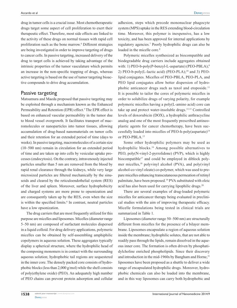
Table 1 Micellar formulations being currently tested in clinical trials
Polymeric micelle Block copolymer Drug Indication Clinical phase
NK012 PeG-PGlu(SN-38) SN-38 Breast cancer IINK105 PeG-P(aspartate) Paclitaxel Advanced stomach cancer IISP1049C Pluronic L61 and F127 Doxorubicin Adenocarcinoma of esophagus,
gastroesophageal junction and stomachIII
NC-6004 PeG-PGlu(cisplatin) Cisplatin Solid tumors I/IIGenexol-PM PeG-P(D,L-lactide) Paclitaxel Breast cancer IvGenexol-PM PeG-P(D,L-lactide) Paclitaxel Pancreatic cancer IIGenexol-PM PeG-P(D,L-lactide) Paclitaxel Non-small-cell lung cancer in
combination with carboplatinII
Genexol-PM PeG-P(D,L-lactide) Paclitaxel Pancreatic cancer in combination with gemcitabine
I/II
Genexol-PM PeG-P(D,L-lactide) Paclitaxel Ovarian cancer in combination with carboplatin
I/II
Abbreviations: PeG, polyethylene glycol; SN38, 7-ethyl-10-hydroxy-camptothecin.
hydrophilic drugs. In the last 20 years, a major development
has been the formulation of polyethylene glycol (PEG)
ylated liposomes (PEG-liposomes), known as stealth lipo-
somes, with a prolonged circulation time in the blood.22
PEG-liposomes contain polyethylene glycol derivatives of
phosphatidylethanolamine (PEG-lipid). The major differ-
ence compared to PEO is the molecular weight of the ethoxyl
chain that is below 20,000 Daltons. Nowadays, eleven drugs
with liposomal delivery systems have been approved by the
US Food and Drug Administration (FDA) and six additional
liposomal drugs are in advanced phase clinical trials. Two of
these liposomal systems are employed in cancer therapy. The
first stealth liposome was approved in 1995 by the US FDA
and is still the only formulation to be approved (in the United
States as DOXIL® [Alza Corporation, Vacaville, CA, USA]
and in Europe as Caelyx® [Janssen Pharmaceutica, Beerse,
Belgium]), for the treatment of Kaposi’s sarcoma23 and recur-
rent ovarian cancer.24 DOXIL liposomes are approximately
100 nm in diameter with the following lipid composi-
tion (expressed as percentage mole ratio): hydrogenated
soybean phosphatidylcholine (56.2%), cholesterol (38.3%),
polyethylene-glycol (molecular weight [MW] 1,900)
derivatized distearoyl- phosphatidylethanolamine (5.3%),
and α-tocopherol (0.2%). Loading of doxorubicin
(0.125 drug/lipid weight ratio) is based on the ammonium
sulfate gradient method.
The combined use of drugs acting on different targets
within cancer cells is widely utilized in oncology to improve
efficacy, overcome undesirable toxicity, reduce the admin-
istered amounts of each agent, and reach multiple targets –
thereby increasing the therapeutic index of the native drugs.24
Supramolecular aggregates are theoretically capable of
loading more than one drug at a time, which would allow
for the simultaneous delivery of multiple drugs.25 Such an
approach may be of additional value for clinical application
of these delivery systems. Several examples of micelles and
liposomes acting as co-delivery transporters are reported in
the literature.26,27
Aside from the aforementioned aggregates generally
belonging to the soft matter category, hard matter carriers,
such as metal nanoparticles and ceramic nanoparticles,
have been developed in recent years for their applications
in diagnostics and therapeutics.28 One carefully studied
metal nanoparticle is iron oxide, which can be used for
such purposes after being coated with dextran, surfactants,
phospholipids, or other compounds that increase its stability.
Also, aminosilane-coated iron oxide nanoparticles have been
utilized in thermotherapy to treat brain tumors.29
Magnetic nanoparticles (MNPs) of iron oxide possess
unique magnetic properties and have the ability to function
at the cellular and molecular level of biological interactions.
Such nanoparticles are attractive for applications in thermo-
therapy, as contrast agents for magnetic resonance imaging
(MRI) and as carriers for drug delivery.30 Other early nano-
technology approaches toward the chance of overcoming
multidrug resistance (MDR) in cancer include covalent
attachment of drug to polymers and solid-core nanopar-
ticles to prevent drug efflux.31 Recently, DOX conjugated
superparamagnetic iron oxide nanoparticles (SPION;
NP-DOX) were developed and examined for susceptibil-
ity to MDR mediated drug efflux, a common mechanism
of resistance to DOX.32 Metal nanoparticles utilizing gold
have good optical and chemical properties and are being
investigated for use in infrared phototherapy applications.
Ceramic nanoparticles such as silica, titania, and alumina
are generally bioinert and have porous structures. These
International Journal of Nanomedicine 2014:9submit your manuscript | www.dovepress.com
Dovepress
Dovepress
1540
Accardo et al
nanoparticles have also been proposed as drug delivery
vehicles for cancer therapy.33
Nanoparticles have several features that make them
appealing for these applications: a large surface area that
allows them to trap an elevated number of active drug mole-
cules; their structural versatility that allows them to obtain
objects of varying sizes and pharmacokinetic properties in
order to optimize drug delivery; and the possibility of cou-
pling with other molecules, such as pharmacokinetic modi-
fiers (PEG) or labels that can be used for tracking (magnetic,
radioactive, or fluorescent).
Active targetingThe currently approved nanoparticle systems have, in some
cases, improved the therapeutic index of approved drugs by
reducing drug toxicity or enhancing drug efficacy. However,
there are data indicating that PEGylated liposomes loaded
with doxorubicin34 do not significantly improve therapeutic
efficacy compared to the native carrier free drug. An explana-
tion for these results may be that PEGylated liposomes only
increase drug concentration in the tumor vasculature, but
there is no significant change in the intracellular drug level,
which is crucial for efficacy.
Therefore, active targeting is being actively pursued in
order to target delivery. This approach is based on utilizing
nanoparticles that have been externally modified with bio-
active molecules capable of selectively recognizing targets
present in cancer.
Different systems are used to provide targeting capa-
bilities and these include monoclonal antibodies, receptor-
specific peptides or proteins, nucleic acids (deoxyribonucleic
acids/ribonucleic acids [DNA/RNA] aptamers), small
mole cules, and even vitamins or carbohydrates. Monoclonal
antibodies or antibody fragments that can be selected with a
high degree of specificity for the target tissue, with elevated
binding affinities, are therefore particularly suitable for this
task. Antibodies are being used to deliver radioisotopes,35
toxins,36 cytokines,37 and other drugs. In certain settings
the targeting antibody also displays therapeutic properties38
giving the added advantage of targeting the cancer cell by
two distinct mechanisms. Despite the recent progress in
antibody engineering, antibody development is still fairly
expensive and use of such biomolecules as drugs presents
stability and storage problems when designing formula-
tions for clinical use. Another issue that may arise with
non-humanized antibodies is immunogenicity, which may
limit repeat administrations due to the risk of significant
side effects.
On the other hand, several non-antibody ligands can be
coupled to larger drug vectors for this same purpose. This
class of compounds may display less selective interaction
with potential targets. Ligands such as folate and transferrin,
which target growth-factor receptors,39,40 have targets that are
expressed not only in cancer cells but also in normal tissues.
There are also physiological concentrations of native ligands
that may compete for the target.
Peptide targetingNatural and synthetic peptides are a class of small ligands
that have great potential for such applications. They offer the
advantage of providing infinite sequence/structure possibili-
ties that can potentially be designed to bind to any cancer
related target. Furthermore, such an approach is expected
to yield fewer problems related to immunogenicity. Among
potential targets, there are several cell surface receptor
systems that have small peptides as ligands that have been
shown to be highly expressed in a variety of neoplastic and
non-neoplastic cells.41 Furthermore, receptor-targeting pep-
tides have shown a high level of internalization within tumor
cells via receptor-mediated endocytosis. Such a feature of
these systems may be of value in facilitating intracellular
delivery of the intended payload. The drawbacks related to the
use of these compounds are the relatively lower target affini-
ties and the metabolic instability of these compounds that may
be extremely sensitive to protease degradation. Improving
metabolic stability and pharmacokinetics can be attempted by
modifying peptide sequences using specific coded or uncoded
amino acids or amino acids with D configurations. Cycling of
the N-terminal with the C-terminal or with a side-chain, or the
C-terminal with a side-chain and the side-chain with another
side-chain, can also be utilized for such purpose. Another
advantage is the possibility of designing analogs that can act
as antagonists. Cell surface receptor antagonists show the dual
advantage of not activating the biological pathways following
receptor binding and have also been shown to have higher
binding capacities to their agonist counterparts.42,43 These
attractive physical properties coupled with their smaller size
make peptides very appealing candidates for developing new
target-specific nanoparticles.
Most peptide based targeting ligands are derived from
known endogenous proteins capable of binding the target
receptor with high affinity. Molecular modeling of new
peptide sequences based on the known three-dimensional
structure of the target receptor is also a possible strategy
for rational design of new compounds, although such an
approach requires thorough knowledge of the structure of
International Journal of Nanomedicine 2014:9 submit your manuscript | www.dovepress.com
Dovepress
Dovepress
1541
Receptor binding peptides for nanoparticle encapsulated drugs
ligand/ receptor interaction.44 A further possibility for identify-
ing new peptide sequences for recognizing tumor- associated
proteins is the use of phage display techniques.45
Once the binding sequence is identified a number of syn-
thetic strategies have been put in place in order to modify the
surface of micelles, liposomes, or nanoparticles in order to
display the targeting peptide sequence. One main concern in
this part of development is to achieve high coupling efficiency
while distancing the bioactive peptide from the nanostruc-
ture surface in order to maintain the specific conformation
required for high affinity binding to the target. The bioactive
peptide may be introduced on the aggregate surface directly
during nanostructure preparation by coupling the peptide
to an amphiphilic moiety (pre-functionalization strategy;
Figure 1A), or introducing the peptide on the surface of
the nanostructures after they have been obtained (post-
functionalization; Figure 1B).
The first method, usually employed for the obtainment
of peptide containing micelles and liposomes, needs a
well-purified amphiphilic peptide molecule; it is mixed
in appropriate solvents and in the chosen ratio with other
amphiphilic molecules and phospholipids; then micelles or
liposomes are obtained by evaporating the solvent or using
extrusion procedures. The advantage of this approach is
that one obtains a well-defined amount of bioactive mole-
cules in the aggregates and there are no impurities. With
phospholipid
peptide sequence
A
==
+
biotin-peptide
1)
2)
A
A
a)
b)aliphatic - PEG-
-peptide
liposome
Peptide sequences:
CCK8: DYMGWMDF-NH2
QWAVGHLM-NH2
QLYENKPRRPYIL-NH2
pyroEHWSTGLRPG-NH2
fCFwKTCT-OH
[7–14]BN:
Octreotide:
Lutein:
NT1–13:
A
A
biotinylated amphiphile
avidin
O2N
N3
O
OO
O
O
S SN
R
R
COOH
R·SHR·SH
= =H
B
=
=
=
==+
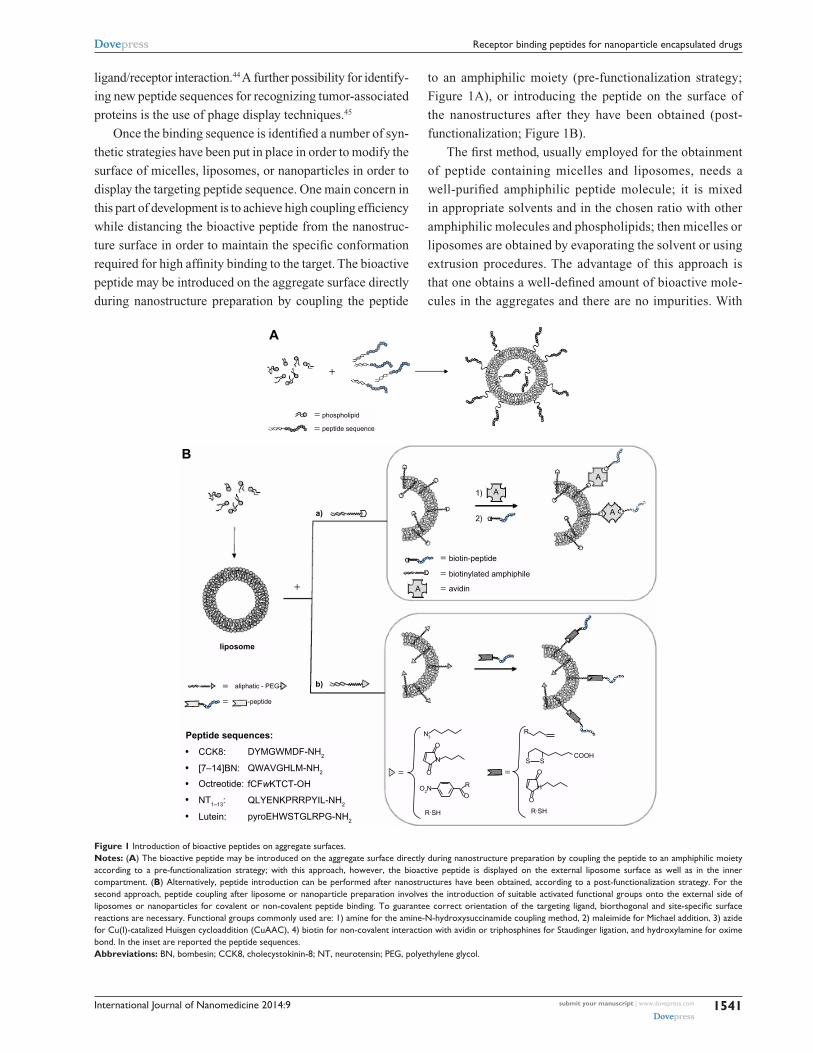
Figure 1 Introduction of bioactive peptides on aggregate surfaces.Notes: (A) The bioactive peptide may be introduced on the aggregate surface directly during nanostructure preparation by coupling the peptide to an amphiphilic moiety according to a pre-functionalization strategy; with this approach, however, the bioactive peptide is displayed on the external liposome surface as well as in the inner compartment. (B) Alternatively, peptide introduction can be performed after nanostructures have been obtained, according to a post-functionalization strategy. For the second approach, peptide coupling after liposome or nanoparticle preparation involves the introduction of suitable activated functional groups onto the external side of liposomes or nanoparticles for covalent or non-covalent peptide binding. To guarantee correct orientation of the targeting ligand, biorthogonal and site-specific surface reactions are necessary. Functional groups commonly used are: 1) amine for the amine-N-hydroxysuccinamide coupling method, 2) maleimide for Michael addition, 3) azide for Cu(I)-catalized Huisgen cycloaddition (CuAAC), 4) biotin for non-covalent interaction with avidin or triphosphines for Staudinger ligation, and hydroxylamine for oxime bond. In the inset are reported the peptide sequences.Abbreviations: BN, bombesin; CCK8, cholecystokinin-8; NT, neurotensin; PeG, polyethylene glycol.
International Journal of Nanomedicine 2014:9submit your manuscript | www.dovepress.com
Dovepress
Dovepress
1542
Accardo et al
this approach, however, the bioactive peptide is displayed
on the external liposome surface as well as in the inner
compartment.
For the second approach, peptide coupling after lipo-
some or nanoparticle preparation involves the introduction
of suitable activated functional groups onto the external side
of liposomes or nanoparticles for covalent or non-covalent
peptide binding. To guarantee correct orientation of the
targeting ligand, biorthogonal and site-specific surface reac-
tions are necessary. The synthetic strategy should be aimed at
optimizing reproducibility and yield of the coupling reaction.
Functional groups commonly used are: a) amine for the amine-
N-Hydroxysuccinimide coupling method; b) maleimide for
Michael addition; c) azide for Cu(I)-catalyzed Huisgen
cycloaddition (CuAAC); and d) biotin for non-covalent inter-
action with avidin or triphosphines for Staudinger ligation,
and recently, hydroxylamine for oxime bond.46
A considerable number of molecular targets for peptides
are either exclusively expressed or overexpressed on both
cancer vasculature and cancer cells. They can be classified
into three wide categories: integrins; growth factor recep-
tors (GFRs); and G-protein coupled receptors (GPCRs).
These receptors offer attractive targets for anticancer
therapeutics as they are often implicated in tumor growth
and progression. Many nanoparticles and liposomes have
been labeled with peptides capable of interacting with these
receptors and have been reported in the literature in the last
decade. Nanoparticles grafted with the RGD sequence able
to bind integrin receptors have been widely evaluated for
the treatment of different cancers, such as ovarian cancer,
melanoma, and breast carcinoma.47–49 Peptides targeting
growth factor receptors have been utilized to functionalize
liposomes encapsulating chemo-therapeutics.50 Peptides have
also been developed to target the extracellular matrix of the
diseased tissues, and this is an important alternative strategy
to target unhealthy tissues which can also be incorporated
with nanomedicine.51,52
This review will focus on delivery systems containing
peptides that recognize GPCRs. GPCRs constitute a mem-
brane protein family involved in the recognition and transduc-
tion of signals as diverse as light, Ca2+, and small molecule
signaling, including peptides, nucleotides, and proteins. The
general structural features, obtained by indirect studies as
well as X-ray crystallography, indicate the presence of seven
transmembrane helices connected by three intracellular and
three extracellular loops. The N-terminal domain is directed
into the extracellular space and C-terminal points to the intra-
cellular space. Ligand binding to receptor is a crucial event
in initiating signals, and the study of how ligands interact
with their receptors can reveal the molecular basis for both
binding and receptor activation. The ligand binding site for
peptides has been found in the N-terminal extradomain or on
the portion of the extracellular loops adjacent to the extracel-
lular moiety of the transmembrane helices. Knowledge of
the structural details of this interaction could be very useful
for designing ligands for targeted delivery. Unfortunately,
detailed structural characterization of the ligand-receptor
complex for most systems is very difficult to obtain. However
several approaches, such as biochemical affinity, photoaffin-
ity labeling,53 and site-directed mutagenesis54 have allowed us
to determine which amino acid residues are involved in bind-
ing. The interest in developing agonist or antagonist peptides
against these receptors is based on the biological role these
receptor pathways have in specific cancer types.
Overexpression of small peptide receptors has been
documented for a wide number of cancers.41 As many as
105–106 receptor molecules per cell or receptor densities in
the pmol ⋅ mg−1 protein range have been reported for a variety
of systems, such as somatostatin receptors in neuroendocrine
tumors, cholecystokinin (CCK) receptors in medullary
thyroid cancer, bombesin receptors in prostate and breast
carcinoma, and several others.
We will review delivery systems targeting a family of
regulatory peptide receptors overexpressed in specific cancer
types, focusing particularly on receptors for somatostatin
(SST), cholecystokinin (CCK), gastrin-releasing peptides
(GRP/Bombesin), lutein, and neurotensin.
Somatostatin based delivery systemsAt least five subtypes of somatostatin receptors (SSTRs;
SSTRs 1–5) have been discovered: they belong to a distinct
group within the superfamily of G-protein-coupled receptors.
SST binds these receptors with high affinity, with the main
physiologic purpose of inhibiting some functions of the
target cell, for example blocking growth-hormone release in
the hypothalamus. This endogenous peptide is preferentially
produced in neurons and secretory cells in the central and
peripheral nervous system and in the gastrointestinal tract.55
The different receptor subtypes show 50% sequence homol-
ogy, which is particularly evident in the transmembrane
regions. Aside from the expression in normal tissues, SSTRs
have been found in many different types of tumors, mostly
of neuroendocrine origin, such as gastroenteropancreatic
tumors, neuroblastomas, medulloblastomas, breast can-
cers, meningiomas, paragangliomas, renal cell carcinomas,

International Journal of Nanomedicine 2014:9 submit your manuscript | www.dovepress.com
Dovepress
Dovepress
1543
Receptor binding peptides for nanoparticle encapsulated drugs
lymphomas, hepatocellular carcinomas, and small cell lung
cancers. In general, SSTR2 is the most common SSTR
subtype found in human tumors, followed by SSTR1, with
SSTR3, 4 and 5 being less common. The high frequency of
SSTR expression in human tumors has been exploited for
diagnostic and therapeutic applications.
The wild type SST tetradecapeptide has a limited thera-
peutic value due to its short biological half-life (,3 minutes).55
This feature pressed scientists to develop peptide analogs with
improved stability to enzymatic cleavage and therefore with
prolonged circulation time. The most successful derivative is
octreotide (OCT).56 This eight amino acid analog, developed
by Sandoz (now Novartis) is able to induce endocytosis by
binding to SSTR2 with high affinity (inhibitory concentra-
tion [IC]50
=2nM), and SSTR 3 (IC50
=376 nM) and SSTR
5 (IC50
=299 nM) to lesser degrees. OCT has been a subject
of extensive structural studies, including nuclear magnetic
resonance (NMR),57 in order to design peptide conjugates as
vehicles for contrast agents or drugs. OCT peptide binding
to receptors is not affected when chemical modifications
are introduced on its N-terminus. Radiolabeled OCT conju-
gates are commonly used in clinical tumor diagnosis58 and
in clinical trials for peptide receptor radiotherapy (PRRT).59
OCT has been further used to enhance the delivery of drugs
to tumor cells by chemically conjugating it with anti-tumor
drugs.60 These promising results prompted many research-
ers to develop OCT as a specific targeting moiety to deliver
nanocarriers incorporating anti-tumor drugs into tumor cells
via SSTRs endocytosis (Table 2).
Liposomes and micellesOne of the most relevant issues for chemotherapeutic drugs
is poor solubility in water and/or in buffers, which limits the
quantities of drug that can be administered. Supramolecular
aggregates can improve the biodistribution and pharmacoki-
netics of these drugs.8 Moreover, as previously reported,
severe side effects of these drugs can be reduced by enhanc-
ing delivery to the target tissue.13 In the last few years, many
different aggregates have been developed to carry chemo-
therapeutic drugs to SSTR2 expressing tumors by coupling
to the OCT peptide.61
Octreotide labeled aggregates may be obtained following
the two approaches presented above. One strategy was based
on synthesizing the OCT on trityl resin in solid phase and
coupling the other molecular building blocks step by step. The
advantage of this approach is to supervise all synthetic steps
protecting all reactive functions in order to avoid collateral
products. The most relevant disadvantage is the difficulty in
Table 2 Octreotide labeled supramolecular aggregates or nanoparticles
Peptide conjugation methods Formulation Drug or nanoparticles References
OCT versus NHS-PeG-b-PCL Micelle: OCT-PeG-b-PCL PTX-salinomycin 65OCT versus p-nitrophenylcarbonyl- PeG(100) monostearate
NLC: OCT-polyethylene glycol(100) monostearate (PGMS)
HCPT 67
OCT versus p-nitrophenylcarbonyl- PeG(100) monostearate
NLC: OCT-polyethylene glycol(100) monostearate (PGMS)
HCPT 68
OCT-PeG3400-DSPe Liposome: DSPC OCT-PeG3400-DSPe (different ratio)
Irinotecan CPT 11 70
OCT versus BocNHPeG-NHS Micelle OCT(Phe)-PeG-SA (OPS)/ (OCC) (in different ratio)
DOX 73
OCT versus BocNHPeG-NHS Micelles (OCT(Phe)-PeG-DOCA) (DAHC) 1:5 (molar ratio)
DOX 74
OCT versus pNP-PeG-Pe Liposome PC:Chol:OCT-PeG-Pe 5:1:0.5 (molar ratio)
DOX 75
OCT versus DSPe-PeG-NHS Liposome ePC/chol/ DSPe-PeG/DSPe-PeG-OCT (15.9:4.1:5.7:0.3, w/w)
DOX 76
OCT amphiphilc solid phase synthesis Liposome (C18)2(AdOO)5OCT/ Peg1500Lys(Pt-aminoetGly)-Lys(C18)2 1:9 (molar ratio)
Pt(II), DOX 77
DSPe-PeG2000-OCT (not declared) Liposome ePC/Chol/DSPe-PeG-OCT/CA- 4;25:1.28:6:2,w/w
CA-4 and DOX 78
TOC-Boc AuNPs AuNPs 80
Octreotide versus AuNPs (∼20 nm) AuNPs 81
Abbreviations: AuNP, gold nanoparticles; Boc, tert-Butyl carbamates; DOX, doxorubicin; HCPT, 10-hydroxycamptothecin; NHS, succinimidyl carboxymethyl ester; OCT, octreotide; PeG, polyethylene glycol; PTX, paclitaxel; TOC, Tyr3-octreotide.
International Journal of Nanomedicine 2014:9submit your manuscript | www.dovepress.com
Dovepress
Dovepress
1544
Accardo et al
purifying these molecules that are poorly soluble and which
need to be solubilized in organic solvents, as they would
aggregate in water-based buffers. An alternative strategy con-
sists in assembling the amphiphilic molecule in solution. The
hydrophobic moiety and the hydrophilic linker are coupled on
the N-terminus of the OCT after peptide purification. In this
case side reactions are of concern as OCT has two primary
amino groups (the N-terminus and the side chain of Lys)
and the coupling reaction may get mono- or di-substituted
derivatives. To limit the undesired products, reactions must
be conducted at a pH value below 10. In certain instances
a test of α Lys-C digestion is necessary to further confirm
the coupling site. Trypsin cleaves peptide chains mainly at
the carboxyl side of lysine or arginine, except when either
is followed by proline. If the conjugation occurs at the Lys
residue, there would be no change in the mass spectrum
after trypsinization; otherwise the modification occurring
at the N-terminus would exhibit a reduced mass fragment.
The OCT amphiphilic molecules can self-assemble, or
generate micelles or liposomes by mixing with a surfactant.
Hydrophobic drugs are preferentially loaded in the core of
micelles,8 whereas water soluble drugs could be carried in
the inner compartment of liposomes or in the hydrophilic
shell of micelles.
Important issues in the development of OCT coupled
aggregates are confirming that there is adequate exposure on
the aggregate surface, and also confirming the ability of the
OCT peptides in recognizing and binding the target receptor.
In order to characterize these aggregates for their suitability for
in vivo use as selective targeting tools, it is possible to study
peptide properties on the aggregate surface through classical
chemical physical methods. Morisco et al61 developed OCT
containing aggregates for use as drug carriers and magnetic
resonance imaging (MRI) contrast agents. The monomers,
synthesized on solid phase, contain, in the same molecule,
three different functions: the chelating agent (DTPAGlu or
DOTA); OCT; and a hydrophobic moiety based on two C18
hydrophobic chains. These monomers (OCA-DTPAGlu, OCA-
DOTA) self-assemble in water solution, giving stable micelles.
Fluorescence studies indicate, for the two compounds as well
as for their gadolinium complexes (OCA-DOTA[Gd] and
OCA-DTPAGlu[Gd]), the complete exposure of OCT on the
micelle surface. In fact, the tryptophan emission at 345–350
nm suggests a hydrophilic environment for this residue.
Circular dichroism measurements show the predominant
presence of an antiparallel beta-sheet peptide conformation
characterized by a beta-like turn. This conformation has been
demonstrated to be suitable for receptor binding.
The same group has also studied62 mixed aggregates
formulated by co-assembling: a first monomer containing
the OCT peptide, an ethoxyl spacer bound to the peptide
N-terminus, and the hydrophobic moiety; a second monomer
containing the same hydrophobic chains bound through a
lysine residue to different polyamino-polycarboxy ligands;
and a chelating agent such as DTPAGlu, DTPA, or DOTA
to allow coordination of metal ions. Structural character-
ization of the aggregates indicates a shape and size of the
supramolecular aggregates suitable for in vivo use. For these
aggregates, fluorescent emission of the tryptophan residue
at 340 nm also suggests exposure of the peptide to the water
environment, thus available to interact with the SSTR2.
Later work by the group of Helbok et al63 demonstrated
the in vitro and in vivo selective aggregate binding of OCT
coupled PEGylated liposomal nanoparticles radiolabeled
with indium-111. The OCT derivative was synthesized
by cross-linking of the S-acetyl-mercaptopropionic acid
peptide with Mal-DSPE-PEG2000. Liposomes were
obtained by mixing the OCT derivative with adequate
amounts of palmitoyl oleoyl-phosphatidylcholine (POPC),
lyso-stearyl- phosphatidylglycerol (Lyso-PG), distearyl-
phosphatidylcholine–polyethyleneglycol-2000 (DSPE-
PEG2000), and dimyristoyl phosphoethanolamine-DTPA
(DMPE-DTPA) in a molar ratio of 0.1:11:7.5:0.9:2,
respectively. Targeting properties of the OCT labeled lipo-
somes were evaluated in vitro on rat pancreatic tumor cells
(AR42J), demonstrating specific binding and IC50
values
in the low nanomolar range. Unfortunately only moderate
uptake was observed when in vivo experiments were per-
formed in animals; this may be explained by the limited and
slow accessibility of target receptors on tumor cells by large
constructs such as these, compared to small peptides that
show much more rapid diffusion and binding to the receptors
and cellular internalization.
Similar proof of concept was reported by Petersen et al.64
Liposomes (DSPC/Chol/DSPE-PEG2000/DSPE-PEG2000-
TATE in a molar ratio 50:40:9:1, respectively) with an
encapsulated positron emitter 64Cu for positron emission
tomography (PET) imaging were tested in vivo in a mouse
model. [Tyr3]-octreotate (TATE), an OCT analog, function-
alized with maleimide, was covalently attached to the distal
end of DSPE-PEG2000 via a thioether bond. Biodistribution
and pharmacokinetic properties of TATE coupled liposomes
were compared with peptide free liposomes and with the
radiolabeled peptide alone. 64Cu-loaded PEGylated liposomes
derivatized with the TATE peptide displayed significantly
higher tumor-to-muscle (T/M) ratio (12.7±1.0) compared

International Journal of Nanomedicine 2014:9 submit your manuscript | www.dovepress.com
Dovepress
Dovepress
1545
Receptor binding peptides for nanoparticle encapsulated drugs
to control-liposomes without TATE (8.9±0.9) and to 64Cu-
DOTA-TATE peptide (7.2±0.3). These results demonstrate
the feasibility of utilizing somatostatin analogs for specific
targeting of the above described aggregates to tumors over-
expressing somatostatin receptors.
Paclitaxel (PTX) is a mitotic inhibitor used to treat
patients with lung, ovarian, breast, head and neck cancers,
and advanced forms of Kaposi’s sarcoma. This drug is poorly
soluble in water and thus is a suitable candidate for loading in
micelles. Zhang et al studied a combination of PTX and sal-
inomycin (SAL), an experimental drug recently found to be
very effective on breast cancer stem cells.65 Both drugs were
loaded in polyethylene glycol-b-polycaprolactone (PEG-b-
PCL) polymeric micelles obtaining OCT-(PTX)-PEG-b-PCL
(OCT-M-PTX) and salinomycin (SAL)-loaded PEG-b-PCL
(M-SAL). OCT was coupled to NHS-PEG-b-PCL through
the activated NHS group in dimethyl sulfoxide (DMSO) solu-
tion. The prepared micelles have a diameter of approximately
25–30 nm, and the encapsulation efficiency of the drug was
.90%. The presence of the OCT peptide favors uptake of
micelles in SSTR overexpressing MCF-7 breast cancer cells.
Moreover, free OCT can inhibit such interaction confirm-
ing that cellular uptake is indeed occurring by a receptor-
mediated mechanism. The efficacy of combination therapy
using OCT-M-PTX plus M-SAL was confirmed in vitro and
in MCF-7 xenografts in mice: the combination treatment
results in a stronger inhibitory effect on tumor survival by
killing both non-stem cancer cells and cancer stem cells.
Another water insoluble chemotherapeutic in a broad
spectrum of cancers, including leukemias and cancers of
the liver, stomach, breast, and colon, is a natural derivative
of camptothecin, the 10-hydroxycamptothecin (HCPT) in
lactone form. One way to improve the solubility of HCPT is
to change the lactone form to the carboxylate form by adding
NaOH. However, this leads to less activity and more unwanted
toxicity.66 At the same time, HCPT has a short half-life in
vivo and poor biodistribution. Obviously, pharmacokinetics
of this molecule is improved by using drug carriers. Su et al67
formulated HCPT-loaded nanostructured lipid carriers (NLC)
made from poly(ethylene glycol)-poly(γ-benzyl-L-glutamate)
(PEG-PBLG). At this amphiphilic polymer the conjugate
OCT labeled polyethylene glycol monostearate (OPMS) was
added. The labeling procedure was carried out in a solution
of p-nitrophenyl-PMS adding OCT and incubating at pH 9.
The OCT binding on PMS was determined by bicinchoninic
assay (BCA) protein assay kit. Nanoparticle size depends on
the different molar ratio of their components. In a more recent
study, the authors demonstrated that surface density of the
targeting moiety was crucial to determine physicochemical
properties, drug release, cellular uptake, and cytotoxicity.
Compared to pharmacokinetic studies, modified NLCs
had a longer circulation than NLC due to PEGylation effect,
and OPMS-modified NLCs had larger mean residence
time than PGMS-modified NLCs, showing 58.5 ng/mL at
24 hours of drugs versus 15.8 ng/mL. Furthermore, qualita-
tive observation of cellular uptake by florescence microscopy
showed higher uptake of OCT-modified NLCs on tumor cells
(SMMC-7721) overexpressing somatostatin receptors, in
comparison to OCT-modified NLCs uptake on control cells
after incubation at 37°C for 2 hours.68
Irinotecan (CPT-11), another analog of camptothecin,
induces a growth inhibition of tumor cells in medullary
thyroid carcinoma (MTC).69 This derivative is water soluble
but its use is limited because of many side effects. Iwase and
Maitani70 overcame these problems by loading this drug in
OCT decorated liposomes. Liposomes were formulated by
mixing DSPC lipids with OCT-PEG3400
-DSPE amphiphilic
molecules in different ratios. The association of modified
OCT-targeted liposomes with TT cells was significantly
higher than non-targeted PEGylated liposomes and was
significantly inhibited by empty OCT-targeted liposomes but
not by free OCT. The authors suggest that the affinity of free
OCT and OCT-CL to SSTR are not the same.70 After 96 hours
of exposure, cytotoxicity of OCT-targeted liposomal CPT-11
(IC50
: 1.05 µM) was higher than free CPT-11 (IC50
: 3.76 µM)
or PEGylated liposomal CPT-11 (IC50
: 3.05 µM). Moreover,
OCT-targeted liposomal CPT-11 led to significantly higher
antitumor activity and prolonged survival time compared
with non-targeted liposomal and free CPT-11.
The major efforts in target delivery mediated by soma-
tostatin analogs have been devoted to carry DOX on tumor
cells. DOX is a hydrophilic drug and can be loaded in
micelles or in liposome inner compartments. The approval
of DOXIL in 1995 opened a route to new formulations in
order to improve efficacy and tolerability of the drug as
compared with the non-liposomal counterparts or passive
targeting aggregates.
Hydrophobilized polysaccharides polymeric micelles
are currently very attractive for researchers due to their
well-known nontoxicity and excellent biocompatibility
and biodegradability.71 In the last few years, Zou et al72
studied N-octyl-O,N-carboxymethyl chitosan (OCC)
and N- deoxycholic acid-O,N-hydroxyethylation chitosan
(DAHC) micelles. OCC and DAHC micelles exhibited good
loading capacities for DOX, with a drug loading content
(DLC) in the 22%–30% range. The first attempt to graft them

International Journal of Nanomedicine 2014:9submit your manuscript | www.dovepress.com
Dovepress
Dovepress
1546
Accardo et al
BlankA
B
350.00 637.50 925.00 1,212.50 1,500.00
1 h 6 h 12 h 24 h
Figure 2 In vivo imaging of tumor-bearing mice after administration of Cy-7 loaded DAHC micelles (A) and Cy-7 loaded OPD(20%)-DAHC micelles (B) at 1, 6, 12, and 24 hours.Note: Reprinted from Biomaterials, 33(27), Huo M, Zou A, Yao C, et al, Somatostatin receptor-mediated tumor-targeting drug delivery using octreotide-PeG-deoxycholic acid conjugate-modified N-deoxycholic acid-O, N-hydroxyethylation chitosan micelles, 6393–6407, Copyright (2012) with permission from elsevier.74
Abbreviations: DAHC, N-deoxycholic acid-O,N-hydroxyethylation chitosan; OPD, OCT(Phe)-PeG-DOCA; h, hours.
with OCT was carried out, conjugating the N-terminal moiety
to the free carboxylic groups of OCT.72 The reaction had an
extremely low (about 3%) yield, which is largely due to the
high molecular weights of OCT and chitosan derivatives, the
strong hydrogen bonds in the chitosan backbone, and poor
solubility of chitosan derivatives in organic solvent. This
result pushed toward alternative mixed aggregates, adding
to DAHC a ligand-PEG-lipid conjugate able to guarantee
same time long circulation time in blood and ligand targeting.
Therefore the peptide N-terminal function was anchored in
solution to a PEG fragment and this moiety was conjugated
to an aliphatic chain obtaining the OCT(Phe)-PEG-SA (OPS)
monomer or to deoxycholic acid obtaining the OCT(Phe)-
PEG-DOCA (OPD).73
Micelles formulated by adding OPS to the final formu-
lation were not significantly affected with respect to size or
shape. Their diameter is less than 120 nm with spherical
shape and zeta potential of 30 mV. Enhanced tumor-targeting
capacity was observed in BALB/c nude mice bearing MCF-7
cancer xenografts as compared with the self-assembling OCC
micelles. Moreover, pharmacodynamic studies demonstrated
that DOX-OCC-OCT presented a stronger inhibition of tumor
growth (86.7% versus 33.3%) and lower systemic toxicity
compared to free DOX and DOX-OCC micelles.
Insertion of OPD in aggregate formulations showed no
significant effect on drug loading properties while slightly
increasing particle size (230 nm average diameter versus
200 nm) and partly shielded the positive charges on the
surface of micelles.7 Accelerated release rate of DOX
from micelles were also observed after OPD modification,
the release profile also exhibited pH-sensitive properties.
Compared to DAHC-DOX micelles, OPD-DAHC-DOX
micelles exhibited significantly stronger cytotoxicity to
human breast cancer cells (MCF-7; SSTRs overexpression)
but had almost the same effect on human embryonic lung
fibroblasts (WI-38 cells; no SSTRs expression). The results
of flow cytometry and confocal laser scanning microscopy
further revealed that OPD-DAHC-DOX micelles could
be selectively taken into tumor cells by SSTRs-mediated
endocytosis. In vivo investigation on nude mice confirmed that
OPD-DAHC micelles possessed much higher tumor-targeting
capacity than the DAHC control and exhibited enhanced
anti-tumor efficacy and decreased systemic toxicity. Figure 2
shows images of micelles in the tumor-bearing mice at 1,
6, 12, and 24 hours after administration of fluorescent dye,
Cyanine 7, encapsulated into DAHC (Figure 2A) micelles
and OPD (20%)-DAHC micelles (Figure 2B). During the live
imaging test, most of the Cy7 accumulated in liver and tumor
after intravenous administration of both micellar formulations.
However, preferential accumulation of fluorescence was obvi-
ous in the tumor site compared to the liver or other normal
tissues at 12 and 24 hours after injection. Moreover, the
OPD-DAHC micelles showed higher tumor-targeting effi-
ciency, which led to higher accumulation of micelles in the
tumors than DAHC micelles. These results provide decisive
evidence that the designed OPD-DAHC micelles are suitable
for tumor-specific drug delivery. This high tumor targetability
of micelles might be due to a combination of an EPR effect
and receptor-mediated uptake of micelles (Figure 2).
OCT-polyethylene glycol-phosphatidylethanolamine (OCT-
PEG-PE) was developed for the assembling of liposomes; the
effect of OCT modification on the enhancement of the delivery
and targeting of DOX-loaded liposomes was investigated in
vitro and in vivo.75 OCT-PEG-PE was synthesized by a three-
step reaction. DOX loading was carried out by the well assessed
ammonium sulfate gradient method. Both drug uptake assays
and cell apoptosis assays suggested that octreotide-labeled lipo-
some (DOX-OL) noticeably increased the uptake of DOX by
International Journal of Nanomedicine 2014:9 submit your manuscript | www.dovepress.com
Dovepress
Dovepress
1547
Receptor binding peptides for nanoparticle encapsulated drugs
fluorescent measurement (about 100% higher than that in unla-
beled liposome [DOX-CL] cases) in SMMC-7721 cells and
showed a more significant cytotoxicity compared to DOX-CL.
The effect of DOX-OL was remarkably inhibited by free OCT.
In contrast, no significant difference in drug cytotoxicity was
found between DOX-OL and DOX-CL in CHO cells without
obvious expression of SSTRs. The study of ex vivo fluorescence
tissues imaging of BALB/c mice and in vivo tissue distribution
of B16 tumor-bearing mice indicated that DOX-OL caused
remarkable accumulation of DOX in melanoma tumors and the
pancreas, in which the SSTRs are highly expressed. In another
study,76 DOX-loaded OCT-DSPE-PEG monomer containing
sterically stabilized liposomes (SSL) increased intracellular
delivery of DOX in SSTR2-positive cells, through a mechanism
of receptor-mediated endocytosis, as demonstrated by fluores-
cence spectrophotometry, confocal laser scanning microscopy,
and flow cytometry studies. Confocal microscopy studies were
carried out on NCI-H446, MCF-7, and Chinese hamster ovary
(CHO) cells. After 3 hours of incubation with SSL-DOX, OCT-
SSL-DOX, or free DOX at DOX concentration of 10 µM at
37°C, NCI-H446 and MCF-7 displayed more red fluorescence
of DOX than SSL-DOX ones. In terms of CHO, there was no
red fluorescence in both passive and active targeting liposome
groups, proving no expression of SSTR2 on the cells. The active
targeting was confirmed by treating with excess free OCT (5
mg/mL). In this case, the uptake of OCT-SSL-DOX by NCI-
H446 cells at 37°C was significantly inhibited because of the
preoccupation of receptors.
Compared to SSL, OCT modification on SSL exhibited
little effect on the physicochemical properties of SSL.
However, it reduced the circulation time of loaded-DOX to
some extent in rats, increased cytotoxicity in SSTR2-positive
tumor cells, enhanced drug accumulation in tumor tissue,
and improved anticancer efficacy in SSTR2-overexpressing
tumor model. The antitumor effect in vivo of OCT-SSL-DOX
was demonstrated inhibiting tumor growth better than that
of SSL-DOX (P,0.05).
Cis platinum is frequently used in combination with other
drugs such as PTX, bleomycin, vinblastine, and in several
trials with DOX.
As proof of concept of combined therapy based on DOX
and platinum complexes, OCT grafted liposomal aggregates
were recently formulated and studied.77 Mixed aggregates were
formulated by co-assembling, at a 10:90 molar ratio, a first
monomer containing two C18 hydrophobic moieties bound to
the N-terminus of the cyclic OCT peptide, and spaced from
the bioactive peptide by five units of dioxoethylene linkers,
(C18)2(AdOO)
5-OCT, and a second amphiphilic monomer
containing a platinum complex anchored to the lipophilic
tail, (C18)2PKAG-Pt. Mixed aggregates (C18)
2-PKAG-Pt/
(C18)2(AdOO)
5-OCT give large liposomes with a diameter
of 168 nm. DOX encapsulation in the inner compartment
was obtained by using the pH gradient method.
Another example of combined therapy was the use, at the
same time, of DOX and combretastatin. Combretastatin A-4,
the principal cancer cell growth-inhibitory constituent of the
Zulu medicinal plant Combretum caffrum, has been undergo-
ing preclinical development.78 However, the very limited water
solubility of this phenol has complicated drug formation.
Loading in aggregates could be an important improvement
for its use. Both combretastatin A-4 (CA-4) and DOX were
loaded in OCT-modified stealth liposomes in order to achieve
the active delivery of these two drugs, followed by sequentially
suppressing tumor vasculature and tumor cells. The drug
loading efficiency of DOX was consistently greater than 95%,
while it was 70%–80% for CA-4. The drug encapsulation effi-
ciency in liposomes was not affected by OCT modification. A
rapid release of CA-4 followed by a slow release of DOX was
observed in vitro. In fact, the release of CA-4 was more than
60% at 8 hours, while DOX released less than 20% at 48 hours.
The active targeted liposomes OCT-L[CD] showed a specific
cellular uptake through ligand-receptor interaction and a higher
antitumor effect in vitro against SSTR positive cell line. The
in vivo sequential killing effect of such systems was found
as evidenced by the fast inhibition of blood vessels and slow
apoptosis-inducing of tumor cells. The anticancer efficacy of
different formulations is displayed in Figure 3. As seen in Fig-
ure 3A, the tumor volume was always the smallest at each test
point in OCT-L[CD] group, suggesting its stronger inhibition
effect on solid tumor compared to other groups (P,0.05). The
excised tumors in OCT-L[CD] group were also the smallest at
the end of the test (Figure 3B). The results were in accordance
with the antitumor study and cell uptake in vitro.
Metal nanoparticlesMetal nanoparticles have been extensively studied and offer
extraordinary features for diagnostic as well as therapeutic
applications.79 Multifunctional systems of gold nanoparti-
cles (AuNPs) capped by the [Tyr3]Octreotide (TOC) peptide
were prepared and characterized by transmission electron
microscopy (TEM) and UV-Vis (ultraviolet- visible),
infrared, and fluorescence spectroscopy.80 AuNPs and
AuNP-TOC fluorescence emission spectra were obtained
both in solution and in murine AR42J-tumor tissues. Results
suggest that AuNP were functionalized with TOC through
interactions with the N-terminal amine function of the

International Journal of Nanomedicine 2014:9submit your manuscript | www.dovepress.com
Dovepress
Dovepress
1548
Accardo et al
A B800
PBS
L[CD]
OCT-L[CD]
Antitumor effect
2 cm
ControlL[C]+L[D] L[CD] OCT-L[CD]
700
600
500T
um
or
volu
me
(m
m3 )
400
300
200
100
00 2 4 6 8 10 12 14
Day after tumor implantation
Figure 3 Antitumor efficiency of different treatments in MCF-7-bearing subcutaneous tumor models in nude mice. (A) Tumor volumes versus time. Data represent mean ± standard deviation (n=6). (B) Tumors excised at the end of the tests.Note: Springer and Pharm Res, 29, 2012, 2902–2911, Spatiotemporally controlled co-delivery of anti-vasculature agent and cytotoxic drug by octreotide-modified stealth liposomes, Dai w, Jin w, Zhang J, et al, Figure 10.78 with kind permission from Springer Science and Business Media.Abbreviations: PBS, phosphate buffer solution; OCT, octreotide.
phenylalanine, the amide groups, and possibly with the
indole group of the tryptophan residue. The fluorescence
analyses in tissue revealed a recognition of the AuNP-TOC
conjugate for the neuroendocrine tumor because of the
lower energy position of the fluorescence resonance (692
nm) with respect to that of the AuNP in the same tumoral
tissue (684 nm). The emission band observed in the near
infrared region (692 nm) opens, for AuNP-TOC, a potential
use as theranostics.
The effect of laser heating, a well-characterized AuNP-
OCT system on HeLa cell viability, was evaluated as a suitable
agent for plasmonic photothermal therapy in the treatment
of cervical cancer.81 The peptide was conjugated to AuNPs
(∼20 nm) by spontaneous reaction of thiol groups. HeLa cells
were incubated at 37°C with AuNP-citrate, with AuNP-OCT,
or without nanoparticles. After laser irradiation, the presence
of AuNP caused a significant increase in the temperature
of the medium (48°C versus 38.3°C without AuNP). The
AuNP-OCT system resulted in a significant decrease in cell
viability of up to 6% compared to the AuNP-citrate system
(15.8%±2.1%). Two possible mechanisms could be at play:
1) OCT alone exerts an effect on survival HeLa cells, or 2) the
release of heat (∼727°C per nanoparticle) in the membranes
or cytoplasm of the cells caused by the interaction between
AuNP-OCT and somatostatin receptors reduced viability.
Cholecystokinin based delivery systemsThe gastrointestinal peptides gastrin and cholecystokinin
(CCK) exist in different molecular forms of variable length
with the same five terminal amino acid sequences at their
carboxyl termini. They act as neurotransmitters in the brain
and as regulators of various functions of the gastrointestinal
tract, primarily at the level of the stomach, pancreas, and
gallbladder.82 CCK and gastrin actions are mediated by sev-
eral receptor subtypes, the best characterized being CCK1
and CCK2 receptors.83 The overexpression of either or both
subtypes of these receptors has been found in certain human
tumors and particularly in tumors of neuroendocrine origin.
In particular, CCK2-R is overexpressed in a large percent-
age (90%) of medullary thyroid cancers, and to a lesser level
in small cell lung cancers and in gastroenteropancreatic
(GEP) tumors. Development of CCK2-R targeting radiop-
harmaceuticals for imaging and for radionuclide therapy has
gained great interest. A wide number of CCK and gastrin
derivatives displaying high affinity for the CCK2-R have
been characterized over the past years for the purpose of in
vivo receptor targeting for imaging and for therapy.84 In all
derivatives, the chelating agents able to coordinate radioac-
tive metals are bound on the peptide N-terminus. In fact,
modifications on peptide N-terminus do not affect receptor
binding that is essentially due to the interaction of recep-
tor N-terminal extradomain with C-terminal fragment of
the peptide ligand, as demonstrated by NMR studies85 and
theoretical calculations.86
On the basis of these data, Accardo et al, in the last
10 years, developed a wide class of CCK8 decorated
supramolecular aggregates (Naposomes) in order to delivery
contrast agents and drugs, thus acting like theranostics
(Table 3).87 Naposomes are formulated by amphiphilic
molecules containing a hydrophobic moiety with two C18
aliphatic chains able to stabilize the aggregates in water
International Journal of Nanomedicine 2014:9 submit your manuscript | www.dovepress.com
Dovepress
Dovepress
1549
Receptor binding peptides for nanoparticle encapsulated drugs
solution. The hydrophilic shell contains a chelating agent
such as DOTA or DTPA or their metal complexes, and the
CCK8 bioactive peptide. The chelating agent plays a double
task: i) it gives the aggregation driving force for the presence
of negative charges; and ii) it acts as polydentate ligand by
complexing with high stability paramagnetic (Gd[III]) or
radioactive (111In[III], 67Ga[III], 68Ga[III], 99mTc[V], 177Lu[III],
or 64Cu[II]) metal ions for imaging application by MRI,
PET, and scintigraphy. Naposomes can be obtained by self-
assembling amphiphilic monomers containing in the same
molecule: i) the hydrophobic moiety with two C18 aliphatic
chains; ii) the chelating agent or its metal complexes; iii) the
bioactive CCK8 peptide; and iv) PEG spacers of appropri-
ate length to allow the exposure of the bioactive moiety on
the external surface of the resulting aggregate.88 The shape
and the size of the resulting Naposomes can be modulated
by adding commercial phospholipids, such as DOPC, to the
synthetic amphiphilic monomer.
Another class of Naposomes can be formulated by
combining together two amphiphilic monomers (Figure 4).
The first monomer contains the CCK8 peptide, a PEG spacer
and two C18 hydrocarbon chains, while the second monomer
contains the DOTA or DTPA chelating agent and the same
hydrophobic moiety (general formula (C18)2LCCK8 and
(C18)2CA, respectively). The morphology and size of the
resulting aggregates (micelles, liposomes, or open bilay-
ers) are influenced by several parameters, such as pH, ionic
strength, monomer structure (length of polioxiethylene
spacers), and composition and formulation procedure (dis-
solution in buffered solution or well-assessed procedures
based on sonication and extrusion).87
All aggregates are able to act as theranostics, carrying
contrast agents like Gd ions for MRI imaging, radioactive
metals for nuclear medicine techniques, and chemother-
apy drugs.
Theranostic effects were demonstrated as proof of
concept for the aggregate based on (C18)2DTPAGlu and
(C18)2PEG
2000CCK8 monomers in 70:30 ratio.89 The uptake
of 111In-radiolabeled aggregates by A431 cells overexpress-
ing CCK2-R via transfection was demonstrated by in vitro
experiments at 4°C and at 37°C. In vivo biodistribution
showed that the overall retention of radiolabeled aggre-
gates in mice at 18 hours is very high, with essentially
no excretion of radioactivity over the observation period.
Moreover, the radioactivity retention of the receptor-
positive xenografts was always higher than in their respec-
tive controls (Figure 4). Finally, cytotoxicity assays were
performed by incubating the cells with peptide-containing
aggregates filled with DOX in ratio 2:1 per aggregate. The
overexpressing receptor cells survive significantly less than
the control cells.
DOX has been also encapsulated in micelles obtained
by self-assembling of (C18)2(AdOO)
5CCK8 monomers.90
These nanostructures, fully characterized by structural
measurements, are able to encapsulate poorly water soluble
molecules, such as pyrene, and DOX drug in their hydro-
phobic compartment. The encapsulation process, followed
and quantified by fluorescence techniques, shows a strong
preference of DOX for the inner hydrophobic environment
of these nanostructures.
Further aggregates were formulated by adding the
same (C18)2(AdOO)
5CCK8 monomer to (C18)
2DOTA in a
10:90 molar ratio.91 (C18)2DOTA monomer that is respon-
sible for aggregate shape and size allows the obtainment of
stable liposomes in water solution. DOX loading content is
above 95% of the total drug added with a drug/lipid weight
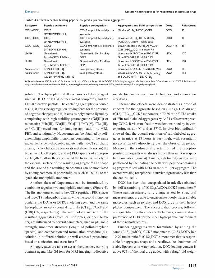
Table 3 Others receptor binding peptide coupled supramolecular aggregates
Receptor Peptide sequence Peptide conjugation Aggregates and lipid composition Drug References
CCK1–CCK2 CCK8 DYMGwMDF-NH2
CCK8 amphiphilic solid phase synthesis
Micelle: (C18)2(AdOO)5CCK8 DOX 90
CCK1–CCK2 CCK8 DYMGwMDF-NH2
CCK8 amphiphilic solid phase synthesis
Liposome: (C18)2DOTA, (C18)2
(AdOO)5CCK8 9:1 molar ratioDOX 91
CCK1–CCK2 CCK8 DYMGwMDF-NH2
CCK8 amphiphilic solid phase synthesis
Bilayer-liposome: (C18)2DTPAGlu/ (C18)2PeG2000CCK8 in ratio 7:3
DOX 111In 89
LHRH Gonadorelin Pyr-HwSTGLRPGNH2
Gonadorelin-SH- Mal-Peg- DSPe
Liposome: HSPC/Chol/mPeG-DSPe/ Gon-PeG-DSPe 90:10:0.4 0.1%
MTX 107
LHRH Gonadorelin Pyr-HwSTGLRPGNH2
Gonadorelin-SH- Mal-Peg- DSPe
Liposome: HSPC/Chol/mPeG-DSPe/ Gon-PeG-DSPe 90:10:0.4 0.1%
MTX 108
Neurotensin RRPYIL Nt[8–13] Solid phase synthesis Liposome: DOPC-NT4Lys(C18)2 97:3 DOX 111Neurotensin RRPYIL Nt[8–13],
QLYeNKPRRPYIL Nt[1–13]Solid phase synthesis Liposome: DOPC (NT8–13)L-(C18)2
and DOPC (NT1-13)L-(C18)2
DOX 112
Abbreviations: AdOO, 8-amino-3,6-dioxaoctanoic acid; CCK, cholecystokinin; DOPC, 1,2-Dioleoyl-sn-glycero-3-phosphocholine; DOX, doxorubicin; DSPe, 1,2-distearoyl-sn-glycero-3-phosphoethanolamine; LHRH, luteinizing hormone releasing hormone; MTX, methotrexate; PeG, polyethylene glycol.

International Journal of Nanomedicine 2014:9submit your manuscript | www.dovepress.com
Dovepress
Dovepress
1550
Accardo et al
Figure 4 Scheme of Naposomes formulation and their in vitro and in vivo behavior. Flow cytometric analysis of association of liposomal DOX and free DOX with human cells.Notes: A431 cells (A) and HUveC cells (B) at a density of 1.3 ⋅ 106 cells/mL were incubated with CCK8/DOTA-DOX, DOTA-DOX, and free DOX at a final concentration of 1 µg DOX/mL for 1 hour at 48°C. Untreated cells served as negative control while free doxorubicin solution was used as positive control. The untreated cells (negative controls) and cells incubated with non-specific DOTA-DOX give identical behavior with overlapping curves. (C and D) Cytotoxicity of liposomal DOX against human cells on 431 cells and HUveC, respectively. Cells were incubated with CCK8/DOTA-DOX and DOTA-DOX at different concentration ranging between 0 and 1,000 ng/mL at 37°C. After 8 hours, the medium was removed and after an additional 72 hours, an MTT assay was performed. Data are expressed as percent of negative control. (E) γ-camera image (dorsal view) obtained prior to dissection of one of the animals 18 h after injection of radiolabeled aggregates clearly shows higher concentration of the radiolabel in the receptor positive xenograft (+, left flank) compared with the control tumor (−, right flank). (A–D) Reproduced with permission from John wiley and Sons. Morisco A, Accardo A, Tesauro D, Palumbo R, Benedetti e, Morelli G. Peptide-labeled supramolecular aggregates as selective doxorubicin carriers for delivery to tumor cells. Biopolymers. 2011;96:88–96.91 Copyright © 2011 wiley Periodicals, Inc. (E) Reproduced with permission from John wiley and Sons. Accardo A, Tesauro D, Aloj L, et al. Peptide-containing aggregates as selective nanocarriers for therapeutics. ChemMedChem. 2008;3(4):594–602.89 Copyright © 2008 wILeY-vCH verlag GmbH & Co. KGaA, weinheim.Abbreviations: CCK, cholecystokinin; DOTA, 1,4,7,10-tetraazacyclododecane-N,N’,N’’,N’’’-tetraacetic acid; DOX, doxorubicin; HUveC, human umbilical vein endothelial cell; MTT, 3-(4,5-dimethyl-2-thiazolyl)-2,5-diphenyl-2H-tetrazolium bromide.
(w)/w ratio of 0.134. The cellular uptake of the peptide
containing targeted liposomal DOX on A431 and HUVEC
cells was 70- and 8-fold higher than that for non-targeted
liposomes, respectively, indicating that the bioactive CCK8
peptide is able to enhance uptake into the A431 carcinoma
cells and, at lower amounts, in the endothelial HUVEC cells
(Figure 4).
Bombesin based delivery systemsFour receptor-subtypes associated with the Bombesin
like peptides (BLP) family have been identified: sub-
type 1 (termed GRP-R or BB2); subtype 2 (termed NMB-R
or BB1); subtype 3 (termed BRS-3) classified as an orphan
receptor because its natural ligand is yet to be identified; and
subtype 4 (termed BB4). In addition to their physiological
functions, these receptors have been found overexpressed in
prostate, breast, small cell lung,92 ovarian, and gastrointestinal
stromal tumors.93
Peptides able to bind these receptors belong to a family
of brain-gut peptides. BN (bombesin) is a 14-amino-acid
peptide present in amphibian tissues, whereas GRP, its human
counterpart, consists of 27 amino acids. GRP and BN differ
International Journal of Nanomedicine 2014:9 submit your manuscript | www.dovepress.com
Dovepress
Dovepress
1551
Receptor binding peptides for nanoparticle encapsulated drugs
by only one of the ten C-terminal residues playing similar
biological activities.94 GRP acts primarily in the central and
enteric nervous systems where it regulates several physiologi-
cal processes including satiety, thermoregulation, circadian
rhythm, smooth muscle contraction, immune function, as
well as the release of other peptide hormones.
The fourteen BN residues, its eight-residues C-terminal
peptide sequence ([7–14]BN), and many other BN analogs
acting as agonist or antagonists, have been modified to
selectively carry diagnostic or therapeutic agents to their
receptors. Many studies demonstrate that the [7–14]BN
fragment conjugated on the N-terminus with amino acid
linkers, aliphatic or hydrophilic moiety preserves the affin-
ity for receptors.95,96 Moreover, the pan-bombesin analog
[β-Ala11, Phe13, Nle14] BN[7–14] conjugated through a
linker to dextran covalently cross-linked to iron oxide (CLIO)
is able to bind to PC-3 cells overexpressing GPR receptors,
as indicated by MRI studies.97
Liposomes and micellesOn the basis of MRI results, Accardo et al developed
new bombesin based supramolecular aggregates acting as
theranostic agents (Table 4).98 They were obtained by the
combination of two amphiphilic synthetic monomers: a first,
more abundant, monomer based on a lysine residue carrying
a DOTA chelating agent on the epsilon amino function and
an hydrophobic moiety with two C18 chains on the alpha
amino function; and a second monomer containing the same
hydrophobic moiety, PEG spacers, and the 7–14 BN peptide
fragment. The DOTA containing monomer drives to form
stable liposomes in water solution independently from the
presence of 10% in peptide monomer, as demonstrated by
SANS (small angle neutron scattering) and DLS (dynamic
light scattering) techniques. The liposome hydrodynamic
radius and bilayer thickness were found to be around 200 nm
and 4 nm, respectively. This structure is different from that
observed for similar aggregates, in which the presence of
DTPAGlu chelating agent in the most abundant amphiphilic
monomer produces highly polydisperse aggregates (rod-like
micelles, open bilayers, and vesicles). This behavior could be
explained on the basis of the lower negative charge (−3) of
DOTA versus DTPAGlu (−5); a decrease of the electrostatic
repulsion between the headgroups favors the formation
of large and low curvature aggregates, such as liposomes.
Different systems were studied depending on the length
of the PEG spacer in the peptide containing monomer.
In vitro data of radioactive labeled 111In-(C18)2DOTA/
(C18)2AdOO
5-[7–14]BN liposomes show specific binding
to receptor expressing cells, while the presence of a longer
PEG (Peg3000) on the external liposomal surface hides the
bioactive peptide, preventing receptor binding. In vivo experi-
ments display the expected biological behavior of aggregates
of such size and molecular composition, and preliminarily
confirm the aggregates’ ability to specifically target receptor
expressing xenografts. At later stages, liposomes based on co-
aggregation of 1,2-distearoyl-sn-glycero-3-phosphocholine
(DSPC) phospholipid with the amphiphilic synthetic mono-
mer MonY-BN were developed.99 This monomer contains,
in a single molecule, the [7–14]BN peptide fragment, the
DTPA chelating agent, the hydrophobic moiety with two
C18 alkyl chains, and a PEG spacer of 1500 Daltons. DOX
loading capability of DSPC/MonY-BN (97:3 molar ratio)
liposomes is 0.20 (drug/lipid, w/w), higher than that found for
the approved liposomal drug DOXIL (0.125). The selective
liposome binding was evaluated in vitro by gamma count-
ing experiments after radiolabeling of liposomes with 111In
isotopes. Cytotoxic assays showed significantly lower cell
survival after cell incubation with DSPC/MonY-BN/DOX
liposomes, compared to DSPC/DOX treated cells. Intrave-
nous treatment of PC-3 xenograft-bearing mice produced
Table 4 Bombesin analogs labeled supramolecular aggregates or nanoparticles
Peptide sequence Peptide conjugation Aggregates and lipid composition Drug References
[7–14] BN QwAvGHML-NH2
BN amphiphilic solid phase synthesis
Liposome: (C18)2DOTA/ (C18)2(AdOO)5BN and (C18)2DOTA/ (C18)2Peg3000BN (9:1 molar ratio)
DOX 98
[7–14] BN QwAvGHML-NH2
BN amphiphilic solid phase synthesis
Liposome: DSPC/MonY-BN (1:0.03 molar ratio)
DOX 99
AhoH-DPheQwAvNMeGHSta- L-NH2
BN analog amphiphilic solid phase synthesis
Liposome: DSPC/MonY-BN-AA1 (1:0.03 molar ratio)
DOX 101
Ac-Cys-Ahx-QwAvGHLMNH2 Ac-Cys-Ahx-BN NH2-AuNP
AuNPs 104
7–14 BN QwAvGHML-NH2
7–14 BN functionalized on N-terminus with lipoic acid AuNPs
AuNPs 103
Abbreviations: AdOO, 8-amino-3,6-dioxaoctanoic acid; AhOH, 21-amino-4,7,10,13,16,19-hexaoxaheneicosanoic acid; AuNP, gold nanoparticles; Ahx, aminohexanoic acid; BN, bombesin; DOX, doxorubicin; DSPC, 1,2-Distearoyl-sn-glycero-3-phosphocholine.

International Journal of Nanomedicine 2014:9submit your manuscript | www.dovepress.com
Dovepress
Dovepress
1552
Accardo et al
higher tumor growth inhibition (60%) compared with non-
specific liposomes (36%) relative to control animals.
The relatively short in vivo circulation time of this natural
peptide fragment suggested to many researchers that modi-
fying peptide sequences can enhance protease stability.100
A very recent attempt was carried out, replacing the Leu13-Met14
C-terminal sequence with Sta13-Leu14, for stabilization against
aminopeptidase, and inserting N-methyl-glycine in place of
natural glycine in order to reinforce the Val-Gly bond that
could be sensitive to carnitine enzyme.100 Moreover, Mansi
et al100 demonstrated that replacement of Leu13 with the Sta13
residue provides antagonist properties to the peptide. Finally,
the presence of D-Phe residue on the N-terminal end of the
BN bioactive sequence increases binding and stability. DSPC/
MonY-BN-AA1/DOX liposomes containing the modified
BN-AA1 bombesin sequence were able to target PC-3 cells
in a selective way and provide therapeutic efficacy in PC-3
cells and PC-3 xenograft bearing mice to a slightly greater
extent than DSPC/MonY-BN/DOX liposomes.101
Metal nanoparticlesSeveral metal nanoparticles were labeled to target BN recep-
tors for tumor therapy and treatment monitoring.102 AuNPs
have been used in photothermal therapy for the destruction
or molecular surgery of cancer cells or tumors. When irradi-
ated with a focused laser in the near-infrared region (NIR) of
suitable wavelength, targeted aggregates of AuNPs can kill
cancer cells. At the same time, they are carriers of anticancer
drugs or contrast agents, providing synergistic advantages in
oncology as it relates to molecular imaging and therapy.
Chanda et al103 synthesized a library of GRP receptor
nanoplatforms by conjugating AuNPs with BN peptides. The
7–14 BN peptide was functionalized on N-terminus with lipoic
acid, which contains a disulfide group able to bind AuNPs.
Reactive sites on AuNPs surface allow the incorporation of
varying amounts of BN peptides and provide a library of
AuNP-BN conjugates with different ratios. The hydrodynamic
diameter of AuNP-BNs (115–155 nm) is compatible for effec-
tive penetration within tumor vasculature, which has porosity
in the 150–300 nm range. In vitro cellular interactions and
binding affinities (IC50
) toward GRP receptors on human pros-
tate cancer cells and in vivo studies using AuNP-BN and its
radiolabeled surrogate198 exhibited high binding affinity (IC50
in microgram ranges), providing unambiguous evidences that
AuNP-BN constructs are GRP-receptor-specific. Indeed, the
nanoparticles were accumulated with high selectivity in GRP-
receptor-rich pancreatic acne in normal mice and in tumor cells
of prostate-tumor-bearing, severe combined immunodeficient
mice. More recently, Hosta-Rigau et al104 exploited the ability
of BN labeled AuNPs to vehicle an analog of the RAF peptide.
This pharmaceutical active peptide ligand is able to inhibit
Rb-Raf-1 binding in vivo and therefore inhibits tumor growth
and angiogenesis.105 BN and RAF peptides were conjugated to
nanoparticles by modifying gold surface with Cys residues and
an aminohexanoic acid (Ahx) acting as spacer. Internalization
mechanism of peptide-AuNP conjugates and enhancement of
activity and selectivity of peptide multifunctionalized conju-
gates was observed by confocal laser scanning microscopy.
Preliminary results confirm that conjugates in which BN is
present penetrate GRPr overexpressing cells, as indicated by
coloration of nanoconjugates of Ac-Cys-Ahx-BN and Ac-Cys-
Ahx-RAF inside cells due to the accumulation and reflection
of the AuNP. The enhancement in activity and selectivity could
contribute to a potential improvement of the efficacy of RAF
for therapy by reducing the therapeutic index. Furthermore,
this strategy provides an opportunity for the controlled deliv-
ery of AuNPs used as cargoes for a localized (nanometrically)
therapy like the so-called molecular surgery.
Other systemsLutein releasing hormone based delivery systemsLuteinizing hormone (LH)-releasing hormone (also referred
to as GNRH or LHRH) is the central regulator of reproduc-
tion via its action upon the hypothalamic-pituitary axis. The
LHRH receptors are characteristically overexpressed in
many different tumors, such as breast, ovarian, endometrial,
and prostate cancers, but barely expressed in healthy vis-
ceral organs. The elevated expression of LHRH receptors
in various cancers makes it possible to use them as target
moieties to deliver cytotoxic agents to these tumors.106 Some
small peptide LHRH analogs were able to recognize a broad
variety of tumors, but not normal cells. Targeting these small
peptides has certain advantages, including ease of prepara-
tion, lower antigenicity, and increased stability over the use
of conventional protein macromolecules.
Liposomes were prepared using a lipid molar ratio HSPC/
Chol/mPEG–DSPE 90:10:0.4, and 0.1% mol Mal-PEG–DSPE
was further inserted for ligand conjugation. Gonadorelin
(Pyr-His-Trp-Ser-Tyr-Gly-Leu-Arg-Pro-Gly-NH2) was first
thiolated through incubation with Traut’s reagent. Thiolated
gonadorelin was chemically coupled to N-[(3-maleimide-
1-oxopropyl) aminopropyl polyethylene glycol-carbamyl]
distearoylphosphatidylethanolamine via a thioether bond
and subsequently inserted into polyethylene glycol-grafted
liposomes. Efficient transfer of gonadorelin-PEG-DSPE from
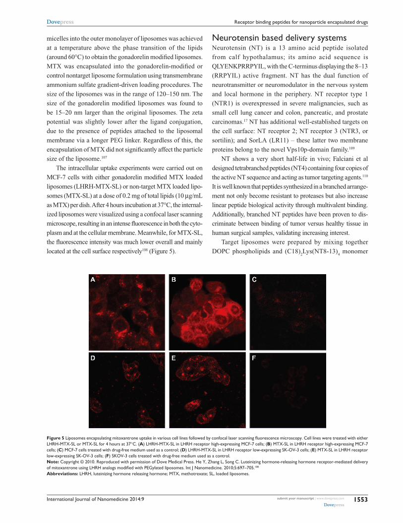
International Journal of Nanomedicine 2014:9 submit your manuscript | www.dovepress.com
Dovepress
Dovepress
1553
Receptor binding peptides for nanoparticle encapsulated drugs
micelles into the outer monolayer of liposomes was achieved
at a temperature above the phase transition of the lipids
(around 60°C) to obtain the gonadorelin modified liposomes.
MTX was encapsulated into the gonadorelin-modified or
control nontarget liposome formulation using transmembrane
ammonium sulfate gradient-driven loading procedures. The
size of the liposomes was in the range of 120–150 nm. The
size of the gonadorelin modified liposomes was found to
be 15–20 nm larger than the original liposomes. The zeta
potential was slightly lower after the ligand conjugation,
due to the presence of peptides attached to the liposomal
membrane via a longer PEG linker. Regardless of this, the
encapsulation of MTX did not significantly affect the particle
size of the liposome.107
The intracellular uptake experiments were carried out on
MCF-7 cells with either gonadorelin modified MTX loaded
liposomes (LHRH-MTX-SL) or non-target MTX loaded lipo-
somes (MTX-SL) at a dose of 0.2 mg of total lipids (10 µg/mL
as MTX) per dish. After 4 hours incubation at 37°C, the internal-
ized liposomes were visualized using a confocal laser scanning
microscope, resulting in an intense fluorescence in both the cyto-
plasm and at the cellular membrane. Meanwhile, for MTX-SL,
the fluorescence intensity was much lower overall and mainly
located at the cell surface respectively108 (Figure 5).
Neurotensin based delivery systemsNeurotensin (NT) is a 13 amino acid peptide isolated
from calf hypothalamus; its amino acid sequence is
QLYENKPRRPYIL, with the C-terminus displaying the 8–13
(RRPYIL) active fragment. NT has the dual function of
neurotransmitter or neuromodulator in the nervous system
and local hormone in the periphery. NT receptor type 1
(NTR1) is overexpressed in severe malignancies, such as
small cell lung cancer and colon, pancreatic, and prostate
carcinomas.17 NT has additional well-established targets on
the cell surface: NT receptor 2; NT receptor 3 (NTR3, or
sortilin); and SorLA (LR11) – these latter two membrane
proteins belong to the novel Vps10p-domain family.109
NT shows a very short half-life in vivo; Falciani et al
designed tetrabranched peptides (NT4) containing four copies of
the active NT sequence and acting as tumor targeting agents.110
It is well known that peptides synthesized in a branched arrange-
ment not only become resistant to proteases but also increase
linear peptide biological activity through multivalent binding.
Additionally, branched NT peptides have been proven to dis-
criminate between binding of tumor versus healthy tissue in
human surgical samples, validating increasing interest.
Target liposomes were prepared by mixing together
DOPC phospholipids and (C18)2Lys(NT8-13)
4 monomer
Figure 5 Liposomes encapsulating mitoxantrone uptake in various cell lines followed by confocal laser scanning fluorescence microscopy. Cell lines were treated with either LHRH-MTX-SL or MTX-SL for 4 hours at 37°C. (A) LHRH-MTX-SL in LHRH receptor high-expressing MCF-7 cells; (B) MTX-SL in LHRH receptor high-expressing MCF-7 cells; (C) MCF-7 cells treated with drug-free medium used as a control; (D) LHRH-MTX-SL in LHRH receptor low-expressing SK-Ov-3 cells; (E) MTX-SL in LHRH receptor low-expressing SK-Ov-3 cells; (F) SKOv-3 cells treated with drug-free medium used as a control.Note: Copyright © 2010. Reproduced with permission of Dove Medical Press. He Y, Zhang L, Song C. Luteinizing hormone-releasing hormone receptor-mediated delivery of mitoxantrone using LHRH analogs modi fied with PEGylated liposomes. Int J Nanomedicine. 2010;5:697–705.108
Abbreviations: LHRH, luteinizing hormone releasing hormone; MTX, methotrexate; SL, loaded liposomes.

International Journal of Nanomedicine 2014:9submit your manuscript | www.dovepress.com
Dovepress
Dovepress
1554
Accardo et al
Figure 6 Confocal microscopy of (A) HT29 and (B) Te671 cells incubated with DOPC-(C18)2Lys(NT8-13)4-DOX liposomes (200 nm, right) and with DOPC-DOX liposomes (200 nm, left) for 2 hours at 37°C. Plasma membranes were stained with lectin-FITC (green).Note: Reproduced with permission from Falciani C, Accardo A, Brunetti J, et al. Target-selective drug delivery through liposomes labeled with oligobranched neurotensin pep tides. ChemMedChem. 2011;6(4):678–685.111 Copyright © 2011 wILeY-vCH verlag GmbH & Co. KGaA, weinheim.Abbreviations: DOPC, 1,2-dioleoyl-sn-glycero-3-phosphocholine; DOX, doxo ru - bicin; FITC, fluorescein isothiocyanate.
obtained by using a solid phase synthesis.111 In vitro cytotoxic
results of functionalized liposomes loaded with DOX show a
clear advantage, in comparison to native liposomes, in tumor
cell drug internalization, both in HT29 and TE671 cells.
Fluorescent-activated cell sorting (FACS) analyses are in line
with these results, showing a fluorescence signal increase in
both cell lines when NT4 decorated liposomes are compared
to the non-functionalized analogs (Figure 6). All of these
effects can be attributed to a higher rate of internalization
of the decorated liposomes.
Recently, the comparison of the branched (NT4) versus
linear (NT) peptides demonstrated liposomes decorated with
branched peptides present a better profile in drug delivery,
with respect to liposomes decorated with the correspondent
monomeric peptides.112
Conclusion and future perspectivesBeside new therapies and new drugs, the innovative adminis-
tration methods of well tested active principles can represent
an additional weapon in the fight against cancer. Compared to
conventional small molecule-based therapy, nano-therapeutic
systems have several potential advantages: they can remain in
the circulation for an extended period of time when injected
intravenously, and present high payload capacity, reduced
toxicity to healthy tissues, and improved antitumor efficacy.
The active targeting by means of drug encapsulated nanopar-
ticles decorated with targeting bioactive moieties represents
the next frontier in drug delivery: it reduces drug side effects
and increases the therapeutic index. Peptides, based on their
chemical and biological properties, could have a prevalent
role in directing drug encapsulated nanoparticles, such as
liposomes, micelles, or hard nanoparticles, toward tumor tis-
sues. Therapeutic agents based on nanovectors decorated with
peptides targeting GPCRs membrane receptors overexpressed
by cancer cells have been reviewed in this article. Despite the
promising in vitro and in vivo results here described, all com-
pounds reported in literature are still in preclinical phases. For
most of the described systems, it is possible to schedule Phase
I clinical trials, which can definitively legitimize the use of
peptide decorated nanoparticles as target selective delivery
systems for cancer therapy. Moreover, many efforts should
be made to search for other peptide sequences to decorate
drug encapsulated nanovectors.
AcknowledgmentsThe authors would like to thank Italian Minister for Research
(MIUR) for financial support under PRIN 2009WCNS5C
and FIRB RBRN07BMCT founding projects.
DisclosureThe authors report no conflicts of interest in this work.
References1. Richardson PF. Nanotechnology therapeutics in oncology- recent
Developments and future outlook. Annu Rep Med Chem. 2012;47: 239–252.
2. Levchenko TS, Hartner WC, Torchilin VP. Liposomes for cardiovascular targeting. Ther Deliv. 2012;3(4):501–514.
3. Maradana MR, Thomas R, O’Sullivan BJ. Targeted delivery of curcumin for treating type 2 diabetes. Mol Nutr Food Res. 2013;57(9): 1550–1556.
4. Levesque JP, Winkler IG. It takes nerves to recover from chemotherapy. Nat Med. 2013;19(6):669–671.
5. Matsumura Y, Maeda H. A new concept for macromolecular therapeutics in cancer chemotherapy: mechanism of tumoritropic accumulation of proteins and the antitumor agent smancs. Cancer Res. 1986;46(12 Pt 1):6387–6392.
6. Salmaso S, Caliceti P. Stealth properties to improve therapeutic efficacy of drug nanocarriers. J Drug Deliv. 2013;2013:1–19.
7. Gabizon AA. Liposome circulation time and tumor targeting: implications for cancer chemotherapy. Adv Drug Deliv Rev. 1995;16:285–294.
8. Aliabadi HM, Shahin M, Brocks DR, Lavasanifar A. Disposition of drugs in block copolymer micelle delivery systems: from discovery to recovery. Clin Pharmacokinet. 2008;47(10):619–634.
9. Kwon GS, Kataoka K. Block copolymer micelles as long-circulating drug vehicles. Adv Drug Delivery Rev. 1995;16:295–309.
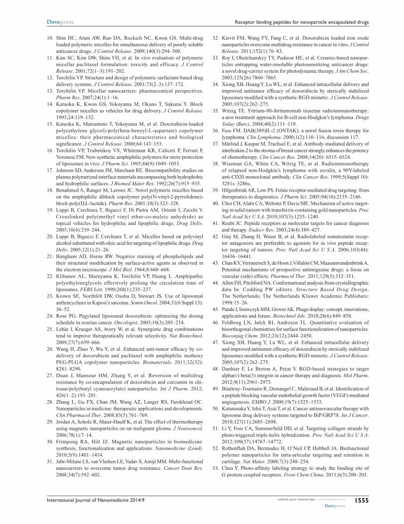
International Journal of Nanomedicine 2014:9 submit your manuscript | www.dovepress.com
Dovepress
Dovepress
1555
Receptor binding peptides for nanoparticle encapsulated drugs
10. Shin HC, Alani AW, Rao DA, Rockich NC, Kwon GS. Multi-drug loaded polymeric micelles for simultaneous delivery of poorly soluble anticancer drugs. J Control Release. 2009;140(3):294–300.
11. Kim SC, Kim DW, Shim YH, et al. In vivo evaluation of polymeric micellar paclitaxel formulation: toxicity and efficacy. J Control Release. 2001;72(1–3):191–202.
12. Torchilin VP. Structure and design of polymeric surfactant-based drug delivery systems. J Control Release. 2001;73(2–3):137–172.
13. Torchilin VP. Micellar nanocarriers: pharmaceutical perspectives. Pharm Res. 2007;24(1):1–16.
14. Kataoka K, Kwon GS, Yokoyama M, Okano T, Sakurai Y. Block copolymer micelles as vehicles for drug delivery. J Control Release. 1993;24:119–132.
15. Kataoka K, Matsumoto T, Yokoyama M, et al. Doxorubicin-loaded poly(ethylene glycol)-poly(beta-benzyl-L-aspartate) copolymer micelles: their pharmaceutical characteristics and biological significance. J Control Release. 2000;64:143–153.
16. Torchilin VP, Trubetskoy VS, Whiteman KR, Caliceti P, Ferruti P, Veronese FM. New synthetic amphiphilic polymers for steric protection of liposomes in vivo. J Pharm Sci. 1995;84(9):1049–1053.
17. Johnson SD, Anderson JM, Marchant RE. Biocompatibility studies on plasma polymerized interface materials encompassing both hydrophobic and hydrophilic surfaces. J Biomed Mater Res. 1992;26(7):915–935.
18. Benahmed A, Ranger M, Leroux JC. Novel polymeric micelles based on the amphiphilic diblock copolymer poly(N-vinyl-2-pyrrolidone)-block-poly(D,L-lactide). Pharm Res. 2001;18(3):323–328.
19. Luppi B, Cerchiara T, Bigucci F, Di Pietra AM, Orienti I, Zecchi V. Crosslinked poly(methyl vinyl ether-co-maleic anhydride) as topical vehicles for hydrophilic and lipophilic drugs. Drug Deliv. 2003;10(4):239–244.
20. Luppi B, Bigucci F, Cerchiara T, et al. Micelles based on polyvinyl alcohol substituted with oleic acid for targeting of lipophilic drugs. Drug Deliv. 2005;12(1):21–26.
21. Bangham AD, Horne RW. Negative staining of phospholipids and their structural modification by surface-active agents as observed in the electron microscope. J Mol Biol. 1964;8:660–668.
22. Klibanov AL, Maruyama K, Torchilin VP, Huang L. Amphipathic polyethyleneglycols effectively prolong the circulation time of liposomes. FEBS Lett. 1990;268(1):235–237.
23. Krown SE, Northfelt DW, Osoba D, Stewart JS. Use of liposomal anthracyclines in Kaposi’s sarcoma. Semin Oncol. 2004;31(6 Suppl 13): 36–52.
24. Rose PG. Pegylated liposomal doxorubicin: optimizing the dosing schedule in ovarian cancer. Oncologist. 2005;10(3):205–214.
25. Lehár J, Krueger AS, Avery W, et al. Synergistic drug combinations tend to improve therapeutically relevant selectivity. Nat Biotechnol. 2009;27(7):659–666.
26. Wang H, Zhao Y, Wu Y, et al. Enhanced anti-tumor efficacy by co- delivery of doxorubicin and paclitaxel with amphiphilic methoxy PEG-PLGA copolymer nanoparticles. Biomaterials. 2011;32(32): 8281–8290.
27. Duan J, Mansour HM, Zhang Y, et al. Reversion of multidrug resistance by co-encapsulation of doxorubicin and curcumin in chi-tosan/poly(butyl cyanoacrylate) nanoparticles. Int J Pharm. 2012; 426(1–2):193–201.
28. Zhang L, Gu FX, Chan JM, Wang AZ, Langer RS, Farokhzad OC. Nanoparticles in medicine: therapeutic applications and developments. Clin Pharmacol Ther. 2008;83(5):761–769.
29. Jordan A, Scholz R, Maier-Hauff K, et al. The effect of thermotherapy using magnetic nanoparticles on rat malignant glioma. J Neurooncol. 2006;78(1):7–14.
30. Frimpong RA, Hilt JZ. Magnetic nanoparticles in biomedicine: synthesis, functionalization and applications. Nanomedicine (Lond). 2010;5(9):1401–1414.
31. Jabr-Milane LS, van Vlerken LE, Yadav S, Amiji MM. Multi-functional nanocarriers to overcome tumor drug resistance. Cancer Treat Rev. 2008;34(7):592–602.
32. Kievit FM, Wang FY, Fang C, et al. Doxorubicin loaded iron oxide nanoparticles overcome multidrug resistance in cancer in vitro. J Control Release. 2011;152(1):76–83.
33. Roy I, Ohulchanskyy TY, Pudavar HE, et al. Ceramic-based nanopar-ticles entrapping water-insoluble photosensitizing anticancer drugs: a novel drug-carrier system for photodynamic therapy. J Am Chem Soc. 2003;125(26):7860–7865.
34. Xiong XB, Huang Y, Lu WL, et al. Enhanced intracellular delivery and improved antitumor efficacy of doxorubicin by sterically stabilized liposomes modified with a synthetic RGD mimetic. J Control Release. 2005;107(2):262–275.
35. Witzig TE. Yttrium-90-ibritumomab tiuxetan radioimmunotherapy: a new treatment approach for B-cell non-Hodgkin’s lymphoma. Drugs Today (Barc). 2004;40(2):111–119.
36. Foss FM. DAB(389)IL-2 (ONTAK): a novel fusion toxin therapy for lymphoma. Clin Lymphoma. 2000;1(2):110–116; discussion 117.
37. Mårlind J, Kaspar M, Trachsel E, et al. Antibody-mediated delivery of interleukin-2 to the stroma of breast cancer strongly enhances the potency of chemotherapy. Clin Cancer Res. 2008;14(20): 6515–6524.
38. Wiseman GA, White CA, Witzig TE, et al. Radioimmunotherapy of relapsed non-Hodgkin’s lymphoma with zevalin, a 90Y-labeled anti-CD20 monoclonal antibody. Clin Cancer Res. 1999;5(Suppl 10): 3281s–3286s.
39. Hilgenbrink AR, Low PS. Folate receptor-mediated drug targeting: from therapeutics to diagnostics. J Pharm Sci. 2005;94(10):2135–2146.
40. Choi CH, Alabi CA, Webster P, Davis ME. Mechanism of active target-ing in solid tumors with transferrin-containing gold nanoparticles. Proc Natl Acad Sci U S A. 2010;107(3):1235–1240.
41. Reubi JC. Peptide receptors as molecular targets for cancer diagnosis and therapy. Endocr Rev. 2003;24(4):389–427.
42. Ginj M, Zhang H, Waser B, et al. Radiolabeled somatostatin recep-tor antagonists are preferable to agonists for in vivo peptide recep-tor targeting of tumors. Proc Natl Acad Sci U S A. 2006;103(44): 16436–16441.
43. Chan KY, Vermeersch S, de Hoon J, Villalón CM, Maassenvandenbrink A. Potential mechanisms of prospective antimigraine drugs: a focus on vascular (side) effects. Pharmacol Ther. 2011;129(3):332–351.
44. Allen FH, Pitchford NA. Conformational analysis from crystallographic data In: Codding PW editors. Structure Based Drug Design. The Netherlands; The Netherlands Kluwer Academic Publishers: 1998:15–26.
45. Pande J, Szewczyk MM, Grover AK. Phage display: concept, innovations, applications and future. Biotechnol Adv. 2010;28(6):849–858.
46. Feldborg LN, Jølck RI, Andresen TL. Quantitative evaluation of bioorthogonal chemistries for surface functionalization of nanoparticles. Bioconjug Chem. 2012;23(12):2444–2450.
47. Xiong XB, Huang Y, Lu WL, et al. Enhanced intracellular delivery and improved antitumor efficacy of doxorubicin by sterically stabilized liposomes modified with a synthetic RGD mimetic. J Control Release. 2005;107(2):262–275.
48. Danhier F, Le Breton A, Préat V. RGD-based strategies to target alpha(v) beta(3) integrin in cancer therapy and diagnosis. Mol Pharm. 2012;9(11):2961–2973.
49. Binétruy-Tournaire R, Demangel C, Malavaud B, et al. Identification of a peptide blocking vascular endothelial growth factor (VEGF)-mediated angiogenesis. EMBO J. 2000;19(7):1525–1533.
50. Katanasaka Y, Ishii T, Asai T, et al. Cancer antineovascular therapy with liposome drug delivery systems targeted to BiP/GRP78. Int J Cancer. 2010;127(11):2685–2698.
51. Li Y, Foss CA, Summerfield DD, et al. Targeting collagen strands by photo-triggered triple-helix hybridization. Proc Natl Acad Sci U S A. 2012;109(37):14767–14772.
52. Rothenfluh DA, Bermudez H, O’Neil CP, Hubbell JA. Biofunctional polymer nanoparticles for intra-articular targeting and retention in cartilage. Nat Mater. 2008;7(3):248–254.
53. Chen Y. Photo-affinity labeling strategy to study the binding site of G protein-coupled receptors. Front Chem China. 2011;6(3):200–205.
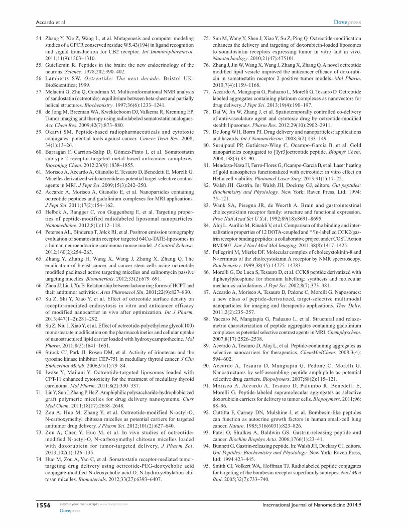
International Journal of Nanomedicine 2014:9submit your manuscript | www.dovepress.com
Dovepress
Dovepress
1556
Accardo et al
54. Zhang Y, Xie Z, Wang L, et al. Mutagenesis and computer modeling studies of a GPCR conserved residue W5.43(194) in ligand recognition and signal transduction for CB2 receptor. Int Immunopharmacol. 2011;11(9):1303–1310.
55. Guiellemin R. Peptides in the brain: the new endocrinology of the neurons. Science. 1978;202:390–402.
56. Lamberts SW. Octreotide: The next decade . Bristol UK: BioScientifica; 1999.
57. Melacini G, Zhu Q, Goodman M. Multiconformational NMR analysis of sandostatin (octreotide): equilibrium between beta-sheet and partially helical structures. Biochemistry. 1997;36(6):1233–1241.
58. de Jong M, Breeman WA, Kwekkeboom DJ, Valkema R, Krenning EP. Tumor imaging and therapy using radiolabeled somatostatin analogues. Acc Chem Res. 2009;42(7):873–880.
59. Okarvi SM. Peptide-based radiopharmaceuticals and cytotoxic conjugates: potential tools against cancer. Cancer Treat Rev. 2008; 34(1):13–26.
60. Barragán F, Carrion-Salip D, Gómez-Pinto I, et al. Somatostatin subtype-2 receptor-targeted metal-based anticancer complexes. Bioconjug Chem. 2012;23(9):1838–1855.
61. Morisco A, Accardo A, Gianolio E, Tesauro D, Benedetti E, Morelli G. Micelles derivatized with octreotide as potential target-selective contrast agents in MRI. J Pept Sci. 2009;15(3):242–250.
62. Accardo A, Morisco A, Gianolio E, et al. Nanoparticles containing octreotide peptides and gadolinium complexes for MRI applications. J Pept Sci. 2011;17(2):154–162.
63. Helbok A, Rangger C, von Guggenberg E, et al. Targeting proper-ties of peptide-modif ied radiolabeled liposomal nanoparticles. Nanomedicine. 2012;8(1):112–118.
64. Petersen AL, Binderup T, Jølck RI, et al. Positron emission tomography evaluation of somatostatin receptor targeted 64Cu-TATE-liposomes in a human neuroendocrine carcinoma mouse model. J Control Release. 2012;160(2):254–263.
65. Zhang Y, Zhang H, Wang X, Wang J, Zhang X, Zhang Q. The eradication of breast cancer and cancer stem cells using octreotide modified paclitaxel active targeting micelles and salinomycin passive targeting micelles. Biomaterials. 2012;33(2):679–691.
66. Zhou JJ, Liu J, Xu B. Relationship between lactone ring forms of HCPT and their antitumor activities. Acta Pharmacol Sin. 2001;22(9):827–830.
67. Su Z, Shi Y, Xiao Y, et al. Effect of octreotide surface density on receptor-mediated endocytosis in vitro and anticancer eff icacy of modified nanocarrier in vivo after optimization. Int J Pharm. 2013;447(1–2):281–292.
68. Su Z, Niu J, Xiao Y, et al. Effect of octreotide-polyethylene glycol(100) monostearate modification on the pharmacokinetics and cellular uptake of nanostructured lipid carrier loaded with hydroxycamptothecine. Mol Pharm. 2011;8(5):1641–1651.
69. Strock CJ, Park JI, Rosen DM, et al. Activity of irinotecan and the tyrosine kinase inhibitor CEP-751 in medullary thyroid cancer. J Clin Endocrinol Metab. 2006;91(1):79–84.
70. Iwase Y, Maitani Y. Octreotide-targeted liposomes loaded with CPT-11 enhanced cytotoxicity for the treatment of medullary thyroid carcinoma. Mol Pharm. 2011;8(2):330–337.
71. Liu Y, Sun J, Zhang P, He Z. Amphiphilic polysaccharide- hydrophobicized graft polymeric micelles for drug delivery nanosystems. Curr Med Chem. 2011;18(17):2638–2648.
72. Zou A, Huo M, Zhang Y, et al. Octreotide-modified N-octyl-O, N-carboxymethyl chitosan micelles as potential carriers for targeted antitumor drug delivery. J Pharm Sci. 2012;101(2):627–640.
73. Zou A, Chen Y, Huo M, et al. In vivo studies of octreotide-modified N-octyl-O, N-carboxymethyl chitosan micelles loaded with doxorubicin for tumor-targeted delivery. J Pharm Sci. 2013;102(1):126–135.
74. Huo M, Zou A, Yao C, et al. Somatostatin receptor-mediated tumor-targeting drug delivery using octreotide-PEG-deoxycholic acid conjugate-modified N-deoxycholic acid-O, N-hydroxyethylation chi-tosan micelles. Biomaterials. 2012;33(27):6393–6407.
75. Sun M, Wang Y, Shen J, Xiao Y, Su Z, Ping Q. Octreotide-modification enhances the delivery and targeting of doxorubicin-loaded liposomes to somatostatin receptors expressing tumor in vitro and in vivo. Nanotechnology. 2010;21(47):475101.
76. Zhang J, Jin W, Wang X, Wang J, Zhang X, Zhang Q. A novel octreotide modified lipid vesicle improved the anticancer efficacy of doxorubi-cin in somatostatin receptor 2 positive tumor models. Mol Pharm. 2010;7(4):1159–1168.
77. Accardo A, Mangiapia G, Paduano L, Morelli G, Tesauro D. Octreotide labeled aggregates containing platinum complexes as nanovectors for drug delivery. J Pept Sci. 2013;19(4):190–197.
78. Dai W, Jin W, Zhang J, et al. Spatiotemporally controlled co-delivery of anti-vasculature agent and cytotoxic drug by octreotide-modified stealth liposomes. Pharm Res. 2012;29(10):2902–2911.
79. De Jong WH, Borm PJ. Drug delivery and nanoparticles: applications and hazards. Int J Nanomedicine. 2008;3(2):133–149.
80. Surujpaul PP, Gutiérrez-Wing C, Ocampo-García B, et al. Gold nanoparticles conjugated to [Tyr3]octreotide peptide. Biophys Chem. 2008;138(3):83–90.
81. Mendoza-Nava H, Ferro-Flores G, Ocampo-García B, et al. Laser heating of gold nanospheres functionalized with octreotide: in vitro effect on HeLa cell viability. Photomed Laser Surg. 2013;31(1):17–22.
82. Walsh JH. Gastrin. In: Walsh JH, Dockray GJ, editors. Gut peptides: Biochemistry and Physiology. New York: Raven Press, Ltd; 1994: 75–121.
83. Wank SA, Pisegna JR, de Weerth A. Brain and gastrointestinal cholecystokinin receptor family: structure and functional expression. Proc Natl Acad Sci U S A. 1992;89(18):8691–8695.
84. Aloj L, Aurilio M, Rinaldi V, et al. Comparison of the binding and inter-nalization properties of 12 DOTA-coupled and ¹¹¹In-labelled CCK2/gas-trin receptor binding peptides: a collaborative project under COST Action BM0607. Eur J Nucl Med Mol Imaging. 2011;38(8):1417–1425.
85. Pellegrini M, Mierke DF. Molecular complex of cholecystokinin-8 and N-terminus of the cholecystokinin A receptor by NMR spectroscopy. Biochemistry. 1999;38(45):14775–14783.
86. Morelli G, De Luca S, Tesauro D, et al. CCK8 peptide derivatized with diphenylphosphine for rhenium labelling: synthesis and molecular mechanics calculations. J Pept Sci. 2002;8(7):373–381.
87. Accardo A, Morisco A, Tesauro D, Pedone C, Morelli G. Naposomes: a new class of peptide-derivatized, target-selective multimodal nanoparticles for imaging and therapeutic applications. Ther Deliv. 2011;2(2):235–257.
88. Vaccaro M, Mangiapia G, Paduano L, et al. Structural and relaxo-metric characterization of peptide aggregates containing gadolinium complexes as potential selective contrast agents in MRI. Chemphyschem. 2007;8(17):2526–2538.
89. Accardo A, Tesauro D, Aloj L, et al. Peptide-containing aggregates as selective nanocarriers for therapeutics. ChemMedChem. 2008;3(4): 594–602.
90. Accardo A, Tesauro D, Mangiapia G, Pedone C, Morelli G. Nanostructures by self-assembling peptide amphiphile as potential selective drug carriers. Biopolymers. 2007;88(2):115–121.
91. Morisco A, Accardo A, Tesauro D, Palumbo R, Benedetti E, Morelli G. Peptide-labeled supramolecular aggregates as selective doxorubicin carriers for delivery to tumor cells. Biopolymers. 2011;96: 88–96.
92. Cuttitta F, Carney DN, Mulshine J, et al. Bombesin-like peptides can function as autocrine growth factors in human small-cell lung cancer. Nature. 1985;316(6031):823–826.
93. Patel O, Shulkes A, Baldwin GS. Gastrin-releasing peptide and cancer. Biochim Biophys Acta. 2006;1766(1):23–41.
94. Bunnett G. Gastrin-releasing peptide. In: Walsh JH, Dockray GJ, editors. Gut Peptides: Biochemistry and Physiology. New York: Raven Press, Ltd; 1994:423–445.
95. Smith CJ, Volkert WA, Hoffman TJ. Radiolabeled peptide conjugates for targeting of the bombesin receptor superfamily subtypes. Nucl Med Biol. 2005;32(7):733–740.

International Journal of Nanomedicine
Publish your work in this journal
Submit your manuscript here: http://www.dovepress.com/international-journal-of-nanomedicine-journal
The International Journal of Nanomedicine is an international, peer-reviewed journal focusing on the application of nanotechnology in diagnostics, therapeutics, and drug delivery systems throughout the biomedical field. This journal is indexed on PubMed Central, MedLine, CAS, SciSearch®, Current Contents®/Clinical Medicine,
Journal Citation Reports/Science Edition, EMBase, Scopus and the Elsevier Bibliographic databases. The manuscript management system is completely online and includes a very quick and fair peer-review system, which is all easy to use. Visit http://www.dovepress.com/ testimonials.php to read real quotes from published authors.
International Journal of Nanomedicine 2014:9 submit your manuscript | www.dovepress.com
Dovepress
Dovepress
Dovepress
1557
Receptor binding peptides for nanoparticle encapsulated drugs
96. Parry JJ, Kelly TS, Andrews R, Rogers BE. In vitro and in vivo evaluation of 64Cu-labeled DOTA-linker-bombesin(7–14) analogues containing different amino acid linker moieties. Bioconjug Chem. 2007;18(4):1110–1117.
97. Martin AL, Hickey JL, Ablack AL, Lewis JD, Luyt LG, Gillies ER. Synthesis of bombesin-functionalized iron oxide nanoparticles and their specific uptake in prostate cancer cells. J Nanopart Res. 2009;12(5):1599–1608.
98. Accardo A, Mansi R, Morisco A, et al. Peptide modified nanocarriers for selective targeting of bombesin receptors. Mol Biosyst. 2010;6(5): 878–887.
99. Accardo A, Salsano G, Morisco A, et al. Peptide-modified liposomes for selective targeting of bombesin receptors overexpressed by can-cer cells: a potential theranostic agent. Int J Nanomedicine. 2012;7: 2007–2017.
100. Mansi R, Wang X, Forrer F, et al. Evaluation of a 1,4,7,10- tetraazacyclododecane-1,4,7,10-tetraacetic acid-conjugated bombesin-based radioantagonist for the labeling with single-photon emission computed tomography, positron emission tomography, and therapeutic radionuclides. Clin Cancer Res. 2009;15(16):5240–5249.
101. Accardo A, Mansi R, Salzano G, et al. Bombesin peptide antagonist for target-selective delivery of liposomal doxorubicin on cancer cells. J Drug Target. 2013;21:240–249.
102. Zharov VP, Galitovskaya EN, Johnson C, Kelly T. Synergistic enhancement of selective nanophotothermolysis with gold nanoclusters: potential for cancer therapy. Lasers Surg Med. 2005;37(3): 219–226.
103. Chanda N, Kattumuri V, Shukla R, et al. Bombesin functionalized gold nanoparticles show in vitro and in vivo cancer receptor specificity. Proc Natl Acad Sci U S A. 2010;107(19):8760–8765.
104. Hosta-Rigau L, Olmedo I, Arbiol J, Cruz LJ, Kogan MJ, Albericio F. Multifunctionalized gold nanoparticles with peptides targeted to gastrin-releasing peptide receptor of a tumor cell line. Bioconjug Chem. 2010;21(6):1070–1078.
105. Dasgupta P, Sun J, Wang S, et al. Disruption of the Rb – Raf-1 interaction inhibits tumor growth and angiogenesis. Mol Cell Biol. 2004;24(21):9527–9541.
106. Nagy A, Schally AV. Targeting of cytotoxic luteinizing hormone-releasing hormone analogs to breast, ovarian, endometrial, and prostate cancers. Biol Reprod. 2005;73(5):851–859.
107. He Y, Zhang L, Song C. Luteinizing hormone-releasing hormone recep-tor-mediated delivery of mitoxantrone using LHRH analogs modified with PEGylated liposomes. Int J Nanomedicine. 2010;5:697–705.
108. He Y, Zhang L, Song C. Luteinizing hormone-releasing hormone receptor-mediated delivery of mitoxantrone using LHRH analogs modified with PEGylated liposomes. Int J Nanomedicine. 2010;5: 697–705.
109. Hermey G. The Vps10p-domain receptor family. Cell Mol Life Sci. 2009;66(16):2677–2689.
110. Falciani C, Fabbrini M, Pini A, et al. Synthesis and biological activity of stable branched neurotensin peptides for tumor targeting. Mol Cancer Ther. 2007;6(9):2441–2448.
111. Falciani C, Accardo A, Brunetti J, et al. Target-selective drug delivery through liposomes labeled with oligobranched neurotensin pep-tides. ChemMedChem. 2011;6(4):678–685.
112. Falciani C, Brunetti J, Lelli B, et al. Nanoparticles exposing neurotensin tumor-specific drivers. J Pept Sci. 2013;19(4):198–204.
Related Documents