http://dx.doi.org/10.4068/cmj.2013.49.2.55 Ⓒ Chonnam Medical Journal, 2013 Chonnam Med J 2013;49:55-64 55 Review Article www.cmj.ac.kr Recent Trends in Nuclear Cardiology Practice Kyoung Sook Won * and Bong-Il Song Department of Nuclear Medicine, Dongsan Medical Center, Keimyung University School of Medicine, Daegu, Korea Over the past three decades, radionuclide myocardial perfusion scintigraphy (MPS) has become established as the main functional cardiac imaging technique for ischemic heart disease. It is currently appropriate for all aspects of detecting and managing is- chemic heart disease, including diagnosis, risk assessment and stratification, assess- ment of myocardial viability, and evaluation of left ventricular function. The purpose of this article was to review recent trends in nuclear cardiology practice, excluding posi- tron emission tomography. The past few years have brought several rapid develop- ments that have increased photon sensitivity in nuclear cardiology scanner hardware. Additionally, software applying new methods of single photon emission tomography (SPECT) reconstruction on conventional and dedicated systems has preserved or even improved SPECT image quality with lower count statistics. On the other hand, much interest has been shown in lowering the radiation dose by the stakeholders of MPS. Key Words: Myocardial perfusion imaging; SPECT; Radiation This is an Open Access article distributed under the terms of the Creative Commons Attribution Non-Commercial License (http://creativecommons.org/licenses/by-nc/3.0) which permits unrestricted non-commercial use, distribution, and reproduction in any medium, provided the original work is properly cited. Article History: received 22 July, 2013 revised 24 July, 2013 accepted 25 July, 2013 Corresponding Author: Kyoung Sook Won Department of Nuclear Medicine, Dongsan Medical Center, Keimyung University School of Medicine, 56 Dalseong-ro, Jung-gu, Daegu 700-712, Korea TEL: +82-53-250-7022 FAX: +82-53-250-8128 E-mail: [email protected] INTRODUCTION Over the past three decades, radionuclide myocardial perfusion scintigraphy (MPS) has become established as the main functional cardiac imaging technique for measur- ing and assessing the manifestations of ischemic heart dis- ease (IHD). Cardiac single photon emission tomography (SPECT) is a widely used noninvasive nuclear image ac- quisition technique for investigating IHD. SPECT is cur- rently appropriate for all aspects of detecting and manag- ing IHD, including diagnosis, risk assessment and strat- ification, assessment of myocardial viability, and evalua- tion of left ventricular function. 1 More recently, hybrid im- ages combining the functional imaging of SPECT and com- puted tomography (CT) coronary angiography have en- abled improved diagnosis, risk stratification, and treat- ment planning for patients with suspected coronary artery disease. 2 The field of nuclear cardiology is ever expanding with newer indications for imaging the heart. Technical ad- vancements in instrumentation together with better radio- pharmaceuticals have revolutionized the field. On the oth- er hand, much interest has been paid to lowering the radia- tion dose by the stakeholders of MPS. In this article, recent developments and trends in nuclear cardiology practice, with the exception of positron emission tomography, are reviewed. HARDWARE In current practice, SPECT imaging has superseded pla- nar perfusion imaging. SPECT is defined as tomographic scintigraphy from three-dimensional images of radio- active tracer distribution that are produced by the de- tection of single photons (γ-rays) from acquired multiple planar images. The cross-sectional images have high reso- lution and can delineate myocardial perfusion in various regions. It is widely used to evaluate myocardial perfusion as well as ventricular function in patients with suspected IHD. Since its introduction for routine clinical practice in the late 1980s, the principle has remained the same. Although MPS is widely used in current clinical practice, it has some fundamental limitations, including a long im- age acquisition time, low image resolution, and the dose of patient radiation exposure. Therefore, reducing patient ra- diation exposure, as well as shortening acquisition time, has become an important issue in MPS. The radiation dose and acquisition time are intrinsically linked, because lon- ger acquisition times can be used with lower injected doses and higher doses can be used to shorten acquisition times.

Welcome message from author
This document is posted to help you gain knowledge. Please leave a comment to let me know what you think about it! Share it to your friends and learn new things together.
Transcript
http://dx.doi.org/10.4068/cmj.2013.49.2.55Ⓒ Chonnam Medical Journal, 2013 Chonnam Med J 2013;49:55-6455
Review Article
www.cmj.ac.kr
Recent Trends in Nuclear Cardiology PracticeKyoung Sook Won* and Bong-Il SongDepartment of Nuclear Medicine, Dongsan Medical Center, Keimyung University School of Medicine, Daegu, Korea
Over the past three decades, radionuclide myocardial perfusion scintigraphy (MPS) has become established as the main functional cardiac imaging technique for ischemic heart disease. It is currently appropriate for all aspects of detecting and managing is-chemic heart disease, including diagnosis, risk assessment and stratification, assess-ment of myocardial viability, and evaluation of left ventricular function. The purpose of this article was to review recent trends in nuclear cardiology practice, excluding posi-tron emission tomography. The past few years have brought several rapid develop-ments that have increased photon sensitivity in nuclear cardiology scanner hardware. Additionally, software applying new methods of single photon emission tomography (SPECT) reconstruction on conventional and dedicated systems has preserved or even improved SPECT image quality with lower count statistics. On the other hand, much interest has been shown in lowering the radiation dose by the stakeholders of MPS.
Key Words: Myocardial perfusion imaging; SPECT; Radiation
This is an Open Access article distributed under the terms of the Creative Commons Attribution Non-Commercial License (http://creativecommons.org/licenses/by-nc/3.0) which permits unrestricted non-commercial use, distribution, and reproduction in any medium, provided the original work is properly cited.
Article History:received 22 July, 2013revised 24 July, 2013accepted 25 July, 2013
Corresponding Author:Kyoung Sook WonDepartment of Nuclear Medicine, Dongsan Medical Center, Keimyung University School of Medicine, 56 Dalseong-ro, Jung-gu, Daegu 700-712, KoreaTEL: +82-53-250-7022FAX: +82-53-250-8128E-mail: [email protected]
INTRODUCTION
Over the past three decades, radionuclide myocardial perfusion scintigraphy (MPS) has become established as the main functional cardiac imaging technique for measur-ing and assessing the manifestations of ischemic heart dis-ease (IHD). Cardiac single photon emission tomography (SPECT) is a widely used noninvasive nuclear image ac-quisition technique for investigating IHD. SPECT is cur-rently appropriate for all aspects of detecting and manag-ing IHD, including diagnosis, risk assessment and strat-ification, assessment of myocardial viability, and evalua-tion of left ventricular function.1 More recently, hybrid im-ages combining the functional imaging of SPECT and com-puted tomography (CT) coronary angiography have en-abled improved diagnosis, risk stratification, and treat-ment planning for patients with suspected coronary artery disease.2
The field of nuclear cardiology is ever expanding with newer indications for imaging the heart. Technical ad-vancements in instrumentation together with better radio-pharmaceuticals have revolutionized the field. On the oth-er hand, much interest has been paid to lowering the radia-tion dose by the stakeholders of MPS. In this article, recent developments and trends in nuclear cardiology practice,
with the exception of positron emission tomography, are reviewed.
HARDWARE
In current practice, SPECT imaging has superseded pla-nar perfusion imaging. SPECT is defined as tomographic scintigraphy from three-dimensional images of radio-active tracer distribution that are produced by the de-tection of single photons (γ-rays) from acquired multiple planar images. The cross-sectional images have high reso-lution and can delineate myocardial perfusion in various regions. It is widely used to evaluate myocardial perfusion as well as ventricular function in patients with suspected IHD. Since its introduction for routine clinical practice in the late 1980s, the principle has remained the same. Although MPS is widely used in current clinical practice, it has some fundamental limitations, including a long im-age acquisition time, low image resolution, and the dose of patient radiation exposure. Therefore, reducing patient ra-diation exposure, as well as shortening acquisition time, has become an important issue in MPS. The radiation dose and acquisition time are intrinsically linked, because lon-ger acquisition times can be used with lower injected doses and higher doses can be used to shorten acquisition times.

56
Recent Trends in Nuclear Cardiology Practice
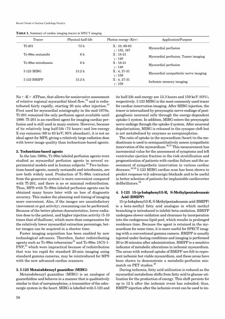
FIG. 2. Case example of MPI of a patientwith 70% stenosis of the proximal mid-left anterior descending coronary artery.(A) Perfusion images by conventional (top) and high-speed (bottom) SPECT demonstrating a moderate reversible perfusion defect in the left anterior de-scending coronary artery territory. Total acquisition time is indicated on the left-hand side. (B) Quantitative analysis yielded similar results for high-speed and conventional SPECT. From Ref. 5.
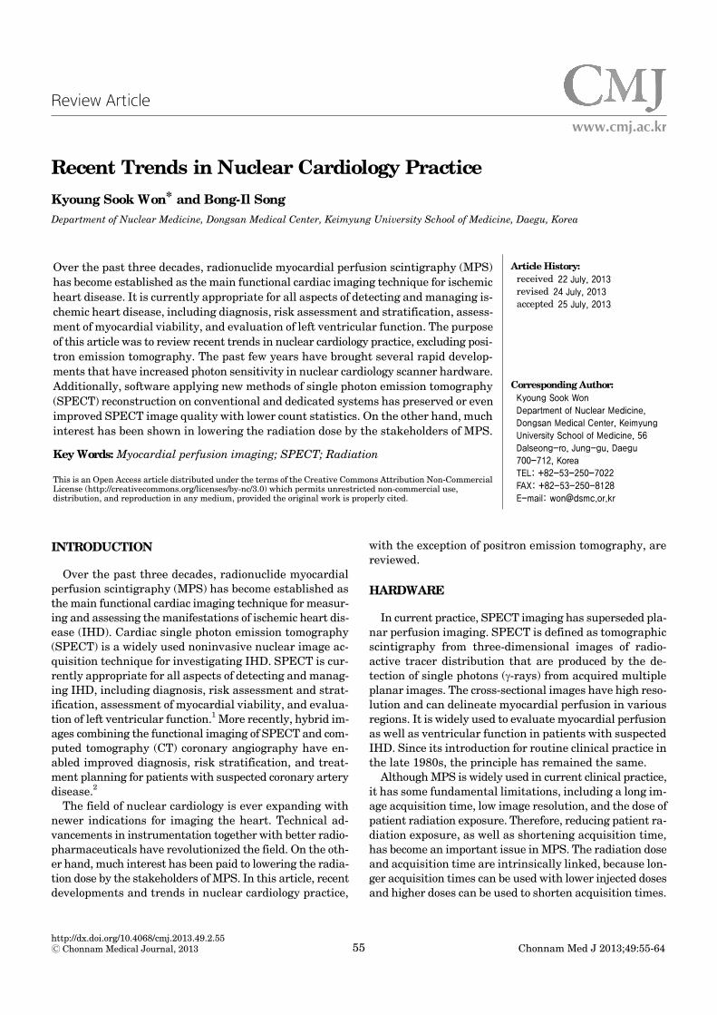
FIG. 1. D-SPECT cardiac system and detector-head configuration. (A) D-SPECT cardiac scanner with seated patient. (B) Each detectorcolumn is composed of CZT sensor (39×39×5 mm) with four 16×16 pixel detectors and tungsten collimator with 0.2-mm septa and squareopening (pitch, 2.46 mm; length, 21.7 mm). (C) Nine detector columns, each capable of rotation and translation, are used to scan themyocardium. From Ref. 3.

57
Kyoung Sook Won and Bong-Il Song
High-sensitivity dedicated cardiac camera systems pro-vide an opportunity to lower the injected doses for SPECT MPS. The past few years have brought several rapid devel-opments that have increased photon sensitivity in nuclear cardiology scanner hardware (Figs. 1 and 2). Several new dedicated camera systems with optimized acquisition ge-ometry and collimator design resulting in increased count sensitivity have been introduced by vendors.3-9 As a result, acquisition times for MPS can be significantly shortened without a significant sacrifice of accuracy. In particular, a recent study on dedicated cardiac scanners with cad-mium-zinc-telluride detectors found that a 10-min stress scan could be performed with an effective average radia-tion dose of less than 1 mSv.10
Attenuation correction to reduce soft-tissue attenuation artifacts was introduced by using external beam (transmission) images with the use of either a γ-emitting source or, more commonly, low-dose CT in conjunction with SPECT imaging (SPECT/CT). This has inherent problems, but it is generally accepted that attenuation correction helps to improve the accuracy in reporting.11 The practice of combining MPS with other modalities such as coronary CT angiography for better diagnostic certainty12,13 has in-tensified concerns regarding the total radiation dose deliv-ered to the patient.14
There are now several camera systems that combine the fast acquisition gamma cameras with high-resolution car-diac CT (hybrid cardiac cameras). Image co-registration using these hybrid systems enables more informed and bet-ter interpretation of both coronary CT findings as well as nuclear medicine perfusion imaging.15,16 Combined images enable better diagnosis, stratification, and treatment plan-ning for patients with suspected coronary artery disease.
SOFTWARE
Backprojection reconstruction of images has now been mostly replaced by iterative reconstruction. Iterative re-construction requires high computing power because the calculation is intensive. Although it has been routinely available in nuclear medicine since the 1990s, its use has been widespread only in the last decade. This method of re-construction has reduced the artifacts from adjacent struc-tures with high signal intensity such as the liver and gut. Software development for image acquisition and re-construction has led to faster image acquisition and im-proved image quality while decreasing radiation exposure to the patient. Several manufacturers have introduced software applications that incorporate iterative recon-struction, resolution recovery, and noise reduction, and these solutions have provided good-quality SPECT images despite reduced count statistics. Owing to the new methods of SPECT reconstruction applied in the software, SPECT image quality has been preserved or even improved with lower count statistics.17-24
Reliable automatic normalization of myocardial activity for rest and stress images is necessary for comparison and
interpretation of MPS. For example, extracardiac hot spots such as gallbladder or gastrointestinal tract activity can se-riously disturb the cardiac image quality. The development of cardiac packages has enabled reliable automatic nor-malization of myocardial activity.25 In addition, compar-ison with normal libraries can help to identify artifacts due to breast or diaphragm and aid in correct interpretation of images.26
The reporting aid software has enabled more accurate and objective assessment of the heart. Several new soft-ware packages are now available for displaying images. The new display software packages have better edge de-tection algorithms with better display of normalized images. The introduction of extensive normal libraries has enabled more accurate semiquantitative assessment of left ventricular perfusion. There is now a growing literature on the uses of MPS in heart failure. In addition to providing information on per-fusion, MPS can give an accurate assessment of both sys-tolic and diastolic left ventricular function, including phase and movement asynchrony, which, in conjunction with CT anatomy information from hybrid imaging, can be used in planning for positioning of various intracardiac devices.27-29 With new developments in cameras and soft-ware, these studies can be performed in a few minutes with-out the need for the patient to be in a supine position for long periods, which can be difficult for heart failure patients. Patient motion is a common cause of artifacts on MPS. Motion on SPECT studies can be detected in several ways. Breaks or irregularities in the sinogram indicate motion. This display method is not always easy to interpret, however. Furthermore, sinograms do not show horizontal motion. A simple and more commonly used method for de-tecting patient motion involves inspection of the cine dis-play of all planar projection images, i.e., rotating images. If motion is detected, a motion-correction program can be applied.30
RADIOPHARMACEUTICALS
Various radiopharmaceuticals are available for imaging the myocardium (Table 1). They all play an important role in the diagnosis and management of ischemic heart disease. The choice of radiopharmaceuticals should be based on the clinical questions to be answered. The choice of compound depends on a weighing of the advantages and disadvanta-ges of each tracer and a clinical decision as to which will give the best information.
1. Thallium (Tl-201) Tl-201 chloride was the first radiopharmaceutical to be widely used clinically for imaging myocardial perfusion.31 Tl-201 is a cyclotron-produced radionuclide with a half-life of 73 hours. Extensive research with cell culture, isolated heart, and intact animal models showed that thallous ion has a relatively high myocardial extraction, in part via
58
Recent Trends in Nuclear Cardiology Practice
TABLE 1. Summary of cardiac imaging tracers in SPECT imaging
Tracer Physical half-life Photon energy (Kev) Application/Purpose
Tl-201
Tc-99m sestamibi
Tc-99m tetrofosmin
I-123 MIBG
I-123 BMIPP
73 h
6 h
6 h
13.2 h
13.2 h
X : 10, 69-83γ : 135, 167X : 18-21γ : 140X : 18-21γ : 140X : 4, 27-31γ : 159X : 4, 27-31γ : 159
Myocardial perfusion
Myocardial perfusion, Tumor imaging
Myocardial perfusion
Myocardial sympathetic nerve imaging
Ischemic memory imaging
Na+-K+ ATPase, that allows for noninvasive assessment of relative regional myocardial blood flow,32 and is redis-tributed fairly rapidly, starting 20 min after injection.33 First used for myocardial scintigraphy in the mid-1970s, Tl-201 remained the only perfusion agent available until 1990. Tl-201 is an excellent agent for imaging cardiac per-fusion and is still used in many centers. However, because of its relatively long half-life (73 hours) and low-energy X-ray emission (69 to 83 keV; 95% abundant), it is not an ideal agent for MPS, giving a relatively large radiation dose with lower image quality than technetium-based agents.
2. Technetium-based agents In the late 1980s, Tc-99m labeled perfusion agents were studied as myocardial perfusion agents in several ex-perimental models and in human subjects.34 Two techne-tium-based agents, namely sestamibi and tetrofosmin, are now both widely used. Production of Tc-99m (extracted from the generator system) is more convenient compared with Tl-201, and there is no or minimal redistribution. Thus, MPS with Tc-99m-labeled perfusion agents can be obtained many hours later with no loss of diagnostic accuracy. This makes the planning and timing of imaging more convenient. Also, if the images are unsatisfactory (movement or gut activity), rescanning can be performed. Because of the better photon characteristics, lower radia-tion dose to the patient, and higher injection activity (5-10 times that of thallium), which more than compensates for the relatively lower myocardial extraction percentage, bet-ter images can be acquired in a shorter time. Faster imaging acquisition has been enabled by new technological advances. Therefore, faster redistributing agents such as Tc-99m teboroxime34 and Tc-99m 15C5-1- PNP,35 which were impractical because of redistribution that was too rapid for standard 20-min imaging using standard gamma cameras, may be reintroduced for MPS with the new advanced cardiac scanners.
3. I-123 Metaiodobenzyl guanidine (MIBG) Metaiodobenzyl guanidine (MIBG) is an analogue of guanethidine and behaves in a manner that is qualitatively similar to that of norepinephrine, a transmitter of the adre-nergic system in the heart. MIBG is labelled with I-123 and
its half-life and energy are 13.3 hours and 159 keV (83%), respectively. I-123 MIBG is the most commonly used tracer for cardiac innervation imaging. After MIBG injection, the tracer is internalized by presynaptic nerve endings of post-ganglionic neuronal cells through the energy-dependent uptake-1 system. In addition, MIBG enters the presynaptic nerve endings through the uptake-2 system. After neuronal depolarization, MIBG is released in the synapse cleft but is not metabolized by enzymes as norepinephrine. The ratio of uptake in the myocardium (heart) to the me-diastinum is used to semiquantitatively assess sympathetic innervation of the myocardium.36,37 This measurement has incremental value for the assessment of symptoms and left ventricular ejection fraction in the risk stratification and prognostication of patients with cardiac failure and the as-sessment of sympathetic innervation in various cardiac diseases.38,39 I-123 MIBG cardiac scan has been shown to predict response to β-adrenergic blockade and to be useful in better selection of patients for implantable cardioverter defibrillators.36
4. I-123 15-(p-Iodophenyl)3-R, S-Methylpentadecanoic Acid (BMIPP)
15-(p-Iodophenyl)3-R, S-Methylpentadecanoic acid (BMIPP) is a beta-methyl fatty acid analogue in which methyl branching is introduced to inhibit beta-oxidation. BMIPP undergoes slower oxidation and clearance by incorporation into the endogenous lipid pool, which results in prolonged residence time. Because the agent is retained in the my-ocardium for some time, it is more useful for SPECT imag-ing with a conventional gamma camera. BMIPP is usually injected under fasting conditions and imaging is performed 20 to 30 minutes after administration. BMIPP is a sensitive indicator of metabolic alterations in ischemic myocardium. The areas with reduced uptake of BMIPP are felt to repre-sent ischemic but viable myocardium, and these areas have been shown to demonstrate a metabolic-perfusion mis-match on PET studies.40
During ischemia, fatty acid utilization is reduced as the myocardial metabolism shifts from fatty acid to glucose uti-lization for the production of energy. This shift persists for up to 12 h after the ischemic event has subsided; thus, BMIPP injection after the ischemic event can be used to im-
59
Kyoung Sook Won and Bong-Il Song
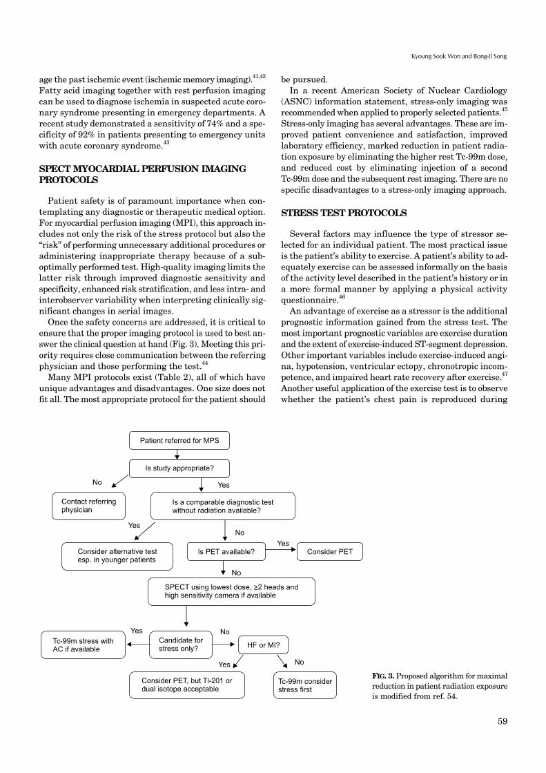
FIG. 3. Proposed algorithm for maximalreduction in patient radiation exposureis modified from ref. 54.
age the past ischemic event (ischemic memory imaging).41,42 Fatty acid imaging together with rest perfusion imaging can be used to diagnose ischemia in suspected acute coro-nary syndrome presenting in emergency departments. A recent study demonstrated a sensitivity of 74% and a spe-cificity of 92% in patients presenting to emergency units with acute coronary syndrome.43
SPECT MYOCARDIAL PERFUSION IMAGING PROTOCOLS
Patient safety is of paramount importance when con-templating any diagnostic or therapeutic medical option. For myocardial perfusion imaging (MPI), this approach in-cludes not only the risk of the stress protocol but also the ‘‘risk’’ of performing unnecessary additional procedures or administering inappropriate therapy because of a sub-optimally performed test. High-quality imaging limits the latter risk through improved diagnostic sensitivity and specificity, enhanced risk stratification, and less intra- and interobserver variability when interpreting clinically sig-nificant changes in serial images. Once the safety concerns are addressed, it is critical to ensure that the proper imaging protocol is used to best an-swer the clinical question at hand (Fig. 3). Meeting this pri-ority requires close communication between the referring physician and those performing the test.44
Many MPI protocols exist (Table 2), all of which have unique advantages and disadvantages. One size does not fit all. The most appropriate protocol for the patient should
be pursued. In a recent American Society of Nuclear Cardiology (ASNC) information statement, stress-only imaging was recommended when applied to properly selected patients.45 Stress-only imaging has several advantages. These are im-proved patient convenience and satisfaction, improved laboratory efficiency, marked reduction in patient radia-tion exposure by eliminating the higher rest Tc-99m dose, and reduced cost by eliminating injection of a second Tc-99m dose and the subsequent rest imaging. There are no specific disadvantages to a stress-only imaging approach.
STRESS TEST PROTOCOLS
Several factors may influence the type of stressor se-lected for an individual patient. The most practical issue is the patient’s ability to exercise. A patient’s ability to ad-equately exercise can be assessed informally on the basis of the activity level described in the patient’s history or in a more formal manner by applying a physical activity questionnaire.46
An advantage of exercise as a stressor is the additional prognostic information gained from the stress test. The most important prognostic variables are exercise duration and the extent of exercise-induced ST-segment depression. Other important variables include exercise-induced angi-na, hypotension, ventricular ectopy, chronotropic incom-petence, and impaired heart rate recovery after exercise.47 Another useful application of the exercise test is to observe whether the patient’s chest pain is reproduced during
60
Recent Trends in Nuclear Cardiology Practice
TABLE 2. SPECT myocardial perfusion imaging protocols and patient radiation dose
Protocol Effective radiation dose
One-Day Tc-99m Rest (10 mCi)/Stress (30 mCi) ProtocolOne-Day Tc-99m Stress (10 mCi)/Rest (30 mCi) ProtocolTwo-Day Stress (25 mCi)/Rest Tc-99m (25 mCi) ProtocolTwo-Day Rest (25 mCi)/Stress Tc-99m (25 mCi) ProtocolStress-Only Tc-99m (10 mCi or 25 mCi) Protocol
One-Day Rest Tl-201 (3.5 mCi)/Stress Tc-99m (25 mCi) Dual Isotope Protocol
One-Day Tl-201 Stress (3.5 mCi)/Redistribution ProtocolTl-201 Stress (3.0 mCi)/Redistribution with Optional Additional Imaging Protocols
(1) additional delayed imaging (2) re-injection of Tl-201 (1.5 mCi) after redistribution imagingSPECT Protocols for Assessing Patients with Congestive Heart Failure and LV Dysfunction
(1) Rest/Delayed Tl-201 (3.5 mCi) Protocol (2) Nitrate-Enhanced Tc-99m SPECTSPECT Protocols in Acute Chest Pain: Rest Tc-99m Imaging (25 mCi)
11.4 mSv for Tc-99m sestamibi and 9.3 mSv for Tc-99m tetrofosmin12.3 mSv for Tc-99m sestamibi and 9.3 mSv for Tc-99m tetrofosmin14.8 mSv for Tc-99m sestamibi and 11.6 mSv for Tc-99m tetrofosmin14.8 mSv for Tc-99m sestamibi and 11.6 mSv for Tc-99m tetrofosmin2.7 mSv for Tc-99m sestamibi and 2.3 mSv for Tc-99m tetrofosmin with 10 mCi
6.8 mSv for Tc-99m sestamibi and 5.8 mSv for Tc-99m tetrofosmin with 25 mCi
22.1 mSv for Tc-99m sestamibi and 21.2 mSv for Tc-99m tetrofosmin
15.3 mSv19.7 mSv in case of (2)
(1) 15.3 mSv (2) 12.7 mSv for Tc-99m sestamibi and 10.4 mSv using Tc-99m te-
trofosmin, based on 10.0 mCi and 35.0 mCi doses, respectively
8.0 mSv for Tc-99m sestamibi and 5.8 mSv for Tc-99m tetrofosmin
Modified from Ref. 44.
stress to determine whether its etiology is cardiac or noncardiac. For these reasons, exercise stress is preferred over pharmacologic testing when an adequate exercise ef-fort can be achieved. The most commonly used type of exercise in the United States is treadmill testing. The Bruce protocol is most com-monly used in which most patients complete a workload of 7-13 METs over 6-12 minutes. Several other protocols (modified Bruce, Naughton, Balke) increase workload in smaller increments, which is an advantage in elderly or frail patients. Bicycle ergometry is widely applied in Europe. Other forms of exercise testing, such as arm ergo-metry, are rarely used. Beyond the inability to exercise, additional indications for vasodilator stress are the presence of left bundle branch block (LBBB) or a paced ventricular rhythm. Another unique application of vasodilator stress testing is very ear-ly risk stratification of stable patients with AMI. In the Adenosine Sestamibi SPECT Post-Infarction Evaluation (INSPIRE) study, which enrolled patients with both ST- and non-ST-segment elevation AMI, vasodilator stress imaging was safely performed as early as 12 hours after presentation.48
Three pharmacologic agents are currently FDA ap-proved in the United States: dipyridamole, adenosine, and regadenoson. These agents demonstrate similar diag-nostic accuracy.49 Adenosine is a direct nonselective ad-enosine receptor agonist. The major advantage of ad-enosine over dipyridamole and regadenoson is its very short half-life (<10 s). For this reason, adenosine is the pre-ferred agent in patients who are at greatest risk for develop-ing side effects (e.g., a patient with borderline low blood
pressure). Dipyridamole is an indirect coronary artery vasodilator. It blocks the intracellular reuptake and deam-ination of adenosine and thereby boosts adenosine levels in the intravascular space. Intravenous dipyridamole can be safely administered to a patient taking oral dipyri-damole. Regadenoson is the first selective adenosine A2A receptor agonist to be approved for pharmacologic stress testing. Its affinity for the adenosine A1 receptor is at least 10-fold lower than for the A2 receptor, minimizing the risk for AV block. In the phase 3 Adenoscan Versus Regadenoson Comparative Evaluation for MPI (ADVANCE MPI) 2 tri-als, regadenoson had a lower composite side effect profile than did adenosine but with comparable imaging results.49 The half-life of regadenoson is triphasic, and aminophyl-line can be used to treat side effects. Physiologic effects of this medication can persist for up to 30 minutes after administration. Regadenoson is logistically easier to ad-minister than adenosine or dipyridamole because it is giv-en as an intravenous bolus injection over 10 s without the need for an infusion pump. Dobutamine is the most commonly used synthetic cat-echolamine for pharmacologic stress testing in the United States. The half-life of dobutamine is approximately 2 mi-nutes, but its physiologic effect may persist for 5 to 20 mi-nutes and may require the administration of a short-acting beta-blocker for reversal. Given the variable physiologic response to dobutamine in individual patients and the more labor-intensive requirements of administering this agent, dobutamine is designated as a secondary pharmaco-logic stress agent in nuclear cardiology. Its use is reserved for patients who cannot adequately exercise and who have a contraindication to performing vasodilator stress testing.

61
Kyoung Sook Won and Bong-Il Song
TABLE 3. Inappropriate indications (median score 1-3)
IndicationAppropriateness
score (1-9)
Detection of CAD: Symptomatic evaluation of ischemic equivalent (non-acute) Low pretest probability of CAD or ECG interpretable AND able to exerciseDetection of CAD: Symptomatic acute chest pain Define ACSDetection of CAD/risk assessment without ischemic equivalent: Asymptomatic Low CHD Risk (ATP III Risk Criteria) Intermediate CHD risk (ATP III Risk Criteria)/ECG interpretableDetection of CAD/risk assessment without ischemic equivalent: Syncope Low CHD risk (ATP III Risk Criteria)Risk assessment with prior test results and/or known chronic stable CAD, asymptomatic OR stable symptoms, normal prior stress imaging study Low CHD risk (ATP III Risk Criteria) or Last stress imaging study done less than 2 years ago Intermediate to high CHD risk (ATP III Risk Criteria)/Last stress imaging study done less than 2 years ago Low CHD risk (ATP III Risk Criteria) or Last stress imaging study done more than or equal to 2 years agoRisk assessment with prior test results and/or known chronic stable CAD, asymptomatic OR stable symptoms, abnormal coronary angiography OR abnormal prior stress imaging study, no prior revascularization Known CAD on coronary angiography OR prior abnormal stress imaging study/Last stress imaging study done less
than 2 years agoRisk assessment with prior test results and/or known chronic stable CAD, asymptomatic, prior coronary calcium Agatston score Agatston score less than 100Risk assessment with prior test results and/or known chronic stable CAD, Duke treadmill score Low-risk Duke treadmill scoreRisk assessment: Preoperative evaluation for non-cardiac surgery without active cardiac conditions, low-risk surgery Preoperative evaluation for non-cardiac surgery risk assessmentRisk assessment: Preoperative evaluation for non-cardiac surgery without active cardiac conditions, intermediate-risk surgery Moderate to good functional capacity (greater than or equal to 4 METs) No clinical risk factors Asymptomatic up to 1 year post normal catheterization, non-invasive test, or previous revascularizationRisk assessment: Preoperative evaluation for non-cardiac surgery without active cardiac conditions, vascular surgery Moderate to good functional capacity (greater than or equal to 4 METs) No clinical risk factors Asymptomatic up to 1 year post normal catheterization, non-invasive test, or previous revascularizationRisk assessment: Within 3 months of an acute coronary syndrome, STEMI Primary PCI with complete revascularization Hemodynamically unstable, signs of cardiogenic shock, or mechanical complicationsRisk assessment: Within 3 months of an acute coronary syndrome, ACS-asymptomatic post-revascularization (PCI or CABG) Evaluation prior to hospital dischargeRisk assessment: Within 3 months of an acute coronary syndrome, cardiac rehabilitation Prior to initiation of cardiac rehabilitation (as a stand-alone indication)Risk assessment: Post-revascularization (PCI or CABG), asymptomatic Less than 2 years after PCIRisk assessment: Post-revascularization (PCI or CABG), cardiac rehabilitation Prior to initiation of cardiac rehabilitation (as a stand-alone indication)Evaluation of ventricular function, evaluation of left ventricular function Routine use of stress FP RNA in conjunction with rest/stress gated SPECT MPI
I(3)
I(1)
I(1)I(3)
I(3)
I(1)I(3)I(3)
I(3)
I(2)
I(2)
I(1)
I(3)I(2)I(2)
I(3)I(2)I(2)
I(2)I(1)
I(1)
I(3)
I(3)
I(3)
I(3)
Modified from Ref. 51.
The administration of anti-ischemic medications (i.e., nitrates, beta blockers, and calcium channel antagonists) either alone or in combination can reduce the diagnostic sensitivity of stress SPECT or PET MPI, irrespective of the stressor modality utilized.50
APPROPRIATE USE OF MPI
Over the past few years, major cardiac societies such as the American College of Cardiology/American Heart Association in collaboration with many other imaging soci-
62
Recent Trends in Nuclear Cardiology Practice
eties have taken proactive steps to guide the use of cardiac imaging. Appropriate use criteria for nuclear cardiology have been published to ensure that clinicians in most in-stances are ‘‘doing the right test for the right patient at the right time.’’51 The recent comprehensive ASNC preferred practice statement ‘‘Patient Centered Imaging’’ by Depuey et al.44 outlines various MPI protocols with existing iso-topes and their advantages and disadvantages and best use in daily clinical practice. This document reiterates an im-portant point that physicians practicing nuclear cardiol-ogy should embrace: ‘‘One size does not fit all.’’ An appropriate study, by definition, provides incre-mental information, combined with clinical judgment, which exceeds the expected negative consequences, such as the risk of ionizing radiation from the procedure. Thus, when appropriately utilized, the clinical benefits of MPI far outweigh the lifetime potential risk of ionizing radiation exposure. In patients with inappropriate indications for MPI (Table 3), the estimated life-attributable risk of cancer from radiation exposure, although minimal, should still be considered to be unacceptable. Hence, the discussion re-garding the use of low-level ionizing radiation for MPI should focus on the appropriate selection of patients and the application of the As Low As Reasonably Achievable (ALARA) principle for the administration of a radiotracer to obtain high-quality, clinically interpretable images.52-54
In conclusion, these past few years have brought several rapid developments that have increased photon sensitivity in nuclear cardiology scanner hardware. Additionally, software applying new methods of SPECT reconstruction on conventional and dedicated systems has preserved or even improved SPECT image quality with lower count statistics. Several new dedicated camera systems with op-timized acquisition geometry and collimator design result-ing in increased count sensitivity have been introduced by vendors. Although it is convenient for patients to have imaging completed in 2 to 4 min, the efficiency of these sys-tems provides the opportunity to reduce both the duration of the acquisition and the radiation dose to the patient. MPI protocols should be optimized to achieve the lowest possible radiation exposure while maintaining diagnostic performance. Key aspects of this optimization include the use of radionuclides with shorter half-lives (such as Tc-99m and PET tracers), stress-only imaging when feasible, and weight-based dosing. Supporting research and develop-ment of camera technology, image reconstruction and ac-quisition techniques, and approaches to collimation are cru-cial to continued progress in the field of nuclear cardiology.
REFERENCES
1. Health Quality Ontario. Single photon emission computed tomography for the diagnosis of coronary artery disease: an evidence-based analysis. Ont Health Technol Assess Ser 2010; 10:1-64.
2. 2006 image of the year: focus on cardiac SPECT/CT. J Nucl Med
2006;47:14N-5N.3. Gambhir SS, Berman DS, Ziffer J, Nagler M, Sandler M, Patton
J, et al. A novel high-sensitivity rapid-acquisition single-photon cardiac imaging camera. J Nucl Med 2009;50:635-43.
4. Garcia EV, Faber TL, Esteves FP. Cardiac dedicated ultrafast SPECT cameras: new designs and clinical implications. J Nucl Med 2011;52:210-7.
5. Sharir T, Slomka PJ, Hayes SW, DiCarli MF, Ziffer JA, Martin WH, et al. Multicenter trial of high-speed versus conventional single-photon emission computed tomography imaging: quanti-tative results of myocardial perfusion and left ventricular function. J Am Coll Cardiol 2010;55:1965-74.
6. Sharir T, Ben-Haim S, Merzon K, Prochorov V, Dickman D, Ben-Haim S, et al. High-speed myocardial perfusion imaging initial clinical comparison with conventional dual detector anger camera imaging. JACC Cardiovasc Imaging 2008;1:156-63.
7. Herzog BA, Buechel RR, Katz R, Brueckner M, Husmann L, Burger IA, et al. Nuclear myocardial perfusion imaging with a cadmium-zinc-telluride detector technique: optimized protocol for scan time reduction. J Nucl Med 2010;51:46-51.
8. Buechel RR, Herzog BA, Husmann L, Burger IA, Pazhenkottil AP, Treyer V, et al. Ultrafast nuclear myocardial perfusion imaging on a new gamma camera with semiconductor detector technique: first clinical validation. Eur J Nucl Med Mol Imaging 2010;37:773-8.
9. Esteves FP, Raggi P, Folks RD, Keidar Z, Askew JW, Rispler S, et al. Novel solid-state-detector dedicated cardiac camera for fast myocardial perfusion imaging: multicenter comparison with standard dual detector cameras. J Nucl Cardiol 2009;16:927-34.
10. Nakazato R, Berman DS, Hayes SW, Fish M, Padgett R, Xu Y, et al. Myocardial perfusion imaging with a solid-state camera: simulation of a very low dose imaging protocol. J Nucl Med 2013;54:373-9.
11. Garcia EV. SPECT attenuation correction: an essential tool to realize nuclear cardiology's manifest destiny. J Nucl Cardiol 2007;14:16-24.
12. Araujo LI, Jimenez-Hoyuela JM, McClellan JR, Lin E, Viggiano J, Alavi A. Improved uniformity in tomographic myocardial perfusion imaging with attenuation correction and enhanced acquisition and processing. J Nucl Med 2000;41:1139-44.
13. Slomka PJ, Berman DS, Germano G. Applications and software techniques for integrated cardiac multimodality imaging. Expert Rev Cardiovasc Ther 2008;6:27-41.
14. Einstein AJ, Henzlova MJ, Rajagopalan S. Estimating risk of cancer associated with radiation exposure from 64-slice computed tomography coronary angiography. JAMA 2007;298:317-23.
15. Schaap J, Kauling RM, Boekholdt SM, Nieman K, Meijboom WB, Post MC, et al. Incremental diagnostic accuracy of hybrid SPECT/CT coronary angiography in a population with an intermediate to high pre-test likelihood of coronary artery disease. Eur Heart J Cardiovasc Imaging 2013;14:642-9.
16. Di Carli MF, Dorbala S, Meserve J, El Fakhri G, Sitek A, Moore SC. Clinical myocardial perfusion PET/CT. J Nucl Med 2007;48: 783-93.
17. DePuey EG, Gadiraju R, Clark J, Thompson L, Anstett F, Shwartz SC. Ordered subset expectation maximization and wide beam reconstruction "half-time" gated myocardial perfusion SPECT

63
Kyoung Sook Won and Bong-Il Song
functional imaging: a comparison to "full-time" filtered backpro-jection. J Nucl Cardiol 2008;15:547-63.
18. Borges-Neto S, Pagnanelli RA, Shaw LK, Honeycutt E, Shwartz SC, Adams GL, et al. Clinical results of a novel wide beam reconstruction method for shortening scan time of Tc-99m cardiac SPECT perfusion studies. J Nucl Cardiol 2007;14:555-65.
19. Venero CV, Heller GV, Bateman TM, McGhie AI, Ahlberg AW, Katten D, et al. A multicenter evaluation of a new post-processing method with depth-dependent collimator resolution applied to full-time and half-time acquisitions without and with simulta-neously acquired attenuation correction. J Nucl Cardiol 2009;16: 714-25.
20. Maddahi J, Mendez R, Mahmarian JJ, Thomas G, Babla H, Bai C, et al. Prospective multicenter evaluation of rapid, gated SPECT myocardial perfusion upright imaging. J Nucl Cardiol 2009;16:351-7.
21. Druz RS, Phillips LM, Chugkowski M, Boutis L, Rutkin B, Katz S. Wide-beam reconstruction half-time SPECT improves diagno-stic certainty and preserves normalcy and accuracy: a quantita-tive perfusion analysis. J Nucl Cardiol 2011;18:52-61.
22. DePuey EG, Bommireddipalli S, Clark J, Leykekhman A, Thompson LB, Friedman M. A comparison of the image quality of full-time myocardial perfusion SPECT vs wide beam reconstruction half-time and half-dose SPECT. J Nucl Cardiol 2011;18:273-80.
23. DePuey EG, Ata P, Wray R, Friedman M. Very low-activity stress/high-activity rest, single-day myocardial perfusion SPECT with a conventional sodium iodide camera and wide beam reconstruction processing. J Nucl Cardiol 2012 ;19:931-44.
24. DePuey EG, Bommireddipalli S, Clark J, Thompson L, Srour Y. Wide beam reconstruction "quarter-time" gated myocardial perfusion SPECT functional imaging: a comparison to "full-time" ordered subset expectation maximum. J Nucl Cardiol 2009;16: 736-52.
25. Burrell S, MacDonald A. Artifacts and pitfalls in myocardial perfusion imaging. J Nucl Med Technol 2006;34:193-211; quiz 212-4.
26. Garcia E, Maddahi J, Berman D, Waxman A. Space/time quan-titation of thallium-201 myocardial scintigraphy. J Nucl Med 1981;22:309-17.
27. Chen J, Boogers MJ, Bax JJ, Soman P, Garcia EV. The use of nuclear imaging for cardiac resynchronization therapy. Curr Cardiol Rep 2010;12:185-91.
28. Boogers MJ, Chen J, van Bommel RJ, Borleffs CJ, Dibbets- Schneider P, van der Hiel B, et al. Optimal left ventricular lead position assessed with phase analysis on gated myocardial perfusion SPECT. Eur J Nucl Med Mol Imaging 2011;38:230-8.
29. Schindler TH, Quercioli A. Left ventricular dyssynchrony asse-ssment by phase analysis from gated myocardial perfusion SPECT: moving beyond conventional criteria. Heart 2011;97:4-5.
30. Niu X, Yang Y, Jin M, Wernick MN, King MA. Effects of motion, attenuation, and scatter corrections on gated cardiac SPECT reconstruction. Med Phys 2011;38:6571-84.
31. Lebowitz E, Greene MW, Fairchild R, Bradley-Moore PR, Atkins HL, Ansari AN, et al. Thallium-201 for medical use. I. J Nucl Med 1975;16:151-5.
32. Nielsen AP, Morris KG, Murdock R, Bruno FP, Cobb FR. Linear
relationship between the distribution of thallium-201 and blood flow in ischemic and nonischemic myocardium during exercise. Circulation 1980;61:797-801.
33. Beller GA, Watson DD, Ackell P, Pohost GM. Time course of thallium-201 redistribution after transient myocardial ischemia. Circulation 1980;61:791-7.
34. Dahlberg ST. Assessment of myocardial perfusion with Tc-99m: image is everything. J Nucl Cardiol 2009;16:493-6.
35. Liu Z, Chen L, Liu S, Barber C, Stevenson GD, Furenlid LR, et al. Kinetic characterization of a novel cationic (99m)Tc(I)- tricarbonyl complex, (99m)Tc-15C5-PNP, for myocardial perfu-sion imaging. J Nucl Cardiol 2010;17:858-67.
36. Patel AD, Iskandrian AE. MIBG imaging. J Nucl Cardiol 2002;9:75-94.
37. Agostini D, Verberne HJ, Burchert W, Knuuti J, Povinec P, Sambuceti G, et al. I-123-mIBG myocardial imaging for assess-ment of risk for a major cardiac event in heart failure patients: insights from a retrospective European multicenter study. Eur J Nucl Med Mol Imaging 2008;35:535-46.
38. Flotats A, Carrió I. Cardiac neurotransmission SPECT imaging. J Nucl Cardiol 2004;11:587-602.
39. Jacobson AF, Senior R, Cerqueira MD, Wong ND, Thomas GS, Lopez VA, et al; ADMIRE-HF Investigators. Myocardial iodine-123 meta-iodobenzylguanidine imaging and cardiac events in heart failure. Results of the prospective ADMIRE-HF (AdreView Myocardial Imaging for Risk Evaluation in Heart Failure) study. J Am Coll Cardiol 2010;55:2212-21.
40. Shi CQ, Young LH, Daher E, DiBella EV, Liu YH, Heller EN, et al. Correlation of myocardial p-(123)I-iodophenylpentadecanoic acid retention with (18)F-FDG accumulation during experimental low-flow ischemia. J Nucl Med 2002;43:421-31.
41. Herrero P, Gropler RJ. Imaging of myocardial metabolism. J Nucl Cardiol 2005;12:345-58.
42. Dilsizian V, Bateman TM, Bergmann SR, Des Prez R, Magram MY, Goodbody AE, et al. Metabolic imaging with beta-methyl- p-[(123)I]-iodophenyl-pentadecanoic acid identifies ischemic memory after demand ischemia. Circulation 2005;112:2169-74.
43. Kawai Y, Tsukamoto E, Nozaki Y, Morita K, Sakurai M, Tamaki N. Significance of reduced uptake of iodinated fatty acid analogue for the evaluation of patients with acute chest pain. J Am Coll Cardiol 2001;38:1888-94.
44. Depuey EG, Mahmarian JJ, Miller TD, Einstein AJ, Hansen CL, Holly TA, et al. Patient-centered imaging. J Nucl Cardiol 2012;19: 185-215.
45. Cerqueira MD, Allman KC, Ficaro EP, Hansen CL, Nichols KJ, Thompson RC, et al. Recommendations for reducing radiation exposure in myocardial perfusion imaging. J Nucl Cardiol 2010; 17:709-18.
46. Hlatky MA, Boineau RE, Higginbotham MB, Lee KL, Mark DB, Califf RM, et al. A brief self-administered questionnaire to determine functional capacity (the Duke Activity Status Index). Am J Cardiol 1989;64:651-4.
47. Kligfield P, Lauer MS. Exercise electrocardiogram testing: beyond the ST segment. Circulation 2006;114:2070-82.
48. Mahmarian JJ, Shaw LJ, Filipchuk NG, Dakik HA, Iskander SS, Ruddy TD, et al; INSPIRE Investigators. A multinational study to establish the value of early adenosine technetium-99m
64
Recent Trends in Nuclear Cardiology Practice
sestamibi myocardial perfusion imaging in identifying a low-risk group for early hospital discharge after acute myocardial infarction. J Am Coll Cardiol 2006;48:2448-57.
49. Cerqueira MD, Nguyen P, Staehr P, Underwood SR, Iskandrian AE; ADVANCE-MPI Trial Investigators. Effects of age, gender, obesity, and diabetes on the efficacy and safety of the selective A2A agonist regadenoson versus adenosine in myocardial perfusion imaging integrated ADVANCE-MPI trial results. JACC Cardio-vasc Imaging 2008;1:307-16.
50. Mahmarian JJ. Monitoring medical therapy: the role of nonin-vasive imaging. In: Dilsizian V, Narula J, Braunwald E, eds. Atlas of nuclear cardiology. 2nd ed. Philadelphia, PA: Current Medi-cine, 2006;191-210.
51. Hendel RC, Berman DS, Di Carli MF, Heidenreich PA, Henkin RE, Pellikka PA, et al; American College of Cardiology Foundation Appropriate Use Criteria Task Force; American Society of Nuclear Cardiology; American College of Radiology; American Heart Association; American Society of Echocardio-logy; Society of Cardiovascular Computed Tomography; Society for Cardiovascular Magnetic Resonance; Society of Nuclear
Medicine. ACCF/ASNC/ACR/AHA/ASE/SCCT/SCMR/SNM 2009 appropriate use criteria for cardiac radionuclide imaging: a report of the American college of cardiology foundation appropriate use criteria task force, the American society of nuclear cardiology, the American college of radiology, the American heart association, the American society of echocardiography, the society of cardio-vascular computed tomography, the society for cardiovascular magnetic resonance, and the society of nuclear medicine. J Am Coll Cardiol 2009;53:2201-29.
52. Shaw LJ, Achenbach S, Chandrashekhar Y, Dilsizian V, Hundley WG, Kern MJ, et al. Imaging modalities and radiation: benefit has its risks. JACC Cardiovasc Imaging 2010;3:550-2.
53. Laskey WK, Feinendegen LE, Neumann RD, Dilsizian V. Low-level ionizing radiation from noninvasive cardiac imaging: can we extrapolate estimated risks from epidemiologic data to the clinical setting? JACC Cardiovasc Imaging 2010;3:517-24.
54. Cerqueira MD, Allman KC, Ficaro EP, Hansen CL, Nichols KJ, Thompson RC, et al. Recommendations for reducing radiation exposure in myocardial perfusion imaging. J Nucl Cardiol 2010;17:709-18.
Related Documents