Recent Insights Into the Biomedical Applications of Shape-memory Polymers Maria C. Serrano, Guillermo A. Ameer* 1. Introduction Shape-memory polymers (SMP) are polymeric materials that have the ability to switch from a temporary to a permanent shape due to an external stimulation. The temporary shapes are stable until the appropriate stimulus (i.e. shape memory-driving stimulus) is applied to the material to induce the recovery of the permanent, original shape. [1] The acquisition of shape-memory properties is a combination of both a suitable polymer network architec- ture and a tailored processing and programming techno- logy, rather than an intrinsic property. [2] The first SMP, polynorbornene, was developed by CdF Chimie Company (France) in 1984. [3] One year later, this material was commercialized by Nippon Zeon Company (Japan) under the trade name of Norsorex. [4] A few more commercial SMP came later, including Kurare TP-301 by Kurare Corporation (Japan) and Asmer by Asahi Company (Japan). [5] Polyurethane-based SMP developed by Mitsubishi Heavy Industries [6] and others have been the most relevant SMP commercialized for the past two decades. Shape-memory research was first built on the thermally- induced dual-shape effect, where heat is directly driving the shape change through a transition temperature (T trans ) that could be a melting temperature or a glass transition temperature. [1] Melting temperatures are generally pre- ferred over glass transitions as they induce a sharper transition that is easier to predict and control. The heat- induced shape-memory effect (SME) can also be indirectly triggered by diverse stimuli, ranging from IR-light irradia- tion, [7] electrical stimulation, [8] exposure to alternating magnetic fields, [9,10] or immersion in water. [11] Further investigation has also led to the synthesis of polymers with a light-induced SME that is independent of tempera- ture. [12,13] Additionally, and depending on the SME-driving stimulus and the desired application, SMP can be triggered by direct or indirect contact, the latter when remote actuation is pursued. SME is conventionally quantified in dynamic mechanical experiments in order to determine the strain fixity rate (R f ), the strain recovery rate (R r ), and the switching temperature for those thermally-induced (T trans ), Review Prof. G. A. Ameer Chemistry of Life Processes Institute, Evanston, Illinois 60208, USA E-mail: [email protected] Dr. M. C. Serrano Instituto de Ciencia de Materiales de Madrid, Consejo Superior de Investigaciones Cientı ´ficas, Cantoblanco, Madrid 28049, Spain Prof. G. A. Ameer Biomedical Engineering Department, Northwestern University, Evanston, Illinois 60208, USA Institute for BioNanotechnology in Medicine, Northwestern University, Chicago, Illinois 60611, USA Division of Vascular Surgery, Feinberg School of Medicine, Northwestern University, Chicago, Illinois 60611, USA Shape-memory polymers (SMP) are versatile stimuli-responsive materials that can switch, upon stimulation, from a temporary to a permanent shape. This advanced functionality makes SMP suitable and promising materials for diverse technological applications, including the fabrication of smart biomedical devices. In this paper, advances in the design of SMP are dis- cussed, with emphasis on materials investigated for medical applications. Future directions necess- ary to bring SMP closer to their clinical application are also highlighted. ß 2012 WILEY-VCH Verlag GmbH & Co. KGaA, Weinheim wileyonlinelibrary.com Macromol. Biosci. 2012, DOI: 10.1002/mabi.201200097 1 Early View Publication; these are NOT the final page numbers, use DOI for citation !! R

Welcome message from author
This document is posted to help you gain knowledge. Please leave a comment to let me know what you think about it! Share it to your friends and learn new things together.
Transcript

Review
Recent Insights Into the BiomedicalApplications of Shape-memory Polymers
Maria C. Serrano, Guillermo A. Ameer*
Shape-memory polymers (SMP) are versatile stimuli-responsive materials that can switch,upon stimulation, from a temporary to a permanent shape. This advanced functionality makesSMP suitable and promising materials for diverse technological applications, including thefabrication of smart biomedical devices. In thispaper, advances in the design of SMP are dis-cussed, with emphasis on materials investigatedfor medical applications. Future directions necess-ary to bring SMP closer to their clinical applicationare also highlighted.
1. Introduction
Shape-memory polymers (SMP) are polymeric materials
that have the ability to switch from a temporary to a
permanent shape due to an external stimulation. The
temporary shapes are stable until the appropriate stimulus
(i.e. shape memory-driving stimulus) is applied to the
material to induce the recovery of the permanent, original
shape.[1] The acquisition of shape-memory properties is a
combination of both a suitable polymer network architec-
ture and a tailored processing and programming techno-
logy, rather than an intrinsic property.[2] The first SMP,
polynorbornene, was developed by CdF Chimie Company
(France) in 1984.[3] One year later, this material was
Prof. G. A. AmeerChemistry of Life Processes Institute, Evanston, Illinois 60208,USAE-mail: [email protected]. M. C. SerranoInstituto de Ciencia de Materiales de Madrid, Consejo Superior deInvestigaciones Cientıficas, Cantoblanco, Madrid 28049, SpainProf. G. A. AmeerBiomedical Engineering Department, Northwestern University,Evanston, Illinois 60208, USAInstitute for BioNanotechnology in Medicine, NorthwesternUniversity, Chicago, Illinois 60611, USADivision of Vascular Surgery, Feinberg School of Medicine,Northwestern University, Chicago, Illinois 60611, USA
� 2012 WILEY-VCH Verlag GmbH & Co. KGaA, Weinheim wileyonlin
Early View Publication; these are NOT
commercialized by Nippon Zeon Company (Japan) under
the trade name of Norsorex.[4] A few more commercial
SMP came later, including Kurare TP-301 by Kurare
Corporation (Japan) and Asmer by Asahi Company
(Japan).[5] Polyurethane-based SMP developed by
Mitsubishi Heavy Industries[6] and others have been the
most relevant SMP commercialized for the past two
decades.
Shape-memory research was first built on the thermally-
induced dual-shape effect, where heat is directly driving the
shape change through a transition temperature (Ttrans) that
could be a melting temperature or a glass transition
temperature.[1] Melting temperatures are generally pre-
ferred over glass transitions as they induce a sharper
transition that is easier to predict and control. The heat-
induced shape-memory effect (SME) can also be indirectly
triggered by diverse stimuli, ranging from IR-light irradia-
tion,[7] electrical stimulation,[8] exposure to alternating
magnetic fields,[9,10] or immersion in water.[11] Further
investigation has also led to the synthesis of polymers with
a light-induced SME that is independent of tempera-
ture.[12,13] Additionally, and depending on the SME-driving
stimulus and the desired application, SMP can be triggered
by direct or indirect contact, the latter when remote
actuation is pursued. SME is conventionally quantified in
dynamic mechanical experiments in order to determine the
strain fixity rate (Rf), the strain recovery rate (Rr), and the
switching temperature for those thermally-induced (Ttrans),
elibrary.com Macromol. Biosci. 2012, DOI: 10.1002/mabi.201200097 1
the final page numbers, use DOI for citation !! R
Guillermo A. Ameer, a native of Panama City,Panama, is Professor of Biomedical Engineeringand Surgery at Northwestern University. Hereceived his PhD in Chemical and BiomedicalEngineering from the Massachusetts Instituteof Technology. Dr. Ameer’s research interestsinclude biomaterials, tissue engineering, andbio/nanotechnology for improved therapeuticsand diagnostics. He has co-authored over100 peer-reviewed publications, conferenceabstracts and book chapters, 27 patents issuedand pending, and has co-founded severalmedical devices companies.
Marıa C. Serrano received her PhD in Biochem-istry and Molecular Biology from the UniversidadComplutense de Madrid in 2006 (Spain). After apostdoctoral training period in Dr Ameer’sresearch laboratory for over 2 years, she isnow a postdoctoral fellow Juan de la Cierva inthe Bioinspired Materials Group leaded by Pro-fessor del Monte in the Instituto de Ciencia deMateriales de Madrid (ICMM-CSIC). Her researchinterests are focused on biomaterials for cardi-ovascular, bone and neural tissue regeneration.
Figure 1. Example of a stress–strain-temperature diagram for athermoplastic SMP with thermally-induced SME. Reproducedwith permission.[60] Copyright 2011, American Chemical Society.
2
REa
www.mbs-journal.de
M. C. Serrano, G. A. Ameer
all these parameters that define the particular SME of a
concrete material (Figure 1).[14]
From a practical point of view, SMP are considered
versatile stimuli-sensitive polymers,[15] with advanced
functionalities that make them suitable for diverse
applications such as self-deployable sun-sails or antenna
in spacecraft, wrinkle free fabrics, morphing wing struc-
tures, cell phones with active disassembly when obsolete,
and heat-shrinkable tubes for electric isolation or films for
packing.[16–19] For instance, polyurethanes have been
extensively explored for the development of materials
with tunable responsiveness and even patented for their
use as threads in fabrics to increase their crease resis-
tance.[20,21] More recently, SMP are being introduced in a
variety of new applications such as microfoldable vehicles
and microtags.[22]
In this article, the design of SMP will be discussed
from a biomedical perspective, with emphasis on those
materials with potential for medical applications.
Future directions necessary to bring these materials closer
to their clinical application will be also highlighted. For
further details on shape-memory composites, materials
with self-healing characteristics, and stimuli-responsive
systems not included herein, readers are referred else-
where.[23–39]
2. Introducing the Shape-memory Effect IntoMaterials
Since its discovery in alloys in 1932 by Chang and Read,[40]
the SME has been extensively investigated in metal alloys
for its potential use in medicine.[41] This phenomenon was
later observed in polymers,[42,43] thus introducing a variety
Macromol. Biosci. 2012, DOI: 1
� 2012 WILEY-VCH Verlag Gmb
rly View Publication; these are NOT the final pag
of materials with stimuli responsiveness that represented a
cheaper and more efficient alternative to established shape-
memory alloys. More recently, the synthesis of SMP has
found inspiration in biological substances, as naturally
occurring bile acids have been used to fabricate amorphous,
thermoplastic polyesters with shape-memory proper-
ties.[44,45] Interestingly, these bioinspired polymers dis-
played rubber-like elasticity, glass transitions close to body
temperature, high strains at low temperatures, and low
systemic toxicity. Biodegradable polymers synthesized
by coupling amino acids and a-hydoxy acids, such as
polydepsipeptides, with poly(e-caprolactone) have also
demonstrated interesting shape-memory properties and
promise for biomedical applications.[46,47]
Shape-memory properties have also been incorporated
into polymer composites,[48–50] thus resulting in materials
that can generally recover from relatively large mechanical
strains by combining the advanced functionality of
stimuli-responsiveness with the benefits of using particles
embedded in the polymer network.[51] Five major objectives
are pursued when designing SMP composites: improving
shape recovery stress and mechanical properties,
speeding up shape recovery by increasing thermal con-
ductivity, fabricating novel formulations, tuning particular
properties in existing SMP, and designing materials with
additional responsiveness to electricity, magnetic fields,
light, or moisture.[27] For instance, Mohr et al. introduced
magnetic nanoparticles in thermoplastic polymers to
develop SMP composites able to be actuated by inductive
0.1002/mabi.201200097
H & Co. KGaA, Weinheim www.MaterialsViews.com
e numbers, use DOI for citation !!
Recent Insights Into the Biomedical Applications . . .
www.mbs-journal.de
heating slightly above body temperature in an alternating
magnetic field.[9]
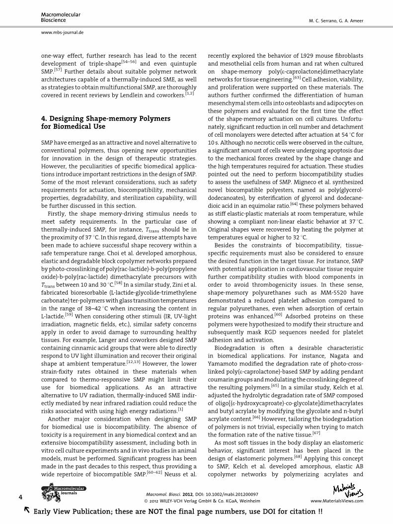
3. Unraveling the Shape-memory Effectin Polymers
SMP contain actively moving polymer networks (i.e.
able to perform movements by themselves)[1] in which
the permanent shape is determined by netpoints that are
connected with stimuli-sensitive molecular switches.[52] In
general terms, SME requires three major stages: processing,
programming, and recovery. Conventionally, the polymer
network is formed into the initial, permanent shape.
Afterwards, programming leads to the deformation of
the polymer network into the temporary, fixed shape by the
action of an external stress. Finally, upon application of
the appropriate external stimulus, the polymer recovers its
original shape.[1] In these networks, a certain orientation of
the chain segments must be permitted in order to obtain
deformation when a stress is externally applied. Cross-
linking of either chemical or physical nature is responsible
for controlling the shape transitions in these materials.
When physical crosslinking is involved, domains with the
Figure 2. Scheme of thermally-induced shape-memory properties dsimplification, switch structures have been represented as diamondtemporary to a permanent shape is controlled by the transition tem2011, John Wiley and Sons.
www.MaterialsViews.com
Macromol. Biosci. 2012, DOI: 10.1
� 2012 WILEY-VCH Verlag Gmb
Early View Publication; these are NOT
highest thermal transition are conventionally the net-
points (hard domains) while the chain segments act as
switching domains.[2] By forming additional reversible
crosslinks driven by solidification, these molecular switch-
ing domains may fix the deformed temporary shape.
Figure 2 illustrates the shape memory properties of an
elastomeric SMP based on physical crosslinking. It is
believed that permanent shape recovery in SMP is mainly
lead by the entropic elasticity of the switching domains,[2]
although more complex, theoretical models are required to
interpret the behavior of some SMP.[2,53]
During the tailored processing and programming
required for the acquisition of the thermally-induced
shape-memory properties (also known as dual-shape
creation process) in elastomeric polymers, an external
stress is applied to the SMP in the amorphous state to
deform the material to a predetermined elongation em.
When cooling the material below the thermal transition
temperature Ttrans of the switching domains, these domains
solidify and form physical crosslinks. At this stage, these
additional physical crosslinks dominate the netpoints that
originally determine the permanent shape and then the
elongated shape is temporarily fixed. Reheating will drive
the recovery of the permanent shape.[2] On the basis of this
riven by physical crosslinking in citric acid-based elastomers. Fors and netpoints, as lines. As shown in the figure, transition from aperature of the material. Reproduced with permission.[77] Copyright
002/mabi.201200097
H & Co. KGaA, Weinheim3
the final page numbers, use DOI for citation !! R
4
REa
www.mbs-journal.de
M. C. Serrano, G. A. Ameer
one-way effect, further research has lead to the recent
development of triple-shape[54–56] and even quintuple
SMP.[57] Further details about suitable polymer network
architectures capable of a thermally-induced SME, as well
as strategies to obtain multifunctional SMP, are thoroughly
covered in recent reviews by Lendlein and coworkers.[1,2]
4. Designing Shape-memory Polymersfor Biomedical Use
SMP have emerged as an attractive and novel alternative to
conventional polymers, thus opening new opportunities
for innovation in the design of therapeutic strategies.
However, the peculiarities of specific biomedical applica-
tions introduce important restrictions in the design of SMP.
Some of the most relevant considerations, such as safety
requirements for actuation, biocompatibility, mechanical
properties, degradability, and sterilization capability, will
be further discussed in this section.
Firstly, the shape memory-driving stimulus needs to
meet safety requirements. In the particular case of
thermally-induced SMP, for instance, Ttrans should be in
the proximity of 37 8C. In this regard, diverse attempts have
been made to achieve successful shape recovery within a
safe temperature range. Choi et al. developed amorphous,
elastic and degradable block copolymer networks prepared
by photo-crosslinking of poly(rac-lactide)-b-poly(propylene
oxide)-b-poly(rac-lactide) dimethacrylate precursors with
Ttrans between 10 and 30 8C.[58] In a similar study, Zini et al.
fabricated bioresorbable (L-lactide-glycolide-trimethylene
carbonate) ter-polymers with glass transition temperatures
in the range of 38–42 8C when increasing the content in
L-lactide.[59] When considering other stimuli (IR, UV-light
irradiation, magnetic fields, etc.), similar safety concerns
apply in order to avoid damage to surrounding healthy
tissues. For example, Langer and coworkers designed SMP
containing cinnamic acid groups that were able to directly
respond to UV light illumination and recover their original
shape at ambient temperature.[12,13] However, the lower
strain-fixity rates obtained in these materials when
compared to thermo-responsive SMP might limit their
use for biomedical applications. As an attractive
alternative to UV radiation, thermally-induced SME indir-
ectly mediated by near infrared radiation could reduce the
risks associated with using high energy radiations.[1]
Another major consideration when designing SMP
for biomedical use is biocompatibility. The absence of
toxicity is a requirement in any biomedical context and an
extensive biocompatibility assessment, including both in
vitro cell culture experiments and in vivo studies in animal
models, must be performed. Significant progress has been
made in the past decades to this respect, thus providing a
wide repertoire of biocompatible SMP.[60–62] Neuss et al.
Macromol. Biosci. 2012, DOI: 1
� 2012 WILEY-VCH Verlag Gmb
rly View Publication; these are NOT the final pag
recently explored the behavior of L929 mouse fibroblasts
and mesothelial cells from human and rat when cultured
on shape-memory poly(e-caprolactone)dimethacrylate
networks for tissue engineering.[63] Cell adhesion, viability,
and proliferation were supported on these materials. The
authors further confirmed the differentiation of human
mesenchymal stem cells into osteoblasts and adipocytes on
these polymers and evaluated for the first time the effect
of the shape-memory actuation on cell cultures. Unfortu-
nately, significant reduction in cell number and detachment
of cell monolayers were detected after actuation at 54 8C for
10 s. Although no necrotic cells were observed in the culture,
a significant amount of cells were undergoing apoptosis due
to the mechanical forces created by the shape change and
the high temperatures required for actuation. These studies
pointed out the need to perform biocompatibility studies
to assess the usefulness of SMP. Migneco et al. synthesized
novel biocompatible polyesters, named as poly(glycerol-
dodecanoates), by esterification of glycerol and dodecane-
dioic acid in an equimolar ratio.[64] These polymers behaved
as stiff elastic-plastic materials at room temperature, while
showing a compliant non-linear elastic behavior at 37 8C.
Original shapes were recovered by heating the polymer at
temperatures equal or higher to 32 8C.
Besides the constraints of biocompatibility, tissue-
specific requirements must also be considered to ensure
the desired function in the target tissue. For instance, SMP
with potential application in cardiovascular tissue require
further compatibility studies with blood components in
order to avoid thrombogenicity issues. In these sense,
shape-memory polyurethanes such as MM-5520 have
demonstrated a reduced platelet adhesion compared to
regular polyurethanes, even when adsorption of certain
proteins was enhanced.[60] Adsorbed proteins on these
polymers were hypothesized to modify their structure and
subsequently mask RGD sequences needed for platelet
adhesion and activation.
Biodegradation is often a desirable characteristic
in biomedical applications. For instance, Nagata and
Yamamoto modified the degradation rate of photo-cross-
linked poly(e-caprolactone)-based SMP by adding pendant
coumarin groups and modulating the crosslinking degree of
the resulting polymers.[65] In a similar study, Kelch et al.
adjusted the hydrolytic degradation rate of SMP composed
of oligo[(e-hydroxycaproate)-co-glycolate]dimethacrylates
and butyl acrylate by modifying the glycolate and n-butyl
acrylate content.[66] However, tailoring the biodegradation
of polymers is not trivial, especially when trying to match
the formation rate of the native tissue.[67]
As most soft tissues in the body display an elastomeric
behavior, significant interest has been placed in the
design of elastomeric polymers.[68] Applying this concept
to SMP, Kelch et al. developed amorphous, elastic AB
copolymer networks by polymerizing acrylates and
0.1002/mabi.201200097
H & Co. KGaA, Weinheim www.MaterialsViews.com
e numbers, use DOI for citation !!
Recent Insights Into the Biomedical Applications . . .
www.mbs-journal.de
poly[(L-lactide)-ran-glycolide]dimethylacrylate using UV-
light.[69] In these studies, SMP with Ttrans varying from 9
to 45 8C were obtained depending on the choice of acrylate
monomer and the monomer ratio, with higher elasticity
than pure dimethylacrylate networks and potential to
show triple-shape functionality.
By controlling polymer composition and integrating
several single components to create multifunctional SMP,
additional properties can also be acquired and/or modu-
lated in SMP to benefit their biomedical application (e.g.,
permeability, electrical conductivity, mechanical proper-
ties, biodegradability, magnetic properties, transparency,
radio-opacity, etc).[2] For instance, Alteheld et al. described
amorphous and transparent copolyester-urethane net-
works with shape-memory properties and potential
applicability as devices for minimally invasive surgery
in ophthalmic applications.[70] Unfortunately, the Ttrans
(which varied from 48 to 66 8C depending on composition)
limited their biomedical use. More recent advances by
these authors have achieved a lower transition tempera-
ture range for these polymers (between 14 and 56 8C) by the
selection of the comonomer (e.g., p-dioxanone, diglycolide,
or e-caprolactone) and its content (wt%) without affecting
the elastic properties of the materials.[71] Finally, the effects
of standard sterilization methods on the SME must be
considered and assessed as these methods may modify the
switch mechanisms within the SMP.
5. Biomedical Applications of Shape-memoryPolymers
Since early attempts by Echigo et al. to occlude the cardiac
ductus arteriosus by using a thermally-activated SMP based
on polynorbornene,[72] significant experience has been
obtained in the design and application of SMP in biomedicine.
In this section, the most relevant advances in the biomedical
application of SMP are discussed, with special emphasis on
those works where evidence of functionality either in vitro or
in vivo has been obtained. Particularly, recent insights in the
use of SMP as drug delivery systems and medical devices
intended for minimally invasive surgery, including endo-
vascular clot removal, aneurysm occlusion, degradable
sutures, fasteners and removable stents, and orthodontic
appliances, are highlighted. All these approaches have been
summarized in Table 1. For further details about these and
other biomedical applications of SMP, readers are referred to
other comprehensive reviews.[28,39]
5.1. Drug Delivery Systems
Controlled drug delivery has been a major goal in the design
of smart materials for biomedical applications.[73] Neffe
et al. have recently developed degradable SMP networks
www.MaterialsViews.com
Macromol. Biosci. 2012, DOI: 10.1
� 2012 WILEY-VCH Verlag Gmb
Early View Publication; these are NOT
prepared by UV-curing oligo[(e-caprolactone)-co-glycolide]-
dimethacrylates precursors.[74] Ethacridine lactate and
enoxacin were loaded in the resulting polymer networks
by swelling and in situ incorporation as examples of
hydrophilic and hydrophobic test drugs, respectively. In
addition to achieving drug delivery and SME at relevant
temperatures (28–42 8C), further studies demonstrated the
accomplishment of three major requirements: mainte-
nance of shape-memory functionality after drug incorpora-
tion, diffusion-controlled release independent of polymer
biodegradation, and drug release profiles independent of
programming and recovery processes. The authors antici-
pated the potential use of this drug-releasing SMP for the
fabrication of ureter stents with bactericidal activity or
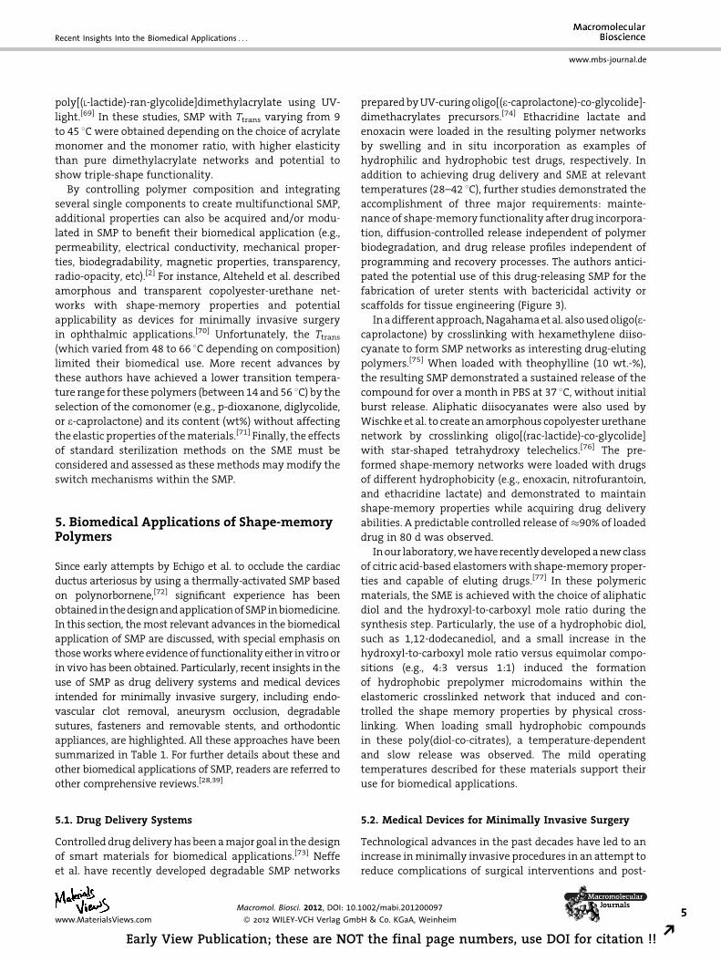
scaffolds for tissue engineering (Figure 3).
In a different approach, Nagahama et al. also used oligo(e-
caprolactone) by crosslinking with hexamethylene diiso-
cyanate to form SMP networks as interesting drug-eluting
polymers.[75] When loaded with theophylline (10 wt.-%),
the resulting SMP demonstrated a sustained release of the
compound for over a month in PBS at 37 8C, without initial
burst release. Aliphatic diisocyanates were also used by
Wischke et al. to create an amorphous copolyester urethane
network by crosslinking oligo[(rac-lactide)-co-glycolide]
with star-shaped tetrahydroxy telechelics.[76] The pre-
formed shape-memory networks were loaded with drugs
of different hydrophobicity (e.g., enoxacin, nitrofurantoin,
and ethacridine lactate) and demonstrated to maintain
shape-memory properties while acquiring drug delivery
abilities. A predictable controlled release of�90% of loaded
drug in 80 d was observed.
In our laboratory, we have recently developed a new class
of citric acid-based elastomers with shape-memory proper-
ties and capable of eluting drugs.[77] In these polymeric
materials, the SME is achieved with the choice of aliphatic
diol and the hydroxyl-to-carboxyl mole ratio during the
synthesis step. Particularly, the use of a hydrophobic diol,
such as 1,12-dodecanediol, and a small increase in the
hydroxyl-to-carboxyl mole ratio versus equimolar compo-
sitions (e.g., 4:3 versus 1:1) induced the formation
of hydrophobic prepolymer microdomains within the
elastomeric crosslinked network that induced and con-
trolled the shape memory properties by physical cross-
linking. When loading small hydrophobic compounds
in these poly(diol-co-citrates), a temperature-dependent
and slow release was observed. The mild operating
temperatures described for these materials support their
use for biomedical applications.
5.2. Medical Devices for Minimally Invasive Surgery
Technological advances in the past decades have led to an
increase in minimally invasive procedures in an attempt to
reduce complications of surgical interventions and post-
002/mabi.201200097
H & Co. KGaA, Weinheim5
the final page numbers, use DOI for citation !! R
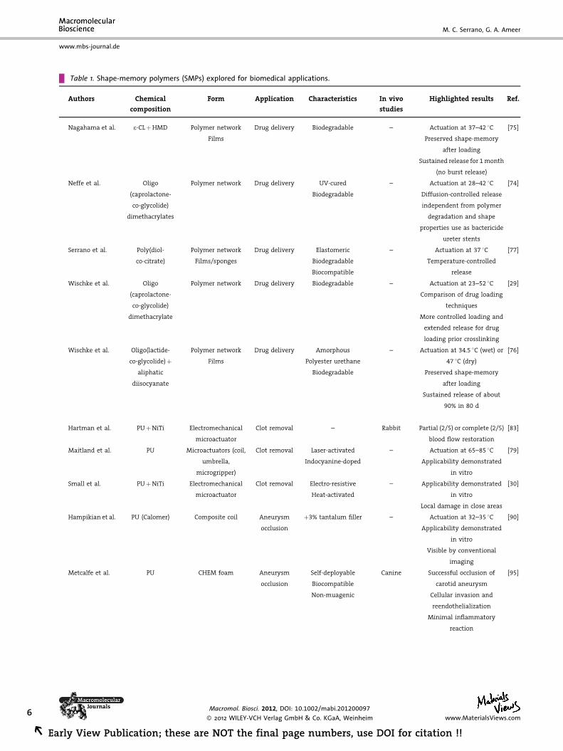
Table 1. Shape-memory polymers (SMPs) explored for biomedical applications.
Authors Chemical
composition
Form Application Characteristics In vivo
studies
Highlighted results Ref.
Nagahama et al. e-CLþHMD Polymer network
Films
Drug delivery Biodegradable – Actuation at 37–42 8C [75]
Preserved shape-memory
after loading
Sustained release for 1 month
(no burst release)
Neffe et al. Oligo
(caprolactone-
co-glycolide)
dimethacrylates
Polymer network Drug delivery UV-cured – Actuation at 28–42 8C [74]
Biodegradable Diffusion-controlled release
independent from polymer
degradation and shape
properties use as bactericide
ureter stents
Serrano et al. Poly(diol-
co-citrate)
Polymer network
Films/sponges
Drug delivery Elastomeric
Biodegradable
Biocompatible
– Actuation at 37 8C
Temperature-controlled
release
[77]
Wischke et al. Oligo
(caprolactone-
co-glycolide)
dimethacrylate
Polymer network Drug delivery Biodegradable – Actuation at 23–52 8C [29]
Comparison of drug loading
techniques
More controlled loading and
extended release for drug
loading prior crosslinking
Wischke et al. Oligo(lactide-
co-glycolide)þaliphatic
diisocyanate
Polymer network
Films
Drug delivery Amorphous
Polyester urethane
Biodegradable
– Actuation at 34.5 8C (wet) or
47 8C (dry)
Preserved shape-memory
after loading
Sustained release of about
90% in 80 d
[76]
Hartman et al. PUþNiTi Electromechanical
microactuator
Clot removal – Rabbit Partial (2/5) or complete (2/5) [83]
blood flow restoration
Maitland et al. PU Microactuators (coil,
umbrella,
microgripper)
Clot removal Laser-activated – Actuation at 65–85 8C [79]
Indocyanine-doped Applicability demonstrated
in vitro
Small et al. PUþNiTi Electromechanical
microactuator
Clot removal Electro-resistive
Heat-activated
– Applicability demonstrated
in vitro
[30]
Local damage in close areas
Hampikian et al. PU (Calomer) Composite coil Aneurysm
occlusion
þ3% tantalum filler – Actuation at 32–35 8C [90]
Applicability demonstrated
in vitro
Visible by conventional
imaging
Metcalfe et al. PU CHEM foam Aneurysm
occlusion
Self-deployable
Biocompatible
Non-muagenic
Canine Successful occlusion of
carotid aneurysm
Cellular invasion and
reendothelialization
Minimal inflammatory
reaction
[95]
6 Macromol. Biosci. 2012, DOI: 10.1002/mabi.201200097
� 2012 WILEY-VCH Verlag GmbH & Co. KGaA, Weinheim www.MaterialsViews.com
REarly View Publication; these are NOT the final page numbers, use DOI for citation !!
www.mbs-journal.de
M. C. Serrano, G. A. Ameer
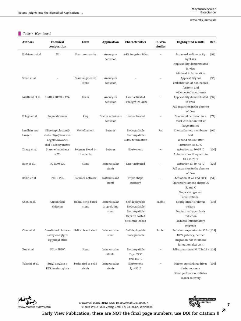
Table 1. (Continued)
Authors Chemical
composition
Form Application Characteristics In vivo
studies
Highlighted results Ref.
Rodriguez et al. PU Foam composite Aneurysm
occlusion
þ4% tungsten filler – Improved radio-opacity
by X-ray
[98]
Applicability demonstrated
in vitro
Minimal inflammation
Small et al. – Foam-augmented
stent
Aneurysm
occlusion
– – Applicability for
embolization of non-necked
fusiform and
wide-necked aneurysms
[96]
Maitland et al. HMDþHPEDþ TEA Foam Aneurysm
occlusion
Laser-activated
þEpolightTM 4121
– Applicability demonstrated
in vitro
Full expansion in the absence
of flow
[97]
Echigo et al. Polynorbornene Ring Ductus arteriosus
occlusion
Heat-activated – Successful occlusion in a
mock-circulation test of
large arteries
[72]
Lendlein and
Langer
Oligo(caprolactone)-
diolþ oligo(dioxaxon-
oligo(dioxaxone)-
diolþdiisocyanates
Monofilament Sutures Biodegradable
Biocompatible
400% deformation
Rat Chorioallantoic membrane
test
Wound closure after
actuation at 41 8C
[99]
Zhang et al. Styrene-butadiene Polymer blend in
filaments
Sutures Elastomeric – Actuation at 56–57 8C [100]
þPCL Automatic knotting within
10 s at 70 8C
Baer et al. PU MM5520 Stent Intravascular
stents
Laser-activated – Actuation at 40–45 8C [120]
Full expansion in the absence
of flow
Bellin et al. PEGþPCL Polymer network Fasteners and
stents
Triple-shape
memory
– Actuation at 40 and 60 8C [54]
Transitions among shapes A,
B, and C
Shape changes not
unidirectional
Chen et al. Crosslinked
chitosan
Helical strip-based
stent
Intravascular
drug-eluting
stent
Self-deployable
Biodegradable
Biocompatible
Heparin-coated
Sirolimus-loaded
Rabbit Nearly linear sirolimus
release
Neointima hyperplasia
reduction
Reduced inflammatory
response
[119]
Chen et al. Crosslinked chitosan
þethylene glycol
diglycidyl ether
Helical blend stent Intravascular
stent
Self-deployable
Biodegradable
Rabbit Full stent expansion in 150 s
100% patency, neither
migration nor thrombus
formation after 24 h
[118]
Xue et al. PCLþ PHBV Stent Intravascular
stents
Biocompatible – Self-expansion at 37 8C in 25 s [114]
Tm� 39 8C
and 142 8C
Yakacki et al. Butyl acrylateþPEGdimetracrylate
Perforated or solid
stents
Intravascular
stents
Elastomeric
Tg� 50 8C
– Higher crosslinking drives
faster recovery
Stent perforation initiates
sooner recovery
[105]
www.MaterialsViews.com
Macromol. Biosci. 2012, DOI: 10.1002/mabi.201200097
� 2012 WILEY-VCH Verlag GmbH & Co. KGaA, Weinheim7
Early View Publication; these are NOT the final page numbers, use DOI for citation !! R
Recent Insights Into the Biomedical Applications . . .
www.mbs-journal.de

Table 1. (Continued)
Authors Chemical
composition
Form Application Characteristics In vivo
studies
Highlighted results Ref.
Lendlein and
Langer
Diverse composition Self-expanding
implant
Gastrointestinal
or urogenital
device
Diverse stimuli for
actuation
– Promise for curbing appetite
in obesity
[121]
or drug-delivery in other
pathologies
Patent
Marco Diverse composition Self-inflating
implant
Gastrointestinal
device
Actuated by acidic
pH or at 3.7 8C
– Promise for curbing appetite [122]
Patent
Mather et al. Diverse composition Ligatures, brackets,
modules, appliances
Orthodontic
therapy
Diverse stimuli for
actuation
– Promise to secure arch wire
to brackets, self-ligating
brackets, SMP-coated wire
springs to
display human teeth
[124]
Patent
Nakasima et al. Polynorbornene Appliances Orthodontic
therapy
Heat-activated – Promise to displace human
teeth
[123]
e-CL, oligo(e-caprolactone); HMD, hexamethylene diisocyanate; HPED, hydroxypropyl ethylenediamine; NiTi, nitinol (nickel-titanium
alloy); PCL, poly(e-caprolactone); PEG, poly(ethylene glycol); PHBV, poly(hydroxybutyrate-co-hydroxyvalerate); PU, polyurethane; TEA,
triethanolamine.
8
REa
www.mbs-journal.de
M. C. Serrano, G. A. Ameer
surgery costs.[78] By using the dual-shape properties of
SMP, implants could be potentially delivered to the
desired target in a compressed, temporary shape and then
allowed to acquire the functional, permanent shape once
set in place and only after application of the appropriate
stimulus.[1] Metallic shape-memory alloys such as nitinol
(Ni–Ti alloy) are already used as cardiovascular stents,
guide wires, and orthodontic wires.[41] Unfortunately, these
alloys display important limitations, such as a limited
range of mechanical deformation (up to 8%) and a time-
consuming programming often with temperatures over
several hundreds of degrees Celsius,[14] thus opening new
opportunities for improvement by using SMP.
5.2.1. Microactuators for Endovascular Clot Removal
In the USA stroke is the primary cause of long-term
disability, with �730 000 cases annually. Their treatment
and rehabilitation costs exceed $40 billion per year.[79]
Intravenous recombinant tissue plasminogen activator
(tPA) is the only therapy currently approved for the clinical
treatment of acute ischemic stroke. Unfortunately, positive
outcomes are linked to the early administration of tPA (i.e.
within 3 h of the onset of symptoms), increasing the risk of
intracranial hemorrhage outside of this window.[80] In this
context, the design of microdevices to enable clot removal
has recently attracted significant attention in minimally
invasive surgery as an alternative to conventional clot-
dissolving drug treatments. However, critical engineering
Macromol. Biosci. 2012, DOI: 1
� 2012 WILEY-VCH Verlag Gmb
rly View Publication; these are NOT the final pag
of both the polymer and the resulting device is required to
satisfy actuation and clot removal under physiological
pressure and flow conditions.[80,81] Maitland and coworkers
designed an electromechanical microactuator composed of
a SMP and shape memory nitinol to remove stroke-causing
thrombus in brain blood vessels.[82] After delivery through a
catheter and penetration of the clot in a straight rod shape,
the device transformed to a corkscrew shape when actuated
with electro-resistive heating caused by an electrical
current that allowed clot grabbing and extraction. The
authors demonstrated the applicability of this prototype in
a water-filled silicone neurovascular model and found
potential thermal damage only localized in artery wall
areas adjacent to the device. When these endovascular
actuators were tested in vivo for the extraction of induced
thrombus that occluded the common carotid artery in
rabbits,[83] in four out of five cases partial or complete blood
flow restoration was confirmed by angiography. These
authors also reported on the design of optically-actuated
urethane-based SMP for the removal of neurovascular
occlusions causing ischemic strokes.[84] In an alternative
approach, this research group described a novel SMP
actuated by diode laser heating (A¼ 800 nm) as a
microactuator for endovascular thrombus removal.[85,86]
In vitro studies in a thrombotic vascular occlusion model
demonstrated the feasibility of its clinical use versus
conventional clot-dissolving drugs such as tPA. To guaran-
tee successful deployment, the microactuator was doped
with indocyanine green dye in order to increase adsorption
0.1002/mabi.201200097
H & Co. KGaA, Weinheim www.MaterialsViews.com
e numbers, use DOI for citation !!
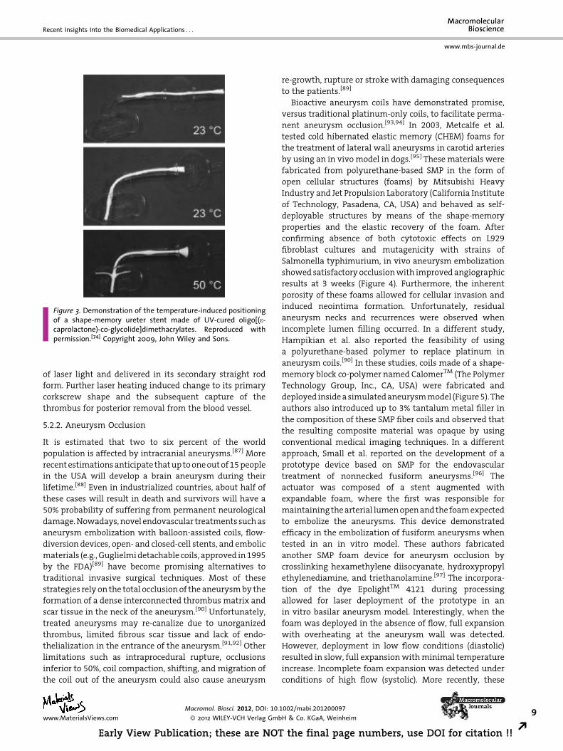
Figure 3. Demonstration of the temperature-induced positioningof a shape-memory ureter stent made of UV-cured oligo[(e-caprolactone)-co-glycolide]dimethacrylates. Reproduced withpermission.[74] Copyright 2009, John Wiley and Sons.
Recent Insights Into the Biomedical Applications . . .
www.mbs-journal.de
of laser light and delivered in its secondary straight rod
form. Further laser heating induced change to its primary
corkscrew shape and the subsequent capture of the
thrombus for posterior removal from the blood vessel.
5.2.2. Aneurysm Occlusion
It is estimated that two to six percent of the world
population is affected by intracranial aneurysms.[87] More
recent estimations anticipate that up to one out of 15 people
in the USA will develop a brain aneurysm during their
lifetime.[88] Even in industrialized countries, about half of
these cases will result in death and survivors will have a
50% probability of suffering from permanent neurological
damage. Nowadays, novel endovascular treatments such as
aneurysm embolization with balloon-assisted coils, flow-
diversion devices, open- and closed-cell stents, and embolic
materials (e.g., Guglielmi detachable coils, approved in 1995
by the FDA)[89] have become promising alternatives to
traditional invasive surgical techniques. Most of these
strategies rely on the total occlusion of the aneurysm by the
formation of a dense interconnected thrombus matrix and
scar tissue in the neck of the aneurysm.[90] Unfortunately,
treated aneurysms may re-canalize due to unorganized
thrombus, limited fibrous scar tissue and lack of endo-
thelialization in the entrance of the aneurysm.[91,92] Other
limitations such as intraprocedural rupture, occlusions
inferior to 50%, coil compaction, shifting, and migration of
the coil out of the aneurysm could also cause aneurysm
www.MaterialsViews.com
Macromol. Biosci. 2012, DOI: 10.1
� 2012 WILEY-VCH Verlag Gmb
Early View Publication; these are NOT
re-growth, rupture or stroke with damaging consequences
to the patients.[89]
Bioactive aneurysm coils have demonstrated promise,
versus traditional platinum-only coils, to facilitate perma-
nent aneurysm occlusion.[93,94] In 2003, Metcalfe et al.
tested cold hibernated elastic memory (CHEM) foams for
the treatment of lateral wall aneurysms in carotid arteries
by using an in vivo model in dogs.[95] These materials were
fabricated from polyurethane-based SMP in the form of
open cellular structures (foams) by Mitsubishi Heavy
Industry and Jet Propulsion Laboratory (California Institute
of Technology, Pasadena, CA, USA) and behaved as self-
deployable structures by means of the shape-memory
properties and the elastic recovery of the foam. After
confirming absence of both cytotoxic effects on L929
fibroblast cultures and mutagenicity with strains of
Salmonella typhimurium, in vivo aneurysm embolization
showed satisfactory occlusion with improved angiographic
results at 3 weeks (Figure 4). Furthermore, the inherent
porosity of these foams allowed for cellular invasion and
induced neointima formation. Unfortunately, residual
aneurysm necks and recurrences were observed when
incomplete lumen filling occurred. In a different study,
Hampikian et al. also reported the feasibility of using
a polyurethane-based polymer to replace platinum in
aneurysm coils.[90] In these studies, coils made of a shape-
memory block co-polymer named CalomerTM (The Polymer
Technology Group, Inc., CA, USA) were fabricated and
deployed inside a simulated aneurysm model (Figure 5). The
authors also introduced up to 3% tantalum metal filler in
the composition of these SMP fiber coils and observed that
the resulting composite material was opaque by using
conventional medical imaging techniques. In a different
approach, Small et al. reported on the development of a
prototype device based on SMP for the endovascular
treatment of nonnecked fusiform aneurysms.[96] The
actuator was composed of a stent augmented with
expandable foam, where the first was responsible for
maintaining the arterial lumen open and the foam expected
to embolize the aneurysms. This device demonstrated
efficacy in the embolization of fusiform aneurysms when
tested in an in vitro model. These authors fabricated
another SMP foam device for aneurysm occlusion by
crosslinking hexamethylene diisocyanate, hydroxypropyl
ethylenediamine, and triethanolamine.[97] The incorpora-
tion of the dye EpolightTM 4121 during processing
allowed for laser deployment of the prototype in an
in vitro basilar aneurysm model. Interestingly, when the
foam was deployed in the absence of flow, full expansion
with overheating at the aneurysm wall was detected.
However, deployment in low flow conditions (diastolic)
resulted in slow, full expansion with minimal temperature
increase. Incomplete foam expansion was detected under
conditions of high flow (systolic). More recently, these
002/mabi.201200097
H & Co. KGaA, Weinheim9
the final page numbers, use DOI for citation !! R

Figure 4. Macroscopic and microscopic appearance of healed aneurysms after embolization with CHEM sponges in the lateral wall ofcommon carotid arteries at 3 and 12 weeks. (A–C) Aneurysms with a small crescent of neocanalization (arrows). (D–I) Aneurysms completelyhealed. Images of aneurysm necks (A, D, G) and axial sections (B, E, H). Microscopic views of axial sections (C,F,I). Reproduced withpermission.[95] Copyright 2003, Elsevier.
10
REa
www.mbs-journal.de
M. C. Serrano, G. A. Ameer
authors published on the opacification of a similar
polyurethane-based SMP foam by incorporation of a
tungsten particulate filler (4 vol.-%).[98] This SMP composite
foams demonstrated improved radio-opacity in situ by
X-ray through a pig skull while maintaining their original
Figure 5. Deployment of two SMP coils for aneurysm occlusion under s2006, Elsevier.
Macromol. Biosci. 2012, DOI: 1
� 2012 WILEY-VCH Verlag Gmb
rly View Publication; these are NOT the final pag
mechanical, physical, and chemical properties. Additional
implantation studies confirmed minimal inflammation
caused by these tungsten-doped SMP. Further experimental
and computational simulation studies by Maitland and
coworkers emphasized the interest in SMP polyurethane-
imulated flow conditions. Reproduced with permission.[90] Copyright
0.1002/mabi.201200097
H & Co. KGaA, Weinheim www.MaterialsViews.com
e numbers, use DOI for citation !!
Recent Insights Into the Biomedical Applications . . .
www.mbs-journal.de
based foams as safe treatments for intracranial saccular
aneurysms in humans.[89] In these studies, oversized
shape memory foams were predicted as a better filling
for the entire aneurysm cavity while causing stresses below
those that induce rupture of aneurysm walls.
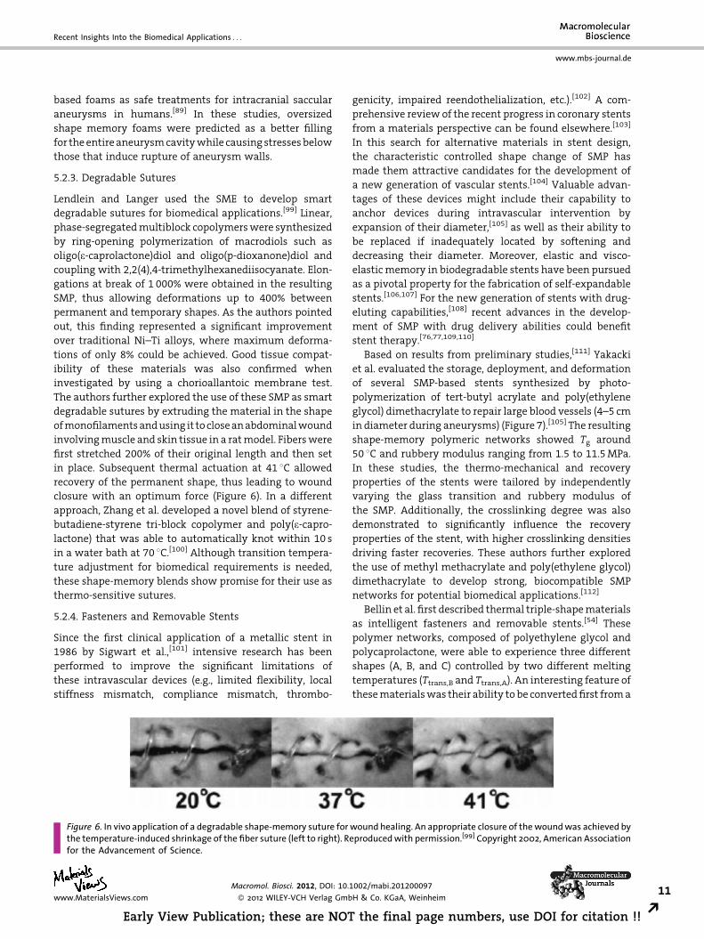
5.2.3. Degradable Sutures
Lendlein and Langer used the SME to develop smart
degradable sutures for biomedical applications.[99] Linear,
phase-segregated multiblock copolymers were synthesized
by ring-opening polymerization of macrodiols such as
oligo(e-caprolactone)diol and oligo(p-dioxanone)diol and
coupling with 2,2(4),4-trimethylhexanediisocyanate. Elon-
gations at break of 1 000% were obtained in the resulting
SMP, thus allowing deformations up to 400% between
permanent and temporary shapes. As the authors pointed
out, this finding represented a significant improvement
over traditional Ni–Ti alloys, where maximum deforma-
tions of only 8% could be achieved. Good tissue compat-
ibility of these materials was also confirmed when
investigated by using a chorioallantoic membrane test.
The authors further explored the use of these SMP as smart
degradable sutures by extruding the material in the shape
of monofilaments and using it to close an abdominal wound
involving muscle and skin tissue in a rat model. Fibers were
first stretched 200% of their original length and then set
in place. Subsequent thermal actuation at 41 8C allowed
recovery of the permanent shape, thus leading to wound
closure with an optimum force (Figure 6). In a different
approach, Zhang et al. developed a novel blend of styrene-
butadiene-styrene tri-block copolymer and poly(e-capro-
lactone) that was able to automatically knot within 10 s
in a water bath at 70 8C.[100] Although transition tempera-
ture adjustment for biomedical requirements is needed,
these shape-memory blends show promise for their use as
thermo-sensitive sutures.
5.2.4. Fasteners and Removable Stents
Since the first clinical application of a metallic stent in
1986 by Sigwart et al.,[101] intensive research has been
performed to improve the significant limitations of
these intravascular devices (e.g., limited flexibility, local
stiffness mismatch, compliance mismatch, thrombo-
Figure 6. In vivo application of a degradable shape-memory suture forthe temperature-induced shrinkage of the fiber suture (left to right). Refor the Advancement of Science.
www.MaterialsViews.com
Macromol. Biosci. 2012, DOI: 10.1
� 2012 WILEY-VCH Verlag Gmb
Early View Publication; these are NOT
genicity, impaired reendothelialization, etc.).[102] A com-
prehensive review of the recent progress in coronary stents
from a materials perspective can be found elsewhere.[103]
In this search for alternative materials in stent design,
the characteristic controlled shape change of SMP has
made them attractive candidates for the development of
a new generation of vascular stents.[104] Valuable advan-
tages of these devices might include their capability to
anchor devices during intravascular intervention by
expansion of their diameter,[105] as well as their ability to
be replaced if inadequately located by softening and
decreasing their diameter. Moreover, elastic and visco-
elastic memory in biodegradable stents have been pursued
as a pivotal property for the fabrication of self-expandable
stents.[106,107] For the new generation of stents with drug-
eluting capabilities,[108] recent advances in the develop-
ment of SMP with drug delivery abilities could benefit
stent therapy.[76,77,109,110]
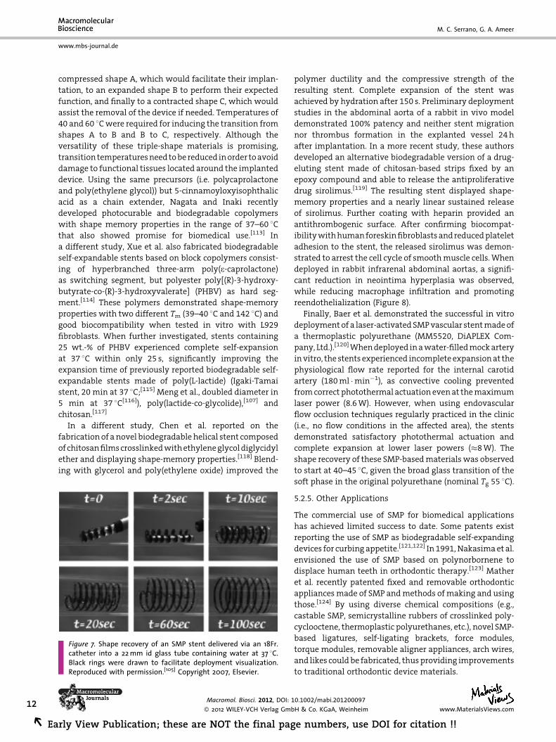
Based on results from preliminary studies,[111] Yakacki
et al. evaluated the storage, deployment, and deformation
of several SMP-based stents synthesized by photo-
polymerization of tert-butyl acrylate and poly(ethylene
glycol) dimethacrylate to repair large blood vessels (4–5 cm
in diameter during aneurysms) (Figure 7).[105] The resulting
shape-memory polymeric networks showed Tg around
50 8C and rubbery modulus ranging from 1.5 to 11.5 MPa.
In these studies, the thermo-mechanical and recovery
properties of the stents were tailored by independently
varying the glass transition and rubbery modulus of
the SMP. Additionally, the crosslinking degree was also
demonstrated to significantly influence the recovery
properties of the stent, with higher crosslinking densities
driving faster recoveries. These authors further explored
the use of methyl methacrylate and poly(ethylene glycol)
dimethacrylate to develop strong, biocompatible SMP
networks for potential biomedical applications.[112]
Bellin et al. first described thermal triple-shape materials
as intelligent fasteners and removable stents.[54] These
polymer networks, composed of polyethylene glycol and
polycaprolactone, were able to experience three different
shapes (A, B, and C) controlled by two different melting
temperatures (Ttrans,B and Ttrans,A). An interesting feature of
these materials was their ability to be converted first from a
wound healing. An appropriate closure of the wound was achieved byproduced with permission.[99] Copyright 2002, American Association
002/mabi.201200097
H & Co. KGaA, Weinheim11
the final page numbers, use DOI for citation !! R
12
REa
www.mbs-journal.de
M. C. Serrano, G. A. Ameer
compressed shape A, which would facilitate their implan-
tation, to an expanded shape B to perform their expected
function, and finally to a contracted shape C, which would
assist the removal of the device if needed. Temperatures of
40 and 60 8C were required for inducing the transition from
shapes A to B and B to C, respectively. Although the
versatility of these triple-shape materials is promising,
transition temperatures need to be reduced in order to avoid
damage to functional tissues located around the implanted
device. Using the same precursors (i.e. polycaprolactone
and poly(ethylene glycol)) but 5-cinnamoyloxyisophthalic
acid as a chain extender, Nagata and Inaki recently
developed photocurable and biodegradable copolymers
with shape memory properties in the range of 37–60 8Cthat also showed promise for biomedical use.[113] In
a different study, Xue et al. also fabricated biodegradable
self-expandable stents based on block copolymers consist-
ing of hyperbranched three-arm poly(e-caprolactone)
as switching segment, but polyester poly[(R)-3-hydroxy-
butyrate-co-(R)-3-hydroxyvalerate] (PHBV) as hard seg-
ment.[114] These polymers demonstrated shape-memory
properties with two different Tm (39–40 8C and 142 8C) and
good biocompatibility when tested in vitro with L929
fibroblasts. When further investigated, stents containing
25 wt.-% of PHBV experienced complete self-expansion
at 37 8C within only 25 s, significantly improving the
expansion time of previously reported biodegradable self-
expandable stents made of poly(L-lactide) (Igaki-Tamai
stent, 20 min at 37 8C;[115] Meng et al., doubled diameter in
5 min at 37 8C[116]), poly(lactide-co-glycolide),[107] and
chitosan.[117]
In a different study, Chen et al. reported on the
fabrication of a novel biodegradable helical stent composed
of chitosan films crosslinked with ethylene glycol diglycidyl
ether and displaying shape-memory properties.[118] Blend-
ing with glycerol and poly(ethylene oxide) improved the
Figure 7. Shape recovery of an SMP stent delivered via an 18Fr.catheter into a 22 mm id glass tube containing water at 37 8C.Black rings were drawn to facilitate deployment visualization.Reproduced with permission.[105] Copyright 2007, Elsevier.
Macromol. Biosci. 2012, DOI: 1
� 2012 WILEY-VCH Verlag Gmb
rly View Publication; these are NOT the final pag
polymer ductility and the compressive strength of the
resulting stent. Complete expansion of the stent was
achieved by hydration after 150 s. Preliminary deployment
studies in the abdominal aorta of a rabbit in vivo model
demonstrated 100% patency and neither stent migration
nor thrombus formation in the explanted vessel 24 h
after implantation. In a more recent study, these authors
developed an alternative biodegradable version of a drug-
eluting stent made of chitosan-based strips fixed by an
epoxy compound and able to release the antiproliferative
drug sirolimus.[119] The resulting stent displayed shape-
memory properties and a nearly linear sustained release
of sirolimus. Further coating with heparin provided an
antithrombogenic surface. After confirming biocompat-
ibility with human foreskin fibroblasts and reduced platelet
adhesion to the stent, the released sirolimus was demon-
strated to arrest the cell cycle of smooth muscle cells. When
deployed in rabbit infrarenal abdominal aortas, a signifi-
cant reduction in neointima hyperplasia was observed,
while reducing macrophage infiltration and promoting
reendothelialization (Figure 8).
Finally, Baer et al. demonstrated the successful in vitro
deployment of a laser-activated SMP vascular stent made of
a thermoplastic polyurethane (MM5520, DiAPLEX Com-
pany, Ltd.).[120] When deployed in a water-filled mock artery
in vitro, the stents experienced incomplete expansion at the
physiological flow rate reported for the internal carotid
artery (180 ml �min�1), as convective cooling prevented
from correct photothermal actuation even at the maximum
laser power (8.6 W). However, when using endovascular
flow occlusion techniques regularly practiced in the clinic
(i.e., no flow conditions in the affected area), the stents
demonstrated satisfactory photothermal actuation and
complete expansion at lower laser powers (�8 W). The
shape recovery of these SMP-based materials was observed
to start at 40–45 8C, given the broad glass transition of the
soft phase in the original polyurethane (nominal Tg 55 8C).
5.2.5. Other Applications
The commercial use of SMP for biomedical applications
has achieved limited success to date. Some patents exist
reporting the use of SMP as biodegradable self-expanding
devices for curbing appetite.[121,122] In 1991, Nakasima et al.
envisioned the use of SMP based on polynorbornene to
displace human teeth in orthodontic therapy.[123] Mather
et al. recently patented fixed and removable orthodontic
appliances made of SMP and methods of making and using
those.[124] By using diverse chemical compositions (e.g.,
castable SMP, semicrystalline rubbers of crosslinked poly-
cyclooctene, thermoplastic polyurethanes, etc.), novel SMP-
based ligatures, self-ligating brackets, force modules,
torque modules, removable aligner appliances, arch wires,
and likes could be fabricated, thus providing improvements
to traditional orthodontic device materials.
0.1002/mabi.201200097
H & Co. KGaA, Weinheim www.MaterialsViews.com
e numbers, use DOI for citation !!

Figure 8. Photographs of a test stent after deployment in the rabbit infrarenal abdominal aorta (a) and patency test of the stent-implantedvessel at 4 weeks (b). Confocal fluorescence images show reendothelialization (c, top) and macrophage infiltration after stent implantation(c, bottom). Reproduced with permission.[119] Copyright 2009, Elsevier.
Recent Insights Into the Biomedical Applications . . .
www.mbs-journal.de
6. Challenges and Future Perspectives
SMP present significant advantages when compared to
traditional shape-memory alloys, including lower density,
lightweight, lower cost of raw materials, lower cost of
fabrication and processing, and easy tailoring. Further-
more, the resulting materials have higher versatility in
shapes, higher recovery strains (>300%) and lower recovery
stresses (1–10 MPa). SMP also allow for the use of more
diverse stimuli for actuation, which could be remote, and
show ability to be converted on drug carriers. Moreover,
SMP are versatile to be combined with other materials to
form composites and blends for the acquisition of addi-
tional properties. Finally, their chemical stability, along
with biocompatibility and tunable biodegradability,
opens their use for biomedical applications, with a higher
potential for cheaper reuse and recycle. All these properties
have encouraged significant research on the use of SMP for
diverse biomedical applications. Particularly, as exten-
sively discussed in this article, minimally invasive surgery
may clearly benefit from SMP technology as it provides a
smart platform to deploy therapeutic devices through small
incisions and narrow conducts to reach the target in the
body. Although investigated for the fabrication of degrad-
able sutures or self-expanding stents, additional biomedical
applications for SMP could be further explored. For instance,
the design of ‘‘growth’’ stents[125] that could be deployed
in infant patients, whose blood vessels will grow after
implantation, could benefit from the progress in bio-
degradable SMP-based stents if designed as polymers with
www.MaterialsViews.com
Macromol. Biosci. 2012, DOI: 10.1
� 2012 WILEY-VCH Verlag Gmb
Early View Publication; these are NOT
the ability to be remodeled with the surrounding tissue.
Additionally, in the orthodontic practice, the application of
these actively moving materials show promise, as they
provide lighter materials and more constant forces that
may cause less pain to the patient, along with the esthetic
advantages that these materials offer (e.g., transparency,
colorable, and/or stain resistant). In some of these
applications, SMP with Tg slightly higher (e.g., 50 8C) would
be of interest, as they will resist hot beverages and
foods.[124]
Although promising, significant limitations still remain
when considering the long-term success of SMP in
biomedical applications. In the particular case of stenting,
for instance, larger radial strength is necessary to with-
stand vessel pulsation pressure at 37 8C, as well as broader
ranges of mechanical properties to completely fulfill the
vascular mechanical requirements (e.g., tensile stress
values of 0.069–1.17 MPa in different human vascular
conducts).[68] Hydration effects on material performance
should be addressed by tests in a physiological environ-
ment as the plasticization by water could significantly
affect network switching temperature at 37 8C and then
change polymer actuation.[76] Moreover, ageing after
implantation should also be carefully considered as it
can increase switching temperatures by more than
10 8C.[126] As the SME will occur under complex environ-
mental constraints generated by the surrounding tissues,
constitutive modeling studies might facilitate clinical
implementation of this technology by predicting impor-
tant limitations before clinical use.[127]
002/mabi.201200097
H & Co. KGaA, Weinheim13
the final page numbers, use DOI for citation !! R

14
REa
www.mbs-journal.de
M. C. Serrano, G. A. Ameer
The exploration of SMP in human trials is still rare and
their use in current clinical practice, negligible. For instance,
Pelissier et al. explored Polysoft1 patches for the surgical
repair of inguinal hernias allowing for reducing post-
operative complications and pain and shortening the
recovery time of the patients.[128,129] Although made of a
light monofilament polypropylene mesh, this patch con-
tained a recoil ring with shape memory properties to
facilitate the patch placement and spreading. The feasi-
bility of using this patch containing a memory coil is being
further investigated in humans.[130] In a different clinical
trial, SMP composites made of poly(lactide-co-e-caprolac-
tone) and b-tricalcium phosphate (chronOS Strips) are
currently under investigation as part of a synthetic bone
void filler.[131] The shape memory of these composites
facilitates site-specific placement as the strips conform the
specific peculiarities of the implant site of each patient.
Although diverse non-degradable thermally-induced
SMP are commercially available (e.g., thermoplastic and
thermoset polyurethanes DiAPLEX, Veriflex1, VerilyteTM,
VeritexTM, Tecoflex1, and TEMBO1, among others),[30]
none of these materials is available in medical grade
quality yet, thus limiting their translation in clinical
applications.
In summary, the main requirements for SMP envisioned
for biomedical use are: (i) actuation, preferentially remote,
within biologically safe ranges to avoid damage of
surrounding tissues, (ii) long-term stability that guarantee
durability of the outcome of the therapeutic procedure,
(iii) biocompatibility, (iv) compressed temporary shape to
facilitate minimally invasive deployment, (v) biodegrad-
ability by gradual erosion to avoid explantation, (vi) non-
toxicity, and (vii) non-immunogenicity. Multidisciplinary
research including efforts from chemists, physicians,
biologists, engineers, and clinicians will contribute to
significant advances in the biomedical use of SMP from
bench to bedside.
Acknowledgements: M. C. S. is greatly in debt with MINECO fora Juan de la Cierva fellowship.
Received: March 20, 2012; Revised: June 4, 2012; Publishedonline: DOI: 10.1002/mabi.201200097
Keywords: biomedical applications; minimally invasive surgery;poly(diol-co-citrates); polymers; shape-memory effect
[1] M. Behl, A. Lendlein, Mater. Today 2007, 10, 20.[2] M. Behl, M. Y. Razzaq, A. Lendlein, Adv. Mater. 2010, 22, 3388.[3] J. A. Hiltz, Shape Memory Polymers – Literature Review. TM
2002-127. Defence R&D Canada – Atlantique. http://pub-
Macromol. Biosci. 2012, DOI: 1
� 2012 WILEY-VCH Verlag Gmb
rly View Publication; these are NOT the final pag
s.drdc.gc.ca/PDFS/xtr9/p518446.pdf. Last retrieved: 07/18/2012.
[4] C. Liang, C. A. Rogers, E. Malafeew, J. Intell. Mater. Syst. Struct.1997, 8, 380.
[5] K. Nakayama, Int. Polym. Sci. Technol. 1991, 18, 43.[6] Y. Shirai, S. Hayashi, Development of Polymeric Shape Mem-
ory Material, MTB184, Mitsubishi Heavy Industries, Inc.,Japan 1988.
[7] H. Koerner, W. D. Liu, M. Alexander, P. Mirau, H. Dowty, R. A.Vaia, Polymer 2005, 46, 4405.
[8] J. W. Cho, J. W. Kim, Y. C. Jung, N. S. Goo, Macromol. RapidCommun. 2005, 26, 412.
[9] R. Mohr, K. Kratz, T. Weigel, M. Lucka-Gabor, M. Moneke,A. Lendlein, Proc. Natl. Acad. Sci. U. S. A. 2006, 103, 3540.
[10] T. Weigel, R. Mohr, A. Lendlein, Smart Mater. Struct. 2009, 18,025011.
[11] B. Wang, W. M. Huang, C. Li, C. M. Lee, L. Li, Smart Mater.Struct. 2004, 13, 191.
[12] A. Lendlein, H. Y. Jiang, O. Junger, R. Langer, Nature 2005,434, 879.
[13] H. Y. Jiang, S. Kelch, A. Lendlein, Adv. Mater. 2006, 18, 1471.[14] A. Lendlein, S. Kelch, Angew. Chem., Int. Ed. 2002, 41, 2034.[15] A. Lendlein, V. P. Shastri, Adv. Mater. 2010, 22, 3344.[16] A. Charlesby, in: Atomic Radiation and Polymers, Pergamon
Press, Oxford 1960, p. 198.[17] R. Vaia, J. Baur, Science 2008, 319, 420.[18] J. Hu, Shape Memory Polymers and Textiles, Woodhead
Publishing Limited, Cambridge, UK 2007.[19] X. X. Liu, J. L. Hu, K. M. Babu, S. Y. Wang, Text. Res. J. 2008, 78,
1048.[20] K. Kobayashi, S. Shunichi, (Mitsubishi Heavy Industries), US-
A 5128197, 1992, (http://www.uspto.gov/patft/).[21] Q. Meng, J. Hu, Polym. Adv. Technol. 2008, 19, 131.[22] W. M. Huang, C. W. Lee, H. P. Teo, J. Intell. Mater. Syst. Struct.
2006, 17, 753.[23] J. F. Mano, Adv. Eng. Mater. 2008, 10, 515.[24] S. A. Madbouly, A. Lendlein, Adv. Polym. Sci. 2010, 226, 41.[25] R. V. Ulijn, N. Bibi, V. Jayawarna, P. D. Thornton, S. J. Todd,
R. J. Mart, A. M. Smith, J. E. Gough, Mater. Today 2007, 10, 40.[26] D. Ratna, J. K. Kocsis, J. Mater. Sci. 2008, 43, 254.[27] Q. Meng, J. Hu, Compos.: Part A 2009, 40, 1661.[28] C. M. Yakacki, K. Gall, Adv. Polym. Sci. 2010, 226, 147.[29] C. Wischke, A. T. Neffe, A. Lendlein, Adv. Polym. Sci. 2010,
226, 177.[30] W. Small, IV, P. Singhal, T. S. Wilson, D. J. Maitland, J. Mater.
Chem. 2010, 20, 3356.[31] A. Lendlein, J. Mater. Chem. 2010, 20, 3332.[32] A. Lendlein, Ed., Shape-memory Polymers, Springer-Verlag,
Berlin, Heidelberg 2010.[33] L. Sun, W. M. Huang, Z. Ding, Y. Zhao, C. C. Wang,
H. Purnawali, C. Tang, Mater. Des. 2012, 33, 577.[34] J. Leng, S. Du, Eds., Shape-memory Polymers and Multifunc-
tional Composites, CRC Press/Taylor & Francis, Boca Raton2010.
[35] F. El Feninat, G. Laroche, M. Fiset, D. Mantovani, Adv. Eng.Mater. 2002, 4, 91.
[36] M. Yoshida, R. Langer, A. Lendlein, J. Lahann, J. Macromol.Sci., Part C: Polym. Rev. 2006, 46, 347.
[37] W. M. Huang, Z. Ding, C. C. Wang, J. Wei, Y. Zhao,H. Purnawali, Mater. Today 2010, 13, 54.
[38] E. Smela, Adv. Mater. 2003, 15, 481.[39] A. Lendlein, M. Behl, B. Hiebl, C. Wischke, Exp. Rev. Med.
Devices 2010, 7, 357.
0.1002/mabi.201200097
H & Co. KGaA, Weinheim www.MaterialsViews.com
e numbers, use DOI for citation !!
Recent Insights Into the Biomedical Applications . . .
www.mbs-journal.de
[40] L. C. Chang, T. A. Read, Trans. AIME 1951, 189, 47.[41] P. Lipscomb, L. D. M. Nokes, The Application of Shape Mem-
ory Alloys in Medicine, Mechanical Engineering Pudicotions,limited, Suffolk, UK 1996.
[42] B.-K. Kim, S.-Y. Lee, M. Xu, Polymer 1996, 37, 5781.[43] A. Lendlein, A. M. Schmidt, R. Langer, Proc. Nat. Acad. Sci. U. S.
A. 2001, 98, 842.[44] J. E. Gautrot, X. X. Zhu, Angew. Chem., Int. Ed. 2006, 45,
6872.[45] J. E. Gautrot, X. X. Zhu, Macromolecules 2009, 42, 7324.[46] Y. Feng, M. Behl, S. Kelch, A. Lendlein, Macromol. Biosci. 2009,
9, 45.[47] Y. Feng, J. Lu, M. Behl, A. Lendlein, Macromol. Biosci. 2010, 10,
1008.[48] X. Zheng, S. Zhou, X. Li, J. Weng, Biomaterials 2006, 27, 4288.[49] P. Miaudet, A. Derre, M. Maugey, C. Zakri, P. M. Piccione,
R. Inoubli, P. Poulin, Science 2007, 318, 1294.[50] M. Behl, U. Ridder, Y. Feng, S. Kelch, A. Lendlein, Soft Matter
2009, 5, 676.[51] K. Gall, M. L. Dunn, Y. Liu, D. Finch, M. Lake, N. A. Munshi,
Acta Mater. 2002, 50, 5115.[52] M. Behl, A. Lendlein, Soft Matter 2007, 3, 58.[53] J. S. Sodhi, I. J. Rao, Int. J. Eng. Sci. 2010, 48, 1576.[54] I. Bellin, S. Kelch, R. Langer, A. Lendlein, PNAS 2006, 103,
18043.[55] J. Zotzmann, M. Behl, D. Hofmann, A. Lendlein, Adv. Mater.
2010, 22, 3424.[56] M. Behl, A. Lendlein, J. Mater. Chem. 2010, 20, 3335.[57] J. Li, T. Liu, S. Xia, Y. Pan, Z. Zheng, X. Ding, Y. Peng, J. Mater.
Chem. 2011, 21, 12213.[58] N. Y. Choi, S. Kelch, A. Lendlein, Adv. Eng. Mater. 2006, 8, 439.[59] E. Zini, M. Scandola, P. Dobrzynski, J. Kasperczyk, M. Bero,
Biomacromolecules 2007, 8, 3661.[60] S. Fare, V. Valtulina, P. Petrini, E. Alessandrini, G. Pietrocola,
M. C. Tanzi, P. Speziale, L. Visai, J. Biomed. Mater. Res. A 2005,73, 1.
[61] B. Guo, Y. Chen, Y. Lei, L. Zhang, W. Y. Zhou, A. B. M. Rabie,J. Zhao, Biomacromolecules 2011, 12, 1312.
[62] S. H. Ajili, N. G. Ebrahimi, M. Soleimani, Acta Biomater. 2009,5, 1519.
[63] S. Neuss, I. Blomenkamp, R. Stainforth, D. Boltersdorf,M. Jansen, N. Butz, A. Perez-Bouza, R. Knuchel, Biomaterials2009, 30, 1697.
[64] F. Migneco, Y. C. Huang, R. K. Biria, S. J. Hollister, Biomaterials2009, 30, 6479.
[65] M. Nagata, Y. Yamamoto, J. Polym. Sci., A: Polym. Chem. 2009,47, 2422.
[66] S. Kelch, S. Steuer, A. M. Schmidt, A. Lendlein, Biomacromo-lecules 2007, 8, 1018.
[67] D. Hofmann, M. Entrialgo-Castano, K. Kratz, A. Lendlein, Adv.Mater. 2009, 21, 3237.
[68] M. C. Serrano, E. J. Chung, G. A. Ameer, Adv. Funct. Mater.2010, 20, 192.
[69] S. Kelch, N.-Y. Choi, Z. Wang, A. Lendlein, Adv. Eng. Mater.2008, 10, 494.
[70] A. Alteheld, Y. Feng, S. Kelch, A. Lendlein, Angew. Chem., Int.Ed. 2005, 44, 1188.
[71] A. Lendlein, J. Zotzmann, Y. Feng, A. Alteheld, S. Kelch,Biomacromolecules 2009, 10, 975.
[72] S. Echigo, T. Matsuda, T. Kamiya, E. Tsuda, K. Suda, K. Kuroe,Y. Ono, K. Yazawa, ASAIO Trans. 1990, 36, M195.
[73] R. Langer, D. A. Tirrell, Nature 2004, 428, 487.[74] A. T. Neffe, B. D. Hanh, S. Steuer, A. Lendlein, Adv. Mater.
2009, 21, 3394.
www.MaterialsViews.com
Macromol. Biosci. 2012, DOI: 10.1
� 2012 WILEY-VCH Verlag Gmb
Early View Publication; these are NOT
[75] K. Nagahama, Y. Ueda, T. Ouchi, Y. Ohya, Biomacromolecules2009, 10, 1789.
[76] C. Wischke, A. T. Neffe, S. Steuer, A. Lendlein, J. ControlledRelease 2009, 138, 243.
[77] M. C. Serrano, L. Carbajal, G. A. Ameer, Adv. Mater. 2011, 23,2211.
[78] J. G. Hunter, Ed., Minimally Invasive Surgery, McGraw Hill,New York 1993.
[79] D. J. Maitland, M. F. Metzger, D. Schumann, A. Lee, T. S.Wilson, Lasers Surg. Med. 2002, 30, 1.
[80] J. R. Marler, N Engl. J. Med. 1995, 333, 1581.[81] M. F. Metzger, T. S. Wilson, D. Schumann, D. L. Matthews, D. J.
Maitland, Biomed. Microdevices 2002, 4, 89.[82] W. Small, IV, T. S. Wilson, P. R. Buckley, W. J. Benett, J. M. Loge,
J. Hartman, D. J. Maitland, IEEE Trans. Biomed. Eng. 2007, 54,1657.
[83] J. Hartman, W. Small, IV, T. S. Wilson, J. Brock, P. R. Buckley,W. J. Benett, J. M. Loge, D. J. Maitland, Am. J. Neuroradiol.2007, 28, 872.
[84] T. S. Wilson, W. Small, IV, W. J. Benett, J. P. Bearinger, D. J.Maitland, Proc. SPIE Int. Soc. Opt. Eng. 2005, 6007, 60070R.
[85] W. Small, IV, T. S. Wilson, W. J. Benett, J. M. Loge, D. J.Maitland, Opt. Express 2005, 13, 8204.
[86] W. Small, IV, M. F. Metzger, T. S. Wilson, D. J. Maitland, IEEE J.Sel. Top. Quantum Electron. 2005, 11, 892.
[87] M. Horowitz, D. Samson, P. Purdy, AJNR Am. J. Neuroradiol.1997, 18, 510.
[88] http://www.brainaneurysm.com/index.html. �ASITN. Lastretrieved: 01/13/2012.
[89] W. Hwang, B. L. Volk, F. Akberali, P. Singhal, J. C. Criscione, D.J. Maitland, Biomech. Model Mechanobiol. 2012, 11, 715.
[90] J. M. Hampikian, B. C. Heaton, F. C. Tong, Z. Zhang, C. P. Wong,Mater. Sci. Eng. C 2006, 26, 1373.
[91] J. Byrne, J. K. Hope, N. Hubbard, J. H. Morris, AJNR Am. J.Neuroradiol. 1997, 18, 29.
[92] D. Kallmes, G. Helm, S. Hudson, T. Altes, H. Do, J. Mandell,H. Cloft, Radiology 1999, 213, 217.
[93] D. Kallmes, N. Fujiwara, D. Yuen, D. Dai, S. Li, AJNR Am. J.Neuroradiol. 2003, 24, 591.
[94] H. Cloft, D. Kallmes, AJNR Am. J. Neuroratiol. 2004, 25, 60.[95] A. Metcalfe, A.-C. Desfaits, I. Salazkin, L’H. Yahia, W. M.
Sokolowski, J. Raymond, Biomaterials 2003, 24, 491.[96] W. Small, IV, P. R. Buckley, T. S. Wilson, W. J. Benett,
J. Hartman, D. Saloner, D. J. Maitland, IEEE Trans. Biomed.Eng. 2007, 54, 1157.
[97] D. J. Maitland, W. Small, IV, J. M. Ortega, P. R. Buckley,J. Rodriguez, J. Hartman, T. S. Wilson, J. Biomed. Opt. 2007,12, 030504–1.
[98] J. N. Rodriguez, Y.-J. Yu, M. W. Miller, T. S. Wilson, J. Hartman,F. J. Clubb, B. Gentry, D. J. Maitland, Ann. Biomed. Eng. 2012,40, 883.
[99] A. Lendlein, R. Langer, Science 2002, 296, 1673.[100] H. Zhang, H. Wang, W. Zhong, Q. Du, Polymer 2009, 50, 1596.[101] U. Sigwart, J. Puel, V. Mirkovitch, F. Joffre, L. Kappenberger,
N. Engl. J. Med. 1987, 316, 701.[102] J. C. Palmaz, J. Endovasc. Ther. 2004, 11, 200.[103] G. Mani, M. D. Feldman, D. Patel, C. M. Agrawal, Biomaterials
2007, 28, 1689.[104] F. Jung, C. Wischke, A. Lendlein, MRS Bull. 2010, 35, 607.[105] C. M. Yakacki, R. Shandas, C. Lanning, B. Rech, A. Eckstein,
K. Gall, Biomaterials 2007, 28, 2255.[106] T. Valimaa, S. Laaksovirta, T. L. Tammela, P. Laippala,
M. Talja, T. Isotalo, A. Petas, K. Tarri, P. Tormala, Biomaterials2002, 23, 3575.
002/mabi.201200097
H & Co. KGaA, Weinheim15
the final page numbers, use DOI for citation !! R
16
REa
www.mbs-journal.de
M. C. Serrano, G. A. Ameer
[107] S. S. Venkatraman, L. P. Tan, J. F. D. Joso, Y. C. F. Boey, X. Wang,Biomaterials 2006, 27, 1573.
[108] T. F. Luscher, J. Steffel, F. R. Eberli, M. Joner, G. Nakazawa, F. C.Tanner, R. Virmani, Circulation 2007, 115, 1051.
[109] C. Wischke, A. Lendlein, Pharm. Res. 2010, 27, 527.[110] H. M. Wache, D. J. Tartakowska, A. Hentrich, M. H. Wagner,
J. Mater. Sci.: Mater. Med. 2003, 14, 109.[111] K. Gall, C. M. Yakacki, Y. Liu, R. Shandas, N. Willett, K. S.
Anseth, J. Biomed. Mater. Res., A 2005, 73, 339.[112] C. M. Yakacki, R. Shandas, D. Safranski, A. M. Ortega,
K. Sassaman, K. Gall, Adv. Funct. Mater. 2008, 18, 2428.[113] M. Nagata, K. Inaki, J. Appl. Polym. Sci. 2011, 120, 3556.[114] L. Xue, S. Dai, Z. Li, Biomaterials 2010, 31, 8132.[115] H. Tamai, K. Igaki, E. Kyo, K. Kosuga, A. Kawashima,
S. Matsui, H. Komori, T. Tsuji, S. Motohara, H. Uehata,Circulation 2000, 102, 399.
[116] B. Meng, J. Wang, N. Zhu, Q.-Y. Meng, F.-Z. Cui, Y.-X. Xu,J. Mater. Sci.: Mater. Med. 2006, 17, 611.
[117] A. Lauto, M. Ohebshalom, M. Esposito, J. Mingin, P. S. Li,D. Felsen, M. Goldstein, D. P. Poppas, Biomaterials 2001, 22,1869.
[118] M.-C. Chen, H.-W. Tsai, Y. Chang, W.-Y. Lai, F.-L. Mi, C.-T. Liu,H.-S. Wong, H.-W. Sung, Biomacromolecules 2007, 8, 2774.
[119] M.-C. Chen, Y. Chang, C.-T. Liu, W.-Y. Lai, S.-F. Peng, Y.-W.Hung, H.-W. Tsai, H.-W. Sung, Biomaterials 2009, 30, 79.
[120] G. M. Baer, W. Small, IV, T. S. Wilson, W. J. Benett, D. L.Matthews, J. Hartman, D. J. Maitland, Biomed. Eng. Online2007, 6, n.43.
Macromol. Biosci. 2012, DOI: 1
� 2012 WILEY-VCH Verlag Gmb
rly View Publication; these are NOT the final pag
[121] WO 2004073690 A1 (2004), Self-expanding device for thegastrointestinal or urogenital area, invs.: A. Lendlein, R. S.Langer.
[122] WO 2006/092789 A2 (2006), Biodegradable self-inflatingintragastric implants for curbing appetite, invs.: D. Marco.
[123] A. Nakasima, J. R. Hu, M. Ichinose, H. Shimada, Eur. J. Orthod.1991, 13, 179.
[124] WO 2006/071520 A2 (2006), Shape memory polymer ortho-dontic appliances, and methods of making and using thesame, invs.: P. Mather, C. Liu, C. Burstone.
[125] P. Ewert, E. Riesenkampff, M. Neuss, O. Kretschmar,N. Nagdyman, P. E. Lange, Catheter. Cardiovasc. Int. 2004,62, 506.
[126] V. Lorenzo, A. Dıaz-Lantada, P. Lafont, H. Lorenzo-Yustos,C. Fonseca, J. Acosta, Mater. Des. 2009, 30, 2431.
[127] Y. Liu, K. Gall, M. L. Dunn, A. R. Greenberg, J. Diani, Int. J. Plast.2006, 22, 279.
[128] E. P. Pelissier, Hernia 2006, 10, 248.[129] E. P. Pelissier, O. Monek, D. Blum, P. Ngo, Hernia 2007, 11, 229.[130] F. Orcalli, Preperitoneal versus pre-transversalis hernia
repair, Clinical trial Phase IV: NCT01350830, San BonifacioHospital, Italy 2011, http://clinicaltrials.gov/ct2/show/study/NCT01350830?term=NCT01350830&rank=1. Lastretrieved: 07/13/2012.
[131] Prospective, multi-center clinical outcomes study evalu-ating the chronOS strip combined with bone marrowaspirate, Clinical trial: NCT00943384, Synthes USA HQ,Inc. 2011.
0.1002/mabi.201200097
H & Co. KGaA, Weinheim www.MaterialsViews.com
e numbers, use DOI for citation !!
Related Documents