Citation: Alam, M.R.; Abdul-Ghafar, J.; Yim, K.; Thakur, N.; Lee, S.H.; Jang, H.-J.; Jung, C.K.; Chong, Y. Recent Applications of Artificial Intelligence from Histopathologic Image-Based Prediction of Microsatellite Instability in Solid Cancers: A Systematic Review. Cancers 2022, 14, 2590. https://doi.org/10.3390/ cancers14112590 Academic Editor: Damiano Caruso Received: 18 April 2022 Accepted: 22 May 2022 Published: 24 May 2022 Publisher’s Note: MDPI stays neutral with regard to jurisdictional claims in published maps and institutional affil- iations. Copyright: © 2022 by the authors. Licensee MDPI, Basel, Switzerland. This article is an open access article distributed under the terms and conditions of the Creative Commons Attribution (CC BY) license (https:// creativecommons.org/licenses/by/ 4.0/). cancers Systematic Review Recent Applications of Artificial Intelligence from Histopathologic Image-Based Prediction of Microsatellite Instability in Solid Cancers: A Systematic Review Mohammad Rizwan Alam 1 , Jamshid Abdul-Ghafar 1 , Kwangil Yim 1 , Nishant Thakur 1 , Sung Hak Lee 1 , Hyun-Jong Jang 2 , Chan Kwon Jung 1 and Yosep Chong 1, * 1 Department of Hospital Pathology, College of Medicine, The Catholic University of Korea, Seoul 06591, Korea; [email protected] (M.R.A.); [email protected] (J.A.-G.); [email protected] (K.Y.); [email protected] (N.T.); [email protected] (S.H.L.); [email protected] (C.K.J.) 2 Catholic Big Data Integration Center, Department of Physiology, College of Medicine, The Catholic University of Korea, Seoul 06591, Korea; [email protected] * Correspondence: [email protected]; Tel.: +82-2-2258-1620; Fax: +82-2-783-6648 Simple Summary: Although the evaluation of microsatellite instability (MSI) is important for im- munotherapy, it is not feasible to test MSI in all cancers due to the additional cost and time. Recently, artificial intelligence (AI)-based MSI prediction models from whole slide images (WSIs) are being developed and have shown promising results. However, these models are still at their elementary level, with limited data for validation. This study aimed to assess the current status of AI applications to WSI-based MSI prediction and to suggest a better study design. The performance of the MSI prediction models were promising, but a small dataset, lack of external validation, and lack of a mul- tiethnic population dataset were the major limitations. Through a combination with high-sensitivity tests such as polymerase chain reaction and immunohistochemical stains, AI-based MSI prediction models with a high performance and appropriate large datasets will reduce the cost and time for MSI testing and will be able to enhance the immunotherapy treatment process in the near future. Abstract: Cancers with high microsatellite instability (MSI-H) have a better prognosis and respond well to immunotherapy. However, MSI is not tested in all cancers because of the additional costs and time of diagnosis. Therefore, artificial intelligence (AI)-based models have been recently developed to evaluate MSI from whole slide images (WSIs). Here, we aimed to assess the current state of AI application to predict MSI based on WSIs analysis in MSI-related cancers and suggest a better study design for future studies. Studies were searched in online databases and screened by reference type, and only the full texts of eligible studies were reviewed. The included 14 studies were published between 2018 and 2021, and most of the publications were from developed countries. The commonly used dataset is The Cancer Genome Atlas dataset. Colorectal cancer (CRC) was the most common type of cancer studied, followed by endometrial, gastric, and ovarian cancers. The AI models have shown the potential to predict MSI with the highest AUC of 0.93 in the case of CRC. The relatively limited scale of datasets and lack of external validation were the limitations of most studies. Future studies with larger datasets are required to implicate AI models in routine diagnostic practice for MSI prediction. Keywords: artificial intelligence; neoplasm; microsatellite instability; deep learning; systematic review; whole slide images 1. Introduction Colorectal cancers (CRCs) with high microsatellite instability (MSI-H) have a better prognosis and respond very well to immunotherapy [1–3]. MSI-H cancers generally show certain distinctive clinicopathological features, such as younger age, tumor location in Cancers 2022, 14, 2590. https://doi.org/10.3390/cancers14112590 https://www.mdpi.com/journal/cancers

Welcome message from author
This document is posted to help you gain knowledge. Please leave a comment to let me know what you think about it! Share it to your friends and learn new things together.
Transcript
Citation: Alam, M.R.; Abdul-Ghafar,
J.; Yim, K.; Thakur, N.; Lee, S.H.; Jang,
H.-J.; Jung, C.K.; Chong, Y. Recent
Applications of Artificial Intelligence
from Histopathologic Image-Based
Prediction of Microsatellite Instability
in Solid Cancers: A Systematic
Review. Cancers 2022, 14, 2590.
https://doi.org/10.3390/
cancers14112590
Academic Editor: Damiano Caruso
Received: 18 April 2022
Accepted: 22 May 2022
Published: 24 May 2022
Publisher’s Note: MDPI stays neutral
with regard to jurisdictional claims in
published maps and institutional affil-
iations.
Copyright: © 2022 by the authors.
Licensee MDPI, Basel, Switzerland.
This article is an open access article
distributed under the terms and
conditions of the Creative Commons
Attribution (CC BY) license (https://
creativecommons.org/licenses/by/
4.0/).
cancers
Systematic Review
Recent Applications of Artificial Intelligence fromHistopathologic Image-Based Prediction of MicrosatelliteInstability in Solid Cancers: A Systematic ReviewMohammad Rizwan Alam 1, Jamshid Abdul-Ghafar 1 , Kwangil Yim 1 , Nishant Thakur 1 , Sung Hak Lee 1 ,Hyun-Jong Jang 2 , Chan Kwon Jung 1 and Yosep Chong 1,*
1 Department of Hospital Pathology, College of Medicine, The Catholic University of Korea, Seoul 06591, Korea;[email protected] (M.R.A.); [email protected] (J.A.-G.); [email protected] (K.Y.);[email protected] (N.T.); [email protected] (S.H.L.); [email protected] (C.K.J.)
2 Catholic Big Data Integration Center, Department of Physiology, College of Medicine, The Catholic Universityof Korea, Seoul 06591, Korea; [email protected]
* Correspondence: [email protected]; Tel.: +82-2-2258-1620; Fax: +82-2-783-6648
Simple Summary: Although the evaluation of microsatellite instability (MSI) is important for im-munotherapy, it is not feasible to test MSI in all cancers due to the additional cost and time. Recently,artificial intelligence (AI)-based MSI prediction models from whole slide images (WSIs) are beingdeveloped and have shown promising results. However, these models are still at their elementarylevel, with limited data for validation. This study aimed to assess the current status of AI applicationsto WSI-based MSI prediction and to suggest a better study design. The performance of the MSIprediction models were promising, but a small dataset, lack of external validation, and lack of a mul-tiethnic population dataset were the major limitations. Through a combination with high-sensitivitytests such as polymerase chain reaction and immunohistochemical stains, AI-based MSI predictionmodels with a high performance and appropriate large datasets will reduce the cost and time for MSItesting and will be able to enhance the immunotherapy treatment process in the near future.
Abstract: Cancers with high microsatellite instability (MSI-H) have a better prognosis and respondwell to immunotherapy. However, MSI is not tested in all cancers because of the additional costs andtime of diagnosis. Therefore, artificial intelligence (AI)-based models have been recently developedto evaluate MSI from whole slide images (WSIs). Here, we aimed to assess the current state of AIapplication to predict MSI based on WSIs analysis in MSI-related cancers and suggest a better studydesign for future studies. Studies were searched in online databases and screened by reference type,and only the full texts of eligible studies were reviewed. The included 14 studies were publishedbetween 2018 and 2021, and most of the publications were from developed countries. The commonlyused dataset is The Cancer Genome Atlas dataset. Colorectal cancer (CRC) was the most commontype of cancer studied, followed by endometrial, gastric, and ovarian cancers. The AI models haveshown the potential to predict MSI with the highest AUC of 0.93 in the case of CRC. The relativelylimited scale of datasets and lack of external validation were the limitations of most studies. Futurestudies with larger datasets are required to implicate AI models in routine diagnostic practice forMSI prediction.
Keywords: artificial intelligence; neoplasm; microsatellite instability; deep learning; systematicreview; whole slide images
1. Introduction
Colorectal cancers (CRCs) with high microsatellite instability (MSI-H) have a betterprognosis and respond very well to immunotherapy [1–3]. MSI-H cancers generally showcertain distinctive clinicopathological features, such as younger age, tumor location in
Cancers 2022, 14, 2590. https://doi.org/10.3390/cancers14112590 https://www.mdpi.com/journal/cancers

Cancers 2022, 14, 2590 2 of 18
the ascending colon, histologic features of mucinous or areas of signet ring cells, andtumor-infiltrating lymphocytes [4,5]. Microsatellite instability (MSI) is induced by somaticinactivation of mismatch repair genes, and it is approximately 15% in CRC, includingsporadic (12%) and germline mutations (Lynch syndrome, 3%) [6–9]. CRC carcinogenesisalso follows the chromosomal instability pathway, which is accompanied by the loss ofheterozygosity (LOH) and chromosomal rearrangement [10]. Circulating tumor DNA(ctDNA) may be detected as LOH in DNA microsatellites, and it is also useful in detectingmolecular heterogeneity [11]. Moreover, MSI-H has been observed in many other solidcancers, such as endometrial, gastric, breast, prostate, and pancreatic cancers [2,12,13].The European Society for Medical Oncology (ESMO) also recommended the testing ofthe BRCA1/2 gene mutation and MSI-H in patients with metastatic castration-resistantprostate cancer, as it is related to the predictivity of therapeutic success [14,15].
Recently, immunotherapy has emerged as a promising approach for the treatment ofmalignancy, with many tumor-infiltrating lymphocytes such as metastatic melanoma, lungcancer, and other MSI-H cancers [3,16–18]. As melanoma has high immunogenicity and anabundance of adjacent immune cells, immunotherapy has been shown to be effective [19,20].Similarly to melanoma, MSI-H cancers show abundant infiltrating lymphocytes and canalso be a target for immunotherapy [21,22]. Because of this broad clinical importance,testing for MSI or mismatch repair deficiency (dMMR) has been recommended for morecancer types [23,24]. Moreover, the guidelines of many scientific societies recommendtesting the MSI/dMMR universally [25].
MSI is not tested unanimously in all cancers due to the additional cost and timefor molecular tests such as polymerase chain reaction (PCR) or immunohistochemistry(IHC), and sometimes it may also require additional biopsy [26–30]. Moreover, the resultsof MSI/dMMR are not fully reliable, as previous studies reported various sensitivityranges for IHC and PCR (85–100% and 67–100%, respectively) [31–33]. A recent reviewarticle reported the discordance rate between IHC and PCR to be as high as 1–10% [10].MSI/dMMR identification using only one method might lead to misinterpretation andusing both methods can raise the cost [34]. In addition, immunotherapy itself is alsocostly and shows beneficial effects only in the MSI-H cancers; therefore, the accurateidentification of eligible patients is important [35]. Owing to these limitations, a morerobust and universally applicable method is required to predict the MSI with high accuracyand low cost.
Recently, artificial intelligence (AI)-based models were developed to predict MSI fromhematoxylin and eosin (H&E) whole-slide images (WSIs), and have shown promisingresults [29,30]. AI-based models are emerging in many medical fields, including radiology,dermatology, ophthalmology, and pathology, with promising results [36–40]. In pathology,deep learning- (DL) based models have also shown surprising results in cancer detection,classification, and grading [29,41–44]. More recently, AI models are now being applied,even to molecular subtyping and treatment response prediction that surpasses humanability and can change the whole pathology practice in the future [44,45]. Pathologists havetried to find out the characteristic morphological features of MSI-H cancers such as tumor-infiltrating lymphocytes and mucinous morphology on H&E stained slides. However, it ishard to quantify these features manually, and the interpretation can vary widely accordingto the observers. To overcome these limitations, researchers started to develop AI modelsthat can predict MSI status using the WSIs from many cancers [29,46,47]. Currently, AItechnology for MSI prediction is at the basic level and the training data is still insufficientfor validation.
Therefore, we designed a systematic review to assess the current status of AI applica-tion on the MSI prediction using WSIs analysis and to suggest a better study design forfuture studies.
Cancers 2022, 14, 2590 3 of 18
2. Materials and Methods2.1. Search Strategy
The protocol of this systematic review follows the standard guidelines for a system-atic review of the Preferred Reporting Items for Systematic Reviews and Meta-Analyses(PRISMA) statement. A systematic search of online databases including EMBASE, MED-LINE, and Cochrane was conducted. Articles published in English up to August 2021 wereincluded. The following queries were used in the search; “deep learning”, “microsatelliteinstability”, “gene mutation”, “prognosis prediction”, ”solid cancers”, “whole slide im-age”, “image analysis”, “artificial intelligence”, and “machine learning”. We also manuallysearched the eligible studies, and the included studies were managed using EndNote (ver.20.0.1, Bld. 15043, Thomson Reuters, New York, NY, USA). The protocol of this system-atic review is registered with PROSPERO (282422).The Institutional Review Board of theCatholic University of Korea approved the ethical clearance for this study (UC21ZISI0129).
2.2. Article Selection and Data Extraction and Analysis
The combined search results from online databases were retrieved and transferredto the EndNote, and duplicates were removed. Original studies with full text on AI andMSI prediction from WSIs in solid cancers were included. To identify eligible studies,two independent reviewers (MRA and YC) first screened the studies by title and abstract.Finally, the full text of each eligible study was reviewed. Any discrepancy between theauthors (MRA and YC) regarding study selection was resolved by consulting a third author(JAG). Case studies, editorials, conference proceedings, letters to the editor, review articles,poster presentations, and articles not written in English were excluded.
3. Results3.1. Characteristics of Eligible Study
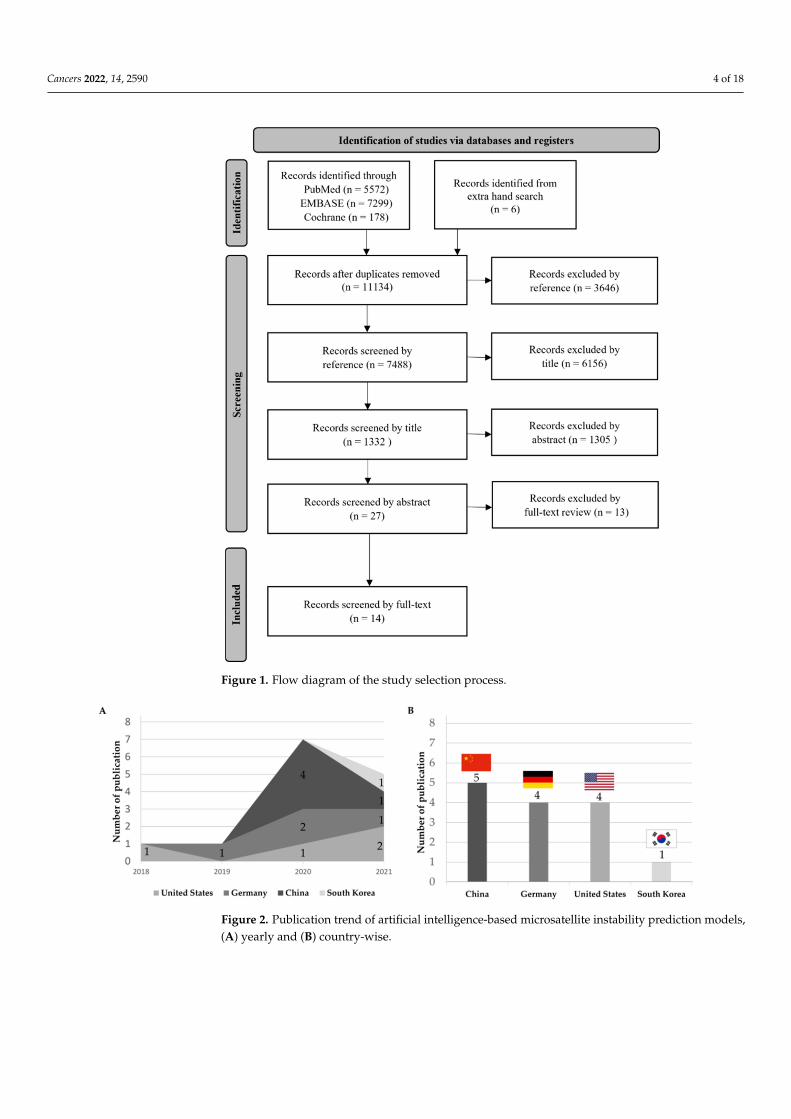
The detailed criteria for selecting and reviewing the articles are shown in Figure 1.The initial search from online databases yielded 13,049 records and six articles identifiedthrough a hand search. After removing duplicates, a total of 11,134 records remained.Following that, 3646 records were removed owing to an irrelevant reference type, whichwas reduced to 7488 records. Next, 6156 records were excluded by title, which was reducedto 1332 records. After 1305 records were removed by abstract, 27 records were selected forfull-text review. In the process of full-text review, only 14 studies met the inclusion criteriaand were included in the systematic review.
3.2. Yearly and Country-Wise Trend of Publication
The yearly and country-wise trends of publications are illustrated in Figure 2. TheAI models for MSI prediction was first reported in 2018 and slightly increased so far. Theincluded 14 studies were published from China (n = 5), followed by Germany (n = 4), theUnited States (n = 4), and South Korea (n = 1).
3.3. MSI Prediction Models by Cancer Types
The number of publications on MSI models according to cancer types is shownin Figure 3. Most studies were from CRC (57.9%; n = 11), followed by endometrial(21.0%; n = 4), gastric (15.9%; n = 3), and ovarian cancers (5.2%; n = 1).
Cancers 2022, 14, 2590 4 of 18
Figure 1. Flow diagram of the study selection process.
Figure 2. Publication trend of artificial intelligence-based microsatellite instability prediction models,(A) yearly and (B) country-wise.

Cancers 2022, 14, 2590 5 of 18
Figure 3. Artificial intelligence-based MSI prediction models according to target organs.
3.4. Prediction of MSI Status in CRC
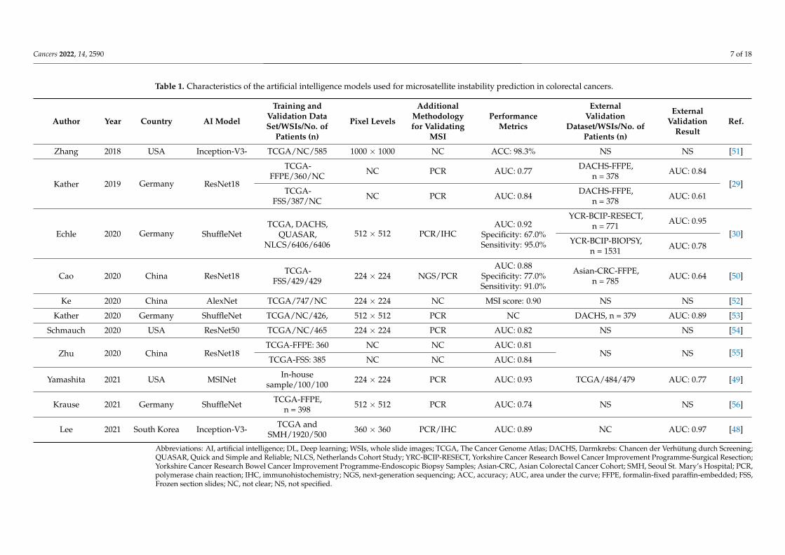
The key characteristics of the AI models included in the CRC are summarized inTable 1. Most of the studies used the TCGA dataset for training and validation of their AImodels. The study by Echle et al. used data from a large-scale international collaborationrepresenting the European population for training, validation, and testing, which includes6406 patients from Darmkrebs: Chancen der Verhütung durch Screening (DACHS), Quickand Simple and Reliable (QUASAR), and Netherlands Cohort Study (NLCS) datasetsin addition to the TCGA dataset [30]. DACHS is a dataset of CRC patients with stageI-IV from the German Cancer Research Center. QUASAR is a clinical trial data of CRCpatients, mainly with stage II tumors, from the United Kingdom. NLCS is a dataset fromthe Netherlands that includes patients of any tumor stage. The study by Lee et al. used anin-house dataset along with the TCGA dataset, and the study by Yamashita et al. used onlyan in-house dataset for training, validation, and testing of their AI models [48,49]. A studyby Co et al. and Lee et al. used an Asian dataset for external validation, which is differentfrom the population dataset used for training and testing their models [48,49].
The comparison of the AUC of their tests is shown in Figure 4. The performance metricAUC of AI models ranged 0.74–0.93. The highest AUC 0.93 was reported by Yamashita et al.with a small data set, but a study by Echle et al. with a large international dataset alsoshowed good AUC 0.92. Kather et al. and Coa et al. trained and tested their models onfrozen section slides (FSS) and compared their model performance with the results of aformalin-fixed paraffin-embedded (FFPE) slide dataset [29,50]. Their results showed thatAUC is slightly higher in the model trained and tested on FSS in comparison to that trainedand tested on FFPE.
A comparison of the sensitivity and specificity of the AI models of CRC is shownin Figure 4. Echle et al.’s study with a large-scale international dataset showed a goodsensitivity of 95.0%, although its specificity was slightly low (67.0%) [30]. A study byCoa et al. showed good sensitivity and a specificity of 91.0% and 77.0%, respectively [50].

Cancers 2022, 14, 2590 6 of 18
Figure 4. Comparison of the performance metric of microsatellite instability prediction models incolorectal cancers. (A). Area under the ROC curve. (B). Sensitivity and specificity.
The type of AI models used for MSI prediction in each study is shown in the supple-mentary Table S1. We also compared the AUCs of AI models that used the same datasetand that is shown in Supplementary Figure S1A,B. Our data showed that the averageperformance of ResNet18 model in CRC was better in FSS (AUC 0.85) compared to FFPE(AUC 0.79). The next commonly used AI model for CRC was ShuffleNet, which was usedby three studies. However, due to heterogeneity in their data, we were able to compare onlytwo studies, which showed an average AUC of 0.83. The average AUCs of both ResNet18and ShuffleNet classifiers were almost similar.
Cancers 2022, 14, 2590 7 of 18
Table 1. Characteristics of the artificial intelligence models used for microsatellite instability prediction in colorectal cancers.
Author Year Country AI Model
Training andValidation DataSet/WSIs/No. of
Patients (n)
Pixel Levels
AdditionalMethodologyfor Validating
MSI
PerformanceMetrics
ExternalValidation
Dataset/WSIs/No. ofPatients (n)
ExternalValidation
ResultRef.
Zhang 2018 USA Inception-V3- TCGA/NC/585 1000 × 1000 NC ACC: 98.3% NS NS [51]
Kather 2019 Germany ResNet18
TCGA-FFPE/360/NC NC PCR AUC: 0.77 DACHS-FFPE,
n = 378 AUC: 0.84
[29]TCGA-
FSS/387/NC NC PCR AUC: 0.84 DACHS-FFPE,n = 378 AUC: 0.61
Echle 2020 Germany ShuffleNetTCGA, DACHS,
QUASAR,NLCS/6406/6406
512 × 512 PCR/IHCAUC: 0.92
Specificity: 67.0%Sensitivity: 95.0%
YCR-BCIP-RESECT,n = 771 AUC: 0.95
[30]YCR-BCIP-BIOPSY,
n = 1531 AUC: 0.78
Cao 2020 China ResNet18 TCGA-FSS/429/429 224 × 224 NGS/PCR
AUC: 0.88Specificity: 77.0%Sensitivity: 91.0%
Asian-CRC-FFPE,n = 785 AUC: 0.64 [50]
Ke 2020 China AlexNet TCGA/747/NC 224 × 224 NC MSI score: 0.90 NS NS [52]
Kather 2020 Germany ShuffleNet TCGA/NC/426, 512 × 512 PCR NC DACHS, n = 379 AUC: 0.89 [53]
Schmauch 2020 USA ResNet50 TCGA/NC/465 224 × 224 PCR AUC: 0.82 NS NS [54]
Zhu 2020 China ResNet18TCGA-FFPE: 360 NC NC AUC: 0.81
NS NS [55]TCGA-FSS: 385 NC NC AUC: 0.84
Yamashita 2021 USA MSINet In-housesample/100/100 224 × 224 PCR AUC: 0.93 TCGA/484/479 AUC: 0.77 [49]
Krause 2021 Germany ShuffleNet TCGA-FFPE,n = 398 512 × 512 PCR AUC: 0.74 NS NS [56]
Lee 2021 South Korea Inception-V3- TCGA andSMH/1920/500 360 × 360 PCR/IHC AUC: 0.89 NC AUC: 0.97 [48]
Abbreviations: AI, artificial intelligence; DL, Deep learning; WSIs, whole slide images; TCGA, The Cancer Genome Atlas; DACHS, Darmkrebs: Chancen der Verhütung durch Screening;QUASAR, Quick and Simple and Reliable; NLCS, Netherlands Cohort Study; YRC-BCIP-RESECT, Yorkshire Cancer Research Bowel Cancer Improvement Programme-Surgical Resection;Yorkshire Cancer Research Bowel Cancer Improvement Programme-Endoscopic Biopsy Samples; Asian-CRC, Asian Colorectal Cancer Cohort; SMH, Seoul St. Mary’s Hospital; PCR,polymerase chain reaction; IHC, immunohistochemistry; NGS, next-generation sequencing; ACC, accuracy; AUC, area under the curve; FFPE, formalin-fixed paraffin-embedded; FSS,Frozen section slides; NC, not clear; NS, not specified.
Cancers 2022, 14, 2590 8 of 18
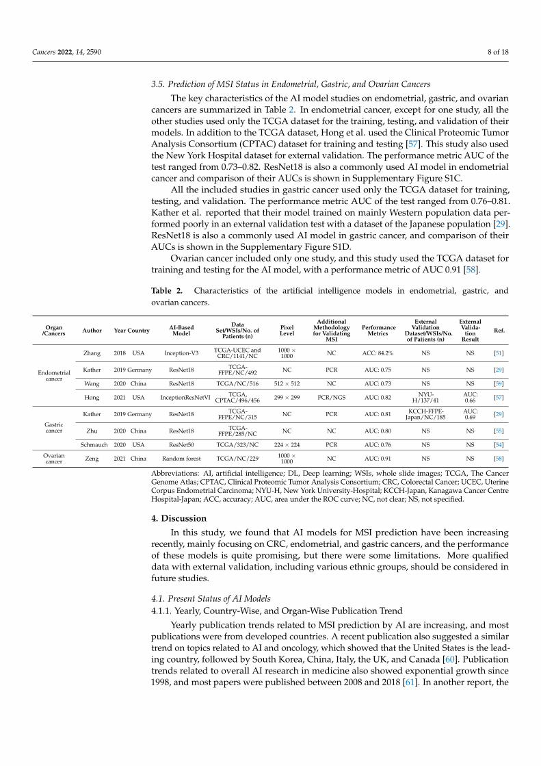
3.5. Prediction of MSI Status in Endometrial, Gastric, and Ovarian Cancers
The key characteristics of the AI model studies on endometrial, gastric, and ovariancancers are summarized in Table 2. In endometrial cancer, except for one study, all theother studies used only the TCGA dataset for the training, testing, and validation of theirmodels. In addition to the TCGA dataset, Hong et al. used the Clinical Proteomic TumorAnalysis Consortium (CPTAC) dataset for training and testing [57]. This study also usedthe New York Hospital dataset for external validation. The performance metric AUC of thetest ranged from 0.73–0.82. ResNet18 is also a commonly used AI model in endometrialcancer and comparison of their AUCs is shown in Supplementary Figure S1C.
All the included studies in gastric cancer used only the TCGA dataset for training,testing, and validation. The performance metric AUC of the test ranged from 0.76–0.81.Kather et al. reported that their model trained on mainly Western population data per-formed poorly in an external validation test with a dataset of the Japanese population [29].ResNet18 is also a commonly used AI model in gastric cancer, and comparison of theirAUCs is shown in the Supplementary Figure S1D.
Ovarian cancer included only one study, and this study used the TCGA dataset fortraining and testing for the AI model, with a performance metric of AUC 0.91 [58].
Table 2. Characteristics of the artificial intelligence models in endometrial, gastric, andovarian cancers.
Organ/Cancers Author Year Country AI-Based
ModelData
Set/WSIs/No. ofPatients (n)
PixelLevel
AdditionalMethodologyfor Validating
MSI
PerformanceMetrics
ExternalValidation
Dataset/WSIs/No.of Patients (n)
ExternalValida-
tionResult
Ref.
Endometrialcancer
Zhang 2018 USA Inception-V3 TCGA-UCEC andCRC/1141/NC
1000 ×1000 NC ACC: 84.2% NS NS [51]
Kather 2019 Germany ResNet18 TCGA-FFPE/NC/492 NC PCR AUC: 0.75 NS NS [29]
Wang 2020 China ResNet18 TCGA/NC/516 512 × 512 NC AUC: 0.73 NS NS [59]
Hong 2021 USA InceptionResNetVI TCGA,CPTAC/496/456 299 × 299 PCR/NGS AUC: 0.82 NYU-
H/137/41AUC:0.66 [57]
Gastriccancer
Kather 2019 Germany ResNet18 TCGA-FFPE/NC/315 NC PCR AUC: 0.81 KCCH-FFPE-
Japan/NC/185AUC:0.69 [29]
Zhu 2020 China ResNet18 TCGA-FFPE/285/NC NC NC AUC: 0.80 NS NS [55]
Schmauch 2020 USA ResNet50 TCGA/323/NC 224 × 224 PCR AUC: 0.76 NS NS [54]
Ovariancancer Zeng 2021 China Random forest TCGA/NC/229 1000 ×
1000 NC AUC: 0.91 NS NS [58]
Abbreviations: AI, artificial intelligence; DL, Deep learning; WSIs, whole slide images; TCGA, The CancerGenome Atlas; CPTAC, Clinical Proteomic Tumor Analysis Consortium; CRC, Colorectal Cancer; UCEC, UterineCorpus Endometrial Carcinoma; NYU-H, New York University-Hospital; KCCH-Japan, Kanagawa Cancer CentreHospital-Japan; ACC, accuracy; AUC, area under the ROC curve; NC, not clear; NS, not specified.
4. Discussion
In this study, we found that AI models for MSI prediction have been increasingrecently, mainly focusing on CRC, endometrial, and gastric cancers, and the performanceof these models is quite promising, but there were some limitations. More qualifieddata with external validation, including various ethnic groups, should be considered infuture studies.
4.1. Present Status of AI Models4.1.1. Yearly, Country-Wise, and Organ-Wise Publication Trend
Yearly publication trends related to MSI prediction by AI are increasing, and mostpublications were from developed countries. A recent publication also suggested a similartrend on topics related to AI and oncology, which showed that the United States is the lead-ing country, followed by South Korea, China, Italy, the UK, and Canada [60]. Publicationtrends related to overall AI research in medicine also showed exponential growth since1998, and most papers were published between 2008 and 2018 [61]. In another report, the

Cancers 2022, 14, 2590 9 of 18
number of publications in overall AI and machine learning in oncology remained stableuntil 2014, but increased enormously from 2017 [60], which is consistent with our results.
Our data showed that the number of publications on MSI models is higher in CRCcompared to endometrial, gastric and ovarian cancers. It may be because the CRC is thesecond most lethal cancer worldwide, and approximately 15% of CRC is caused by theMSI [6–9,62,63]. MSI-high tumors are widely considered to have a large neoantigen burden,making them especially responsive to immune checkpoint inhibitor therapy [64,65]. Inrecent years, MSI has gained much attention because of its involvement in predicting theresponse to immunotherapy for many types of tumors [66]. An example of the AI modelfor CRC is shown in Figure 5.
Figure 5. Example of an artificial intelligence model for colorectal cancer. Figure 1. Overview of theEnsemble Patch Likelihood Aggregation (EPLA) model. A whole slide image (WSI) of each patientwas obtained and annotated to highlight the regions of carcinoma (ROIs). Next, patches were tiledfrom ROIs, and the MSI likelihood of each patch was predicted by ResNet-18, during which a heatmap was shown to visualize the patch-level prediction. Then, patch likelihood histogram (PALHI)pipelines and bags of words (BoW) pipelines integrated the multiple patch-level MSI likelihoods intoa WSI-level MSI prediction, respectively. Finally, ensemble learning combined the results of the twopipelines and made the final prediction of the MS status. Reprinted from Ref. [50].
AI models using WSI showed great potential for prediction of MSI in CRCs, whichcan be used as a low-cost screening method for these patients. It also can be used as aprescreening tool to select MSI-H probability for patients before testing with the currentcostly available PCR/IHC methods. However, further validation of these models on a largedataset is necessary to improve their performance to an acceptable level of clinical usage.Most of the MSI models for CRC were developed on a dataset of surgical specimens. Moremodels from endoscopic biopsy samples using more datasets from various ethnic popula-tions should be developed in the future, which can reduce the possibility of missing MSI-Hcases, particularly in advanced CRCs, where resection is not possible. Another limitationof these AI modes is that they cannot distinguish between hereditary and sporadic MSIcases. Therefore, to improve the performance of these models, training and validation witha large dataset is required in future research studies.
As immunotherapy and MSI testing gets more and more importance in other solidcancers such as gastric, endometrial, and ovarian cancers, we can see that the AI-basedMSI prediction models have also been applied in these cancers recently. They showedpromising results for a potential application, although the evidence is still insufficient. Alarge dataset with external validation should follow in the future.
4.1.2. Performance of AI Models and Their Cost Effectiveness
The sensitivity and specificity of AI models were comparable to that of routinely usedmethods such as PCR and IHC. The study by Echle et al. and Coa et al. showed 91.0–95.0%

Cancers 2022, 14, 2590 10 of 18
of sensitivity and 67.0–77.0% of specificity [30,50]. In the literature, IHC sensitivity rangesfrom 85–100% and the specificity ranges from 85–92% [31,32]. MSI PCR showed 85–100%sensitivity and 85–92% specificity [31]. According to a recent study assessing the cost-effectiveness of these molecular tests and the AI models, the accuracy of MSI predictionmodels was similar to that of commonly used PCR and IHC methods [67]. NGS technologyis useful for the testing of many gene mutations, such as for epithelial ovarian cancerpatients with BRCA mutation or for HR deficiency that might benefit from a therapeuticoption of platinum agents and PARP inhibitors, whereas immune checkpoint inhibitors areeffective in tumors with the MSI-H [68].
In this study, the authors predicted the net medical costs of six different clinicalscenarios using the combination of different MSI testing methods including PCR, IHC, NGSand AI models and corresponding treatment in the United States. An overview of the costeffectiveness comparison of their study is shown in Figure 6. They reported that AI modelswith high PCR or IHC can save up to $400 million annually [67]. As the cancer burden isincreasing, a precise diagnosis of MSI is essential to identify appropriate candidates forimmunotherapy and to reduce the medical costs.
Figure 6. The cost effectiveness of MSI prediction models. Comparison of total testing and treatment-related costs by clinical scenario. AI, artificial intelligence; IHC, immunohistochemistry; NGS,next-generation sequencing; PCR, polymerase chain reaction. Reprinted from Ref. [67].
4.2. Limitation and Challenge of AI Models4.2.1. Data, Image Quality and CNN Architecture
To obtain the best results from any convolutional neural network (CNN) model, alarge dataset from various ethnic groups is required for training, testing, and validation.Most studies in this review had a relatively small number of TCGA datasets for appropriatetraining and validation. Without a large-scale validation, the performance of these AImodels cannot be generalized, and it is not feasible for routine diagnosis. One studycould not perform further subgroup analysis due to limited clinical information of TCGAdatasets [49]. Another study raised the potential limitation that the TCGA datasets maynot represent the real situation [55]. Another group of researchers raised the potentiallimitation of technical artifacts such as blurred images in TCGA datasets [30]. Although theTCGA dataset includes patients from various institutes/hospitals, but all are the patientsare from similar ethnic group, which is primarily from the North America. A few studiesby Echle et al., Kather et al., Yamashita et al., and Lee et al. used European datasets (DACH)
Cancers 2022, 14, 2590 11 of 18
and local in-house datasets for training or external validation [29,30,48,49]. However, forhigh generalizability, the datasets from various ethnic groups should be explored further.
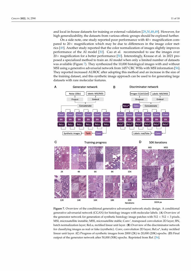
On a side note, one study reported poor performance with 40× magnification com-pared to 20× magnification which may be due to differences in the image color met-rics [49]. Another study reported that the color normalization of images slightly improvesperformance of the AI model [30]. Cao et al. recommended to use the images over20× magnification for a better performance [50]. Interestingly, Krause et al. in 2021 pro-posed a specialized method to train an AI model when only a limited number of datasetswas available (Figure 7). They synthesized the 10,000 histological images with and withoutMSI using a generative adversarial network from 1457 CRC WSIs with MSI information [56].They reported increased AUROC after adopting this method and an increase in the size ofthe training dataset, and this synthetic image approach can be used to for generating largedatasets with rare molecular features.
Figure 7. Overview of the conditional generative adversarial network study design. A conditionalgenerative adversarial network (CGAN) for histology images with molecular labels. (A) Overview ofthe generator network for generation of synthetic histology image patches with 512 × 512 × 3 pixels.MSI, microsatellite instable; MSS, microsatellite stable; Conv’, transposed convolution 2D layer; BN,batch normalization layer; ReLu, rectified linear unit layer. (B) Overview of the discriminator networkfor classifying images as real or fake (synthetic). Conv, convolution 2D layer; ReLu*, leaky rectifiedlinear unit layer. (C) Progress of synthetic images from 2000 (2K) to 20,000 (20K) epochs. (D) Finaloutput of the generator network after 50,000 (50K) epochs. Reprinted from Ref. [56].

Cancers 2022, 14, 2590 12 of 18
The choice of CNN also affects the performance of the AI models; commonly usednetworks such as ResNet18, ShuffleNet, and Inception-V3 have been used in most of thestudies. The ResNet model has many other variations as per the number of layers used,such as ResNet18, ResNet34, ResNet50, and many others. The ResNet18 model has 72-layerarchitecture with 18 deep layers, which may degrade the output result due to multiple deeplayers in the network [69]. However, if the output result is degraded it can be fixed throughback propagation. ShuffleNet has a simple design architecture, and it is also optimized formobile devices [53]. Therefore, it can show good performance with a high accuracy at alow training time [53].
A study observed that lightweight neural network models performed on par withmore complex models [53]. Performance comparison including three to six of these modelsis essential for enhancing the performance of the final model.
4.2.2. External Validation and Multi-Institutional Study
In CRC cases, six out of 11 studies included an external validation. The performancemetric AUC for external validation ranged from 0.61–0.97. In endometrial and gastriccancer cases, only one study for each group performed external validation. AI modelsthat are trained and tested on a single dataset may overfit and perform well on internaldatasets. However, these models show low performance when tested for external datasets.Therefore, external validation on different datasets is always necessary in order to have awell-trained AI model.
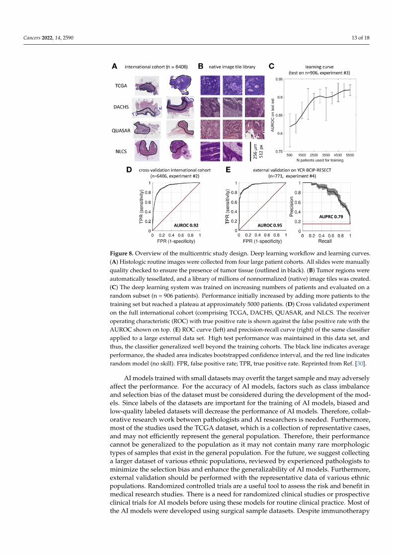
Studies also suggested that a large sample size, multiple institutions data, and patientswith different populations are needed to determine the generalization performance oftheir AI models. An overview of the multicentric study deign is shown in Figure 8.AI models trained mainly on data from Western populations performed poorly whenvalidated on Asian populations [29]. Another study suggested that transfer learning formodel fine-tuning in different ethnic populations may improve the generalizability of theirAI models [50]. Previous researchers argued that datasets from multi-institutional andmultinational models enhanced the generalizability of DL models [70,71].
4.2.3. MSI Prediction on Biopsy Samples
Most studies only use WSIs of surgical specimens for the development of their AImodels. However, MSI prediction on small colonoscopic biopsy samples is more practi-cally useful in the clinical setting if it is feasible. A recent study observed relatively lowperformance on biopsy samples with their surgical specimen trained AI model [30]. Thus,further research on small biopsy samples is required to increase the performance.
4.2.4. Establishment of Central Facility
AI technology in medical applications is still growing recent study showed increasingtrend of patent related to AI and pathological images [72]. The lack of installed slidescanners in hospitals can hinder the implementation of DL models. The WSIs are largefiles which cannot be stored in a routine hospital setting. The whole slide scanners and theviewing and archiving system along with an appropriate server is expensive equipmentthat cannot be easily established. The establishment of central slide scanner facilities with aserver with a larger data storage capacity can overcome this challenge [45,73].
4.3. Future Direction
Originally, AI applications in the pathology field focused on mimicking or replacinghuman pathologists’ tasks, such as segmentation, classification, and grading. The main goalof these studies was to reduce intra- or inter-observer variability in pathologic interpretationto support or augment human ability.
Cancers 2022, 14, 2590 13 of 18
Figure 8. Overview of the multicentric study design. Deep learning workflow and learning curves.(A) Histologic routine images were collected from four large patient cohorts. All slides were manuallyquality checked to ensure the presence of tumor tissue (outlined in black). (B) Tumor regions wereautomatically tessellated, and a library of millions of nonnormalized (native) image tiles was created.(C) The deep learning system was trained on increasing numbers of patients and evaluated on arandom subset (n = 906 patients). Performance initially increased by adding more patients to thetraining set but reached a plateau at approximately 5000 patients. (D) Cross validated experimenton the full international cohort (comprising TCGA, DACHS, QUASAR, and NLCS. The receiveroperating characteristic (ROC) with true positive rate is shown against the false positive rate with theAUROC shown on top. (E) ROC curve (left) and precision-recall curve (right) of the same classifierapplied to a large external data set. High test performance was maintained in this data set, andthus, the classifier generalized well beyond the training cohorts. The black line indicates averageperformance, the shaded area indicates bootstrapped confidence interval, and the red line indicatesrandom model (no skill). FPR, false positive rate; TPR, true positive rate. Reprinted from Ref. [30].
AI models trained with small datasets may overfit the target sample and may adverselyaffect the performance. For the accuracy of AI models, factors such as class imbalanceand selection bias of the dataset must be considered during the development of the mod-els. Since labels of the datasets are important for the training of AI models, biased andlow-quality labeled datasets will decrease the performance of AI models. Therefore, collab-orative research work between pathologists and AI researchers is needed. Furthermore,most of the studies used the TCGA dataset, which is a collection of representative cases,and may not efficiently represent the general population. Therefore, their performancecannot be generalized to the population as it may not contain many rare morphologictypes of samples that exist in the general population. For the future, we suggest collectinga larger dataset of various ethnic populations, reviewed by experienced pathologists tominimize the selection bias and enhance the generalizability of AI models. Furthermore,external validation should be performed with the representative data of various ethnicpopulations. Randomized controlled trials are a useful tool to assess the risk and benefit inmedical research studies. There is a need for randomized clinical studies or prospectiveclinical trials for AI models before using these models for routine clinical practice. Most ofthe AI models were developed using surgical sample datasets. Despite immunotherapy

Cancers 2022, 14, 2590 14 of 18
being the best treatment choice for CRC patients with stage IV tumors, the endoscopicbiopsy sample is the only available tissue from these patients due to the inability of surgicalresection. Future studies are needed to accurately estimate MSI based on biopsy samples,which will aid in the selection of immunotherapy for patients with advanced CRC cancer.Currently available AI models can not specifically differentiate between Lynch syndromeand MSI-H in sporadic cancer patients. The Development of an AI model for detectingLynch syndrome may help in selecting better therapeutic options for these patients. Itis difficult to understand how the AI models arrive at a conclusion. This is because AIalgorithms process data in a “black box”. Therefore, the AI models should be validatedagainst the currently available quality standards to ensure their efficiency.
However, scientists are increasingly focusing on the “superpower” from AI modelsthat can surpass human abilities, such as mutation, prognosis, and treatment responsepredictions in cancer patients. Our research group has already developed an AI model forMSI prediction in CRC, and the results is quite promising [48]. These findings motivatedus to initiate a multi-institutional research project for the MSI prediction from CRC WSIs.Our first aim is to collect a large image dataset of CRC patients and verify the quality of theimage by experienced pathologists. Second, we will develop an AI model using this largeimage dataset and test the generalized performance of AI models so that it may be feasibleto use it in routine practice. At present, we are in the process of scanning the H&E slides ofCRC patients in collaboration with 14 hospitals/institutions around the country.
5. Conclusions
This study showed that in the future, AI models can be an alternative and effectivemethod for the prediction of MSI-H from WSIs. Overall, AI models showed promisingresults and have the potential to predict MSI-H in a cost-effective manner. However, thelack of a large dataset, multiethnic population sample, and lack of external validation weremajor limitations of the previous studies. Currently, the AI models are not approved forclinical use to replace routine molecular tests. As the cancer burden is increasing, thereis need for the precise diagnostic method for predicting MSI-H and identify appropriatecandidates for immunotherapy and to reduce the medical costs. AI models also can beused as a prescreening tool to select MSI-H probability for patients before testing withthe current costly available PCR/IHC methods. Future studies are needed to accuratelyestimate MSI based on biopsy samples, which will aid in the selection of immunotherapyfor patients advance stages of CRC. Moreover, currently available AI models can notspecifically differentiate between Lynch syndrome and MSI-H in sporadic cancer patients.The development of an AI model for detecting Lynch syndrome may help in selectingbetter therapeutic options for these patients. As a result, to ensure efficiency, AI modelsshould be tested against currently existing quality standards before being used in clinicalpractice. Well-designed AI models in the future can improve their performance withoutcompromising diagnostic accuracy. Training and validation with a larger dataset andexternal validation on new datasets may improve the performance of AI models to anacceptable level.
Supplementary Materials: The following supporting information can be downloaded at: https://www.mdpi.com/article/10.3390/cancers14112590/s1, Figure S1: Comparison of AUCs of AImodels. (A) Comparison of ResNet18 in colorectal cancer. (B) comparison of ShuffleNet in colorectalcancer. (C) Comparison of ResNet18 in endometrial cancer. (D) Comparison of ResNet18 in gastriccancer; Table S1: Artificial intelligence models used for microsatellite.
Author Contributions: Conceptualization, M.R.A. and Y.C.; methodology, M.R.A. and Y.C.; software,M.R.A., Y.C., K.Y., S.H.L., J.A.-G., H.-J.J., N.T. and C.K.J.; validation, M.R.A., J.A.-G., K.Y. and Y.C.;formal analysis, M.R.A., J.A.-G., K.Y. and Y.C.; investigation, M.R.A. and Y.C.; resources, M.R.A.and Y.C.; data curation, M.R.A., Y.C., J.A-G. and K.Y.; writing—original draft preparation, M.R.A.;writing—review and editing, M.R.A., Y.C., K.Y., S.H.L., J.A.-G., H.-J.J., N.T. and C.K.J.; visualization,M.R.A. and Y.C.; supervision, Y.C., K.Y., S.H.L., J.A.-G., H.-J.J. and C.K.J.; project administration, Y.C.,K.Y. and J.A.-G. All authors have read and agreed to the published version of the manuscript.
Cancers 2022, 14, 2590 15 of 18
Funding: This research was supported by a grant of the Korea Health Technology R&D Projectthrough the Korea Health Industry Development Institute (KHIDI), funded by the Ministry of Health& Welfare, Republic of Korea (grant number: HI21C0940).
Institutional Review Board Statement: The study was conducted in accordance with the Declarationof Helsinki and approved by the Institutional Review Board of the Catholic University of Korea(UC21ZISI0129) (18 October 2021).
Informed Consent Statement: Not applicable.
Data Availability Statement: The data presented in this study are available upon request from the cor-responding author (https://www.researchgate.net/profile/Yosep-Chong (accessed on 17 April 2022)).The data are not publicly available due to institutional policies.
Acknowledgments: We thank Na Jin Kim for performing the strategic literature search. We wouldalso like to thank Ah Reum Kim for arranging the documents related to this research project.
Conflicts of Interest: The authors declare that they have no conflict of interest.
References1. Popat, S.; Hubner, R.; Houlston, R. Systematic review of microsatellite instability and colorectal cancer prognosis. J. Clin. Oncol.
2005, 23, 609–618. [CrossRef]2. Boland, C.R.; Goel, A. Microsatellite instability in colorectal cancer. Gastroenterology 2010, 138, 2073–2087. [CrossRef]3. Le, D.T.; Uram, J.N.; Wang, H.; Bartlett, B.R.; Kemberling, H.; Eyring, A.D.; Skora, A.D.; Luber, B.S.; Azad, N.S.; Laheru, D. PD-1
blockade in tumors with mismatch-repair deficiency. N. Engl. J. Med. 2015, 372, 2509–2520. [CrossRef]4. Greenson, J.K.; Bonner, J.D.; Ben-Yzhak, O.; Cohen, H.I.; Miselevich, I.; Resnick, M.B.; Trougouboff, P.; Tomsho, L.D.; Kim, E.;
Low, M. Phenotype of microsatellite unstable colorectal carcinomas: Well-differentiated and focally mucinous tumors and theabsence of dirty necrosis correlate with microsatellite instability. Am. J. Surg. Path. 2003, 27, 563–570. [CrossRef]
5. Smyrk, T.C.; Watson, P.; Kaul, K.; Lynch, H.T. Tumor-infiltrating lymphocytes are a marker for microsatellite instability incolorectal carcinoma. Cancer 2001, 91, 2417–2422. [CrossRef]
6. Tariq, K.; Ghias, K. Colorectal cancer carcinogenesis: A review of mechanisms. Cancer Biol. Med. 2016, 13, 120–135. [CrossRef]7. Devaud, N.; Gallinger, S. Chemotherapy of MMR-deficient colorectal cancer. Fam. Cancer 2013, 12, 301–306. [CrossRef]8. Cheng, L.; Zhang, D.Y.; Eble, J.N. Molecular Genetic Pathology, 2nd ed.; Springer: New York, NY, USA, 2013.9. Hewish, M.; Lord, C.J.; Martin, S.A.; Cunningham, D.; Ashworth, A. Mismatch repair deficient colorectal cancer in the era of
personalized treatment. Nat. Rev. Clin. Oncol. 2010, 7, 197–208. [CrossRef]10. Evrard, C.; Tachon, G.; Randrian, V.; Karayan-Tapon, L.; Tougeron, D. Microsatellite instability: Diagnosis, heterogeneity,
discordance, and clinical impact in colorectal cancer. Cancers 2019, 11, 1567. [CrossRef]11. Revythis, A.; Shah, S.; Kutka, M.; Moschetta, M.; Ozturk, M.A.; Pappas-Gogos, G.; Ioannidou, E.; Sheriff, M.; Rassy, E.; Boussios, S.
Unraveling the wide spectrum of melanoma biomarkers. Diagnostics 2021, 11, 1341. [CrossRef]12. Bailey, M.H.; Tokheim, C.; Porta-Pardo, E.; Sengupta, S.; Bertrand, D.; Weerasinghe, A.; Colaprico, A.; Wendl, M.C.; Kim, J.;
Reardon, B. Comprehensive characterization of cancer driver genes and mutations. Cell 2018, 173, 371–385. [CrossRef] [PubMed]13. Bonneville, R.; Krook, M.A.; Kautto, E.A.; Miya, J.; Wing, M.R.; Chen, H.-Z.; Reeser, J.W.; Yu, L.; Roychowdhury, S. Landscape of
microsatellite instability across 39 cancer types. JCO Precis. Oncol. 2017, 2017, PO.17.00073. [CrossRef] [PubMed]14. Ghose, A.; Moschetta, M.; Pappas-Gogos, G.; Sheriff, M.; Boussios, S. Genetic Aberrations of DNA Repair Pathways in Prostate
Cancer: Translation to the Clinic. Int. J. Mol. Sci. 2021, 22, 9783. [CrossRef] [PubMed]15. Mosele, F.; Remon, J.; Mateo, J.; Westphalen, C.; Barlesi, F.; Lolkema, M.; Normanno, N.; Scarpa, A.; Robson, M.; Meric-Bernstam, F.
Recommendations for the use of next-generation sequencing (NGS) for patients with metastatic cancers: A report from the ESMOPrecision Medicine Working Group. Ann. Oncol. 2020, 31, 1491–1505. [CrossRef]
16. Khalil, D.N.; Smith, E.L.; Brentjens, R.J.; Wolchok, J.D. The future of cancer treatment: Immunomodulation, CARs and combinationimmunotherapy. Nat. Rev. Clin. Oncol. 2016, 13, 273–290. [CrossRef]
17. Mittal, D.; Gubin, M.M.; Schreiber, R.D.; Smyth, M.J. New insights into cancer immunoediting and its three component phases—Elimination, equilibrium and escape. Curr. Opin. Immunol. 2014, 27, 16–25. [CrossRef] [PubMed]
18. Darvin, P.; Toor, S.M.; Nair, V.S.; Elkord, E. Immune checkpoint inhibitors: Recent progress and potential biomarkers. Exp. Mol.Med. 2018, 50, 165. [CrossRef]
19. Herbst, R.S.; Soria, J.-C.; Kowanetz, M.; Fine, G.D.; Hamid, O.; Gordon, M.S.; Sosman, J.A.; McDermott, D.F.; Powderly, J.D.;Gettinger, S.N. Predictive correlates of response to the anti-PD-L1 antibody MPDL3280A in cancer patients. Nature 2014,515, 563–567.
20. Zou, W.; Wolchok, J.D.; Chen, L. PD-L1 (B7-H1) and PD-1 pathway blockade for cancer therapy: Mechanisms, responsebiomarkers, and combinations. Sci. Transl. Med. 2016, 8, 328rv324. [CrossRef]
Cancers 2022, 14, 2590 16 of 18
21. Jenkins, M.A.; Hayashi, S.; O’shea, A.-M.; Burgart, L.J.; Smyrk, T.C.; Shimizu, D.; Waring, P.M.; Ruszkiewicz, A.R.; Pollett, A.F.;Redston, M. Pathology features in Bethesda guidelines predict colorectal cancer microsatellite instability: A population-basedstudy. Gastroenterology 2007, 133, 48–56. [CrossRef]
22. Alexander, J.; Watanabe, T.; Wu, T.-T.; Rashid, A.; Li, S.; Hamilton, S.R. Histopathological identification of colon cancer withmicrosatellite instability. Am. J. Pathol. 2001, 158, 527–535. [CrossRef]
23. Benson, A.B.; Venook, A.P.; Al-Hawary, M.M.; Arain, M.A.; Chen, Y.-J.; Ciombor, K.K.; Cohen, S.A.; Cooper, H.S.; Deming, D.A.;Garrido-Laguna, I. Small bowel adenocarcinoma, version 1.2020, NCCN clinical practice guidelines in oncology. J. Natl. Compr.Canc. Netw. 2019, 17, 1109–1133. [PubMed]
24. Koh, W.-J.; Abu-Rustum, N.R.; Bean, S.; Bradley, K.; Campos, S.M.; Cho, K.R.; Chon, H.S.; Chu, C.; Clark, R.; Cohn, D. Cervicalcancer, version 3.2019, NCCN clinical practice guidelines in oncology. J. Natl. Compr. Cancer Netw. 2019, 17, 64–84. [CrossRef][PubMed]
25. Sepulveda, A.R.; Hamilton, S.R.; Allegra, C.J.; Grody, W.; Cushman-Vokoun, A.M.; Funkhouser, W.K.; Kopetz, S.E.; Lieu, C.;Lindor, N.M.; Minsky, B.D. Molecular Biomarkers for the Evaluation of Colorectal Cancer: Guideline From the American Societyfor Clinical Pathology, College of American Pathologists, Association for Molecular Pathology, and American Society of ClinicalOncology. J. Mol. Diagn. 2017, 19, 187–225. [CrossRef]
26. Percesepe, A.; Borghi, F.; Menigatti, M.; Losi, L.; Foroni, M.; Di Gregorio, C.; Rossi, G.; Pedroni, M.; Sala, E.; Vaccina, F. Molecularscreening for hereditary nonpolyposis colorectal cancer: A prospective, population-based study. J. Clin. Oncol. 2001, 19, 3944–3950.[CrossRef] [PubMed]
27. Aaltonen, L.A.; Salovaara, R.; Kristo, P.; Canzian, F.; Hemminki, A.; Peltomäki, P.; Chadwick, R.B.; Kääriäinen, H.; Eskelinen, M.;Järvinen, H. Incidence of hereditary nonpolyposis colorectal cancer and the feasibility of molecular screening for the disease. N.Engl. J. Med. 1998, 338, 1481–1487. [PubMed]
28. Singh, M.P.; Rai, S.; Pandey, A.; Singh, N.K.; Srivastava, S. Molecular subtypes of colorectal cancer: An emerging therapeuticopportunity for personalized medicine. Genes Dis. 2021, 8, 133–145. [CrossRef]
29. Kather, J.N.; Pearson, A.T.; Halama, N.; Jäger, D.; Krause, J.; Loosen, S.H.; Marx, A.; Boor, P.; Tacke, F.; Neumann, U.P. Deeplearning can predict microsatellite instability directly from histology in gastrointestinal cancer. Nat. Med. 2019, 25, 1054–1056.[CrossRef]
30. Echle, A.; Grabsch, H.I.; Quirke, P.; van den Brandt, P.A.; West, N.P.; Hutchins, G.G.; Heij, L.R.; Tan, X.; Richman, S.D.; Krause, J.Clinical-grade detection of microsatellite instability in colorectal tumors by deep learning. Gastroenterology 2020, 159, 1406–1416.[CrossRef]
31. Coelho, H.; Jones-Hughes, T.; Snowsill, T.; Briscoe, S.; Huxley, N.; Frayling, I.M.; Hyde, C. A Systematic Review of Test AccuracyStudies Evaluating Molecular Micro-Satellite Instability Testing for the Detection of Individuals With Lynch Syndrome. BMCCancer 2017, 17, 836. [CrossRef]
32. Snowsill, T.; Coelho, H.; Huxley, N.; Jones-Hughes, T.; Briscoe, S.; Frayling, I.M.; Hyde, C. Molecular testing for Lynch syndromein people with colorectal cancer: Systematic reviews and economic evaluation. Health Technol Assess 2017, 21, 1–238. [CrossRef][PubMed]
33. Zhang, X.; Li, J. Era of universal testing of microsatellite instability in colorectal cancer. World. J. Gastrointest. Oncol. 2013, 5, 12–19.[CrossRef] [PubMed]
34. Cohen, R.; Hain, E.; Buhard, O.; Guilloux, A.; Bardier, A.; Kaci, R.; Bertheau, P.; Renaud, F.; Bibeau, F.; Fléjou, J.-F. Association ofprimary resistance to immune checkpoint inhibitors in metastatic colorectal cancer with misdiagnosis of microsatellite instabilityor mismatch repair deficiency status. JAMA Oncol. 2019, 5, 551–555. [CrossRef] [PubMed]
35. Andre, T.; Shiu, K.-K.; Kim, T.W.; Jensen, B.V.; Jensen, L.H.; Punt, C.J.; Smith, D.M.; Garcia-Carbonero, R.; Benavides, M.; Gibbs, P.Pembrolizumab versus chemotherapy for microsatellite instability-high/mismatch repair deficient metastatic colorectal cancer:The phase 3 KEYNOTE-177 Study. J. Clin. Oncol. 2020, 38, LBA4. [CrossRef]
36. Ardila, D.; Kiraly, A.P.; Bharadwaj, S.; Choi, B.; Reicher, J.J.; Peng, L.; Tse, D.; Etemadi, M.; Ye, W.; Corrado, G. End-to-end lungcancer screening with three-dimensional deep learning on low-dose chest computed tomography. Nat. Med. 2019, 25, 954–961.[CrossRef]
37. Bejnordi, B.E.; Veta, M.; Van Diest, P.J.; Van Ginneken, B.; Karssemeijer, N.; Litjens, G.; Van Der Laak, J.A.; Hermsen, M.; Manson,Q.F.; Balkenhol, M. Diagnostic assessment of deep learning algorithms for detection of lymph node metastases in women withbreast cancer. JAMA 2017, 318, 2199–2210. [CrossRef]
38. Esteva, A.; Kuprel, B.; Novoa, R.A.; Ko, J.; Swetter, S.M.; Blau, H.M.; Thrun, S. Dermatologist-level classification of skin cancerwith deep neural networks. Nature 2017, 542, 115–118. [CrossRef]
39. Nam, S.; Chong, Y.; Jung, C.K.; Kwak, T.-Y.; Lee, J.Y.; Park, J.; Rho, M.J.; Go, H. Introduction to digital pathology and computer-aided pathology. J. Pathol. Transl. Med. 2020, 54, 125–134. [CrossRef]
40. De Fauw, J.; Ledsam, J.R.; Romera-Paredes, B.; Nikolov, S.; Tomasev, N.; Blackwell, S.; Askham, H.; Glorot, X.; O’Donoghue, B.;Visentin, D. Clinically applicable deep learning for diagnosis and referral in retinal disease. Nat. Med. 2018, 24, 1342–1350.[CrossRef]
41. Diao, J.A.; Wang, J.K.; Chui, W.F.; Mountain, V.; Gullapally, S.C.; Srinivasan, R.; Mitchell, R.N.; Glass, B.; Hoffman, S.; Rao, S.K.Human-interpretable image features derived from densely mapped cancer pathology slides predict diverse molecular phenotypes.Nat. Commun. 2021, 12, 1613. [CrossRef]

Cancers 2022, 14, 2590 17 of 18
42. Sirinukunwattana, K.; Domingo, E.; Richman, S.D.; Redmond, K.L.; Blake, A.; Verrill, C.; Leedham, S.J.; Chatzipli, A.; Hardy, C.;Whalley, C.M. Image-based consensus molecular subtype (imCMS) classification of colorectal cancer using deep learning. Gut2021, 70, 544–554. [CrossRef] [PubMed]
43. Skrede, O.-J.; De Raedt, S.; Kleppe, A.; Hveem, T.S.; Liestøl, K.; Maddison, J.; Askautrud, H.A.; Pradhan, M.; Nesheim, J.A.;Albregtsen, F. Deep learning for prediction of colorectal cancer outcome: A discovery and validation study. Lancet 2020, 395,350–360. [CrossRef]
44. Chong, Y.; Kim, D.C.; Jung, C.K.; Kim, D.-c.; Song, S.Y.; Joo, H.J.; Yi, S.-Y. Recommendations for pathologic practice using digitalpathology: Consensus report of the Korean Society of Pathologists. J. Pathol. Transl. Med. 2020, 54, 437–452. [CrossRef] [PubMed]
45. Kim, H.; Yoon, H.; Thakur, N.; Hwang, G.; Lee, E.J.; Kim, C.; Chong, Y. Deep learning-based histopathological segmentation forwhole slide images of colorectal cancer in a compressed domain. Sci. Rep. 2021, 11, 22520. [CrossRef]
46. Tizhoosh, H.R.; Pantanowitz, L. Artificial intelligence and digital pathology: Challenges and opportunities. J. Pathol. Inform. 2018,9, 38. [CrossRef]
47. Greenson, J.K.; Huang, S.-C.; Herron, C.; Moreno, V.; Bonner, J.D.; Tomsho, L.P.; Ben-Izhak, O.; Cohen, H.I.; Trougouboff, P.;Bejhar, J. Pathologic predictors of microsatellite instability in colorectal cancer. Am. J. Surg. Path. 2009, 33, 126–133. [CrossRef]
48. Lee, S.H.; Song, I.H.; Jang, H.J. Feasibility of deep learning-based fully automated classification of microsatellite instability intissue slides of colorectal cancer. Int. J. Cancer 2021, 149, 728–740. [CrossRef]
49. Yamashita, R.; Long, J.; Longacre, T.; Peng, L.; Berry, G.; Martin, B.; Higgins, J.; Rubin, D.L.; Shen, J. Deep learning model for theprediction of microsatellite instability in colorectal cancer: A diagnostic study. Lancet Oncol. 2021, 22, 132–141. [CrossRef]
50. Cao, R.; Yang, F.; Ma, S.-C.; Liu, L.; Zhao, Y.; Li, Y.; Wu, D.-H.; Wang, T.; Lu, W.-J.; Cai, W.-J. Development and interpretation of apathomics-based model for the prediction of microsatellite instability in Colorectal Cancer. Theranostics 2020, 10, 11080. [CrossRef]
51. Zhang, R.; Osinski, B.L.; Taxter, T.J.; Perera, J.; Lau, D.J.; Khan, A.A. Adversarial deep learning for microsatellite instabilityprediction from histopathology slides. In Proceedings of the 1st Conference on Medical Imaging with Deep Learning (MIDL2018), Amsterdam, The Netherlands, 4–6 July 2018; pp. 4–6.
52. Ke, J.; Shen, Y.; Guo, Y.; Wright, J.D.; Liang, X. A prediction model of microsatellite status from histology images. In Proceedingsof the 2020 10th International Conference on Biomedical Engineering and Technology, Tokyo, Japan, 15–18 September 2020;pp. 334–338.
53. Kather, J.N.; Heij, L.R.; Grabsch, H.I.; Loeffler, C.; Echle, A.; Muti, H.S.; Krause, J.; Niehues, J.M.; Sommer, K.A.; Bankhead, P.Pan-cancer image-based detection of clinically actionable genetic alterations. Nat. Cancer 2020, 1, 789–799. [CrossRef]
54. Schmauch, B.; Romagnoni, A.; Pronier, E.; Saillard, C.; Maillé, P.; Calderaro, J.; Kamoun, A.; Sefta, M.; Toldo, S.; Zaslavskiy, M.A deep learning model to predict RNA-Seq expression of tumours from whole slide images. Nat. Commun. 2020, 11, 3877.[CrossRef] [PubMed]
55. Zhu, J.; Wu, W.; Zhang, Y.; Lin, S.; Jiang, Y.; Liu, R.; Wang, X. Computational analysis of pathological image enables interpretableprediction for microsatellite instability. arXiv 2020, arXiv:2010.03130.
56. Krause, J.; Grabsch, H.I.; Kloor, M.; Jendrusch, M.; Echle, A.; Buelow, R.D.; Boor, P.; Luedde, T.; Brinker, T.J.; Trautwein, C. Deeplearning detects genetic alterations in cancer histology generated by adversarial networks. J. Pathol. 2021, 254, 70–79. [CrossRef][PubMed]
57. Hong, R.; Liu, W.; DeLair, D.; Razavian, N.; Fenyö, D. Predicting endometrial cancer subtypes and molecular features fromhistopathology images using multi-resolution deep learning models. Cell Rep. Med. 2021, 2, 100400. [CrossRef]
58. Zeng, H.; Chen, L.; Zhang, M.; Luo, Y.; Ma, X. Integration of histopathological images and multi-dimensional omics analysespredicts molecular features and prognosis in high-grade serous ovarian cancer. Gynecol. Oncol. 2021, 163, 171–180. [CrossRef]
59. Wang, T.; Lu, W.; Yang, F.; Liu, L.; Dong, Z.; Tang, W.; Chang, J.; Huan, W.; Huang, K.; Yao, J. Microsatellite instability predictionof uterine corpus endometrial carcinoma based on H&E histology whole-slide imaging. In Proceedings of the 2020 IEEE 17thinternational symposium on biomedical imaging (ISBI), Iowa City, IA, USA, 3–7 April 2020; pp. 1289–1292.
60. Musa, I.H.; Zamit, I.; Okeke, M.; Akintunde, T.Y.; Musa, T.H. Artificial Intelligence and Machine Learning in Oncology: HistoricalOverview of Documents Indexed in the Web of Science Database. EJMO 2021, 5, 239–248. [CrossRef]
61. Tran, B.X.; Vu, G.T.; Ha, G.H.; Vuong, Q.-H.; Ho, M.-T.; Vuong, T.-T.; La, V.-P.; Ho, M.-T.; Nghiem, K.-C.P.; Nguyen, H.L.T. Globalevolution of research in artificial intelligence in health and medicine: A bibliometric study. J. Clin. Med. 2019, 8, 360. [CrossRef]
62. Yang, G.; Zheng, R.-Y.; Jin, Z.-S. Correlations between microsatellite instability and the biological behaviour of tumours. J. CancerRes. Clin. Oncol. 2019, 145, 2891–2899. [CrossRef]
63. Carethers, J.M.; Jung, B.H. Genetics and genetic biomarkers in sporadic colorectal cancer. Gastroenterology 2015, 149, 1177–1190.[CrossRef]
64. Kloor, M.; Doeberitz, M.V.K. The immune biology of microsatellite-unstable cancer. Trends Cancer 2016, 2, 121–133. [CrossRef]65. Chang, L.; Chang, M.; Chang, H.M.; Chang, F. Microsatellite instability: A predictive biomarker for cancer immunotherapy. Appl.
Immunohistochem. Mol. Morphol. 2018, 26, e15–e21. [CrossRef] [PubMed]66. Le, D.T.; Durham, J.N.; Smith, K.N.; Wang, H.; Bartlett, B.R.; Aulakh, L.K.; Lu, S.; Kemberling, H.; Wilt, C.; Luber, B.S. Mismatch
repair deficiency predicts response of solid tumors to PD-1 blockade. Science 2017, 357, 409–413. [CrossRef] [PubMed]67. Kacew, A.J.; Strohbehn, G.W.; Saulsberry, L.; Laiteerapong, N.; Cipriani, N.A.; Kather, J.N.; Pearson, A.T. Artificial intelligence can
cut costs while maintaining accuracy in colorectal cancer genotyping. Front. Oncol. 2021, 11, 630953. [CrossRef]

Cancers 2022, 14, 2590 18 of 18
68. Boussios, S.; Mikropoulos, C.; Samartzis, E.; Karihtala, P.; Moschetta, M.; Sheriff, M.; Karathanasi, A.; Sadauskaite, A.; Rassy, E.;Pavlidis, N. Wise management of ovarian cancer: On the cutting edge. J. Pers. Med. 2020, 10, 41. [CrossRef] [PubMed]
69. He, K.; Zhang, X.; Ren, S.; Sun, J. Deep residual learning for image recognition. In Proceedings of the IEEE Conference onComputer Vision and Pattern Recognition, Las Vegas, NV, USA, 27–30 June 2016; pp. 770–778.
70. Djuric, U.; Zadeh, G.; Aldape, K.; Diamandis, P. Precision histology: How deep learning is poised to revitalize histomorphologyfor personalized cancer care. NPJ Precis. Oncol. 2017, 1, 22. [CrossRef]
71. Serag, A.; Ion-Margineanu, A.; Qureshi, H.; McMillan, R.; Saint Martin, M.-J.; Diamond, J.; O’Reilly, P.; Hamilton, P. TranslationalAI and deep learning in diagnostic pathology. Front. Med. 2019, 6, 185. [CrossRef]
72. Ailia, M.J.; Thakur, N.; Abdul-Ghafar, J.; Jung, C.K.; Yim, K.; Chong, Y. Current Trend of Artificial Intelligence Patents in DigitalPathology: A Systematic Evaluation of the Patent Landscape. Cancers 2022, 14, 2400. [CrossRef]
73. Chen, J.; Bai, G.; Liang, S.; Li, Z. Automatic image cropping: A computational complexity study. In Proceedings of the IEEEConference on Computer Vision and Pattern Recognition, Las Vegas, NV, USA, 27–30 June 2016; pp. 507–515.
Related Documents


![Artificial Intelligence · Artificial Intelligence 2016-2017 Introduction [5] Artificial Brain: can machines think? Artificial Intelligence 2016-2017 Introduction [6] ... Deep Blue](https://static.cupdf.com/doc/110x72/5f0538917e708231d411e192/artificial-intelligence-artificial-intelligence-2016-2017-introduction-5-artificial.jpg)