Real Time Imaging of Fluorescent Flagellar Filaments Chiara Decaroli s1035375

Welcome message from author
This document is posted to help you gain knowledge. Please leave a comment to let me know what you think about it! Share it to your friends and learn new things together.
Transcript
Real Time Imaging of Fluorescent Flagellar Filaments
Chiara Decarolis1035375
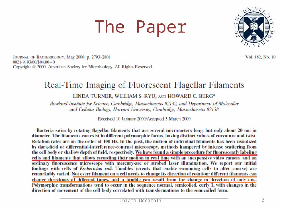
The Paper
2Chiara Decaroli
Objectives
1.Find an innovative way to image E. coli and Salmonella enterica;
2. Study the motility of bacteria: polymorphic transformations on E. Coli related to runs and tumbles.
3Chiara Decaroli
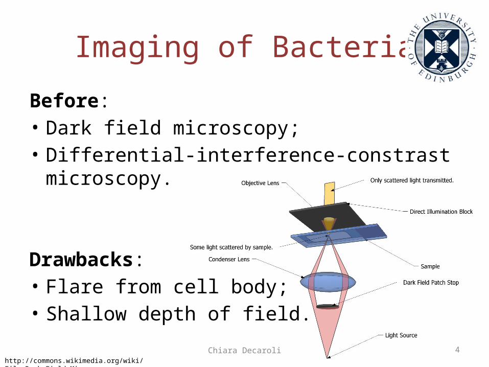
Imaging of BacteriaBefore:• Dark field microscopy;• Differential-interference-constrast microscopy.
Drawbacks:• Flare from cell body;• Shallow depth of field.
4Chiara Decarolihttp://commons.wikimedia.org/wiki/File:Dark_Field_Microscope.png
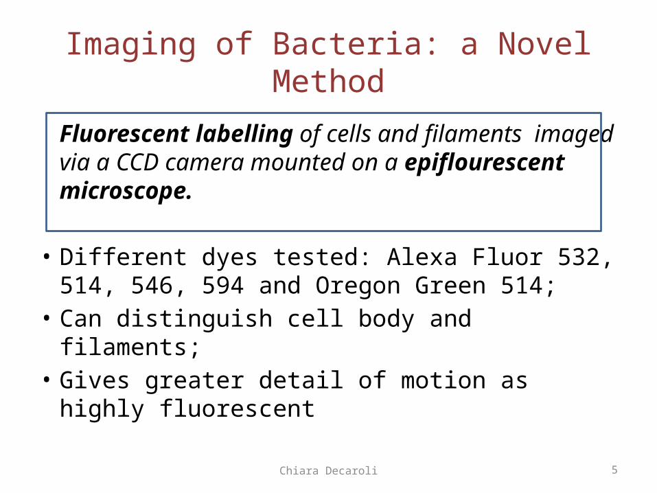
Imaging of Bacteria: a Novel Method
Fluorescent labelling of cells and filaments imaged via a CCD camera mounted on a epiflourescent microscope.
• Different dyes tested: Alexa Fluor 532, 514, 546, 594 and Oregon Green 514;
• Can distinguish cell body and filaments;
• Gives greater detail of motion as highly fluorescent
5Chiara Decaroli
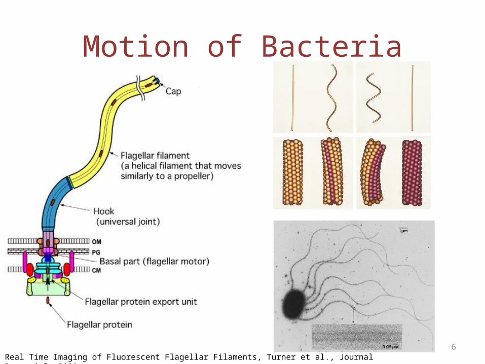
Motion of Bacteria
6Real Time Imaging of Fluorescent Flagellar Filaments, Turner et al., Journal Bacteriol, 2000
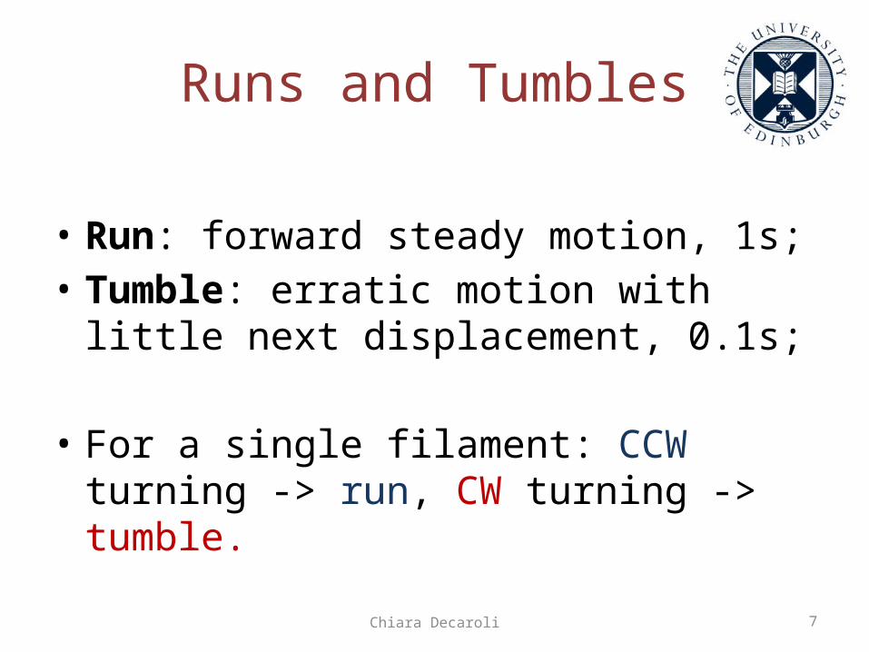
Runs and Tumbles
• Run: forward steady motion, 1s;• Tumble: erratic motion with little next displacement, 0.1s;
• For a single filament: CCW turning -> run, CW turning -> tumble.
7Chiara Decaroli
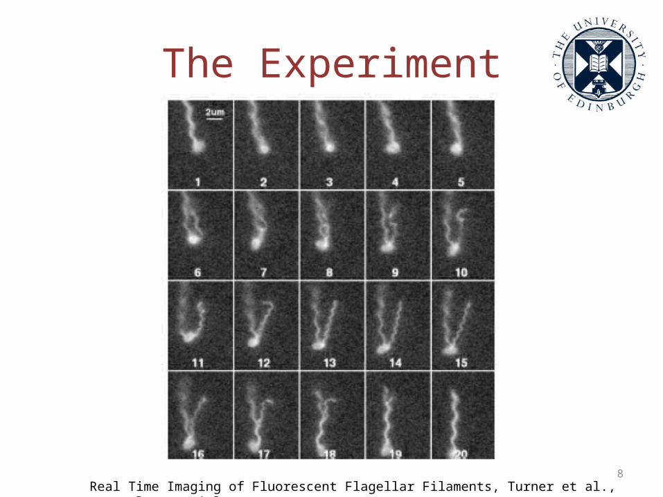
The Experiment
8Real Time Imaging of Fluorescent Flagellar Filaments, Turner et al., Journal Bacteriol, 2000
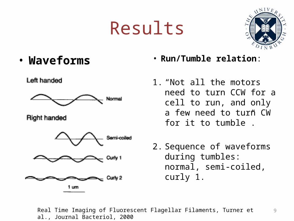
Results• Waveforms • Run/Tumble relation:
1. “Not all the motors need to turn CCW for a cell to run, and only a few need to turn CW for it to tumble”.
2. Sequence of waveforms during tumbles: normal, semi-coiled, curly 1.
9Real Time Imaging of Fluorescent Flagellar Filaments, Turner et al., Journal Bacteriol, 2000

Questions?
Chiara Decaroli 10
Related Documents


